hypopituitarism caused by melanoma of pituitary · hypopituitarism causedbya...
TRANSCRIPT

J. clin. Path. (1963), 16. 144
Hypopituitarism caused by a melanoma of thepituitary gland
JAMES McE. NEILSON AND A. D. MOFFAT
From the Departments of Medicine and Pathology, Stobhill General Hospital, Glasgow
SYNOPSIS A clinical and pathological description is given of a case of panhypopituitarism causedby a melanoma of the pituitary gland. The possible origins of a melanoma in this site are discussed.
Neoplastic causes of hypopituitarism may beclassified into three groups: 1, Primary tumours ofthe pituitary gland (chromophobe adenoma, malig-nant chromophobe adenoma (carcinoma of thepituitary), craniopharyngioma (adamantinoma,ameloblastoma, Rathke's pouch tumour, suprasellarcyst, teratoma, tumour of the hypophyseal duct));2, tumours of closely related structures (meningioma,carcinoma of the nasopharynx, tumours of the thirdventricle, glioma of the optic nerve and chiasma,parapituitary epidermoid and dermoid tumours);3, metastatic carcinoma.
CASE REPORT
The patient was aged 62 when he first attended theHaematology Clinic in August 1956. Since 1946 he hadcomplained ofsymptoms of rheumatoid arthritis affectingchiefly both wrists, the joints of both hands, both knees,and the left shoulder. He had been a miner for more than30 years when, in 1949, he was awarded a 40% pensionfor coal-miner's pneumoconiosis. On admission to theward on 15 August 1956 his major complaints were ofchronic dyspnoea related to pneumoconiosis and of arecent exacerbation in the rheumatoid arthritis. He hadnot at any time noticed tiredness or weakness but forapproximately 10 years he had observed that he requiredto shave less frequently and that his body hair haddecreased. Oral iron therapy had been prescribed by hisown doctor because of pallor of the skin. The patient hadfailed to note any recent increase in pallor and claimedthat he had always been pale. His wife had had fivechildren, four of whom were alive and well in 1956. Theyoungest was aged 20 years.The patient looked older than his stated age. The skin
and mucosae showed some pallor, and the former wasvery fine and soft. Changes attributable to rheumatoidarthritis affected the joints previously mentioned. Hisnails were beaked. There was slight bilateral ankleoedema. Suprapubic, axillary, and beard-area hair wasReceived for publication 17 September 1962
very sparse. The hair on the scalp was thin but evenlydistributed. The external genitalia were normal inappearance. The tongue was reddened and showedatrophy of the mucosa along the middle of the dorsum.The pulse rate was 60 per minute and the rhythm wasregular. There was no cardiac enlargement. The heartsounds were faint but clear-cut. His blood pressure was120/70 mm. of mercury. There was clinical evidence ofpulmonary emphysema. The percussion note was im-paired at the left base posteriorly and the respiratorymurmur was reduced over the same area. There were noneurological abnormalities. In particular, the ocularfundi were normal and perimetry showed no visualfield defect.
His I.Q. was not of a high order.Haemoglobin was 119 g. per 100 ml., the M.C.H.C.
31-3 %. The sternal marrow was relatively hypoplastic buterythropoiesis was normoblastic. Basal metabolic rateson two occasions were -10% and -6% of standard.The serum urea estimated on several occasions was withinnormal limits. Serum electrolytes, apart from slighthyponatraemia, were also within normal limits. A chestradiograph showed an opacity at the left base suggestiveof pneumonia with an associated small pleural effusion.There were no diagnostic features of pneumoconiosis.Radiological changes of rheumatoid arthritis were presentin both knees. After a preliminary few days of a highcarbohydrate diet a 50 g. oral glucose tolerance testshowed a flat curve beginning with a fasting level of78 mg. % and rising to 124 mg. % one hour later. At theend of two hours the blood sugar dropped to 74 mg. %.An intravenous glucose tolerance test showed normalmaximal values but the blood sugar at 120 minutes wasonly 57 mg. %. An intravenous insulin sensitivity testwas not performed but the blood sugar after an oral doseof 1 0 g. of carbutamide fell to hypoglycaemic levels andremained there for six hours. A 24-hour urine volumeof 1,140 ml. contained only 0-47 mg. of 17-ketosteroids.A skull radiograph showed enlargement of the sellaturcica with erosion of the posterior clinoid processes.The Wassermann and Kahn reactions were both negative.These features were sufficient to establish the diagnosis
of pneumonia, rheumatoid arthritis, and hypopituitarism.144
on May 22, 2020 by guest. P
rotected by copyright.http://jcp.bm
j.com/
J Clin P
athol: first published as 10.1136/jcp.16.2.144 on 1 March 1963. D
ownloaded from

Hypopituitarism caused by a melanoma of the pituitary gland
He was seen by a neurosurgeon who thought that thelikeliest cause of the last condition was a chromophobeadenoma although a Rathke pouch tumour was anotherpossibility.The patient was given soluble aspirin for rheumatoid
arthritis and the pneumonia was treated with penicillin.The hypopituitarism was treated with monthly intra-muscular injections of methyl testosterone microcrystules(100 mg.), oral thyroid (60 mg. per day), and cortisone(25 mg. per day). He was discharged in October of thesame year subjectively and objectively much improved.He was kept under review at intervals as an out-
patient but in March 1957 had to be readmitted. InJanuary of 1957 he had been treated for a recurrence otpneumonia in another hospital where, apparently,supportive endocrine therapy had been discontinued.When this was re-instituted his symptoms disappearedand he returned to good health. He was discharged on22 March 1957.He attended as an out-patient for a further seven
months remaining on the therapy noted above. In addition,on several occasions he was given intra-articular hydro-cortisone into both knee joints.
In April 1958 he gave a history of an episode of severeanterior chest pain some five weeks previously. An E.C.G.showed evidence of minimal restricted anterior myo-cardial damage, and a chest radiograph showed somecardiac enlargement. A skull radiograph showed nochange in the appearance of the sella turcica. He was notadmitted. For more than a year thereafter he was com-paratively well although requiring repeated intra-articular trimethyl prednisolone for rheumatoid arthritis.On 27 July 1959 he was readmitted for the last time.
Before admission he had been complaining of markedfrequency and his general practitioner had prescribedsulphadiazine for cystitis. The patient, afraid of confusingthe various tablets he was receiving, decided to discon-tinue thyroid and cortisone. Four days after he did so,probably as a consequence of the combined effect of theurinary tract infection and the withdrawal of replacementendocrine therapy, he became semi-comatose and wasadmitted to hospital. His blood pressure was 85/65 mm.of mercury. A chest radiograph showed generalizedcardiac enlargement with some pulmonary congestion,and an E.C.G. evidence of anterior and posteriorischaemic myocardial damage. The serum urea was27 mg. %, serum protein 6-3 g. %, serum chloride91 mEq./litre, serum sodium 120 mEq./litre, potassium4-0 mEq./litre, and alkali reserve 17 mEq./litre. Pus cellswere still present in the urine and culture produced agrowth of B. coli. Initially he responded well to treatmentfor pyuria and to an increased dosage of cortisone but on8 August he suddenly became dyspnoeic and lapsed intocoma with an unrecordable pulse or blood pressure. Hedied shortly afterwards.
NECROPSY
There was rheumatoid deformity of the joints pre-viously indicated. The features of chronic bronchitisand emphysema with early cor pulmonale were
FIG. 1. Pituitary gland and adjacent part of sphenoid. Ablock has been removed from the middle of the gland.Natural size.
present. There was no histological evidence ofpneumoconiosis. The coronary arteries were athero-matous with moderate encroachment on theirlumina but no evidence of myocardial fibrosis wasfound. There was benign hyperplasia of the prostate.The urinary bladder was moderately distended butthere was no evidence of pyogenic infection in theurinary tract.The pituitary gland was grossly enlarged (3-5 x
3 x 2-5 cm.) and jet-black throughout (Fig. 1)except for a thin rind of whitish tissue on the rightpostero-lateral aspect. The enlarged gland haderoded the bony floor of the sella turcica which wasrepresented by a thick, tough, fibrous membrane.The diaphragma sellae did not appear to be infil-trated but was displaced upwards to impinge uponand erode the surface of the medial aspect of theleft optic tract. There was no evidence of pigmenta-tion or tumour in the eyeballs, optic nerves, tracts,or meninges. A careful search of the suprarenals,nails, and skin also failed to show a primary growth.
HISTOLOGY OF THE PITUITARY GLAND
Step sections were examined from a series of blocksrepresenting the entire pituitary gland and thisdescription is a summary of the findings.The whitish rind of tissue on the right postero-
lateral aspect of the gland was found to consist ofpars nervosa and somewhat atrophic anteriorpituitary lobe tissue with intercellular fibrosis. Asmall non-encapsulated chromophobe adenoma,
145
on May 22, 2020 by guest. P
rotected by copyright.http://jcp.bm
j.com/
J Clin P
athol: first published as 10.1136/jcp.16.2.144 on 1 March 1963. D
ownloaded from

James McE. Neilson and A. D. Moffat
TABLEHISTOCHEMICAL REACTIONS OF PIGMENT GRANULES
Nile Blue Sulphatefor Lipofuscins(Pearse, 1953)
Completely bleached in Negative42 hours
Discoid bodies Completely bleached in Negative48 hours
1 mm. in diameter, lay within the anterior lobetissue. The cells of this adenoma were mainly smalland rounded with scanty cytoplasm (Fig. 2), but insome parts the cells had a larger proportion ofcytoplasm. A few of both types of cells containeddark pigment granules (Fig. 3), whilst some of thecells in the atrophic anterior pituitary tissue alsocontained similar granules of pigment. The mass ofblack tissue showed an adenomatous pattern(Fig. 4) although areas of more papilliform structurewere seen. In some areas both patterns intermingled(Fig. 5). The cellular detail was usually completelyobscured by a heavy deposit of dark brown coarse
and fine granules which seemed to distend the cells,many of which appeared to be ruptured so that thepigment was extruded into the intercellular spaces.
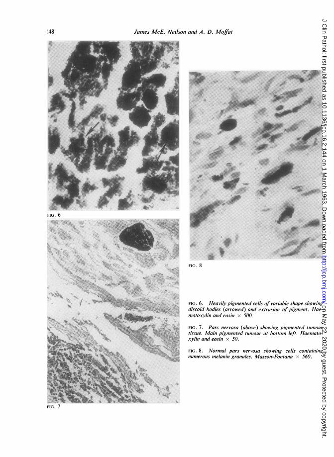
Elsewhere most of the cells were rounded and oftencontained dark, brownish, structureless, discoidbodies (Fig. 6) but sometimes they were columnaror even triangular. Occasionally, there were isletsof smaller rounded cells with scanty cytoplasmlightly pigmented with fine brown granules (Fig. 5).Pigmented tumour tissue had penetrated the
capsular lymphatics of the pituitary (Fig. 4) and thepars nervosa (Fig. 7).The histochemical reactions given by the pigment
granules and the intracellular discoid bodies are
summarized in the Table and show conclusively thatthe pigment in both instances was melanin. A fewsmall deposits of brown pigment around some ofthe blood vessels gave negative results in all theabove tests but a positive Perls' iron-staining re-
action.
DISCUSSION
A search of the literature shows that a melanin-containing tumour of the pituitary gland has onlyonce before been reported. This was a secondarymelanoma mentioned by Biggart (1961). The site ofthe primary growth was not recorded nor was itnoted whether the tumour was a single secondary or
only one of multiple deposits. In the present case
two different tumours were found in the pituitary.The larger tumour was heavily pigmented withmelanin. The smaller showed the features of a
Ziehl-Neelsen Masson-Fontana P.A.S.
NegativeStrongly positive Mainly brown, occasionally~~~~~~~~~~~~~~~~~~~~~~~~~~~~~~~~~~~~~~~~~~~~~~~~~~~~~~~~~~~~~~~~~~~~~~~~~~~~~~
Negative Strongly positive Mainly brown, occasionallyreddish-brown
Negative Strongly positive Usually positive
chromophobe adenoma with occasional cells whichcontained melanin. Examination of step sectionsshowed that the two tumours were always separatedby anterior lobe tissue with no continuity betweenthem. It is recognized that pituitary adenomata are
common, and estimates vary between 8% and 10%
in routine necropsies (Kraus quoted by Anderson,1953; Biggart and Dott, 1936). The chromophobetype is the most common (Anderson, 1953). Thesmall adenoma (1 mm. in diameter) may thereforebe regarded as an incidental finding only. Before it isdismissed, however, it should be noted that, althoughmelanin pigmentation was extreme in the largertumour, a few cells of the small adenoma also con-
tained melanin granules. Melanosis and melanomatamay occur in sites where normally no melanin isformed (Willis, 1959), and, since melanin pig-mentation was common to both tumours, it ispossible that they were variants of the same growth,the chromophobe being the cell type unrecognizablein the larger tumour because of extensive melanosis.The more plausible explanation, however, is thatthese were distinct and separate lesions, the largertumour being a true melanoma.
It has already been emphasized that a carefulpost-mortem search of the usual sites of primarymelanomata was unrewarding. Although the eye-balls were not opened at necropsy, they wereexamined frequently and carefully during the threeyears the patient was under observation and on
several occasions in the days immediately precedinghis death without showing any evidence of a
melanoma. In addition the optic nerves, optictracts, and suprarenals showed no tumour tissue.The possibility cannot be excluded that the patientmight have had a melanoma removed from the skinyears before his death but there were no obviousscars to suggest this. The onset of symptoms due tohypopituitarism cannot be dated accurately but itcan be accepted that this state was present for threeyears before death. The fact that the tumour was
present for this duration of time and remainedsolitary makes it seem improbable that it was a
metastatic growth.Primary melanomata of the leptomeninges have
been described (Gibson, Burrows, and Weir, 1957;
146
Bleaching with 10%H2 02
Pigment granules
on May 22, 2020 by guest. P
rotected by copyright.http://jcp.bm
j.com/
J Clin P
athol: first published as 10.1136/jcp.16.2.144 on 1 March 1963. D
ownloaded from

Hypopituitarism caused by a melanoma of the pituitary gland
FIG. 2 FIG. 3
FIG. 2. Chromophobe adenoma (a) at right. Pigmented tumour (b) bottom left. Atrophic anterior lobe tissue (c) between.Mallory trichrome x 50.
FIG. 3. Chromophobe adenoma with pigmented cells arrowed. Mallory trichrome x 500.
FIGl. Q
FIG. 4. Adenomatous pattern ofpigmented tumour and penetration of capsular lymphatic. Masson-Fontana x 50.FIG. 5. Adenomatous andpapilliform patterns. Islets ofsmaller, lightly pigmented cells arrowed. Haematoxylin and eosinx 125.
on May 22, 2020 by guest. P
rotected by copyright.http://jcp.bm
j.com/
J Clin P
athol: first published as 10.1136/jcp.16.2.144 on 1 March 1963. D
ownloaded from
James McE. Neilson and A. D. Moffat
_ .
_ 8~~~~~~~~~~.-AF
*7: JIM
.:
Is.
FIG. 8
I
- 4-
NS. -Y
FIG. 6. Heavily pigmented cells of variable shape showingdiscoid bodies (arrowed) and extrusion of pigment. Hae-matoxylin and eosin x 500.
FIG. 7. Pars nervosa (above) showing pigmented tumourtissue. Main pigmented tumour at bottom left. Haemato-xylin and eosin x 50.
FIG. 8. Normal pars nervosa showing cells containingnumerous melanin granules. Masson-Fontana x 560.
:..;
FIG. 7
148
4...-, -A%IC
i.-'
"'t !u",.
:JRF.... .0:
lamba
1, .QPis,
t.
Ar.
on May 22, 2020 by guest. P
rotected by copyright.http://jcp.bm
j.com/
J Clin P
athol: first published as 10.1136/jcp.16.2.144 on 1 March 1963. D
ownloaded from

Hypopituitarism caused by a melanoma of the pituitary gland
Bouton, 1958), and Gibson et al. agreed with Willis(1950) that the source of these tumours is theelongated pigment cells of the pia mater. While itcould be postulated that the tumour described hereoriginated from such cells of the pia mater, one of ushas frequently observed the presence of similarpigment-containing cells in the pars nervosa ofnormal pituitary glands (Fig. 8). The staining re-actions of the pigment in these cells are those ofmelanin. Kernohan and Sayre (1956) observe that'often a golden brown pigment is scattered irregularlythroughout the pars nervosa of the pituitary gland'.It seems that the tumour described by us may thushave two possible origins. Either it arose from thepigment cells of the pia mater or from the pigment-containing cells of the pars nervosa. but whatever itsorigin, from consideration of the facts we believethat the tumour was a primary melanoma and thecause of hypopituitarism in this patient.
We wish to thank Dr. J. B. Rennie, consultant physician.and Dr. J. C. Dick, consultant pathologist, for permissionto publish this case. We are also grateful to Mr. P. S.Waldie for the illustrations and photographs.
REFERENCES
Anderson, W. A. D. (1953). Pathology, 2nd ed., p. 967. Mosby, St.Louis.
Biggart, J. H. (1961). Pathology of the Nervous System, 3rd ed., p. 334.Livingstone, Edinburgh.and Dott, N. M. (1936). Brit. med. J., 2, 1153.
Bouton, J. (1958). J. clin. Path., 11, 122.Gibson, J. B., Burrows, D., and Weir, W. P. (1957). J. Path. Bact..
74, 419.Kernohan, J. W., and Sayre, G. P. (1956). Tumors of the Pituitary'
Gland and Infundibulum. Atlas of Tumor Pathology. Section 10,Fascicle 36, p. 11. Armed Forces Institute of Pathology.Washington.
Pearse, A. G. E. (1953). Histochemistry Theoretical and Appliedp. 479. Churchill, London.
Willis, R. A. (1950). Principles of Pathology, p. 564. Butterworth,London.
(1959). In Modern Trends in Pathology, edited by D. H. Collins,p. 117. Butterworth, London.
149
on May 22, 2020 by guest. P
rotected by copyright.http://jcp.bm
j.com/
J Clin P
athol: first published as 10.1136/jcp.16.2.144 on 1 March 1963. D
ownloaded from