hypoyirus manipulation of nonself recognition-associated
TRANSCRIPT
Hypoyirus manipulation of Nonself Recognition-Associated Programmed Cell Death
in the Chestnut Blight Fungus, Cryphonectria parasitica
A thesis presented to the Faculty of Graduate Studies of
Carleton University
By
Fuad Tanha
In partial fulfillment of the requirements for the degree of
Master of Science
2008
Copyright 2008, Fuad Tanha
1*1 Library and Archives Canada
Published Heritage Branch
395 Wellington Street Ottawa ON K1A0N4 Canada
Bibliotheque et Archives Canada
Direction du Patrimoine de I'edition
395, rue Wellington Ottawa ON K1A0N4 Canada
Your file Votre reference ISBN: 978-0-494-44144-2 Our file Notre reference ISBN: 978-0-494-44144-2
NOTICE: The author has granted a nonexclusive license allowing Library and Archives Canada to reproduce, publish, archive, preserve, conserve, communicate to the public by telecommunication or on the Internet, loan, distribute and sell theses worldwide, for commercial or noncommercial purposes, in microform, paper, electronic and/or any other formats.
AVIS: L'auteur a accorde une licence non exclusive permettant a la Bibliotheque et Archives Canada de reproduire, publier, archiver, sauvegarder, conserver, transmettre au public par telecommunication ou par Plntemet, prefer, distribuer et vendre des theses partout dans le monde, a des fins commerciales ou autres, sur support microforme, papier, electronique et/ou autres formats.
The author retains copyright ownership and moral rights in this thesis. Neither the thesis nor substantial extracts from it may be printed or otherwise reproduced without the author's permission.
L'auteur conserve la propriete du droit d'auteur et des droits moraux qui protege cette these. Ni la these ni des extraits substantiels de celle-ci ne doivent etre imprimes ou autrement reproduits sans son autorisation.
In compliance with the Canadian Privacy Act some supporting forms may have been removed from this thesis.
Conformement a la loi canadienne sur la protection de la vie privee, quelques formulaires secondaires ont ete enleves de cette these.
While these forms may be included in the document page count, their removal does not represent any loss of content from the thesis.
Canada
Bien que ces formulaires aient inclus dans la pagination, il n'y aura aucun contenu manquant.

ABSTRACT
Introduction of the pathogenic fungus, Cryphonectria parasitica, to North
America devastated the population of American chestnut, reducing it from a dominant
overstory tree into a shrub. The appearance of hypo virulent strains provided an
opportunity for biological control, due to their ability to transmit dsRNA viral elements
into virulent strains. Unfortunately, this program was not a success, in part, at least,
because of the fungal vegetative incompatibility system that impeded transmission of the
virus from infected to uninfected strains. Vegetative incompatibility in C. parasitica is
associated with six unlinked vie loci (vie 1,2,3, 4, 6, and 7), each with at least two
alleles. Fusion of two strains that differ at one or more vie loci can result in Programmed
Cell Death (PCD). The focus of this thesis was to characterize the factor(s) encoded by
the viral genome that interacts with the vjc-associated PCD in C. parasitica. Here, I
report a significant reduction in PCD upon fusion of strains differing at vie3 is associated
with hypo virus infection. Using chimeric hypo viruses of CHV1-EP713 and CHV1-
Euro7, this PCD reduction effect was assigned to the virus ORF A region that encodes
proteins p29 and p40. The p29-effect was studied in some detail. In addition to vic3, p29
caused a significant reduction in PCD rates triggered by differences at all other vie loci
studied, including vic2,6 and 7. p29 is a multifunctional protein that was previously
documented to alter fungal pigmentation patterns, conidiation and enzyme expression,
and p29 acts as suppressor of RNA silencing. My results further showed that the vic3-
associated PCD reduction is associated with production of p29 RNA rather that the p29
protein. Using dicer-like mutant strains I also showed that RNA silencing is involved in
vz'ci-associated PCD reduction. This information suggests a model in which p29
iii
interferes with RNA silencing, that in turn reduces v/c-associated PCD. Finally,
microscopy analyses of strains with multiple vie differences suggested that there are
interactions among vie genes involving independent pathways.
IV
ACKNOWLEDGEMENTS
I would like to express my sincere gratitude to my supervisor, Dr. Myron L.
Smith for all his support and encouragement throughout my academic and personal life.
This thesis could not have been written without his help and guidance.
I would like to thank my advisors, Dr. Linda Bonen and Dr. P. John Vierula, for
their feedback and suggestions; Dr. Donald, L. Nuss (University of Maryland) and Dr.
Michael Milgroom (Cornell University) for providing me with constructs and strains;
Robert Smith for helping with Western blotting and protein work; Melissa Begin for
teaching me real time PCR; Isabel Cruz, the "Adobe-photoshop wizard", for her help in
figure editing; and Denis Lafontaine for assisting me with reverse transcription. My
deepest thanks go to all these people who had some part in this project.
A special thank you also goes to my family. My mother, Mahboubeh Talebi, for
all the love and support that she has been giving me throughout my life and all the
sacrifices that she has made for me. Mom, I could never thank you enough. My brothers
Jamshid, Freidoon, Farzad and Reza, my sisters Hadigheh and Fatemeh, and my sister in
law Ramesh for their help and encouragement. I am truly grateful. I know for a fact that
this would not have been possible without them. On a final note, I am much indebted to
Negar, my wife, whose support, patience, and love was a big factor in completing the
project.
v

TABLE OF CONTENTS
ACCEPTANCE SHEET ii
ABSTRACT iii
ACKNOWLEDGEMENTS v
TABLE OF CONTENTS vi
LIST OF TABLES viii
LIST OF FIGURES ix
LIST OF ABBREVIATES xi
INTRODUCTION 1
Chestnut blight 1
Genome analysis ofhypovirus 6
Nonself recognition in C. parasitica 16
Relationship between virus transmission and VC in C. parasitica 19
8.5 Objectives 23
MATERIALS AND METHODS 23
Isolates of C. parasitica and growth conditions 23
C. parasitica spheroplasts preparation 31
Fungal transfection 32
in vitro transcription 32 Virus transfection ofC. parasitica 33
Fungal Transformation 34
Long term storage of strains 35
Microscopy Assay 35
Slide preparation 35 Slide analysis 38
Heterokaryon incompatibility test 41
Mycelial incompatibility assay 44
Polymerase chain reactions 47
DNA digestion and ligation 53
vi

DNA recovery from agarose gels 53
Fungal protein extraction 54
Affinity Purification using M2 Anti-flag Agarose Beads 54
SDS polyacrylamide gel electrophoresis (SDS-PAGE) 55
Staining SDS-PAGE gels with coomassie brilliant blue in preparation for mass
spectroscopy 56
Western blotting 57
RESULTS 58
Effect of viral infection on C. parasitica phenotype 58
Effect of chimeric recombinant viruses on PCD 64
Vegetative incompatibility 68
Heterokaryon incompatibility tests 68 Mycelial incompatibility assay 73
Protein-protein interaction 76
p29 sequence analysis 81
p29, RNA silencing and nonself recognition 84
vie pathway dissection 92
DISCUSSION 98
p29 protein and nucleotide sequence analysis 109
A possible p29-RNA silencing-nonself recognition connection 110
Dissecting the vie pathway using hypo virus p29 factor as a probe 114
SUMMARY AND CONCLUSIONS 118
LITERATURE CITED 119
vii
LIST OF TABLES Table 1. Strains used in this study 45 Table 2. Transformation plasmids along with their oligonucleotide primers used in this
study 51 Table 3. Effect of chimeric hypo virus transfection on colony morphology 62 Table 4: Effect of p29 expression on cell death frequencies between pairs of isolates that
differ at the single or multiple vie loci 94
vni
LIST OF FIGURES Figure 1. Diagram of Cryphonectria parasitica on chestnut tree 4 Figure 2. Schematic representation of the organization of four fully sequenced CHV
strains, 8 Figure 3. Comparison of cankers formed in chestnut tissues inoculated with virus-free C.
parasitica strain EP155 (left), EP155 transfected with either CHVl-Euro7 (center) or CHV1-EP713 (right) 12
Figure 4. Colony morphology of virus-free C. parasitica (left), C. parasitica transfected with either CHVl-Euro7 (center) or CHV 1-EP713 (right) 14
Figure 5. Schematic diagram of pLDST plasmid used for synthesizing RNA transcripts of inserted hypo virus sequence 25
Figure 6. Schematic representation of parental hypo viruses CHV1-EP713 and CHV1-Euro7 and their chimeric constructs used in this study 27
Figure 7. pCPXHYl vector used for plasmid transformations 29 Figure 8. Schematic diagram of slide culture technique used for microscopy assays 36 Figure 9. Types of interactions observed under 40x magnification 39 Figure 10. Schematic representation of technique used for heterokaryon incompatibility
test 42 Figure 11. Schematic representation of technique used for construction of chimeras 49 Figure 12. Colony morphologies of C. parasitica isolates with and without transfected
viruses 60 Figure 13: Clustered bar representation of viral constructs (left) and associated cell death
frequencies (right) 66 Figure 14: Heterokaryon outgrowth (hk) from EP155B and 22.1H pairing (vic-
compatible) (a). In comparison, the outgrowth is absent in pairing of strains 22.1H and P74-3 that differ at vic3 (b) 69
Figure 15: Growth rates of heterokaryotic sectors obtained from confronting hygromycin-resistant isolates of 22.1H, PXH1 or PXH3 against each of the 13 benomyl-resistant isolates 71
Figure 16. Mycelial incompatibility tests between strain pairs with defined vie characteristics 74
Figure 17: Western blot of flagged-tag constructs of CHVl-EP713-p29 and CHV1-Euro7-p29 77
Figure 18: Affinity purified samples of CHVl-EP713-p29 and CHVl-Euro7-p29 using M2 anti-flag agarose beads followed by SDS-PAGE and Coomassie Brilliant Blue staining 79
Figure 19: Clustered bar representation of viral constructs (left) and associated PCD frequencies (right) 82
Figure 20: Illustration of PCD frequencies in P74-3 (recipient) and EP155 (donor) 86 Figure 21. Sequence alignment of the wild-type CHVl-EP713-p29 and CHV1-EP713-
p29stop for first 60 bp of coding regions with corresponding amino acid sequences for each of three forward frames 88
Figure 22: Colony morphology of EP155 virus-free (left), EP155 (CHV1-EP713-p29stop) (middle) and EP155 (CHVl-EP713-p29) (right) 90
IX
Figure 23. Scatter plot representing a comparison between cell death ratio of isolates involving single and double vie differences of vic6 and 7 (A) and vic2 and 3 (B). ..96
Figure 24. Schematic diagram of the CHV1-EP713 genome, showing essential domains and residues in p29 coding region 103
Figure 25. Hypothetical representation showing the difference between the frequency (F) and rate (R) of v/c-associated PCD compared to virus-free confrontation (WT).... 107
Figure 26. Illustrates two different models for v/c-incompatibility pathway 116
x
LIST OF ABBREVIATES bmlR
benR
bp cm cDNA CHV °C DEPC DNA ddH20 dsRNA EDTA g gpd het hph hyg hygR
KDa Tm uL mL mm mM M NTP ORF % PCR PDA PDB PDAg PMSF PCD RNA RPM RT SDS VC VCG vie VSR V
benomyl resistant beta-tubulin benomyl resistant base pair centimeters complementary deoxyribonucleic acid cryphonectria parasitica hypovirus degree Celsius diethyl pyrocarbonate deoxyribonucleic acid double distilled water double-stranded ribonucleic acid ethylenediaminetetracetic acid grams glyceraldehyde-3-phosphate dehydrogenase heterokaryon incompatibility locus hygromycin B phosphotransferase hygromycin hygromycin resistant kilodaltons melting temperature microlitre millilitre millimeter millimolar moles per litre nucleoside triphosphate open reading frame percent polymerase chain reaction potato dextrose agar potato dextrose broth green potato dextrose agar phenyl methyl sulforyl fluoride programmed cell death ribonucleic acid revolutions per minute reverse transcription sodium dodecyl sulfate vegetative compatibility vegetative compatibility group vegetative incompatibility locus viral suppressors of RNA silencing volts
gene promoter
xi

1
INTRODUCTION
Chestnut blight
For the past century researchers have been investigating the mechanisms by
which some microorganisms inhibit the growth or metabolic activity of other
microorganisms. In the past decade, interest in using these antagonistic microorganisms
as biological controls of plant pathogens has increased with the aim of improving the
sustainability of agriculture, horticulture and forestry with minimal side-effects to the
environment (Duffy et ah, 2003). The attempted treatment of the chestnut blight fungus,
Cryphonectria parasitica, represents one of the classical cases of biological control of a
plant disease.
The American chestnut tree, Castanea dentata, is a member of the family
Fagaceae, closely related to oaks and beeches. The chestnut was one of the important
forest crops in eastern United States before the 1900's. The wood was valued for
building materials and the nuts constituted a major food source for humans as well as
deer, wild turkey, bear and other wildlife (Nuss, 1992). The chestnut was the dominant
tree in the uplands of North American eastern hardwood forests until being decimated by
the chestnut blight. The first evidence of chestnut blight in North America was reported
in 1904 in the New York Botanic Gardens. Within 50 years the disease had extended
from Maine to Alabama and west to the Mississippi River. The chestnut blight epidemic
was caused by the ascomycete fungus Cryphonectria parasitica, which was imported to
North America in the late 1800s from the Japanese chestnut tree, Castanea crenata, and
accidentally introduced to American chestnut trees (Anagnostakis, 2001). The loss of the
American chestnut had major ecological, social and economical consequences. It was
estimated that from 1910 to 1950 approximately 3.5 billion trees were lost (Cox, 1991).
2
At this point 9 million acres of American chestnut were dead (Anagnostakis, 1982).
Financial loss from the disease for only three states, Pennsylvania, South Carolina and
West Virginia, for the year of 1912 was estimated at $82.5 million (Cox, 1991).
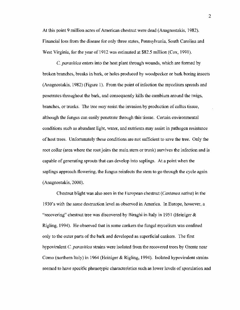
C. parasitica enters into the host plant through wounds, which are formed by
broken branches, breaks in bark, or holes produced by woodpecker or bark boring insects
(Anagnostakis, 1982) (Figure 1). From the point of infection the mycelium spreads and
penetrates throughout the bark, and consequently kills the cambium around the twigs,
branches, or trunks. The tree may resist the invasion by production of callus tissue,
although the fungus can easily penetrate through this tissue. Certain environmental
conditions such as abundant light, water, and nutrients may assist in pathogen resistance
of host trees. Unfortunately these conditions are not sufficient to save the tree. Only the
root collar (area where the root joins the main stem or trunk) survives the infection and is
capable of generating sprouts that can develop into saplings. At a point when the
saplings approach flowering, the fungus reinfects the stem to go through the cycle again
(Anagnostakis, 2000).
Chestnut blight was also seen in the European chestnut (Castanea sativa) in the
1930's with the same destruction level as observed in America. In Europe, however, a
"recovering" chestnut tree was discovered by Biraghi in Italy in 1951 (Heiniger &
Rigling, 1994). He observed that in some cankers the fungal mycelium was confined
only to the outer parts of the bark and developed as superficial cankers. The first
hypovirulent C. parasitica strains were isolated from the recovered trees by Grente near
Como (northern Italy) in 1964 (Heiniger & Rigling, 1994). Isolated hypovirulent strains
seemed to have specific phenotypic characteristics such as lower levels of sporulation and

3
they lacked orange pigmentation compared to normal, virulent strains. Grente also
noticed that inoculation of infected tissue with these hypovirulent strains as well as co-
inoculation with normal isolates resulted in reduced virulence levels. Preliminary
investigations on several European hypovirulent C. parasitica strains showed that
double-stranded RNA (dsRNA) was responsible for the conversion to a hypovirulence
phenotype (Dawe & Nuss, 2001).
4
Figure 1. Diagram of Cryphonectriaparasitica on chestnut tree. Conidia from the
infected stock are transferred by wind or animals to wounds on otherwise healthy trees.
Pathogen invasion through a wound results in canker formation and damaged vasculature
of the tree, which consequently destroys the leaves and branches beyond the site of
canker formation. Orange pigmented pycnidia bearing conidia appear on the surface of
the cankers followed by production of ascospores via the sexual cycle. (Modified from V.
Mortensen, ontained at http://chestnut.cas.psu.edu/PDFs/cryphonectria_life_cycle.pdf)
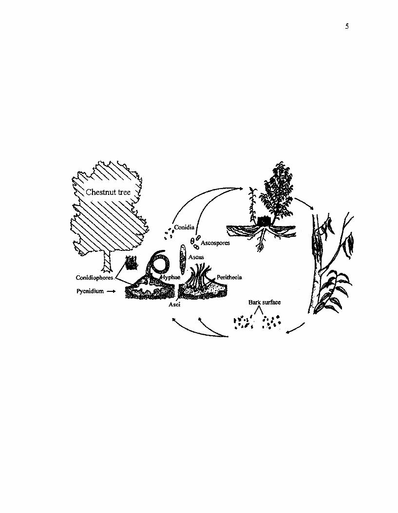
5
Conidiophores
Pycnidium —+
Conidia / 'SOBS
9 Ascospores
[yphae
Asct
Perithecia
Bark surface
A
6
Genome analysis of hypo virus
In order to understand the basis of hypo virulence and develop strategies for using
it in biological control, a comprehensive knowledge of the structural properties of
hypovirulence-related dsRNA is essential. Complete nucleotide sequences and analysis
of dsRNA isolated from European and North American hypovirulent C. parasitica strains
allowed the International Committee of Taxonomy of Viruses to create a new family,
Hypoviridae, with a single genus, Hypovirus (Dawe & Nuss, 2001). Four "species" of
Cryphonectria parasitica hypovirus (CHV) have been described (Milgroom & Cortesi,
2004): CHV-1 CHV-2, CHV-3 and CHV-4. CHV-1 is common in Europe and thought to
have been introduced from Asia. It was also found in a few areas in North America,
where it was used for biological control. CHV-1 has been studied more thoroughly than
other species due to its role in biological control of chestnut blight in Europe. CHV-2
and CHV-3 are found in New Jersey and Michigan, respectively. In addition, CHV-2 has
been found at one location in Asia. Because of high sequence similarity, there is a
possibility that CHV2 strains were introduced into China from North America. Due to its
genome organization, CHV-4 belongs to the hypovirus genus. However this hypovirus
has little or no effect on the host organism. It is mostly found in eastern North America
(Milgroom & Cortesi, 2004).
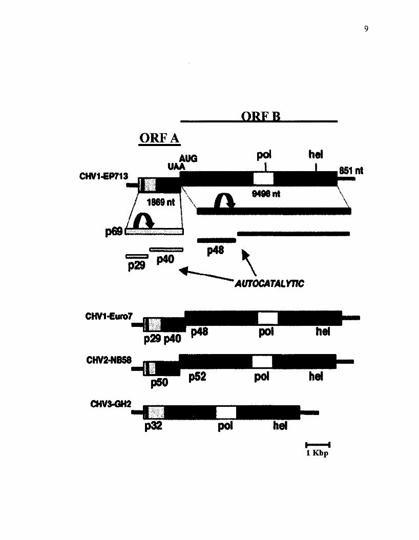
To date the genomes of three of the four species have been fully sequenced.
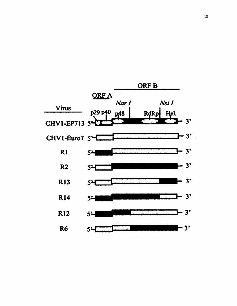
Representing two strains of CHV-1, comparative analyses show that CHV1-EP713 and
CHVl-Euro7 are comprised of two large open reading frames designated as ORF A and
ORF B (Figure 2). ORF A codes for the polyprotein, p69, which is autocatalytically
cleaved to produce p29 and p40 proteins. This cleavage is facilitated by the presence of

7
papain-like cysteine protease domain located within the p29 sequence. Similarly, ORF B
is autocatalytically processed to release a p48 protein and a second, larger peptide that
contains polymerase and helicase domains (Dawe & Nuss, 2001). The junction between
ORFs A and B is comprised of a sequence 5'-UAAUG-3', where UAA and AUG
segments serve as translation termination and initiation codons for ORF A and ORF B
respectively.
8
Figure 2. Schematic representation of the organization of four fully sequenced CHV
strains, two of CHV-1 and one each of CHV-2 and CHV-3. A fourth species, CHV-4,
has not been fully characterized but appears to have a similar genomic structure.
(Modified from Dawe & Nuss, 2001).
OKFR
ORFA
CHV1-EP713
AUTOCATALWC
CHV1-Euro7
p29p40
CHV2-NB58
CHV3-GH2
p32 pol he!
10
CHV2-NB58 has a similar genomic organization to that of CHV1 (Figure 2).
However, in contrast to CHV1-EP713, in vitro translation studies on CHV2-NB58
revealed that ORF A, which encodes a 50 kDa protein does not undergo autocatalytic
cleavage (Hillman et al, 1994). As opposed to CHV1 and CHV2, the CHV3-GH2
genome consists of a single ORF. Amino acid sequence alignment of the three species
showed a closer relation between CHV1 and CHV2, with CHV3 being more distantly
related (Dawe & Nuss, 2001).
Among the Hypoviridae affecting C. parasitica, CHV-1 has been given the most
attention due to its role as a biological control agent of chestnut blight in Europe. Most
relevant to this thesis, two strains, CHV-1-EP713 and CHV-1-Euro7, are known as
severe and mild hypovirus genotypes, respectively. This classification is based on
differences in their effects on C. parasitica virulence and associated characteristics such
as canker morphology, colony growth and sporulation level. Cankers on chestnut bark
produced by virus-free strains expand quickly, and bear multiple pycnidia that contain
orange conidia (Figure 3). However, the same C. parasitica strains infected with CHV1-
EP713 produce small cankers that contain a very small number or no pycnidia. The size
of cankers produced by strains infected with CHVl-Euro7 are observed to be three to
four times larger than the ones produced by EP713 (Cummings-Carlson, 1998). Effects
on C. parasitica growth rate and morphology also differentiate these two virus strains.
As shown under defined laboratory conditions (Figure 4), C. parasitica strains infected
by CHV1-EP713 grow slower and produce few or no asexual spores compared to the
corresponding virus-free strain. In contrast, the CHVl-Euro7-infected strain has a higher
11
growth rate than the virus-free strain but lacks pigmentation and asexual spore production
(Dawe&Nuss,2001).
Identification of a viral basis to C. parasitica hypovirulence in European
chestnuts provided the impetus for application of CHV1 strains as biological control
agents of North American chestnut blight. However, this strategy did not succeed,
apparently because North American strains of C. parasitica have a more complex nonself
recognition system than their European counterparts. The following sections briefly
provide some background into nonself recognition systems of filamentous fungi and how
they relate to viral transmission patterns.

12
Figure 3. Comparison of cankers formed in chestnut tissues inoculated with virus-free C.
parasitica strain EP155 (left), EP155 transfected with either CHVl-Euro7 (center) or
CHV1-EP713 (right) (Modified from Chen et al., 2000).

13

14
Figure 4. Colony morphology of virus-free C. parasitica (left), C. parasitica transfected
with either CHVl-Euro7 (center) or CHV1-EP713 (right) (Modified from Dawe & Nuss,
2001).

15
< • • , * « ; < * * " "
16
Nonself recognition in C. parasitica
Filamentous fungi grow through hyphal tip extension. This process is facilitated
by a structure located near the hyphal tip known as the Spitzenkorper, which is
responsible for delivery of the required components for the extension of the cell wall
(Glass et ah, 2000). Fusion between hyphae is known as anastomosis. It occurs during
the growth process to result in the formation of a mycelium, an interconnected network of
hyphae. Described for the first time by Buller (Buller, 1933), anastomosis involves the
breakdown of two hyphal cell walls and fusion of the plasma membranes. Upon fusion
the cells are capable of exchanging nuclei and other organelles, which is important for
maintaining the communication and homeostasis within a mycelium during growth and
reproduction (Glass et al, 2000).
Distinct fungal individuals with genetically different nuclei are also capable of
anastamosis; a process that results in the formation of a heterokaryon. Heterokaryon
formation may be beneficial for facilitating mitotic genetic exchange as well as
increasing mycelial biomass to facilitate physiological processes such as sexual/asexual
reproduction and accessing resources (Gregory, 1984). In many fungi, sexual and
asexual incompatibility are quite distinct processes. Two strains that have the ability to
fuse and form a heterokaryon during the sexual cycle may not be able to form a
successful heterokaryon during the vegetative cycle and vice versa (Leslie, 1993). Sexual
compatibility is controlled by the mating-type locus at which cells of compatible mating
type contain different, "idiomorphic" DNA sequences. Expression of compatible mating-
type idiomorphs establishes the cell-type sexual identity and subsequent expression of

17
different classes of transcriptional elements that lead to meiosis and meiospore
production (Fraser & Heitman, 2003).
In contrast, during the asexual phase, fusion between two individuals that are
different at one or more het loci (/zeferokaryon incompatibility, also called vie for
vegetative mcompatibility), will result in cell death as a result of lytic processes. This
phenomenon is known as heterokaryon, vegetative or somatic incompatibility and may,
in theory, take place before, during, or after hyphal fusion (Micali & Smith, 2003; Glass
et ah, 2000). Post-fusion vegetative incompatibility mechanisms are best characterized in
Podospora anserina and Neurospora crassa. In both species vegetative incompatibility
was shown to be regulated by either allelic or nonallelic mechanisms. Allelic
incompatibility occurs upon fusion of two individuals that carry different alleles at a
single het (or vie) locus. Nonallelic incompatibility results from interactions between
specific allelic combinations between two different loci.
Vegetative incompatibility is, therefore, a nonself recognition system that is
analogous to graft rejection in animals and plants. All filamentous fungi examined have
several, usually more than six, vie loci (Saupe, 2000). Vegetative incompatibility in C.
parasitica is associated with at least six unlinked vie loci (vie 1,2,3, 4, 6, and 7), each
with at least two alleles and each is thought to involve allelic interactions (Cortesi &
Milgroom, 1998). Hence, vegetative compatibility (VC) type can be defined based on the
presence of alleles at all vie loci. Fungal strains belonging to different VC groups (VCG)
can be identified by three techniques: heterokaryon incompatibility tests, barrage tests,
and partial diploid analyses (Micali & Smith, 2003).

18
Heterokaryon incompatibility testing examines whether or not two strains can
fuse to form stable heterokaryons. In practice, auxotrophic or antibiotic forcing markers
or complementary pigmentation markers can be used to test for heterokaryon formation.
For example, heterokaryon formation in Fusarium oxysporum is tested by using strains
containing different nit mutations (incapable of reducing nitrate), nit complementation
allows a compatible, but not an incompatible, heterokaryon to grow on medium
containing nitrate as the only source of nitrogen (Leslie, 1993). In addition to a reduced
growth rate, incompatible forced heterokaryons can be recognized by an aberrant
appearance such as reduced conidiation and accumulation of dark brown pigment (Micali
& Smith, 2005). Barrage tests are done by simply co-inoculating two strains onto the
same plate and allowing them to grow together. The term Barrage was introduced by
Vandendries to explain the response that occurred upon hyphal fusion between the two
different strains of fungi. A barrage is composed of an area containing dead or dying
cells with deposition of dark pigment. It may also appear as a clear zone or other types of
demarcation. However such morphological alterations are absent in vegetatively
compatible individuals. This phenomenon is seen in many fungi such as Podospora and
Neurospora and is the method most widely used for assigning strains to VC groups in C.
parasitica. Finally, partial diploid analyses can either be done through crosses with a
strain bearing a translocated het locus or through transformation of DNA containing the
het locus. In either case, having two copies of the duplicated het loci in the nucleus
results in a strain that is referred to as a partial diploid and if the two copies represent
incompatible alleles then the strain will be "self-incompatible".
19
Vegetative incompatibility and barrage tests are often thought to represent
different manifestations of the same process. However, at least two studies have shown
that this is not always the case (Micali & Smith, 2003; Smith et al, 2006). These studies
showed that differences at loci that are associated with barrage line formation do not
necessarily cause heterokaryon incompatibility, and vice versa. The distinct nature of
heterokaryon incompatibility and barrage formation may be explained if barrage
formation occurs in some cases without hyphal fusion, unlike heterokaryon
incompatibility in which fusion is necessary. Barrage formation may occur through
inhibitory effects of substances that diffuse from one or both strains, or may involve
diffusible substances and cell-surface receptors that interact to trigger barrage formation.
These observations suggest that heterokaryon incompatibility represents a "subset" of the
vegetative incompatibility system (Smith et al, 2006).
Relationship between virus transmission and VC in C. parasitica
Extensive studies by the French mycologist Jean Grente illustrated that the
hypovirulence phenotype could be transmitted from one strain to another through hyphal
fusion (Grente & Sauret, 1969). This finding set the stage for attempts at biological
control of C. parasitica through artificial introduction of hypo virulent strain. However,
as mentioned above, this program was not successful in North America, presumably due
to the restriction of viral elements by the fungus vegetative incompatibility system. The
transmission of cytoplasmic elements between strains is thought to be modulated by
vegetative incompatibility (Caten, 1972; Liu & Milgroom, 1996). That biocontrol in
Europe was more successful compared to North America is thought to be due to
differences in the diversity of VC groups in the two fungal populations. Thirty-three VC
groups were identified from analysis of the strains obtained from several regions in
France and Italy (Anagnostakis & Kranz, 1986). Strain diversity in some sites in Italy
was reported to be from four in Modena to 11 in Calabria, and other European regions
were reported to have only one VC type. On the other hand, 67 VC groups were
identified in Connecticut alone and, in 1978, a total of 37 VC types was reported among
202 isolates from West Virginia (Anagnostakis & Kranz, 1987).
The main source of VC diversity in North American C. parasitica is due to
heteroallelism at multiple vie loci combined with sexual recombination. The presence of
perithecia, the sexual fruiting body of this fungus, was reported by investigators in a
number of European countries, but they are uncommon in most areas (Heiniger &
Rigling, 1994). The absence of perithecia does not necessary preclude the occurrence of
genetic recombination. Data from six unlinked restriction fragment length polymorphism
(RFLP) loci and DNA fingerprinting suggest that C. parasitica may have a mixed mating
system with self-fertilization and outcrossing in the same population (Milgroom et al.,
1993). Work of Rizwana and Powell also showed that UV light may induce changes in
vegetative compatibility in C. parasitica, which may result in increased diversity of VC
groups in natural populations (Rizwana & Powell, 1992). Besides diversity of VC types
in natural populations, other factors may contribute to the unsuccessful spread and
persistence of hypo virulence such as overall reduction of robustness of hypovirulent
strains and reduction of sporulation by some hypovirulent strains (Nuss, 1992).
Nevertheless, the relatively higher VCG diversity in North America may explain the

21
observed inability to control chestnut blight using VCG as a biological control as evident
from laboratory transmission studies.
Cortesi el al. (2001) quantified the effect of heteroallelism at each of six vie loci
on virus transmission. Significant variation was associated with respect to the different
loci. It was shown that hypovirus transmission occurred in 100% of paired isolates that
had no vie differences, or when vic4 differed between strains. Heteroallelism at vic3 and
6 resulted in 76% and 32% transmission, respectively. However, when strain pairs were
heteroallelic at vicl, 2 or 7 the frequency of virus transmission was dependent on the
allelic constitution (1 or 2) of the donor and recipient isolates. That is, virus transmission
was significantly asymmetrical in these cases. The average transmission frequencies with
pairs of strains heteroallelic at one of vicl, 2 ox 7 were 56%, 25% or 78%, respectively.
Microscopic examinations with fungi have revealed some common characteristics
of cells undergoing incompatible fusions, including septal plugging, vacuolization of the
cytoplasm, organelle degradation and shrinkage of the cytoplasm away from cell wall
(Glass et al, 2000). These alterations in cells are similar to characteristics associated
with eukaryotic Programmed Cell Death (PCD) in higher eukaryotes. In higher
eukaryotes PCD is involved in tissue development and immune system functions by
eliminating non-functional cells or the cells that are infected with viral pathogens. Due to
the presence of internal sensors, normal cells are prevented from entering into the PCD
pathway. Cell death as a result of PCD is associated with chromatin condensation, DNA
fragmentation, membrane blebbing, cell shrinkage, and compartmentalization of the dead
cells into apoptotic bodies. The correlations between Programmed Cell Death (PCD) and
vegetative incompatibility associated with six vie genes was investigated by Biella et al.

22
(2002). Cytological results illustrated that all six vie genes elicit what appears to be a
common PCD pathway based on similar appearance of cells leading up to death. The
analysis of incompatibility reactions in N. crassa at the ultrastructural level showed
similar phases leading up to PCD as were observed in C. parasitica (Jacobson et al.
1998), which may suggest that the PCD pathway is conserved in fungi. DNA
fragmentation, one of the early features of PCD, was observed by TUNEL assays
(terminal deoxyribonucleotidyl transferase) ofhet-c and un-24 incompatible cells of N.
crassa (Marek et al, 2003; A. Moss and ML Smith, unpublished).
Biella et al. (2002) also demonstrated that there is a negative correlation between
the frequencies of cell death and virus transmission with pairs of isolates that differ at
single vie loci. This suggested that PCD inhibited virus transmission. This study also
revealed that the asymmetric cell death in pairs of individuals that were heteroallelic at
vicl, 2, and 7 correlated with asymmetric virus transmission. So, delay or reduced
frequencies of cell death in a recipient isolate provides more opportunity for transmission
of viral elements from an incompatible infected donor isolate. In addition, Biella and
coauthors provided preliminary evidence that virus infection may have a negative or
positive effect on the frequency of PCD, and suggested that there may be a virus-v/c gene
interaction in this host-pathogen system (Biella et al, 2002). For example, a lower
frequency of cell death in a virus-free recipient upon fusion with an infected donor could
be the result of suppression of the incompatibility system by the virus. On the other
hand, higher rates of cell death could be a response by the fungus to reduce virus
transmission.
23
8.5 Objectives
The objectives of this project were sequentially built upon to: (1) verify
preliminary findings by Biella et al. (2002) that virus-v/c gene interaction occur in this
host-pathogen system, (2) use chimeric recombinant viruses from CHV1-EP713 and
CHVl-Euro7 to identify factors, which interact with v/c3-associated PCD in C.
parasitica, (3) investigate the effects of ORF A from CHV1 genome on heterokaryon
incompatibility and barrage formation, (4) determine the mechanism by which p29
affects PCD using analysis of protein-protein interactions, (5) analyze the p29 sequence
to determine the domain responsible for PCD reduction, (6) investigate whether there is a
connection between the role of p29 altering RNA silencing and nonself recognition, and
(7) dissect the vie pathway using hypovirus p29 factor as a probe.
MATERIALS AND METHODS
Isolates of C. parasitica and growth conditions
Isolates used in this study are given in Table 1. Strains containing hygromycin B
resistance or benomyl resistance plasmids are designated by "B" or "H", respectively,
following the original strain number. All the strains were grown on a laboratory
benchtop at ~22°C. Benomyl resistant strains were obtained by transformation of
spherop lasts of original strains with plasmids carrying the bmf gene, as explained in
Smith el al. (2006) and maintained on potato dextrose agar (PDA; Becton, Dickinson and
Company, Sparks, MD) supplemented with 0.15 ug/mL benomyl (PDA+ben; Bonide
Products, Inc., Yorkville NY). The hygromycin B-resistant isolate, 22.1H, carries the E.
coli hygromycin B phosphotransferase gene hph and was acquired from A. Churchill
24
(Boyce Thompson Institute, Ithaca, New York). It was maintained on PDA
supplemented with 30 (ig/mL hygromycin (PDA+hyg; Roche Molecular Biochemicals,
Mannheim Germany).
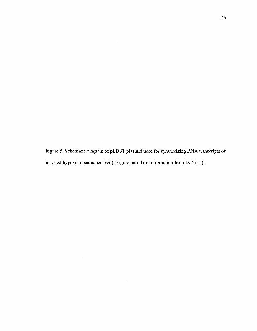
Plasmid pLDST (Figure 5) carries the full-length infectious cDNA clone of the
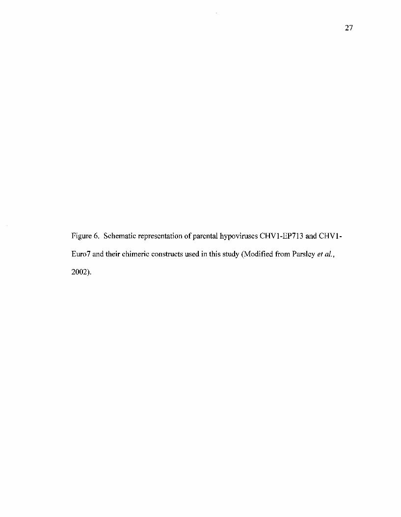
parental hypovirus CHV1-EP713. Chimeric hypoviruses (Chen et al. 2000); Rl, R2, R6,
R12, R13, R14 (Figure 6) and mutants of Ap29 (deletion of p29 region of CHV1-EP713),
Ap40 (deletion of p40 region of CHV1-EP713), and Ap69 (deletion of p60 region of
CHV1-EP713) are all described in Suzuki et al. (2003). pE7T-N plasmid contains the
full length cDNA clone of CHVl-Euro7. Infection of each virus construct was
established by transfection of virus-free EP155 isolate with in vitro synthesized viral
transcripts, as explained by Chen et al (1994) with minor modifications. Plasmids PXH1
and PXH3 were constructed by insertion of p29 and p40 coding regions of CHV1-EP713
ORF A (Craven et al, 1993) into the pCPXHYl transformation vector (Figure 7),
respectively. This vector has unique cloning sites between the C. parasitica
glyceraldehyde-3-phosphate dehydrogenase gene promoter (Vgpd) and terminator (Tgpd)
for expression of cDNA inserts. This vector was used for construction of transformation
plasmids CHVl-EP713-p29-flag, CHVl-Euro7-p29-fiag, CHI, CH2, CH4, CH8 and
CHVl-EP713-p29stop. Viral infection was achieved via transformation into a virus-free
EP155 isolate. All C. parasitica isolates that were transfected or transformed were
maintained on PDA, and then stored on glass microfibre filters at -20 °C.
25
Figure 5. Schematic diagram of pLDST plasmid used for synthesizing RNA transcripts of
inserted hypo virus sequence (red) (Figure based on information from D. Nuss).

26
Amp
/T7 polymerase promoter
FolyAtail Spel
viral sequence kpnl
Xbal
Sad
Sspl
EcoRI
PLDST (15432hp)
27
Figure 6. Schematic representation of parental hypoviruses CHV1-EP713 and CHV1-
Euro7 and their chimeric constructs used in this study (Modified from Parsley et al.,
2002).
28
ORFB ORFA
Nor I V i n , s ?on40 i fl 1
p2yp|W p4g 1 CHV1-EP713 5 * c d 3 ^ - * > "
Nsil
RdRpl Hel. 3'
CHVl«Euro7 5*"CZZf jr™ 3
Rl 3'
R2
R13 5H C 3'
R14 J- 3*
R12 y-r
R6

29
Figure 7. pCPXHYl vector used for plasmid transformations (Craven et al., 1993)

30
PUC19
pCPXHYl (8342bp)
JKpiiI t t l tul
Split
S Topd
31
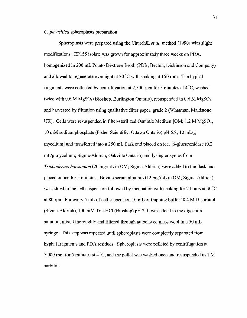
C. parasitica spheroplasts preparation
Spheroplasts were prepared using the Churchill et al. method (1990) with slight
modifications. EP155 isolate was grown for approximately three weeks on PDA,
homogenized in 200 mL Potato Dextrose Broth (PDB; Becton, Dickinson and Company)
and allowed to regenerate overnight at 30 C with shaking at 150 rpm. The hyphal
fragments were collected by centrifugation at 2,500 rpm for 5 minutes at 4 C, washed
twice with 0.6 M MgSC>4 (Bioshop, Burlington Ontario), resuspended in 0.6 M MgS04,
and harvested by filtration using qualitative filter paper, grade 2 (Whatman, Maidstone,
UK). Cells were resuspended in filter-sterilized Osmotic Medium [OM; 1.2 M MgS04,
10 mM sodium phosphate (Fisher Scientific, Ottawa Ontario) pH 5.8; 10 mL/g
mycelium] and transferred into a 250 mL flask and placed on ice. (3-glucuronidase (0.2
mL/g mycelium; Sigma-Aldrich, Oakville Ontario) and lysing enzymes from
Trichoderma harzianum (20 mg/mL in OM; Sigma-Aldrich) were added to the flask and
placed on ice for 5 minutes. Bovine serum albumin (12 mg/mL in OM; Sigma-Aldrich)
was added to the cell suspension followed by incubation with shaking for 2 hours at 30 C
at 80 rpm. For every 5 mL of cell suspension 10 mL of trapping buffer [0.4 M D-sorbitol
(Sigma-Aldrich), 100 mM Tris-HCl (Bioshop) pH 7.0] was added to the digestion
solution, mixed thoroughly and filtered through autoclaved glass wool in a 50 mL
syringe. This step was repeated until spheroplasts were completely separated from
hyphal fragments and PDA residues. Spheroplasts were pelleted by centrifugation at
5,000 rpm for 5 minutes at 4 C, and the pellet was washed once and resuspended in 1 M
sorbitol.

Fungal transfection
32
in vitro transcription
Plasmid pLDST was used for synthesis of viral coding strand transcripts.
Transcripts that were synthesized using this system included the full-length infectious
cDNA clone of the parental hypovirus CHV1-EP713, chimeric hypoviruses (Rl, R2, R6,
R12, R13 and R14) and deletion mutants (Ap29, Ap40 and Ap69). These transcribed
segments are represented in Figure 13. The transcribed region within the pLDST plasmid
is flanked at the 5' end by a T7 bacteriophage promoter (TAATACGACTCACTATAG)
and at the 3' terminus by 22 adenosine residues, which coincides with that of the L-
dsRNA natural polyadenylate tail. Plasmids were amplified in DH5a cells (Invitrogen,
Burlington, Ontario), followed by extraction using Wizard plus miniprep DNA
purification system (Promega, Nepean Ontario). For transcription, plasmids were
linearized by digestion with Spel (New England biolabs, Pickering Ontario),
phenol/chloroform-extracted, EtOH-precipitated and resuspended in nuclease-free water.
Prior to the transcription step glass instruments and centrifuge tubes were baked
overnight at 240 °C in order to inactive any contaminating RNase. In addition, RNase-
free pipette tips were used and all the solutions were treated with 0.1% diethyl
pyrocarbonate (DEPC; Sigma-Aldrich) for 24 hours and autoclaved. Transcripts were
generated using MEGAscript T7 Kit (Applied Biosystems, Streetsville Ontario).
Reagents were mixed in the following order; nuclease free water (to final volume of 20
\xL), NTPs (8 |^L), S^el-linearized pLDST plasmid (1 ug), 10X reaction buffer (2 uL),
and enzyme mix (2 uL). The reactions were incubated at 37 °C for 2 hours followed by
addition of 1 uL DNAsel and 15 minutes incubation at 37 °C. 15 uL of nuclease-free
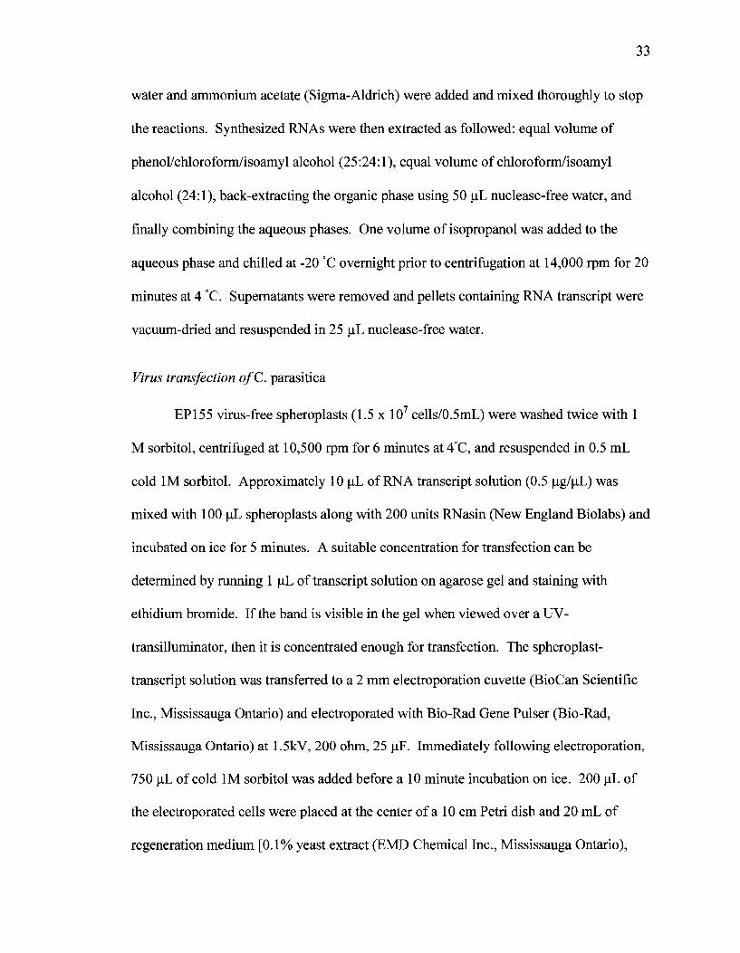
33
water and ammonium acetate (Sigma-Aldrich) were added and mixed thoroughly to stop
the reactions. Synthesized RNAs were then extracted as followed: equal volume of
phenol/chloroform/isoamyl alcohol (25:24:1), equal volume of chloroform/isoamyl
alcohol (24:1), back-extracting the organic phase using 50 uL nuclease-free water, and
finally combining the aqueous phases. One volume of isopropanol was added to the
aqueous phase and chilled at -20 °C overnight prior to centrifugation at 14,000 rpm for 20
minutes at 4 °C. Supernatants were removed and pellets containing RNA transcript were
vacuum-dried and resuspended in 25 uL nuclease-free water.
Virus transfection ofC. parasitica
EP155 virus-free spheroplasts (1.5 x 10⁷ cells/0.5mL) were washed twice with 1
M sorbitol, centrifuged at 10,500 rpm for 6 minutes at 4°C, and resuspended in 0.5 mL
cold 1M sorbitol. Approximately 10 uL of RNA transcript solution (0.5 ug/uL) was
mixed with 100 uL spheroplasts along with 200 units RNasin (New England Biolabs) and
incubated on ice for 5 minutes. A suitable concentration for transfection can be
determined by running 1 uL of transcript solution on agarose gel and staining with
ethidium bromide. If the band is visible in the gel when viewed over a UV-
transilluminator, then it is concentrated enough for transfection. The spheroplast-
transcript solution was transferred to a 2 mm electroporation cuvette (BioCan Scientific
Inc., Mississauga Ontario) and electroporated with Bio-Rad Gene Pulser (Bio-Rad,
Mississauga Ontario) at 1.5kV, 200 ohm, 25 uF. Immediately following electroporation,
750 uL of cold 1M sorbitol was added before a 10 minute incubation on ice. 200 uL of
the electroporated cells were placed at the center of a 10 cm Petri dish and 20 mL of
regeneration medium [0.1% yeast extract (EMD Chemical Inc., Mississauga Ontario),
34
0.1% casein hydrolysate (Fluka, Oakville Ontario), 1M sucrose (Bioshop), and 1.6% agar
(Bioshop)] was poured into the dish from the edge of the plate towards the center. This
keeps the cells at the center of the dish during the regeneration process. Plates were
incubated overnight at room temperature, at which time excess sorbitol was discarded
and plates were inverted and incubated at room temperature. After 2-4 days, when
hyphal growth was evident, small pieces of agar containing hyphae from the edge of the
cultures were transferred to PDA plates for observation and further work.
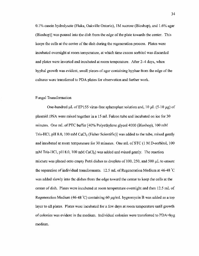
Fungal Transformation
One-hundred uL of EP155 virus-free spheroplast solution and, 10 uL (5-10 |j,g) of
plasmid DNA were mixed together in a 15 mL Falcon tube and incubated on ice for 30
minutes. One mL of PTC buffer [40% Polyethylene glycol 4000 (Bioshop), 100 mM
Tris-HCl, pH 8.0, 100 mM CaCb (Fisher Scientific)] was added to the tube, mixed gently
and incubated at room temperature for 30 minutes. One mL of STC (1 M D-sorbitol, 100
mM Tris-HCl, pH 8.0, 100 mM CaC^) was added and mixed gently. The reaction
mixture was placed onto empty Petri dishes as droplets of 100, 250, and 500 uL to ensure
the separation of individual transformants. 12.5 mL of Regeneration Medium at 46-48 °C
was added slowly into the dishes from the edge toward the center to keep the cells at the
center of dish. Plates were incubated at room temperature overnight and then 12.5 mL of
Regeneration Medium (46-48 °C) containing 60 jj,g/mL hygromycin B was added as a top
layer to all plates. Plates were incubated for a few days at room temperature until growth
of colonies was evident in the medium. Individual colonies were transferred to PDA+hyg
medium.
35
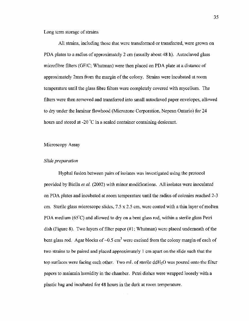
Long term storage of strains
All strains, including those that were transformed or transfected, were grown on
PDA plates to a radius of approximately 2 cm (usually about 48 h). Autoclaved glass
microfibre filters (GF/C; Whatman) were then placed on PDA plate at a distance of
approximately 2mm from the margin of the colony. Strains were incubated at room
temperature until the glass fibre filters were completely covered with mycelium. The
filters were then removed and transferred into small autoclaved paper envelopes, allowed
to dry under the laminar flowhood (Microzone Corporation, Nepean Ontario) for 24
hours and stored at -20 °C in a sealed container containing desiccant.
Microscopy Assay
Slide preparation
Hyphal fusion between pairs of isolates was investigated using the protocol
provided by Biella et al. (2002) with minor modifications. All isolates were inoculated
on PDA plates and incubated at room temperature until the radius of colonies reached 2-3
cm. Sterile glass microscope slides, 7.5 x 2.5 cm, were coated with a thin layer of molten
PDA medium (65°C) and allowed to dry on a bent glass rod, within a sterile glass Petri
dish (Figure 8). Two layers of filter paper (#1; Whatman) were placed underneath of the
bent glass rod. Agar blocks of ~0.5 cm3 were excised from the colony margin of each of
two strains to be paired and placed approximately 1 cm apart on the slide such that the
top surfaces were facing each other. Two mL of sterile ddFbO was poured onto the filter
papers to maintain humidity in the chamber. Petri dishes were wrapped loosely with a
plastic bag and incubated for 48 hours in the dark at room temperature.
36
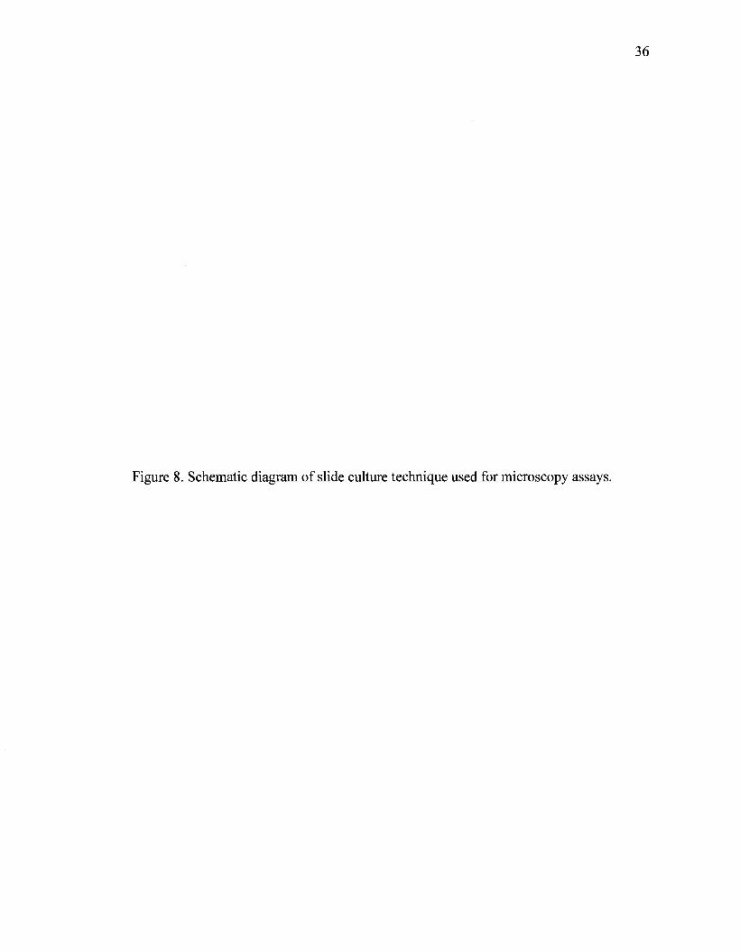
Figure 8. Schematic diagram of slide culture technique used for microscopy assays.

37
Bent glass rod
Wet Whatman filter paper
Glass microscope slide covered with PDA
Glass Petri dish
38
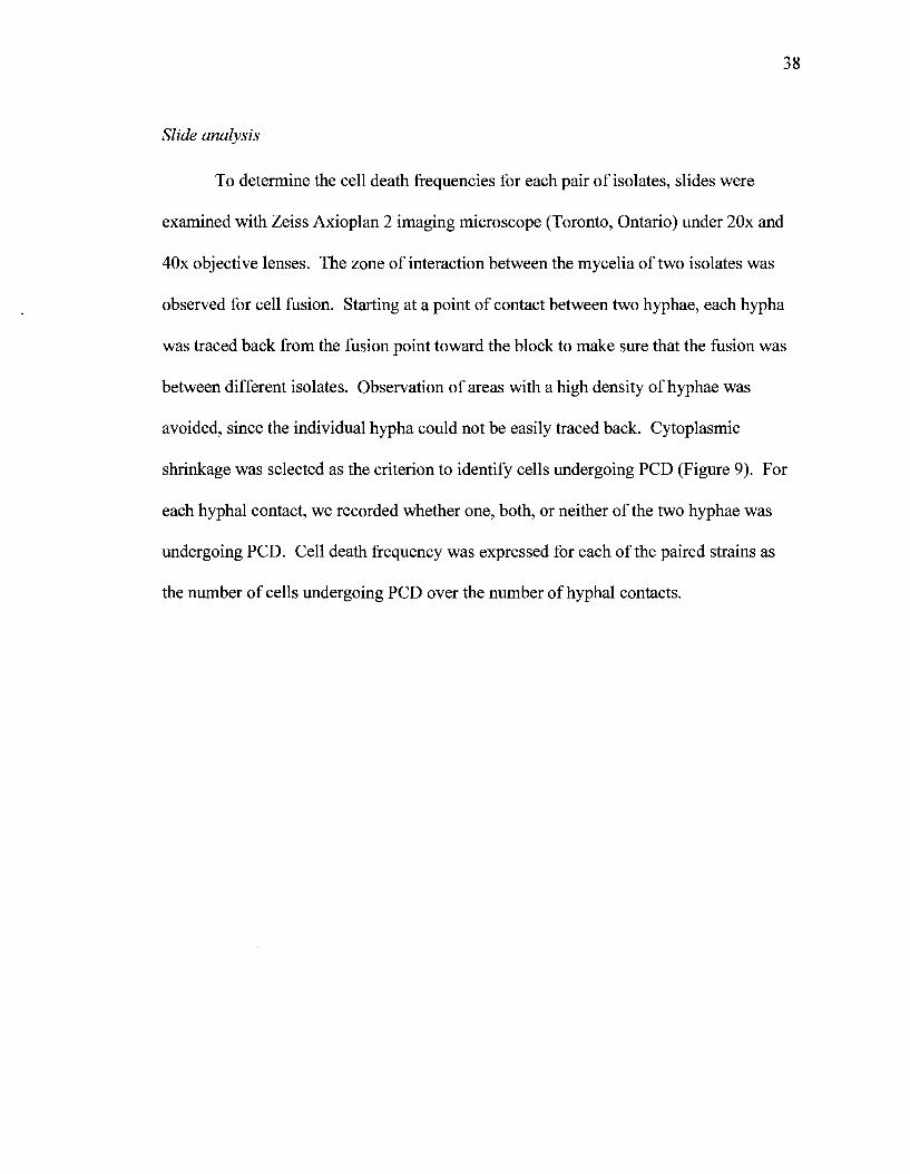
Slide analysis
To determine the cell death frequencies for each pair of isolates, slides were
examined with Zeiss Axioplan 2 imaging microscope (Toronto, Ontario) under 20x and
40x objective lenses. The zone of interaction between the mycelia of two isolates was
observed for cell fusion. Starting at a point of contact between two hyphae, each hypha
was traced back from the fusion point toward the block to make sure that the fusion was
between different isolates. Observation of areas with a high density of hyphae was
avoided, since the individual hypha could not be easily traced back. Cytoplasmic
shrinkage was selected as the criterion to identify cells undergoing PCD (Figure 9). For
each hyphal contact, we recorded whether one, both, or neither of the two hyphae was
undergoing PCD. Cell death frequency was expressed for each of the paired strains as
the number of cells undergoing PCD over the number of hyphal contacts.
39
Figure 9. Types of interactions observed under 40x magnification. Point of fusion in
each panel is shown with arrow, (a) Compatible interaction without sign of PCD in
hyphae of either strain, (b & c) Incompatible fusions between p74-3 and EP155 infected
with CHVlEuro7. In panel B isolate EP155-CHVlEuro7 shows vacuolization (v). In
panel C, EP155-CHVlEuro7 shows cytoplasmic collapse (c). (d) Fusion of P74-3 and
EP155-CHV1EP713 with both isolates displaying different stages of cell death, [v =
vacuolization; s = cytoplasmic shrinkage; c = collapse of cell contents]

40
41
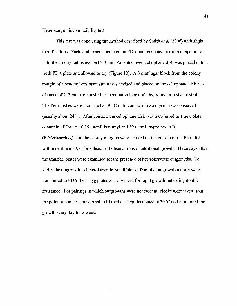
Heterokaryon incompatibility test
This test was done using the method described by Smith et al (2006) with slight
modifications. Each strain was inoculated on PDA and incubated at room temperature
until the colony radius reached 2-3 cm. An autoclaved cellophane disk was placed onto a
fresh PDA plate and allowed to dry (Figure 10). A3 mm3 agar block from the colony
margin of a benomyl-resistant strain was excised and placed on the cellophane disk at a
distance of 2-3 mm from a similar inoculation block of a hygromycin-resistant strain.
The Petri dishes were incubated at 30 °C until contact of two mycelia was observed
(usually about 24 h). After contact, the cellophane disk was transferred to a new plate
containing PDA and 0.15 ug/mL benomyl and 30 ug/mL hygromycin B
(PDA+ben+hyg), and the colony margins were marked on the bottom of the Petri dish
with indelible marker for subsequent observations of additional growth. Three days after
the transfer, plates were examined for the presence of heterokaryotic outgrowths. To
verify the outgrowth as heterokaryotic, small blocks from the outgrowth margin were
transferred to PDA+ben+hyg plates and observed for rapid growth indicating double
resistance. For pairings in which outgrowths were not evident, blocks were taken from
the point of contact, transferred to PDA+ben+hyg, incubated at 30 °C and monitored for
growth every day for a week.
42
Figure 10. Schematic representation of technique used for heterokaryon incompatibility
test.
43
benR strain grown on PDA plate
PDA plate overlayed with cellophane disk and inoculated with benR
and hygR strains
one day
Transfer of Cellophane disk after growth of strains to
PDA+Hyg + Ben
hygR strain grown on PDA plate
Examine for heterokaryon sectors (hk) on PDA + Hyg + Ben plate
44
Mycelial incompatibility assay
Strains to be paired were inoculated separately onto PDA and incubated at room
temperature until the colony radius reached 2-3 cm. Two mm agar blocks from the
colony margin of each strain were placed approximately 5 mm apart on PDAg plates
(Powell, 1995). One litre of this medium contained 39 g PDA, 7 g malt extract (Becton,
Dickinson and Company), 2 g yeast extract, 0.8 g tannic acid (BDH Chemicals Ltd, Poole
England), 0.1 g methionine (Life Technologies Inc., Grand Island NY), 2 mg biotin (Life
Technologies Inc.), 2 mg thiamine (added after autoclaving; Life Technologies Inc.), 20 g
agar and 50 mg bromocresol green (BDH Chemicals Ltd). Each PDAg plate contained
eight pairs of isolates. Plates were incubated at 25 °C for 10 days in the dark. Pairings
that did not form a dark line were scored as compatible and pairs that did form a dark line
were scored as incompatible.

45
Table 1. Strains used in this study.

isolate
EP155B 22.1H P1-11B P10-2B P11-23B P1-5B P1-6B P42-7B P74-3B P78-6B P4-4B P5-1B P10-18B MJ1-3-20B P24-33B P32-35B EP29 16L1 EP392 39L1 P67-7 P21-14
vie genotype1
2211-22 2211-22 2211-22 2211-22 1211-22 1211-22 2111-22 2111-22 2221-22 2221-22 2212-22 2212-22 2211-12 2211-12 2211-21 2211-21 2221-11 2111-11 2121-22 2122-22
EU-typ(
EU-5(v( EU-5(vc EU-5(V( EU-5(vc EU-31 EU-31 EU-6 EU-6 EU-60 EU-60 EU-l(vc EU-l(vc EU-21 EU-21 EU-18 EU-18 EU-46 EU-9 EU-59 EU-38
;40) ;40) ;40) ;40)
;10) ;10)
origin2
ATCC#38755 from A. Churchill hy^ of EP155-2 from A. Churchill PC17xPC7 V029 x PC7 P l - l l x P 3 - 3 PC17xPC7 PC17xPC7 P l - l l x L I 1 3 P l - l lxP50-16 P50-4xP74-7 V054 x PC7 V 0 5 4 x V 0 6 4 V029 x PC7 P17-8xJA17 P4-4 x SA26 PC39 x VA35 MG Milgroom MG Milgroom Pl-6xP25-6 P49-6 x P54-5
1 Alleles of each vie locus are given in order from vie J, 2, 3, 4, -, 6, 7. Alleles that differed from those of EP155 are in bold.
2 All strains expect EP155B and 22.1H are from crosses set up by MG Milgroom of Cornell University, Ithaca NY.
47
Polymerase chain reactions
All constructs along with their specific primers prepared by polymerase chain
reactions (PCRs) are listed in Table 2. Primers were manually designed and the melting
temperatures were adjusted using OligoAnalyzer program (Integrated DNA
Technologies, http://www.idtdna.com/home/home.aspx). All the primers were
synthesized by Sigma-Aldrich Company. Expected amplicon sizes were verified by
agarose gel electrophoresis in 1% (w/v) agarose (Amresco, Guelph, Ontario) and
ethidium bromide (Sigma-Aldrich) in lx TAE [0.04 M Tris-acetate, 0.001 M EDTA
(Sigma-Aldrich)] and visualized by Alpha-Innotech UV illuminator (San Leandro, CA).
Amplicons of the correct size were then sequenced at StemCore Laboratories (OHPJ,
Ottawa Ontario). PCR reactions were done in a final volume of 20 uL and contained 2
uL of template (5 ng/uL), IX PCR buffer (Bioshop), forward and reverse primers, 1.5 uL
MgCl2 (25 mM; Bioshop), 0.4 uL dNTPs (100 mM; Bioshop) and 0.2 uL Taq DNA
polymerase (5 units/uL; Bioshop). PCR reactions were performed in a Biometra
(Goettingen, Germany) TGradient thermocycler with the following reaction conditions:
initial denaturation at 95 C for 5 minutes then 30 cycles of 95 C for 30 seconds,
annealing for 30 seconds (temperature varies depending on the primer pairs), and
o o
polymerization at 72 C for 30 seconds. These cycle parameters were followed by 72 C
for 10 minutes and 4 C for an indefinite time. CHI and CH2 chimeras were constructed
by the site-directed mutagenesis technique with sequence overlap extension as shown in
(Figure 11). The procedure consisted of two rounds of PCR. The first round resulted in
generation of two PCR products with overlapping ends from two different templates in
separate reactions. In the subsequent round, PCR fragments were combined in a reaction

48
to allow the overlapping ends to anneal and form a new template. The fused product was
amplified further by PCR with external primers.

49
Figure 11. Schematic representation of technique used for construction of chimeras.
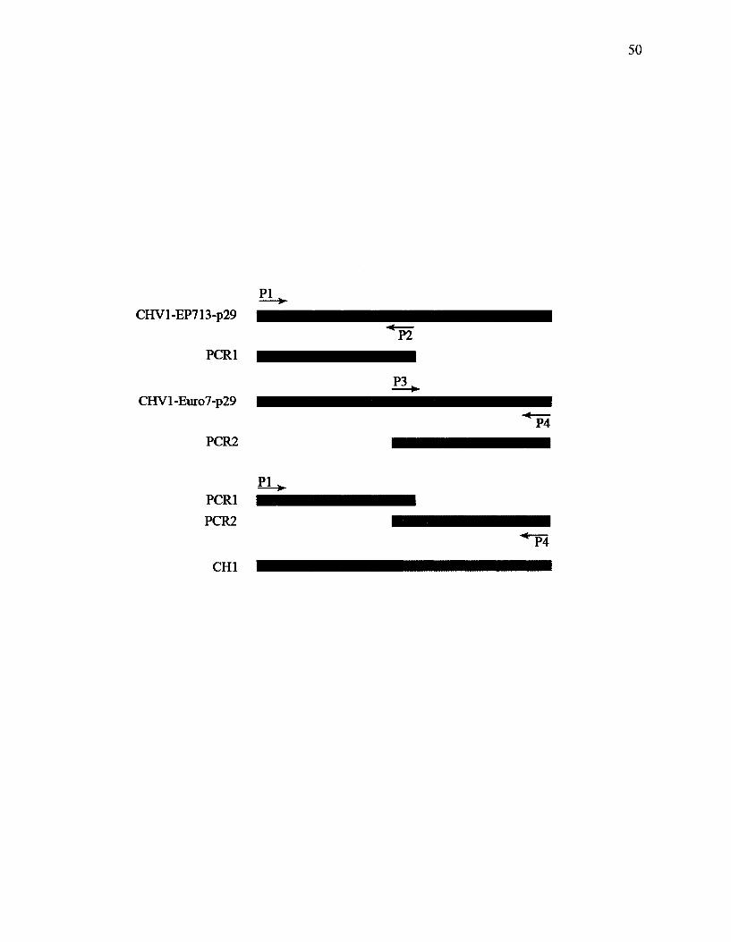
50
PI
CHVl-EP713-p29
PCR1
CHVl-Euro7-p29
PCR2
PCR1
PCR2
PI
CHI
P2
P3
P4
P4

Table 2. Transformation plasmids along with their oligonucleotide primers used in this
study.

Tra
nsfa
mat
ion
plas
mid
EP7
13-p
29-f
lag
Eur
o7^>
29-il
ag
CH
I
cm
CH
4
CH
8
EP7
13-p
29st
op
Tra
nsfo
nnan
t de
sign
atio
n
EP1
55 (C
HV
1-E
P713
-p29
-fl
ag)
EP1
55 (C
HV
1-E
uro7
-p29
-fl
ag)
EP1
55 (
CH
I)
EP1
55 (C
H2)
EP1
55 (C
H4)
EP1
55 (C
H8)
EP1
55(E
P713
-p2
9sto
p)
Prim
ers
(5'>
3')
GC
CA
GG
TA
CC
AT
GG
CT
CA
AT
TA
AG
AA
AA
CC
CA
GT
JAkC
GC
AT
XX
CIT
GT
CA
IVG
TC
GT
CC
WG
TA
lTC
GC
CQ
CC
AA
l C
CG
GG
CA
AG
G
CC
AG
GT
AC
CA
TG
TC
TT
GT
CT
TA
GA
AA
AC
CC
lAkO
XA
TX
XC
Tm
TC
ArC
GT
CG
TC
CT
rGT
AA
rCG
TIG
CC
GA
T
OC
GG
GC
GA
GG
GC
CA
GG
TA
CC
AT
GG
CT
TC
Crr
CG
TA
CG
TT
GT
TC
CA
A
TT
GG
AA
CA
AC
GT
AC
GA
AG
GA
TA
AC
GC
AT
GC
CT
TG
TC
AT
CG
G
CC
AG
GT
AC
CA
TG
TC
TT
GT
TC
CT
TC
GT
AC
GT
rGrr
CC
AA
T
TG
GA
AC
AA
CG
TA
CG
AA
GG
A
TA
AC
GC
AT
GC
CT
TG
TC
AT
CG
TC
GT
CC
TT
GT
AA
TC
GC
CG
CC
AA
T
CC
GG
GC
AA
G
GC
CA
G6T
AC
CA
TG
0CT
CA
AT
TA
AG
AA
AA
CC
CA
GT
AA
OX
AT
GC
TT
XC
TIO
TC
AT
CG
TC
GT
CC
TT
arA
AT
CQ
TC
TQ
CC
GC
CA
GG
TA
CC
AT
GT
TG
CaC
AC
CC
CT
GA
CG
GG
GT
AT
GT
AA
CG
CA
TG
CT
TA
CT
TO
rC^T
CG
rcG
rCC
ITG
W^r
cGC
CG
CC
A
GC
CA
GG
TA
CC
AT
GG
CT
! AA
TG
AA
GA
IAG
(XC
AG
T
TA
A(X
K:A
TG
CC
7TC
rC47
UC
?rC
GrC
C7T
G7:
44rC
GC
CG
CC
AA
T
CC
GG
GC
AA
G
Tm
Co
55.0
62
0
50.0
64.0
60.0
63.0
63.0
66.9
60
.0
63.0
63
.0
62
0
55.0
62
0
67.0
66.0
56.0
62
0
Com
men
ts
kpnl
site
is s
how
n in
bol
d
SpJi
site
and
flag
seq
uenc
e ar
e sh
own
in b
old
and
ital
ic r
espe
ctiv
ely
KpA
site
is s
how
n in
bol
d
Spft
i. si
te a
nd fl
ag s
eque
nce
are
show
n in
bol
d an
d ita
lic, r
espe
ctiv
ely
Forw
ard
and
reve
rse
pmne
rs t
o am
plify
1-3
44
posi
tion
from
EP7
13-p
29 te
mpl
ate
Forw
ard
and
reve
rse
prim
ers
to a
mpl
ify 3
24-7
47
posi
tion
fiom
Eur
o7-p
29 te
mpl
ate
Forw
ard
and
reve
rse
prim
ers
to a
mpl
ify 1
-344
po
sitio
n fr
om E
uix>
7-p2
9 te
mpl
ate
Forw
ard
and
reve
rse
pmne
rs t
o am
plify
324
-747
po
sitio
n fr
om E
P713
-p29
tem
plat
e
Kpn
l site
is s
how
n in
bol
d
SpK
site
, sto
p co
don
and
flag
sequ
ence
are
sho
wn
in
bold
, gre
y co
la' a
nd it
alic
, re
spec
tivel
y
Kpn
l site
and
star
t cod
on a
re sh
own
in b
old
and
italic
, re
spec
tivel
y Sp
li s
ite, s
top
codo
n an
d fl
ag se
quen
ce a
re s
how
n in
bo
ld, g
rey
cola
- and
ital
ic, r
espe
ctiv
ely
Kpt
i. si
te a
nd su
bstit
uted
nuc
leot
ides
are
sho
wn
in
bold
and
gre
y co
lor,
res
pctiv
ley
Spli
site
and
flag
seq
uenc
e ai
e sh
own
in b
old
and
italic
, res
pect
ivel
y
to

53
DNA digestion and ligation
All PCR products were cloned into pCR2.1-TOPO vector using the TOPO TA
cloning kit and the manufacture's protocol (Invitrogen). Inserts were then excised from
TOPO TA by mixing 1 ug of DNA, 5 uL of digestion buffer (#1; New England Biolabs),
1 uL BSA (10 mg/mL; New England Biolabs), 2 units each ofSphl and Knpl (New
o
England) and up to 50 uL of ddHaO. Digests were incubated at 37 C for 2 hours,
confirmed for expected fragment sizes and recovered following agarose gel
electrophoresis. After DNA recovery, inserts were ligated into vector pCPXHYl. Each
ligation reaction was composed of 30 ng of insert, 90 ng of vector digested as described
above with Sphl and Knpl, 2 uL of 10X T4 DNA ligase buffer (New England Biolabs)
and 5 units of T4 DNA ligase (New England Biolabs). Reactions were brought to 25 uL o
of ddtkO and incubated overnight at 4 C.
DNA recovery from agarose gels
Following agarose gel electrophoresis, DNA bands were excised and mixed with
3 volumes of Nal (Sigma-Aldrich) and incubated at 55 C to melt the gel material. Ten
|oL of Glass Milk was then added to the solution and incubated overnight at 4 C. The
sample was centrifuged at 14,000 rpm for 30 seconds, the supernatant discarded and the
pellet was washed twice with new wash buffer (50 mM NaCl, 10 mM Tris pH 7.6, 2.5
mM EDTA, 50% EtOH). The pellet was dried briefly and then resuspended in 15 \xL
ddHiO, incubated for 5 minutes at 55 °C and centrifuged at 14,000 rpm for 1 minute.
This step was repeated after transferring the supernatant to a fresh tube. Any glass milk

54
residue was removed by centrifugation followed by measuring the DNA concentration
with a NanoDrop 1000 spectrophotometer (Thermo Scientific, Wilmington, DE).
Fungal protein extraction
Total protein was extracted from each of three isolates of EP155 transformed with
either CHVl-EP713-p29-flag, CHVl-Euro7-p29-flag or CHVl-EP713-p29. For this,
each strain was inoculated onto a cellophane membrane that was overlayed on PDA and
incubated at room temperature for 15 to 20 days. The mycelium was scraped off the
membrane using a sterile spatula, and ground in a mortar and pestle with liquid nitrogen.
Powder was transferred to an eppendorf tube along with 100 mg of 0.5 mm glass beads
(BioSpec Products Inc., Bartlesville OK) and 1 volume of disruption buffer [20 mM
Tris.Cl (pH 7.9), 10 mM MgCl2 (Sigma-Aldrich), 1 mM EDTA, 5% glycerol (Sigma-
Aldrich), 0.1 M DTT (Sigma-Aldrich), 0.3 M ammonium sulfate (BDH Chemicals Ltd.),
100 mM PMSF (MP biomedicals, Solon OH) and 1 protease inhibitor cocktail tablets
(Roche)]. The tubes were vortexed at 13,000 rpm for 30 seconds and incubated on ice for
2 minutes. This step was repeated five times and tubes were centrifuged for one hour at
12,000 rpm at 4 C. Supernatant was removed and the protein concentration of each
sample was measured using the Quick Start Bradford Protein Assay protocol from Bio-
Rad.
Affinity Purification using M2 Anti-flag Agarose Beads
The following components were mixed together in a 15 mL Falcon tube on ice:
100 uL of anti-FLAG M2 antibodies bound to agarose beads (Sigma-Aldrich) along with
55
1 mL of Buffer A [50% 2X Buffer {40 mM Tris-HCl (pH 8.0), 10 mM MgCl2, 20%
glycerol, 0.2% Tween 20}, 25% 2 M KC1, 0.1% PMSF, 0.07% (3-mercaptoethanol
(Sigma-Aldrich), and one mini-protease tablet]. The mixture was centrifuged for 3
minutes at 1000 rpm and the supernatant was removed. The pellet was further
centrifuged for 30 seconds at 1000 rpm to completely remove the buffer. Extracted
protein (250 fig) was added to the tube, mixed gently and incubated for one hour at 4 C
on a platform shaker (Reliable Scientific Inc., Nesbit MS). After incubation, samples
were centrifuged at 1000 rpm for 3 minutes and supernatant was removed. The agarose
beads were washed three times with Buffer A to remove any unbound protein. Bound
protein was then eluted by incubating the beads with 30 ug/mL FLAG peptide (Sigma-
Aldrich) for 30 minutes at 4 C on a platform shaker followed by centrifugation at 1000
o
rpm for 3 minutes at 4 C.
SDS polyacrylamide gel electrophoresis (SDS-PAGE)
In this section a BioRad Mini Protean 3 gel system (Bio-Rad) was used. The
glass plates were cleaned using ethanol and assembled according to the manufacturer's
instructions. The resolving gel (12% acrylamide) was prepared by mixing the following
components in order in a 50 mL falcon tube; 4800 uL ddtbO, 6300 uL of 40%
acrylamide/Bis solution (Bio-Rad), 3800 uL of 1.5 M Tris (pH 8.8), 150 uL 10% sodium
dodecyl sulfate (SDS; Bioshop), and 150 uL of 10% ammonium persulfate (APS; Sigma-
Aldrich). Ten uL of N, N, N', N'-Tetramethylethylenediamine (TEMED; Sigma-
Aldrich) was added to the mixture, swirled quickly and poured into the gap between the
glass plates. One cm of space at the top was left empty and overlaid with ddHiO. The
56
gel was allowed to polymerize for 20 minutes at room temperature. After
polymerization, water was removed and the stacking gel was added. The stacking gel
was prepared by mixing; 4060 uL of ddH20, 880 uL of 40% acrylamide/Bis solution,
1660 uL of 1.5 M Tris (pH 8.8), 66 uL 10% SDS and 66.8 uL of 10% APS. After
addition of 10 uL of TEMED, the solution was swirled quickly and added onto the
polymerized resolving gel. A clean comb was inserted into the stacking gel without
creating any air bubbles and the gel was incubated at room temperature for another 20
minutes. After polymerization and removing the comb, the gel was mounted to the
electrophoresis apparatus, placed in the chamber and filled with reservoir buffer [25 mM
Tris, 250 mM glycine (Sigma-Aldrich), 0.1% SDS]. Each protein sample was prepared
by mixing 15 ug of protein with 10 uL of SDS gel-loading buffer (50 mM Tris, pH 6.8,
100 mM dithiothreitol, 2% SDS, 0.1 % bromophenol blue, 10% glycerol) and incubating
at 80 C for 10 minutes. Samples were centrifuged at high speed for one minute and
loaded into wells of the gel along with 10 uL of precision plus protein kaleidoscope
standard (Bio-Rad). After attaching the apparatus to the power supply, a voltage of 150
V was applied until the dye moved into the resolving gel. The voltage was then increased
to 225 V and allowed to run until the bromophenol blue reached to the bottom of the
resolving gel.
Staining SDS-PAGE gels with coomassie brilliant blue in preparation for mass
spectroscopy
All the following steps should be carried out using clean dishes and the gel should
be covered with saran wrap at all times and never touched with bare hands. The SDS-
57
PAGE gel was fixed in 100 mL of methanol/acetic acid (Anachemia, Mississauga ON)
(25%: 10% v/v). A staining solution was prepared by dissolving 0.25 g of Coomassie
Brilliant blue (Fluka) in 90 mL of 50% methanol and 10 mL of glacial acetic acid
followed by filtration using 25 mm syringe filters (Cellulose Acetate membrane; Ultident,
St. Laurent QC). The gel was immersed in 100 mL of staining solution and incubated for
2 hours on a platform shaker. After removing the staining solution, the gel was destained
for 4 hours in 25% methanol with 10% acetic acid and 65% ddLkO. The destaining
solution was changed 3 to 4 times during this period. The gel was stored in 7.5% acetic
acid, wrapped in saran wrap at 4 C. Mass spectroscopy was performed at the National
Research Council Canada (Ottawa, Ontario) using Liquid Chromatography coupled to
electrospray ionization mass spectrometry (LC/MS).
Western blotting
Two porous pads, two Mini Trans-Blot filter papers (Bio-Rad) and one piece of
nitrocellulose membrane (Bio-Rad) cut to similar size as the SDS-PAGE gel were soaked
in transfer buffer (39 mM glycine, 48 mM Tris base, 20% methanol) prior to assembly.
After running SDS-PAGE gels, the apparatus was disassembled and the glass plates were
slowly separated from the gel. The stacking gel was removed from the resolving gel and
the transfer apparatus was set up on a gel holder cassette in the order from anode (+) to
cathode (-): porous pad, filter paper, nitrocellulose filter, gel, filter paper and porous pad.
A glass pipette was used to roll and remove air bubbles as each layer was applied. The
cassette was placed into the trans-blotting module and positioned into the buffer tank
along with an ice pack and fresh transfer buffer. Thirty volts was applied overnight at
58
o
4 C. On the following day, the transfer apparatus was disassembled and the protein
standard markers were marked on the membrane using a fine pencil. The membrane was
incubated at room temperature in the following solutions: 1 hour with gentle shaking in
blocking solution [5% fat-free dried milk in 1% Tris-Buffered Saline Tween-20 (TBST; 9
mL of Tris HC1 pH8.0, 27 mL of 5 M NaCl and 900 mL Tween-20 in 900 ddH20)];
vigorous shaking 4x15 min each in 1% TBST; very gently shaking for 1 hour in 10 mL
of blocking solution with 10 x uL anti-FLAG M2 peroxidase conjugate antibody
(1:1000) (Sigma-Aldrich); finally, vigorous shaking for 15 min in 1% TBST. After
removing the excess buffer, the membrane was incubated with Chemiluminescent
peroxidase substrate 3 (Sigma-Aldrich) according to manufacture's protocol, wrapped in
plastic wrap and exposed to Kodak BioMax light film (Sigma-Aldrich) for 2 minutes and
developed with Kodak developer (Sigma-Aldrich) and Kodak fixer (Sigma-Aldrich)
according to manufacture's instructions.
RESULTS
Effect of viral infection on C. parasitica phenotype
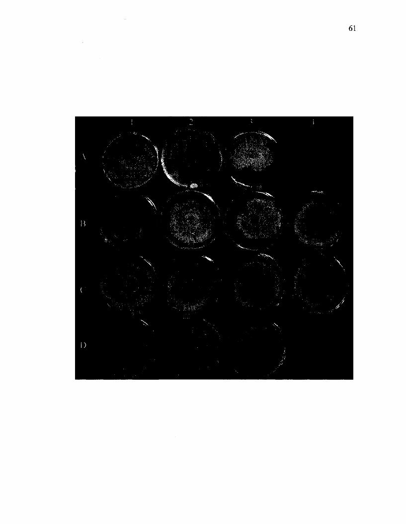
The effects of wild-type and chimeric hypoviruses in altering the phenotypic traits
of C. parasitica are shown in Figure 12. The key characteristics that were evaluated were
growth rate and mycelial pigmentation patterns. Compared to the EP155 virus-free strain
(A2 in Figure 12), the EP155 strain transfected with CHV1-EP713 had a slower growth
rate and lacked conidia resulting in a non-pigmented colony (data not shown). In
contrast, the CHVl-Euro7 transfected EP155 strain (A3) had an intermediate level of
pigmentation and slightly slower growth rate compared to the EP155 strain. The
59
phenotypes of EP155 strains bearing chimeric viruses were compared with the above
virus-free, and transfected CHV1-EP713 and CHVl-Euro7 forms of EP155 (Table 3).
The transfected isolates all had similar growth rates to CHVl-Euro7, except for those
bearing the Rl (Bl) and R6 (B3) chimeras, which grew more slowly, similar to EP155-
CHV1EP713. In terms of pigmentation, most isolates had a similar pattern as EP155-
CHVlEuro7. Among transfected strains only EP155 (R6) and EP155 (R13) (CI) had
phenotypes that were similar to those described in the papers published by Nuss's group
(Chen et al, 2000; Suzuki et al, 2003).

60
Figure 12. Colony morphologies of C. parasitica isolates with and without transfected
viruses. Across the top are isolates of virus-free P74-3 (Al) and EP155 (A2) and EP155
transfected with CHVl-Euro7 (A3) and chimeric viruses (Bl, Rl; B2, R2; B3, R6; B4,
R12; CI, R13; C2, R14; C3, PXH3; C4, PXH1; Dl, Ap29; D2, Ap69; D3, Ap40). Isolates
were maintained on PDA for 10 days on the laboratory bench at room temperature prior
to photographing.
61
62
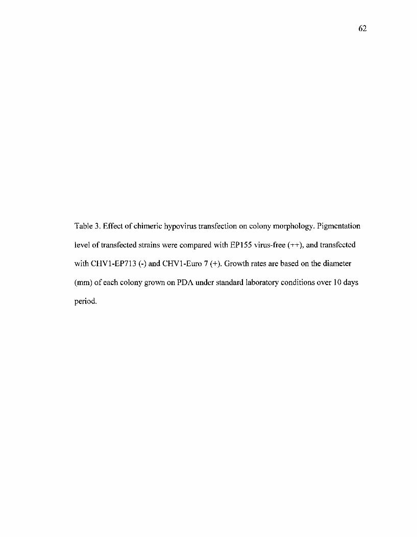
Table 3. Effect of chimeric hypo virus transfection on colony morphology. Pigmentation
level of transfected strains were compared with EP155 virus-free (++), and transfected
with CHV1-EP713 (-) and CHVl-Euro 7 (+). Growth rates are based on the diameter
(mm) of each colony grown on PDA under standard laboratory conditions over 10 days
period.
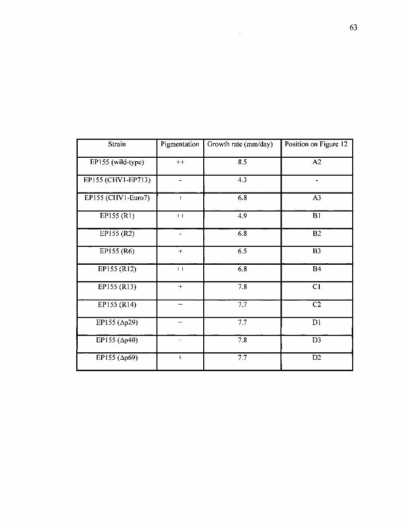
63
Strain
EP155 (wild-type)
EP155 (CHV1-EP713)
EP155 (CHVl-Euro7)
EP155(Rl)
EP155(R2)
EP155(R6)
EP155(R12)
EP155(R13)
EP155(R14)
EP155(Ap29)
EP155(Ap40)
EP155 (Ap69)
Pigmentation
++
-
+
++
-
+
++
+
+
+
+
+
Growth rate (mm/day)
8.5
4.3
6.8
4.9
6.8
6.5
6.8
7.8
7.7
7.7
7.8
7.7
Position on Figure 12
A2
-
A3
Bl
B2
B3
B4
CI
C2
Dl
D3
D2
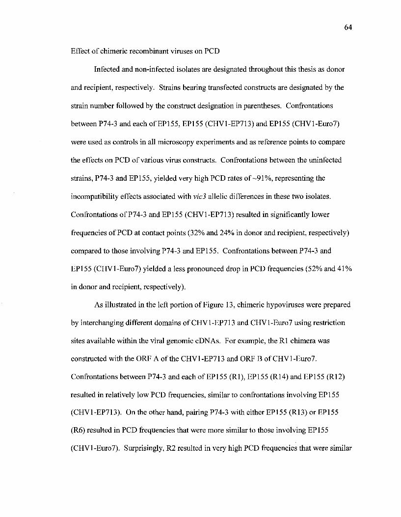
64
Effect of chimeric recombinant viruses on PCD
Infected and non-infected isolates are designated throughout this thesis as donor
and recipient, respectively. Strains bearing transfected constructs are designated by the
strain number followed by the construct designation in parentheses. Confrontations
between P74-3 and each of EP155, EP155 (CHV1-EP713) and EP155 (CHVl-Euro7)
were used as controls in all microscopy experiments and as reference points to compare
the effects on PCD of various virus constructs. Confrontations between the uninfected
strains, P74-3 and EP155, yielded very high PCD rates of-91%, representing the
incompatibility effects associated with vicS allelic differences in these two isolates.
Confrontations of P74-3 and EP155 (CHV1-EP713) resulted in significantly lower
frequencies of PCD at contact points (32% and 24% in donor and recipient, respectively)
compared to those involving P74-3 and EP155. Confrontations between P74-3 and
EP155 (CHVl-Euro7) yielded a less pronounced drop in PCD frequencies (52% and 41%
in donor and recipient, respectively).
As illustrated in the left portion of Figure 13, chimeric hypoviruses were prepared
by interchanging different domains of CHV1-EP713 and CHVl-Euro7 using restriction
sites available within the viral genomic cDNAs. For example, the Rl chimera was
constructed with the ORF A of the CHV1-EP713 and ORF B of CHVl-Euro7.
Confrontations between P74-3 and each of EP155 (Rl), EP155 (R14) and EP155 (R12)
resulted in relatively low PCD frequencies, similar to confrontations involving EP155
(CHV1-EP713). On the other hand, pairing P74-3 with either EP155 (R13) or EP155
(R6) resulted in PCD frequencies that were more similar to those involving EP155
(CHVl-Euro7). Surprisingly, R2 resulted in very high PCD frequencies that were similar
65
to those involving the non-infected strains. Nevertheless, the combined results suggested
that v/c3-associated PCD reduction is correlated to ORF A characteristics.
Strains transfected with CHV1-EP713 constructs with deletions in ORF A,
namely Ap69, Ap40 and Ap29, suggested that regions encoding both p29 and p40 proteins
are involved in reducing the rate (or frequency) of v/cJ-associated PCD (Figure 13). The
Ap69 construct had negligible effects on PCD compared to uninfected pairings.
Deletions of either p29 or p40 had intermediate reductions in PCD compared to the
noninfected control confrontations. In any event, analysis of these deletion constructs
further support our previous results that ORF A is associated with reduction in vic3-
associated PCD. Further supporting this ORF A role, EP155 strains were transformed to
constitutively express copies of either p29 or p40. Confronting either of these two strains
with P74-3 yielded very few hyphal contacts that had evidence of PCD (Figure 13).
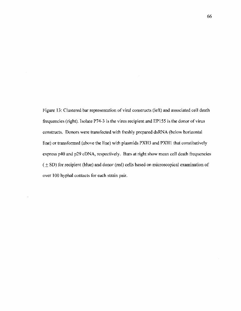
66
Figure 13: Clustered bar representation of viral constructs (left) and associated cell death
frequencies (right). Isolate P74-3 is the virus recipient and EP155 is the donor of virus
constructs. Donors were transfected with freshly prepared dsRNA (below horizontal
line) or transformed (above the line) with plasmids PXH3 and PXH1 that constitutively
express p40 and p29 cDNA, respectively. Bars at right show mean cell death frequencies
(+ SD) for recipient (blue) and donor (red) cells based on microscopical examination of
over 100 hyphal contacts for each strain pair.

67
Virus/Construct Recipient Donor
EP713-p40
EP713-p29
ORFB
~i T"
1 0.8 0.6 0.4 0.2 0 0.2 0.4 0.6 0.8 Cell Death Frequency (+SD)
I a Q.

68
Vegetative incompatibility
Heterokaryon incompatibility tests
This assay was performed by confronting each of the hygromycin-resistant
isolates 22.1H, EP155 (PXH1) or EP155 (PXH3) against each of 13 benomyl-resistant
isolates containing the same or different alleles at one of each of six vie loci. Strain
22.1H is a hygromycin resistant derivative of EP155. As detailed in Figure 7 plasmids
PXH1 and PXH3 confer hygromycin resistance and constitutively express cDNAs of the
p29 and p40 components of ORF A, respectively. The colonies were inoculated as pairs
and allowed to grow together on cellophane overlaid on PDA. After contact, the
cellophane with paired colonies was transferred to PDA+hyg+ben medium and observed
for 1 week for the presence or absence of heterokaryotic outgrowths (Figure 14).
Outgrowths were observed when pairs of isolates contained the same alleles at all six vie
loci or differed only at vic4, as previously reported by Smith et al. (2006). In order to
verify the outgrowths as heterokaryotic, small blocks from the margins of outgrowths
were transferred to the PDA+ben+hyg plates and the growth rates were recorded (Figure
15). In cases where no outgrowths were observed, blocks were taken from the point of
contact between the pairs of isolates. Pairings that differed at any of vie 1, 2, 3, 6, or 7 did
not show any outgrowths regardless of whether p29 or p40 were expressed, and were not
observed to grow upon transfer to fresh PDA+ben+hyg. Heterokaryon incompatibility
characteristics were not, therefore, altered by the viral components that had effectively
reduced PCD.
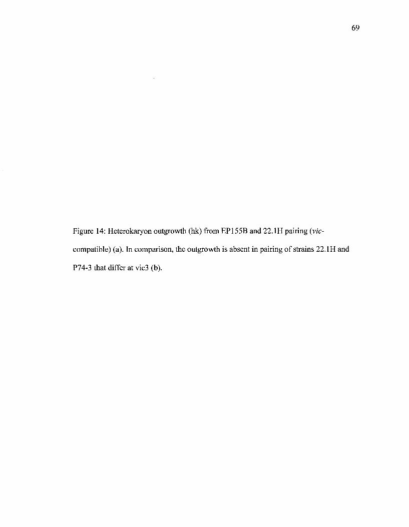
69
Figure 14: Heterokaryon outgrowth (hk) from EP155B and 22.1H pairing (vic-
compatible) (a). In comparison, the outgrowth is absent in pairing of strains 22.1H and
P74-3 that differ at vic3 (b).
70
71
Figure 15: Growth rates of heterokaryotic sectors obtained from confronting hygromycin-
resistant isolates of 22.1H, PXH1 or PXH3 against each of the 13 benomyl-resistant
isolates labeled across the bottom, vie differences for pairings are specified across the
top.
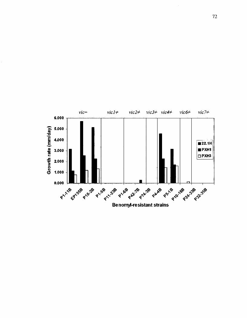
72
v/c= v/cif4 vic2¥i vic3J= vic4+ vic6i= vie 71= c Ann
(mm
/day
)
i i i
£ 3.000 (0
£ 2.000
O 1-000
0.000
h 1 1 1
|L LI MJ $
f <v •
1
$ < "
1 1 | IL 1 I.
f -n
• 22.1H • PXH1 • PXH3
f «
i
Benomyl-resistant strains
73
Mycelial incompatibility assay
This test was performed on PDAg plates containing bromocresol green by
confronting each of the three isolates EP155, EP155 (PXH1) and EP155 (PXH3)
separately with each of 14 benomyl-resistant isolates having specific vie genotype
differences (Figure 16). The formation of a dark line (barrage) is associated with the
fusion and subsequent cell death between two incompatible mycelia. As expected, no
barrage was seen in pairings of v/ocompatible strains, vz'c-incompatible strains
(including those expressing p29 or p40) developed well-defined barrages with the
exception of EP155 (PXH1) + P4-4B, which differ at vic4. vic4 is considered the
"weakest" vie locus (Biella et al, 2002), since vic4 heteroallelism does not interfere with
heterokaryon formation (see Figure 15). That the barrage line between vzW-incompatible
strains EP155 (PXH1) and P4-4B was poorly defined or absent (based on multiple
barrage tests) suggests that expression of the p29 element has a subtle effect on mycelial
incompatibility. Overall, based on these barrage tests neither p29 nor p40 significantly
alters the mycelial incompatibility characteristics associated with vie gene differences.

74
Figure 16. Mycelial incompatibility tests between strain pairs with defined vie
characteristics. Each of three strains, EP155, EP155 transformed with PXH1, and EP155
transformed with PXH3, was paired separately to 14 strains that have same vie genotype
to EP155 or differ at a single vie locus, as designated across the top. Barrage lines are
evident as dark lines where the paired colonies grow together.

*
75
76
Protein-protein interaction
To study how the p29 protein effects PCD rates based on our microscopy assays,
we made flag-tagged constructs of p29 from each of CHV1-EP713 and CHVl-Euro7 and
expressed these in the fungal host, EP155. It was hypothesized that these constructs may
allow for the isolation and identification of fungal protein(s) that interact with p29.
Crude protein extracts of strains containing CHVl-EP713-p29-flag and CHVl-Euro7-
p29-flag constructs had different banding patterns from each other as visualized through
Western blotting (Figure 17). The molecular weight of both of these p29-flag proteins
was expected to be -30 KDa. Three bands with molecular weights of ~60 KDa, 42 KDa
and 30 KDa were found to be common in both flag-tagged protein samples. No immuno-
reactive proteins were observed in protein from a no-flag control. Affinity purification
using M2 anti-flag agarose beads yielded two bands with molecular weights of 60 KDa
and 30 KDa (Figure 18). Unfortunately sequence analysis of these bands mostly revealed
antibody fragments.
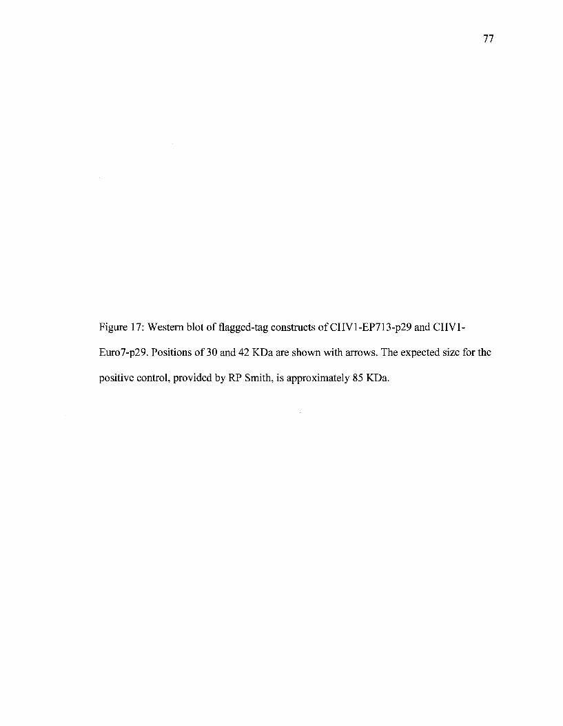
77
Figure 17: Western blot of flagged-tag constructs of CHVl-EP713-p29 and CHV1-
Euro7-p29. Positions of 30 and 42 KDa are shown with arrows. The expected size for the
positive control, provided by RP Smith, is approximately 85 KDa.

78
EP155 transf EP155 transf
60 «$ i
i—H
ON <N •
o
w w
ON
I
CO
SP i
ON CN OH I cn T—I
8P a i
ON
OH I
O O
ON
m r--P-t W
i
o o o o o
o o
U U U +
-85KDa 42KDa ~30KDa
Long exp (-30 min)
Short exp (~2 min)

Figure 18: Affinity purified samples of CHVl-EP713-p29 and CHVl-Euro7-p29 using
M2 anti-flag agarose beads followed by SDS-PAGE and Coomassie Brilliant Blue
staining revealed two bands with molecular weights of -60 KDa and -30 KDa.
80
81
p29 sequence analysis
The p29 sequence was analyzed in order to identify the domain responsible for
vz'ci-associated PCD reduction. CHI and CH2 chimeras of p29 were constructed by
replacing the first 300 bp of CHVl-Euro7-p29-flag with the first 300bp of CHV1-EP713-
p29 (Figure 19). CH4 and CH8 constructs were made by deleting the last and first 150
nucleotides, respectively, from CHVl-EP713-p29. These fragments were inserted behind
the Pgpd promoter within the plasmid pCPXHYl and then transformed into EP155.
Microscopy assays were performed to examine PCD frequencies in contacts between
these transformed strains and P74-3 (Figure 19). Expression of each of these constructs
resulted in a reduction in observed PCD frequencies over that of no-virus controls. All of
the constructs, except chimera CH2, had PCD rates that resembled those associated with
expression of the p29-Euro7 construct. Of these modified forms of p29, CH2, which has
the 5' end of p29-Euro7 and the 3' end of p29-EP713, had the greatest effect on PCD
reduction. This suggested that the 3' portion of the p29 gene is the major player in
reducing PCD, but this is not supported by results with the CH8 construct. The CH8
construct has the 5' region of p29-EP713 deleted but maintains all of its 3' region. In
general, these results suggest that there is no well-defined subdomain within p29 that is
involved in the reduction of vz'cJ-associated PCD.

82
Figure 19: Clustered bar representation of viral constructs (left) and associated PCD
frequencies (right). These results are means (±SD) based on a minimum of 50 contacts
for each pairing. The constructs were expressed in EP155 (the donor) while P74-3 (the
recipient) was virus free.

83
Virus/Construct Recipient Donor
EP155 (CHS)
EP155 (CH4)
EP155(CH2)
EP155(CH1)
EP155(p29-Euro7)
EP155(p29-EP713)
EP155
T 1 r 0.5 0 0.5
Cell Death Frequency (+SD)

84
p29, RNA silencing and nonself recognition
Segers el al. (2007) showed that dicer-like protein dcl-2, but not dcl-1 is essential
for RNA silencing in C. parasitica. Our microscopy analysis of v/c3-associated PCD
using dcl-1 and dcl-2 mutants revealed interesting parallels between RNA silencing and
Wc-associated PCD (Figure 20). Confrontations between strains P74-3 and EP155 (Adcl-
2), and between P74-3 and EP155 (A.dcl-l/Adcl-2), resulted in reduced v/c3-associated
PCD rates that were similar to those observed when the EP155 partner expresses CHV1-
EP713-p29. However, hyphal contacts between P74-3 and EP155 (Adcl-1) had high rates
of PCD in cells surrounding hyphal contact points. This suggested that the virus may be
using the RNA silencing pathway to affect reduced PCD rates in incompatible fusion
events. This possible link suggested that the p29 effect on PCD could be mediated by an
RNA effector.
In order to determine if the effect of p29 on PCD reduction is mediated by RNA
or by protein, three stop codons were inserted into the beginning of the CHV1-EP713-
p29 sequence (Figure 21). Introduction of these translation termination sites eliminated
the p29 phenotypic effects of reduced colony growth rate and decreased pigmentation and
conidiation in EP155 colonies (Figure 22), but did not compromise the cell death
frequency effect (Figure 20). These results strongly suggest that reduction in vic3-
associated PCD is mediated by RNA synthesized from ORF A, and specifically from the
p29-encoding region. Furthermore, the data indicates that the p29 effects on colony
morphology (pigmentation patterns, etc.) are independent of reduction in vz'ci-associated
PCD; the former is dependent with p29 protein synthesis while the later is likely
associated with p29 RNA. Furthermore, the involvement of p29 RNA in reducing PCD
85
is consistent with the idea that this element interferes somehow with the RNA silencing
pathway.
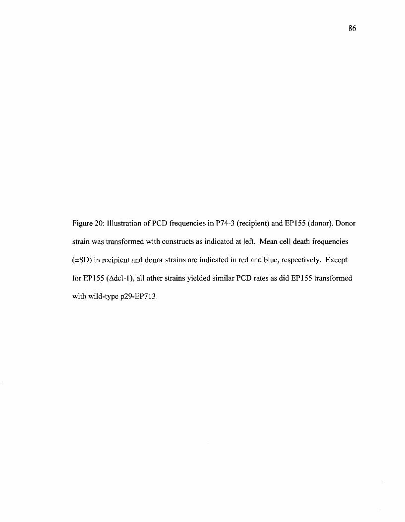
86
Figure 20: Illustration of PCD frequencies in P74-3 (recipient) and EP155 (donor). Donor
strain was transformed with constructs as indicated at left. Mean cell death frequencies
(±SD) in recipient and donor strains are indicated in red and blue, respectively. Except
for EP155 (Adcl-1), all other strains yielded similar PCD rates as did EP155 transformed
with wild-type p29-EP713.
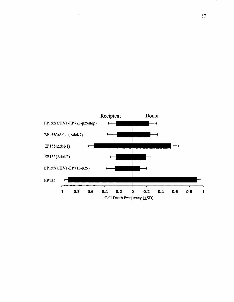
87
Recipient Donor EP155(CHVl-EP713-p29stop)
EP155(Adcl-l/,Adcl-2)
EP155(Adcl-l)
EP155(Adcl-2)
EP155(CHVl-EP713-p29)
EP155
0.8 0.6 0.4 0.2 0 0.2 0.4 0.6 0.8 1 Cell Death Frequency (±SD)
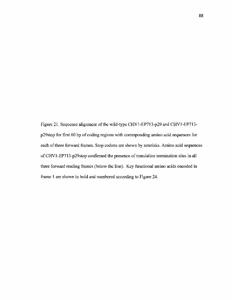
88
Figure 21. Sequence alignment of the wild-type CHVl-EP713-p29 and CHV1-EP713-
p29stop for first 60 bp of coding regions with corresponding amino acid sequences for
each of three forward frames. Stop codons are shown by asterisks. Amino acid sequences
of CHVl-EP713-p29stop confirmed the presence of translation termination sites in all
three forward reading frames (below the line). Key functional amino acids encoded in
frame 1 are shown in bold and numbered according to Figure 24.

ON
0
0
MA
QL
RK
PS
QS
LV
LS
ES
VD
PT
CH
Vl-E
P7
13
-p2
9 A
TG
GC
TC
AA
TT
AA
GA
AA
AC
CC
AG
TC
AG
AG
TC
TG
GT
GC
TC
TC
TG
AA
AG
TG
TT
GA
TC
CT
AC
T
CH
Vl-E
P7
13
-p2
9s
top
AT
GG
CT
TA
AT
GA
AG
AT
AG
CC
CA
GT
CA
GA
GT
CT
GG
TG
CT
CT
CT
GA
AA
GT
GT
TG
AT
CC
TA
CT
MA
**
R*
PS
QS
LV
LS
ES
VD
PT
Frame 1
25
70 72
73
MA* * R*
PSQSLVLSESVDPTTVDPFVSVRTEEVVPAGCITLWEYRSCGDVPGPLSHGDLRRLRTPDGVCKCQVHFELPTVLKS
162
GSTGTVPEHPAVLAAFIGRPRRCSLEQRTKELDSRFLQLVHGGLPARPSYMIARPPRPVRGLCSSRNGSLAQFGQGYCYLSAI
215
VDSARWRVARTTGWCVRVADYLRLLQWVGRRSFGSFQIEKSAVDHVYHVVVDAEYQSEQDGALFYQAILGLAEKDPLARJPG
248 249
Frame 2
WLNEDSPVRVWCSLKVLILLQWILSSA*GRKRWSLRVA*PYGSTETHVATCLARCRMVISGVCAPLTGYVSRSTLSCRPCSRVV
PPERSRNTPRCWLPSLAGLAGVPWNNVRRSWIPDSFSWCMVASLRGHHT*LRARPGQFEACAHLGTVPLLSLGRATVISRPL*T
AQDGASPVPPVGVYVWQTILGCYNGLVAGPLAPFRLRKAPSTMFITWSSTLSISLSRMGPFSTKPFWVWPRRIPLPGLA
Frame 3
GLMKIAQSESGAL* KC* SYYSGSFRQREDGRGGPCGLHNPMGVQRLMWRRAWPVVAW* SPAFAHP* RGM* VPGPL* VADRAQEW
FHRNGPGTPRGVGCLHWQASPVFLGTTYEGVGFPIPSVGAWWPPCEAIIHDCAPAQASSRPVLISERFPCSVWAGLLLSLGHCR
QRKMARRPYHRLVCTCGRLS* AATMGWSPVLWLLSD* EKRRRPCLSRGRRR* VSV* AGWGPFLPSHFGFGREGSPCPDWR

90
Figure 22: Colony morphology of EP155 virus-free (left), EP155 (CHV1-EP713-
p29stop) (middle) and EP155 (CHVl-EP713-p29) (right). All isolates were grown at
room temperature for 10 days.

91

vie pathway dissection
In this section cell death frequencies were measured using pairs of isolates that
differed at one of vic2, 3, 6 or 7, as well as strain pairs having multiple vie differences.
PCD frequencies for each pair were analyzed in the presence and absence of CHV1-
EP713-p29, expressed by the EP155 strain. All PCD frequencies were converted to a
ratio of 1-i/ni, where "i" is the PCD frequency from pairing in which p29 was expressed
in EP155 strain and "ni" is the PCD frequency from the corresponding non-infected
pairing (Table 4). These ratios are also plotted in Figure 23 where a higher value
represents a more pronounced effect of the virus element on cell death reduction.
From Table 4 and Figure 23 it can be seen that the presence of p29 resulted in
significant reductions in PCD for all confrontations tested in which there was a single vie
gene difference. In some cases, but not in all, where there were multiple vie differences
an increased rate of PCD was observed. For example, paired isolates that differed at one
of vic2 or vic3 showed significant cell death reductions compared to the pairing in which
there were differences at both vie2 and vie3. Likewise, vie3£, 6£, 7^ differences had
relatively high PCD frequencies compared to the effects of single differences at each of
these three loci. This suggests that there is a "weakening" of the virus (p29) effect on
PCD-reduction when there are multiple vie differences. In contrast, pairings with
differences at both vic6 and vic7 did not apparently differ in PCD frequencies from
pairings that were incompatible for one of vic6 or vic7. While the interactions between
multiple vie gene products and hypovirus factors are likely complex, this preliminary
analysis suggests that insights into the pathways may be gained using hypovirus as a type
of genetic probe. It may be hypothesized based on this data, for example, that vic2 and

93
vie3 act independently, and thus additively, while vic6 and vic7 operate within a common
pathway.
94
Table 4: Effect of p29 expression on cell death frequencies between pairs of isolates that
differ at the single or multiple vie loci. For each confrontation, EP155 (donor) had or did
not have p29 from CHV1-EP713, as indicated, i = PCD frequency when donor contains
and expresses p29 construct, ni = PCD frequency when donor does not contain p29
construct
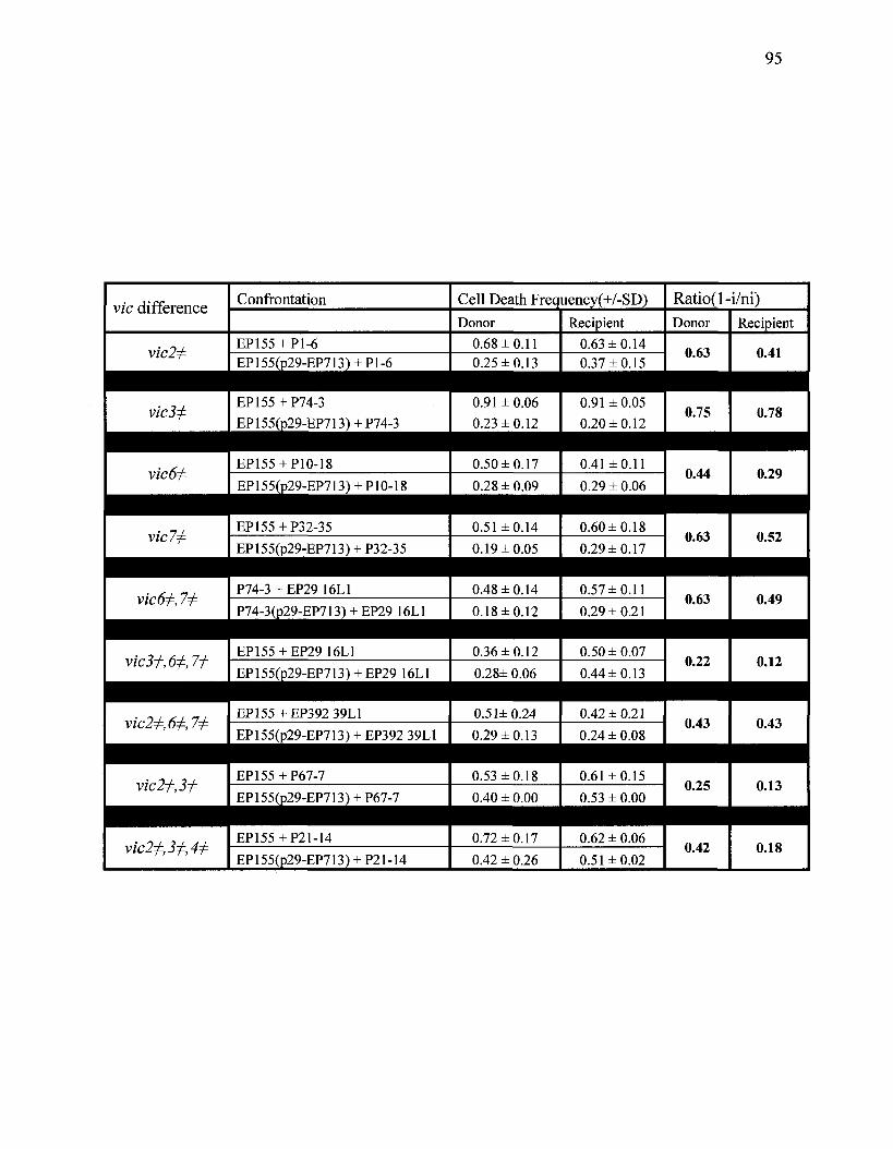
95
vie difference
vic2^
Confrontation
EP155 + P1-6 EP155(p29-EP713) + Pl-6
Cell Death Frequency(+/-SD)
Donor
0.68 ±0.11 0.25 ±0.13
Recipient
0.63 ±0.14 0.37 ±0.15
Ratio(l-i/ni) Donor
0.63
Recipient
0.41
vic3j:
vic6^
vie 7^
vic6^, 7^
vic3j^, 6^, 7^
vic2+M,7±
vie 2^,3^
vic2±,3+A*
EP155+P74-3
EP155(p29-EP713) + P74-3 0.91 ± 0.06
0.23 ±0.12
EP155+P32-35
EP155(p29-EP713) + P32-35
P74-3 + EP29 16L1
0.51 ±0.14
P74-3(p29-EP713) + EP29 16L1
EP155 + EP29 16L1
0.19 ±0.05
0.48 ±0.14
EP155(p29-EP713) + EP29 16L1
EP155 + EP392 39L1
0.18±0.12
0.36 ±0.12
EP155(p29-EP713) + EP392 39L1
0.28± 0.06
0.51± 0.24
0.29 ±0.13
EP155 + P21-14
EP155(p29-EP713) + P21-14
0.72 ±0.17
0.91 ± 0.05
0.20 ±0.12
0.60 ±0.18
0.29 ±0.17
0.57 ±0.11
0.29 ±0.21
0.50 ± 0.07
0.44 ±0.13
0.42 ±0.21
0.24 ± 0.08
0.62 ± 0.06
0.75 0.78
EP155+P10-18
EP155(p29-EP713) + P10-18
0.50 ±0.17
0.28 ± 0.09
0.41 ±0.11
0.29 ± 0.06
EP155 + P67-7
EP155(p29-EP713) + P67-7
0.53 ±0.18
0.40 ± 0.00
0.61 ±0.15
0.53 ± 0.00
0.42 ± 0.26 0.51 ±0.02
0.44
0.63
0.63
0.22
0.43
0.25
0.42
0.29
0.52
0.49
0.12
0.43
0.13
0.18
96
Figure 23. Scatter plot representing a comparison between cell death ratio of isolates
involving single and double vie differences of vic6 and 7 (A) and vic2 and 3 (B).
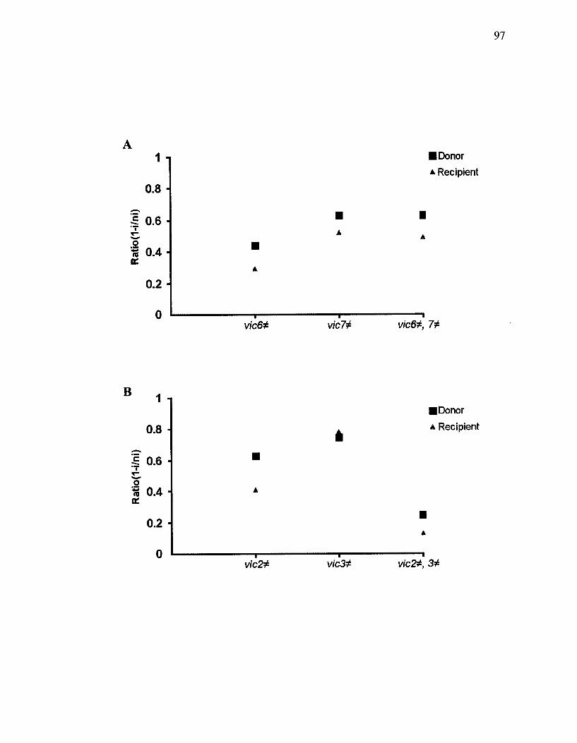
97
1
0.8
1 0.6 H
H 0.4 DC
0.2
0
•Donor
* Recipient
vied* vic72 vic6+, 7*
B 1
0.8
• Donor
* Recipient
•5 0.6 "T T -
% 0.4
0.2
0 vic2* vic3* vic&, 3*
98
DISCUSSION
Devastation of the American chestnut tree by the parasitic fungus, C. parasitica,
is one of the classical examples of a plant disease caused by introduction of an introduced
species. The appearance of the hypovirulent fungal strains and their subsequent effects
on chestnut blight prompted researchers to investigate this phenomenon. Preliminary
investigations revealed that hypovirulent phenotype conversion is caused by the presence
of dsRNA that was transmitted cytoplasmically from a hypovirulent to a virulent strain,
and this set the stage for large scale biological control efforts. However, this program did
not succeed apparently because of the fungal vegetative incompatibility system that
slowed or prevented virus transfer between strains. Vegetative incompatibility in C.
parasitica is associated with at least six unlinked vie loci (vie 1, 2,3, 4, 6, and 7), each
with at least two alleles (Cortesi & Milgroom, 1998). Microscopic examination of hyphal
incompatible fusions revealed some common characteristics associated with vegetatively
incompatible reactions such as; septal plugging, vacuolization of the cytoplasm, organelle
degradation and shrinkage of the cytoplasm away from cell wall (Jacobson et al, 1998;
Biella et al, 2002). These alterations in cells are similar to characteristics associated
with eukaryotic PCD. Therefore, PCD in fungi can occur as a result of fusion between
two vegetatively incompatible strains.
The occurrence of PCD under specific physiological and pathological conditions
has been widely reported for the past 150 years (Bursch el al., 2000). PCD has been
categorized into two main types: apoptosis and autophagy. Apoptosis was classified as a
type of cell death that is triggered by physiological or pathological stimuli to regulate the
cell population. Cells involved in apoptosis undergo specific biochemical and
99
physiological changes. These changes can take place in the cytoplasm and nucleus, as
well as plasma membrane. During the early stages of apoptosis, the cell integrity will be
altered. This transformation is identified as shrinkage and distancing of cells from
neighboring cells. Chromatin in the nucleus aggregates and is digested into fragments by
endonucleases. The nucleus will break down into several fragments that become
enclosed within apoptotic bodies. These apoptotic bodies are formed from intact
membranes that prevent leakage during the process of apoptosis (Lawen, 2003).
Autophagy is another type of programmed cell death that is involved in a wide range of
biological processes such as pathogen attack or other environmental stimuli, and cellular
remodeling during development (Yorimitsu & Klionsky, 2005). This process involves
sequestration of abnormal organelles in the cytoplasm followed by degradation and
recycling by the lysosome/vacuole.
The correlation between vegetative incompatibility and PCD associated with the
six vie genes in C. parasitica was investigated by Biella et al. (2002). They demonstrated
that there is a negative correlation between cell death and viral transmission when pairs
of isolates differed at single vie loci. Preliminary observations were also made by Biella
and co-workers that indicated that viral activity altered v/c-associated PCD. Our work
first set out to verify these earlier findings. This was done through a series of
microscopical examinations with virus infected and uninfected strains of EP155 paired
with P74-3, two stains that carry different alleles at vic3. A significant reduction in cell
death frequencies was observed in both the donor and recipient isolates upon fusion of
P74-3 with EP155 (CHV1-EP713), in comparison to the corresponding virus-free
isolates. Similarly, when P74-3 was paired with EP155 (CHVl-Euro7) a reduction in
PCD frequency was observed, although this effect was less dramatic than for P74-3 +
EP155 (CHV1-EP713) pairings. These observations corroborated earlier findings that a
virus element(s) was interacting with the fungal nonself recognition system to reduce
PCD.
In order to identify the virus element(s) responsible for vz'ci-associated PCD
reduction, a set of chimeric hypo viruses of CHVl-Euro7 and CHV1-EP713 was used.
These constructs were previously used by Chen et al. (2000) to determine the region of
the viral genome responsible for modifying fungal growth rate, canker and colony
morphology. In their study chimeras carrying specific regions of the virus genome
showed similar phenotypic alteration to the parental virus type. These similarities
allowed them to narrow down the genome to identify specific hypovirus domains that
contribute to particular morphological characteristics.
Our microscopy analyses revealed that the presence of chimeras Rl, R12 or R14
resulted in cell death frequencies that were similar to those of the parental virus, CHV1-
EP713. On the other hand, PCD frequencies in pairings with chimeras R13 and R6 were
similar to those involving CHVl-Euro7. These data indicated that the ORF A region was
involved in vz'cJ-associated PCD reduction. This was further supported by results with
Ap69, wherein the viral ORF A region was deleted. Expression of this construct resulted
in no appreciable reduction in PCD in pairings of EP155 (Ap69) + P74-3. Pairings with
Ap29 and Ap40 mutants from the CHV1-EP713 virus genome resulted in intermediate
cell death frequencies, suggesting that these proteins interact cooperatively with a
component(s) of the vic3-associated PCD pathway. Interestingly, compared to
experiments with transfected strains, strains that express transformed copies of CHV1-
101
EP713-p29 or CHVl-EP713-p40 had a more pronounced effect in reducing PCD
frequencies.
One possible explanation for these minor discrepancies between pairings with
transfected mutants and transformed strains could be due to the variable and unstable
nature of transfected strains, compared to the stable nature of the transformants. Other
evidence of instability of transfected strains is based on our morphological observations
of EP155 infected with chimeras carrying the CHV1-EP713 ORF B (R2, R14, R12 and
R6). Of these, only the morphology of EP155 carrying R2 was consistent with the
previous reports (Chen et ah, 2000). Discrepancies in colony morphology characteristics
could be due to virus abundance following the transfection stage or to variable rates of
loss of virus activity during subculturing since transfected strains tend to be quite
unstable (Nuss, personal communications). For these reasons, we used strains for our
experiments as soon as possible after transfection. Further instability of virus elements
was documented when the p29 or p40 regions were deleted from the viral genome
(Suzuki el ah, 2003). Such deletions were previously observed to result in a reduced
accumulation of viral RNA. Changes in the viral RNA content in some of our deletion
mutants may also have modified cell death frequency rates for some of the transfected
strains. The same study by Suzuki el ah showed that expressing p29 protein in Ap29
mutant strains increased the RNA accumulation level to that observed in wild-type virus
(Suzuki el ah, 2003). Analogous work could be done with our system to investigate if
p29 expression in Ap29 mutant strains would result in a more pronounced reduction in
cell death frequencies. Regardless of these possible instability issues, our results with
transfected strains were internally consistent, and were further supported by work with
102
transformed strains in implicating ORF A as the region responsible for altering vic3-
associated PCD rates.
Within ORF A, the p29 protein has been given a lot of attention due to its
multifunctionality (Suzuki et al, 1999, 2003; Choi et al. 1991; Konnin et al. 1991).
Figure 24 shows a summary of the domains and essential residues identified for p29 from
CHV1-EP713, including alteration of the host phenotype and autocatalytic cleavage.
Studies on Ap29 (lacking 88% of the P29 coding region) showed that, in addition to
p29's contribution to virus replication, p29 plays a major role in altering fungal
pigmentation patterns, conidiation, and laccase accumulation (Suzuki el al. 1999). Using
a gain of function assay and through progressive repair of the Ap29 mutant, Suzuki et al.
(2003) assigned the symptom determinant domain to the region extending from Phe25
through Gln73. The N-terminus portion of p29 and the HC-Pro papain-like protease
encoded by potyviruses were also shown to share sequence identity, specifically in four
conserved cysteine residues; Cys38, Cys48, Cys70, and Cys72 (Choi et al. 1991; Konnin
et al. 1991). Site-directed mutations of either residues Cys70 or Cys72 resulted in colony
morphology changes as well as substantial reduction in mycelial growth (Suzuki el al.
1999). Therefore, it was proposed that the specific secondary or tertiary conformational
constraints required for the functionality of the Phe25-Gln73 domain were dependent on
Cys70 and Cys72 residues. Our results provide evidence for an additional function of the
CHV1 ORF A, and specifically to the p29 protein coding region; that of modifying the
host nonself recognition response.
103
Figure 24. Schematic diagram of the CHV1-EP713 genome, showing essential domains
and residues in p29 coding region (modified from Suzuki et al, 1999). See Figure 21
(Frame 1) for amino acid sequence.
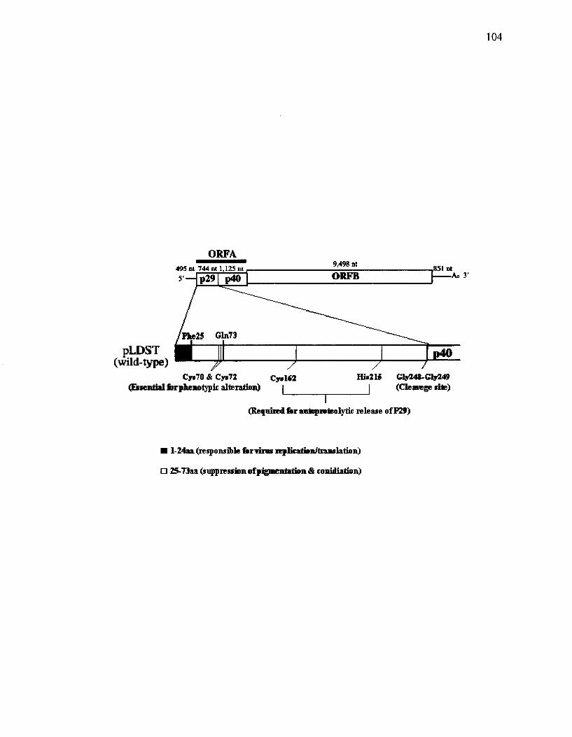
ORFA 495 M 744 nt 1,125 nt 5'-
9,498nt 851 nl
An 3'
|>LDST (wild-type) f
Cys70 & Cys72 (Essential forphenotypic alteration)
His215 Cly248-Gly249 I (Cleavege site)
(Required for autoproteolytic release of P29)
• l-24aa(responsible for virus replkation/translation)
D 25-73aa (suppression of pigmentation & conidiation)
105
Vegetative incompatibility
Vegetative incompatibility between fungal strains can be evaluated by
heterokaryon incompatibility testing and barrage tests. Heterokaryon incompatibility
tests determine whether two complementary nuclear types can be maintained in the same
hypha to enable stable heterokaryotic growth. Barrage testing is done by confronting two
strains to determine whether or not dead or dying cells and/or deposition of dark
pigments develop in the zone of contact. Unlike heterokaryon incompatibility, barrage
formation does not necessarily involve hyphal fusion. Contact between substances
diffused from either, or both strains and their cellular receptors may facilitate barrage
formation (Micali & Smith, 2005).
The results presented in this thesis are consistent with those of Smith et al. (2006),
which showed that heteroallelism at vie loci was associated with heterokaryon
incompatibility function for all vie loci with the exception of vic4. Both studies found
that differences at vic4, while causing barrage lines to form, did not impede heterokaryon
formation, growth or stability. One explanation for this vic4 anomoly is that this locus
may express extracellular factors that are toxic for isolates carrying the other allele. Such
factors have not been identified, but Rizwana and Powell (1995) suggested that cell
surface particles are involved in triggering heterokaryon incompatibility, by some vie
loci. This hypothesis was based on their observation that vicl and vic2 heterokaryon
incompatibility was bypassed upon protoplast fusion. In contrast to our observation that
expression of either p29 or p40 in the nuclear type significantly reduced v/ci-associated
PCD, heterokaryon and barrage test results were not significantly altered by virus factors.
The only possible exception to this was that the barrage line between EP155(PXH1)+ P4-

106
4, which differ at vic4, was indistinct or absent. This effect may be enhanced by
expressing the virus factors in both strains, rather than in just EP155, as was done in this
study.
Based on our comparisons of microscopy, heterokaryon and barrage assays, we
hypothesize that p29 and p40 operate by reducing the "rate" rather than "frequency" of
PCD. Figure 25 explains this point graphically. In this figure we plot hypothetical
functions of the number of PCD through time. For "WT" pairings of uninfected strains,
the number of cells undergoing PCD increases to a maximum and then levels out.
Confrontations that involve an infected strain have fewer PCD evident at the time of
microscopic examination. This could be accounted for by either a reduced rate of PCD or
a reduced frequency of cells undergoing PCD. We surmise, however, that a reduced
frequency of PCD would be evident as reduced severity in heterokaryon incompatibility
test results and/or a less pronounced barrage. With time, barrages and heterokaryon
incompatibility would still develop, however, even with a reduced rate of PCD. This
hypothesis could be tested by monitoring the confrontation over the period of time using
our microscopy assay. Unfortunately, slides cannot be re-examined after placing the
cover slip over the colonies since this seems to inhibit further growth. Also, there is a
narrow viewing window after hyphal contact, before the contact zone is overgrown and
obscured. Heterokaryon and barrage incompatibility tests support the rate argument in
the sense that presence of p29 did not change the final outcome. Therefore, we can
speculate that p29 delays cell death and allows for some viral transmission even in
incompatible fusions.

107
Figure 25. Hypothetical representation showing the difference between the frequency (F)
and rate (R) of v/c-associated PCD compared to virus-free confrontation (WT). Region A
represents the time that microscopy analysis was performed. Region B corresponds to the
period in which barrage and heterokaryon tests are observed.

00
o
<L>
B
# of Cell D
eath

109
p29 protein and nucleotide sequence analysis
Following our finding that both p29 and p40 reduced Wc3-associated PCD, we
focused in some detail on the effects of the p29 region. The mechanism of the p29-v/c3
interaction was investigated by attempting to identify any possible host protein(s) that
may associate with p29. For this purpose flag-tagged constructs of p29 from each of
CHV1-EP713 and CHVl-Euro7 were made and expressed in the fungal host, EP155.
The molecular weight of the p29-flag protein is approximately 30 KDa. Affinity
purification from both constructs showed two bands with molecular weights of 60 KDa
and 30 KDa. Mass spectrometry of both bands revealed antibody fragment sequences.
Therefore, it was not possible to identify any protein interaction. In a parallel study, Nuss
and co-workers performed the yeast two-hybrid technique which only revealed
dimerization of p29 (Nuss, personal communications) and may explain our observation of
a 30 KDa (monomer) band and a 60 KDa (dimer) band provided that the dimmer is not
dissociated by the reducing condition used in SDS-PAGE. In addition, this test should be
performed under a non-denaturing condition to prevent the disruption of any nonvalent
interactions involving p29.
The p29 nucleotide sequence was analyzed in order to determine the domain
responsible for PCD reduction. For this purpose four constructs were generated, either
by interchanging a segment between CHVl-EP713-p29 and CHVl-Euro7-p29 (CHI and
CH2) or by deleting a section of CHVl-EP713-p29 (CH4 and CH8). Expression of each
of these constructs resulted in PCD frequency reductions compared to no-virus controls.
However, no clear correlation was observed between regions of p29 and differences in

110
PCD reduction characteristics, rather, the indications were that the entire region functions
to reduce v/ci-asociated PCD.
A possible p29-RNA silencing-nonself recognition connection
PCD is involved in different cellular processes during development, homeostasis
and interaction with other system (Hamann et al, 2008). Here, we showed that PCD
contributes to nonself recognition functions and interacts with pathogenicity factors.
Similar to PCD, RNA silencing is a pathway that is involved in cellular development and
functions by regulation of the gene expression system. RNA silencing occurs in a wide
range of eukaryotic organisms including nematodes, flies, protozoa, insects and human
cell lines, plants and fungi. This mechanism regulates the level of gene transcripts either
through transcriptional gene silencing (TGS), or by posttranscriptional gene silencing
(PTGS) involving a sequence-specific RNA degradation process (Agrawal elal, 2003).
The initial step of RNA silencing (recognition step) involves processing of
double-stranded RNA or single-stranded RNA with step-loop structures and cleavage of
RNA into small interfering RNA (siRNA) molecules ranging from 21 to 30 nucleotides
by RNase III ribonuclease family (dicer proteins). Subsequently target RNA is degraded
by incorporation of siRNA into a multi-protein complex, known as effector, through a
homology dependent manner. There are three types of effector complexes: RNA-induced
transcriptional silencing complex (RITS), siRNA-dependent RNA-induced silencing
complex (siRISC) and miRNA-dependent RNA-induced silencing complex (miRISC)
and each of them direct different pathways (Li & Ding, 2006). The effector complex is
composed of single stranded small RNAs known as guide strand, a protein from

I l l
Argonaute family (AGO) and two RNA-binding proteins (Sioud, 2007; Ischizuka et al,
2002; Caudy etal, 2002).
In general RNA silencing can be triggered through different sources of RNA
molecules such as RNA viruses, transposons, siRNA (which may be introduced
exogenously or endogenously) and miRNA (Rana, 2007). Viruses are able to induce
RNA silencing either by using their own RNA or encode a miRNA that engages the
silencing machinery of the host. The notion of RNA silencing as an antiviral defense
mechanism was supported by two important observations based on the plant genetic
studies. First, RNA silencing appears to protect plant hosts against invasion by viruses.
Key observations in support of this are based on mutant analyses of critical elements of
the silencing pathway such as dicer and argonaute proteins (Morel et al, 2002). Such
mutants are extremely susceptible to viral infection. Presence of Viral Suppressors of
RNA silencing (VSRs) is the second indication to establish the RNA silencing as
antiviral mechanism. VSR particles are encoded by viruses, which invade plants,
vertebrates and invertebrates (Thornbury et al, 1985). VSRs mainly target steps of RNA
silencing that involve small RNAs, and thus interfere with the production of siRNA, by
sequestering siRNA or preventing long or short distance spread of RNA silencing. For
example, pi9 protein of tombusviruses interacts with 21-25 nucleotides double stranded
siRNA and forms pl9-siRNA complexes, which results in reducing the free siRNA in
cells (Silhavy el al, 2002 and Lakatos el al, 2004). VSRs interfere with siRNA
production either by binding to dsRNA, which consequently prevents the dicing process
or by direct binding to dicer protein. Our results suggest the possibility that CHV1
112
viruses produce RNA factors that interact with the host RNA silencing pathway to
modulate v/c-associated PCD, as discussed below.
Existing knowledge of quelling, a form of RNA silencing in fungi, has been
mostly initiated from studies of N. crassa. Extensive analysis of mutant isolates of JV.
crassa identified some fundamental components of RNA silencing pathway. However,
whether RNA silencing acts as a viral defence mechanism is difficult to experimentally
test in N. crassa because of the absence of a well-developed virus pathogen system. On
the other hand C. parasitica, a closely related species to N. crassa, provides a suitable
experimental system due to the existence of hypo viruses. The p29 sequence of CHV1
shares similarities with HC-pro, a protein encoded by plant potyviruses. HC-pro protein
of Tobacco etch virus interferes with RNA silencing by disrupting of dsRNA processing
into siRNA through interfering with dicer function (Mallory et al, 2002). An important
distinction between the two systems is our finding that it is likely the p29 RNA that acts
to reduce v/cJ-associated PCD. HC-pro was also reported to interfere with protease
function (Jin et al, 2007). The proteasome is part of the ATP-dependent proteolytic
pathway in eukaryotic cells which is involved in protein degradation associated with
many cellular processes as well as with disrupting protein synthesis from viral RNAs in
infected cells (Coux et al, 1996).
A recent study by Segers el al (2007) provided evidence that RNA silencing in C.
parasitica may act as an antiviral defense mechanism. In their study of two dicer-like
proteins, dcl-2 mutants, but not dcl-1 mutants, showed a high level of susceptibility to
viral infection. However, infection of dcl-2 mutants with a hypo virus CHV1-EP713
mutant that lacked p29 showed wild-type susceptibility levels. Similar viral RNA levels
113
were observed in these Ap29/ Adcl-2 infections as were observed in wild-type hosts
infected with wild-type viruses. It was concluded that p29 acts as a suppressor of RNA
silencing through direct or indirect neutralization of dcl-2 function. Our microscopy
analysis of v/c3-associated PCD using dcl-1 and dcl-2 mutants revealed similar patterns.
Confrontations between strain P74-3 and the EP155-derived Adcl-2 and Adcl-llAdcl-2
double mutants resulted in similar cell death frequencies as observed with the p29-EP713
infected strain. Similarity in outcome between disruption of dcl-2 gene and p29 may
suggest that RNA silencing is engaged in nonself recognition system.
Unexpectedly, given its homology to HC-pro, the plant potyviral protein
described above, inserting stop codons at the beginning of the p29 sequence did not
compromise its effect and it showed a similar cell death frequency as the wild-type p29.
This would suggest that in contrast to the HC-pro protein-based factor, RNA from the
p29 region interferes with PCD. Whether this stop-construct also interferes with RNA
silencing is not known, and should be tested. Such cases of RNA function were reported
for members of the pathogenic human herpesvirus family that use small size RNAs with
specific secondary structure to manipulate the RNA silencing pathway (Cai et al., 2005
and Pfeffer et al., 2004). Currently, we are taking two approaches to further test this
result. First, we are using quantitative reverse transcriptase PCR to measure dcl-2
transcript levels in three isolates, EP155, EP155 (CHVl-EP713-p29) and EP155 (CHV1-
EP713-p29stop). We hypothesize that dcl-2 transcript levels in EP155 (CHV1-EP713-
p29) and EP155 (CHVl-EP713-p29stop) should be similar to each other but higher than
EP155 wild-type. Our second approach involves the construction of small nucleotide
segments of CHVl-EP713-p29 of approximately 100 nucleotides long to determine if
114
any specific region(s) are involved in RNA silencing suppression. We note that
involvement of p29 at the RNA level may explain our protein work in which no p29-
host-protein interaction was detected.
Dissecting the vie pathway using hypovirus p29 factor as a probe
As mentioned in the introduction, vegetative incompatibility in C. parasitica is
governed by at least six unlinked vie loci, each having at least two allelic forms. Pairs of
isolates differing only at the vic3 locus were used for previous studies in this report. In
this section cell death frequencies were measured using pairs of isolates that differed at
one or more of vic2, 3, 6 or 7. PCD frequencies for each pair were analyzed in the
presence and absence of CHVl-EP713-p29, expressed by the EP155 strain. The
objective of this part of the study was three-fold. First, to see whether the p29 factor
influenced the rate of PCD elicited by vie genes in addition to vic3. Second, to
investigate whether multiple vie differences would overcome the virus effects. Finally,
we wanted to investigate whether we could learn something about the v/c-incompatibility
pathway(s) using the hypovirus as a "molecular probe". In this section, pairings
involving isolates with vicl and 4 were not tested. We omitted these comparisons since
the previous study by Biella et al. (2002) showed that there was no significant effect on
vicl- or vzW-associated PCD rates when isolates were, or were not, infected with CHV1
virus. In addition, cell death frequencies between two non-infected isolates with vic4
difference were low to begin with, as previously reported. From confrontations involving
single vie gene differences it can be concluded that p29 significantly reduces PCD
associated with vicl, 3, 6 and 7
115
While preliminary, microscopy analysis involving single and multiple vie
differences suggest at least two different v/'c-incompatibility pathways. Non-additive
effects of vic6 and vic7 suggested that these two loci relate to a single PCD pathway. The
observed "additive" effects of vic2 and vic3 differences demonstrated that some vie genes
may control the incompatibility pathway independently. Here, it appears that the virus
effect of reducing PCD is diluted.

116
Figure 26. Illustrates two different models for v/c-incompatibility pathway, (a) Potentially
epistatic interactions between vic6 and vic7 that results in non-additive vie effects. A
virus factor would be able to modulate the entire pathway by interacting with vic7, for
example. Such an interaction with this pathway would result in a PCD rates that are
similar to when vic7 alone differs between the paired strains, (b) Involvement of vic2 and
vie 3 genes in two independent pathways. A viral signal would be diluted since the virus
has to manipulate the system at two different points.

117
118
SUMMARY AND CONCLUSIONS
This thesis investigated how the nonself recognition system in C. parasitica is
manipulated by CHV1 hypovirus. In fungi vegetative incompatibility is a nonself
recognition system that triggers PCD as a defense mechanism. Using microscopy
analysis, I characterized cases where the presence of CHV1-EP713 virus in one strain
resulted in a significant reduction in rate of PCD associated with specific vie locus
differences. I confirmed that the presence of hypovirus CHV1-EP713 clearly reduces the
rate of v/ci-associated PCD, as suggested previously by Biella et al. (2002), and similarly
reduces PCD rates associated with differences at each of vic2, 6 and 7. I took advantage
of what is known about the genome organization of hypovirus to further identify factors
that interact with vz'ci-associated PCD. Using chimeric recombinant viruses I determined
that ORF A is responsible for PCD reduction. Furthermore I identified that p29 and p40
regions of ORF A reduced the rate of Wc3-associated PCD. I analyzed p29 further and
determined that it is likely the p29 RNA that affects v/ci-associated PCD rates. In spite
of these significant reductions in PCD rates based on microscopy analyses, my results
clearly indicated that p29 did not significantly alter heterokaryon incompatibility nor
barrage reactions in v/c-incompatible pairings. Microscopy analysis using EP155-derived
del mutants revealed the involvement of dcl-2 in v/cJ-associated PCD. Insertion of stop
codons at the beginning of the p29 sequence showed similar cell death frequency as wild-
type p29. Therefore, we may propose that the p29 RNA, rather than the protein,
interferes with RNA silencing, possibly with dcl-2 function, and consequently reduces
the rate of v/c3-associated PCD. Finally, preliminary data was presented that indicates
that vie genes operate independently on different pathways involved in triggering PCD.

119
LITERATURE CITED
Agrawal, N., Dasaradhi, P. V. N., Mohmmed, A., Malhotra, P., Bhatnagar, R. K. and Mukherjee, S. K. 2003. RNA Interference: Biology, Mechanism, and Applications. Microbiology and Molecular Biology Review. 67: 657-685.
Anagnostakis, S. L. 2000. Revitalization of the Majestic Chestnut: Chestnut Blight Disease.Retrieved:Febraaryl6/ 2006 from: http://www.apsnet.org/online/feature/chestnut/top.html
Anagnostakis, S. L. & Kranz, J. 1987. Population dynamics of Cryphonectriaparasitica in mixed-hardwood forest in Connecticut. Phytopathology. 11: 751-54.
Anagnostakis, S. L. 1982. Biological control of chestnut blight. Science. 215: 466-71.
Anagnostakis, S. L. 2001. American chestnut sprout survival with biological control of the chestnut-blight fungus population. Forest Ecology and Management. 152:225-233.
Anagnostakis, S. L., Hau, B., Kranz, J. 1986. Diversity of vegetative compatibility groups ofCryphonectria parasitica in Connecticut and Europe. Plant Dis. 70: 536-38.
Biella, S., Smith, M. L., Aist, J. R., Cortesi, P. & Milgroom, M. G. 2002. Programmed Cell Death correlates with virus transmission in a filamentous fungus. Proc, R. Soc. Lond. B. 269: 2269-2276.
Bio-Rad"Quick Start Bradford Protein Assay Instruction Manual" retrieved July 14, 2008 from: http://www.bio-rad.eom/cmc_upload/Literature/55685/4110065A.pdf.
Buller, A. H. R. 1933. Researches on Fungi. London: Longman. Vol. 5, 416pp.
Bursch, W., Ellinger, a., Gerner, CH., Frohwein, U. and Schulte-hermann, R. 2000. Programmed cell death (PCD): Apoptosis, autophagic PCD, or others?. Ann. N.Y. Acad. Sci. 926: 1-12.
Cai, X., Lu, S., Zhang, Z., Gonzales, C. M., Damania, B. and Cullen, B. R. 2005. Kaposi's sarcoma-associated herpesvirus expresses an array of viral microRNAs in latently infected cells. PNAS. 102: 5570-5575.
Caten, C. E. 1972. Vegetative incompatibility and cytoplasmic infection in fungi. J. Gen. Microbiol. 72: 221-229.
Caudy, A. A., Myers, M. Hannon, G. J. and Hammond, S. M. 2002. Fragile X-related protein and VIG associate with RNA interference machinery. Genes Dev. 16:2491-2496.
Chen, B., Choi, G. H. & Nuss, D. L. 1994. Attenuation of fungal virulence by synthetic infectious hypovirus transcripts. Science. 264: 1762-1764.
120
Chen, B., Geletka, L. M. & Nuss, D. L. 2000. Using chimeric hypoviruses to fine-tune the interaction between a pathogenic fungus and its plant host. J. virol. 74: 7562-7567.
Choi, G. H. R., Pawlyk, D. M. & Nuss, D. L. 1991. The autocatalytic protease P29 encoded by a hypovirulence-associated virus of the chestnut blight fungus resembles the potyvirus-encoded protease HC-Pro. Virology. 183: 747-752.
Churchill, A. C. L., Ciuffetti, L. M., Hansen, D. R., Van Etten, H. D. & Van Alfen, N. K. 1990. Transformation of the fungal pathogen Cryphonectria parasitica with a variety of heterologous plasmids. Current Genetics. 17: 25-31.
Cogoni, C. and Macino, G. 1999. Posttranscriptional gene silencing in Neurospora by a RecQ DNA Helicase. Science. 286: 2342-2344.
Cogoni, C. and Macino, Giuseppe. 1997. Isolation of quelling-defective (qde) mutants impaired in posttranscriptional transgene-induced gene silencing in Neurospora crassa. Proc. Natl. Acad. Sci. 94: 10233-10238.
Cortesi, P. & Milgroom, M. G. 1998. Genetics of Vegetative Incompatibility in Cryphoneteria parasitica. Applied and Environmental Microbiology. 64: 2988-94.
Cortesi, P., McCulloch, C. E., Song, H., Lin, H., Migroom, M. G. 2001. Genetic control of horizontal virus transmission in the chestnut blight fungus, Cryphonecteria parasitica. Genetics. 159: 107-118.
Coux, O., Tanaka, K. and Goldberg, A. L. 1996. Structure and functions of the 20S and 26S proteasomes. Annu Rev Biochem. 65: 801-847.
Cox, S. 1999. The American Chestnut Story. Retrieved February 15, 2006 from: http://lamar.colostate.edu/~samcox/chestnut.htm.
Craven, M. G., pawlyk, D. M., Choi, G. H. & Nuss, D. L. 1993. Papain-like protease p29 as a symptom determinant encoded by a hypovirulence-associated virus of the chestnut blight fungus. J. Virology. 67: 6513-6521.
Cummings-Carlson, J., Fulbright, D. W., MacDonald, W. L., Milgroom, M. G. 1998. West Salem: a research update. J. Am. Chestnut Found. 12: 24-26.
Dawe, A. L., and Nuss, D. L. 2001. Hypoviruses and Chestnut Blight: Exploiting Viruses to Understand and Modulate Fungal pathogenesis. Annu. Rev. Genet. 35:1-29.
Duffy, B., Schouten, A. & Raaijmakers, J. M. 2003. Pathogen Self-Defense: mechanisms to Counterac Microbial Antagonism. Annu. Rev. Phytopathol. 41:501-538.
Fraser, J. A. & Heitman, J. 2003. Fungal mating-type loci. Curr. Biol. 13: R792-R795.
Fulci, V. and Macino, G. 2007. Quelling: post-transcriptional gene silencing guided by small RNAs in Neurospora crassa. Current Opinion in Microbiology. 10: 199-203.
Glass, N. L., Jacobson, D. J. & Shiu, P. K. T. 2000. The genetics of hyphal fusion and vegetative incompatibility in filamentous ascomycete fungi. Annu. Rev. Genet. 34: 165-86.
Gregory, P. H. 1984. The fungal mycelium: an historical perspective. Trans. Br. Mycol. Soc. 82: 1-11.
Grente, J. & Sauret, S. 1969. L'hpovirulence exclusive, est-elle controlee par des determinants cytoplasmiques ? C. R. Acad. Sci. Paris Ser. 268:3173-76.
Hamann, A., Brust, D. and Osiewacz, H. D. 2008. Apoptosis pathways in fungal growth, development and ageing. Trends in Microbiology. 16: 276-283.
Hannon, G. J. 2002. RNA interference. Nature. 418: 244-251.
Heiniger, U. & Rigling, D. 1994. Biological control of chestnut blight in Europe. Annu. Rev. Phytopathol. 32: 581-99.
Hillman B. I., Halpern, B. T. & Brown, M. P. 1994. A viral dsRNA element of the chestnut blight fungus with a distinct genetic organization. Virology. 201: 241-250.
Ischizuka, A., Siomi, M. C. and Siomi, H. 2002. A Drosophila fragile X protein interacts with components of RNAi and ribosomal proteins. Gene Dev. 16:2497-2508.
Jacobson, D. J., Beurkens, K. & Klomparens, K. L. 1998. Microscopic and ultrastructural examination of vegetative incompatibility in partial diploids heterozygous at het loci in Neurospora crasssa. Fungal Genet. Biol. 23: 45-56.
Jin, Y., Ma, D., Dong, J., Jin, J., Li, D., Deng, C. and Wang, C. 2007. HC-Pro protein of Potato Virus Y can interact with three Arabidopsis 20S proteasome subunits in planta. Journal of Virology. 81:12881-12888.
Kerr, J.F.R., Wyllie, A. H. and Currie, A. R. 1972. Apoptosis: a basic biological phenomenon with wide-ranging implications in tissue kinetics. Br. J. Cancer. 26: 239-257.
Koonin, E. V., Choi, G. H., Nuss, D. L., Shapira, R. & Carrington, J. C. 1991. Evidence for common ancestry of the chestnut blight hypovirulence-associated double-stranded RNA and a group of positive-strand RNA plant viruses. Proc. Natl. Acad. Sci. USA. 88:10647-10651.
122
Lakatos, L., Szittya, G., Silhavy, D. and Burgyan, J. 2004. Molecular mechanism ofRNAsilencing suppression mediated by pi9 protein of tombusvirases. EMBOJ. 23:876-84.
Lawen, A. 2003. Apoptosis: An introduction. Bioessays. 25:888-896. Leslie, J. F. 1993. Fungal vegetative compatibility. Annu. Rev. Phytopathol. 31: 127-50 Li, F. and Ding, S. W. 2006. Virus counterdefense: diverse strategies for evading the RNA-silencing immunity. Annu. Rev. Microbiol. 60: 503-531.
Li, H., Li, W. X. and Ding, S. W. 2002. Induction and suppression of RNA silencing by an animal virus. Science. 296:1319-21.
Liu, Y. C. & Milgroom, M. G. 1996. Correlation between hypovirus transmission and the number of vegetative incompatibility (vie) genes different among isolates from a natural population of Cryphonectria parasitica. Phytopathology. 86: 79-86.
Mallory, A. C , Reinhart, B. J., Bartel, D., Vance, V. B., Bowman, L. H. 2002. A viral suppressor of RNA silencing differentially regulates the accumulation of short interfering RNAs and micro-RNAs in tobacco. Proc. Natl. Acad. Sci. USA 99:15228-33.
Marek, S. M., Wu, J., Glass, N. L., Gilchrist, D .G., Bostock, R. M. 2003. Nuclear DNA degradation during heterrokaryon incompatibility in Neurospora crassa. Fungal Genetics andBiology. 40: 126-137.
Micali, C. O. & Smith, M. L. 2003. On the independence of barrage formation and heterokaryon incompatibility in Neurospora crassa. Fungal Genetics and Biology. 38: 209-219.
Micali, O. C, & Smith, M. L. 2005. Biological concepts of vegetative self and nonself recognition in fungi. In: Xu J. ed. Evolutionary Genetics of Fungi. Wymondham, UK: Horizon Bioscience: 63-85.
Milgroom, M. G. & Cortesi, P. 2004. Biological Control of Chestnut Blight With Hypo virulence : A Critical Analysis. Annu. Rev. Phytopahol. 42: 311-38.
Milgroom, M. G., Lipari, S. E., Ennos, R. A. & Liu, Y. 1993. Estimation of the outcrossing rate in the chestnut blight fungus, Cryphonecteria parasitica. Heredity. 70: 385-392.
Morel, J. B., Godon, C, Mourrain, P., Beclin, C , Boutet, S., Feuerback, F., Proux, F. and Vaucheret, H. 2002. Fertile hypomorphic ARGONAUTE (agol) mutants impaired in post-transcriptional gene silencing and virus resistance. Plant Cell. 14:629-39.
Nuss, D. L. 1992. Biological control of chestnut blight: an example of virus-mediated attenuation of fungal pathogenesis. Microbio. Rev. 56: 561-576.
123
Pfeffer, S., Zavolan, M., Gra'sser, F. A., Chien, M., Russo, J. J., Ju, J., John, B., Enright, A. J., Marks, D., Sander, C. and Tuschl, T. 2004. Identification of Virus-Encoded MicroRNAs. Science. 304: 734-736.
Mortenson, V. Disease diagram of chestnut blight caused by Cryphonectria parasitica. Retrieved August 17, 2008 from: http://lamar.colostate.edu/~samcox/chestnut.htm.
Powell, W. A. 1995. Vegetative incompatibility and mycelial death of Cryphonectria parasitica detected with a pH indicator. Mycologia. 87: 738-741.
Rana, T. M. 2007. Illuminating the silence: understanding the structure and function of small RNAs. Molecular cell biology. 8: 23-36.
Rizwana, R. & Powell, W. A. 1992. Ultraviolet light-induced instability of vegetative compatibility groups of Cryphonecteria parasitica. Phytopathology. 82: 1206-1211.
Rizwana, R. & Powell, W. A. 1995. Ultraviolet light-induced heterokaryon formation and pansexuality in Cryphonectria parasitica. Exp. Mycol. 19: 48-60.
Romano, N. and Macino, G. 1992. Quelling: transient inactivation of gene expression in Neurospora crassa by transformation with homologous sequences. Mol Microbiol. 6:3343-3353.
Saupe, S. J. 2000. Molecular Genetics of Heterokaryon Incompatibility in Filamentous Ascomycetes. Microbiology and molecular biology reviews. 64:489-502.
Segers, G. C , Zhang, X., Deng, F., Sun, Q. and Nuss, D. L. 2007. Evidence that RNA silencing functions as an antiviral defense mechanism in fungi. PNAS. 104: 12902-12906.
Silhavy, D., Molnar, A., Lucioli, A., Szittya, G., Hornyik, C, Tavazza, M. and Burgyan, J. 2002. A viral protein suppresses RNA silencing and binds silencing-generated, 21- to 25-nucleotide doublestranded RNAs. EMBOJ. 21:3070-80.
Sioud, M. 2007. RNA interference and innate immunity. Advanced drug delivery reviews. 59: 153-163.
Smith, M. L., Gibbs, C. C. & Milgroom, M. G. 2006. Heterokaryon incompatibility function of barrage-associated vegetative incompatibility gene (vie) in Cryphonectria parasitica. Mycologia. 98: 49-56.
Suzuki, N. & Nuss, D. L. 2002. Contribution of protein p40 to hypovirus-mediated modulation of fungal host phenotype and viral RNA accumulation. J. Virol. 76: 7747-7759.
124
Suzuki, N., Chen, B. & Nuss, D. L. 1999. Mapping of a hypovirus P29 protease symptom determinant domain with sequence similarity to potyvirus HC-Proprotease. J. Virol. 73:9478-9484.
Suzuki, N., Maruyama, K., Moriyama, M. & Nuss, D. L. 2003. Hypovirus papain-like protease P29 functions in trans to enhance viral double-stranded RNA accumulation and vertical transmission. J. Virol. 11: 11697-11707.
Thornbury, D. W., Hellmann, G. M., Rhoads, R. E. and Pirone, T. P. 1985. Purification and characterization of potyvirus helper component. Virology, 144: 260-267.
Waterhouse, P. M. and Fusaro, A. F. 2006. Plant science. Viruses face a double defense by plant small RNAs. Science. 313: 54-55.
Yorimitsu, T. and Klionsky, D. J. 2005. Autophagy: Molecular machinery for self-eating. Cell death and differentiation. 12: 1542-1552.