hysteroscopy newsletter vol2 issue 4 english
TRANSCRIPT

Jul-Ago 2016 | vol. 2 | issue 4 www.hysteroscopy.info
WELCOME 1
Interview of the month 3
Highlights articles 5
Endometrial Polyps 6
What's your diagnosis? 9
Conundrums 10
Devices 14
Brief review 15
Original article 19
1
HYSTEROSCOPY PICTURES
2
INSIDE THIS ISSUE
Jose “Tony” Carugno
he summer season is here… and with the hot weather, summer camps and the pleasure of having the kids at home for vacation, the arrival of the “fun sunny season” has a different flavor inside the hospital. The beginning of summer,
specifically July 1st, sets the start of a new academic year in all the teaching hospitals across the country (USA). This transition from the end of the current and the beginning of the next academic year generates conflicting feelings in most of us, academic physicians. We see with pride how our graduating chief residents leave the hospital, taking with them all the clinical knowledge and surgical skills that they obtained from us, as they are ready to take the challenge that is ahead of them to initiate their career wherever life will take them… On the other hand, their spot at the hospital will be replaced by a set of new faces that come with an empty glass but full of enthusiasm to start their new life as Obstetrics and Gynecology residents.
Seeing all the new residents wandering around the hospital, asking for directions and ready to actively participate in taking care of our patients, remind us as teachers that we have another challenge ahead of us; which is to provide the new residents with all the available tools to become the best gynecologists that they could be. It is here where, in mi opinion, we have the greatest chance to pass our passion for hysteroscopy to the new generation of incoming residents.
This new generation of technically savvy young physicians who come with gifted skilled hands that quickly learn to perform surgical procedures, probably as a result of many hours spent playing videogames during their childhood, give us the opportunity to teach them the art of hysteroscopy. I encourage all of us, mentors, to have them understand the value of hysteroscopy, and to make the hysteroscope an essential tool of their gynecologic practice. This new generation must understand the concept that is promoted by Dr Bradley “the hysteroscope is the stethoscope of the gynecologist” and should include hysteroscopy in their armamentarium since the very beginning of their training.
So, lets all embrace the challenge. Lets take this opportunity to show the new generation of gynecologists the real value of hysteroscopy, lets share with all of them our passion, our enthusiasm for what we know is the future of gynecology.
Lets be generous and share with this new generation of young physician all that we know about hysteroscopy. I encourage all of you who like me, have a passion for hysteroscopy, to become mentors, to spread the word, to take the new residents under your wings and to create a new generation of skilled hysteroscopists that will take the art of hysteroscopy to a new level.
With all that… I wish you a “Happy New Academic Year”
T

TEAM COODINATORSPAIN
L. Alonso
EDITORIAL COMMITTEE
SPAINE. Cayuela
L. Nieto
ITALYG. Gubbini
A. S. Laganà
USAJ. CarugnoL. Bradley
MEXICOJ. Alanis-Fuentes
PORTUGALJ. Metello
ARGENTINA A. M. Gonzalez
VENEZUELAJ. Jimenez
SCIENTIFIC COMMITTEEA. Tinelli (ITA)A. Úbeda (Spa)A. Arias (Ven)
M. Rodrigo (Spa)A. Di Spiezio Sardo (Ita)
E. de la Blanca (Spa)A. Favilli (Ita)
M. Bigozzi (Arg)S. Haimovich (Spa)
R. Lasmar (Bra)A. Garcia (USA)N. Malhotra (Ind)
J. Dotto (Arg)I. Alkatout (Ger)
R. Manchanda (Ind)M. Medvediev (Ukr)
All rights reserved. The responsibility of the signed contributions is primarily of the authors and does not necessarily reflect the views of the editorial
or scientific committees.
HYSTEROSCOPY
PICTURES
www.hysteroscopy.info
2
Endometrial hyperplasia is an overgrowth of endometrial glands, with different shapes and sizes, which causes increased endometrial thickness. There is a higher proportion in the gland/stromal ratio than observed in normal endometrium. Under the spectrum of endometrial hyperplasia different pathologies that have the common feature of increasing endometrial thickness. Some of these injuries have virtually no malignant potential while others are clearly premalignant lesions. Tissue evaluation plays a key role in the diagnosis of this entity.
The diagnosis of endometrial hyperplasia should be suspected in women with heavy and frequent menstrual periods or in women with abnormal uterine bleeding, especially if they have risk factors such as anovulation, polycystic ovaries, obesity or taking estrogen therapy. Endometrial hyperplasia produces abnormal uterine bleeding in both premenopausal and postmenopausal patients being the cause of 10% of abnormal uterine bleeding and 15% of postmenopausal vaginal bleeding.
If you are interested in sharing your cases or have a hysteroscopy image that you consider unique and want to share, send it to [email protected]
Jul-Ago 2016 | vol. 2 | issue 4
Superficial vaginal endometriotic implant
Detailed aspect of the cystic area with retained blood
Hysteroscopic aspect of simple endometrial
Hyperplasia
Overgrowth which causes increased endometrial
thickness

3
www.hysteroscopy.info
INTERVIEW WITH... The history of the hysteroscopy and Prof. Bettocchi are linked forever. The vaginoscopic approach as well as his 5 mm hysteroscope were the first step of the modern hysteroscopy.
How did you developed the vaginoscopy approach? The vaginoscopic approach was developed in ’92 as an answer to my experience abroad and to the need to overcome the shortage of anaesthetists we used to have; actually in those days we still had to access the operating theatres for the anaesthesia and due to the shortage of anaesthetists and the growing number of patients, we decided to find a way to hysteroscopy and finally strip down our patients’ discomfort. Back then, there were more and more nuns accessing our institute and this fact motivated us even more to find a non-invasive access to the cervical canal.
The first vaginoscopies were executed even before to develop an outpatient procedure, when the hysteroscopies were still executed with CO2, so just imagine how difficult it could be to do vaginoscopies with gas! Only during the following years, with the new hysteroscopes, we could use the liquid and so standarise the technique to make it reproducible.
You have design some new devices for hysteroscopy, do you have any other tool in mind? Yes, actually we have many projects going on, but, actually, for the company it is impossible to manage all them at the same time, so we are now prioritising them and I hope you will see something new very soon. Anyway, the latest innovations have been the Integrated Hysteroscope (B.I.O.H.) and the amazing suction/irrigation device (pump) called Hysteromat E.A.S.I.
There is a growing interest in hysteroscopy, what can we do to promote the hysteroscopy? Well, non-enthusiasts commonly consider hysteroscopy just a secondary and minor procedure. So we shall first of all “convert” them and make them to understand that they are in front of a very important and valuable procedure. Furthermore, hysteroscopy is in the hands of the youngest gynaecologists who consider this technique suitable for them against laparoscopy; then we shall try to have an effect on the young blood!
Stefano BettocchiAssociate Professor Dipartimento
di Ginecologia Ostetricia e Neonatologia, I U.O. di
Ginecologia e Ostetricia,
Università degli studi di BariItaly
This is the starting point of the modern hysteroscopy
”Hysteroscopy is in the hands of the youngest gynaecologists”
Jul-Ago 2016 | vol. 2 | issue 4

4
www.hysteroscopy.info
In your opinion, which is the best way to become a skilled hysteroscopist? Definitely not the do-it- yourself. In fact many colleagues, due to the lack of skilled hysteroscopists, they rely just on info they can obtain during some congresses and courses and on their own “attempts”; they try their own luck. Well, we should make sure we have experts in each country so that we can have experts everywhere able to teach; we can not limit the teaching task to super-experts coming from abroad.
Has hysteroscopy reached its limits? I don’t think so. The clinical pathologies are well defined because we know the uterine cavity very well, but we have to keep working on the improvement of the technology to solve even more rapidly and efficiently these pathologies
Please give us your future reflections in regards to hysteroscopy.As I have just said, my reflections are not just based on pathologies, but on what we should do in order to standardise these procedures and research new procedures for our daily activities.
I personally think that this question can be of interest for too many people. Do you have any advice for the young physician who is starting out in the world of surgery? First of all, I would suggest him to be passionate: passion can make the difference. The young physician should learn and listen to experts but, at the same time he should not be passive in the learning process. He should try to be innovative also when he is onlyvrepeating activities he has learned or seen from experts. This is my own story, I could have been a clone of my maestro, but I was always looking for new solutions in my reality.So, do respect your teaching experts, but always look for something new discovering and sometimes overcoming your limits!
“We should make sure we have experts in each country so that we can have experts everywhere able to teach”
“Passion can make the difference”
Jul-Ago 2016 | vol. 2 | issue 4

www.hysteroscopy.info
5
HIGHLIGHT ARTICLESPublished on different medias
Intrauterine adhesion prevention after hysteroscopy: a systematic review and meta-analysis.
Healy MW, Schexnayder B, Connell MT, Terry N, DeCherney AH, Csokmay JM, Yauger BJ, Hill MJ. Am J Obstet Gynecol. 2016 May. [Epub ahead of print]
BACKGROUND: Despite years of studies evaluating prevention strategies for intrauterine adhesion formation after operative hysteroscopy, it is still unclear which strategies are most effective.OBJECTIVE: The objective of the study was to perform a systematic review and meta-analysis to evaluate the effectiveness of postoperative prevention strategies on intrauterine adhesion formation following operative hysteroscopy.STUDY DESIGN: Literature searches were conducted in MEDLINE, Embase, ClinicalTrials.gov, and Cochrane Library databases. Inclusion criteria were published randomized controlled clinical trials from 1989 to 2014 comparing any postoperative preventative measures of intrauterine adhesion after hysteroscopy. The main outcome measure was a reduction in postoperative intrauterine adhesion. Heterogeneity of the studies was evaluated using a Q test and an I2 index. Analyses were performed using a random-effects model with outcome data reported as relative risk with 95% confidence interval.RESULTS: Twelve studies were included in the systematic review. Eight studies compared similar treatment methods and were included in the meta-analysis. Three studies evaluated hyaluronic acid gel, of which 2 reported a significant decrease in intrauterine adhesion with treatment. The meta-analysis demonstrated a significant reduction of intrauterine adhesion when using hyaluronic acid gel. Two studies evaluated polyethylene oxide-sodium carboxymethylcellulose gel, 1 of which demonstrated a decrease in intrauterine adhesion with treatment. A meta-analysis showed a significant reduction of intrauterine adhesion with polyethylene oxide-sodium carboxymethyl cellulose gel. However, these 3 studies demonstrating a benefit of the gels in preventing adhesion formation were all conducted by the same research group. Other research groups have not confirmed these results. A sensitivity analysis excluding these trials from this single group demonstrated no benefit to adhesion prevention with either gel formation. Three studies investigated oral estrogen therapy after hysteroscopy and found no difference in intrauterine adhesion. A meta-analysis showed no decrease in intrauterine adhesion with estrogen therapy after hysteroscopy. Data were lacking to perform metaanalyses on the use of intrauterine balloon, intrauterine device, and other adhesion prevention barriers in preventing intrauterine adhesion.CONCLUSION: There was a lack of definitive evidence to conclude that any treatment is effective in preventing posthysteroscopy uterine adhesion formation. The available literature has significant heterogeneity and a high risk of bias, making any definitive conclusions difficult.
Does adding endometrial scratching to diagnostic hysteroscopy improve pregnancy rates in women with recurrent in-vitro fertilization failure?
Seval MM , Şükür YE, Özmen B, Berker B, Sönmezer M, Atabekoğlu C.Gynecol Endocrinol. 2016 Jun 3:1-4. [Epub ahead of print]
OBJECTIVE: To investigate the effect of additional endometrial scratching procedure during hysteroscopy on assisted reproductive technology (ART) cycle outcomes in repeated implantation failure (RIF) patients without endometrial or uterine abnormalities on hysteroscopic evaluation.MATERIALS AND METHODS: Three hundred and forty-five RIF patients who underwent ART at a university-based infertility clinic between January 2011 and June 2015 were recruited in this retrospective cohort study. Uterine cavities of all included patients were evaluated by diagnostic hysteroscopy 7-14 days prior to the subsequent ART cycle. Women without endometrial abnormalities were allocated into two groups; the scratching group was consisted of patients who underwent endometrial scratching by using monopolar electric energy with needle forceps during hysteroscopy, and the control group was consisted of patients who underwent only diagnostic hysteroscopy.RESULTS: The implantation rate was significantly higher in the scratching group than the control group (37.7% versus 24.5%; p = 0.04). Clinical and ongoing pregnancy rates were also found to be significantly higher in the scratching group than the control group (37.7% versus 27.6%; p = 0.03; and 33.3% versus 23%; p = 0.03, respectively).CONCLUSION: Endometrial scratching during diagnostic hysteroscopy seems to enhance implantation and as well pregnancy rates in comparison to diagnostic hysteroscopy alone.
Jul-Ago 2016 | vol. 2 | issue 4

www.hysteroscopy.info
6
Endometrial Polyps: Should they always be removed?
Alicia ÚbedaDepartment of Obstetrics, Gynecology and Reproduction. Hospital Universitario Dexeus Quirón, Barcelona
The simplest answer to this clinical question would be a resounding yes. The concern is the possibility of malignancy located in the polyp. However, the presence of cancer cells in endometrial polyps is very rare. Dockerti Ferris in 1944 established diagnostic criteria for adenocarcinoma originated in an endometrial polyp. First, the carcinoma must be limited to a portion of the polyp, second, the base of the polyp should be free of cancer cells and third, the endometrium surrounding the base of the polyp should be normal.
The recommended procedure is hysteroscopy polypectomy and the risk of encountering a malignant lesion should not be the only criteria to value when deciding to offer polypectomy. Therefore, several questions arise when considering a polypectomy:1. What are the suggested clinical indications for endometrial polypectomy?• Abnormal uterine bleeding, that causes discomfort to the patient, frequently described as "it is just not normal" by the patients.• Desire of future fertility: several authors claim that removing endometrial polyps, when present, improves the rates of both spontaneous pregnancy and fertility rate when using assisted reproduction techniques.
a. Up to 4 times increase the success rate of IVF (Kodaman, 2016)b. Reported 63% pregnancy rate after polypectomy (P <0.00001) (Bosteels, 2015; Cochrane)c. Decreased expression of NF-κB1 p65 and NF-kB in the luteal phase of the menstrual cycle (Bozkurt, 2015)d. The location of the polyp may influence the surgical decision. e. Hysteroscopic suspicion of atypiaf. Patient request.
2. What is the rate of atypical endometrial hyperplasia and endometrial cancer in asymptomatic women with endometrial lining less than 4mm?
It was reported a rate of 3.3% atypical endometrial hyperplasia and 2.9% of endometrial cancer (Yasa et al, 2016). These findings are morefrequent if the endometrial lining is greater than 15 mm (Famuvide et al, 2014). However, when a cut off is set at 8 mm, the evidence is inconclusive for predicting the presence of endometrial cancer (Seckin, 2016) According Ates et al (2014) in postmenopausal women with abnormal uterine bleeding (AUB) there is increased risk of malignancy if the endometrial lining is thicker than 6.5mm. Instead, in the absence of AUB, they found no value of measuring the endometrial thickness on ultrasound as a screening method for endometrial cancer.
Benign Polyp Benign Polyposis
Hysteroscopy Newsletter Hysteroscopy Newsletter
Jul-Ago 2016 | vol. 2 | issue 4

www.hysteroscopy.info
7
3. What is the incidence of atypical endometrial hyperplasia and endometrial cancer in patients with hysteroscopically benign appearing endometrial polyps? According to Ricciardi et al (2014), in 1,027 cases of patient who underwent hysteroscopic polypectomy, they found 2.7% with atypical hyperplasia and 1.5% with endometrial cancer (total 4.2%). The risk was higher in postmenopausal patients and in patients with AUB, and lower in premenopausal women with AUB and in asymptomatic postmenopausal patients . Meanwhile, Ugglietti et al (2014) observed among 2,245 cases a risk of malignancy 0.3% in patients under the age of 50 years but 11.8% in postmenopausal patients with AUB and as low as 3% in the absence of AUB.In our experience in 2 different periods:
►Between October 1995 and May 2005, we performed 1986 hysteroscopic polypectomies. We found 6 cases of cancer inside the polyp (0.3%) The mean age of the patient with cancer was 61 years old. (Range 50-71)
►In a later study of cases performed between 2010 and 2015, in 1998 hysteroscopic polypectomy we found 3 cases of neoplasia inside the polyp (0.6%) (ages 45-53-77 years) and 9 cases of polypoid neoplasia configuration with a mean age 48 (ages 32-60 years).
4-Cost of hysteroscopy compared to expectant managementSeveral aspects must be considered when contemplating to have an expectant management of the patient with endometrial polyps.
►What is the cost to the health system of serial ultrasound compared with one in office hysteroscopic procedure?►What is the practicality of performing one hysteroscopic therapeutic procedure versus a series of diagnostic
ultrasounds?►What is the total cost of a case of endometrial cancer if present?
Final comments:The rationale for performing hysteroscopic polypectomy is supported by several factors: the age and symptoms of
women, future fertility, anxiety and fear of cancer. Scientific evidence indicates that the risk of malignancy is greater in postmenopausal women presenting with abnormal uterine bleeding. If expectant management is contemplated, a good quality ultrasound or sonohysterogram should be serially performed to monitor the polyps. Lastly, the capacity of performing in office hysteroscopy will decrease cost and facilitate the procedure.
References:Yasa C, Dural O, Bastu E, Ugurlucan FG, Nehir A, İyibozkurt AC. Evaluation of the diagnostic role of transvaginal ultrasound measurements of endometrial thickness to detect endometrial malignancy in postmenopausal asymptomatic women. Arch Gynecol Obstet 2016 [epub ahead of print].
Kodman PH. Hysteroscopic polypectomy Undergoing IVF treatment for women: when is it necessary? Curr Opin Obstet Gynecol. 2016; 28: 184-90.
Karakuş SS, Özdamar Ö, Karakuş R, Gün I, Sofuoğlu K, Muhcu M, Polat outcomes following hysteroscopic M. Reproductive resection of endometrial polyps of different location, number and size in Patients With J Obstet Gynaecol infertility. 2016; 36: 395-8.
Seckin B, Cicek MN, Dikmen AU, Bostancı EI, Müftüoğlu KH. Diagnostic value of sonography for detecting endometrial pathologies in postmenopausal Women with and without bleeding. J Clin Ultrasound. 2016; 44: 339-46
Bosteels J, J Kasius, Weyers S, Broekmans FJ, Mol BW, D'Hooghe TM. Hysteroscopy for treating subfertility Associated With Suspected major uterine cavity abnormalities. Cochrane Database Syst Rev. 2015; 2: CD009461.
Bozkurt M, L Şahin, Ulaş M. Hysteroscopic polypectomy decreases NF-κB1 expression in the mid-secretory endometrium of Women with endometrial polyp .. Eur J Obstet Gynecol Reprod Biol 2015; 189:. 96-100. Famuyide AO, Breitkopf DM, Hopkins MR, Laughlin-Tommaso SK. Asymptomatic postmenopausal women thickened endometrium in: risk .. J Minim malignancy Invasive Gynecol. 2014; 21: 782-6.
Ates S, Sevket O, S Sudolmus, Ozel A, Molla T, Dane B, Dansuk R. The value of transvaginal sonography in detecting endometrial pathologies in postmenopausal women bleeding .. With or Without Minerva Gynecol. 2014; 66: 335-40.
Ricciardi E, Vecchione A, Marci R, Schimberni M, Frega A, Maniglio P, D Caserta, Moscarini M. Clinical factors and malignancy in endometrial polyps. Analysis of 1027 cases .. Eur J Obstet Gynecol Reprod Biol 2014; 183:. 121-4.
Uglietti A, C Mazzei, Deminico N, Somigliana E, Vercellini P, Fedele L. Endometrial polyps detected at ultrasound and rate of Arch Gynecol Obstet .. malignancy. 2014; 289: 839-43. Tresserra F, Labastida R., Pascual MA, Ubeda A, S. Dexeus endometrioid adenocarcinoma in endometrial polyp. Prog Obstet Gynecol. 2005; 48: 69-73
Polypoid like endometrial tumorPolypoid adenocarcinoma of the endometrium
Hysteroscopy Newsletter Hysteroscopy Newsletter
Jul-Ago 2016 | vol. 2 | issue 4

www.hysteroscopy.info
8
DID YOU KNOW...?
It is estimated that 88% of septum resections, 76% of synechiae resections, and 40% of myomectomies will have postoperative
intrauterine adhesions.
Sugimoto first categorized the morphologic features of endometrialadenocarcinoma by hysteroscopy
Jul-Ago 2016 | vol. 2 | issue 4

www.hysteroscopy.info
9
Comprehensive Pocket Atlas Of Hysteroscopy
W. Fried, R.M. Bernstein, E.Y. Krim and L. Lipkin
Year 2010; 152 pages
Comprehensive Pocket Atlas of Hysteroscopy is an essential resource providing basic techniques involved in diagnostic and operative hysteroscopy for practicing clinicians and students. This informative and visually appealing guide also provides an overview of the common pathology captured by hundreds of actual uterine procedures. Each pathological finding is presented in a clear, high-quality photograph. This manual serves as a quick reference with authoritative guidance and includes a CD-ROM that demonstrates real time procedures.
WHAT'S YOUR DIAGNOSIS?
Sometimes, when performing hysteroscopy, it is important to pay attention to every corner of the uterus, as Vasari stated «cerca trova», «he who
seeks finds»
Answer to the previous issue: Detailed view of “micropolyps” in C.E.
Hysteroscopy Newsletter
Jul-Ago 2016 | vol. 2 | issue 4
www.hysteroscopy.info
10
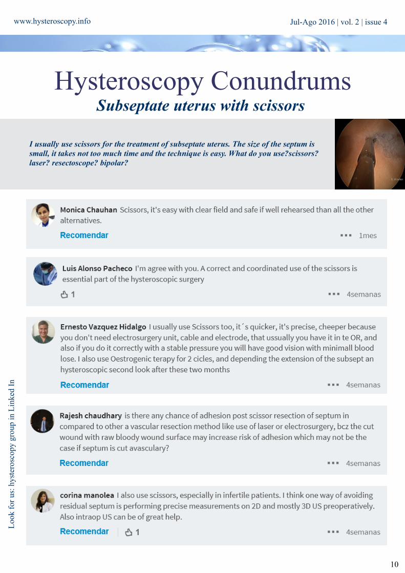
Hysteroscopy ConundrumsSubseptate uterus with scissors
I usually use scissors for the treatment of subseptate uterus. The size of the septum is small, it takes not too much time and the technique is easy. What do you use?scissors? laser? resectoscope? bipolar?
Loo
k fo
r us
: hys
tero
scop
y gr
oup
in L
inke
d In
Jul-Ago 2016 | vol. 2 | issue 4
www.hysteroscopy.info
11
Jul-Ago 2016 | vol. 2 | issue 4

www.hysteroscopy.info
12
Intrauterine synechiae is the presence of fibrous adhesions inside the uterine cavity. In over 90% of cases, uterine adhesions formation is caused by uterine curettage especially if performed during the postpartum period or surgical abortions. Uterine curettage performed during these periods can damage the lining of the endometrium, allowing myometrial areas to contact each other, forming intrauterine adhesions. Other less common causes include uterine surgery such as hysteroscopic myomectomy or metroplasty and infectious endometritis, that rarely produces adhesions except when caused by tuberculosis. Uterine adhesions were initially described by Asherman in 1950 as a result of filling defects at the level of the endometrial cavity observed by hysterography, may become so extensive that could lead to a complete obliteration of the uterine cavity, resulting in amenorrea.
If a patient with uterine sinequia becomes pregnant, the intrauterine synechia gets rodeated by amnion, which is seen on ultrasound as a band inside the uterine cavity. These sonographic findings were originally described by Mahony who described 7 cases. They created the term "Amniotic sheet” to describe the image in a cross-sectional sinequia seen encompassed by the amnion. This image of an undulating band with an oval image at its free end, which is corresponding to the sinequia, has also been called "The sperm sign."
The amniotic sheet is usually asymptomatic and are discovered incidentally during the routine obstetric ultrasound. The prevalence of amniotic sheet is reported between 0.14% and 0.75%. Previous interventions on the uterus appear to play an important role in the formation of these structures. Finberg, described 28 cases of uterine synechiae among which 78% of patients had a history of uterine curettage, in addition, this group had a significantly higher prevalence of cesarean sections. Some authors propose the use of color Doppler to differenciate membranes exclusively of fetal origin (amniotic bands) from amniotic sheet. These authors reported three cases in which upon using color Doppler they visualized a vessel inside the adhesion with concordant arterial pulse similar to maternal heart rate, concluded that it was a tissue of maternal origin, therefore a uterine synechiae.
Neither uterine synechiae nor the amniotic sheet post not any risk to the fetus, as they are covered by amnion and structures that are not in contact with the fetus.
Hysteroscopy Newsletter
Intrauterine adhesionThe sperm sign
Curiosities: Amniotic sheet
Hysteroscopy Newsletter
Jul-Ago 2016 | vol. 2 | issue 4

www.hysteroscopy.info
13
CongresSINTERNATIONAL
ESGE 25th Annual Congress Brussels, Belgium |Oct 2-5 |2016
43 International Forum. Update in Obstetrics, Gynecology and Reproductive MedicineBarcelona, Spain | Oct 26-28| 2016
The 24th World Congress on Controversies in Obstetrics, Gynecology & Infertility Amsterdam, Netherlands |Nov 10-13|2016
63 Congreso Mexicano de Ginecología y ObstetriciaMérida, Mexico |Nov 6-10|2016
ESHRE 32nd Annual Meeting Helsinki, Finland |Jul 3-6 |2016
APAGE and TAMIG Annual Congress Taipei, Taiwan |Nov 3-6|2016
Fertility Society of Australia Annual Conference Western Australia, Australia|Sep 4-7|2016
23rd Annual Summer Conference on Obstetrics & Gynecology South Carolina,USA |Ago 3-6|2016
RANZCOG 2016 Annual Scientific Meeting Perth, Australia |Oct 16-19 |2016
Kongress der Deutschen Gesellschaft für Gynäkologie und Geburtshilfe Stuttgart, Deutschland |Oct 19-22 |2016
American Society for Reproductive Medicine Annual MeetingSalt Lake City, USA |Oct 15-19|2016
3rd International Conference on Gynecology & Obstetrics Dubai, EAU Nov 24-26 |2016
Jul-Ago 2016 | vol. 2 | issue 4

14
www.hysteroscopy.info
HYSTEROSCOPY
DEVICESDisposable Hysteroscopic Polyp Snare
The disponsable Hysteroscopic Polyp Snared (DHPS) is used to cut and coagulated polyps and fibroids for removal from the uterus under direct vision. The Duckbill shape of open snare allows enhanced control and capture of pedunculated uterine
polyps.Main caracteristics are:
Snare and handle as a single unitDisposable. Intended for one-time use
Can be used through the working channel of a rigid or flexible hysteroscopeCan be used with or without electrocautery
Handle accepts a 2 mm monopolar plug for connection to a power source
Ambulatory transcervical resection of polyps with the Duckbill polyp snare: a modality for
treatment of endometrial polyps.J Minim Invasive Gynecol. 2005 Jan-Feb;12(1):37-9.
Timmermans A1, Veersema S.
We performed a retrospective analysis of cases in which polypectomy was performed with the Duckbill polyp snare and a prospective pain analysis in patients undergoing office-based hysteroscopy using a visual analog scale (VAS, range 0-10). The patients, both pre- and postmenopausal, underwent office hysteroscopy for abnormal uterine bleeding. In all, 116 cases of endometrial polyps were diagnosed and removed with the Duckbill polyp snare. This technique was easy to set up and allowed therapeutic hysteroscopy in a see-and-treat fashion during office hysteroscopy. In 188 patients, pain was evaluated using a VAS. Polypectomy with the Duckbill snare was well
tolerated by patients, with a pain score of 4.8 compared with a pain score of 4.2 for diagnostic hysteroscopy. Therefore, we conclude that the Duckbill polyp snare is useful for operative office hysteroscopy and is well tolerated by patients.
http
://w
ww
.aqd
med
ical
.com
/abo
ut_u
s/
Jul-Ago 2016 | vol. 2 | issue 4

In recent years we are witnessing the emergence of different devices and surgical techniques for in office hysteroscopic treatment of submucosal fibroid. In many cases the procedure can be performed without any anesthesia, allowing the use of different types of energy through instrument of 4-5mm in diameter with a working channel of 5Fr such as Versapoint® with bipolar energy or laser, and apply different techniques that allows even the excision deeper myomas. Still, currently 40% of the hysteroscopic myomectomy are carried out in the operating room.
Important aspects for performing in office myomectomy are: - Availability of hysteroscopes with working channels of small diameter. - Complexity of fibroid: size, intra-myometrial component, location, etc. (Lasmar classification) - Hysteroscopist skill level and experience: myomas type G1-G2 need for complete resection in a short operative time that is only achieved by expert hysteroscopists - Operating time: It is related to the size and location of the fibroid, the device used and the skill of hysteroscopist. Usually the operative time varies between 15 to 30 minutes. - Patient ability to tolerate the procedure.
Within all existing classifications for the prediction of success for hysteroscopic myomectomy, the Lasmar classification is the most accurate. Even more if it is associated the concept continent/content proposed by Haimovich
Therefore, when addressing a myoma, the hysteroscopist must take into account: - Surgical approach: Dictated by the complexity of the myoma - Treatment Sequence: ONE STEP “see and treat”
TWO STEPS. Diagnostic first and then treatment.
15
www.hysteroscopy.info
Techniques for in-office hysteroscopic myomectomyDra. Cinta Vidal, H.U Juan Ramón Jiménez. Huelva. Spain
Brief Review
Hysteroscopy Newsletter
Jul-Ago 2016 | vol. 2 | issue 4

www.hysteroscopy.info
16
Approach to fibroid type G0 Initially we recommend proceeding to cut the vascular pedicle and after removal of the fibroid, by extraction with graspers used for fibroids of small size or morcellation, vaporizing, if larger. Some authors recommend leaving the fragmets of the fibroid within the uterine cavity and its spontaneous expulsion after several menstrual cycles. Other authors advise against it for bleeding and cramping pain until its spontaneous expulsion.
Approach to fibroids type G1-G2 Adequate excision of fibroid type G1-G2 requires total myomectomy and careful enucleation of the fibroid. The right dissection plane is delineated by the pseudo-capsule which is an independent identity represented by a layer between the myometrium and myoma. It consists of collagen fibers and a network of small blood vessels that form a vascular ring. With the exception of pedunculated fibroids, the neurovascular pseudocapsule is responsible for blood supply to the fibroid. When the correct plane of pseudocapsule is entered, there is loose connective tissue bridges and multiple capillaries or small vessels. Dissection of this plane is easy and decreases blood loss during surgery. Another advantage of entering this plane is the preservation of the integrity of the underlying myometrium, thus avoiding scars on it. The scars on the myometrium affect subsequent fertility and contribute to the formation of post surgical adhesions. This factor is the reason for the low rate of adhesions preserving the plane of dissection of the pseudo-capsule. Proper surgical technique for submucosal fibroids should always keep the pseudo-capsule intact.
There are different techniques to address pseudocapsule:
- Bettochi technique (OPPIuM). This technique involves making an incision in the endometrial lining of the fibroid with hysteroscopic scissors or bipolar electrode, in line with the reflection of fibroid uterine wall to the surface of cleaving the fibroid with the capsule. This procedure promotes fibroid protrusion into the uterine cavity in the following menstrual cycles, thereby facilitating subsequent surgery in the future, increasing the chance of success and reducing complications.
- Myomectomy in toto. The technique is similar to the above but the incision made in the endometrial mucosa is elliptical, achieving the same effect as the above.
- Haimovich technique. With enucleation of the psedo-capsule - Mazzon technique. Also known as the “Coldloop” technique, which has a low complication rate of 2% and
allows successful myomectomy in one step of more than 80% of myomas. First, is to carry out the resection of the intracavitary portion of the fibroid. This is followed by enucleation of the intramural component.
- Technique of hydromassage. Changing intrauterine distention pressures, achieves the same effect as with the mechanical instrument, once resected intracavitary component is achieved.
In conclusion for adequate excision of fibroid with small intra-cavitary component, perform first an incision of the endometrial mucosa with either technique above described. On the other hand, to excise fibroids with large intra-cavitary component, first resect the intra-cavitary portion of the fibroid and then perform the enucleation of the intramural component
The concept Continent/content take into consideration the relation between the continent (uterine cavity) and
the content (Myoma).
The myoma inside the red uterus is similar in size to that of the blue one, but the working space is quite
different.
Jul-Ago 2016 | vol. 2 | issue 4
www.twitter.com/hysteronews
HYSTEROscopy group
Hysteroscopy newsletter
Hysteroscopy newsletter
www.facebook.com/hysteronews
17
www.hysteroscopy.info
SURGICAL DEVICES
VERSAPOINT The Versapoint® hysteroscopy with bipolar energy system is a minimally invasive procedure that allows the treatment of uterine lesions in the outpatient setting without general anesthesia. It is a system that uses bipolar electrosurgical energy with small caliber electrodes (1.7 mm) through the hysteroscope to visualize the uterine cavity through an optical system connected to a camera. The hysteroscope may be as small as 5 mm diameter, which would not require dilating the cervix. The VersaPoint® technology is based on the basic principles of electrosurgery. It has been a breakthrough in hysteroscopic surgery, because although theoretically it is a monopolar system, the arrangement of the electrodes allows it to behave like a circuit of bipolar energy offering the versatility of the monopolar energy (cut and coagulation) and bipolar security. To this the small size of its electrodes, which allows performing outpatient procedures that previously, could only be performed in the operating room. The design of the electrodes is special in that the active electrode is located at the tip, and the return electrode on the handle. They are placed in separate with an insulating line. The required distension medium is normal saline, which does not alter the sodium concentration of cells as occurs with hypotonic solutions such as sorbitol or glycine thus avoiding the risk of fluid overload. Also provides a low resistance path that allows the energy generated to go back to the return electrode, without becoming part of the electrical circuit the patient's body. Currently there are 5 bipolar electrodes available on the market, three electrodes with different terminals for use with hysteroscopes with working channel of 1.6 to 2 mm each of which is designed for a specific task: spring (for vaporization), Twizzle (for cut) and ball (to coagulate), and two electrodes which can only be used with a resectoscope. (bipolar handle).
LASER This device is capable of transforming other type of energy into electromagnetic radiation emitting light beams of different wavelengths. According to the wavelength, it achieves different effects: cutting, coagulation, vaporisation. The lasers most commonly used in hysteroscopy have been Neodymium large wavelength that has a depth of penetration up to 10 mm that can be used for cutting and coagulating.
MINIRESECTOSCOPEThe Gubbini Miniresectoscope of small diameter uses bipolar energy performing the same function as the traditional bipolar handle. There are different electrodes with different tips. (Ball, loop, blade)
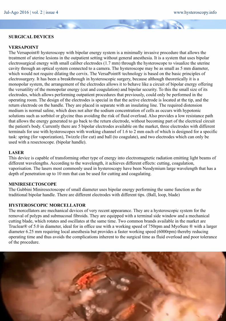
HYSTEROSCOPIC MORCELLATOR The morcellators are mechanical devices of very recent appearance. They are a hysteroscopic system for the removal of polyps and submucosal fibroids. They are equipped with a terminal side window and a mechanical cutting blade, which rotates and oscillates at the same time. Two common brands available in the market are Truclear® of 5.0 in diameter, ideal for in office use with a working speed of 750rpm and MyoSure ® with a larger diameter 6.25 mm requiring local anesthesia but provides a faster working speed (6000rpm) thereby reducing operating time and thus avoids the complications inherent to the surgical time as fluid overload and poor tolerance of the procedure.
Jul-Ago 2016 | vol. 2 | issue 4

18
www.hysteroscopy.info
Approach to fibroid type G0 Initially we recommend proceeding to cut the vascular pedicle and after removal of the fibroid, by extraction with graspers used for fibroids of small size or morcellation, vaporizing, if larger. Some authors recommend leaving the fragmets of the fibroid within the uterine cavity and its spontaneous expulsion after several menstrual cycles. Other authors advise against it for bleeding and cramping pain until its spontaneous expulsion.
Approach to fibroids type G1-G2 Adequate excision of fibroid type G1-G2 requires total myomectomy and careful enucleation of the fibroid. The right dissection plane is delineated by the pseudo-capsule which is an independent identity represented by a layer between the myometrium and myoma. It consists of collagen fibers and a network of small blood vessels that form a vascular ring. With the exception of pedunculated fibroids, the neurovascular pseudocapsule is responsible for blood supply to the fibroid. When the correct plane of pseudocapsule is entered, there is loose connective tissue bridges and multiple capillaries or small vessels. Dissection of this plane is easy and decreases blood loss during surgery. Another advantage of entering this plane is the preservation of the integrity of the underlying myometrium, thus avoiding scars on it. The scars on the myometrium affect subsequent fertility and contribute to the formation of post surgical adhesions. This factor is the reason for the low rate of adhesions preserving the plane of dissection of the pseudo-capsule. Proper surgical technique for submucosal fibroids should always keep the pseudo-capsule intact.
There are different techniques to address pseudocapsule:
- Bettochi technique (OPPIuM). This technique involves making an incision in the endometrial lining of the fibroid with hysteroscopic scissors or bipolar electrode, in line with the reflection of fibroid uterine wall to the surface of cleaving the fibroid with the capsule. This procedure promotes fibroid protrusion into the uterine cavity in the following menstrual cycles, thereby facilitating subsequent surgery in the future, increasing the chance of success and reducing complications.
- Myomectomy in toto. The technique is similar to the above but the incision made in the endometrial mucosa is elliptical, achieving the same effect as the above.
- Haimovich technique. With enucleation of the psedo-capsule - Mazzon technique. Also known as the “Coldloop” technique, which has a low complication rate of 2% and
allows successful myomectomy in one step of more than 80% of myomas. First, is to carry out the resection of the intracavitary portion of the fibroid. This is followed by enucleation of the intramural component.
- Technique of hydromassage. Changing intrauterine distention pressures, achieves the same effect as with the mechanical instrument, once resected intracavitary component is achieved.
In conclusion for adequate excision of fibroid with small intra-cavitary component, perform first an incision of the endometrial mucosa with either technique above described. On the other hand, to excise fibroids with large intra-cavitary component, first resect the intra-cavitary portion of the fibroid and then perform the enucleation of the intramural component
Congress Committee Chair: Sergio Haimovich (Spa)
CoChairs: Andrea Tinelli (Ita) Luis Alonso (Spa)
Congress Committee: Jose Alanis Fuentes (Mex) Linda Bradley (USA) Jorge Dotto (Arg) Ricardo Lasmar (Bra) Narendra Malhotra (Ind) Osama Shawki (Egy) Honorary Committee: Stefano Bettocchi (Ita)
Jul-Ago 2016 | vol. 2 | issue 4
19
www.hysteroscopy.info
Original Article Treatment of hydrosalpinx before IVF
Jose Rios. Juan Lorente Unidad de Reproducción Asistida. Hospital Universitario Reina Sofía. Córdoba. Spain
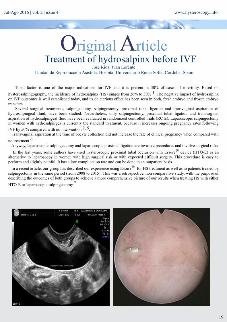
Tubal factor is one of the major indications for IVF and it is present in 30% of cases of infertility. Based on
hysterosalpingography, the incidence of hydrosalpinx (HS) ranges from 26% to 30% 1. The negative impact of hydrosalpinx on IVF outcomes is well established today, and its deleterious effect has been seen in both, fresh embryo and frozen embryo transfers. Several surgical treatments, salpingectomy, salpingostomy, proximal tubal ligation and transvaginal aspiration of hydrosalpingeal fluid, have been studied. Nevertheless, only salpingectomy, proximal tubal ligation and transvaginal aspiration of hydrosalpingeal fluid have been evaluated in randomized controlled trials (RCTs). Laparoscopic salpingectomy in women with hydrosalpinges is currently the standard treatment, because it increases ongoing pregnancy rates following
IVF by 50% compared with no intervention 2, 5. Transvaginal aspiration at the time of oocyte collection did not increase the rate of clinical pregnancy when compared with
no treatment 6. Anyway, laparoscopic salpingectomy and laparoscopic proximal ligation are invasive procedures and involve surgical risks
In the last years, some authors have used hysteroscopic proximal tubal occlusion with Essure® device (HTO-E) as an alternative to laparoscopy in women with high surgical risk or with expected difficult surgery. This procedure is easy to perform and slightly painful. It has a low complication rate and can be done in an outpatient basis.
In a recent article, our group has described our experience using Essure® for HS treatment as well as in patients treated by salpingectomy in the same period (from 2008 to 2015). This was a retrospective, non comparative study, with the purpose of describing the outcomes of both groups to achieve a more comprehensive picture of our results when treating HS with either
HTO-E or laparoscopic salpingectomy.7
Hysteroscopy Newsletter Hysteroscopy Newsletter
Jul-Ago 2016 | vol. 2 | issue 4

20
www.hysteroscopy.info
There were no significant differences in the clinical features of both groups, except age that was significantly older in Essure® group (36.12 ± 3.42 vs 32.47 ± 4.16, p= 0.003). During the study period (2008-2015), 31 patients with hydrosalpinx
were subjected to treatment with Essure®. Of these, 22 began a cycle of IVF. 13 embryo transfers were performed. The other cases were cancelled due to low ovarian response. During the same period of time, 40 patients with unil- or bilateral HS underwent laparoscopic salpingectomy. No complication occurred during surgical procedure in this group. 23 of them started a cycle of IVF. Only 3 did not reach embryo transfer due to low ovarian response.
There was no case of late miscarriage in any group. The results of our study suggest that women undergoing HTO-E show acceptable clinical pregnancy rate (33.3%)
and embryo implantation rate (16.3 %), considering patients features. However, abortion rates are remarkably high (57.1 %). In order to a better assessment of these results, we have also included the outcomes of patients who underwent salpingectomy during the same time period in our unit. This group of patients had a higher clinical pregnancy rate (70.6%) and a higher implantation rate (34.1 %) with a lower abortion rate, very similar to women without HS who underwent IVF/ICSI in our unit in the same period of time (18.2 % vs 21.0 % respectively). Live birth rate per patient was also higher in patients who underwent salpingectomy (52.9% vs 14.3% in the Essure® group). Both groups aren’t comparable, so that we cannot draw any firm conclusion, but our data suggest a slight trend to better outcomes and less abortion rate when HS is
treated by salpingectomy. This fact has been also reported in a recent meta-analysis by Barbosa et al 8, which included our own data.
In the only RCT published to the date by Dreyer et al 9, 85 women with uni- or bilateral hydrosalpinx were randomized to HTO (N=42) and laparoscopic salpingectomy (N=43) in an unblinded study. The ongoing pregnancy rate following one IVF/ICSI cycle was 11/42 (26.2%) in the Essure® group compared with 24/43 (55.8%) in the laparoscopy group (p=0.008). Miscarriage rate was not statistically different (2/27, 7.4% in the Essure® group vs 1/32, 3.1% in the salpingectomy group). Live birth rate per patient was 29.6% with Essure® and 50% with salpingectomy. Differences in ongoing pregnancy rates can be attributed to the differences in the implantation rate (18% compared with 41.7%). The authors suggest that these differences may be caused by the presence of the Essure® device itself. They speculate that Essure® may have a negative influence on the endometrial environment leading to lower endometrial receptivity and subsequently lower implantation rates. Therefore, the conclusion of this study is that salpingectomy remains the procedure of choice for women with hydrosalpinges who are planned for IVF/ICSI.
With all these data we can conclude that today, laparoscopic salpingectomy is the better option for patients with uni- or bilateral hydrosalpinx prior to an IVF cycle. Hysteroscopic occlusion with Essure® is an attractive alternative but it should be reserved only for those patients with increased surgical risk.
REFERENCES:1-Evers JLH. Female subfertility. Lancet. 2002.360. 151-92-Johnson N, van Voorst S, Sowter MC, Strandell A, Mol BWJ. Surgical treatment for tubal disease in women due to undergo in vitro fertilisation.
Cochrane Database of Systematic Reviews 2010, Issue 1. Art. No.: CD002125. 3-Kontoravdis A, Makrakis E, Pantos K, Botsis D, Deligeoroglu E, Creatsas G). Laparoscopic proximal tubal occlusion (LPTO) and salpingectomy result in similar
improvement in IVF outcome in patients with hydrosalpinx. Fertil Steril 2006;86:1642–84-Moshin V, Hotineanu A. Reproductive outcome of the proximal tubal occlusion prior to IVF in patients with hydrosalpinx. Hum Reprod 2006;21:i193–i1945-Vignarajan CP, Singh N. Salpingectomy versus proximal tubal occlusion for hydrosalpinges prior to in-vitro-fertilization (IVF) cycle -Is there a difference in
ovarian reserve or response to gonadotropins? Fertil Steril 2014;102:e136-e137.6-Hammadieh N, Coomarasamy A, Ola B, Papaioannou S, Afnan M, Sharif K. Ultrasound-guided hydrosalpinx aspiration during oocyte collection improves
pregnancy outcome in IVF: a randomized controlled trial. Hum Reprod 2008;23:1113 – 11177-Lorente J, Ríos JE, Pomares E, Romero MI, Castelo-Branco C, Arjona JE. Essure a novel option for the treatment of hydrosalpinx: a case series and
literature review. Gynecol Endocrinol 2016;32(2):166-708-Barbosa M, Sotiriadis A, Papatheodorou SI, Mijatovic V, Nastri CO, Martins WP. High miscarriage rate in women submitted to Essure for
hydrosalpinx before embryo transfer: a systematic review and meta-analysis. Ultrasound Obstet Gynecol. 2016. [Epub ahead of print]9-Dreyer K, Lier MCI, Emanuel MH, Twisk JWR, Mol BWJ, Schats R, Hompes PGA, Mijatovic V. Hysteroscopic proximal tubal occlusion versus laparoscopic
salpingectomy as a treatment for hydrosalpinges prior to IVF or ICSI: an RCT
Jul-Ago 2016 | vol. 2 | issue 4

21
www.hysteroscopy.info
HYSTEROSCOPY
Editorial teaM Progress in any area of the medical field, must have a good communication channel to allow all professionals to be at the forefront of new techniques and procedures, and to promote sharing of information among professionals to improve the different techniques and procedures.
Therefore, if we are talking about innovation, we must talk about the need to be globally connected.
When doing a search in PubMed with the term "Hysteroscopy" we see the increasing number of publications in recent years. The 291 papers published in 2015 shows a growing interest in hysteroscopy, driven largely by the ability to make increasingly less invasive procedures and more accurate diagnoses.
Nowadays, a common reflection arises between patients, with which most of us can relate: "They are always wondering what's new?". It would be great if we, as gynecologists with an interest in hysteroscopy, could know what is the current state of hysteroscopy in every part of the world?
This growing interest in hysteroscopy, and the absence of a specialized journal in this field, was the need that drove us to initiate this ambitious project. Our goal was clear: to create the first journal dedicated exclusively to hysteroscopy, with free distribution over the Internet, so that we could easily reach the largest number of hysteroscopists and thus contribute to the development and continuous improvement of our specialty. Therefore, we invite the active collaboration of peers who shared the same concerns, firmly believing in the present need to contribute to the dissemination of knowledge and experience in hysteroscopy. Thus Hysteroscopy Newsletter was born!. A, bi-monthly publication, advertisements free, ensuring the rigor of the publication; and with a website for unlimited dissemination, and completely free to access. Social media networks and blogs have contributed greatly to increase the dissemination and contact among professionals, facilitating exchange of ideas, skills and knowledge from anywhere in the world and at any time.
Today, 16 months after starting this adventure, we can quantify the results. Neswletter discharges have been increasing significantly every month; reaching more than 2700 downloads only during the first 25 days of May. Our most active channel of distribution (web, YouTube, Linkedin, MedTube, Facebook and Twitter) have more and more subscribers, more video views and more traffic, allowing us to interconnect with other physicians and be able to reach every corner of the world. We are excited to share with you that we have received visits to the site from 103 different countries!, Being in the "Top 5 countries" USA (27.5%), Spain (10.8%), Mexico (3.9%), India (3.8%) and Canada (3.5%),. Also, taking advantage of the interconnection network that has been generated with the Newsletter, we have launched multicenter trials and other very interesting projects.
This is our current balance, a dream fulfilled, our passion. What does the future hold? 'Help us find out, join the Hysteroscopy World !!
#WeLoveHysteroscopy
Laura Nieto
Hysteroscopy Newsletter is an opened forum to all professionals who want to contribute with their
knowledge and even share their doubts with a word-wide gynecological
community
www.facebook.com/hysteronews
www.twitter.com/hysteronews
Hysteroscopy newsletter
HYSTEROscopy group
Hysteroscopy newsletter
FIND US ON
Jul-Ago 2016 | vol. 2 | issue 4
www.medtube.net