i have no relevant financial relationships with the
TRANSCRIPT

I have no relevant financial relationships with the manufacturer(s) of any commercial product(s) and/or provider(s) of commercial services discussed in this CME activity.

Coombs and Gell classification
Type I – immediate
Type II - antibody-dependent
Type III - immune complex
Type IV - cell-mediated or delayed

A 23yo male comes in after camping over the weekend. He was out in the woods and now has a rash. It is very itchy and came up after about 3d after his trip. The lesions consist of small yellowish blisters that show swelling. There are no other people who are affected in the group. What type of hypersensitivity reaction is he having?
A: Type I
B: Type II
C: Type III
D: Type IV
E: Type V

Type I hypersensitivity is an allergic reaction provoked by re-exposure to a specific antigen.
The reaction is mediated by IgE antibodies and produced by the immediate release of histamine, tryptase, arachidonate and derivatives by mast cells.
Exposure may be by ingestion, inhalation, injection, or direct contact.

This causes an inflammatory response leading to an immediate (within seconds to minutes) reaction.
The reaction may be either local or systemic. Symptoms vary from mild irritation to sudden death from anaphylactic shock.
Treatment usually involves epinephrine, antihistamines, and corticosteroids

In type II hypersensitivity, the antibodies produced by the immune response bind to antigens on the patient's own cell surfaces. Blood is the medium
The antigens recognized in this way may either be intrinsic ("self" antigen, innately part of the patient's cells) or extrinsic (absorbed onto the cells during exposure to some foreign antigen, possibly as part of infection with a pathogen
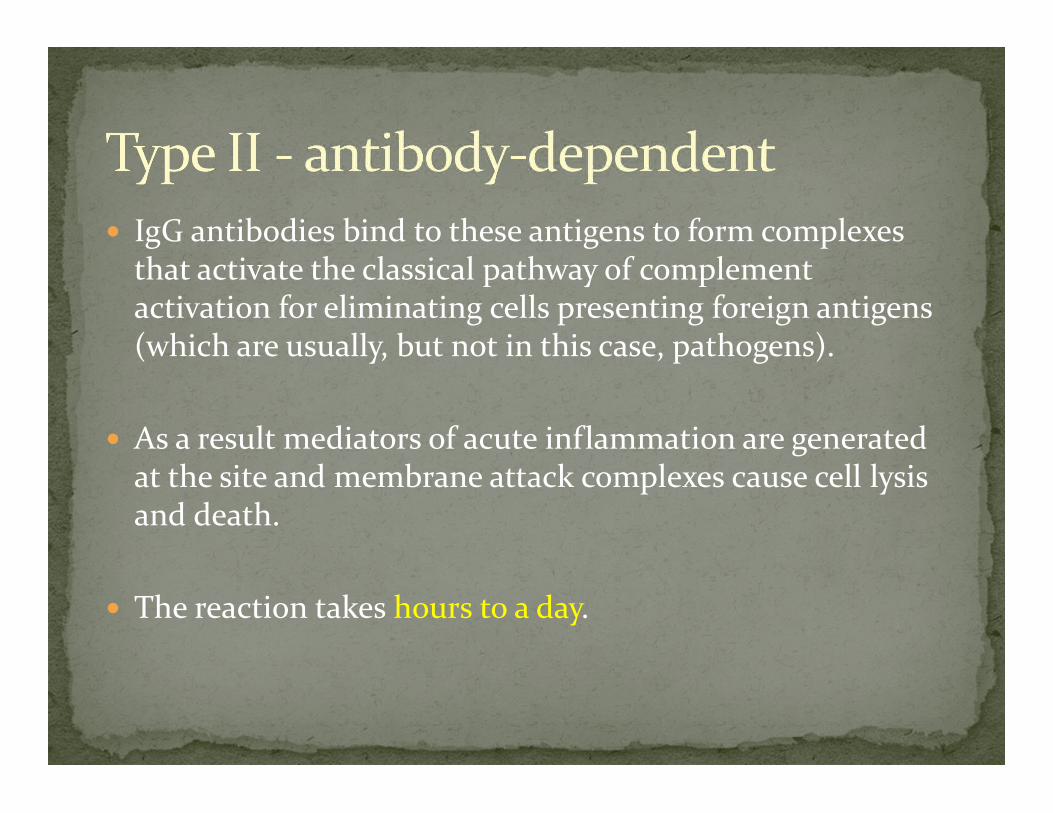
IgG antibodies bind to these antigens to form complexes that activate the classical pathway of complement activation for eliminating cells presenting foreign antigens (which are usually, but not in this case, pathogens).
As a result mediators of acute inflammation are generated at the site and membrane attack complexes cause cell lysisand death.
The reaction takes hours to a day.
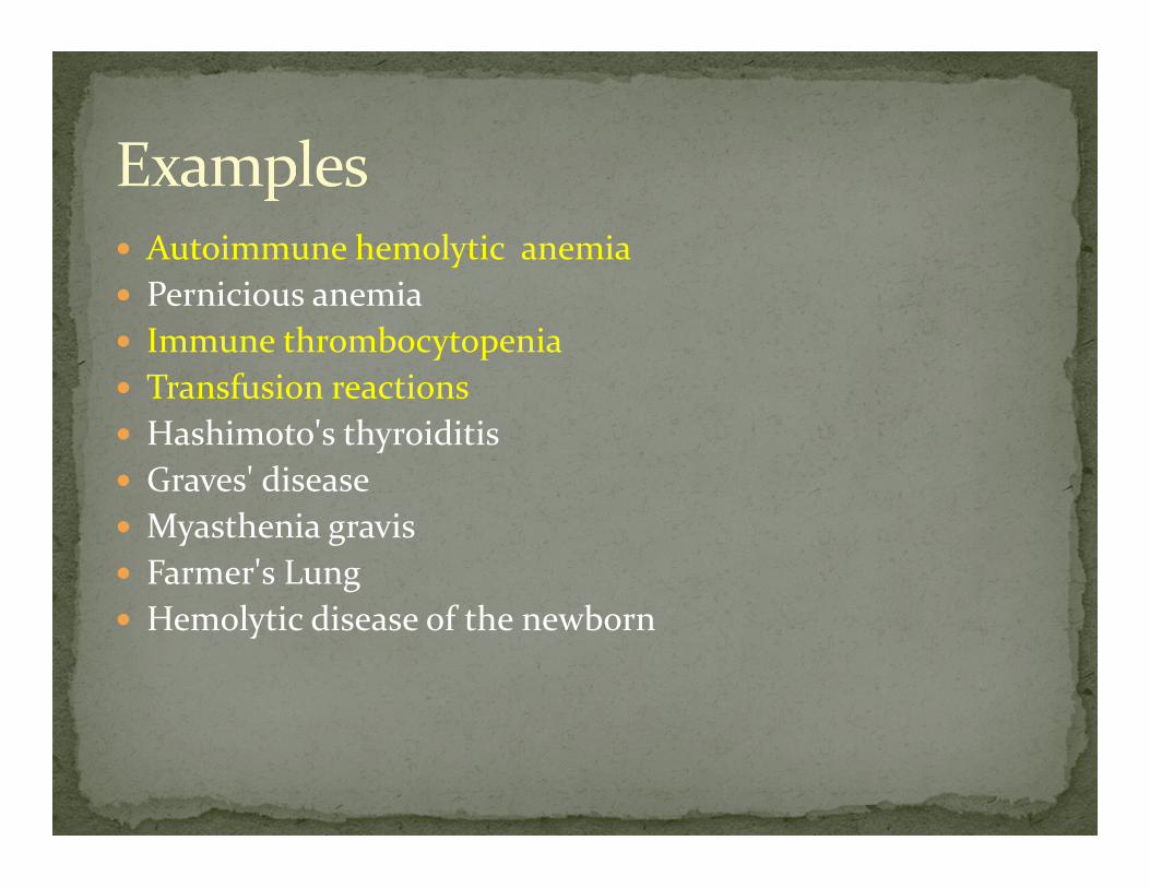
Autoimmune hemolytic anemia
Pernicious anemia
Immune thrombocytopenia
Transfusion reactions
Hashimoto's thyroiditis
Graves' disease
Myasthenia gravis
Farmer's Lung
Hemolytic disease of the newborn

In type III hypersensitivity soluble immune complexes (aggregations of antigens and IgG antibodies) form and are deposited in various tissues Tissue is the medium
Deposition is usually in: skin, kidney and joints
This may trigger an immune response according to the classical pathway of complement activation.
The reaction takes hours to days to develop
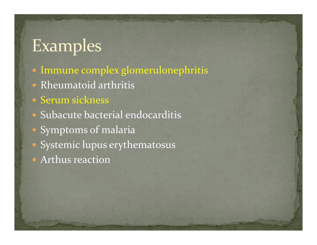
Immune complex glomerulonephritis
Rheumatoid arthritis
Serum sickness
Subacute bacterial endocarditis
Symptoms of malaria
Systemic lupus erythematosus
Arthus reaction

Type IV hypersensitivity is often called delayed type as the reaction takes two to three + days to develop.
Unlike the other types, it is not antibody mediated but rather is a type of cell-mediated response.

Contact dermatitis - poison ivy rash Temporal arteritis
Symptoms of leprosy
Symptoms of tuberculosis
Transplant rejection


Hapten a small separable part of an antigen that reacts specifically with an
antibody but is incapable of stimulating antibody production except in combination with a carrier protein molecule
Direct haptenation e.g. Beta-lactam antibiotics
Haptenation by drug metabolites e.g. Sulfonamides
Complete antigens e.g. Insulin

Application of proteomics in the elucidation of chemical-mediated allergic contact dermatitis . Toxicology Research. Tessa Höper
Diagnosis History Skin testing In Vitro testing Challenges

Nature of the reaction Is this an allergic reaction?
If so what kind?
Temporal sequence! When did drug and other exposures occur in relation to
the onset and remission of the reaction?
When did the itching start
When did the rash come up

Propensity of the exposures to cause such a reaction.
Discontinuation effect
Readministration effect

0
2
4
6
8
10
12
14
16
18
1 2 3 4 5 6 7 8 9 10 11 12 13 14 15 16 17 18 19 20 21
Onset
Hours

Exposure-Response Intervals Onset within 15 minutes -- 85% Onset within 30-60 minutes -- 96% Onset within 12 hours -- ~100%
Anaphylactoid reactions are defined as those reactions that produce the same clinical picture with anaphylaxis but are not IgE mediated, occur through a direct nonimmune-mediated release of mediators from mast cells and/or basophils or result from direct complement activation. “Pseudoallergic” reactions can occur after any dose:
Vancomycin, opiates, NSAID, IV contrast dye
Idsoe, Bull WHO 1968;38:159

While many drug reactions are immunologic (such as with penicillins, cephalosporins, sulfonamides, quinine), others are idiosyncratic and depend on pharmacogenetics (such as with G6PD deficiency and Primaquine, or with acetylator status and Isoniazid)
Glucose-6-phosphate dehydrogenase(G6PD) deficiency is a genetic disorder that is most common in males. G6PD deficiency mainly affects red blood cells. The most common result is hemolytic anemia.
In G6PD deficiency, pts may not have symptoms. Symptoms happen if red blood cells are exposed to certain chemicals in food or medicine, certain bacterial or viral infections, or stress. They may include: Paleness Jaundice Dark urine Fatigue Shortness of breath Enlarged spleen
Drugs to avoid: Chloroquine Mefloquine Pamaquine. Primaquine. Quinidine. Quinine.

IgE to medications
Epicutaneous & ID skin tests
In vitro tests for specific IgE
T-cells reactive with drug determinants
Patch tests
Intradermal tests
In vitro tests for lymphocyte reactivity

This 27 year old insulin dependent diabetic man had done well on insulin for 2 years. For the past week he has had severe generalized urticaria immediately after each of his twice daily NPH insulin injections. The urticaria would be nearly gone before his next injection.
What type of hypersensitive is this and how would you test for this?
A: Type I with patch testing
B: Type III with intradermal skin testing
C: Type IV with blood IgE testing
D: Type I with intradermal skin testing
E: Type V with intradermal skin testing

Sharply circumscribed
Raised
Pruritic
Transient lesions
No residual abnormalities

A 32 year old man developed generalized urticaria, periodic flushing, a systemic rash, and wheezing about 1wk after a surgical procedure. He has been having itching systemically along with fatigue. He was in semi-good health but dealing with diet control DM-II. There was no hx of asthma or other lung related issues in his PMHx.
He was having a hernia fixed and tolerated the procedure well. He and his surgeon want to know if this has something to do with the surgical procedure or if there is something else going on?

What steps should be taken? Obtain the anesthesiologist record of the case for temporal
verifications
Obtain hospital records
Compile a list of exposures
Do not forget about latex allergy

There was no documentation of a rash or any abnormal vital signs at the start of the surgery. Records show that Iodine was
used to prep the area. He was given a dosage of
Cefalexin just after he had anesthesia for the surgery.
He did have some HTN during the case and was given Hydralazine for this.
No latex was used.
He did develop a rash when he was in post op but he was given Benadryl and this resolved.
Labs: CBC – WNL CMP – elevated glucose, Cr
and BUN
What would you do next? A: CXR B: Skin culture of the wound C: Monitor D: Order immune labs E: Treat with steroids

Immunological labs show: ANA – Elevated Antihistones – Elevated SED – Elevated
What type of hypersensitivity reaction is this man having: A: Type I B: Type II C: Type III D: Type IV E: Type V
Drug-Induced Glomerular Disease: Immune-Mediated Injury. Clin J Am Soc Nephrol. 2015 Jul 7; 10(7): 1300–1310.
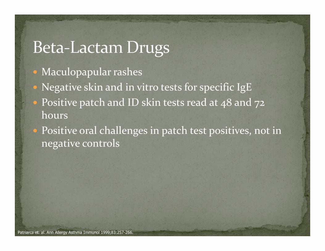
Maculopapular rashes
Negative skin and in vitro tests for specific IgE
Positive patch and ID skin tests read at 48 and 72 hours
Positive oral challenges in patch test positives, not in negative controls
Patriarca et. al. Ann Allergy Asthma Immunol 1999;83:257-266.

Relatively non-pruritic
Resolve without residual damage
Benign
Drug associated may be specific lymphocyte mediated

26 year-old man admitted to the hospital with cc of having a very bad rash after he started to take some Carbamazepine for his seizure disorder.
Methylprednisoloneineffective
Oral mucosa denuded, diffuse target bullouslesions.
What type of reaction is he having and how would you treat this:
A: Type III – but change to dexamethasone
B: Type IV - IVIg
C: Type III - Cyclosporin
D: Type IV - Cyclosporin
E: None of the above
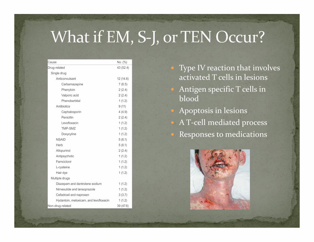
Type IV reaction that involves activated T cells in lesions
Antigen specific T cells in blood
Apoptosis in lesions
A T-cell mediated process
Responses to medications

Cyclosporine or other agents for T-lymphocyte mediated reactions Glucocorticoids are logical, but weak
Cyclosporine 5 mg/kg/day (15 mg/kg full immunosuppressive dose)
Prograf and others as alternatives
IVIg – Not effective in some reported trials

Management Avoidance
Desentization Referral to allergy can help establish what drugs could
be causing the issue and if there is a way to overcome the allergy Treating through false reactions Acute desensitization Slow desensitization Treatment of reactions
Desentization is a short term fix for a problem. The allergy will re-occur s/p discontinuation of the drug in question.

Drug allergy diagnosis is very difficult
History is key when determining what drugs are players and eventually what to test.
Establish what type of hypersensitivity the reaction is in order to determine what course of action to treat.
Consider desentization if applicable to the case.