identification of a new phenotype of tolerogenic human ...¬cation of a new phenotype of...
TRANSCRIPT

of June 4, 2018.This information is current as
Aspergillus oryzaeby Fungal Proteases from Tolerogenic Human Dendritic Cells Induced Identification of a New Phenotype of
Philippe MoingeonPallardy, Laurence Van Overtvelt, Laurent Mascarell andNony, Marie Naveau, Armelle Biola-Vidamment, Marc Aline Zimmer, Sonia Luce, Fanny Gaignier, Emmanuel
http://www.jimmunol.org/content/186/7/3966doi: 10.4049/jimmunol.1003184March 2011;
2011; 186:3966-3976; Prepublished online 2J Immunol
Referenceshttp://www.jimmunol.org/content/186/7/3966.full#ref-list-1
, 32 of which you can access for free at: cites 76 articlesThis article
average*
4 weeks from acceptance to publicationFast Publication! •
Every submission reviewed by practicing scientistsNo Triage! •
from submission to initial decisionRapid Reviews! 30 days* •
Submit online. ?The JIWhy
Subscriptionhttp://jimmunol.org/subscription
is online at: The Journal of ImmunologyInformation about subscribing to
Permissionshttp://www.aai.org/About/Publications/JI/copyright.htmlSubmit copyright permission requests at:
Email Alertshttp://jimmunol.org/alertsReceive free email-alerts when new articles cite this article. Sign up at:
Print ISSN: 0022-1767 Online ISSN: 1550-6606. Immunologists, Inc. All rights reserved.Copyright © 2011 by The American Association of1451 Rockville Pike, Suite 650, Rockville, MD 20852The American Association of Immunologists, Inc.,
is published twice each month byThe Journal of Immunology
by guest on June 4, 2018http://w
ww
.jimm
unol.org/D
ownloaded from
by guest on June 4, 2018
http://ww
w.jim
munol.org/
Dow
nloaded from
The Journal of Immunology
Identification of a New Phenotype of Tolerogenic HumanDendritic Cells Induced by Fungal Proteases fromAspergillus oryzae
Aline Zimmer,* Sonia Luce,* Fanny Gaignier,* Emmanuel Nony,* Marie Naveau,*
Armelle Biola-Vidamment,† Marc Pallardy,† Laurence Van Overtvelt,* Laurent Mascarell,*
and Philippe Moingeon*
We characterized a new pathway to induce tolerogenic dendritic cells (DCs) following treatment of human monocyte-derived DCs
with proteases from the fungus Aspergillus oryzae (ASP). ASP-treated DCs (ASP-DCs) exhibit a CD802CD832CD862Ig-like
transcript (ILT)22ILT32ILT4+ phenotype, do not secrete cytokines or chemokines, and express tolerogenic markers such as
glucocorticoid-induced leucine zipper, NO synthetase-2, retinaldehyde dehydrogenase-1 or retinaldehyde dehydrogenase-2. When
cocultured with naive CD4+ T cells, ASP-DCs induce an anergic state that can be reversed by IL-2. Generated T cells mediate
a suppressive activity in third-party experiments that is not mediated by soluble factors. A comparison between dexamethasone-
treated DCs used as a reference for regulatory T cell-inducing DCs and ASP-DCs reveals two distinct phenotypes. In contrast to
dexamethasone, ASP treatment induces glucocorticoid-induced leucine zipper independently of glucocorticoid receptor engage-
ment and leads to NF-kB p65 degradation. Abrogation of protease activities in ASP using specific inhibitors reveals that aspartic
acid-containing proteases are key inducers of regulatory genes, whereas serine, cysteine, and metalloproteases contribute to NF-
kB p65 degradation. Collectively, those features correspond to a previously unreported anergizing phenotype for human DCs.
Such regulatory mechanisms may allow fungi to downregulate host immune responses and provide clues for new approaches to
treat proinflammatory disorders. The Journal of Immunology, 2011, 186: 3966–3976.
Dendritic cells (DCs) are specialized APCs playing a piv-otal role in the induction of both immunity and tolerance(1). Depending upon the type of pathogen encountered
and the profile of costimulatory and T cell-polarizing moleculesengaged, DCs drive the development of Th1, Th2, Th17 effector,or suppressive/regulatory T cells (2–7). Regulatory T cell (Treg)generation is currently considered as a potential treatment forautoimmune diseases (8), graft-versus-host disease (9–11), or al-lergy (12, 13). Thus, the induction of tolerogenic DCs supportingsuppressive T cell responses while downregulating effector re-sponses can be considered as an approach to control a variety of in-flammatory disorders.Several studies have demonstrated that Treg-promoting DCs can
be generated in vitro following treatment with biological (e.g., IL-10, TGF-b) or pharmacological agents (recently reviewed in Ref.
14). Most particularly, DCs conditioned with dexamethasone(DEX-DCs) present a semimature phenotype and prevent theupregulation of costimulatory molecules as well as the secretionof proinflammatory cytokines like IL-12 (15–17). Such DEX-DCsinhibit the proliferation of allospecific T cells (18, 19), prolongallograft survival in mice (20), and decrease the number of IFN-g–producing CD4+ T cells, but support the differentiation of IL-10–producing type 1 Tregs (Tr1) (15, 21, 22). Besides their capacityto elicit suppressive T cells, tolerogenic DCs are commonly de-fined based on the expression of various surface markers such asIg-like transcript (ILT) molecules (4, 23–28), transcriptional reg-ulators like glucocorticoid-induced leucine zipper (GILZ) (29–31), or enzymes such as retinaldehyde dehydrogenase (RALDH)(32–36) or NO synthetase-2 (NOS-2) (37), all contributing to theirfunctional properties.In this study, we describe a new pathway to induce tolerogenic
DCs following treatment with proteases from the Aspergillusoryzae fungus. A. oryzae is used for the production of traditionalfermented food such as soy products as well as enzymes, anti-biotics, beverages, and volatile compounds (38). In this study, wedemonstrate that protease from A. oryzae (ASP)-DCs exhibita tolerogenic phenotype distinct from other conventional regula-tory DCs such as DEX-DCs.
Materials and MethodsReagents
ASP is a protease–peptidase complex produced by submerged fermentationof A. oryzae, which contains both endoprotease and exopeptidase activities(Sigma-Aldrich, St. Louis, MO). This product complies with the recom-mended purity specifications for food-grade enzymes issued by the JointFood and Agriculture Organization/World Health Organization ExpertCommittee on Food Additives and the Food Chemicals Codex. In selected
*Stallergenes, Antony Cedex 92183, France; and †Universud, INSERM Unite Mixtede Recherche 996, Faculte de Pharmacie, Universite Paris Sud 11, 92296 Chatenay-Malabry, France
Received for publication September 24, 2010. Accepted for publication January 27,2011.
This work was supported by Stallergenes. A.Z. was supported by a ConventionIndustrielle de Formation par la Recherche fellowship from the Association Natio-nale de la Recherche et de la Technologie.
Address correspondence and reprint requests to Dr. Mascarell Laurent, Stallergenes, 6Rue Alexis de Tocqueville, Antony Cedex 92183, France. E-mail address: [email protected]
Abbreviations used in this article: ASP, protease from Aspergillus oryzae; Ct, cyclethreshold; DC, dendritic cell; DEX, dexamethasone; DNCB, dinitrochlorobenzene;EPNP, 1,2-epoxy-3-(p-nitrophenoxy)propane; GILZ, glucocorticoid-induced leucinezipper; GR, glucocorticoid receptor; ILT, Ig-like transcript; NOS-2, NO synthetase-2;p65, p65 subunit of NF-kB; PI, propidium iodide; RALDH, retinaldehyde dehydro-genase; Tr1, type 1 regulatory T cell; Treg, regulatory T cell.
Copyright� 2011 by TheAmericanAssociation of Immunologists, Inc. 0022-1767/11/$16.00
www.jimmunol.org/cgi/doi/10.4049/jimmunol.1003184
by guest on June 4, 2018http://w
ww
.jimm
unol.org/D
ownloaded from

experiments, culture supernatants were filtered through 10-kDa cutoff fil-ters (Amicon; Millipore, Billerica, MA) to remove small molecules.
DC generation and in vitro stimulation
Human PBMCs were separated out of buffy coats obtained from healthyvolunteers (Etablissement Francais du Sang, Rungis, France) by centrifu-gation over a Ficoll-Paque plus gradient (PAA Laboratories, Les Mureaux,France). To generate monocyte-derived DCs, 5 3 108–8 3 108 cells werecultured at 37˚C, 5% CO2, in RPMI 1640 medium with stable glutaminesupplemented with 10 mg/ml gentamicin, 50 mM 2-ME, 1% nonessentialamino acids (all obtained from Invitrogen, Carlsbad, CA), and 10% FCS(Gentaur, Brussels, Belgium). After 1 h, nonadherent cells were removed,and adherent cells were further cultivated for 6 d in presence of humanrGM-CSF and rIL-4 (Gentaur) using 100 and 50 ng/ml concentrations,respectively. After 6 d, a pure population of DCs was obtained, with.95%CD142 CD1a+ cells detected by flow cytometry using a FC500 cytometerand the CXP analysis software (Beckman Coulter, Villepinte, France). Upto 106 DCs were plated in a 24-well plate in presence of medium, ASP(0.2–20 mg/ml), DEX (1 mg/ml [2.5 mM]; Sigma-Aldrich), or highly pu-rified LPS from Escherichia coli (1 mg/ml; InvivoGen, Toulouse, France)
for 24 h at 37˚C and 5% CO2. In some experiments, LPS and DEX or ASPwere added together. For signaling experiments, RU-486 (kind gift ofMichel Renoir) was added to the culture at a 1 mM concentration.
Analysis of cell-surface markers, cytokine, and chemokineproduction
For immunofluorescence staining, cells were harvested, washed in PBS, andincubated for 20 min at 4˚C with the following mAbs: FITC anti-CD80, PEanti-CD86, PE–cyanin-5 anti-CD83 (Beckman Coulter), FITC anti-ILT2,PE anti-ILT4, and PE–cyanin-5 anti-ILT3 (R&D Systems, Lille, France).Cells were stained with corresponding isotype-matched control mAbs.Samples were analyzed by flow cytometry with results expressed as meanvariations in percentages of positive cells.
Cytokine measurement was performed in supernatants using the cyto-metric bead array technology. IFN-g, IL-1b, IL-2, IL-6, IL-8, IL-9, IL-10,IL-12p70, IL-13, IL-17A, and TNF-a were measured using the HumanInflammatory CBA kit or CBA Flex sets (BD Biosciences, Le Pont deClaix, France) and analyzed by flow cytometry according to the manu-facturer’s instructions using an FACSArray instrument and the FCAPSoftware (BD Biosciences). Chemokine release was measured in DCs
FIGURE 1. ASP-DCs exhibit a tol-
erogenic phenotype. Monocyte-derived
immature DCs were obtained from
PBMCs of healthy donors (n = 4). Cells
were incubated with medium, DEX (1
mg/ml), ASP (20 mg/ml), LPS (1 mg/
ml), or combinations for 24 h. Expres-
sion of various costimulatory (CD80,
CD83, CD86) or inhibitory molecules
(ILT2, ILT3, ILT4) was assessed using
specific Abs and flow cytometry analy-
sis (A). Results are expressed as the
mean variation 6 SEM in percentages
of positive cells when compared with
untreated DCs. Supernatants were tested
for the presence of IL-1b, IL-6, IL-8,
IL-10, IL-12p70, and TNF-a by cyto-
metric bead array (B) or CCL-1, CXCL-
10, CCL-17, CCL-19, CCL-21, and
CCL-22 by ELISA (C). Treatments
were compared with controls (unless
stated otherwise). *p , 0.05, **p ,0.01 were considered significant (Krus-
kall–Wallis test).
The Journal of Immunology 3967
by guest on June 4, 2018http://w
ww
.jimm
unol.org/D
ownloaded from

supernatants using Duoset ELISA kits (R&D Systems) for CCL-1, CXCL-10, CXCL-12, CCL-17, CCL-19, CCL-20, CCL-21, and CCL-22 accord-ing to the manufacturer’s instructions.
RNA isolation and quantitative real-time PCR analysis
Total RNA was extracted from DCs after a 24-h treatment (unless other-wise specified) using the RNeasy Mini kit (Qiagen, Courtaboeuf, France),and cDNAs were synthesized using TaqMan reverse transcription re-agents (Applied Biosystems, Les Ulis, France) as per the manufacturer’sinstructions. mRNA expression was evaluated by quantitative PCR on a7900HT Real-Time PCR system (Applied Biosystems) with predesignedTaqMan gene expression assays and reagents, according to the manu-facturer’s instructions. Expression of the following genes was assessedin DCs: GILZ (Hs00608272_m1), IDO (Hs00158032_m1), NOS-2(Hs00167248_m1), RALDH-1 (Hs00167445_m1), and RALDH-2(Hs00180254_m1). Data were interpreted for each target gene in compari-son with endogenous b-actin (Hs99999903_m1) as a control. The relativeamount of target genes in each sample was calculated in comparison withthe calibrator sample using the DD cycle threshold (Ct) method. Themagnitude of gene induction was calculated using the equation 22DDCt =22(DCt for stimulated cells 2 DCt for unstimulated cells).
DC/T cocultures, anergy, and apoptosis assessment
For DC/T coculture experiments, treated DCs were washed twice withmedium and cultured in a 24-well plate with allogeneic CD4+ naive T cellsat a 1:10 DC/T ratio for 5 d. Naive CD4+ T cells were isolated fromPBMCs by negative selection using the MACS naive CD4 isolation kit II(Miltenyi Biotec, Paris, France), according to the manufacturer’s instruc-tions. Such naive T cells were confirmed to be .95% pure based on CD3,CD4, and CD45RA expression evaluated by flow cytometry. Supernatantswere analyzed for cytokine release as described earlier. FOXP3 expressionwas assessed on cocultured T cells after fixation, permeabilization, andstaining using a FOXP3 staining kit (eBioscience, San Diego, CA). Formeasurement of anergy induced in cocultured T cells, CD4+ T cells were
isolated using a Dynal negative CD4+ T cell isolation kit (Invitrogen) andcultured 2 d in medium (resting state). T cells were CFSE labeled (Invi-trogen) and restimulated with medium, IL-2 (1000 U/ml; Roche AppliedScience, Meylan, France), anti-CD2, -CD3, and -CD28 beads (using onebead per two cells; Miltenyi Biotec), or both during 3 d. Percentages ofCFSE-proliferating T cells were assessed by flow cytometry.
To assess potential apoptosis, DCs or T cells were stained with theVybrant apoptosis kit (Invitrogen) using Annexin V-FITC and propidiumiodide (PI) as per the manufacturer’s recommendations. Dinitrochloro-benzene (DNCB; Sigma-Aldrich) at a 500 mM concentration was used asa positive control for apoptosis and necrosis induction as suggested byothers (39, 40).
T cell suppressive assays
CD4+CD252 responder cells were isolated using the MACS CD4+ CD25+
regulatory T cell isolation kit (Miltenyi Biotec) through collection ofthe negative fraction. Cells were CFSE labeled and plated with anti-CD2,-CD3, and -CD28 beads (ratio 1:1). Cocultured T cells were added atvarious ratios ranging from 1:2 to 8:1 responder/cocultured T cells andfurther incubated for 3 d. As negative controls, responder cells and cocul-tured cells were cultured alone with or without anti-CD2, -CD3, and -CD28beads. All dilution series were carried out in duplicate, and responder cellproliferation was assessed by flow cytometry.
Impact of soluble factors on T cell suppression was analyzed by in-cubating CFSE-labeled CD4+ T cells stimulated with anti-CD2, -CD3, and-CD28 beads with 4, 20, or 100 ml supernatant from treated DCs/T cellscocultures. Percentages of proliferating cells were determined after 5 d byflow cytometry.
Western blot analysis
Cultured DCs (2 3 106 cells/condition) were washed in cold PBS threetimes before lysis in 50 ml lysis buffer (20 mM Tris [pH 7.4], 137 mMNaCl, 2 mM EDTA [pH 7.4], 1% Triton, 25 mM b-glycerophosphate, 1mM sodium vanadate, 2 mM sodium pyrophosphate, 10% glycerol, 1 mM
FIGURE 2. Distinct patterns of tolerogenic genes are expressed by ASP-DCs and DEX-DCs. Human DCs obtained from four healthy donors were
cultured with medium, ultrapure LPS (1 mg/ml), DEX (1 mg/ml), or ASP at various concentrations (0.2–20 mg/ml) for 24 h (A) or at selected time points
during 48 h kinetics experiments (ASP at 20 mg/ml) (B). Total RNAwas isolated, and GILZ, IDO, NOS-2, RALDH-1, and RALDH-2 gene expression was
evaluated by quantitative real-time PCR. Data are expressed as relative amounts of mRNA in treated DCs in comparison with DCs incubated in medium
alone. Data are normalized to amounts of b-actin and shown as mean6 SEM values. Treatments were compared with controls. *p, 0.05, **p, 0.01 were
considered significant (Kruskall–Wallis test).
3968 A. ORYZAE PROTEASES INDUCE TOLEROGENIC DCs
by guest on June 4, 2018http://w
ww
.jimm
unol.org/D
ownloaded from

PMSF, 10 mg/ml aprotinin, and 10 mg/ml leupeptin, all purchased fromSigma-Aldrich). Homogenates were centrifuged at 15,000 rpm for 20 minat 4˚C and supernatants collected. Protein concentration was determinedby the Bradford method (protein assay; Bio-Rad, Marnes-la-Coquette,France), and equal amounts of denatured proteins were loaded onto anSDS-PAGE gel and transferred to a polyvinylidene difluoride membrane(Invitrogen). After a 1 h blocking step with TBS containing 3% BSA,membranes were incubated with either rabbit polyclonal Abs raised againstGILZ (41) or a rabbit anti-p65 subunit of NF-kB mAb (Santa Cruz Bio-technology, Santa Cruz, CA). Using an HRP-conjugated anti-rabbit sec-ondary Ab (Jackson ImmunoResearch Laboratories, Baltimore, MD), im-munoreactive bands were detected by chemiluminescence (SuperSignalWest Pico; Fisher Scientific, Illkirch, France). Membranes were stripped(ReBlot Plus Mild Ab Stripping Solution; Millipore) and reprobed with anmAb against b-tubulin (Sigma-Aldrich) as loading control or stained withamidoblack (Sigma-Aldrich). Band intensity was determined using Bio1Dsoftware (Vilber Lourmat, Marnes-La-Vallee, France).
Protease-specific inhibition
ASP was incubated with protease inhibitors for 1 h prior to incubation withDCs or heat inactivated at 100˚C for 15 min. Specific inhibitors used in-cluded a mix of metalloprotease inhibitors, the serine protease inhibitorPefabloc SC (4 mM; Roche Applied Science), the cysteine protease in-hibitor E64 (12 mM; Roche Applied Science), and the aspartic proteaseinhibitor 1,2-epoxy-3-(p-nitrophenoxy)propane (EPNP; 9 mM; AcrosOrganics, Gell, Belgium).
Statistical analysis
Data are expressed as mean6 SEM. Statistical differences between groupswere assessed using the multiple comparison, nonparametric Kruskall–Wallis test. Treatments were compared with controls (unless stated oth-erwise), and p , 0.05 and p , 0.01 were considered significant. Statisticaland graphical analyses were performed using Prism 5 software (GraphPad,La Jolla, CA).
ResultsASP prevents DC maturation while inducing tolerogenicmarkers
We first investigated the effect of ASP on the expression of co-stimulatory (CD80, CD83, CD86) or inhibitory receptors (ILT2,ILT3, ILT4) in monocyte-derived DCs (Fig. 1A). DCs cultured withmedium, LPS, or DEX were used as controls. Combinations ofLPS and DEX or ASP were also tested to detect potential cross-inhibitory effects of those molecules. As expected, LPS increasedsurface expression of CD80, CD83, and CD86 molecules, whereasDEX had no significant effect on the basal expression of thesemolecules. ASP induced a complete inhibition of CD80 expres-sion on DCs. Regarding inhibitory molecules, DEX-DCs upreg-ulated ILT2, whereas ASP-DCs rather expressed ILT4 whilelosing surface expression of ILT2 and ILT3. When tested incombination with LPS, DEX did not induce any changes in sur-face expression of such molecules when compared with LPSalone. In contrast, LPS-ASP-DCs exhibited a phenotype similar toASP-DCs (i.e., CD802CD83lowCD86lowILT22ILT32ILT4+), in-dicating a dramatic antimaturation effect.We next monitored cytokine and chemokine secretion by treated
DCs. LPS induced IL-6, IL-8, IL-10, IL-12p70, TNF-a, CCL-1,and CXCL-10 production by DCs, whereas DEX did not induceany of these molecules in comparison with untreated DCs (Fig.1B, 1C). ASP-DCs did not secrete cytokines or chemokines (withthe exception of 25 pg/ml IL-8) and even showed a reduced basalsecretion of IL-10, TNF-a, CCL-17, CCL-21, and CCL-22. Theinhibitory effect of ASP was also confirmed on cytokine and
FIGURE 3. ASP does not induce apoptosis in DCs.
Human DCs obtained from four healthy donors were
cultured with medium, ultrapure LPS (1 mg/ml), DEX
(1 mg/ml), ASP (20 mg/ml), or DNCB (500 mM) for
24 h and apoptosis/necrosis were followed after
Annexin V/PI staining. A representative donor out of
four is presented in A, whereas mean 6 SEM values
are presented in B. Differences were not statistically
significant using the Kruskall–Wallis test.
The Journal of Immunology 3969
by guest on June 4, 2018http://w
ww
.jimm
unol.org/D
ownloaded from

chemokine expression because LPS-ASP-DCs secreted sub-stantially less of those molecules when compared with LPS-DCs(Fig. 1B, 1C).The expression of various genes involved in the induction of
suppressive responses was quantified by real-time PCR after in-cubation of DCs with LPS, DEX, or ASP (Fig. 2A) for 24 h andcompared with DCs incubated in medium alone. As expected, LPSdid not induce GILZ, NOS-2, or RALDH-1/2 gene expression inDCs but significantly upregulated IDO. DEX slightly inducedNOS-2, RALDH-1, and IDO but markedly increased GILZ geneexpression. ASP increased GILZ, NOS-2, RALDH-1, RALDH-2,but also IDO gene expression in a dose-dependent manner, thusleading to a tolerogenic phenotype distinct from DEX-DCs.We further compared the kinetics of gene expression within 1–48
h following treatment with either DEX or ASP (Fig. 2B, 2C). DEXtreatment induced an early and rapid upregulation of GILZ, be-ginning 1 h after treatment, whereas GILZ was induced only 16 hafter ASP treatment. RALDH-2 was also significantly upregulatedfollowing ASP treatment, and the latter also induced an earlier andmuch stronger expression of RALDH-1 when compared withDEX treatment. Collectively, those experiments indicate thatASP-DCs exhibit a tolerogenic phenotype distinct from regulatoryDEX-DCs.
ASP treatment does not induce apoptosis of DCs
Because apoptotic DCs have been shown to induce tolerance (42,43), we investigated whether ASP treatment induces DC apopto-sis. DCs were treated with LPS, DEX, ASP, or DNCB (500 mM)as a positive control for 24 h. Annexin V/PI staining indicatedthat, in contrast to DEX, ASP rather reduces apoptosis and ne-crosis of DCs (Fig. 3A, 3B). Thus, tolerance induction by ASP-DCs is not due to apoptosis induction.
ASP-DCs induce FOXP32 anergic CD4+ T cells witha suppressive activity
We next analyzed the polarization of naive allogeneic CD4+ T cellsafter coculture with treated DCs. To this aim, DCs were first in-cubated for 24 h with LPS, DEX, or ASP, then washed andcocultured with CD4+ T cells. Th cell polarization was monitoredafter 5 d by measuring cytokine levels in culture supernatants (Fig.4A). As expected, LPS and DEX-DCs induced IFN-g and IL-10production by T cells, respectively. In contrast, ASP-DCs mark-edly decreased the secretion of most cytokines tested (i.e., IFN-g,IL-9, IL-10, and IL-13) when compared with control CD4+
T cells. TGF-b secretion was slightly increased in ASP-DC/T cellcocultures (Fig. 4B), but this variation was not statistically sig-nificant.To analyze the impact of ASP-DCs on Treg generation, we
monitored FOXP3 intracellular expression in CD4+ T cells after4 d of coculture. As shown in Fig. 4C, no FOXP3 was detected inT cells incubated with DCs treated with either DEX or ASP. Thus,ASP-DCs support the differentiation of CD4+ T cells that do notexpress FOXP3, whereas T cells induced by DEX-DCs produceIL-10.To investigate the functionality of CD4+ T cells differentiated in
presence of ASP-DCs, we quantified IL-2 secretion by coculturedT cells and restimulated them after a resting period of 2 d, with IL-2, a polyclonal stimulus (i.e., anti-CD2, -CD3, and -CD28 beads),or both to assess whether T cells were anergic. T cells generatedafter control or LPS-DCs exposure secreted IL-2 (data not shown)and proliferated in response to all stimuli tested (Fig. 5). In con-trast, T cells cocultured with DEX-DCs or ASP-DCs did notsecrete IL-2 (not shown) and exhibited a lower proliferationfollowing polyclonal stimulation. Interestingly, whereas DEX-
DCs induced a nonreversible anergy, the anergic state of T cellscocultured with ASP-DCs could be reversed by IL-2 supplemen-tation. The T cell unresponsiveness induced by ASP-DCs or DEX-DCs was not due to apoptosis because none of the treatmentstested changed significantly the relative proportion of early apop-
FIGURE 4. CD4+ T cells cocultured with ASP-DCs downregulate cy-
tokine production and do not express FOXP3. Control, LPS, DEX, or ASP-
treated DCs were washed and incubated with purified allogeneic naive
CD4+ T cells during 5 d for T cell polarization assays (A, B) and 4 d for
FOXP3 staining experiments (C). Cytokine expression was analyzed by
cytometric bead array (A) and ELISA (B). FOXP3 expression was followed
by flow cytometry after intracellular staining. Data were obtained from
four independent experiments conducted with samples from distinct
donors. Results in A are expressed as the relative variation of cytokine
concentration 6 SEM between T cells cocultured with either treated or
untreated DCs. Results in B and C are expressed respectively as concen-
trations (pg/ml) and mean variations in percentages or mean fluorescence
intensity of positive cells6 SEM between T cells generated with treated or
untreated DCs.
3970 A. ORYZAE PROTEASES INDUCE TOLEROGENIC DCs
by guest on June 4, 2018http://w
ww
.jimm
unol.org/D
ownloaded from

totic (Annexin V+PI2) and dead cells (Annexin V+PI+) when com-pared with T cells cocultured with untreated DCs (Fig. 6A).To determine the suppressive capacity of T cells generated by
ASP-DCs, the proliferation of responder CD4+CD252 cells wasassessed following polyclonal restimulation in the presence ofvarying ratios of purified cocultured T cells. As shown in Fig. 6B,ASP treatment induced T cells which inhibited responder T cellproliferation at a ratio of 2:1 responder/cocultured cells and below.
In comparison, DEX-DCs induced suppressive T cells inhibitingresponder T cell proliferation at every ratio tested. Thus, ASP-DCs induce suppressive T cells with a reversible anergic pheno-type in contrast to DEX-DCs, which rather promote the differ-entiation of strongly suppressive T cells with an establishedanergic phenotype.To further evaluate mechanisms involved in this suppressive
effect, CFSE-labeled CD4+ T cells stimulated with anti-CD2,
FIGURE 5. ASP-DCs induce a reversible state of anergy in T cells. Cocultured T cells were isolated with Dynal untouched T cell isolation kit after 5 d of
coculture. Cells were rested in medium during 2 d, labeled with CFSE, and reactivated with medium, IL-2, anti-CD2, -CD3, and -CD28 beads, or both
during 3 d. T cell proliferation was followed by flow cytometry by quantifying cells with a decreased CFSE-associated fluorescence. Representative patterns
of CFSE-associated fluorescence are presented in A. Results in B are compiled from four independent experiments with samples from distinct donors. *p ,0.05.
The Journal of Immunology 3971
by guest on June 4, 2018http://w
ww
.jimm
unol.org/D
ownloaded from

-CD3, and -CD28 beads were incubated with conditioned mediumfrom various DC/T cocultures. As shown in Fig. 6C, supernatantsfrom DEX-DC/T cell cocultures inhibit T cell proliferation,whereas ASP-DC/T cell coculture supernatants had no effect.These results confirm that in contrast to Tr1s induced by DEX-DCs, the suppressive effect of T cells generated by ASP-DCs isnot mediated by soluble factors.
Aspartic acid-containing proteases are responsible forregulatory gene induction
We subsequently investigated which components of ASP wereresponsible for the observed tolerogenic effect in DCs. In a first setof experiments, we tested whether small molecules from ASP werepotentially involved in the induction of regulatory genes. ASPsamples were filtered through 10 kDa cutoff filters, and both thefiltrate and retentate were incubated with DCs for 24 h prior tomeasuring GILZ, NOS-2, RALDH-1, and RALDH-2 gene ex-pression. As shown in Fig. 7A, the filtrate did not induce any of thetested genes, whereas the retentate had a preserved capacity toinduce GILZ, NOS-2, RALDH-1, and RALDH-2 genes, demon-strating that small molecules (,10 kDa) were not involved in theinduction of regulatory genes. DCs were subsequently treated withASP, for which protease activity had been inactivated by heat(100˚C for 15 min) as a positive control or by specific protease
inhibitors. Preincubation of ASP with metallo-, serine, or cysteineprotease inhibitors did not prevent the induction of regulatorygenes (Fig. 7B). In contrast, the aspartic acid protease inhibitorEPNP completely inhibited RALDH-2 and partially GILZ, NOS-2, and RALDH-1 gene expression. Together, these results dem-onstrate that aspartic acid-containing proteases from A. oryzae arecritical for the induction of regulatory genes.
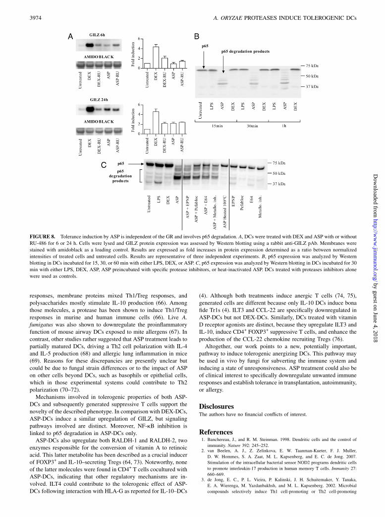
Tolerance induction by ASP is independent of theglucocorticoid receptor and involves p65 degradation
To further document that distinct molecular mechanisms underliethe induction of tolerogenic DCs following DEX or ASP treatment,we compared GILZ induction in ASP-DCs and DEX-DCs inpresence or absence of RU-486, a known glucocorticoid receptor(GR) inhibitor. As shown in Fig. 8A, RU-486 significantlyinhibited GILZ expression in DEX-DCs but had no impact onGILZ expression in ASP-DCs, indicating that GILZ was inducedby a GR-independent pathway in ASP-DCs in contrast to DEX-DCs. These data also confirmed that GILZ was induced later, notonly at a transcriptional but also at a protein level in ASP-DCswhen compared with DEX-DCs. We subsequently investigateda potential involvement of the NF-kB pathway in ASP-mediatedtolerance. Although we did not see any change in IkB expressionin ASP-DCs, we could demonstrate that ASP inhibits LPS-
FIGURE 6. ASP-DCs induce nonapoptotic
T cells with a suppressive capacity. A, For ap-
optosis assays, T cells were harvested at day 6
and labeled with Annexin V/PI. Percentages of
PI2/Annexin V2, PI2/Annexin V+, and PI+/
Annexin V+ cells were determined by flow
cytometry (mean 6 SEM using samples from
four healthy donors). B, To measure T cell
suppressive activity, CD4+CD252 responder
cells were isolated using the CD4+CD25+ reg-
ulatory T cell isolation kit through collection of
the negative fraction. Cells were CFSE labeled
and plated with anti-CD2, -CD3, and -CD28
beads (ratio 1:1). Cocultured T cells were
added at ratios ranging from 1:2 to 8:1 re-
sponder/cocultured T cells and incubated for 3
d. All determinations were carried out in du-
plicate, and responder cell proliferation was
assessed by flow cytometry. C, The potential
involvement of soluble factors on T cell sup-
pression was analyzed by incubating CFSE-la-
beled CD4+ T cells stimulated with anti-CD2,
-CD3, and -CD28 beads, with 4, 20, or 100 ml
supernatant from treated DC/T cell cocultures.
Percentages of proliferating cells were de-
termined after 5 d by flow cytometry. Treat-
ments were compared with controls.
3972 A. ORYZAE PROTEASES INDUCE TOLEROGENIC DCs
by guest on June 4, 2018http://w
ww
.jimm
unol.org/D
ownloaded from

induced NF-kB translocation (not shown). We also observed anearly degradation of p65 (i.e., within 15 min) in ASP-DCs but notin DEX-DCs (Fig. 8B), suggesting that ASP treatment may inhibitthe NF-kB pathway in a distinct manner. To investigate whichproteases were potentially responsible for p65 degradation, weincubated ASP-DCs with specific protease inhibitors. Surprisingly,aspartic acid protease inhibitor did not prevent p65 degradationin contrast to serine, cysteine, and metalloprotease inhibitors, asshown in Fig. 8C. As a control, heat-inactivated ASP completelyprevented p65 degradation in DCs. Collectively, these results in-dicate that proteases from ASP may synergize to induce tolerance,with aspartic acid-containing proteases inducing regulatory genesand serine, cysteine, and metalloproteases contributing to p65degradation.
DiscussionTolerogenic DCs are physiologically important because they caninduce Tregs, preventing or controlling autoimmunity, allergy,and graft rejection (8–13). As of today, various conditioningstrategies are being considered to generate tolerogenic DCs. Thisincludes DC treatment with anti-inflammatory cytokines (IL-10,TGF-b) (4, 44, 45), neuropeptides (46, 47), immunosuppressivedrugs (e.g., dexamethasone, mitomycin C, rapamycin) (16, 19, 48,49), or vitamins (1,25-dihydroxy-vitamin D3, vitamin A) (16, 17,35, 50, 51). Genetic engineering of DCs has also been used toenhance the expression of IL-10 (52), TGF-b (53), soluble TNFR(54), intracellular CTLA-4 (55), or FOXP3 molecules (56) or toprevent activation of the NF-kB pathway (57).In this study, we identified a new pathway to induce tolerogenic
DCs by treating monocyte derived-DCs with proteases from A.oryzae, a fungus commonly used to produce traditional fermented
food, enzymes, antibiotics, beverages, and volatile compounds(16). Our results demonstrate that treatment with ASP inducesDCs with a CD802CD832CD862ILT22ILT32ILT4+ phenotype,which do not secrete cytokines or chemokines and upregulatewell- known tolerogenic markers such as GILZ (29, 30), IDO (58–60), NOS-2 (61, 62), RALDH-1, or RALDH-2 (35, 51, 63, 64).ASP-DCs are clearly different from DEX-DCs because the latterpromote the generation of anergic IL-10–producing Tr1s witha suppressive activity partially mediated by soluble factors, inagreement with the literature (15, 16). In contrast, ASP-DCs ratherinduce anergic suppressive CD4+ T cells, which do not expressFOXP3 or secrete IL-10, but secrete low amounts of TGF-b.Importantly, the suppressive effect of such T cells could not becorrelated with the production of soluble factors. Inhibitionstudies further highlighted the involvement of various proteaseswithin ASP in the induction of regulatory properties in DCs.Our results are in agreement with other studies suggesting
a potential tolerogenic role of Aspergillus-derived molecules.Aspergillus infection was reported to induce the generation ofIDO+ tolerogenic DCs promoting the local recruitment of CD4+
CD25+ Tregs (65). These cells suppressed Th2 responses to thefungus through the combined action of IL-10 and CTLA-4, sug-gesting that such regulatory mechanisms operating at the level ofDCs may be used by fungi to dampen host immune responses andinduce a state of immune tolerance (65). In support of this hy-pothesis, we observed that culture supernatants from other As-pergillus strains such as A. fumigatus or A. niger also inducea regulatory DC phenotype (not shown). Interestingly, various A.fumigatus components have been shown by others to exhibitdramatically different immunomodulatory properties. For exam-ple, secreted proteins induce Th2 cell activation, glycolipids Th17
FIGURE 7. Upregulation of tolerogenic genes by ASP involves aspartic acid-containing proteases. A, ASP was filtered through 10 kDa cutoff filters, and
both filtrates and retentates were incubated with DCs for 24 h. B, ASP was inactivated by heating 15 min at 100˚C or by incubation with protease inhibitors
for 1 h. Specific protease inhibitors used included a mix of metalloprotease inhibitors composed of 1,10-phenanthroline, phosphoramidon, and arphamenine
B, the serine protease inhibitor Pefabloc SC, the cysteine protease inhibitor E64, and the aspartic protease inhibitor EPNP. DCs were cultured with medium
or treated ASP (20 mg/ml) for 24 h. Gene expression of GILZ, NOS-2, RALDH-1, and RALDH-2 was analyzed by quantitative PCR. Data are expressed as
relative amounts of mRNA in DCs treated with either ASP (20 mg/ml) or ASP plus inhibitors in comparison with DCs incubated in medium alone. Data are
normalized to amounts of b-actin and shown as the mean 6 SEM obtained from four healthy donors. **p , 0.01 (Kruskall–Wallis test).
The Journal of Immunology 3973
by guest on June 4, 2018http://w
ww
.jimm
unol.org/D
ownloaded from
responses, membrane proteins mixed Th1/Treg responses, andpolysaccharides mostly stimulate IL-10 production (66). Amongthose molecules, a protease has been shown to induce Th1/Tregresponses in murine and human immune cells (66). Live A.fumigatus was also shown to downregulate the proinflammatoryfunction of mouse airway DCs exposed to mite allergens (67). Incontrast, other studies rather suggested that ASP treatment leads topartially matured DCs, driving a Th2 cell polarization with IL-4and IL-5 production (68) and allergic lung inflammation in mice(69). Reasons for these discrepancies are presently unclear butcould be due to fungal strain differences or to the impact of ASPon other cells beyond DCs, such as basophils or epithelial cells,which in those experimental systems could contribute to Th2polarization (70–72).Mechanisms involved in tolerogenic properties of both ASP-
DCs and subsequently generated suppressive T cells support thenovelty of the described phenotype. In comparison with DEX-DCs,ASP-DCs induce a similar upregulation of GILZ, but signalingpathways involved are distinct. Moreover, NF-kB inhibition islinked to p65 degradation in ASP-DCs only.ASP-DCs also upregulate both RALDH-1 and RALDH-2, two
enzymes responsible for the conversion of vitamin A to retinoicacid. This latter metabolite has been described as a crucial inducerof FOXP3+ and IL-10–secreting Tregs (64, 73). Noteworthy, noneof the latter molecules were found in CD4+ T cells cocultured withASP-DCs, indicating that other regulatory mechanisms are in-volved. ILT4 could contribute to the tolerogenic effect of ASP-DCs following interaction with HLA-G as reported for IL-10–DCs
(4). Although both treatments induce anergic T cells (74, 75),generated cells are different because only IL-10 DCs induce bonafide Tr1s (4). ILT3 and CCL-22 are specifically downregulated inASP-DCs but not DEX-DCs. Similarly, DCs treated with vitaminD receptor agonists are distinct, because they upregulate ILT3 andIL-10, induce CD4+ FOXP3+ suppressive T cells, and enhance theproduction of the CCL-22 chemokine recruiting Tregs (76).Altogether, our work points to a new, potentially important,
pathway to induce tolerogenic anergizing DCs. This pathway maybe used in vivo by fungi for subverting the immune system andinducing a state of unresponsiveness. ASP treatment could also beof clinical interest to specifically downregulate unwanted immuneresponses and establish tolerance in transplantation, autoimmunity,or allergy.
DisclosuresThe authors have no financial conflicts of interest.
References1. Banchereau, J., and R. M. Steinman. 1998. Dendritic cells and the control of
immunity. Nature 392: 245–252.2. van Beelen, A. J., Z. Zelinkova, E. W. Taanman-Kueter, F. J. Muller,
D. W. Hommes, S. A. Zaat, M. L. Kapsenberg, and E. C. de Jong. 2007.
Stimulation of the intracellular bacterial sensor NOD2 programs dendritic cells
to promote interleukin-17 production in human memory T cells. Immunity 27:
660–669.3. de Jong, E. C., P. L. Vieira, P. Kalinski, J. H. Schuitemaker, Y. Tanaka,
E. A. Wierenga, M. Yazdanbakhsh, and M. L. Kapsenberg. 2002. Microbial
compounds selectively induce Th1 cell-promoting or Th2 cell-promoting
FIGURE 8. Tolerance induction by ASP is independent of the GR and involves p65 degradation. A, DCs were treated with DEX and ASP with or without
RU-486 for 6 or 24 h. Cells were lysed and GILZ protein expression was assessed by Western blotting using a rabbit anti-GILZ pAb. Membranes were
stained with amidoblack as a loading control. Results are expressed as fold increases in protein expression determined as a ratio between normalized
intensities of treated cells and untreated cells. Results are representative of three independent experiments. B, p65 expression was analyzed by Western
blotting in DCs incubated for 15, 30, or 60 min with either LPS, DEX, or ASP. C, p65 expression was analyzed by Western blotting in DCs incubated for 30
min with either LPS, DEX, ASP, ASP preincubated with specific protease inhibitors, or heat-inactivated ASP. DCs treated with proteases inhibitors alone
were used as controls.
3974 A. ORYZAE PROTEASES INDUCE TOLEROGENIC DCs
by guest on June 4, 2018http://w
ww
.jimm
unol.org/D
ownloaded from
dendritic cells in vitro with diverse th cell-polarizing signals. J. Immunol. 168:1704–1709.
4. Gregori, S., D. Tomasoni, V. Pacciani, M. Scirpoli, M. Battaglia, C. F. Magnani,E. Hauben, and M. G. Roncarolo. 2010. Differentiation of type 1 T regulatorycells (Tr1) by tolerogenic DC-10 requires the IL-10-dependent ILT4/HLA-Gpathway. Blood 116: 935–944.
5. Ilarregui, J. M., D. O. Croci, G. A. Bianco, M. A. Toscano, M. Salatino,M. E. Vermeulen, J. R. Geffner, and G. A. Rabinovich. 2009. Tolerogenic signalsdelivered by dendritic cells to T cells through a galectin-1-driven immunoreg-ulatory circuit involving interleukin 27 and interleukin 10. Nat. Immunol. 10:981–991.
6. Adorini, L., N. Giarratana, and G. Penna. 2004. Pharmacological induction oftolerogenic dendritic cells and regulatory T cells. Semin. Immunol. 16: 127–134.
7. Pulendran, B., H. Tang, and S. Manicassamy. 2010. Programming dendritic cellsto induce T(H)2 and tolerogenic responses. Nat. Immunol. 11: 647–655.
8. Boissier, M. C., E. Assier, J. Biton, A. Denys, G. Falgarone, and N. Bessis. 2009.Regulatory T cells (Treg) in rheumatoid arthritis. Joint Bone Spine 76: 10–14.
9. Trenado, A., S. Fisson, E. Braunberger, D. Klatzmann, B. L. Salomon, andJ. L. Cohen. 2004. Ex vivo selection of recipient-type alloantigen-specific CD4(+)CD25(+) immunoregulatory T cells for the control of graft-versus-host dis-ease after allogeneic hematopoietic stem-cell transplantation. Transplantation 77(1 Suppl): S32–S34.
10. Turnquist, H. R., R. T. Fischer, and A. W. Thomson. 2010. Pharmacologicalmodification of dendritic cells to promote their tolerogenicity in transplantation.Methods Mol. Biol. 595: 135–148.
11. Trzonkowski, P., M. Bieniaszewska, J. Juscinska, A. Dobyszuk, A. Krzystyniak,N. Marek, J. Mysliwska, and A. Hellmann. 2009. First-in-man clinical results ofthe treatment of patients with graft versus host disease with human ex vivoexpanded CD4+CD25+CD127- T regulatory cells. Clin. Immunol. 133: 22–26.
12. Palomares, O., G. Yaman, A. K. Azkur, T. Akkoc, M. Akdis, and C. A. Akdis.2010. Role of Treg in immune regulation of allergic diseases. Eur. J. Immunol.40: 1232–1240.
13. Bohle, B., T. Kinaciyan, M. Gerstmayr, A. Radakovics, B. Jahn-Schmid, andC. Ebner. 2007. Sublingual immunotherapy induces IL-10-producing T regula-tory cells, allergen-specific T-cell tolerance, and immune deviation. J. AllergyClin. Immunol. 120: 707–713.
14. Rescigno, M. 2010. Dendritic cells in tolerance induction for the treatment ofautoimmune diseases. Eur. J. Immunol. 40: 2119–2123.
15. Bosma, B. M., H. J. Metselaar, N. M. Nagtzaam, R. de Haan, S. Mancham,L. J. van der Laan, E. J. Kuipers, and J. Kwekkeboom. 2008. Dexamethasonetransforms lipopolysaccharide-stimulated human blood myeloid dendritic cellsinto myeloid dendritic cells that prime interleukin-10 production in T cells.Immunology 125: 91–100.
16. Unger, W. W., S. Laban, F. S. Kleijwegt, A. R. van der Slik, and B. O. Roep.2009. Induction of Treg by monocyte-derived DC modulated by vitamin D3 ordexamethasone: differential role for PD-L1. Eur. J. Immunol. 39: 3147–3159.
17. Pedersen, A. W., K. Holmstrøm, S. S. Jensen, D. Fuchs, S. Rasmussen,P. Kvistborg, M. H. Claesson, and M. B. Zocca. 2009. Phenotypic and functionalmarkers for 1alpha,25-dihydroxyvitamin D(3)-modified regulatory dendriticcells. Clin. Exp. Immunol. 157: 48–59.
18. Woltman, A. M., J. W. de Fijter, S. W. Kamerling, L. C. Paul, M. R. Daha, andC. van Kooten. 2000. The effect of calcineurin inhibitors and corticosteroids onthe differentiation of human dendritic cells. Eur. J. Immunol. 30: 1807–1812.
19. Xia, C. Q., R. Peng, F. Beato, and M. J. Clare-Salzler. 2005. Dexamethasoneinduces IL-10-producing monocyte-derived dendritic cells with durable imma-turity. Scand. J. Immunol. 62: 45–54.
20. Emmer, P. M., J. van der Vlag, G. J. Adema, and L. B. Hilbrands. 2006. Den-dritic cells activated by lipopolysaccharide after dexamethasone treatment in-duce donor-specific allograft hyporesponsiveness. Transplantation 81: 1451–1459.
21. Vizzardelli, C., N. Pavelka, A. Luchini, I. Zanoni, L. Bendickson, M. Pelizzola,O. Beretta, M. Foti, F. Granucci, M. Nilsen-Hamilton, and P. Ricciardi-Cas-tagnoli. 2006. Effects of dexamethazone on LPS-induced activationand migra-tion of mouse dendritic cells revealed by a genome-wide transcriptional analysis.Eur. J. Immunol. 36: 1504–1515.
22. Roelen, D. L., D. H. Schuurhuis, D. E. van den Boogaardt, K. Koekkoek,P. P. van Miert, J. J. van Schip, S. Laban, D. Rea, C. J. Melief, R. Offringa, et al.2003. Prolongation of skin graft survival by modulation of the alloimmune re-sponse with alternatively activated dendritic cells. Transplantation 76: 1608–1615.
23. Manavalan, J. S., P. C. Rossi, G. Vlad, F. Piazza, A. Yarilina, R. Cortesini,D. Mancini, and N. Suciu-Foca. 2003. High expression of ILT3 and ILT4 isa general feature of tolerogenic dendritic cells. Transpl. Immunol. 11: 245–258.
24. Chang, C. C., R. Ciubotariu, J. S. Manavalan, J. Yuan, A. I. Colovai, F. Piazza,S. Lederman, M. Colonna, R. Cortesini, R. Dalla-Favera, and N. Suciu-Foca.2002. Tolerization of dendritic cells by T(S) cells: the crucial role of inhibitoryreceptors ILT3 and ILT4. Nat. Immunol. 3: 237–243.
25. Svajger, U., A. Vidmar, and M. Jeras. 2008. Niflumic acid renders dendritic cellstolerogenic and up-regulates inhibitory molecules ILT3 and ILT4. Int. Immu-nopharmacol. 8: 997–1005.
26. Gleissner, C. A., and T. J. Dengler. 2009. Induction of ILT expression on non-professional antigen presenting cells: Clinical applications. Hum. Immunol. 70:357–359.
27. Vlad, G., C. C. Chang, A. I. Colovai, E. R. Vasilescu, R. Cortesini, and N. Suciu-Foca. 2010. Membrane and soluble ILT3 are critical to the generation of Tsuppressor cells and induction of immunological tolerance. Int. Rev. Immunol.29: 119–132.
28. Brenk, M., M. Scheler, S. Koch, J. Neumann, O. Takikawa, G. Hacker, T. Bieber,and D. von Bubnoff. 2009. Tryptophan deprivation induces inhibitory receptorsILT3 and ILT4 on dendritic cells favoring the induction of human CD4+CD25+Foxp3+ T regulatory cells. J. Immunol. 183: 145–154.
29. Cohen, N., E. Mouly, H. Hamdi, M. C. Maillot, M. Pallardy, V. Godot, F. Capel,A. Balian, S. Naveau, P. Galanaud, et al. 2006. GILZ expression in humandendritic cells redirects their maturation and prevents antigen-specificT lymphocyte response. Blood 107: 2037–2044.
30. Hamdi, H., V. Godot, M. C. Maillot, M. V. Prejean, N. Cohen, R. Krzysiek,F. M. Lemoine, W. Zou, and D. Emilie. 2007. Induction of antigen-specificregulatory T lymphocytes by human dendritic cells expressing theglucocorticoid-induced leucine zipper. Blood 110: 211–219.
31. Ayroldi, E., G. Migliorati, S. Bruscoli, C. Marchetti, O. Zollo, L. Cannarile,F. D’Adamio, and C. Riccardi. 2001. Modulation of T-cell activation by theglucocorticoid-induced leucine zipper factor via inhibition of nuclear factorkappaB. Blood 98: 743–753.
32. Sun, C. M., J. A. Hall, R. B. Blank, N. Bouladoux, M. Oukka, J. R. Mora, andY. Belkaid. 2007. Small intestine lamina propria dendritic cells promote de novogeneration of Foxp3 T reg cells via retinoic acid. J. Exp. Med. 204: 1775–1785.
33. Coombes, J. L., K. R. Siddiqui, C. V. Arancibia-Carcamo, J. Hall, C. M. Sun,Y. Belkaid, and F. Powrie. 2007. A functionally specialized population of mu-cosal CD103+ DCs induces Foxp3+ regulatory T cells via a TGF-beta and ret-inoic acid-dependent mechanism. J. Exp. Med. 204: 1757–1764.
34. Geissmann, F., P. Revy, N. Brousse, Y. Lepelletier, C. Folli, A. Durandy,P. Chambon, and M. Dy. 2003. Retinoids regulate survival and antigen pre-sentation by immature dendritic cells. J. Exp. Med. 198: 623–634.
35. Manicassamy, S., and B. Pulendran. 2009. Retinoic acid-dependent regulation ofimmune responses by dendritic cells and macrophages. Semin. Immunol. 21: 22–27.
36. Guilliams, M., K. Crozat, S. Henri, S. Tamoutounour, P. Grenot, E. Devilard,B. de Bovis, L. Alexopoulou, M. Dalod, and B. Malissen. 2010. Skin-draininglymph nodes contain dermis-derived CD103(-) dendritic cells that constitutivelyproduce retinoic acid and induce Foxp3(+) regulatory T cells. Blood 115: 1958–1968.
37. Chen, C., W. H. Lee, L. Zhong, and C. P. Liu. 2006. Regulatory T cells canmediate their function through the stimulation of APCs to produce immuno-suppressive nitric oxide. J. Immunol. 176: 3449–3460.
38. Ogawa, A., A. Yasuhara, T. Tanaka, T. Sakiyama, and K. Nakanishi. 1995.Production of neutral protease by membrane-surface liquid culture of Aspergillusoryzae IAM2704. J. Ferment. Bioeng. 80: 35–40.
39. Manome, H., S. Aiba, and H. Tagami. 1999. Simple chemicals can inducematuration and apoptosis of dendritic cells. Immunology 98: 481–490.
40. Ohtani, T., M. Mizuashi, S. Nakagawa, Y. Sasaki, T. Fujimura, R. Okuyama, andS. Aiba. 2009. TGF-beta1 dampens the susceptibility of dendritic cells to en-vironmental stimulation, leading to the requirement for danger signals for acti-vation. Immunology 126: 485–499.
41. Asselin-Labat, M. L., M. David, A. Biola-Vidamment, D. Lecoeuche,M. C. Zennaro, J. Bertoglio, and M. Pallardy. 2004. GILZ, a new target for thetranscription factor FoxO3, protects T lymphocytes from interleukin-2withdrawal-induced apoptosis. Blood 104: 215–223.
42. Kushwah, R., J. Wu, J. R. Oliver, G. Jiang, J. Zhang, K. A. Siminovitch, andJ. Hu. 2010. Uptake of apoptotic DC converts immature DC into tolerogenic DC,which induce differentiation of Foxp3+ regulatory T cells. Eur. J. Immunol. 40:1022–1035.
43. Kushwah, R., and J. Hu. 2010. Dendritic cell apoptosis: regulation of toleranceversus immunity. J. Immunol. 185: 795–802.
44. Belladonna, M. L., C. Volpi, R. Bianchi, C. Vacca, C. Orabona, M. T. Pallotta,L. Boon, S. Gizzi, M. C. Fioretti, U. Grohmann, and P. Puccetti. 2008. Cuttingedge: Autocrine TGF-beta sustains default tolerogenesis by IDO-competentdendritic cells. J. Immunol. 181: 5194–5198.
45. Steinbrink, K., E. Graulich, S. Kubsch, J. Knop, and A. H. Enk. 2002. CD4(+)and CD8(+) anergic T cells induced by interleukin-10-treated human dendriticcells display antigen-specific suppressor activity. Blood 99: 2468–2476.
46. Delgado, M. 2009. Generating tolerogenic dendritic cells with neuropeptides.Hum. Immunol. 70: 300–307.
47. Pozo, D., P. Anderson, and E. Gonzalez-Rey. 2009. Induction of alloantigen-specific human T regulatory cells by vasoactive intestinal peptide. J. Immunol.183: 4346–4359.
48. Jiga, L. P., T. M. Bauer, J. J. Chuang, G. Opelz, and P. Terness. 2004. Generationof tolerogenic dendritic cells by treatment with mitomycin C: inhibition of al-logeneic T-cell response is mediated by downregulation of ICAM-1, CD80, andCD86. Transplantation 77: 1761–1764.
49. Haidinger, M., M. Poglitsch, R. Geyeregger, S. Kasturi, M. Zeyda,G. J. Zlabinger, B. Pulendran, W. H. Horl, M. D. Saemann, and T. Weichhart.2010. A versatile role of mammalian target of rapamycin in human dendritic cellfunction and differentiation. J. Immunol. 185: 3919–3931.
50. Szeles, L., G. Keresztes, D. Torocsik, Z. Balajthy, L. Krenacs, S. Poliska,A. Steinmeyer, U. Zuegel, M. Pruenster, A. Rot, and L. Nagy. 2009. 1,25-dihydroxyvitamin D3 is an autonomous regulator of the transcriptional changesleading to a tolerogenic dendritic cell phenotype. J. Immunol. 182: 2074–2083.
51. Manicassamy, S., R. Ravindran, J. Deng, H. Oluoch, T. L. Denning, S. P. Kasturi,K. M. Rosenthal, B. D. Evavold, and B. Pulendran. 2009. Toll-like receptor 2-dependent induction of vitamin A-metabolizing enzymes in dendritic cellspromotes T regulatory responses and inhibits autoimmunity. Nat. Med. 15: 401–409.
52. Henry, E., C. J. Desmet, V. Garze, L. Fievez, D. Bedoret, C. Heirman, P. Faisca,F. J. Jaspar, P. Gosset, A. P. Jacquet, et al. 2008. Dendritic cells genetically
The Journal of Immunology 3975
by guest on June 4, 2018http://w
ww
.jimm
unol.org/D
ownloaded from
engineered to express IL-10 induce long-lasting antigen-specific tolerance inexperimental asthma. J. Immunol. 181: 7230–7242.
53. Gorczynski, R. M., J. Bransom, M. Cattral, X. Huang, J. Lei, L. Xiaorong,W. P. Min, Y. Wan, and J. Gauldie. 2000. Synergy in induction of increased renalallograft survival after portal vein infusion of dendritic cells transduced to ex-press TGFbeta and IL-10, along with administration of CHO cells expressing theregulatory molecule OX-2. Clin. Immunol. 95: 182–189.
54. Wang, Q., Y. Liu, J. Wang, G. Ding, W. Zhang, G. Chen, M. Zhang, S. Zheng, andX. Cao. 2006. Induction of allospecific tolerance by immature dendritic cells ge-netically modified to express soluble TNF receptor. J. Immunol. 177: 2175–2185.
55. Tan, P. H., J. B. Yates, S. A. Xue, C. Chan, W. J. Jordan, J. E. Harper,M. P. Watson, R. Dong, M. A. Ritter, R. I. Lechler, et al. 2005. Creation oftolerogenic human dendritic cells via intracellular CTLA4: a novel strategy withpotential in clinical immunosuppression. Blood 106: 2936–2943.
56. Lipscomb, M. W., J. L. Taylor, C. J. Goldbach, S. C. Watkins, A. K. Wesa, andW. J. Storkus. 2010. DC expressing transgene Foxp3 are regulatory APC. Eur. J.Immunol. 40: 480–493.
57. Tomasoni, S., S. Aiello, L. Cassis, M. Noris, L. Longaretti, R. A. Cavinato,N. Azzollini, A. Pezzotta, G. Remuzzi, and A. Benigni. 2005. Dendritic cellsgenetically engineered with adenoviral vector encoding dnIKK2 induce theformation of potent CD4+ T-regulatory cells. Transplantation 79: 1056–1061.
58. Orabona, C., P. Puccetti, C. Vacca, S. Bicciato, A. Luchini, F. Fallarino, R. Bianchi,E. Velardi, K. Perruccio, A. Velardi, et al. 2006. Toward the identification ofa tolerogenic signature in IDO-competent dendritic cells. Blood 107: 2846–2854.
59. Mellor, A. L., and D. H. Munn. 2004. IDO expression by dendritic cells: tol-erance and tryptophan catabolism. Nat. Rev. Immunol. 4: 762–774.
60. Munn, D. H., M. D. Sharma, J. R. Lee, K. G. Jhaver, T. S. Johnson, D. B. Keskin,B. Marshall, P. Chandler, S. J. Antonia, R. Burgess, et al. 2002. Potential reg-ulatory function of human dendritic cells expressing indoleamine 2,3-dioxyge-nase. Science 297: 1867–1870.
61. Yanagawa, Y., K. Iwabuchi, and K. Onoe. 2009. Co-operative action ofinterleukin-10 and interferon-gamma to regulate dendritic cell functions. Im-munology 127: 345–353.
62. Hill, M., R. Zagani, C. Voisine, C. Usal, and I. Anegon. 2007. Nitric oxide andindoleamine 2,3-dioxygenase mediate CTLA4Ig-induced survival in heartallografts in rats. Transplantation 84: 1060–1063.
63. Xiao, S., H. Jin, T. Korn, S. M. Liu, M. Oukka, B. Lim, and V. K. Kuchroo. 2008.Retinoic acid increases Foxp3+ regulatory T cells and inhibits development ofTh17 cells by enhancing TGF-beta-driven Smad3 signaling and inhibiting IL-6and IL-23 receptor expression. J. Immunol. 181: 2277–2284.
64. Kang, S. G., H. W. Lim, O. M. Andrisani, H. E. Broxmeyer, and C. H. Kim.2007. Vitamin A metabolites induce gut-homing FoxP3+ regulatory T cells. J.Immunol. 179: 3724–3733.
65. Montagnoli, C., F. Fallarino, R. Gaziano, S. Bozza, S. Bellocchio, T. Zelante,W. P. Kurup, L. Pitzurra, P. Puccetti, and L. Romani. 2006. Immunity and tol-erance to Aspergillus involve functionally distinct regulatory T cells and tryp-tophan catabolism. J. Immunol. 176: 1712–1723.
66. Bozza, S., C. Clavaud, G. Giovannini, T. Fontaine, A. Beauvais, J. Sarfati,C. D’Angelo, K. Perruccio, P. Bonifazi, S. Zagarella, et al. 2009. Immunesensing of Aspergillus fumigatus proteins, glycolipids, and polysaccharides andthe impact on Th immunity and vaccination. J. Immunol. 183: 2407–2414.
67. Fukahori, S., H. Matsuse, T. Tsuchida, T. Kawano, S. Tomari, C. Fukushima, andS. Kohno. 2010. Aspergillus fumigatus regulates mite allergen-pulsed dendriticcells in the development of asthma. Clin. Exp. Allergy 40: 1507–1515.
68. Lamhamedi-Cherradi, S. E., R. E. Martin, T. Ito, F. Kheradmand, D. B. Corry,Y. J. Liu, and M. Moyle. 2008. Fungal proteases induce Th2 polarization throughlimited dendritic cell maturation and reduced production of IL-12. J. Immunol.180: 6000–6009.
69. Kheradmand, F., A. Kiss, J. Xu, S. H. Lee, P. E. Kolattukudy, and D. B. Corry.2002. A protease-activated pathway underlying Th cell type 2 activation andallergic lung disease. J. Immunol. 169: 5904–5911.
70. Phillips, C., W. R. Coward, D. I. Pritchard, and C. R. Hewitt. 2003. Basophilsexpress a type 2 cytokine profile on exposure to proteases from helminths andhouse dust mites. J. Leukoc. Biol. 73: 165–171.
71. Sokol, C. L., G. M. Barton, A. G. Farr, and R. Medzhitov. 2008. A mechanismfor the initiation of allergen-induced T helper type 2 responses. Nat. Immunol. 9:310–318.
72. Kouzaki, H., S. M. O’Grady, C. B. Lawrence, and H. Kita. 2009. Proteases in-duce production of thymic stromal lymphopoietin by airway epithelial cellsthrough protease-activated receptor-2. J. Immunol. 183: 1427–1434.
73. Maynard, C. L., R. D. Hatton, W. S. Helms, J. R. Oliver, C. B. Stephensen, andC. T. Weaver. 2009. Contrasting roles for all-trans retinoic acid in TGF-beta-mediated induction of Foxp3 and Il10 genes in developing regulatory T cells. J.Exp. Med. 206: 343–357.
74. Ristich, V., W. Zhang, S. Liang, and A. Horuzsko. 2007. Mechanisms of pro-longation of allograft survival by HLA-G/ILT4-modified dendritic cells. Hum.Immunol. 68: 264–271.
75. Ristich, V., S. Liang, W. Zhang, J. Wu, and A. Horuzsko. 2005. Tolerization ofdendritic cells by HLA-G. Eur. J. Immunol. 35: 1133–1142.
76. Adorini, L., and G. Penna. 2009. Dendritic cell tolerogenicity: a key mechanismin immunomodulation by vitamin D receptor agonists. Hum. Immunol. 70: 345–352.
3976 A. ORYZAE PROTEASES INDUCE TOLEROGENIC DCs
by guest on June 4, 2018http://w
ww
.jimm
unol.org/D
ownloaded from