identification and characterization an acyl- coa:triterpene
TRANSCRIPT
Identification and characterization of an acyl- CoA:triterpene acyltransferase activity in rabbit and human tissues
Ira Tabas, '.* Nanda Beatini,' Li-Ling Chen,* Woan-Chyng Su,t Mohindar S. Puar,* * Sundeep Dugar, * * John W. Clader* * Department of Medicine* and Institute of Human Nutrition, t Columbia University, .New York, NY 10032, and Schering-Plough Research," 60 Orange Street, Bloomfield, NJ 07003
Abstract Rabbit and human tissues contain substantial amounts of an unusual lipid, a fatty acid ester of a pentacyclic triterpene, that is a potent in vitro inhibitor of acyl-CoA:choles- terol acyltransferase (ACAT). A possible origin of the triterpene ester is via dietary absorption of plant triterpenes (which have a similar structure to the triterpene moiety of the animal triterpene ester), followed by fatty acid esterification of the triterpene in animal tissues. To support this idea, homogenates of rabbit and human enterocytes and liver are now shown to contain an acyl-CoA:triterpene acyltransferase activity (ATAT) which esterifies triterpene to a fatty acid. The enzyme activity was stimulated by exogenous triterpene and required ATP and coenzyme A when fatty acid was used as substrate; ATP and coenzyme A were not required when fatty acyl-CoA was used. ATAT was not inhibited by two structurally different ACAT inhi- bitors, which may indicate that ACAT and ATAT are different enzymes. Rat enterocytes and liver contained very little ATAT activity, consistent with the finding that rat liver contained very little triterpene ester. To establish that triterpene esterification occurs in vivo, [3H]triterpene was shown to be incorporated into triterpene ester in several organs and tissues from a rabbit given a gastric bolus of the labeled triterpene. These data provide support for the hypothesis that triterpene esters in animal tissues arise from the dietary absorption of triterpenes followed by the esterification of the triterpenes by an enzymatic activity in the animal tissues.-Tabas, I., N. Beatini, L-L. Chen, W-C. Su, M. S. Puar, S. Dugar, and J. W. Clader. Identification and characterization of an acyl-CoA:triterpene acyltransferase ac- tivity in rabbit and human tissues. J Lipid Res. 1991. 32: 1689-1698.
Supplementary key words mogenate ACAT inhibitor plant lipids * ACAT
triterpene ester - enterocytes - liver ho-
Triterpenes are mevalonate-derived plant compounds that contain a skeleton of 30 carbon atoms. When these carbon atoms are arranged in a structure containing five six-member carbon rings, the class of compounds is refer- red to as pentacyclic triterpenes (1). Pentacyclic triter- penes, which are present in numerous species of plants, have been found to have interesting pharmacological
effects on isolated cells and animals, including anti- inflammatory (2) and cytotoxic actions (3, 4). Given these biological effects of pentacyclic triterpenes, an interesting issue related to these compounds regards their dietary ab- sorption, metabolism, and biological actions in animals (including humans) that have ingested plant material.
The discovery of a pentacyclic triterpene fatty acid ester with ACAT inhibitory activity in rabbit and human liver and other tissues has recently been described (5) (see Fig. 1, compound 2, for the structure of the triterpene ester). A preliminary experiment probing the origin of the triterpene ester revealed that when [ 3H]mevalonate, a precursor of triterpenes, was injected into a rabbit, the label failed to become incorporated into triterpene ester (5). Furthermore, the triterpene moiety of the compound (Fig. 1, compound 1) was found to have a structure very similar to several plant triterpenes (3-6) . For these reasons, the idea that the triterpene moiety might ori- ginate from the absorption of dietary plant triterpenes was proposed (5). If this were the case, then a mechanism would be needed to esterify the absorbed triterpene to fat- ty acid.
The goal of this study was to test this hypothesis by identifying an enzymatic activity in animal tissues that catalyzes pentacyclic triterpene esterification. In this report, such a n enzymatic activity from rabbit and hu- man tissues has been identified. This reaction, depicted in Fig. 1, involves the esterification of a pentacyclic triterpene to fatty acid via an acyl-coenzyme A interme-
Abbreviations: ACAT, acy1CoA:cholesterol acyltransferase; ATAT, acyl-CoA:triterpene acyltransferase; ATP, adenosine triphosphate; BSA, bovine serum albumin; CoA, coenzyme A; TLC, thin-layer chromato-
'To whom correspondence should be addressed at: Department of Medicine, Columbia University, 630 West 168th Street, New York, NY 10032.
graphy.
Journal of Lipid Research Volume 32, 1989 1689
by guest, on April 10, 2019
ww
w.jlr.org
Dow
nloaded from
2
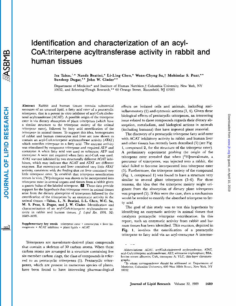
Fig. 1. Scheme for triterpene esterification. The structures of com- pounds 1 (triterpene) and 2 (triterpene ester) were determined previous- ly (5); the latter compound was purified from rabbit liver, and the former compound was obtained by alkaline hydrolysis of the triterpene ester (5). We have proposed and present evidence in this paper for the existence of an enzymatic activity in animal tissues, termed acyl-CoA.triterpene acyltransferase (ATAT), that catalyzes the esterification of triterpene (compound 1) to its fatty acid ester (compound 2) via a fatty acyl-CoA intermediate.
diate and has thus been termed acyl-CoA: triterpene acyl t ran sf erase (ATAT).
MATERIALS AND METHODS
Materials
Egg phosphatidylcholine, ceramide, monoglyceride, diglyceride, oleic acid, stearic acid, and triolein, ATP, coenzyme A, and protease inhibitors (below) were ob- tained from Sigma (St. Louis, MO). Cholesteryl oleate was from Steraloids, Inc. (Wilton, NH). l-14C-labeled fat- ty acids and l-I4C-Iabeled fatty acyl-CoAs (52-58 mCi/mmol) were purchased from DuPont-New England Nuclear. [l-13C]stearic acid (90.1 atom % I3C) was pur- chased from MSD Isotopes (Montreal, Canada). Com- pound 58-035, 3-[decyldimethylsilyl]-N-[2-(4-methyl-
phenyl)-1-phenylethyl] propanamide, was generously pro- vided by Dr. John Heider of Sandoz, Inc., East Hanover, NJ. Compound 277082, a trisubstituted urea, was gen- erously provided by the American Cyanimid Company, Pearl River, NY. Stock solutions (10 mg/ml) of both com- pounds were prepared in dimethylsulfoxide.
Animal tissues
Rabbit and rat tissues and organs, including entero- cytes, as well as human liver were obtained exactly as described previously (5). Samples of normal human small instestine were from endoscopic biopsy specimens.
Triterpene compounds
Rabbit liver triterpene ester was purified by a combina- tion of silicic acid chromatography and preparative TLC from lipid extracts of frozen rabbit liver exactly as described previously (5). The triterpene moiety of this compound was obtained by alkaline hydrolysis of the purified triterpene ester followed by preparative TLC (5). [ 3H]triterpene was prepared as follows: purified triterpene ester (refer to Fig. 1, compound 2) was oxidized to the ketone in ring A by treatment with pyridinium dichro- mate in methylene chloride for 1.5 h at room temperature. The triterpene ester ketone (2.3 mg) was then reduced with NaB3H4 by Du Pont-New England Nuclear Tritium Labeling Services. The resultant [ 3H]triterpene ester was subjected to alkaline hydrolysis (5), and the [3H]triter- pene was purified by preparative TLC as described (5). The final specific activity of the [3H]triterpene was ap- proximately 2.2 mCi/pmol. (In a preliminary experiment, we showed that unlabeled triterpene made from oxidized and NaBH4-reduced triterpene ester, which, by NMR, was a mixture of epimers around the hydroxyl carbon in ring A (refer to Fig. 1, compound l), stimulated rabbit enterocyte ATAT activity [see below] similar to native triterpene.
Standard triterpene esterification (ATAT) assay
Freshly harvested rabbit small intestine was washed with ice-cold normal saline, and the enterocytes were harvested by scraping into 0.1 M phosphate buffer, pH 7.4 (buffer A), containing 0.1 mM TPCK (~-l-chloro-3-[4-tosylamido]-4-phenyl-2-butanone), 1 mM EDTA, 10 pM pepstatin A, 0.1 mM PMSF (phenylmethanesulfonyl fluoride), 2 pM leupeptin, 10 pM iodoacetamide, and 80 p M antipain. The cells were homogenized by 20 strokes in a glass-Teflon cell dis- rupter. In a total volume of 400 pl, 800 pg of this homo- genate was incubated for 1 h at 37OC in buffer A containing 5 mM ATP, 0.5 mM coenzyme-A, 30 p~ [I4C]stearic acid (58 mCi/mmol), 30 pM BSA, and 250 pM triterpene. (In some experiments, 400 pg of homo- genate was assayed in 200 p1 of reaction mixture.) The [ I4C]stearic acid and triterpene were added to the reac-
1690 Journal of Lipid Research Volume 32, 1991
by guest, on April 10, 2019
ww
w.jlr.org
Dow
nloaded from
tion mixture as follows: the appropriate amounts of each compound from stock solutions in ethanol were added together in a test tube, and the solvent was removed under a stream of nitrogen. The dried lipids were then redis- solved in acetone and then added to the rest of the compo- nents in the reaction mixture (8 pl acetone solution per 400-pl assay).
At the end of the 1-h incubation, the reaction was stopped by addition of 4 ml chloroform-methanol 1:2 (v/v). After vortexing, 1.4 ml chloroform and 1.4 ml water were added; the tubes were vortexed again and centrifug- ed at 1000 RPM for 5 min in an IEC CRU-5000 centri- fuge. The bottom organic phase was collected and dried under a nitrogen stream. The dried residue was resus- pended in 50 pl chloroform and applied to 1-cm lanes of 20 x 20-cm analytical TLC plates (Soft Layer Adsorbosil-plus, 250 pm, Alltech Associates, Deerfield, IL). On two side lanes and a middle lane, monoglyceride and triterpene ester standards (20 pg) were spotted. The plates were developed first in diethyl ether-toluene- ethanol-glacial acetic acid 40:50:2:0.2 (v/v/v/v) and then in hexane-diethyl ether 94:6 (v/v). The plates were then dried and placed in an iodine tank to mark the location of the monoglyceride standard. Eight 0.5-cm bands, beginning 1 cm above the monoglyceride spot, were scraped, and the peak of radioactivity appearing in these eight bands was quantified by liquid scintillation count- ing. The 14C cpm were converted to pmol of ["Clstearate incorporated based upon the specific activity of the ['*C]stearic acid added to the reaction. After the plates were scraped, the triterpene ester spots on the remaining standard lanes were visualized by the sulfuric acidkhar- ring method (5) .
Other assays and analyticd methods
Rabbit enterocyte homogenate ACAT activity was assayed in the presence of 0.1 mM ['4C]oleoyl-CoA as previously described for microsomal ACAT activity (7). Protein was determined by the method of Lowry et al. (8). Carbon-18 NMR spectra were obtained on a Varian XL- 300 NMR spectrometer operating at 75.5 MHz as de- scribed previously (5).
RESULTS
Identification of a triterpene-stimulated enzymatic activity in rabbit enterocytes that produces a triterpene-fatty acid ester
To identify the enzymatic activity depicted in Fig. 1, enterocytes were chosen as the initial source of enzyme. This choice was based upon the idea that intestinal cholesterol absorption might require intestinal triterpene esterification just as intestinal cholesterol absorption is thought to require (at least partially) intestinal cholesterol
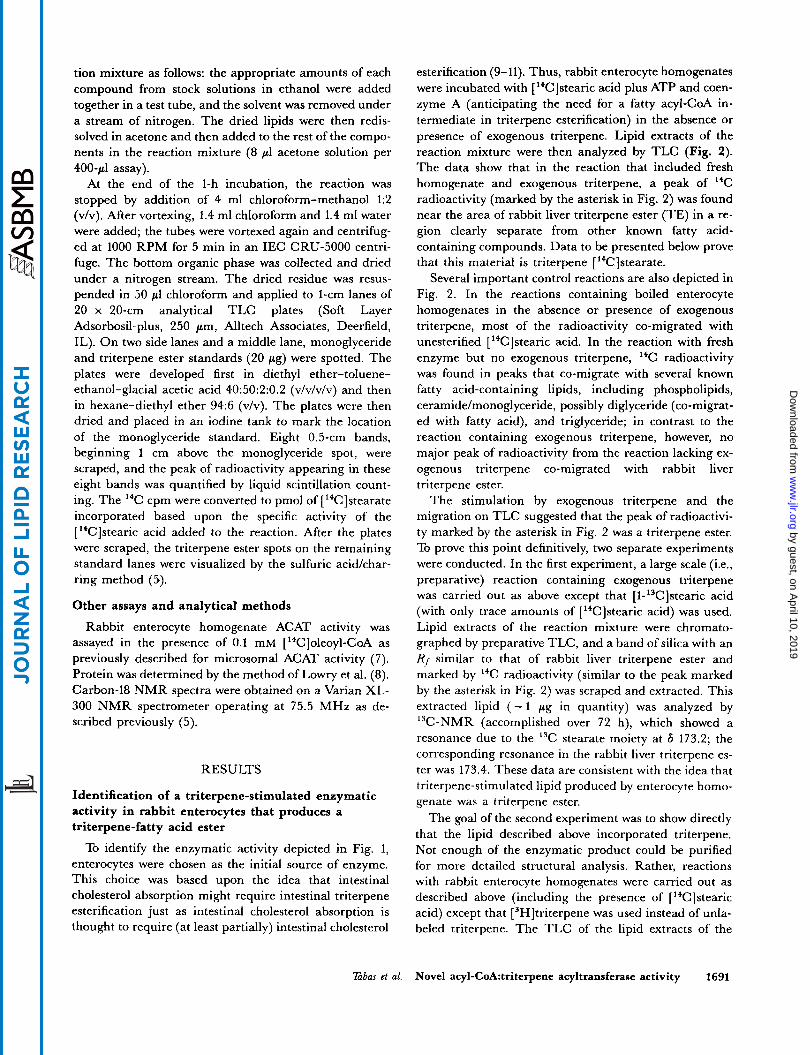
esterification (9- 11). Thus, rabbit enterocyte homogenates were incubated with [ '*C]stearic acid plus ATP and coen- zyme A (anticipating the need for a fatty acyl-CoA in- termediate in triterpene esterification) in the absence or presence of exogenous triterpene. Lipid extracts of the reaction mixture were then analyzed by TLC (Fig. 2). The data show that in the reaction that included fresh homogenate and exogenous triterpene, a peak of "C radioactivity (marked by the asterisk in Fig. 2) was found near the area of rabbit liver triterpene ester (TE) in a re- gion clearly separate from other known fatty acid- containing compounds. Data to be presented below prove that this material is triterpene [ 14C]stearate.
Several important control reactions are also depicted in Fig. 2. In the reactions containing boiled enterocyte homogenates in the absence or presence of exogenous triterpene, most of the radioactivity co-migrated with unesterified [ 14C]stearic acid. In the reaction with fresh enzyme but no exogenous triterpene, I4C radioactivity was found in peaks that co-migrate with several known fatty acid-containing lipids, including phospholipids, ceramide/monoglyceride, possibly diglyceride (co-migrat- ed with fatty acid), and triglyceride; in contrast to the reaction containing exogenous triterpene, however, no major peak of radioactivity from the reaction lacking ex- ogenous triterpene co-migrated with rabbit liver triterpene ester.
The stimulation by exogenous triterpene and the migration on TLC suggested that the peak of radioactivi- ty marked by the asterisk in Fig. 2 was a triterpene ester. To prove this point definitively, two separate experiments were conducted. In the first experiment, a large scale (;.e., preparative) reaction containing exogenous triterpene was carried out as above except that [l-13C]stearic acid (with only trace amounts of [14C]stearic acid) was used. Lipid extracts of the reaction mixture were chromato- graphed by preparative TLC, and a band of silica with an Rf similar to that of rabbit liver triterpene ester and marked by I4C radioactivity (similar to the peak marked by the asterisk in Fig. 2) was scraped and extracted. This extracted lipid ( - 1 pg in quantity) was analyzed by 13C-NMR (accomplished over 72 h), which showed a resonance due to the 13C stearate moiety at 6 173.2; the corresponding resonance in the rabbit liver triterpene es- ter was 173.4. These data are consistent with the idea that triterpene-stimulated lipid produced by enterocyte homo- genate was a triterpene ester.
The goal of the second experiment was to show directly that the lipid described above incorporated triterpene. Not enough of the enzymatic product could be purified for more detailed structural analysis. Rather, reactions with rabbit enterocyte homogenates were carried out as described above (including the presence of [ "Clstearic acid) except that [3H]triterpene was used instead of unla- beled triterpene. The TLC of the lipid extracts of the
Tnbas et al. Novel acyl-CoA:triterpene acyltransferase activity 1691
by guest, on April 10, 2019
ww
w.jlr.org
Dow
nloaded from
PL CER/MG TE DG/FA TG CE 400
300
200
100
0 2 4 6 8 10 12 14 16 0
cm from origin
Fig. 2. Thin-layer chromatogram of ['4C]stearate-labeled lipids from rabbit enterocyte homogenate reaction mix- tures in the absence and presence of exogenous triterpene. Rabbit enterocyte homogenate (fresh or boiled) was in- cubated with ['%]stearate, ATP, CoA, and BSA in the absence and presence of 250 p~ triterpene for 1 h at 37OC. Lipids were extracted as described for the triterpene esterification assay under Experimental Procedures except the entire length of each TLC lane was scraped in 1-cm bands. The four different reactions were as follows: boiled homogenate/no triterpene (open circles), boiled homogenate/plus triterpene (closed circles), fresh homogenateho triterpene (open triangles), fresh homogenate/plus triterpene (closed triangles). The TLC standards were: phospho- lipid (PL), ceramide (CER), monoglyceride (MG), triterpene ester (TE), diglyceride (DG), fatty acid (FA), trigly- ceride (TG) and cholesteryl ester (CE). The peak of radioactivity marked by the asterisk (*) is discussed in the text.
reactions is shown in Fig. 3 . In the upper panel of Fig. 3, a control reaction with boiled homogenate is shown. The 3H radioactivity co-migrated with unesterified triterpene standard, and the I4C radioactivity co- migrated with unesterified fatty acid standard. However, fresh homogenate (Fig. 3, lower panel) resulted in the ap- pearance of a peak of coincident 3H/'4C radioactivity (marked by the asterisk) that co-migrated close to rabbit liver triterpene ester. When this material, produced by a larger scale reaction and purified by preparative TLC, was subjected to alkaline hydrolysis, the 3H radioactivity co-migrated with authentic triterpene (data not shown). These data, together with those above, demonstrate that rabbit enterocyte homogenates, in the presence of stearic acid, ATP, coenzyme A, and exogenous triterpene, cata- lyze the production of a triterpene fatty acid ester. From here on we shall refer to this enzymatic activity as acyl- CoA:triterpene acyltransferase (ATAT).
Initial characterization of rabbit enterocyte homogenate ATAT activity
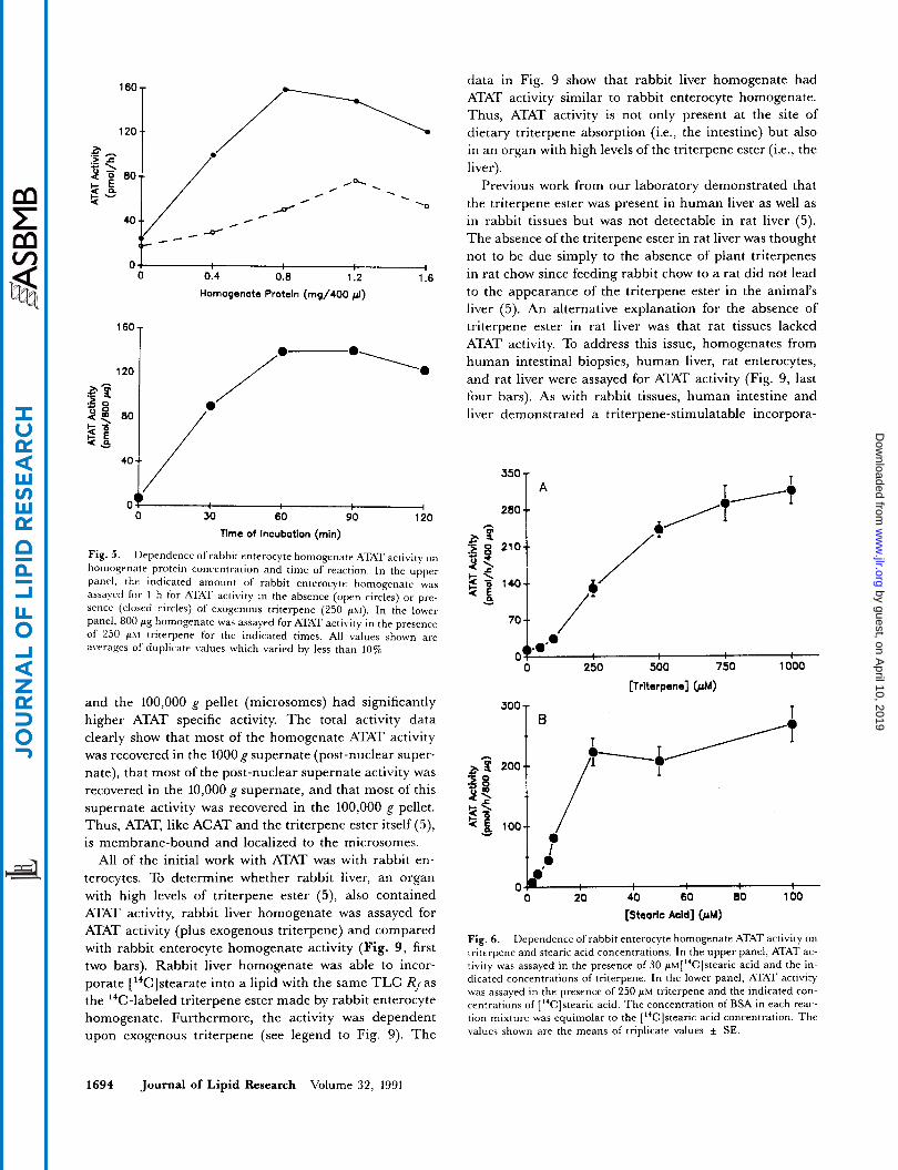
Although both ACAT activity and ATAT activity cata- lyze the fatty acid esterification of a multi-ring, isoprenoid-derived molecule, the fact that ATAT attaches the fatty acid to the fifth ring of a triterpene whereas ACAT works on the first ring of a sterol may suggest that the activities reside on different enzymes. To further ad- dress this issue, rabbit enterocyte homogenate was assayed for both ACAT and ATAT activities in the absence
and presence of each two structurally different ACAT in- hibitors, Sandoz compound 58-035 (12) and Lederle com- pound 277082 (13) (Fig. 4). The data in the top panel show that under the conditions of this experiment, each of the compounds inhibited ACAT activity -80%. In contrast, neither of the compounds inhibited enterocyte ATAT activity (Fig. 4, bottom panel). Pending physical separation of the two activities, these data, together with the substrate differences described above, may indicate that ATAT and ACAT activities represent two different enzymes.
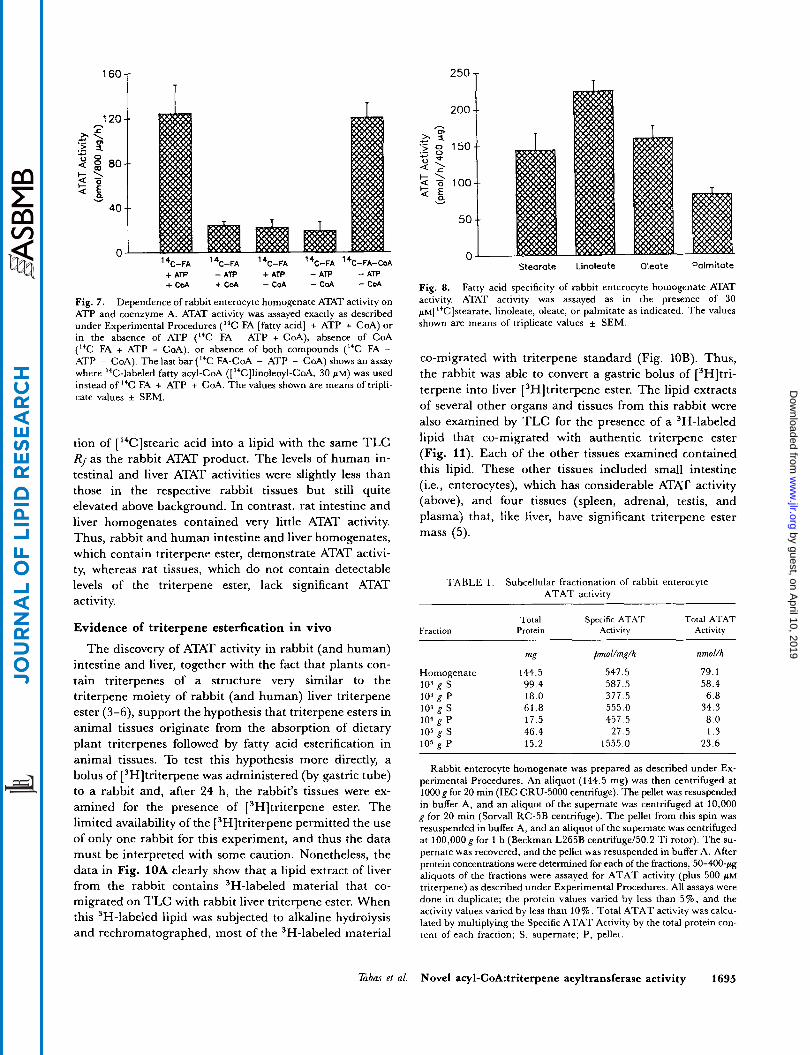
The dependence of ATAT activity on rabbit enterocyte homogenate protein is shown in Fig. 5 , top panel. In the absence of exogenous triterpene, the activity was low com- pared to background (Le., activity with no added protein) and linear up to 1.2 mg homogenate per 400-pl reaction. (In most other homogenate preparatiom that we used, ATAT activity in the absence of exogenous triterpene was even less above background.) In the presence of ex- ogenous triterpene, ATAT activity was substantially above background and peaked at 0.8 mg/400 p1. The de- pendence of ATAT activity (plus exogenous triterpene) on time of incubation i s shown in the lower panel of Fig. 5. The activity continued to increase up to 60 min of incuba- tion; longer periods of incubation did not result in further product formation.
The dose dependency curve for exogenous triterpene is shown in Fig. 6 , upper panel. ATAT activity was not markedly elevated above background until 250 p M
1692 Journal of Lipid Research Volume 32, 1991
by guest, on April 10, 2019
ww
w.jlr.org
Dow
nloaded from
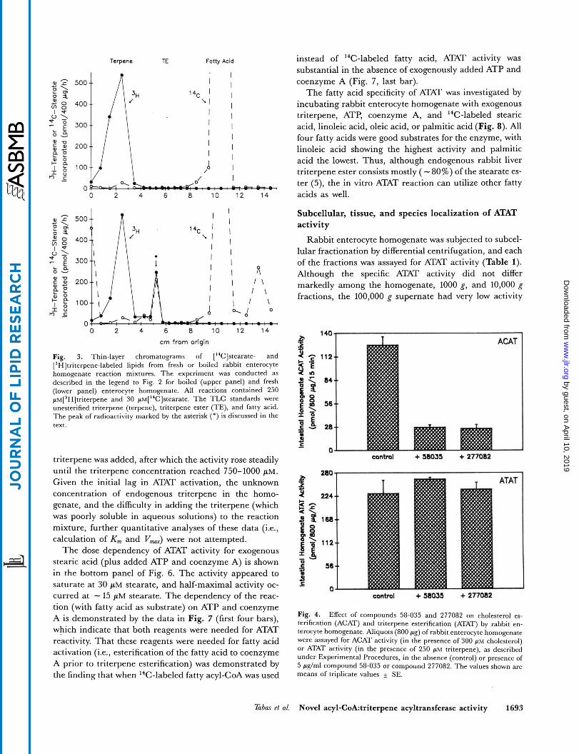
Fig. 3. Thin-layer chromatograms of ['%]stearate- and [3H]triterpene-labeled lipids from fresh or boiled rabbit enterocyte homogenate reaction mixtures. The experiment was conducted as described in the legend to Fig. 2 for boiled (upper panel) and fresh (lower panel) enterocyte homogenate. All reactions contained 250 ~M[~H]tri terpene and 30 p~[I~C]s teara te . The TLC standards were unesterified triterpene (terpene), triterpene ester (TE), and fatty acid. The peak of radioactivity marked by the asterisk (*) is discussed in the text.
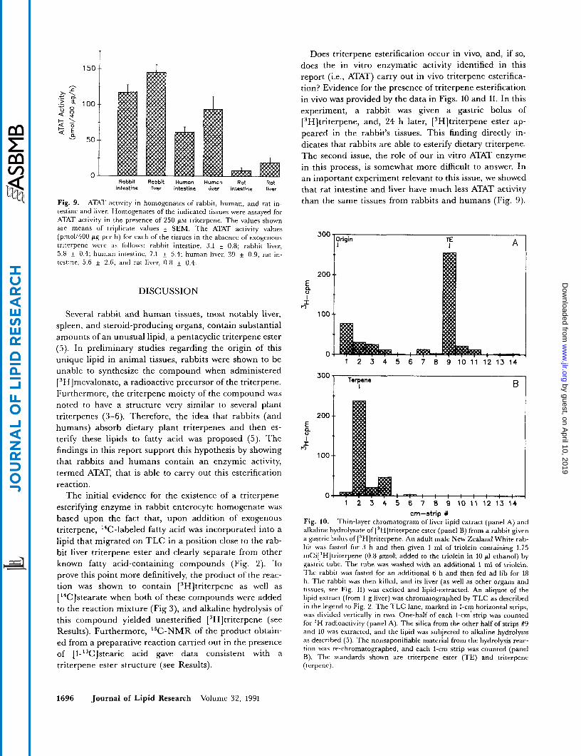
triterpene was added, after which the activity rose steadily until the triterpene concentration reached 750-1000 pM. Given the initial lag in ATAT activation, the unknown concentration of endogenous triterpene in the homo- genate, and the difficulty in adding the triterpene (which was poorly soluble in aqueous solutions) to the reaction mixture, further quantitative analyses of these data (;.e., calculation of K , and V,,) were not attempted.
The dose dependency of ATAT activity for exogenous stearic acid (plus added ATP and coenzyme A) is shown in the bottom panel of Fig. 6 . The activity appeared to saturate at 30 pM stearate, and half-maximal activity oc- curred at - 15 pM stearate. The dependency of the reac- tion (with fatty acid as substrate) on ATP and coenzyme A is demonstrated by the data in Fig. 7 (first four bars), which indicate that both reagents were needed for ATAT reactivity. That these reagents were needed for fatty acid activation (i.e., esterification of the fatty acid to coenzyme A prior to triterpene esterification) was demonstrated by the finding that when 14C-labeled fatty acyl-CoA was used
instead of 14C-labeled fatty acid, ATAT activity was substantial in the absence of exogenously added ATP and coenzyme A (Fig. 7, last bar).
The fatty acid specificity of ATAT was investigated by incubating rabbit enterocyte homogenate with exogenous triterpene, ATP, coenzyme A, and "C-labeled stearic acid, linoleic acid, oleic acid, or palmitic acid (Fig. 8). All four fatty acids were good substrates for the enzyme, with linoleic acid showing the highest activity and palmitic acid the lowest. Thus, although endogenous rabbit liver triterpene ester consists mostly ( - 80%) of the stearate es- ter (5), the in vitro ATAT reaction can utilize other fatty acids as well.
Subcellular, tissue, and species localization of ATAT activity
Rabbit enterocyte homogenate was subjected to subcel- lular fractionation by differential centrifugation, and each of the fractions was assayed for ATAT activity (Table 1). Although the specific ATAT activity did not differ markedly among the homogenate, 1000 g, and 10,000 g fractions, the 100,000 g supernate had very low activity
Fig. 4. Effect of compounds 58-035 and 277082 on cholesterol es- terification (ACAT) and triterpene esterification (ATAT) by rabbit en- terocyte homogenate. Aliquots (800 cg) of rabbit enterocyte homogenate were assayed for ACAT activity (in the presence of 300 p~ cholesterol) or ATAT activity (in the presence of 250 gM triterpene), as described under Experimental Procedures, in the absence (control) or presence of 5 p g h l compound 58-035 or compound 277082. The values shown are means of triplicate values f SE.
T a b u et al. Novel acyl-CoA:triterpene acyltransferase activity 1693
by guest, on April 10, 2019
ww
w.jlr.org
Dow
nloaded from
1204 /
04 I 0 0.4 0.8 1.2 1.6
Homogenate Protein (mg/400 pl)
'""T 120 I
0 I 0 so 60 90 120
Time of Incubation (min)
Fig. 5. Dependence of rabbit enterocyte homogenate ATAT activity on homogenate protein concentration and time of reaction. In the upper panel, the indicated amount of rabbit enterocyte homogenate was assayed for 1 h for ATAT activity in the absence (open circles) or pre- sence (closed circles) of exogenous triterpene (250 psi). In the lower panel, 800 pg homogenate was assayed for ATAT activity in the presence of 250 phl triterpene for the indicated times. All values shown are averages o f duplicate values which varied by less than 10%.
and the 100,000 g pellet (microsomes) had significantly higher ATAT specific activity. The total activity data clearly show that most of the homogenate ATAT activity was recovered in the 1000 g supernate (post-nuclear super- nate), that most of the post-nuclear supernate activity was recovered in the 10,000 g supernate, and that most of this supernate activity was recovered in the 100,000 g pellet. Thus, ATAT, like ACAT and the triterpene ester itself (5), is membrane-bound and localized to the microsomes.
All of the initial work with ATAT was with rabbit en- terocytes. To determine whether rabbit liver, an organ with high levels of triterpene ester (5), also contained ATAT activity, rabbit liver homogenate was assayed for
data in Fig. 9 show that rabbit liver homogenate had ATAT activity similar to rabbit enterocyte homogenate. Thus, ATAT activity is not only present at the site of dietary triterpene absorption (i.e., the intestine) but also in an organ with high levels of the triterpene ester (Le., the liver).
Previous work from our laboratory demonstrated that the triterpene ester was present in human liver as well as in rabbit tissues but was not detectable in rat liver (5). The absence of the triterpene ester in rat liver was thought not to be due simply to the absence of plant triterpenes in rat chow since feeding rabbit chow to a rat did not lead to the appearance of the triterpene ester in the animal's liver (5). An alternative explanation for the absence of triterpene ester in rat liver was that rat tissues lacked ATAT activity. To address this issue, homogenates from human intestinal biopsies, human liver, rat enterocytes, and rat liver were assayed for ATAT activity (Fig. 9, last four bars). As with rabbit tissues, human intestine and liver demonstrated a triterpene-stimulatable incorpora-
280 t
70 4 /
..*e 0 0 250 500 750 1000
300 T
[Trlterpene] (&4)
T
0 0 20 40 60 80 100
[Steark Acid] &hi) ATAT activity (plus exogenous triterpene) and compared with rabbit enterocyte homogenate activity (Fig' ' 9 first two bars). Rabbit liver homogenate was able to incor- porate [ 14Clstearate into a lipid with the Same TLC R, as
homogenate. Furthermore, the activity was dependent upon exogenous triterpene (see legend to Fig. 9). The
Fig. 6. Dependence of rabbit enterocyte homogenate ATAT activity on triterpene and stearic acid concentrations. In the upper panel, ATAT ac- tivity was assayed in the presence of 30 pM["c]SteariC acid and the in- dicated concentrations of triterpene. In the lower panel, ATAT activity was assayed in the presence of 250 p~ triterpene and the indicated con-
tion mixture was equimolar to the ['%]stearic acid concentration. The values shown are the means of triplicate values k SE.
the 14C-1abe1ed triterpene made by rabbit enterocyte centrations of ["CC]stearic acid. The concentration of BSA in each reac-
1694 Journal of Lipid Research Volume 32, 1991
by guest, on April 10, 2019
ww
w.jlr.org
Dow
nloaded from
1607 250 'r
I T
" 14C-FA 14C-FA '*C-FA '*C-FA 14C-FA-C0A
+ATP -ATP +ATP - A T P -ATP + C 0 A +COA - C o A - C 0 A - C 0 A
Fig. 7. Dependence of rabbit enterocyte homogenate ATAT activity on ATP and coenzyme A. ATAT activity was assayed exactly as described under Experimental Procedures (14C FA [fatty acid] + ATP + CoA) or in the absence of ATP (I4C FA - ATP + CoA), absence of CoA (I4C FA + ATP - CoA), or absence of both compounds ("C FA - ATP - CoA). The last bar ('IC FA-CoA - ATP - GOA) shows an assay where "C-labeled fatty acyl-CoA ([14C]linoleoyl-CoA, 30 PM) was used instead of "C FA + ATP + GOA. The values shown are means of tripli- cate values f SEM.
tion of ['4C]stearic acid into a lipid with the same TLC Rf as the rabbit ATAT product. The levels of human in- testinal and liver ATAT activities were slightly less than those in the respective rabbit tissues but still quite elevated above background. In contrast, rat intestine and liver homogenates contained very little ATAT activity. Thus, rabbit and human intestine and liver homogenates, which contain triterpene ester, demonstrate ATAT activi- ty, whereas rat tissues, which do not contain detectable levels of the triterpene ester, lack significant ATAT activity.
Evidence of triterpene esterfication in vivo
The discovery of ATAT activity in rabbit (and human) intestine and liver, together with the fact that plants con- tain triterpenes of a structure very similar to the triterpene moiety of rabbit (and human) liver triterpene ester (3-6), support the hypothesis that triterpene esters in animal tissues originate from the absorption of dietary plant triterpenes followed by fatty acid esterification in animal tissues. To test this hypothesis more directly, a bolus of [3H]triterpene was administered (by gastric tube) to a rabbit and, after 24 h, the rabbit's tissues were ex- amined for the presence of [3H]triterpene ester. The limited availability of the [ 3H]triterpene permitted the use of only one rabbit for this experiment, and thus the data must be interpreted with some caution. Nonetheless, the data in Fig. 10A clearly show that a lipid extract of liver from the rabbit contains 3H-labeled material that co- migrated on TLC with rabbit liver triterpene ester. When this 3H-labeled lipid was subjected to alkaline hydrolysis and rechromatographed, most of the 3H-labeled material
Stearate Linoleate Oleate Palmitate
Fig. 8. Fatty acid specificity of rabbit enterocyte homogenate ATAT activity. ATAT activity was assayed as in the presence of 30 p~[''C]stearate, linoleate, oleate, or palmitate as indicated. The values shown are means of triplicate values SEM.
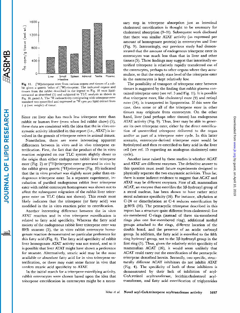
co-migrated with triterpene standard (Fig. 10B). Thus, the rabbit was able to convert a gastric bolus of [3H]tri- terpene into liver [ 3H]triterpene ester. The lipid extracts of several other organs and tissues from this rabbit were also examined by TLC for the presence of a 3H-labeled lipid that co-migrated with authentic triterpene ester (Fig. 11). Each of the other tissues examined contained this lipid. These other tissues included small intestine (i.e., enterocytes), which has considerable ATAT activity (above), and four tissues (spleen, adrenal, testis, and plasma) that, like liver, have significant triterpene ester mass (5).
TABLE 1 . Subcellular fractionation of rabbit enterocyte ATAT activity
Total Specific ATAT Total ATAT Fraction Protein Activity Activity
mg
Homogenate 144.5
103 P 18.0 104 g s 61.8 104 P 17.5 105 g s 46.4 105 P 15.2
1 0 3 ~ s 99.4
pmol/mt/h
547.5 587.5 377.5 555.0 457.5
27.5 1555.0
nmoNh
79.1 58.4 6.8
34.3 8.0 1.3
23.6
Rabbit enterocyte homogenate was prepared as described under Ex- perimental Procedures. An aliquot (144.5 mg) was then centrifuged at lOOOg for 20 min (IEC CRU-5000 centrifuge). The pellet was resuspended in buffer A, and an aliquot of the supernate was centrifuged at 10,000 g for 20 min (Sorvall RC-5B centrifuge). The pellet from this spin was resuspended in buffer A, and an aliquot of the supernate was centrifuged at 100,000 g for 1 h (Beckman L265B centrifuge/50.2 Ti rotor). The su- pernate was recovered, and the pellet was resuspended in buffer A. After protein concentrations were determined for each of the fractions, 50-400-pg aliquots of the fractions were assayed for ATAT activity (plus 500 PM triterpene) as described under Experimental Procedures. All assays were done in duplicate; the protein values varied by less than 5%. and the activity values varied by less than 10%. Total ATAT activity was calcu- lated by multiplying the Specific ATAT Activity by the total protein con- tent of each fraction; s, supernate; P, pellet.
Tabas et al. Novel acyl-CoA:triterpene acyltransferase activity 1695
by guest, on April 10, 2019
ww
w.jlr.org
Dow
nloaded from
Rabbit Rabbit Human Human Rat Rot intestine liver intestine liver intestine liver
Fig. 9. ATAT activity in homogenatcs of rabbit, human, and rat in- testine and liver. Homogenates of the indicated tissues were assayed for ATAT activity in the presence of 250 PM triterpene. The values shown are means of triplicate values k SEM. The ATAT activity values (pmo1/400 pg per h) for each of the tissues in the absence of exogenous triterpene were a s follows: rabbit intestine, 3.1 k 0.8; rabbit liver, 5.8 k 0.4; human intestine, 7.1 + 5.4; human liver, 39 k 0.9; rat in- testine, 5.6 & 2.6. and rat liver, 0.8 k 0.4.
DISCUSSION
Several rabbit and human tissues, most notably liver, spleen, and steroid-producing organs, contain substantial amounts of an unusual lipid, a pentacyclic triterpene ester (5). In preliminary studies regarding the origin of this unique lipid in animal tissues, rabbits were shown to be unable to synthesize the compound when administered [3H]mevalonate, a radioactive precursor of the triterpene. Furthermore, the triterpene moiety of the compound was noted to have a structure very similar to several plant triterpenes (3-6). Therefore, the idea that rabbits (and humans) absorb dietary plant triterpenes and then es- terify these lipids to fatty acid was proposed (5). The findings in this report support this hypothesis by showing that rabbits and humans contain an enzymic activity, termed ATAT, that is able to carry out this esterification reaction.
The initial evidence for the existence of a triterpene- esterifying enzyme in rabbit enterocyte homogenate was based upon the fact that, upon addition of exogenous triterpene, 14C-labeled fatty acid was incorporated into a lipid that migrated on TLC in a position close to the rab- bit liver triterpene ester and clearly separate from other known fatty acid-containing compounds (Fig. 2). To prove this point more definitively, the product of the reac- tion was shown to contain [3H]triterpene as well as [ I4C]stearate when both of these compounds were added to the reaction mixture (Fig 3), and alkaline hydrolysis of this compound yielded unesterified [ 3H]triterpene (see Results). Furthermore, I3C-NMR of the product obtain- ed from a preparative reaction carried out in the presence of [l-'3C]stearic acid gave data consistent with a triterpene ester structure (see Results).
Does triterpene esterification occur in vivo, and, if so, does the in vitro enzymatic activity identified in this report (Le., ATAT) carry out in vivo triterpene esterifica- tion? Evidence for the presence of triterpene esterification in vivo was provided by the data in Figs. 10 and 11. In this experiment, a rabbit was given a gastric bolus of [3H]triterpene, and, 24 h later, [3H]triterpene ester ap- peared in the rabbit's tissues. This finding directly in- dicates that rabbits are able to esterify dietary triterpene. The second issue, the role of our in vitro ATAT enzyme in this process, is somewhat more difficult to answer. In an important experiment relevant to this issue, we showed that rat intestine and liver have much less ATAT activity than the same tissues from rabbits and humans (Fig. 9).
300
200
b I I
m 100
0
300
200 ; I I:
m 100
0
A Irigin TE 1
1 2 3 4 5 6 7 8 9 1 0 1 1 1 2 1 3 1 4
Terpene I
cm-strip 1 Fig. 10. Thin-layer chromatogram of liver lipid extract (panel A) and alkaline hydrolysate of [3H]triterpene ester (panel B) from a rabbit given a gastric bolus of [3H]triterpene. An adult male New Zealand White rab- bit was fasted for 3 h and then given 1 ml of triolein containing 1.75 ~nCi[~H]tr i terpene (0.8 pmol; added to the triolein in 10 p1 ethanol) by gastric tube. The tube was washed with an additional 1 ml of triolein. The rabbit was fasted for an additional 6 h and then fed ad lib for 18 h . The rabbit was then killed, and its liver (as well as other organs and tissues, see Fig. 11) was excised and lipid-extracted. An aliquot of the lipid extract (from 1 g liver) was chromatographed by TLC as described in the legend to Fig. 2. The T L C lane, marked in I-cm horizontal strips, was divided vertically in two. One-half of each I-cm'strip was counted for 'H radioactivity (panel A). The silica from the other half of strips #9 dnd 10 was extracted, and the lipid was subjected to alkaline hydrolysis as described (5). The nonsaponifiable material from the hydrolysis reac- tion was re-chromatographed, and each I-cm strip was counted (panel B). The standards shown are triterpene ester (TE) and triterpene (terpene).
1696 Journal of Lipid Research Volume 32, 1991
by guest, on April 10, 2019
ww
w.jlr.org
Dow
nloaded from
1600 - a.2
3
3
a, 3 VI
a.2 1200
v
.I? aoo a.2
0 7 \ E $ 400 I I
m n -
Liver Small Spleen Adrenal Testis Plasma
Fig. 11. [3H]triterpene ester from various organs and tissues of a rab- bit given a gastric bolus of [3H]triterpene. The indicated organs and tissues from the rabbit described in the legend to Fig. 10 were lipid- extracted as described (5) and subjected to TLC analysis as shown in Fig. 10, panel A. The 3H radioactivity comigrating with triterpene ester standard was quantified and expressed as 3H cpm per lipid extract from 1 g (wet weight) of tissue.
Intestine
Since rat liver also has much less triterpene ester than rabbit or human liver (even when fed rabbit chow) (5), these data are consistent with the idea that the in vitro en- zymatic activity identified in this report (i.e., ATAT) is in- volved in the genesis of triterpene esters in animal tissues.
Nonetheless, there are some interesting apparent differences between in vitro and in vivo triterpene es- terification. First, the fact that the product of the in vitro reaction migrated on our TLC system slightly closer to the origin than either endogenous rabbit liver triterpene ester (Fig. 2) or [3H]triterpene ester generated in vivo by the rabbit given gastric [ 3H]triterpene (Fig. 10) indicated that the in vitro product was slightly more polar than en- dogenous triterpene ester. In a separate experiment, in- cubation of purified endogenous rabbit liver triterpene ester with rabbit enterocyte homogenate was shown not to affect the subsequent migration of the rabbit liver triter- pene ester on TLC (data not shown). This result most likely indicates that the triterpene (or fatty acid) was modified in the in vitro reaction prior to esterification.
Another interesting difference between the in vitro ATAT reaction and in vivo triterpene esterification is related to fatty acid specificity. Whereas the fatty acid moiety of the endogenous rabbit liver triterpene ester was 80% stearate (5), the in vitro rabbit enterocyte homo- genate reaction demonstrated no particular preference for this fatty acid (Fig. 8). The fatty acid specificity of rabbit liver homogenate ATAT activity was not tested, and so it is possible that liver ATAT might have shown a preference for stearate. Alternatively, stearic acid may be the most available or abundant fatty acid for in vivo triterpene es- terification, or there may exist some factor in vivo that confers stearic acid specificity to ATAT.
In the initial search for a triterpene-esterifying activity, rabbit enterocytes were chosen based upon the idea that triterpene esterification in enterocytes might be a neces-
sary step in triterpene absorption just as intestinal cholesterol esterification is thought to be necessary for cholesterol absorption (9- 11). Subsequent work disclosed that there was similar ATAT activity (as expressed per amount of homogenate protein) in enterocytes and liver (Fig. 9). Interestingly, our previous study had demon- strated that the amount of endogenous triterpene ester in enterocytes was much less than that in liver and other tissues (5). These findings may suggest that intestinally es- terified triterpene is relatively rapidly transferred out of the enterocytes, perhaps to other organs where they accu- mulate, so that the steady state level of the triterpene ester in the enterocytes is kept relatively low.
The possibility of transport of triterpene ester between tissues is suggested by the finding that rabbit plasma con- tained triterpene ester (see ref. 5 and Fig. 11). It is possible that triterpene ester, like cholesteryl ester (9) and retinyl ester (14), is transported in lipoproteins. If this were the case, then some or all of the triterpene ester in other tissues may originate from enterocytes. O n the other hand, liver (and perhaps other tissues) has endogenous ATAT activity (Fig. 9). Thus, liver may be able to gener- ate its own triterpene ester, either by the direct esterifica- tion of unesterified triterpene delivered to the organ and/or as part of a triterpene ester cycle. In this latter scenario, enterocyte-derived triterpene ester would be hydrolyzed and then re-esterified to fatty acid in the liver cell (see ref. 15 regarding an analogous cholesteryl ester cycle).
Another issue raised by these studies is whether ACAT and ATAT are different enzymes. The definitive answer to this question must await future experiments designed to physically separate the two enzymatic activities. Thus far, there is some indirect evidence to suggest that ACAT and ATAT may be different enzymes. First of all, mammalian ACAT, an enzyme that esterifies the 30-hydroxyl group of a sterol nucleus, has been shown to have rather strict sterol substrate specificity (16). For instance, alkylation at C-24 or dimethylation at C-4 reduces esterification by 280% (16). The pentacyclic triterpene described in this report has a structure quite different from cholesterol: five six-membered C-rings (instead of three six-membered rings plus one five-membered ring), additional methyl groups attached to the rings, different location of the double bond, and the presence of an acidic carboxyl group. In addition, the fatty acid is esterified to the fifth ring hydroxyl group, not to the 30-hydroxyl group in the first ring (5). Thus, given the relatively strict specificity of mammalian ACAT (16), it would seem unlikely that ACAT could carry out the esterification of the pentacyclic triterpene described herein. Secondly, two specific, struc- turally different ACAT inhibitors do not inhibit ATAT (Fig. 4). The specificity of both of these inhibitors is demonstrated by their lack of inhibition of acyl- CoA:retinol acyltransferase, 1ecithin:cholesterol acyl- transferase, and fatty acid esterification of triglycerides
T a b u et al. Novel acyl-CoA:triterpene acyltransferase activity 1697
by guest, on April 10, 2019
ww
w.jlr.org
Dow
nloaded from
and phospholipids (12, 13). Furthermore, compound 58- 035, a fatty acylamide, is likely to act as a competitive in- hibitor for the fatty acyl-CoA substrate of ACAT (11). Since ATAT also uses a fatty acyl-CoA substrate (Fig. 7) , one would expect that 58-035 would inhibit this enzyme if it were the same as ACAT. Lastly, ra t liver, which lacks significant ATAT activity is a relatively rich source of ACAT activity (16, 17). Thus , pending physical separation of the two activities, indirect evidence accumulated thus far seems to indicate that ACAT a n d ATAT may be different enzymes.
What could be the physiological role of a triterpene- esterifying enzyme? O u r initial report on the discovery of the triterpene ester molecule (5) put forth the idea that triterpene esterification may represent a detoxification reaction since pentacyclic triterpenes a re toxic to cells (3, 6). In this scenario, rabbits a n d humans would incidental- ly absorb triterpenes along with other components from dietary plant material. T h e animals would then require a mechanism to detoxify the triterpenes to prevent their cytotoxic effects, and triterpene esterification may serve this function (just as cholesterol esterification is thought to prevent the cytotoxicity of excess cellular cholesterol (18)).
Alternatively, it is also possible that triterpene es- terification and triterpene esters play other physiological roles. O n e of o u r initial hypotheses regarding the ability of triterpene ester to inhibit ACAT (5) was that if ACAT itself were responsible for triterpene esterification, then the reaction may lead to feedback inhibition of ACAT, However, as discussed above, ACAT a n d ATAT may be different enzymes. Nonetheless, since ATAT, triterpene es- ter, a n d ACAT are all localized to microsomes (ref. 5 a n d Table l), it is still possible that locally generated triterpene ester may lead to microsomal ACAT inhibition. These a n d other possibilities await further investigations of triterpene esters a n d the newly described ATAT enzymatic activity. I
The authors thank Dr. Yechezkiel Stein for a helpful discussion regarding possible tissue sites for triterpene esterification. This work was supported by National Institutes of Health grants HL- 21006 and 39703 and a research grant from Schering-Plough. I.T. is an Established Investigator of the American Heart Asso- ciation and Boehringer Ingelheim, Inc. and a Silberberg Assis- tant Professor of Medicine of Columbia University. Manuscrip! received May I991 and in revtsedform 2 J u h 1991
REFERENCES
Organic Chemistry of Secondary Plant Metabolism. 1969. T. A. Geismann and D. H. G. Crout, editors. Freeman, Cooper, & Co., San Francisco. Fourie, T. G., and F. 0. Snyckers. 1989. A pentacyclic triterpene with anti-inflammatory and analgesic activity from the roots of Commiphora merkeri. J Nat. Prod. 52:
Yamagishi, T., D. C. Zhang, J. J. Chang, D. R. McPhail, A. T. McPhail, and K. H. Lee. 1988. The cytotoxic prin-
1129-1131.
4.
5.
6.
7.
8.
9.
10.
11.
12.
13.
14.
15.
16.
17.
18.
ciples of HyptiC capitata and the structures of the new triterpenes hyptatic acid-A and -B. Phytochemisty 27:
Kitagawa, I., M. Yoshikawa, H. K. Wang, M. Saito, V. Tosirisuk, T. Fujiwara, and K. Tomita. 1982. Revised struc- tures of soyasapogenols A, B, and E, oleanenesapogenols from soy bean. Structure of the soy asaponins I, 11, and 111. Chem. Phannacol. Bull. 30: 2294-2297. Tabas, I., L-L. Chen, J. W. Clader, A. T. McPhail, D. A. Burnett, P. Bartner, P. R. Das, B. N. Pramanik, M. S. Puar, S. J. Feinmark, R. E. Zipkin, G. Boykow, G. Vita, and A. R. Tall. 1990. Rabbit and human liver contain a novel pentacyclic triterpene ester with acyl-CoA:cholesterol acyltransferase inhibitory activity. J. Biol. Chem. 265:
Nozaki, H., H. Suzuki, K-H. Lee, and A. T. McPhail. 1982. Structure and stereochemistry of maytenfolic acid and maytenfoliol, two new antileukemic triterpenes from Maytenus diuersifolia: X-ray crystal structures. J Chem. SOC. Chem. Commun. 1048-1051. Tabas, I., G. C. Boykow, and A. R. Tall. 1987. Foam cell- forming 5774 macrophages have markedly elevated acyl coenzyme A:cholesterol acyl transferase activity compared with mouse peritoneal macrophages in the presence of low density lipoproteins (LDL) despite similar LDL receptor activity. J Clin. Invest. 79: 418-426. Lowry, 0 . H., N. J. Rosebrough, A. L. Farr, and R. J. Ran- dall. 1951. Protein measurement with the Folin phenol rea- gent. J. Biol. Chem. 193: 265-275. Klein, R. L., and L. L. Rude]. 1983. Cholesterol absorption and transport in thoracic duct lymph lipoproteins of nonhuman primates: effect of dietary cholesterol level. J
3213-3216.
8042-8051.
Lipid Res. 2% 343-356. Norum, K. R., A. C. Lilljeqvist, P. Helgerud, E. R. Nor- mann, A. Mo, and B. Selbekk. 1979. Esterification of cholesterol in human small intestine: the importance of acyl-CoA:cholesterol acyltransferase. Eur. J. Clin. Inuest. 9:
Heider, J. G., C. E. Pickens, and L. A. Kelly. 1983. Role of acyl-CoA:cholesterol acyltransferase in cholesterol ab- sorption and its inhibition by 57-118 in the rabbit. J. Lipid Res. 24: 1127-1134. Ross, A. C., K. J. Go, J. G. Heider, and G. H. Rothblat. 1984. Selective inhibition of acyl coenzyme A:cholesterol acyltransferase by compound 58-035. J . Biol. Chem. 259: 815-819. Largis, E. E., C. H. Wang, V. G. DeVries, and S. A. Schaffer. 1989. C L 277,082: a novel inhibitor of ACAT- catalyzed cholesterol esterification and cholesterol absorp- tion. J Lipid Res. 30: 681-690. Goodman, D. S., R. Blomstrand, B. Werner, H. S. Huang, and T. Shiratori. 1966. The intestinal absorption and meta- bolism of vitamin A and beta-carotene in man. J. Clin. In- vest. 45: 1615-1623. Brown, M.S., Y. K. Ho, and J. L. Goldstein. 1980. The cholesteryl ester cycle in macrophage foam cells: continual hydrolysis and re-esterification of cytoplasmic cholesteryl esters. J Biol. Chem. 255: 9344-9352. Tavani, D. M., W. R. Nes, and J. T. Billheimer. 1982. The sterol substrate specificity of acyl CoA:cholesterol acyltransferase from rat liver. J. Lipid Res. 23: 774-781. Tabas, I., G. Boykow, and A. Tall. 1988. Rabbit liver micro- somal ACAT smooth ER enzyme associated with a lipid ACAT inhibitor. Arteriosclerosis. 8: 559A. Spector, A. S., S. N. Mathur, andT. L. Kaduce. 1975. Role of acyl-coenzyme A:cholesterol 0-acyltransferase in cholesterol metabolism, Prop: Lipid Res. 18: 31 -53.
55-62.
1698 Journal of Lipid Research Volume 32, 1991
by guest, on April 10, 2019
ww
w.jlr.org
Dow
nloaded from