identification and characterization of magoand y14genes · pdf fileidentification and...
TRANSCRIPT

Identification and characterization of MAGO and Y14 genesin Hevea brasiliensis
Zi-Ping Yang1,2, Hui-Liang Li1 , Dong Guo1 and Shi-Qing Peng1,2,#
1Key Laboratory of Biology and Genetic Resources of Tropical Crops, Ministry of Agriculture, Institute of
Tropical Bioscience and Biotechnology, Chinese Academy of Tropical Agricultural Sciences, Haikou, China2College of Agriculture, Hainan University, Haikou, China
Abstract
Mago nashi (MAGO) and Y14 proteins are highly conserved among eukaryotes. In this study, we identified twoMAGO (designated as HbMAGO1 and HbMAGO2) and two Y14 (designated as HbY14a and HbY14b) genes in therubber tree (Hevea brasiliensis) genome annotation. Multiple amino acid sequence alignments predicted thatHbMAGO and HbY14 proteins are structurally similar to homologous proteins from other species. Tissue-specific ex-pression profiles showed that HbMAGO and HbY14 genes were expressed in at least one of the tissues (bark,flower, latex, leaf and root) examined. HbMAGOs and HbY14s were predominately located in the nucleus and werefound to interact in yeast two-hybrid analysis (YTH) and bimolecular fluorescence complementation (BiFC) assays.HbMAGOs and HbY14s showed the highest transcription in latex and were regulated by ethylene and jasmonate. In-teraction between HbMAGO2 and gp91phox (a large subunit of nicotinamide adenine dinucleotide phosphate) wasidentified using YTH and BiFC assays. These findings suggested that HbMAGO may be involved in the aggregationof rubber particles in H. brasiliensis.
Keywords: interaction, Hevea brasiliensis, Mago nashi, Y14 proteins
Received: December 24, 2014; Accepted: June 08, 2015.
Introduction
The exon junction complex (EJC) regulates post-
transcriptional events that include mRNA intracellular ex-
port, cytoplasmic localization, non-sense mediated mRNA
decay (NMD) and translation enhancement in metazoans
(Park and Muench, 2007; Lee et al., 2009; Ashton-Beau-
cage et al., 2010; Roignant and Treisman, 2010; Boothby
and Wolniak, 2011; Mufarrege et al., 2011). The EJC is a
multiprotein complex assembled onto mRNAs as a conse-
quence of splicing and is thought to provide a molecular
link between splicing and post-splicing mRNA metabolism
20-24 bases upstream of mRNA exon-exon junctions (Le et
al., 2000, 2003; Tange et al., 2004; Gehring et al., 2009).
Several of the EJC components have been identified and
classified into two groups, namely, EJC “core” and “pe-
ripheral” factors. Four proteins form the EJC core: MAGO
(short form of MAGO NASHI and known as MAGOH in
humans), Y14 (also known as Tsunagi or RBM8A),
eIF4A/eIF4A3 and BTZ (short form of Barentsz, also
known as MLN51), of which MAGO and Y14 constitute a
stable heterodimeric complex in vitro and in vivo (Tange et
al., 2004, 2005; Le and Seraphin, 2008). Additional com-
ponents of the EJC include the splicing factors SRm160,
Pinin and RnpS1, the NMD factors Upf1, Upf2 and Upf3,
the translation factors Skar and Pym, and the export factors
UAP56 and REF/Aly (Roignant and Treisman, 2010).
These proteins are considered to be peripheral EJC compo-
nents that are transiently associated with the EJC core in the
nucleus or cytoplasm, where they are involved in splicing,
export, translation and NMD (Lejeune and Maquat, 2005;
Tange et al., 2005).
The MAGO (meaning “no grandchildren’’ in Japa-
nese) gene was first identified as a strict maternal effect
gene in Drosophila (Boswell et al., 1991; Newmark and
Boswell, 1994; Micklem et al., 1997; Newmark et al.,
1997). MAGO can interact with an RNA binding protein,
Y14, to regulate cell growth and is expressed throughout
the organism (Zhao et al., 1998, 2000; Le et al., 2001; Mohr
et al., 2001; Chen et al., 2007). Based on the crystal struc-
tures of the Drosophila and human MAGO-Y14 complex,
MAGO and Y14 proteins are core components of the EJC
and can form a stable heterodimer that strongly associates
with spliced mRNA (Lau et al., 2003; Shi and Xu, 2003).
Both MAGO and Y14 are evolutionarily highly conserved
Genetics and Molecular Biology, 39, 1, 73-85 (2016)
Copyright © 2016, Sociedade Brasileira de Genética. Printed in Brazil
DOI: http://dx.doi.org/10.1590/1678-4685-GMB-2014-0387
Send correspondence to Shi-Qing Peng. Key Laboratory of Biologyand Genetic Resources of Tropical Crops, Ministry of Agriculture,Institute of Tropical Bioscience and Biotechnology, Chinese Acad-emy of Tropical Agricultural Sciences, Haikou 571101, China.E-mail: [email protected]#Current address: 4 Xueyuan Rd., Haikou 571101, China
Research Article
proteins (Hachet and Ephrussi, 2001; Mohr et al., 2001)
and slow co-evolution of the two protein families is re-
quired for the maintenance of their obligate heterodime-
rization mode (Gong et al., 2014). Comparative sequence
analysis reveals that MAGO is devoid of known structural
motifs while Y14 contains a central RNA-binding domain
(Kataoka et al., 2000; Kim et al., 2001; Shi and Xu, 2003).
The crystal structure of the heterodimeric complexes,
formed in the absence of RNA, shows that the RNA-
binding motif is used for Y14/MAGO protein-protein inter-
action and is not readily available for binding RNA (Fri-
bourg et al., 2003; Lau et al., 2003; Shi and Xu, 2003).
In plants, MAGO proteins have been studied to vary-
ing degrees (Chen et al., 2007; Gong and He, 2014). The
orthologs of MAGO genes are linked to male fertility. For
example, PFMAGO proteins interact with MADS-domain
protein MPF2 and are responsible for male fertility and ca-
lyx development in Physalis (He et al., 2007). In
Arabidopsis, AtMAGO is required for pollen grain devel-
opment (Johnson et al., 2004) and embryo development, as
well as meristem cell death events (Park et al., 2009).
MAGO is essential for spermatogenesis as shown by RNAi
knockdown of MvMAGO in Marsilea (van der Weele et al.,
2007; Boothby and Wolniak, 2011). MAGO genes are also
involved in the development of other organs, such as lea-
ves, stems, roots and seeds. The overexpression of
TcMAGO in transgenic tobacco plants results in longer
roots and a more complex root system (Chen et al., 2007).
In Arabidopsis, RNAi-AtMAGO plants produce a greater
number of leaves, short and fasciated stems, short lateral
roots and non-viable seeds (Park et al., 2009). OsMAGO
knockdown generated short rice plants with abnormal
flowers (Gong and He, 2014). The function of MAGO as a
component of the EJC is well-understood, and some func-
tions of MAGO have been reported for other plants; how-
ever, the existence of a duplicated MAGO gene in rubber
trees is not widely known.
The rubber tree (Hevea brasiliensis) is the main re-
newable world-wide source of commercial natural rubber
(NR), a biopolymer of high economic interest composed
mainly of cis-1,4-polyisoprene. Latex is produced by lati-
cifers after tapping and is a white cytoplasmic colloidal sus-
pension containing mainly rubber particles, but also non-
rubber particles, organelles, proteins and serum. In this
study, the MAGO and Y14 gene families in rubber trees
were characterized. To investigate their evolutionary rela-
tionships and functions, we undertook structural and phylo-
genetic analyses and examined the subcellular locations of
these proteins. The expression profiles of MAGOs and
Y14s in various organs and the response to ethylene (ET)
and jasmonate (JA) were also studied. Furthermore, the in-
teraction partners of HbMAGO were identified in latex and
the HbMAGO2 interaction with gp91phox (a large subunit of
nicotinamide adenine dinucleotide phosphate) was demon-
strated using yeast two-hybrid analysis (YTH) and bimo-
lecular fluorescence complementation (BiFC) assays. Our
results indicate that HbMAGO may be involved in the ag-
gregation of natural rubber in rubber trees.
Materials and Methods
Plant materials
Hevea brasiliensis cultivar RY7-33-97 was planted
on the experimental farm of the Chinese Academy of Trop-
ical Agriculture Sciences, Hainan, China. The shoots were
treated with 0.5% ethylene (ET) or 0.1% jasmonic acid
(JA), as described by Hao and Wu (2000). Latex samples
were collected 1, 3, 6, 9, 24 and 48 h after treatment from 12
shoots for each interval and were immediately stored at
-80 °C for RNA extraction. Three independent biological
replicates were done for each treatment. For latex RNA ex-
traction, the latex was dropped directly into liquid nitrogen
in an ice kettle. Rubber tree flowers, leaves and bark were
washed with double-distilled water to remove latex and
then immediately frozen in liquid nitrogen.
Database search and sequence conservationanalysis of rubber tree MAGO and Y14 genes
A local whole genome shotgun database for rubber
tree was established by Yang et al. (2014). The full-length
cDNA of HbMAGO2 was obtained from a yeast two-
hybrid cDNA library of H. brasiliensis laticifers using
HbWRKY as the bait in the yeast two-hybrid assay (data
unpublished). HbY14a was obtained from the yeast two-
hybrid cDNA library of H. brasiliensis laticifers using
HbMAGO2 as the bait. HbMAGO2 and HbY14a were used
as queries in Blast searches of the rubber tree whole ge-
nome shotgun database. The shotgun sequences that pro-
duced the highest significant alignments judged by the
scores and E-values were used for HMM-based gene struc-
ture prediction (http://linux1.softberry.com/berry.phtml)
based on the Arabidopsis and rubber tree databases. The
genomic DNA and cDNA sequences of four genes were
amplified using appropriate primers (developed based on
gene structure prediction) and sequenced.
Phylogenetic analysis and genomic structure
Analysis and comparison of the HbMAGO and
HbY14 sequences were done using BLAST at the NCBI
database. Clustal X (http://www.ebi.ac.uk/clustalw) was
used for multiple alignments of the nucleic acid and amino
acid sequences of HbMAGOs and HbY14s. A phylogenetic
tree was constructed with MEGA5.2
(http://www.megasoftware.net/) using the maximum likeli-
hood (ML) method with a bootstrap parameter of 1000 rep-
licates. The sequence information for a number of
homologs from other species was retrieved from the NCBI
database and is shown in the explanatory text of Figure 2.
Genomic structures of the HbMAGOs and HbY14s were
analyzed by comparing the cDNA sequences using GSDS
74 Yang et al.

software (http://gsds.cbi.pku.edu.cn/) and the correspond-
ing genomic DNA sequences that were extracted from the
local whole genome shotgun database.
Transient protein expression
To determine the subcellular location of the proteins,
full-length cDNAs were cloned (see Table 1 for primers)
and inserted into the vector pCAMBIAC1302 driven by the
35S promoter. The resulting pCAMBIA1302 constructs
were first transformed into the GV3101 strain of
Agrobacterium tumefaciens by electroporation (Gene
Pulser Xcell, Bio-Rad, USA) and then introduced into on-
ion epidermal cells by agroinfiltration using a water circu-
lating vacuum pump (model SHZ-III B, Shanghai, China)
(Yang et al., 2000, 2014). The empty GFP vector was infil-
trated as a control. For the BiFC assay, the open reading
frames of HbMAGO2 and gp91phox (without their stop
codons) were subcloned into pSPYNE-35S (split YFP
N-terminal fragment expression) or pSPYCE-35S (split
YFP C-terminal fragment expression) vectors driven by the
35S promoter (see Table 1 for primers). The resulting
pSPYNE and pSPYCE plasmids were transformed into the
GV3101 strain and the YNE- and YCE-fused proteins were
then co-expressed in onion epidermal cells by agroinfil-
tration (Walter et al., 2004; Yang et al., 2014). Co-expres-
sions of target genes and YFPN or YFPC were used as
negative controls. The cells were dipped in 30% sucrose so-
lution for 30 min to induce plasmolysis and the green fluo-
rescent protein (GFP) or yellow fluorescent protein (YFP)
signal was then examined with a confocal laser scanning
microscope (Zeiss LSM510, Germany).
Expression analysis
Total RNA was extracted as described by Tang et al.
(2007). First-strand cDNA was synthesized using a
RevertAidTM first-strand cDNA synthesis kit (Fermentas,
Lithuania). qRT-PCR was done using the primers indicated
in Table 1, with the rubber tree actin gene (GenBank
HQ260674.1) being used as a reference gene. The qPCR
fragments were amplified and sequenced to determine
primer specificity. qRT-PCR was done using the fluores-
cent dye SYBR-Green (Takara, China) and the melting
curves of the amplification products were assessed using a
Stratagene Mx3005P real-time thermal cycler (Agilent,
America). The qRT-PCR conditions were as follows: 30 s
at 95 °C for denaturation followed by 40 cycles of 5 s at 94
°C, 20 s at 56 °C and 20 s at 72 °C for amplification. An av-
erage of three independent biological replicates was run for
each time interval. The experimental results were reported
as the mean � SEM of three repli-cates. The data were ana-
lyzed with one-way ANOVA and the level of significance
was set at p < 0.01 or p < 0.05 (Fisher’s protected least sig-
nificant difference). All data analyses were done using Sta-
tistical Product and Service Solutions software (SPSS,
version 16.0).
MAGO and Y14 genes in Hevea brasiliensis 75
Tab
le1
-T
he
pri
mer
sequen
ces
use
din
this
study.
Forw
ard
pri
mer
(5’-
3’)
Rev
erse
pri
mer
(5’-
3’)
For
qR
T-P
CR
assa
yH
bM
AG
O1
GC
TG
TC
CA
TT
CG
AT
TT
GT
AT
CC
CA
GT
TC
TC
GA
AA
GG
CA
GT
AA
HM
AG
O2
GG
AA
CT
GT
TT
GA
GC
TG
TG
TA
AT
GT
CA
TA
GC
AT
TA
GC
AT
TT
CC
GT
TT
C
HbY
14a
AT
CC
CT
CA
CT
TC
TC
TC
CT
AT
CC
TT
GT
CT
CA
CC
GC
TA
AA
TG
GG
HbY
14b
GT
GT
TG
TT
AG
AG
GG
TG
GC
TA
TA
AG
CC
AC
AG
AA
TT
GG
AA
GC
TA
GA
HbA
ctin
CA
GT
GT
CT
GG
AT
AG
GA
GG
AT
CT
AA
AA
TG
GA
CC
GG
AC
TC
AT
CA
TA
C
HbM
AG
O1-B
K/A
DA
GA
GA
AT
TC
AT
GA
TG
AT
GT
TT
GA
TG
AG
AT
GT
GT
GG
AT
CC
GA
AC
CA
GC
CT
GA
AA
CA
TC
CT
G
HbM
AG
O2-B
K/A
DA
GA
GA
AT
TC
AT
GT
TG
AA
GA
TG
AT
GG
AA
GT
TT
GT
GG
AT
CC
GA
AC
CA
GC
CT
GA
AA
CA
TC
CT
G
HbY
14a-B
K/A
DA
CA
GA
AT
TC
AT
GC
GG
TT
TC
CT
CT
AG
AG
AA
AG
GT
GT
GG
AT
CC
GA
AC
CA
GC
CT
GA
AA
CA
TC
CT
G
HbY
14b-B
K/A
DA
CA
GA
AT
TC
AT
GT
AC
CC
TG
AT
TC
CT
GT
CC
AC
CA
TG
TG
GA
TC
CG
AA
CC
AG
CC
TG
AA
AC
AT
CC
TG
For
vec
tor
const
ruct
sgp91-A
DA
CA
GA
AT
TC
AT
GG
TT
CA
AT
GT
CC
TT
AC
AG
CC
AT
GT
GG
AT
CC
GA
AC
CA
GC
CT
GA
AA
CA
TC
CT
G
MA
GO
1-1
302
AG
AG
AA
TT
CA
TG
AT
GA
TG
TT
TG
AT
GA
GA
TG
TG
TG
GA
TC
CA
TA
TT
TT
AA
GA
TA
CC
AT
CT
TG
MA
GO
2-1
302
AC
AG
AA
TT
CA
TG
GT
CC
CA
GG
AG
CA
GG
CT
AG
TG
TG
GA
TC
CG
AA
CC
AG
CC
TG
AA
AC
AT
CC
TG
Y14a-1
302
AG
AG
AA
TT
CA
TG
AT
GA
TG
TT
TG
AT
GA
GA
TG
TG
TG
GA
TC
CT
GC
CG
CT
GC
TT
AG
CA
AT
GT
CG
Y14b-1
302
AC
AC
CA
TG
GA
TG
GC
TA
AC
GC
GG
AT
GC
GG
AA
TG
TA
CT
AG
TG
TA
TC
TC
CT
CC
TA
GG
AC
TC
CT
HbM
AG
O2-Y
CE
/YN
EA
CA
TC
TA
GA
AT
GG
CA
AT
GG
CA
GC
GG
AA
GA
TT
GT
AC
TA
GT
AA
TA
GG
CT
TG
AT
CT
TG
AA
AT
G
gp91-Y
NE
/YC
EA
CA
GT
CG
AC
AT
GA
AG
GG
CT
TA
CC
GA
AA
CA
TT
GT
CC
CG
GG
GC
TA
AA
GT
TG
TT
GC
TA
AT
TG
A

Yeast two-hybrid assays
For yeast mating, the open reading frame of
HbMAGO2 was inserted into pGBKT7 vectors to create a
bait plasmid. The bait plasmid (pGBKT7-HbMAGO2) was
transformed into the yeast strain AH109 by the lithium ace-
tate method, according to the Yeastmaker yeast transforma-
tion system 2 user manual (Clontech); the yeast cells were
subsequently grown on SD/-Trp medium for 3 days. The
bait strain was co-incubated with the library strain to pro-
mote fusion in 2YPDA liquid medium containing kana-
mycin (50 �g/mL) and the intermixture was shaken slowly
at 30 °C. If zygotes were present, the mated culture was
centrifuged and resuspended in 0.5YPDA/Kan liquid me-
dium followed by plating on DDO/X/A (double dropout
medium: SD/-Trp/-Leu with X-�-Gal and aureobasidin A)
medium and incubation at 30 °C for 3-5 days. Plasmids
were rescued from yeast, transformed into E. coli and the
DNA then purified. The interaction was confirmed by co-
transforming the library plasmid and bait into the yeast
strain AH109. The cDNA inserts were subsequently se-
quenced to confirm their identity.
To confirm one-to-one interaction, the open reading
frame of HbMAGOs, HbY14s and gp91phox were inserted
into pGBKT7 or pGADT7 vectors to create bait or prey
(primers listed in Table 1). The indicated combination of
the bait and prey plasmids was co-transformed into yeast
strain AH109 and the yeast cells were plated onto
QDO/X/A medium (quadruple dropout medium:
SD/-Trp/-Leu/-Ade/-His with X-�-Gal and aureobasidin
A). �-Galactosidase activity was measured with o-nitro-
phenyl-�-D-galactopyranoside (ONPG) as the substrate ac-
cording to the Yeast Protocols Handbook (no. PT3024-1,
Clontech). The changes in absorbance were monitored at
OD420nm. The experimental results were reported as the
means � SEM of three repli-cates. The data were analyzed
with one-way ANOVA and the level of significance was set
at p < 0.01 or p < 0.05 (Fisher’s protected least significant
difference). All data analyses were done using Statistical
Product and Service Solutions software (SPSS, version
16.0).
Results
Identification and sequence conservation of rubbertree MAGO and Y14 genes
Two MAGO genes (designated as HbMAGO1 and
HbMAGO2) and two Y14 genes (designated as HbY14a
and HbY14b) were identified in rubber tree. The
HbMAGO1/2 proteins had calculated molecular masses of
17.476 and 17.688 kDa and isoelectric points (pI) of 5.68
and 5.69 (Table 2). Alignment analysis indicated signifi-
cant similarities among homologous proteins. The most
striking feature of MAGO proteins was their highly con-
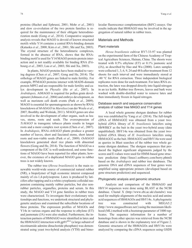
served primary amino acid sequences (Figure 1A). The
HbMAGO1 protein shared 94%, 93%, 92% and 92% ami-
no acid identity with the homologs TcMAGO, PtMAGO,
CsMAGO and VvMAGO, respectively; the HbMAGO2
protein shared 94%, 91%, 92% and 91% amino acid iden-
tity with the homologs of TcMAGO, PtMAGO, CsMAGO
and VvMAGO, respectively. The secondary structure pre-
diction of the MAGO protein revealed that it consisted of
six �-strands (�1-�6) and three �-helices (�1-�3) in a
�1-�2-�3-�4-�1-�5-�6-�2-�3 arrangement (Figure 1A).
The HbY14a/b proteins had calculated molecular masses of
21.737 kDa and 21.903 kDa and isoelectric points (pI) of
4.79 and 4.79 (Table 2).
The HbY14a protein shared 92%, 80%, 72%, 77%,
68% and 72% amino acid identity with the homologs of
RcMAGO, PtaMAGO, PtbMAGO, CsiMAGO, MtMAGO
and GmMAGO, while the HbY14b protein shared 91%,
80%, 71%, 75%, 67% and 69% amino acid identity with the
homologs of RcMAGO, PtaMAGO, PtbMAGO,
CsiMAGO, MtMAGO and GmMAGO. The Y14s protein
possessed seven �-strands (�1-�7) and two �-helices (�1-
�2) in a �1-�2-�1-�3-�4-�5-�2-�6-�7 arrangement (Fig-
ure 1B). Alignment of the Y14 proteins revealed a well-
conserved RNA-binding domain (RBD) in the middle of
the protein (Figure 1B). An exon-intron structure analysis
revealed that HbMAGOs consisted of three introns and two
exons, while the HbY14s had three introns and four exons,
respectively (Figure 1C). Although, the differences in the
cDNA sequences between two MAGOs or two Y14s were
small, the differences in genomic DNA sequences were
enormous.
Phylogenetic analysis of the rubber tree HbMAGOand HbY14 families
To obtain information about the evolutionary rela-
tionships of HbMAGOs and HbY14s, phylogenetic analy-
ses were done based on multiple sequence alignments of all
transcribed proteins from these two gene families in plants.
Phylogenetic analysis based on the alignments of MAGO
family proteins showed that rubber trees and other dicots
were clustered in same group (Figure 2A), whereas mem-
bers of the Y14 family clustered with other dicots in a sin-
gle group (Figure 2B). These analyses also revealed that the
MAGO and Y14 genes were present in algae and that their
differentiation accompanied that of the corresponding spe-
cies.
Subcellular localization of HbMAGOs and HbY14s
MAGO and Y14 are nucleocytoplasmic shuttling
proteins located predominantly in the nucleoplasm and
nuclear speckles (Micklem et al., 1997; Kataoka et al.,
2000; Mohr et al., 2001). To confirm the subcellular loca-
tion of HbMAGOs and HbY14s, the fusion proteins and
the GFP control constructs were introduced into onion
epidermal cells by agroinfiltration using a water circulat-
ing vacuum pump and observed under a fluorescence mi-
76 Yang et al.
croscope. HbMAGOs and HbY14s were targeted
exclusively to the nucleus of the epidermal cells, whereas
the GFP control protein was distributed throughout the
cells (Figure 3).
MAGO and Y14 genes in Hevea brasiliensis 77
Table 2 - Characterization of identified HbMAGO and HbY14 families.
GenBank Gene Exon Exon length (bp) ORF Protein
Gene name (AJJZ010000000) (bp) number E1 E2 E3 E4 3’UTR (bp) aa MW pI
HbMAGO1 AJJZ010970869.1
AJJZ011020394.1
1863 3 219 177 60 161 456 151 17,476 5.68
HbMAGO2 AJJZ010203456.1
AJJZ010251509.1
2190 3 219 177 60 213 456 151 17,688 5.69
HbY14a AJJZ010172128.1
AJJZ010296629.1
3894 4 274 137 140 46 252 597 198 21,737 4.79
HbY14b AJJZ010209074.1
AJJZ010209073.1
3046 4 274 137 140 46 201 597 198 21,903 4.79
aa – amino acids, ORF – open reading frame.
Figure 1 - Sequence alignment of the deduced HbMAGOs (A) and HbY14s (B), and intron-exon organization and exon length (C). Amino acid residues
that are identical in the sequences are darkly shaded and well-conserved residues are shaded in pink. The �-helices and �-strands of the HbMAGOs and
HbY14s are shown as dashed lines and solid lines, respectively. The exons are shown as boxes (open reading frame in black, untranslated region (UTR) in
white) and the introns are represented by lines.

Expression analysis of HbMAGOs and HbY14s indifferent tissues
Real-time quantitative PCR was used to examine the
expression patterns of HbMAGOs and HbY14s in bark,
flowers, latex, leaves and roots. The expression profiles
showed that the four genes (two each for HbMAGO and
HbY14) were transcribed in all of the tissues examined,
with the highest transcription in latex; for HbMAGO1, the
level of transcription in bark was similar to that seen in la-
tex (Figure 4A).
78 Yang et al.
Figure 2 - Phylogenetic trees of HbMAGOs (A) and HbY14s (B). The trees were calculated based on the HbMAGO and HbY14 protein sequences and
other plant MAGOs and Y14s. The GenBank accession numbers of selected homologs used to produce the phylogenetic trees are: CeMAGO
(NP_493025.1), AaMAGO (XP_001660832.1), AmMAGO (XP_001120074.1), DmMAGO (NP_476636.1), WbMAGO (EJW84410.1), HsMAGO
(NP_002361.1), MmMAGO (NP_079840.2), DrMAGO (NP_001017700.1), XtMAGO (XP_002931471.1), CrMAGO (XP_001694745.1), VcMAGO
(XP_002954749.1), OsMAGO1 (EEC82788.1), OsMAGO2 (NP_001066589.1), ZmMAGO1 (NP_001145913.1), ZmMAGO2 (NP_001146966.1),
ZmMAGO3 (ACG28070.1), SiMAGO1 (XP_004972442.1), SiMAGO2 (XP_004957130.1), BdMAGO (XP_003573269.1), AtaMAGO
(EMT32727.1), TuMAGO (EMS54552.1), SbMAGO1 (XP_002443724.1), SbMAGO2 (XP_002462539.1), AthMAGO (NP_171716.1), TcMAGO
(XP_007052335.1), PtMAGO (XP_006375160.1), LrMAGO (ACT33369.1), PpeMAGO (XP_007220423.1), CsMAGO (XP_004133764.1),
PpuMAGO (ABQ11262.1), VvMAGO (XP_002281294.1), CaMAGO (XP_004501210.1), MtMAGO (ACJ86076.1), GmMAGO (NP_001236090.1),
LjMAGO1 (AFK33465.1), LjMAGO2 (AFK48815.1), PvMAGO1 (XP_007147870.1), PvMAGO2 (XP_007134432.1), HoMAGO (AAS20975.1),
FvMAGO (XP_004306981.1), PsMAGO (ABK22137.1), PpaMAGO1 (XP_001770408.1), PpatMAGO2 (XP_001763801.1), CeY14 (NP_497891.1),
AaY14 (XP_001652167.1), AmY14 (XP_395245.2), DmY14 (NP_610454.2), WbY14 (EJW88540.1), HsY14 (NP_005096.1), MmY14
(NP_001095877.1), CrY14 (XP_002953417.1), CrY14 (XP_001696992.1), OsY14a (NP_001051661.1), OsY14b (XP_006654975.1), ZmY14a
(NP_001150559.1), ZmY14b (NP_001150263.1), SiY14a (XP_004960437.1), SiY14b (XP_004981327.1), BdY14 (XP_003568971.1), SbY14a
(XP_002439249.1), SbY14b (XP_002466233.1), RcY14 (XP_002513523.1), PtY14a (XP_002299789.1), PtY14b (XP_002314085.2), CsiY14
(XP_006470660.1), MtY14 (XP_003610955.1), GmY14 (XP_003517521.1), TcaY14 (XP_007015222.1), CsaY14 (XP_004139049.1), StY14a
(XP_006353634.1), StY14b (NP_001274809.1), TcrY14 (ABB91897.1), PsY14 (ABK25331.1), PpY14a (XP_001758256.1) and PpY14b
(XP_001771298.1)

Expression patterns of HbMAGOs and HbY14s inlatex respond to treatment with JA and ET
Since ET and JA have an important role as signaling
molecules that regulate rubber biosynthesis in rubber trees
(Hao and Wu, 2000; Zeng et al., 2009), we examined
whether exposure to ET or JA could influence HbMAGO
and HbY14 expression in latex (Figure 4B), despite the lack
of evidence relating MAGOs and Y14s to hormone signal-
ing pathways. HbMAGO1 was regulated by ET but not by
JA, and HbMAGO2 was induced by JA but not by ET. Two
HbY14s were induced by ET and JA in latex. Gene expres-
sion increased significantly within 9 h and subsequently de-
creased when treated with the plant hormones, i.e.,
HbMAGO1 was influenced by ET, HbMAGO2 by JA,
HbY14a by ET and JA, and HbY14b by JA; HbY14b ex-
pression also increased significantly within 24 h and subse-
quently decreased in response to ET.
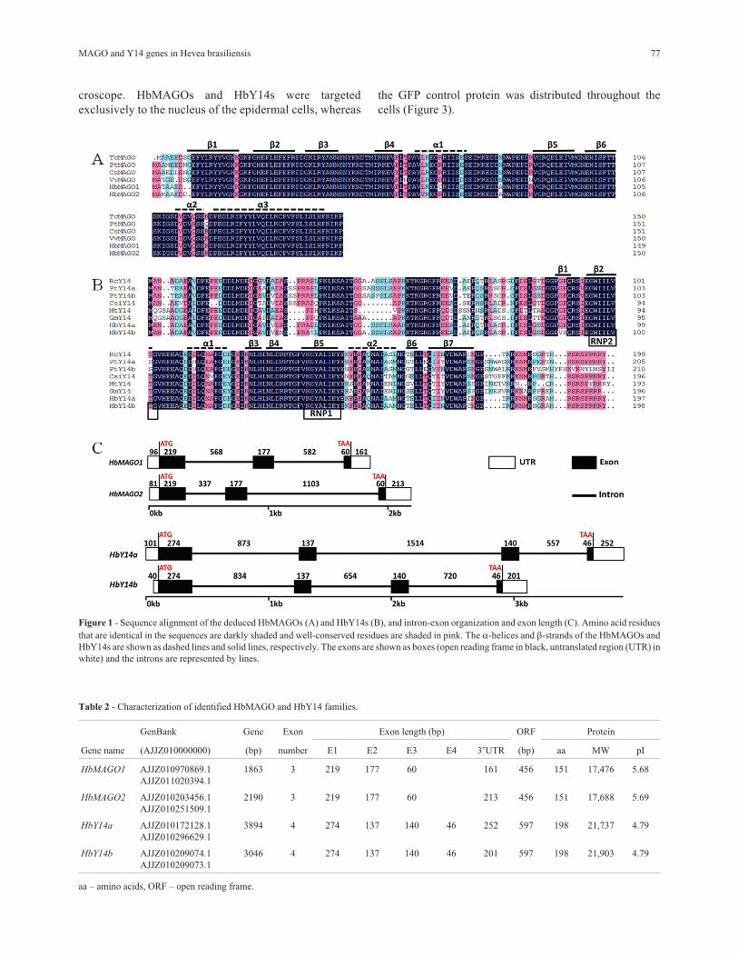
Interaction of HbMAGOs with HbY14s
As the core of EJC, MAGO and Y14 can form hetero-
dimers. Figure 5A shows that HbMAGOs interacted with
HbY14s in the YTH system. The interaction of HbMAGO1
with HbY14s was significantly greater than that of
HbMAGO2 with HbY14s, as assessed by quantifying the
�-galactosidase activity (Figure 5B). This finding suggests
that HbMAGOl interacted specifically with HbY14s.
Interaction of HbMAGO2 with gp91phox
Using BK-MAGO as bait, we obtained gp91phox as
one of the prey proteins screened from a two-hybrid latex
library (Figure 6A). The open reading frame of gp91phox
was cloned (GeneBank: AJJZ010935968.1). To confirm
the subcellular location of gp91phox proteins, gp91phox:GFP
fusion proteins were transiently expressed in onion epider-
mal cells. Confocal imaging of GFP fluorescence revealed
that the gp91phox:GFP fusion protein was located in the
cytomembrane (Figure 6B). The possible interaction be-
tween HbMAGO2 and gp91phox was examined using a
BiFC assay. A strong fluorescence signal was observed in
the cytomembrane of epidermal cells under normal condi-
tions (Figure 6C) and during plasmolysis (Figure 6D), but
not with the negative controls. These data confirmed the in-
teraction between HbMAGO2 and gp91phox.
Discussion
There is increasing evidence that many factors in-
volved in basic cellular processes in animals and plants are
conserved. MAGO and Y14 proteins, which are important
for cellular differentiation in animals, show marked se-
quence conservation across a wide range of species (Chen
et al., 2007). The MAGO and Y14 proteins are encoded by
a small gene family in plants. The Arabidopsis genome
contains one MAGO and one Y14 gene (Park and Muench,
2007), whereas the rice genome contains two MAGO and
two Y14 genes (Gong and He, 2014). We have identified
MAGO and Y14 genes in Hevea brasiliensis 79
Figure 3 - Subcellular localization of HbMAGOs and HbY14s. Bottom
row of panels: fluorescence, bright field and merged fluorescence images
of the GFP control. The other columns indicate the corresponding fluores-
cence, bright field and merged fluorescence images of HbMAGOs and
HbY14s.
Figure 4 - Expression patterns of HbMAGOs and HbY14s in different tis-
sues (A) and the responses to treatment with jasmonic acid (JA) and ethyl-
ene (ET) (B). Relative transcript abundances of HbMAGOs and HbY14s
were examined by RT-qPCR. The Y-axis indicates the relative transcript
abundance level, while the X-axis denotes the rubber tree tissues exam-
ined (A) and the time course of the response to treatment with ET and JA
(B). The rubber tree actin gene (GenBank HQ260674.1) was used as an in-
ternal control. PCR primers were designed to avoid the conserved region
and to amplify 100-300 bp products. The primer sequences are shown in
Table 2. B – bark, L – leaves, F – flowers, La – latex and R – roots. **p <
0.01 (ANOVA).
two MAGO and two Y14 genes in the rubber tree genome.
MAGO and Y14 proteins are highly conserved among
eukaryotes, suggesting that the corresponding genes are es-
sential for eukaryotes. Y14 proteins have a well-conserved
RBD in the middle region. The RBD is a characteristic fea-
ture of the ribonucleo-protein (RNP) family of RNA bind-
ing proteins and is evolutionarily conserved (Nagai et al.,
1995). The homologs of Y14 that have these characteristics
are known to be involved in RNA localization, mRNA
splicing and exon-exon junctions (Kim et al., 2001; Mohr
et al., 2001; Chuang et al., 2013). It has been suggested that
HbY14 may also have these critical functions in plants.
HbMAGOs and HbY14s shared high amino acid sequence
identity with homologs from other species. Although
MAGO and Y14 are highly conserved in eukaryotes, the
clusters of MAGO and Y14 are easily distinguished be-
tween dicots and other clades. The evidence from gene
structure also supports this conclusion. The two MAGO
genes in humans (MAGOH) consist of five exons with a
conserved length and structure, but the coding sequence of
MAGOHB is longer than that of MAGOH (Singh et al.,
2013).
MAGO and Y14 are nucleocytoplasmic shuttling
proteins located predominantly in the nucleoplasm and nu-
clear speckles (Micklem et al., 1997; Kataoka et al., 2000;
Mohr et al., 2001). In Taiwania cryptomerioides,
TcMAGO-EGFP and TcY14-EGFP were detected at low
levels in the cytoplasm at 20 h and 22 h. After 22 h,
TcMAGO-EGFP was located only in the nuclei, whereas
TcY14-EGFP was present in the nuclei and at low levels in
the cytoplasm (Chen et al., 2007). In rice, OsMAGO2 and
OsY14b showed identical distributions in cells and were lo-
cated in the nuclei and cytoplasm. Surprisingly, OsY14a
seemed to be uniquely located in the nuclei, in contrast to
the established subcellular localization of these EJC sub-
units (Gong and He, 2014). As shown here, HbMAGOs and
HbY14s were located in the nuclei, in a manner similar to
their homologs in other species.
In rubber trees, laticifers are tissues that are specifi-
cally involved in the biosynthesis and storage of natural
rubber, as well as in defense against pathogens. In this
study, we found that HbMAGOs and HbY14s had a similar,
constitutive expression pattern in different tissues of rubber
tree. Four genes were transcribed in the tissues examined,
with the highest transcription occurring in latex, a finding
indicative of the vital role of these genes in laticifer cells.
HbMAGO1 was regulated by ET but not by JA, and
HbMAGO2 was induced by JA but not by ET. In contrast,
two HbY14s were regulated by ET and JA in latex. The
biosynthesis of natural rubber is enhanced in rubber trees
by the endogenous accumulation and exogenous applica-
tion of JA (Hao and Wu, 2000). Our findings suggest that
the HbMAGO and HbY14 genes examined may regulate
natural rubber biosynthesis in rubber tree laticifer cells via
ET and JA signal transduction pathways. The differences in
the levels of transcription of the genes in response to ET
and JA also indicated functional differences in laticifer
cells. This is the first demonstration that HbMAGO and
HbY14 are linked to hormone signaling pathways. The pre-
cise relationship will be investigated in future studies.
The MAGO gene was first identified as a strict mater-
nal effect gene in Drosophila, where a single point mutant
80 Yang et al.
Figure 5 - Protein interaction matrices for HbMAGO and HbY14 proteins (A) and quantification of �-galactosidase activity (B). The combination of bait
proteins (BD) and prey proteins (AD) is indicated. Interactions between HbMAGOs and HbY14s were detected in yeast and the same amounts of
co-transformed yeast cells were grown in the highest stringent conditions (QDO/X/A medium). Transformants containing BK-53 and AD-T were used as
positive controls and those containing only BK and AD or fused proteins with BK or AD only were used as negative controls (A). In panel (B),
�-galactosidase activity was used as an indicator of the interaction between HbMAGOs and HbY14s (B). Representative results obtained in at least three
independent experiments are shown. The columns in panel (B) represent the mean � SEM. *p < 0.05 (ANOVA).

in the MAGO locus gave rise to a grandchildless phenotype
because of a defect in the correct cytoplasmic location of
oskar mRNA (Boswell et al., 1991; Newmark and Boswell,
1994; Micklem et al., 1997; Newmark et al., 1997). MAGO
always functions together with an RNA-binding protein,
known as Y14 in Xenopus (Kataoka et al., 2000), RBM8A
in humans (Zhao et al., 2000) and Tsunagi in Drosophila
(Mohr et al., 2001); this protein shuttles between the nu-
cleus and cytoplasm (Hachet and Ephrussi, 2001; Kim et
al., 2001).
The MAGO-Y14 complex is the core of the EJC as-
sembled on RNA 20 nucleotides upstream of exon-exon
junctions (Kataoka et al., 2001; Bono et al., 2004). Deter-
mination of the crystal structure of the MAGO-Y14 com-
plex showed that the MAGO-Y14 interaction was highly
specific and strongly conserved (Zhao et al., 2000; Kataoka
et al., 2001; Le et al., 2001; Shi and Xu, 2003; Stroupe et
MAGO and Y14 genes in Hevea brasiliensis 81
Figure 6 - Functional analysis of HbMAGO2. Interaction of HbMAGO2 with gp91phox in yeast (A), subcellular localization of gp91phox (B), and bimolec-
ular fluorescence complementation (BiFC) assays in plants (C,D). pGBK-HbMAGO2 (bait) and pGAD-gp91phox (prey) were co-transformed into AH109
yeast cells. Aliquots (10 �L) of a 10 diluted yeast suspension culture co-transformed with bait and prey constructs was spotted onto SD/-Trp/-Leu and
SD/-Trp/-Leu/-His/-Ade selection plates. Negative controls consisted of vector with only BK and AD or fused proteins with BK or AD. The intensity of
the interaction was assessed by assaying �-galactosidase activity (A). Panel (B) shows the subcellular localization of gp91phox. The interaction between
HbMAGO2 and gp91phox was confirmed using the BiFC assay (C). Epidermal cells were co-transformed with HbMAGO2 and gp91phox proteins fused to
the N- or C-terminal half of yellow fluorescent protein. HbMAGO2 or gp91phox with vector were used as negative controls. Onion epidermal cells were
observed under normal conditions (C) and after plasmolysis (incubation for 30 min in a 30% sucrose solution) (D). Representative results obtained in at
least three independent experiments are shown. The columns in panel (A) represent the mean � SEM.

al., 2006). In plants, the MAGO-Y14 complex has also
been found in T. cryptomerioides and rice (Chen et al.,
2007; Gong and He, 2014). The functional barrier between
the two protein families was not observed within dicots or
duplicates of rice, but was observed between dicots and
monocot or plants and animals (Gong et al., 2014). The
high specificity and conservation of the MAGO-Y14 inter-
action is clade-specific, and such co-evolution allows the
interaction between these proteins to be maintained across
large evolutionary time scales. In this work, we identified
HbMAGO1/2 and HbY14a/b homologs derived from rub-
ber tree latex and confirmed the interaction between
HbMAGO and HbY14 using YTH analysis. Overall, our
findings indicate that HbMAGO1/2 and HbY14a/b are
evolutionarily highly conserved proteins with similar func-
tions to their homologs.
In rubber trees, NADPH (nicotinamide adenine
dinucleotide phosphate) oxidase is involved in the accumu-
lation of reactive oxygen species (ROS) in tapping panel
dryness (TPD), a physiological disorder characterized by
the spontaneous drying up of the tapping cut that results in
an abnormally low yield or stoppage of latex flow (Jacob et
al., 1994; Chen et al., 2003; Venkatachalam et al., 2009).
ROS are involved in the coagulation of rubber particles that
dramatically reduces natural rubber production. High ROS
production in latex cells triggers oxidative stress leading to
the in situ coagulation of rubber particles (Chrestin et al.,
1984; Li et al., 2010; Leclercq et al., 2012). ROS are also
important signaling molecules in plants.
ROS formation is one of the early physiological re-
sponses of plant cells to biotic and abiotic stress, such as
wounding and pathogens (Bogre et al., 1997; Bolwell et al.,
2002). NADPH oxidase is the main source of ROS in plants
(Asai et al., 2008). In animals, the NADPH oxidase com-
plex consists of two plasma membrane proteins, gp91phox
(phox for phagocyte oxidase) and p22phox. Cytosolic regula-
tory proteins p47phox, p67phox, p40phox and Rac2, translocate
to the plasma membrane to form the active complex after
stimulation (Brandes et al., 2014). However, no homologs
of the p22phox, p67phox, p47phox and p40phox regulators of
phagocyte NADPH oxidase were found in plants.
Plant NADPH oxidases are known as respiratory
burst oxidase homologs (RBOHs) and are homologous to
the catalytic subunit (gp91phox) of mammalian NADPH oxi-
dases (Rodriguez et al., 2007; Suzuki et al., 2011). RBOHs
have been identified and characterized in several species,
including Arabidopsis thaliana (Kawarazaki et al., 2013),
potato (Chen et al., 2013), rice (Yoshie et al., 2005), wheat
(Yamauchi et al., 2014). In TPD trees, high levels of
NADPH oxidase activities lead to the release of O2- , a toxic
form of oxygen (Jacob et al., 1994). Cellular membranes
and especially lutoids, which contain coagulant factors in-
volved in the aggregation of rubber particles, are damaged
by O2- (Chrestin et al., 1984; Li et al., 2010; Leclercq et al.,
2012).
In plants, MAGO can participate in biological pro-
cesses through protein-protein interactions. In Physalis,
MAGO interacts with a MADS-Domain protein that can
regulate plant development through the formation of
dimers and higher order complexes to influence male fertil-
ity and calyx development (He et al., 2007); this finding
suggests that MAGO plays a role in plant biological pro-
cesses independently of the EJC. Little is known about the
function of HbMAGO mediated by interaction with target
proteins in rubber trees. As shown here, the YTH and BiFC
assays indicated that HbMAGO2 interacted with gp91phox.
This finding suggests that HbMAGO2 may regulate
NADPH oxidase-dependent oxidative bursts in rubber tree
latex via protein-protein interactions.
Functional studies have shown that MAGO is in-
volved in development of organization in multicellular or-
ganisms. Over-expression or knockdown of MAGO results
in phenotypic alterations that vary among plants, including
visible longer roots and a more complex root system, a
greater number of leaves, short and fasciated stems, short
lateral roots and non-viable seeds, and short rice plants with
abnormal flowers (Chen et al., 2007; Park et al., 2009;
Gong and He, 2014). In Drosophila, the EJC controls the
splicing of mapk and other long intron-containing tran-
scripts and mitogen-activated protein kinase (MAPK) sig-
naling depends on the regulation of MAPK levels by the
EJC (Ashton-Beaucage et al., 2010). During development
of the eye, MAPK is the primary functional target of
MAGO (Roignant and Treisman, 2010). As core compo-
nents of the EJC linked to MAPK signaling pathway,
MAGOs can control animal cell or tissue development.
Laticifer cells are some of the most important plant cells
that continuously produce natural rubber and are arranged
as concentric sheaths in the phloem (Chen et al., 2000; Han
et al., 2000; Pickard, 2008). There are few reports on the
regulation of laticifer development in general (Tan et al.,
2014), and the development of laticifer cells in rubber trees
remains poorly understood. Our findings suggest that
MAGO may regulate the development of laticifer cells, al-
though further experiments are need to confirm this sugges-
tion.
Conclusions
The present study has provided a comprehensive
genomic analysis of the rubber tree HbMAGO and HbY14
gene families and the first evidence that HbMAGO and
HbY14 are linked with hormone signaling pathways, in-
cluding rubber particle aggregation in laticifer cells. These
data provide important insights into the potential roles of
HbMAGO and HbY14 gene families in rubber trees and
may contribute to clarifying the function of HbMAGO2 in
the aggregation of natural rubber particles.
82 Yang et al.
Acknowledgments
This work was supported by the National Natural Sci-
ence Foundation of China (grant no. 31170634) and Inno-
vation Subject of Hainan province (grants nos. B201301
and Hyb2014-03).
References
Asai S, Ohta K and Yoshioka H (2008) MAPK signaling regulates
nitric oxide and NADPH oxidase-dependent oxidative
bursts in Nicotiana benthamiana. Plant Cell 20:1390-1406.
Ashton-Beaucage D, Udell CM, Lavoie H, Baril C, Lefrancois M,
Chagnon P, Gendron P, Caron-Lizotte O, Bonneil E, Thi-
bault P, et al. (2010) The exon junction complex controls the
splicing of mapk and other long intron-containing transcripts
in Drosophila. Cell 143:251-262.
Bolwell GP, Bindschedler LV, Blee KA, Butt VS, Davies DR,
Gardner SL, Gerrish C and Minibayeva F (2002) The apo-
plastic oxidative burst in response to biotic stress in plants: a
three-component system. J Exp Bot 53:1367-1376.
Bogre L, Ligterink W, Meskiene I, Barker PJ, Heberle-Bors E,
Huskisson NS and Hirt H (1997) Wounding induces the
rapid and transient activation of a specific MAP kinase path-
way. Plant Cell 9:75-83.
Bono F, Ebert J, Unterholzner L, Güttler T, Izaurralde E and Conti
E (2004) Molecular insights into the interaction of PYM
with the Mago-Y14 core of the exon junction complex.
EMBO Rep 5:304-310.
Boothby TC and Wolniak SM (2011) Masked mRNA is stored
with aggregated nuclear speckles and its asymmetric redis-
tribution requires a homolog of Mago nashi. BMC Cell Biol
12:45.
Boswell RE, Prout ME and Steichen JC (1991) Mutations in a
newly identified Drosophila melanogaster gene, mago
nashi, disrupt germ cell formation and result in the formation
of mirror-image symmetrical double abdomen embryos. De-
velopment 113:373-384.
Brandes RP, Weissmann N and Schroder K (2014) Nox family
NADPH oxidases: molecular mechanisms of activation.
Free Radical Biol Med 76c:208-226.
Chen DH, Ye HC, Li GF and Liu Y (2000) Advances in molecular
biology of plant isoprenoid metabolic pathway. Acta Bot Sin
42:551-558.
Chen HJ, Huang CS, Huang GJ, Chow TJ and Lin YH (2013)
NADPH oxidase inhibitor diphenyleneiodonium and re-
duced glutathione mitigate ethephon-mediated leaf senes-
cence, H2O2 elevation and senescence-associated gene ex-
pression in sweet potato (Ipomoea batatas). J Plant Physiol
170:1471-1483.
Chen SC, Peng SQ, Huang GX, Wu KX, Fu XH and Chen ZQ
(2003) Association of decreased expression of a Myb tran-
scription factor with the TPD (tapping panel dryness) syn-
drome in Hevea brasiliensis. Plant Mol Biol 51:51-58.
Chen YR, Shaw JF, Chung MC and Chu FH (2007) Molecular
identification and characterization of Tcmago and TcY14 in
Taiwania (Taiwania cryptomerioides). Tree Physiol
27:1261-1271.
Chrestin H, Bangratz J, d’Auzac J and Jacob JL (1984) Role of the
lutoidic tonoplast in the senescence and degeneration of the
laticifers of Hevea brasiliensis. Zeitschrift für Pflanzen-
physiol 114:261-268.
Chuang TW, Chang WL, Lee KM and Tarn WY (2013) The
RNA-binding protein Y14 inhibits mRNA decapping and
modulates processing body formation. Mol Biol Cell
24:1-13.
Fribourg S, Gatfield D, Izaurralde E and Conti E (2003) A novel
mode of RBD-protein recognition in the Y14-Mago com-
plex. Nat Struct Biol 10:433-439.
Gehring NH, Lamprinaki S, Kulozik AE and Hentze MW (2009)
Disassembly of exon junction complexes by PYM. Cell
137:536-548.
Gong PC, Zhao M and He CY (2014) Slow co-evolution of the
MAGO and Y14 protein families is required for the mainte-
nance of their obligate heterodimerization mode. PLoS One
9:e84842.
Gong PC and He CY (2014) Uncovering divergence of rice exon
junction complex core heterodimer gene duplication reveals
their essential role in growth, development, and reproduc-
tion. Plant Physiol 165:1047-1061.
Hachet O and Ephrussi A (2001) Drosophila Y14 shuttles to the
posterior of the oocyte and is required for oskar mRNA
transport. Curr Biol 11:1666-1674.
Hao BZ and Wu JL (2000) Laticifer differentiation in Hevea
brasiliensis: induction by exogenous jasmonic acid and lino-
lenic acid. Ann Bot 85:37-43.
Han KH, Shin DH, Yang J, Kim IJ, Oh SK and Chow KS (2000)
Genes expressed in the latex of Hevea brasiliensis. Tree
Physiol 20:503-510.
He CY, Sommer H, Grosardt B, Huijser P and Saedler H (2007)
PFMAGO, a MAGO NASHI-like factor, interacts with the
MADS-domain protein MPF2 from Physalis floridana. Mol
Biol Evol 24:1229-1241.
Jacob JL, Prevot JC and Lacrotte R (1994) Tapping panel dryness
in Hevea brasiliensis. Plant Rech Dev 1:15-21.
Johnson MA, von Besser K, Zhou Q, Smith E, Aux G, Patton D,
Levin JZ and Preuss D (2004) Arabidopsis hapless muta-
tions define essential gametophytic functions. Genetics
168:971-982.
Kataoka N, Diem MD, Kim VN, Yong J and Dreyfuss G (2001)
Magoh, a human homolog of Drosophila mago nashi pro-
tein, is a component of the splicing-dependent exon-exon
junction complex. EMBO J 20:6424-6433.
Kawarazaki T, Kimura S, Iizuka A, Hanamata S, Nibori H, Michi-
kawa M, Imai A, Abe M, Kaya H and Kuchitsu K (2013) A
low temperature-inducible protein AtSRC2 enhances the
ROS-producing activity of NADPH oxidase AtRbohF.
Biochim Biophys Acta Mol Cell Res 1833:2775-2780.
Kataoka N, Yong J, Kim VN, Velazquez F, Perkinson RA, Wang
F and Dreyfuss G (2000) Pre-mRNA splicing imprints
mRNA in the nucleus with a novel RNA-binding protein
that persists in the cytoplasm. Mol Cell 6:673-682.
Kim VN, Yong J, Kataoka N, Abel L, Diem MD and Dreyfuss G
(2001) The Y14 protein communicates to the cytoplasm the
position of exon-exon junctions. EMBO J 20:2062-2068.
Lau CK, Diem MD, Dreyfuss G and Van Duyne GD (2003) Struc-
ture of the Y14-Magoh core of the exon junction complex.
Curr Biol 13:933-941.
Le HH and Seraphin B (2008) EJCs at the heart of translational
control. Cell 133:213-216.
MAGO and Y14 genes in Hevea brasiliensis 83

Le HH, Nott A and Moore MJ (2003) How introns influence and
enhance eukaryotic gene expression. Trends Biochem Sci
28:215-220.
Le HH, Izaurralde E, Maquat LE and Moore MJ (2000) The
spliceosome deposits multiple proteins 20-24 nucleotides
upstream of mRNA exon-exon junctions. EMBO J
19:6860-6869.
Le HH, Gatfield D, Braun IC, Forler D and Izaurralde E (2001)
The protein Mago provides a link between splicing and
mRNA localization. EMBO Rep 2:1119-1124.
Lee HC, Choe J, Chi SG and Kim YK (2009) Exon junction com-
plex enhances translation of spliced mRNAs at multiple
steps. Biochem Biophys Res Commun 384:334-340.
Leclercq J, Martin F, Sanier C, Clement-Vidal A, Fabre D, Oliver
G, Lardet L, Ayar A, Peyramard M and Montoro P (2012)
Over-expression of a cytosolic isoform of the HbCuZnSOD
gene in Hevea brasiliensis changes its response to a water
deficit. Plant Mol Biol 80:255-272.
Lejeune F and Maquat LE (2005) Mechanistic links between non-
sense-mediated mRNA decay and pre-mRNA splicing in
mammalian cells. Curr Opin Cell Biol 17:309-315.
Li DJ, Deng Z, Chen CL, Xia ZH, Wu M, He P and Chen SC
(2010) Identification and characterization of genes associ-
ated with tapping panel dryness from Hevea brasiliensis la-
tex using suppression subtractive hybridization. BMC Plant
Biol 10:140.
Micklem DR, Dasgupta R, Elliott H, Gergely F, Davidson C,
Brand A, Gonzalez-Reyes A and St Johnston D (1997) The
mago nashi gene is required for the polarisation of the
oocyte and the formation of perpendicular axes in
Drosophila. Curr Biol 7:468-478.
Mohr SE, Dillon ST and Boswell RE (2001) The RNA-binding
protein Tsunagi interacts with Mago Nashi to establish po-
larity and localize oskar mRNA during Drosophila ooge-
nesis. Gene Dev 15:2886-2899.
Mufarrege EF, Gonzalez DH and Curi GC (2011) Functional in-
terconnections of Arabidopsis exon junction complex pro-
teins and genes at multiple steps of gene expression. J Exp
Bot 62:5025-5036.
Nagai K, Oubridge C, Ito N, Avis J and Evans P (1995) The RNP
domain: a sequence-specific RNA-binding domain involved
in processing and transport of RNA. Trends Biochem Sci
20:235-240.
Newmark PA and Boswell RE (1994) The mago nashi locus en-
codes an essential product required for germ plasm assem-
bly in Drosophila. Development 120:1303-1313.
Newmark PA, Mohr SE, Gong L and Boswell RE (1997) mago
nashi mediates the posterior follicle cell-to-oocyte signal to
organize axis formation in Drosophila. Development
124:3197-3207.
Park NI and Muench DG (2007) Biochemical and cellular charac-
terization of the plant ortholog of PYM, a protein that inter-
acts with the exon junction complex core proteins Mago and
Y14. Planta 225:625-639.
Park NI, Yeung EC and Muench DG (2009) Mago Nashi is in-
volved in meristem organization, pollen formation, and seed
development in Arabidopsis. Plant Sci 176:461-469.
Pickard WF (2008) Laticifers and secretory ducts: two other tube
systems in plants. New Phytol 177:877-888.
Rodriguez AA, Ramiro Lascano H, Bustos D and Taleisnik E
(2007) Salinity-induced decrease in NADPH oxidase activ-
ity in the maize leaf blade elongation zone. J Plant Physiol
164:223-230.
Roignant JY and Treisman JE (2010) Exon junction complex sub-
units are required to splice Drosophila MAP kinase, a large
heterochromatic gene. Cell 143:238-250.
Shi H and Xu RM (2003) Crystal structure of the Drosophila
Mago nashi-Y14 complex. Genes Dev 17:971-976.
Singh KK, Wachsmuth L, Kulozik AE and Gehring NH (2013)
Two mammalian MAGOH genes contribute to exon junc-
tion complex composition and nonsense-mediated decay.
RNA Biol 10:1291-1298.
Stroupe ME, Tange TO, Thomas DR, Moore MJ and Grigorieff N
(2006) The three-dimensional architecture of the EJC core. J
Mol Biol 360:743-749.
Suzuki N, Miller G, Morales J, Shulaev V, Torres MA and Mittler
R (2011) Respiratory burst oxidases: the engines of ROS
signaling. Curr Opin Plant Biol 14:691-699.
Tan DG, Sun XP and Zhang JM (2014) Age-dependent and
jasmonic acid-induced laticifer-cell differentiation in anther
callus cultures of rubber tree. Planta 240:337-344.
Tang CR, Qi JY, Li H, Zhang CL and Wang YK (2007) A conve-
nient and efficient protocol for isolating high-quality RNA
from latex of Hevea brasiliensis (para rubber tree). J
Biochem Biophys Meth 70:749-754.
Tange TO, Nott A and Moore MJ (2004) The ever-increasing
complexities of the exon junction complex. Curr Opin Cell
Biol 16:279-284.
Tange TO, Shibuya T, Jurica MS and Moore MJ (2005) Biochem-
ical analysis of the EJC reveals two new factors and a stable
tetrameric protein core. RNA 11:1869-1883.
van der Weele CM, Tsai CW and Wolniak SM (2007) Mago nashi
is essential for spermatogenesis in Marsilea. Mol Biol Cell
18:3711-3722.
Venkatachalam P, Thulaseedharan A and Raghothama K (2009)
Molecular identification and characterization of a gene asso-
ciated with the onset of tapping panel dryness (TPD) syn-
drome in rubber tree (Hevea brasiliensis Muell.) by mRNA
differential display. Mol Biotechnol 41:42-52.
Walter M, Chaban C, Schutze K, Batistic O, Weckermann K,
Nake C, Blazevic D, Grefen C, Schumacher K, Oecking C,
et al. (2004) Visualization of protein interactions in living
plant cells using bimolecular fluorescence complemen-
tation. Plant J 40:428-438.
Yamauchi T, Watanabe K, Fukazawa A, Mori H, Abe F, Kawa-
guchi K, Oyanagi A and Nakazono M (2014) Ethylene and
reactive oxygen species are involved in root aerenchyma
formation and adaptation of wheat seedlings to oxygen-
deficient conditions. J Exp Bot 65:261-273.
Yang YN, Li RG and Qi M (2000) In vivo analysis of plant pro-
moters and transcription factors by agroinfiltration of to-
bacco leaves. Plant J 22:543-551.
Yang ZP, Li HL, Guo D, Tang X and Peng SQ (2014) Identifica-
tion and characterization of the 14-3-3 gene family in Hevea
brasiliensis. Plant Physiol Biochem 80:121-127.
Yoshie Y, Goto K, Takai R, Iwano M, Takayama S, Isogai A and
Che FS (2005) Function of the rice gp91phox homologs
OsrbohA and OsrbohE genes in ROS-dependent plant im-
mune responses. Plant Biotechnol 22:127-135.
Zeng RZ, Duan CF, Li, XY, Tian WM and Nie ZY (2009)
Vacuolar-type inorganic pyrophosphatase located on the
rubber particle in the latex is an essential enzyme in regula-
84 Yang et al.

tion of the rubber biosynthesis in Hevea brasiliensis. Plant
Sci 176:602-607.
Zhao XF, Colaizzo-Anas T, Nowak NJ, Shows TB, Elliott RW
and Aplan PD (1998) The mammalian homologue of mago
nashi encodes a serum-inducible protein. Genomics
47:319-322.
Zhao XF, Nowak NJ, Shows TB and Aplan PD (2000) MAGOH
interacts with a novel RNA-binding protein. Genomics
63:145-148.
Internet ResourcesSoftberry platform for HMM-based gene structure prediction,
http://linux1.softberry.com/berry.phtml
Multiple alignment software Clustal X,
http://www.ebi.ac.uk/clustalw
MEGA5.2 software for phylogenetic tree construction,
http://www.megasoftware.net/
GSDS software for comparison of cDNA sequences,
http://gsds.cbi.pku.edu.cn/
Associate Editor: Marcia Pinheiro Margis
License information: This is an open-access article distributed under the terms of theCreative Commons Attribution License (type CC-BY), which permits unrestricted use,distribution and reproduction in any medium, provided the original article is properly cited.
MAGO and Y14 genes in Hevea brasiliensis 85