identification of genetic risk variants for deep vein thrombosis by
TRANSCRIPT

Identification of genetic risk variants for deepvein thrombosis by multiplexed next-generationsequencing of 186 hemostatic/pro-inflammatorygenesLotta et al.
Lotta et al. BMC Medical Genomics 2012, 5:7http://www.biomedcentral.com/1755-8794/5/7 (21 February 2012)
RESEARCH ARTICLE Open Access
Identification of genetic risk variants for deepvein thrombosis by multiplexed next-generationsequencing of 186 hemostatic/pro-inflammatorygenesLuca A Lotta1,2, Mark Wang2, Jin Yu2, Ida Martinelli1, Fuli Yu2, Serena M Passamonti1, Dario Consonni3,Emanuela Pappalardo1, Marzia Menegatti1, Steven E Scherer2, Lora L Lewis2, Humeira Akbar2, Yuanqing Wu2,Matthew N Bainbridge2, Donna M Muzny2, Pier M Mannucci1, Richard A Gibbs2* and Flora Peyvandi1*
Abstract
Background: Next-generation DNA sequencing is opening new avenues for genetic association studies incommon diseases that, like deep vein thrombosis (DVT), have a strong genetic predisposition still largelyunexplained by currently identified risk variants. In order to develop sequencing and analytical pipelines for theapplication of next-generation sequencing to complex diseases, we conducted a pilot study sequencing thecoding area of 186 hemostatic/proinflammatory genes in 10 Italian cases of idiopathic DVT and 12 healthy controls.
Results: A molecular-barcoding strategy was used to multiplex DNA target capture and sequencing, whileretaining individual sequence information. Genomic libraries with barcode sequence-tags were pooled (in pools of8 or 16 samples) and enriched for target DNA sequences. Sequencing was performed on ABI SOLiD-4 platforms.We produced > 12 gigabases of raw sequence data to sequence at high coverage (average: 42X) the 700-kilobasetarget area in 22 individuals. A total of 1876 high-quality genetic variants were identified (1778 single nucleotidesubstitutions and 98 insertions/deletions). Annotation on databases of genetic variation and human diseasemutations revealed several novel, potentially deleterious mutations. We tested 576 common variants in a case-control association analysis, carrying the top-5 associations over to replication in up to 719 DVT cases and 719controls. We also conducted an analysis of the burden of nonsynonymous variants in coagulation factor andanticoagulant genes. We found an excess of rare missense mutations in anticoagulant genes in DVT casescompared to controls and an association for a missense polymorphism of FGA (rs6050; p = 1.9 × 10⁻⁵, OR 1.45; 95%CI, 1.22-1.72; after replication in > 1400 individuals).
Conclusions: We implemented a barcode-based strategy to efficiently multiplex sequencing of hundreds ofcandidate genes in several individuals. In the relatively small dataset of our pilot study we were able to identifybona fide associations with DVT. Our study illustrates the potential of next-generation sequencing for the discoveryof genetic variation predisposing to complex diseases.
Keywords: Deep vein thrombosis, venous thromboembolism, next-generation sequencing, target capture, multi-plexing, FGA, rs6025, heamostateome, DVT, VTE
* Correspondence: [email protected]; [email protected] Bianchi Bonomi Hemophilia and Thrombosis Center, U.O.S.Dipartimentale per la Diagnosi e la Terapia delle Coagulopatie, FondazioneIRCCS Cà Granda - Ospedale Maggiore Policlinico, Università degli Studi diMilano and Luigi Villa Foundation, Milan, Italy2Human Genome Sequencing Center, Baylor College of Medicine, Houston,TX, USAFull list of author information is available at the end of the article
Lotta et al. BMC Medical Genomics 2012, 5:7http://www.biomedcentral.com/1755-8794/5/7
© 2012 Lotta et al; licensee BioMed Central Ltd. This is an Open Access article distributed under the terms of the Creative CommonsAttribution License (http://creativecommons.org/licenses/by/2.0), which permits unrestricted use, distribution, and reproduction inany medium, provided the original work is properly cited.

BackgroundDeep vein thrombosis (DVT) of the lower extremities, acommon thrombotic disease often complicated by acutepulmonary embolism [1], has a strong genetic compo-nent as established by family [2-4] and twin studies [5],with a 3-fold increase in disease risk for siblings of indivi-duals with DVT [2] and an estimated hereditary compo-nent of 60% [3]. Genetic risk factors include raremutations in PROC, PROS1 and SERPINC1 leading tothe deficiencies of natural anticoagulant proteins (proteinC, protein S and antithrombin, respectively) and singlenucleotide polymorphisms (SNPs) of F5 (rs6025 or factorV Leiden [FVL]) and of F2 (rs1799963 or prothrombinG20210A) [6]. More recently, genome-wide associationstudies (GWAS) identified associations of common SNPsat 6 different genomic loci (CYP4V2, SERPINC1, GP6,F5, ABO and HIVEP1) [7-9]. In spite of these recentobservations, however, genetic variants established toinfluence the risk for DVT explain only a fraction of dis-ease heritability [10].Next-generation DNA sequencing is opening new ave-
nues for genetic association studies in complex diseases,enabling to sequence large fractions of the human genome(or even the entire genome) at unprecedented speed andper-base costs. Re-sequencing the exome (i.e. the proteincoding area of the genome) or the entire genome isbecoming the gold standard for the identification of dis-ease-causing mutations in Mendelian diseases [11-13].However, the application of these techniques to commondiseases is still limited by the high costs and considerablecomputational burden associated with the analysis ofnumerous samples. Sequencing of a few hundred genes orgenomic loci is a much less expensive alternative towhole-genome (or exome) sequencing, suitable for theanalysis of areas of the genome that are deemed to haveparticular relevance in the pathophysiology of a given dis-ease. However, this regional sequencing approach, whichrequires sample multiplexing in order to achieve efficiency,is technically challenging. Multiplexing on next-generationsequencing platforms entails the use of DNA pools, whichis affected by limitations in downstream genetic associa-tion analysis. These limitations include (a) loss of sensitiv-ity to detect rare genetic variants, (b) uncertainty in allelefrequency estimations determined by the unequal repre-sentation of samples within the pool, and (c) loss of indivi-dual sequence data hampering direct genotype-phenotypeassociations.In this pilot study, we used genomic libraries with bar-
code sequence-tags as a means to overcome the limita-tions of pooling. Using this approach we were able tomultiplex DNA-target capture and SOLiD sequencingwhile retaining individual sequence information. In whatis one of the first practical applications of this technique,
we sequenced the coding area of 186 genes involved inblood hemostasis/inflammation (i.e. two pivotal patho-physiological mechanisms of DVT) in 10 Italian patientswith early-onset idiopathic DVT and 12 thrombosis-freecontrols. The goal of this pilot study was to develop pipe-lines for the application of next-generation sequencing inthe setting of DVT and to test different analyticalapproaches for the identification of disease-associatedvariants.
MethodsPatientsCases of DVT were selected from 2139 unrelated patientsreferred to the Angelo Bianchi Bonomi Hemophilia andThrombosis Center, Milan, (Italy) for diagnostic workoutand thrombophilia testing after a first episode of DVT ofthe lower limbs in the years 1995-2010. Patients wereasked to bring to the center the diagnostic documenta-tion of their thrombotic episodes and underwent a clini-cal interview. DVT had been diagnosed by compressionultrasonography or venography. All patients underwent acomplete thrombophilia screening, including measure-ment of natural anticoagulant proteins, genotyping ofFVL and prothrombin G20210A and search for antipho-spholipid autoantibodies. Coagulation factor VIII andfibrinogen coagulant activities were also measured.A patient selection flow-chart is shown in Additional
file 1 Figure S1. Patients selected for next-generationsequencing were required to have (a) history of idiopathicDVT of the lower limbs, (b) age of disease onset < 55years, (c) wild-type FVL and prothrombin G20210A gen-otypes, (d) absence of natural anticoagulant deficiencies,(e) negative search for anti-phospholipid autoantibodies,(f) been born in Lombardy, the 10 million people regionof Italy that has Milan as a regional capital. Since 42patients matched these criteria, patients were furtherprioritized based on age of onset, familial history ofvenous or arterial thrombosis, completeness of clinicalinformation, DNA amount and quality. Patients includedin the replication were all the remaining 719 patientswith a first episode of idiopathic lower-limb DVT andavailable DNA. Controls included in the study wereselected from a total of 1938 healthy Italian individualsrecruited among friends and non-consanguineous rela-tives who accompanied patients to the Hemophilia andThrombosis Center and agreed to be tested for thrombo-philia. Previous arterial or venous thrombosis in the con-trols was excluded using a validated questionnaire [14].Controls had similar age of the idiopathic DVT cases (±5 years) and the same gender. Controls who underwentnext-generation sequencing were matched with thepatients for geographic provenience (born in Lombardy),in order to minimize population stratification. In case
Lotta et al. BMC Medical Genomics 2012, 5:7http://www.biomedcentral.com/1755-8794/5/7
Page 2 of 11

more than one control matched the same patient, controlinclusion was random. The study was approved by theInstitutional Review Board of the Fondazione IRCCS Ca’Granda - Ospedale Maggiore Policlinico and all subjectsgave their informed consent. Patient recruitment, sam-pling and thrombophilia screening were carried out atthe Angelo Bianchi Bonomi Hemophilia and ThrombosisCenter, Milan (Italy). Next-generation DNA analysis andreplication PCR and Sanger sequencing and associatedanalyses were carried out at the Human Genome Sequen-cing Center (HGSC), Baylor College of Medicine, Hous-ton (USA).
Target area selectionThe protein coding exons, 3’ and 5’ UTRs, and theintron-exon boundaries of 186 genes were chosen astarget area. Target genes included all coagulation factorgenes, anticoagulant genes and genes involved in fibri-nolysis, platelet adhesion and aggregation, cell-cell inter-action, endothelial activation and inflammation. Thegenomic coordinate intervals corresponding to the targetarea were obtained from the UCSC Genome Browserdatabase and sent to NimbleGen for probe design.Probes were designed for all of the submitted intervals,with slightly different genomic coordinates (i.e. tiledregions). The final target area spanned 644, 472 bp.Probes were arrayed on Roche NimbleGen HD2 2.1 M-probe custom chips. The complete lists of target andtiled coordinate-intervals and of the target genes are inthe Additional file 2 and Additional file 1 Table S1.
Experimental designTwo experiments were carried out. In the first experiment,genomic libraries from 4 DVT patients (DVT_P_01,DVT_P_02, DVT_P_03, DVT_P_04) and 4 controls(DVT_C_01, DVT_C_02, DVT_C_03, DVT_C_04) werecaptured on the same Roche NimbleGen HD2 chip andsequenced in the same ABI SOLiD 4 spot (one quarter ofa slide). In the second, libraries from 8 cases (DVT_P_01,DVT_P_05, DVT_P_06, DVT_P_07, DVT_P_08,DVT_P_09, DVT_P_10, DVT_P_11) and 8 controls(DVT_C_05, DVT_C_06, DVT_C_07, DVT_C_08,DVT_C_09, DVT_C_10, DVT_C_11, DVT_C_12) werecaptured on one Roche NimbleGen HD2 chip andsequenced in two ABI SOLiD 4 spots. Sample DVT_P_11failed capture during the second experiment. PatientDVT_P_01 was sequenced twice as a quality controlprocedure.
Genomic library preparation, barcoding and enrichmentof target DNA sequencesTwo micrograms of genomic DNA were used to preparelibraries of DNA fragments that were ligated with ABISOLiD P1-and P2-adaptors [15]. Different modified P2-
adaptors, each containing a specific DNA-tag sequence(molecular barcode), were used for each individuallibrary. Libraries with barcodes were used to prepareequimolar DNA pools of 8 and 16 samples. Four micro-grams of DNA from each pool were hybridized on oneRoche NimbleGen HD2 chip.
ABI SOLiD 4 sequencingCapture products underwent emulsion PCR with P1-adaptor mediated attachment of clonally-amplified tem-plates to loading beads. Beads were covalently attachedon glass slides at the P2-adaptor extremity. Sequencingby oligonucleotide ligation and detection (SOLiD) wasperformed on ABI SOLiD 4 platforms [15].
Read barcode assignment and mapping to the referencegenomeReads with the barcodes were assigned to the corre-sponding sample using custom Perl scripts and mappedto reference human genome, NCBI36/hg18, usingBFAST software [16]. Reads that mapped on the samestarting and end coordinates, considered likely to bePCR duplicates, were marked in the binary alignment/mapping (BAM) files, where mapping information wasstored.
Genetic variant calls and quality control (QC)Sorted BAM files were processed in a variant-calling pipe-line consisting of a BAM filtering process and a variantcalling process. In the first step, duplicate reads wereeliminated from the BAM files, retaining only the readwith the top mapping quality at each pair of start and endmapping coordinates. Also, reads with mapping qualityscore of less than 50 were expunged from the BAM files.In the variant-calling step, Samtools [17] was used to gen-erate PILEUP files with read information at sites wheremismatches from the reference sequence were detected.Consensus in the presence of mismatches, read- and base-quality parameters were used as criteria to distinguishgenetic variants from sequencing errors, filtering high-quality calls. In a final QC process, this set of calls wasfurther cleared from variants with an allele balance (var-iant allele reads/total number of reference plus variantallele reads) below 20% and/or with significant strand bias(i.e. sites at which the variant sequence did not appear onboth forward and reverse reads) in spite of high variantquality.
Variant annotationGenetic variants were annotated on RefSeq database,dbSNP129 and 1000 Genomes pilot release (March2010) [18] using Annovar software [19]. Missense var-iants were also annotated on SIFT [20] and Polyphen 2[21].
Lotta et al. BMC Medical Genomics 2012, 5:7http://www.biomedcentral.com/1755-8794/5/7
Page 3 of 11

Association analysisSeveral pieces of software have been developed to analyzegenotyping result of commercially SNP-genotyping arrays(e.g. those used for GWAS), in particular the successfulPLINK package [22]. On the other hand, few tools areavailable for genetic association analysis of next-genera-tion sequencing datasets. For this reason we developedNxtgen2plink.rb, a software capable of generatingPLINK-compatible input files with phenotypic informa-tion on sequenced individuals and genotype calls at eachsite that was found to be variable in at least one of thesequenced individuals. The software uses read alignmentand coverage information (BAM files), genetic-variantcalls (PILEUP files) and quality control information onvariants with low quality (also in the PILEUP file format)to generate genotype calls across all individuals. Detailsof the workflow of the Nxtgen2plink.rb software are pre-sented in Additional file 1 Figure S2. Association analysiswas the carried out by PLINK and custom Perl andPython scripts.In our dataset we chose to carry out (a) association
analysis by Fisher’s exact test of all identified variantsthat had at least a minor allele frequency (MAF) of 8%in the entire dataset of cases plus controls (i.e. thethreshold MAF enabling a variant to reach a statisticalsignificance with p < 0.05 given the sample size) and agenotype missingness of less than 10% and (b) ‘collap-sing’ analysis comparing the total number of nonsynon-ymous single nucleotide variants (SNVs) in coagulationfactor and anticoagulant protein genes in DVT casesand controls.
ReplicationWe chose to carry over to replication in up to 719 idio-pathic DVT patients and 719 matched controls the top-5 variants from the single-variant association analysis.Replication was carried out by PCR and Sanger sequen-cing, which were chosen as replication techniques giventhe high-throughput capacity of HGSC, their accuracyand their potential to reveal neighboring genetic varia-tion in linkage disequilibrium with or with similar effectto that of the variant undergoing replication. Genotypecalls were performed automatically by SNP-detector [23]and 10% of the calls were verified manually on chroma-tograms. Replication comprised two stages (1) initialreplication in 284 individuals (2) full replication in 1438individuals. We selected for stage 2 variants that giventheir allele frequency and effect size estimated in stage 1had 80% chances to be replicated at p < 0.005 in theentire cohort of 1438 individuals. Genetic associationwas carried out by PLINK. The interaction of rs6050with FVL and prothrombin G20210A was tested usingPLINK with the epistasis option. Association of rs6050with DVT after adjustment for covariates was assessed
by multivariable logistic regression using STATA 10software.
ResultsPatient selection and characteristicsAfter exclusion of patients who were non-Caucasian,refused to give consent, had insufficient DNA amount orlow DNA quality (n = 374), 1765 patients with lowerlimb DVT were included in the selection of idiopathiccases for the study. Of these, 719 patients with idiopathicDVT were further selected for the replication stage ofthis study and another 11 patients were chosen for next-generation sequencing after prioritizing the list of the 42eligible patients selected on the basis of pre-specified cri-teria (see ‘Patients’ in the Methods section). Of 1938thrombosis-free Caucasian individuals recruited at theHemophilia and Thrombosis Center in the same period,1703 who had available DNA were included in the selec-tion of controls. Twelve controls underwent next-genera-tion sequencing and 719 participated to the replicationstage of the study. The individual demographic, clinicaland laboratory features of the patients and controls whounderwent next-generation sequencing are in Additionalfile 1 Table S2. Table 1 reports the characteristics of the1438 individuals that participated to the replication stage.
Sequencing statisticsTo sequence the 700-kilobase target area at an averagedepth of coverage of 42X (after removal of duplicatereads) in 22 samples, 12,040,000,000 bp and ~240 mil-lion reads of raw sequence data were generated. Readswere mapped on the human reference genome,NCBI36/hg18, and non-duplicate reads were retainedand used for genotype calls. An average of 7% of thereads mapped to the target region, corresponding to anenrichment of more than 300-fold (the target area con-stitutes 0.02% of the 3-gigabase human genome). Onaverage, 98, 4% of the target was covered at least once,91% of the target area had at least the 10X coveragerequired for confident variant calls and 80% had cover-age > 20X indicating homogeneous high coverage of thetarget area. Coverage statistics and representative histo-grams can be found in the Additional file 1 (Table S3and S4; Figure S3).
Type and frequency of identified genetic variantsIn the 22 samples, 458 SNVs and 8 indels per individualwere identified in the target region. Additional file 1Tables S5-S7 show the characteristics and annotation ofidentified SNVs and indels. A total of 1778 high-qualitySNVs were identified. Of these, 487 were coding nonsy-nonymous SNVs, which included 102 SNVs not presentin dbSNP129 and 1000 Genomes pilot release. A totalof 5 different nonsense variants were found (p.G2969X
Lotta et al. BMC Medical Genomics 2012, 5:7http://www.biomedcentral.com/1755-8794/5/7
Page 4 of 11

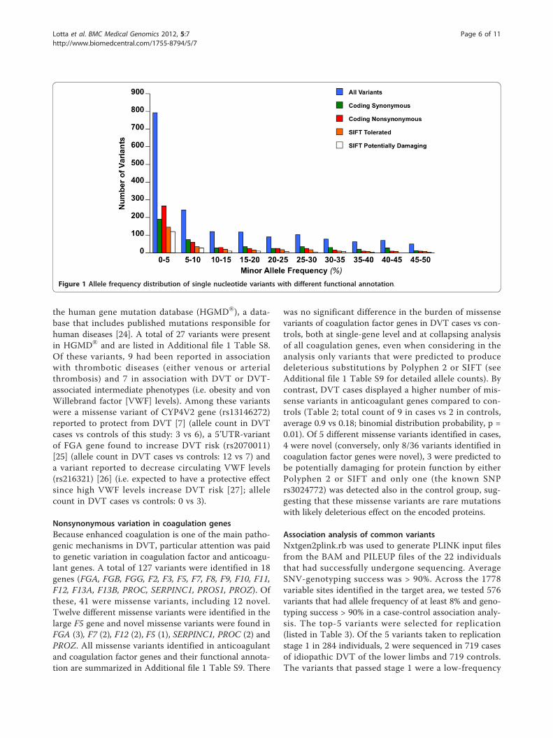
and p.G3170X in COL6A3; p.R412X in KNG1; p.R88Xin SERPINA10 and p.R615X in ZNF544). Ninety-eightindels were called. The most frequent indel was anintronic, single-nucleotide deletion in PLCG2 (chromo-some 16, coordinate 80461934). Figure 1 shows the fre-quency distribution of identified variants. Consistentwith previous observations [11,18], the frequency inter-val between one observation and 5% yielded the highestnumber of variants, the highest nonsynonymous/synon-ymous ratio and the highest SIFT-predicted-damaging/SIFT-predicted-benign ratio. To verify the validity ofvariant calls and QC filters we (a) repeated the sequen-cing of one of the samples (DVT_P_01) (b) sequencedby PCR and Sanger all 22 samples at 35 sites where aSNV was found (c) verified by Pyrosequencing 19
genotypes at sites with variants that did not pass QC.Of 425 SNVs identified after QC in DVT_P_01 at thesecond sequencing experiment, 405 (95.3%) had beenidentified also in the first sequencing run of the samesample, showing the high reproducibility of next-genera-tion sequencing. Of 765 genotypes available from bothSOLiD and Sanger sequencing, 99.8% (697/698) ofhomozygous wild-type genotypes and 97% (65/67) ofeither heterozygous or homozygous variant genotypeswere in agreement. None of the variants that did notpass QC was validated by Pyrosequencing.
Disease-associated variants in the target genesIn order to search for the presence of disease-associatedgenetic variants, all identified SNVs were annotated on
Table 1 Characteristics of the individuals included in the replication stages of the study.
PATIENTS WITH IDIOPATHIC DVT CONTROLS
n= 719 719
GEOGRAPHIC ORIGIN, n (%)
Lombardy 453 (63) 490 (67)
Northern Italy other than Lombardy 89 (13) 84 (12)
Central Italy 30 (4) 20 (3)
Southern Italy and islands 109 (15) 107 (15)
Not available 38 (5) 18 (3)
MALE GENDER, n (%) 325 (45) 325 (45)
AGE, mean years (standard deviation) 42.5 (15) 42.5 (14)
PULMONARY EMBOLISM, n (%) 148 (20) /
REFERRED FOR MORE THAN ONE EPISODE, n (%) 167 (23) /
BODY MASS INDEX, kg/m2 25.9 (4.7) 24 (4)
FACTOR V LEIDEN (rs6025) GENOTYPE, n (%)
GG 611 (85) 696 (97)
AG 102 (14) 23 (3)
AA 6 (1) 0
PROTHROMBIN G20210A (rs1799963) GENOTYPE, n (%)
GG 654 (91) 695 (97)
AG 63 (8) 24 (3)
AA 2 (1) 0
NATURAL ANTICOAGULANT DEFICIENCIES, n (%)
Antithrombin 22 (3) 2 (0.2)
Protein C 26 (3) 2 (0.2)
Protein S 30 (4) 10 (1)
LABORATORY MEASUREMENTS, mean (standard deviation)
Antithrombin,% 97 (12) 99 (15)
Protein C, % 102 (27) 105 (22)
Protein S, % 105 (30) 111 (31)
Prothrombin time, international normalized ratio 1 (0.16) 0.99 (0.15)
Activated partial thromboplastin time, ratio 1 (0.1) 1 (0.1)
Coagulation factor VIII coagulant activity, % 142 (39) 114 (30)
Fibrinogen coagulant activity, % 304 (72) 291 (63)
Lotta et al. BMC Medical Genomics 2012, 5:7http://www.biomedcentral.com/1755-8794/5/7
Page 5 of 11
the human gene mutation database (HGMD®), a data-base that includes published mutations responsible forhuman diseases [24]. A total of 27 variants were presentin HGMD® and are listed in Additional file 1 Table S8.Of these variants, 9 had been reported in associationwith thrombotic diseases (either venous or arterialthrombosis) and 7 in association with DVT or DVT-associated intermediate phenotypes (i.e. obesity and vonWillebrand factor [VWF] levels). Among these variantswere a missense variant of CYP4V2 gene (rs13146272)reported to protect from DVT [7] (allele count in DVTcases vs controls of this study: 3 vs 6), a 5’UTR-variantof FGA gene found to increase DVT risk (rs2070011)[25] (allele count in DVT cases vs controls: 12 vs 7) anda variant reported to decrease circulating VWF levels(rs216321) [26] (i.e. expected to have a protective effectsince high VWF levels increase DVT risk [27]; allelecount in DVT cases vs controls: 0 vs 3).
Nonsynonymous variation in coagulation genesBecause enhanced coagulation is one of the main patho-genic mechanisms in DVT, particular attention was paidto genetic variation in coagulation factor and anticoagu-lant genes. A total of 127 variants were identified in 18genes (FGA, FGB, FGG, F2, F3, F5, F7, F8, F9, F10, F11,F12, F13A, F13B, PROC, SERPINC1, PROS1, PROZ). Ofthese, 41 were missense variants, including 12 novel.Twelve different missense variants were identified in thelarge F5 gene and novel missense variants were found inFGA (3), F7 (2), F12 (2), F5 (1), SERPINC1, PROC (2) andPROZ. All missense variants identified in anticoagulantand coagulation factor genes and their functional annota-tion are summarized in Additional file 1 Table S9. There
was no significant difference in the burden of missensevariants of coagulation factor genes in DVT cases vs con-trols, both at single-gene level and at collapsing analysisof all coagulation genes, even when considering in theanalysis only variants that were predicted to producedeleterious substitutions by Polyphen 2 or SIFT (seeAdditional file 1 Table S9 for detailed allele counts). Bycontrast, DVT cases displayed a higher number of mis-sense variants in anticoagulant genes compared to con-trols (Table 2; total count of 9 in cases vs 2 in controls,average 0.9 vs 0.18; binomial distribution probability, p =0.01). Of 5 different missense variants identified in cases,4 were novel (conversely, only 8/36 variants identified incoagulation factor genes were novel), 3 were predicted tobe potentially damaging for protein function by eitherPolyphen 2 or SIFT and only one (the known SNPrs3024772) was detected also in the control group, sug-gesting that these missense variants are rare mutationswith likely deleterious effect on the encoded proteins.
Association analysis of common variantsNxtgen2plink.rb was used to generate PLINK input filesfrom the BAM and PILEUP files of the 22 individualsthat had successfully undergone sequencing. AverageSNV-genotyping success was > 90%. Across the 1778variable sites identified in the target area, we tested 576variants that had allele frequency of at least 8% and geno-typing success > 90% in a case-control association analy-sis. The top-5 variants were selected for replication(listed in Table 3). Of the 5 variants taken to replicationstage 1 in 284 individuals, 2 were sequenced in 719 casesof idiopathic DVT of the lower limbs and 719 controls.The variants that passed stage 1 were a low-frequency
Figure 1 Allele frequency distribution of single nucleotide variants with different functional annotation.
Lotta et al. BMC Medical Genomics 2012, 5:7http://www.biomedcentral.com/1755-8794/5/7
Page 6 of 11

missense polymorphism of PLCG2 (rs11548656, p.H244R) and a common polymorphism of FGA (rs6050,p.T331A). While PLCG2 p.H244R had no associationafter sequencing in > 1400 individuals, FGA p.T331Ashowed a statistically significant association with DVT ina crude association analysis (p = 1.9 × 10⁻⁵, OR 1.45; 95%CI, 1.22-1.72) and after adjustment for age and gender(p = 0.0081, OR 1.39; 95% CI, 1.10-1.74). PCR and Sangersequencing also identified 7 missense variants in additionto those targeted at these two loci (6 at FGA and 1 atPLCG2). There was no association between these variantsand DVT at both single-variant and collapsing case-con-trol association analysis. Variant features are summarizedin Additional file 1 Table S10. The association of rs6050with DVT was further tested in multivariable logisticregression models adjusting for several covariates. Theassociation between rs6050 and DVT remained statisti-cally significant after adjustment for age, gender and plas-matic levels of fibrinogen (p = 0.0001, OR 1.39; 95% CI,1.10-1.74); and for age, gender, fibrinogen levels, bodymass index, FVL and prothrombin G20210A (p < 0.0001,OR 1.41; 95% CI, 1.10-1.81). We also tested the interac-tion (i.e. deviation of the cumulative effect from an addi-tive model) between rs6050 and FVL and between rs6050and prothrombin G20210A. Both tests were negative(p = 0.44 for interaction with FVL and p = 0.89 for inter-action with prothrombin G20210A).
DiscussionThis pilot study is one of the first applications of next-gen-eration sequencing in DVT. Sequencing by oligonucleotideligation and detection was used to resequence the protein-coding areas of 186 hemostatic/pro-inflammatory genes incases and controls of DVT. This regional sequencingapproach enabled the simultaneous analysis of several doz-ens of genes in many samples at a fraction of the cost andcomputation required for whole-exome and whole-gen-ome analysis. At the current level of multiplexing (i.e. upto 64 samples per SOLiD slide, 16 per spot), the analysis
of our target area in one sample costs one tenth of a high-coverage exome-sequencing and one hundredth of a high-coverage whole-genome sequencing, with similar propor-tions in the saving of computational time (3 hours for readmapping vs the few days of whole-exome or the more than1 week of whole-genome datasets) and information sto-rage-capacity requirements (300 MB per BAM file vs 40GB per BAM for whole-exome or 400 GB for whole-gen-ome sequencing). For these reasons, regional sequencingis ideal to interrogate relatively small genomic areasdeemed of particular functional relevance in a disease.Potential applications of this approach are the sequencingof positional candidate genes or genomic loci identified bygenome-wide linkage or association analysis [28], thelarge-scale replication of the initial findings of exome andwhole-genome resequencing studies and the rapid screen-ing of disease-genes in those Mendelian diseases that haveseveral different causal genes [29].In the field of DVT, the importance of being able to ana-
lyze all known hemostatic genes (i.e. the ‘hemostateome’)has been recently highlighted by Fechtel et al. [30]. Thesimultaneous sequencing of all hemostatic genes inaffected individuals is ideal to study specific combinationsof variants in the hemostatic pathway acting in synergy toconfer DVT-predisposition. This type of approach has thepotential to reveal new disease-predisposing mechanisms,besides the identification of isolated variants that show sta-tistical association with the disease. In this study, the useof molecular barcoding allowed multiplexing without lossof individual sequence information, which is required tofully exploit the potential of sequence data. Sequencing ofthe 186 genes in 22 individuals yielded more than 1700genetic variants of different functional type and frequency.Annotation of identified variants revealed several disease-associated variants, a proportion of which had alreadybeen reported in association with DVT. Many novel var-iants with potentially deleterious effect on the function ofkey hemostatic proteins were also found. These results areconsistent with the recent report by Dewey et al. of the
Table 2 Nonsynonymous single nucleotide variants in anticoagulant genes.
Gene Chromosome Coordinate Substitution TranscriptID
Proteinchange
dbSNP129 1000 GenomesCEU
population,allele
frequency
SIFTa Polyphen2b
Allelescases
Allelescontrols
PROC chr2 127895370 C > T NM_000312 p.R38W Novel not present Dam Prd 2 0
127902716 C > A p.H370Q Novel not present Ben Ben 1 0
SERPINC1 chr1 172150549 G > A NM_000488 p.P58L Novel not present Dam Pod 1 0
PROZ chr13 112861006 C > G NM_003891 p.L11V Novel not present Ben Ben 3 0
112874101 G > A p.R295H rs3024772 not present Ben Prd 2 2
a SIFT-based annotation results, Dam indicates that the mutation is predicted to affect protein function (i.e. ‘damaging’), Ben indicates that the mutation ispredicted to be tolerated (i.e.’benign’).
b Polyphen 2-based annotation results. Prd indicates that the mutation is predicted to be ‘probably damaging’, Pod indicates that the mutation is predicted tobe ‘possibly damaging’, Ben indicates that the mutation is predicted to be ‘benign’.
Lotta et al. BMC Medical Genomics 2012, 5:7http://www.biomedcentral.com/1755-8794/5/7
Page 7 of 11

Table 3 Common variant association results.
Variant information Discovery Replication stage 1 and 2 (combined)
Location Substitution Gene Functionalannotation
Protein dbSNP129 Allelescases
Allelescontrols
p = Effective samplesize cases
MAFcases, %
(n)
Effective samplesize controls
MAFcontrols, %
(n)
p= OR 95%CI
chr4:155727040 T > C FGA exon -missense
p.T331A
rs6050 10 4 0.004 709 32 (453) 702 22 (312) 1.9 ×10⁻⁵
1.45 1.22-1.72
chr16:80474413 A > G PLCG2 exon -missense
p.H244R
rs11548656 4 0 0.013 711 6 (88) 705 6 (83) 0.73 1.05 0.77-1.43
chr4:122837138 T > C ANXA5 Intron Na rs2306416 4 0 0.013 139 15 (41) 138 15 (42) 0.97 0.96 0.60-1.54
chr8:42164111 G > A PLAT exon -synonymous
Na rs1058720 11 3 0.002 139 47 (131) 139 44 (124) 0.61 1.10 0.79-1.54
chr11:47311481 T > C MYBPC3 Intron Na rs11570115 5 0 0.004 137 11 (30) 139 9 (26) 0.63 1.19 0.68-2.07
MAF indicates minor allele frequency; OR, odds ratio; CI, confidence interval; Na, not applicable (the variant does not cause protein sequence changes).
Lottaet
al.BMCMedicalG
enomics
2012,5:7http://w
ww.biom
edcentral.com/1755-8794/5/7
Page8of
11

genome sequence of a family quartet in which the fatherhad a history of DVT and pulmonary embolism [31]. Inthe study, the authors identified four different novel non-synonymous variants in DVT-risk genes and other knownthrombophilia associated variants.In our study, we adopted different analytical
approaches to reveal potential associations with the dis-ease at both single-variant and gene/pathway levels.Although the number of analyzed individuals was verysmall, it was possible to find statistically significant andbiologically plausible associations with DVT. Anincreased burden of rare missense mutations in anticoa-gulant genes was found in DVT cases compared to con-trols. Single-variant association analysis followed byreplication genotyping in > 1400 individuals identifiedan association for the rs6050 SNP in FGA. The excessof rare missense mutations in anticoagulant genes inpatients in whom the deficiencies of natural anticoagu-lants had been excluded with biochemical assays sug-gests that a fraction of idiopathic DVT cases might beaffected by ‘unrecognized’ anticoagulant deficiencies,caused by mutations that impart functional effects towhich currently used biochemical assays are not sensi-tive. The association of rs6050, already described by pre-vious candidate SNP studies, was here identified by anagnostic screening of several dozen genes. The associa-tion of rs6050 was reported in 4 studies focusing onvenous thromboembolism (VTE), therefore includingcases of pulmonary embolism without a diagnosis ofDVT [25,32-34]. Two of these studies were very small,with total sample size (cases and controls) of less than500 individuals [25,32]. Ours is the second largest of allthe studies on rs6050 both in terms of DVT cases inves-tigated and overall statistical power. Thus, along withprevious reports, this study makes of rs6050 one of themost widely replicated variants in DVT. The mechan-isms for the association of rs6050 with DVT/VTE arenot fully understood. FGA rs6050 was reported to resultin enhanced coagulation factor XIII-mediated cross-link-ing of fibrin alpha-chain [35] and to be associated withincreased FGA transcription [32]. In this report wefound no association of FGA rs6050 with plasmaticfibrinogen activity, but the fact that rs6050 is a missenseSNP and that the amino acid substitution is consideredas ‘possibly damaging’ by Polyphen 2 indeed suggestthat the risk for DVT is conferred by an alteration offibrinogen-alpha chain function.
LimitationsThe approach proposed in this study has limitations.The use of DNA target capture may introduce bias inthe detection of genetic variants by allele-selective cap-ture. Both the use of target capture and the short-readlength of next-generation sequencing platforms
constrain comprehensive analysis of copy number varia-tion (CNV), an important source of genetic variability.However, these limitations can be minimized by increas-ing sequence coverage and by using complementarygenetic analyses (e.g. array-based CNV-ascertainment).Restricting the analysis to a few hundred candidategenes may limit the chance to detect novel associations.On the other hand, the efficiency and cost effectivenessof regional sequencing are ideal for the thorough, large-scale analysis of genetic variation in specific biologicalpathways such as hemostasis. This approach may pro-vide deep understanding of the mechanisms by whichmultiple variants in the same biological pathway shapeindividual predisposition to complex phenotypes.
ConclusionsUsing a molecular-barcode based technique for samplemultiplexing on next-generation DNA sequencing plat-forms we sequenced the coding areas of 186 hemo-static/proinflammatory genes in cases and controls ofDVT. We were able to detect known disease-associatedvariants as well as novel potentially-deleterious variantsin disease-associated genes. Our results illustrate thepotential of next-generation gene sequencing for the dis-covery of genetic variation predisposing to common dis-eases and for the study of inherited thrombophilia.
Additional material
Additional file 1: Supplementary Material. Supplementary Tables andFigures.
Additional file 2: target and tiled regions. target and tiled genomiccoordinate-intervals.
AcknowledgementsThe authors would like to thank Dr. Luigi F Ghilardini for the help in draftingfigures and tables, all the former and current members of HGSC MedSeqand Library laboratories for sample analysis, Kyle Chang, Jennifer Drummondand Nipun Kakkar for help in genetic variant calling and annotation, Yu-YeWen for help in study design. FP is recipient of Bayer Hemophilia SpecialProject Award 2011. LAL is recipient of the Bayer Hemophilia ClinicalTraining Award 2009 and of the Associazione Italiana Centri Emofilia (AICE) -De Biasi Prize. Financial support from the Italian Ministry of Health (Grant n.RF-2009-1530493) is gratefully acknowledged. This work has been supportedby Fondazione Cariplo (Grant n. 2011-0524).
Author details1Angelo Bianchi Bonomi Hemophilia and Thrombosis Center, U.O.S.Dipartimentale per la Diagnosi e la Terapia delle Coagulopatie, FondazioneIRCCS Cà Granda - Ospedale Maggiore Policlinico, Università degli Studi diMilano and Luigi Villa Foundation, Milan, Italy. 2Human Genome SequencingCenter, Baylor College of Medicine, Houston, TX, USA. 3Unit of Epidemiology,Fondazione IRCCS Cà Granda - Ospedale Maggiore Policlinico, Universitàdegli Studi di Milano, Milan, Italy.
Authors’ contributionsLAL, RAG and FP designed the research. LAL, YW, DMM and MW designedthe experiments. IM, SMP, EP, MM, FP and PMM gathered samples andclinical information. LAL, DC, MNB, JY and FY analyzed the data. LAL, JY and
Lotta et al. BMC Medical Genomics 2012, 5:7http://www.biomedcentral.com/1755-8794/5/7
Page 9 of 11

FY wrote study-specific software. LAL, EP, MM, SES, LLL, HA conductedexperiments and handled samples. LAL wrote the bulk of the manuscript. Allauthors read and approved the final version of the manuscript.
Competing interestsThe authors declare that they have no competing interests.
Received: 11 October 2011 Accepted: 21 February 2012Published: 21 February 2012
References1. Tapson VF: Acute pulmonary embolism. N Engl J Med 2008, 358:1037-52.2. Sørensen HT, Riis AH, Diaz LJ, Andersen EW, Baron JA, Andersen PK:
Familial risk of venous thromboembolism: a nationwide cohort study. JThromb Haemost 2011, 9:320-4.
3. Souto JC, Almasy L, Borrell M, Blanco-Vaca F, Mateo J, Soria JM, Coll I,Felices R, Stone W, Fontcuberta J, Blangero J: Genetic susceptibility tothrombosis and its relationship to physiological risk factors: the GAITstudy. Genetic Analysis of Idiopathic Thrombophilia. Am J Hum Genet 2000,67:1452-9.
4. Heit JA, Phelps MA, Ward SA, Slusser JP, Petterson TM, De Andrade M:Familial segregation of venous thromboembolism. J Thromb Haemost2004, 2:731-6.
5. Larsen TB, Sørensen HT, Skytthe A, Johnsen SP, Vaupel JW, Christensen K:Major genetic susceptibility for venous thromboembolism in men: astudy of Danish twins. Epidemiology 2003, 14:328-32.
6. Dahlbäck B: Advances in understanding pathogenic mechanisms ofthrombophilic disorders. Blood 2008, 112:19-27.
7. Bezemer ID, Bare LA, Doggen CJ, Arellano AR, Tong C, Rowland CM,Catanese J, Young BA, Reitsma PH, Devlin JJ, Rosendaal FR: Gene variantsassociated with deep vein thrombosis. JAMA 2008, 299:1306-14.
8. Trégouët DA, Heath S, Saut N, Biron-Andreani C, Schved JF, Pernod G,Galan P, Drouet L, Zelenika D, Juhan-Vague I, Alessi MC, Tiret L, Lathrop M,Emmerich J, Morange PE: Common susceptibility alleles are unlikely tocontribute as strongly as the FV and ABO loci to VTE risk: results from aGWAS approach. Blood 2009, 113:5298-303.
9. Morange PE, Bezemer I, Saut N, Bare L, Burgos G, Brocheton J, Durand H,Biron-Andreani C, Schved JF, Pernod G, Galan P, Drouet L, Zelenika D,Germain M, Nicaud V, Heath S, Ninio E, Delluc A, Münzel T, Zeller T, Brand-Herrmann SM, Alessi MC, Tiret L, Lathrop M, Cambien F, Blankenberg S,Emmerich J, Trégouët DA, Rosendaal FR: A follow-up study of a genome-wide association scan identifies a susceptibility locus for venousthrombosis on chromosome 6p24.1. Am J Hum Genet 2010, 86:592-5.
10. Morange PE, Tregouet DA: Deciphering the molecular basis of venousthromboembolism: where are we and where should we go? Br JHaematol 2010, 148:495-506.
11. Ng SB, Turner EH, Robertson PD, Flygare SD, Bigham AW, Lee C, Shaffer T,Wong M, Bhattacharjee A, Eichler EE, Bamshad M, Nickerson DA,Shendure J: Targeted capture and massively parallel sequencing of 12human exomes. Nature 2009, 461:272-6.
12. Ng SB, Buckingham KJ, Lee C, Bigham AW, Tabor HK, Dent KM, Huff CD,Shannon PT, Jabs EW, Nickerson DA, Shendure J, Bamshad MJ: Exomesequencing identifies the cause of a mendelian disorder. Nat Genet 2010,42:30-5.
13. Lupski JR, Reid JG, Gonzaga-Jauregui C, Rio Deiros D, Chen DC, Nazareth L,Bainbridge M, Dinh H, Jing C, Wheeler DA, McGuire AL, Zhang F,Stankiewicz P, Halperin JJ, Yang C, Gehman C, Guo D, Irikat RK, Tom W,Fantin NJ, Muzny DM, Gibbs RA: Whole-genome sequencing in a patientwith Charcot-Marie-Tooth neuropathy. N Engl J Med 2010, 362:1181-91.
14. Frezzato M, Tosetto A, Rodeghiero F: Validated questionnaire for theidentification of previous personal or familial venous thromboembolism.Am J Epidemiol 1996, 143:1257-65.
15. [http://www.appliedbiosystems.com/absite/us/en/home/applications-technologies/solid-next-generation-sequencing/next-generation-systems/solid-sequencing-chemistry.html].
16. Homer N, Merriman B, Nelson SF: BFAST: An alignment tool for largescale genome resequencing. PLoS ONE 2009, 4:e7767.
17. Li H, Handsaker B, Wysoker A, Fennell T, Ruan J, Homer N, Marth G,Abecasis G, Durbin R, 1000 Genome Project Data Processing Subgroup: TheSequence alignment/map (SAM) format and SAMtools. Bioinformatics2009, 25:2078-9.
18. 1000 Genomes Project Consortium, Durbin RM, Abecasis GR, Altshuler DL,Auton A, Brooks LD, Durbin RM, Gibbs RA, Hurles ME, McVean GA: A mapof human genome variation from population-scale sequencing. Nature2010, 467:1061-73.
19. Wang K, Li M, Hakonarson H: ANNOVAR: Functional annotation of geneticvariants from next-generation sequencing data. Nucleic Acids Research2010, 38:e164.
20. Ng PC, Henikoff S: Accounting for human polymorphisms predicted toaffect protein function. Genome Res 2002, 12:436-46.
21. Adzhubei IA, Schmidt S, Peshkin L, Ramensky VE, Gerasimova A, Bork P,Kondrashov AS, Sunyaev SR: A method and server for predictingdamaging missense mutations. Nat Methods 2010, 7:248-249.
22. Purcell S, Neale B, Todd-Brown K, Thomas L, Ferreira MA, Bender D, Maller J,Sklar P, de Bakker PI, Daly MJ, Sham PC: PLINK: a tool set for whole-genome association and population-based linkage analyses. Am J HumGenet 2007, 81:559-75.
23. Zhang J, Wheeler DA, Yakub I, Wei S, Sood R, Rowe W, Liu PP, Gibbs RA,Buetow KH: SNPdetector: a software tool for sensitive and accurate SNPdetection. PLoS Comput Biol 2005, 1:e53.
24. Stenson PD, Mort M, Ball EV, Howells K, Phillips AD, Thomas NS, Cooper DN:The Human Gene Mutation Database: 2008 update. Genome Med 2009,1:13.
25. Ko YL, Hsu LA, Hsu TS, Tsai CT, Teng MS, Wu S, Chang CJ, Lee YS:Functional polymorphisms of FGA, encoding alpha fibrinogen, areassociated with susceptibility to venous thromboembolism in aTaiwanese population. Hum Genet 2006, 119:84-91.
26. Vaidya D, Yanek LR, Herrera-Galeano JE, Mathias RA, Moy TF, Faraday N,Becker LC, Becker DM: A common variant in the Von Willebrand factorgene is associated with multiple functional consequences. Am J Hematol2010, 85:971-3.
27. Smith NL, Rice KM, Bovill EG, Cushman M, Bis JC, McKnight B, Lumley T,Glazer NL, van Hylckama Vlieg A, Tang W, Dehghan A, Strachan DP,O’Donnell CJ, Rotter JI, Heckbert SR, Psaty BM, Rosendaal FR: Geneticvariation associated with plasma von Willebrand factor levels and therisk of incident venous thrombosis. Blood 2011, 117:6007-11.
28. Momozawa Y, Mni M, Nakamura K, Coppieters W, Almer S, Amininejad L,Cleynen I, Colombel JF, de Rijk P, Dewit O, Finkel Y, Gassull MA,Goossens D, Laukens D, Lémann M, Libioulle C, O’Morain C, Reenaers C,Rutgeerts P, Tysk C, Zelenika D, Lathrop M, Del-Favero J, Hugot JP, deVos M, Franchimont D, Vermeire S, Louis E, Georges M: Resequencing ofpositional candidates identifies low frequency IL23R coding variantsprotecting against inflammatory bowel disease. Nat Genet 2011, 43:43-7.
29. Shearer AE, DeLuca AP, Hildebrand MS, Taylor KR, Gurrola J, Scherer S,Scheetz TE, Smith RJ: Comprehensive genetic testing for hereditaryhearing loss using massively parallel sequencing. Proc Natl Acad Sci USA2010, 107:21104-9.
30. Fechtel K, Osterbur ML, Kehrer-Sawatzki H, Stenson PD, Cooper DN:Delineating the Hemostaseome as an aid to individualize the analysis ofthe hereditary basis of thrombotic and bleeding disorders. Hum Genet2011, 130:149-66.
31. Dewey FE, Chen R, Cordero SP, Ormond KE, Caleshu C, Karczewski KJ, Whirl-Carrillo M, Wheeler MT, Dudley JT, Byrnes JK, Cornejo OE, Knowles JW,Woon M, Sangkuhl K, Gong L, Thorn CF, Hebert JM, Capriotti E, David SP,Pavlovic A, West A, Thakuria JV, Ball MP, Zaranek AW, Rehm HL, Church GM,West JS, Bustamante CD, Snyder M, Altman RB, Klein TE, Butte AJ,Ashley EA: Phased whole-genome genetic risk in a family quartet using amajor allele reference sequence. PLoS Genet 2011, 7:e1002280.
32. Carter AM, Catto AJ, Kohler HP, Ariëns RA, Stickland MH, Grant PJ: alpha-fibrinogen Thr312Ala polymorphism and venous thromboembolism.Blood 2000, 96(3):1177-9, Aug 1.
33. Rasmussen-Torvik LJ, Cushman M, Tsai MY, Zhang Y, Heckbert SR,Rosamond WD, Folsom AR: The association of alpha-fibrinogen Thr312Alapolymorphism and venous thromboembolism in the LITE study. ThrombRes 2007, 121:1-7.
34. Arellano AR, Bezemer ID, Tong CH, Catanese JJ, Devlin JJ, Reitsma PH,Bare LA, Rosendaal FR: Gene variants associated with venous thrombosis:confirmation in the MEGA study. J Thromb Haemost 2010, 8:1132-4.
35. Standeven KF, Grant PJ, Carter AM, Scheiner T, Weisel JW, Ariëns RA:Functional analysis of the fibrinogen alpha Thr312Ala polymorphism:effects on fibrin structure and function. Circulation 2003, 107:2326-30.
Lotta et al. BMC Medical Genomics 2012, 5:7http://www.biomedcentral.com/1755-8794/5/7
Page 10 of 11

Pre-publication historyThe pre-publication history for this paper can be accessed here:http://www.biomedcentral.com/1755-8794/5/7/prepub
doi:10.1186/1755-8794-5-7Cite this article as: Lotta et al.: Identification of genetic risk variants fordeep vein thrombosis by multiplexed next-generation sequencing of186 hemostatic/pro-inflammatory genes. BMC Medical Genomics 2012 5:7.
Submit your next manuscript to BioMed Centraland take full advantage of:
• Convenient online submission
• Thorough peer review
• No space constraints or color figure charges
• Immediate publication on acceptance
• Inclusion in PubMed, CAS, Scopus and Google Scholar
• Research which is freely available for redistribution
Submit your manuscript at www.biomedcentral.com/submit
Lotta et al. BMC Medical Genomics 2012, 5:7http://www.biomedcentral.com/1755-8794/5/7
Page 11 of 11