identificationofwheatinflorescencedevelopment-relatedgenes
TRANSCRIPT

Research ArticleIdentification ofWheat Inflorescence Development-Related GenesUsing a Comparative Transcriptomics Approach
Lingjie Ma, Sheng-Wei Ma, Qingyan Deng, Yang Yuan, Zhaoyan Wei, Haiyan Jia ,and Zhengqiang Ma
The Applied Plant Genomics Laboratory of Crop Genomics and Bioinformatics Centre, Nanjing Agricultural University,Jiangsu 210095, China
Correspondence should be addressed to Haiyan Jia; [email protected]
Received 3 August 2017; Revised 26 November 2017; Accepted 3 December 2017; Published 8 February 2018
Academic Editor: Marco Gerdol
Copyright © 2018 Lingjie Ma et al. This is an open access article distributed under the Creative Commons Attribution License,which permits unrestricted use, distribution, and reproduction in any medium, provided the original work is properly cited.
Inflorescence represents the highly specialized plant tissue producing the grains. Although key genes regulating flower initiationand development are conserved, the mechanism regulating fertility is still not well explained. To identify genes and genenetwork underlying inflorescence morphology and fertility of bread wheat, expressed sequence tags (ESTs) from different tissueswere analyzed using a comparative transcriptomics approach. Based on statistical comparison of EST frequencies of individualgenes in EST pools representing different tissues and verification with RT-PCR and RNA-seq data, 170 genes of 59 gene setspredominantly expressed in the inflorescence were obtained. Nearly one-third of the gene sets displayed differentiatedexpression profiles in terms of their subgenome orthologs. The identified genes, most of which were predominantly expressed inanthers, encode proteins involved in wheat floral identity determination, anther and pollen development, pollen-pistilinteraction, and others. Particularly, 25 annotated gene sets are associated with pollen wall formation, of which 18 encodeenzymes or proteins participating in lipid metabolic pathway, including fatty acid ω-hydroxylation, alkane and fatty alcoholbiosynthesis, and glycerophospholipid metabolism. We showed that the comparative transcriptomics approach was effective inidentifying genes for reproductive development and found that lipid metabolism was particularly active in wheat anthers.
1. Introduction
Inflorescences are the reproductive architectures of plants,composed of stems, stalks, bracts, and flowers. Poaceae (alsocalled Gramineae) is one of the largest families in the mono-cotyledonous flowering plants, including the major cerealcrops, such as maize (Zea mays L.), rice (Oryza sativa L.),and wheat (Triticum aestivum L.). Inflorescences of this fam-ily are characterized by their panicle or spike shapes [1],complex branches, and unique spikelets, as well as inconspic-uous and anemophilous flowers without obvious petals andsepals [2, 3]. Species in the genus Triticum takes the shapeof the spike with the spikelets, each containing one or moreflorets, attached to rachis nodes. The wheat floret consistsof a carpel with its ovary, style, and stigma, with three anthersattached to the base through slender filaments, which areenclosed by bract-like organs called lemma and palea.
Inflorescence development and regulation have attractedgreat attention from plant biologists and crop breeders sincethey are crucial for reproduction of flowering plants and forproduction of food grains in cereal crops. Although thekey genes regulating the flower initiation and developmentare conserved in higher plants [4, 5], the diversity of repro-ductive structure and behaviors is still not well explained.Transcriptomes reflect the complete set of RNA transcriptsexpressed by the genome under developmentally or physi-ologically distinct states; therefore, its comparison allowsidentification of genes under common regulation. High-throughput methods, such as serial analysis of geneexpression (SAGE) [6], microarray technology [7], andnext-generation sequencing [8] have enabled transcriptomestudies in an unprecedented scale in many plant species,especially in those with full genome sequence informationavailable. This has led to the discovery of a number of
HindawiInternational Journal of GenomicsVolume 2018, Article ID 6897032, 13 pageshttps://doi.org/10.1155/2018/6897032

genes involved in flower development. These genes displaytempo-spatial expression patterns not only in transcriptomelevel [9–12] but also at the proteome level, such as those inpollen development of tomatoes [13], indicating their strictlyregulated functions. Anther tissues were used in most ofthese studies because they are easy to isolate and specific inbiological roles. The availability of these data has shedsome light on the gene networks that contribute to theformation of unique reproductive structures and flowerdevelopment, although it might still not be enoughbecause of the particularity of different plant species andhighly specific flower components.
In bread wheat (T. aestivum L.), an allohexaploid spe-cies (2n=42) with three closely related subgenomes, thatis, A, B, and D [14], large-scale transcriptomic investiga-tions have been conducted in some tissue types. Crismaniet al. performed microarray-based expression analysis ofanthers across various stages of meiosis in an attempt tobuild the link between the model and wheat and identifyearly meiotic genes [15]. McIntosh et al. investigated thetranscriptome in wheat caryopsis development using datagenerated by SAGE [16]. Yang et al. compared the RNA-seq tags of pistillody stamen and pistil from a pistillodywheat mutant and stamen from the wild-type control toidentify differentially expressed genes [17]. In these stud-ies, the annotation of the identified transcripts was basedon short tags or sequence reads which is impossible toensure accuracy for species such as wheat that has a com-plex genome and does not have detailed sequence infor-mation so far. Moreover, gene identity determinationaccording to short sequence fragments could be problem-atic due to functional diversification of the homoeologousgenes in polyploidy species.
Expressed sequence tags (ESTs) are single-pass sequencereads by sequencing cDNA libraries and usually have >400-base read length. They represent part of the transcriptomein a given tissue and/or at a given developmental stage. Asof January 1, 2013, over one million ESTs are available forwheat in GenBank of the National Center for BiotechnologyInformation (NCBI) and are valuable for identifying genesinvolved in biotic and abiotic stress [18–22], kernel develop-ment and quality [23, 24], and development [21, 25, 26].
In this paper, we reported mining of 170 genespredominantly expressed in floral organs through com-parative transcriptomic profiling using ESTs of 67 wheatcDNA libraries deposited in GenBank genes and theidentification of a few metabolic pathways involved inanther development.
2. Materials and Methods
2.1. Wheat ESTs and Contigs. ESTs were downloaded fromthe NCBI dbEST database (ftp://ncbi.nlm.nih.gov/repository/dbEST). The ESTs used in this study were producedwith 67 cDNA libraries prepared using inflorescences (spikeat flowering or before flowering, anther, pistil, ovary, palea,and lemma), roots, stems, leaves (including seedlings andcrown tissues), and seeds (matured or immature embryos)from normally grown seedlings or plants and represented
434,658 cloning events (Table S1). Libraries subjected toenrichment or normalization treatment and those with lessthan 1000 ESTs were not included in the expression profilinganalysis. The 163rd release of unique wheat transcripts,including 77,657 contigs, was downloaded from the PUTdatabase (http://www.plantgdb.org).
2.2. Gene Mining. To identify putative genes predominantlyexpressed in wheat flowers, BLAST search was conductedusing the PUT contig sequences as queries against the ESTdatabase consisting of the 67 cDNA libraries. ESTs matchingeach PUT contig with ≥90% homology were recorded andclassified according to their library origins from the fiveaforementioned tissue types. The contigs with matched ESTssolely from inflorescences and those with significantly morematched ESTs from inflorescence tissues than from any ofthe other four types of tissues were considered to be the puta-tive genes predominantly expressed in inflorescences. Theprobability to achieve the ESTs matching to a contig in thenoninflorescence libraries was estimated based on the ran-dom sampling principle using the equation: P = Cn
N fn
1 − f N−n, as described by Ding et al. [27], where f is theEST frequency of a contig in the inflorescence libraries esti-mated by dividing the number of matched ESTs by the totalnumber of ESTs in the libraries and n and N are the numberof matched ESTs and the total number of ESTs in libraries ofother tissues types, respectively. A sampling probabil-ity≤ 0.0001 was considered an indication of significant differ-ence in expression levels between the inflorescence tissuesand noninflorescence tissues.
Chromosome-specific genomic DNA sequences corre-sponding to the retained contigs were obtained by retrievingthe chromosome-assigned homologous scaffolds in theTGAC database [28] (http://www.tgac.ac.uk/grassroots-genomics) that showed 95% homology to the contigsequences. Coding DNA sequences (CDS) corresponding tothe contigs in the scaffolds were predicted using the gene-finding program FGENESH [29] (http://linux1.softberry.com) and verified by alignment with the homologous ESTs.Scaffolds that did not contain the full-length target CDS wereextended via in silico walking, using a parameter of 100%match, with the Roche 454 sequence reads of Chinese Spring[30] (http://www.cerealsdb.uk.net).
To confirm the expression predominance of the identi-fied genes in wheat flowers, ESTs with ≥99% homology tothe individual CDS identified from the sixty-seven librarieswere subjected to further analysis. The sampling probabilityto achieve the EST frequency of a CDS in the noninflores-cence libraries was again calculated. A sampling probabil-ity≤ 0.01 was used as the threshold for declaration ofsignificant difference. Expression specificity of the candidategenes was estimated similarly.
The identified genes were coded in numerical order withthe prefix “IDG” (inflorescence development-related gene).Orthologous genes were given a common name but markedwith a chromosome assignment suffix. Multiple copiesderived from duplication of an ancestral gene at the samechromosome were differentiated by a numerical suffix.
2 International Journal of Genomics

2.3. RNA-Seq Data Analysis. RNA-data sets used in the anal-ysis included the developmental time course series in fivetissues (spike or inflorescence, root, leaf, grain, and stem)each with three different developmental stages. The stagesof inflorescence included Z32, two nodes stage; Z39, mei-osis; Z65, anthesis of C.S. [31] (http://wheat.pw.usda.gov/WheatExp), those for C.S. pistil and stamen by Yanget al. [17], and those for anther at meiosis data from theURGI public database (https://wheat-urgi.versailles.inra.fr/Seq-Repository/Expression). After trimmed adapters andremoving vectors and low quality reads with Adapter-Removal in a setting of quality base = 33 [32], the RNA-seqreads were mapped to the sequence database consisting ofgenomic DNA sequences of each identified candidate geneusing HISAT2 [33]. FPKM of a gene was estimated withreads matching to each gene with 100% identity, which werecounted using featureCounts with both readExtension5 andreadExtension3 set at 70 [34].
To estimate the relative expression specificity (RES) of agene in a certain tissue (A), the tissue (B) in which the genewas most highly expressed among all but tissue A was identi-fied. RES was then measured by dividing the difference of thenormalized expression values between A and B by the expres-sion value in A. The higher the RES value is, the more specificthe gene expression is in A. Significance of the expression dif-ference between two tissues was examined via χ2 test. A genewas considered to be differentially expressed when the differ-ence reached significance at P = 0 05.
To reflect the relative expression abundance of each genein each tissue among the identified genes, a heat map wasdrawn with the quotients obtained from dividing the FPKMvalue (the maximal one if two or more developmental stagesof a tissue were involved) of a specific gene in a tissue by theFPKM means of all genes. A log-transformation was appliedto facilitate the mapping drawing.
2.4. RT-PCR. Root, node, internode, flag leaf, glume, lemma,palea, lodicule, stamen, pistil, and rachis tissues of the com-mon wheat landrace “Wangshuibai,” grown in a field duringthe normal growing season, were collected at the headingstage, and developing kernels were collected at the 9th daypostanthesis for RNA extraction. RNA was extracted usingTRIzol reagent (Invitrogen, CA) following the manufac-turer’s protocol and quantified with an Ultrospec 2100 Prospectrometer (Amersham Pharmacia, UK). To eliminateDNA contamination, RNA samples were treated withRNase-free DNase I (Fermentas, Canada), following theproduct manual. First-strand cDNA was synthesized usingoligo(dT) primer with 3μg of total RNA using M-MLVreverse transcriptase (Life Technologies, CA) according tothe manufacturer’s instructions.
Semiquantitative RT-PCR (sqRT-PCR) was performed ina 25μl total reaction volume supplemented with 10–20 ng offirst-strand cDNA as the template, 5 pmol concentration ofeach primer, 5 nmol dNTPs for each, 1U rTaq DNA poly-merase (Takara, Japan), and 1x PCR buffer supplied togetherwith the enzyme. The wheat α-tubulin gene was used incalibrating cDNA templates. The following thermal cycleprofile was observed: 94°C for 3min; 26–32 cycles of 94°C
for 20 s, 56–62°C (depending on the primer sets) for 25 s,and 72°C for 30 s, and a final extension step of 72°C for5min. PCR products were resolved in 2.0% (w/v) agarose gelsand visualized with ethidium bromide staining. RT-PCRreactions were independently repeated three times or moreto ensure reproducibility.
Quantitative real-time RT-PCR (qRT-PCR) amplifica-tions in 20μL volumes, containing 10μL SYBRÒ GreenqRCR Mix (Toyoba), 20 ng template, and 8pmol of eachprimer, were performed using the StepOneTM Real-TimePCR instrument (Applied Biosystems), following the proto-col described in [35]. The reactions were conducted in tripli-cate. The cycle threshold values for each target gene werenormalized based on values obtained in corresponding reac-tions for the wheat α-tubulin gene. The relative expressionwas estimated by employing the 2−ΔΔCT method [36].
The RT-PCR primers used in the present study weredesigned with MacVector 11 (MacVector, NC) and are listedin Table S2. The product length ranged from 150–300 bp.
2.5. Gene Annotation and Pathway Assignment. Genes werefunctionally annotated through homologous search of theNCBI nonredundant (Nr) protein, KEGG [37] (http://www.kegg.jp/blastkoala), and Pfam [38] (http://pfam.xfam.org)databases. Subcellular locations of the proteins werepredicted using the TargetP 1.1 server [39] (http://www.cbs.dtu.dk/services/TargetP). Pathway assignments were basedon KEGG pathway mapping (http://www.kegg.jp/kegg/tool/map_pathway1.html) and keyword search of the plant meta-bolic pathway databases (http://www.plantcyc.org).
3. Results
3.1. Identification of Genes Preferentially Expressed inInflorescence. Of the 67 cDNA libraries in line with thescreening conditions, 29 libraries containing 140,092sequences were from seed tissues; only three cDNA librar-ies including 17,732 sequences were from stem tissues(Table S1). The number of ESTs in each library rangedfrom 1000 to more than 10,000. For identification of theinflorescence development-related genes, these librarieswere classified into five types according to the tissues usedin library preparation, including seedling-stage leaf andstem, seedling to tillering-stage root, seed (from DPA3 tomature), and inflorescence (including premeiotic anthers,anthers at meiosis, pistil and ovary, immature inflores-cence, lemma and palea, spike before flowering, and spikeat flowering).
Alignment of the 77,657 wheat EST contigs, down-loaded from the PlantGDB-assembled unique transcript(PUT) database, with ESTs of the abovementioned cDNAlibraries led to the identification of 335 contigs thatmatched (95% homology) significantly more ESTs frominflorescence tissues, including nearly one-third with ESTsonly from inflorescence tissue libraries. Using these contigsequences as queries in search of the TGAC database [28]with 90% as the homology threshold, 318 nonabundantgenomic DNA sequences were obtained, 294 of whichyield the expected open reading frames (ORF) with EST
3International Journal of Genomics
support in gene prediction with the gene-finding programFGENESH [29]. With 99% homology as the cutoff, 187genes matched significantly more ESTs from the inflores-cence tissues than from any of the other four types oftissues (P = 0 01). We disregarded 17 of these genes thatwere neither more abundant in the inflorescence RNA-seq datasets nor in the anther and pistil data sets. Interest-ingly, most ESTs matching to 14 of the 17 genes camefrom Ogihara’s unpublished cDNA libraries Wh_FL orWh_f, which were constructed with spikelets or spikes atflowering stage. Probably, these genes are expressed at adevelopmental inflorescence stage not included in thetissues for the RNA sequencing. The remaining 170 genesrepresented 59 inflorescence development-related nonre-dundant gene sets, since wheat is an allohexaploid speciesand the majority of the genes have three orthologouscopies (Table 1 and Table S3).
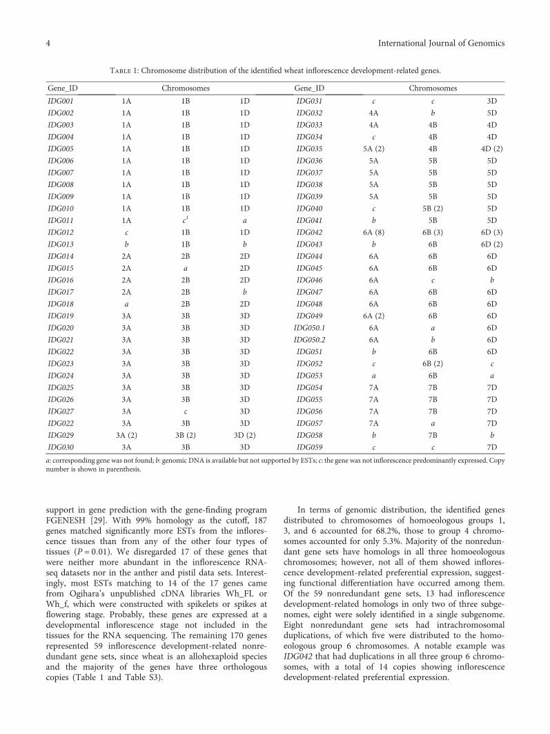
In terms of genomic distribution, the identified genesdistributed to chromosomes of homoeologous groups 1,3, and 6 accounted for 68.2%, those to group 4 chromo-somes accounted for only 5.3%. Majority of the nonredun-dant gene sets have homologs in all three homoeologouschromosomes; however, not all of them showed inflores-cence development-related preferential expression, suggest-ing functional differentiation have occurred among them.Of the 59 nonredundant gene sets, 13 had inflorescencedevelopment-related homologs in only two of three subge-nomes, eight were solely identified in a single subgenome.Eight nonredundant gene sets had intrachromosomalduplications, of which five were distributed to the homo-eologous group 6 chromosomes. A notable example wasIDG042 that had duplications in all three group 6 chromo-somes, with a total of 14 copies showing inflorescencedevelopment-related preferential expression.
Table 1: Chromosome distribution of the identified wheat inflorescence development-related genes.
Gene_ID Chromosomes Gene_ID Chromosomes
IDG001 1A 1B 1D IDG031 c c 3D
IDG002 1A 1B 1D IDG032 4A b 5D
IDG003 1A 1B 1D IDG033 4A 4B 4D
IDG004 1A 1B 1D IDG034 c 4B 4D
IDG005 1A 1B 1D IDG035 5A (2) 4B 4D (2)
IDG006 1A 1B 1D IDG036 5A 5B 5D
IDG007 1A 1B 1D IDG037 5A 5B 5D
IDG008 1A 1B 1D IDG038 5A 5B 5D
IDG009 1A 1B 1D IDG039 5A 5B 5D
IDG010 1A 1B 1D IDG040 c 5B (2) 5D
IDG011 1A c1 a IDG041 b 5B 5D
IDG012 c 1B 1D IDG042 6A (8) 6B (3) 6D (3)
IDG013 b 1B b IDG043 b 6B 6D (2)
IDG014 2A 2B 2D IDG044 6A 6B 6D
IDG015 2A a 2D IDG045 6A 6B 6D
IDG016 2A 2B 2D IDG046 6A c b
IDG017 2A 2B b IDG047 6A 6B 6D
IDG018 a 2B 2D IDG048 6A 6B 6D
IDG019 3A 3B 3D IDG049 6A (2) 6B 6D
IDG020 3A 3B 3D IDG050.1 6A a 6D
IDG021 3A 3B 3D IDG050.2 6A b 6D
IDG022 3A 3B 3D IDG051 b 6B 6D
IDG023 3A 3B 3D IDG052 c 6B (2) c
IDG024 3A 3B 3D IDG053 a 6B a
IDG025 3A 3B 3D IDG054 7A 7B 7D
IDG026 3A 3B 3D IDG055 7A 7B 7D
IDG027 3A c 3D IDG056 7A 7B 7D
IDG022 3A 3B 3D IDG057 7A a 7D
IDG029 3A (2) 3B (2) 3D (2) IDG058 b 7B b
IDG030 3A 3B 3D IDG059 c c 7D
a: corresponding gene was not found; b: genomic DNA is available but not supported by ESTs; c: the gene was not inflorescence predominantly expressed. Copynumber is shown in parenthesis.
4 International Journal of Genomics

3.2. Expression Specificity of the Identified Genes. In mininggenes predominantly expressed in inflorescence, we consid-ered all tissues from inflorescence as a whole. It was noted,in the EST analysis, that some genes were solely expressedin spikes at or before anthesis, some were solely in stamenor in pistil, apart from those expressed more abundantly inspikes than in other tissues. To validate the expressionpatterns of these genes, we tested the significance of expres-sion difference and estimated the expression specificity(RES) of inflorescence (including the stages of Z32, Z39,and Z65), stamen, and pistil relative to the individual vegeta-tive tissues (including kernel) using the RNA-seq data. Basedon the expression profiles, we were able to classify the 170genes into three groups, each with two subgroups (Figure 1and Table S3). Within each subgroup, the expression profileswere similar between genes, but the relative abundances werenot identical even between orthologous genes.
The first group, G1, consisted of 42 genes from 10 genesets. Overall, they had a low expression level and wereexpressed more abundantly and specifically in majority ofthe cases, in the inflorescence as a whole. A few genes, suchas IDG035.2-5A, IDG035.1-4D, IDG035.2-4D, IDG042.1-6A,IDG042.3-6A, IDG042.4-6A, and IDG042.8-6A, were sup-ported by ESTs but were matched to a negligible number ofreads in the RNA-seq datasets. They were classifiedtogether with their orthologous or paralogous homologs,since their expression profiles in EST analysis were similar.Genes in subgroup G1-1 also showed enhanced expres-sion, even though less abundantly, in stamen or pistil.Different from those in G1-1, genes in subgroup G1-2had negligible fragments per kilobase of cDNA modelper million mapped reads (FPKM) values from tissuesother than the inflorescence.
The second group, G2, consisted of 20 gene sets and 48genes. This group was basically characterized by expressionin one or more of the vegetative tissues and an even higherlevel of expression in the stamen and/or pistil. IDG011-1Awas the only exception, which was predominantly expressedin the inflorescence as a whole in spite of a significantlyhigher expression in the stamen and pistil relative to the veg-etative tissues. Genes in subgroup G2-1 all had a significantlyhigher level of expression in the pistil relative to the vegeta-tive tissues, and except for IDG044-6B and IDG044-6D, asignificantly higher level of expression in the stamen as well.A few genes in this subgroup were expressed more abun-dantly in the inflorescence (IDG011-1A) or in the stamen(IDG001-1A, IDG001-1D, and IDG045). Genes in subgroupG2-2 were expressed more abundantly in the stamen relativeto the vegetative tissues, but their expression levels in thepistil were not different from or even lower than those in atleast one of the vegetative tissues.
Group G3 was the largest group, including 80 genes from29 gene sets, characterized by a predominant expression instamen and a negligible level of expression in the vegetativetissues. The RES values (stamen versus vegetative tissues)were high, ranged from 0.82–1.0. G3-1 consisted of 39 genes,all with a negligible FPKM value from the inflorescence. Theremaining genes were different from genes in G3-1 by a sig-nificantly enhanced expression in the inflorescence as well
relative to the vegetative tissues, although the expression levelwas much lower in most cases.
To verify the expression specificity experimentally,sqRT-PCR was performed with tissues of root, node, inter-node, flag leaf, glume, lemma, palea, lodicule, stamen, pis-til, rachis, and kernel 9th day postanthesis, using 27 pairsof primers that corresponds to 64 members of the identi-fied genes (Table S2). All PCR reactions revealed a pattern ofpredominant expression in at least one of the floral tissues ororgans but not in kernels and vegetative organs (Figure 2),which, by and large, were in agreement with results fromthe EST and RNA-seq data analysis. The qRT-PCR of aselected set of genes, including those with a relatively lowerexpression level and those expressed in multiple tissues, fur-ther confirmed these results (Figure S1).
Generally speaking, the tissue expression profilesbetween orthologous genes inferred based on the EST datawere similar to those from the RNA-seq analysis. But a fewexceptions were noted. In most of these cases, a low expres-sion level of certain orthologous members was likely thecause of the discrepancy, for instance, some members ofIDG035 and IDG042.
3.3. Functional Annotation of the Identified Genes. Forty-nineof the identified gene sets were annotated via homologysearch and classified into five categories according to theirputative biological functions (Table 2). It has to bementioned that some genes could functionally fall intomultiple categories.
The first category included only two gene sets; bothencode allergenic proteins. The biological functions of thisclass of proteins in floral development have not been wellcharacterized. IDG035 encodes group 3 grass pollen aller-gens, which have sequence similarity to expansins that pro-mote plant cell wall enlargement and thereby serve as cellwall-loosening agents [40].
The second category included 11 gene sets, most of whichbelonged to the G2 expression type. In this category, five genesets code for proteins related to JA, ET, and GA signaling,three for MADS-box transcription factors and three forH2A and H2B proteins. The MADS-box transcription factorproteins encoded by IDG001 and IDG021 have 87% similar-ity. IDG001 is orthologous to the rice OsMADS4. Accordingto the ABCDE model for floral organ identity specification[41], IDG001 and IDG021 belong to class B MADS-boxgenes. IDG044 encodes an AGL6-like MADS transcriptionfactor and is functionally similar to class E genes [42].
The third category included 28 gene sets, accountingfor 57% of the annotated. They were all predominantlyor specifically expressed in stamen. Most of them wereassociated with substance production, transportation, andassembly for anther and pollen development. Of this cate-gory, 18 gene sets code for proteins associated with fattyacid and lipid metabolism. Homologs in other plants ofmost of these genes have been associated with the processof pollen wall development, such as suberin biosynthesis[43, 44], cutin biosynthesis [45–47], pollen sporopolleninbiosynthesis [48], and pollen exine formation [49–51].Other genes in this category have also been associated
5International Journal of Genomics

Root
Stem
Leaf
Spik
e
Pisti
lSt
amen
Gra
in
Root
Stem
Leaf
Spik
e
Pisti
l
Stam
en
Gra
in
IDG008-1AIDG008-1BIDG008-1DIDG026-3AIDG026-3BIDG026-3DIDG033-4AIDG033-4BIDG035-4BIDG035.1-5AIDG043-6BIDG043.1-6DIDG043.2-6DIDG046-6AIDG005-1AIDG005-1BIDG005-1DIDG014-2AIDG014-2BIDG014-2DIDG033-4DIDG042.2-6AIDG042.5-6AIDG042.6-6AIDG042.7-6AIDG042.1-6BIDG042.2-6BIDG042.3-6BIDG042.1-6DIDG042.2-6DIDG042.3-6DIDG049.1-6AIDG049.2-6AIDG049-6BIDG049-6DIDG001-1AIDG001-1BIDG001-1DIDG011-1AIDG012-1BIDG012-1DIDG023-3AIDG023-3BIDG023-3DIDG028-3AIDG028-3BIDG028-3DIDG044-6AIDG044-6BIDG044-6DIDG045-6AIDG045-6BIDG045-6DIDG058-7BIDG059-7DIDG009-1AIDG009-1BIDG009-1DIDG010-1AIDG010-1BIDG010-1DIDG021-3AIDG021-3BIDG021-3DIDG022-3AIDG022-3BIDG022-3DIDG027-3AIDG027-3DIDG031-3DIDG034-3BIDG034-3DIDG039-5AIDG039-5BIDG039-5DIDG040.1-5BIDG040.2-5BIDG040-5DIDG048-6AIDG048-6BIDG048-6DIDG052.1-6BIDG052.2-6B
10.009.008.007.006.005.004.003.002.001.000.00
IDG003-1AIDG003-1BIDG003-1DIDG004-1AIDG004-1BIDG013-1BIDG017-2AIDG017-2BIDG019-3AIDG019-3BIDG019-3DIDG025-3AIDG025-3BIDG025-3DIDG029.1-3AIDG029.2-3AIDG029.1-3BIDG029.1-3DIDG029.2-3DIDG032-4AIDG032-5DIDG036-5AIDG036-5BIDG036-5DIDG037-5AIDG037-5BIDG037-5DIDG041-5BIDG041-5DIDG047-6AIDG051-6BIDG051-6DIDG053-6BIDG054-7AIDG054-7BIDG054-7DIDG055-7AIDG055-7BIDG055-7DIDG002-1AIDG002-1BIDG002-1DIDG004-1DIDG006-1AIDG006-1BIDG006-1DIDG007-1AIDG007-1BIDG007-1DIDG015-2AIDG015-2DIDG016-2AIDG016-2BIDG016-2DIDG018-2BIDG018-2DIDG020-3AIDG020-3BIDG020-3DIDG024-3AIDG024-3BIDG024-3DIDG029.2-3BIDG030-3AIDG030-3BIDG030-3DIDG038-5AIDG038-5BIDG038-5DIDG047-6BIDG047-6DIDG050.1-6AIDG050.2-6AIDG050.1-6DIDG050.2-6DIDG056-7AIDG056-7BIDG056-7DIDG057-7AIDG057-7D
G2-2
G2
G2-1
G1
G2-2
G1-1
G3-1
G3
G3-2
Figure 1: Relative expression abundance of the identified genes in different tissues based on RNA-seq data. Apart from seven genes with anegligible number of reads, the remaining 163 genes were divided according to their expression profiles into three groups, each of whichhad two subgroups.
6 International Journal of Genomics

with anther and pollen development. IDG038 encodes anacidic peroxidase that might participate in the synthesis ofphenylpropanoids present in sporopollenin [52]. The prod-ucts encoded by IDG007, IDG018, IDG050, IDG055, andIDG056 were related to pollen wall formation [53–55]. BothIDG004 and IDG025 code for anther-specific RTS-likeproteins, required for male fertility and affecting tapetaldevelopment [56]. IDG006 encodes a galactosyltransferase,which is implicated in the accumulation control of glyco-sylated flavonols in pollen [57]. In Arabidopsis, a type IIβ-(1,3)-galactosyltransferase is required for pollen exinedevelopment [58]. IDG020 codes for a late cornified enve-lope- (LCE-) like proline-rich protein. LCE proteins areinvolved in the cornified cell envelope assembly of skinsand associated with ROS detoxification [59].
The fourth category only has four gene sets and is associ-ated with pollination and pollen-stigma interactions. Forinstance, the subtilisin-like protease encoded by IDG002was related to anther dehiscence [60]; the products ofIDG008 were related to pollen tube growth [61]; the che-mocyanins encoded by IDG040 could be involved in thepollination process [62] and induce pollen tube chemotro-pism as a diffusible chemotropic factor [63]. IDG028 codesfor a protodermal factor 1-like protein mainly in the pistil.This protein is related to reactive oxygen species (ROS)homeostasis [64]. ROS are involved in pollen tube growth
and rupture [65, 66], implying the role of modulating ROSlevels in male reproductive development [67].
The fifth category, the “other” in Table 2, had only fourannotated gene sets. Their specific functions in reproductivedevelopment still require clarification.
4. Discussion
Inflorescence represents a highly specialized plant tissueproducing seeds for propagation. Deciphering genesinvolved in its development is the first step to understandthe essence of reproduction and of great importance forseed production manipulation. In this study, we identified59 nonredundant wheat gene sets that were differentiallyexpressed in wheat inflorescence and encode proteins withdiverse functions. Majority of the identified genes wereassociated with metabolic activities and wall assemblyrequired for the specialized process of pollen maturationand pollination, while few showed predominance or spec-ificity in macrosporogenesis. On one hand, this could beattributed to the fact that much more ESTs from librariesmade with anthers were used in the analysis, which hadlimitation of development stage and tissue-type coverage;on the other hand, it was probably due to the specificstructure of pollen grains whose formation requiresexpression of a specific set of genes or gene network thatmade the related genes easily recognized through thedifferential analysis. Our results complemented well withprevious studies involved in wheat floral development.The microarray-based transcriptomic analysis of anthersby Crismani et al. was mainly focused on identificationof early meiotic genes [15]. In the RNA-seq data compar-ison of pistillody stamen versus pistil, pistillody stamenversus stamen, and pistil versus stamen, Yang et al. identi-fied 206 genes highly correlated with stamen and pistildevelopment [17]. Among them, however, only a few werefunctionally annotated as identically as the genes presentedin this paper. It was noted that nearly one-third of theidentified gene sets in the present study displayed differen-tiated expression profiles in terms of their subgenomeorthologs, implying functional diversification in polyploidywheat for the inflorescence development.
The whole process of inflorescence development is underregulatory control. A set of MADS transcription factors reg-ulate floral organ identity specification [41, 68, 69]. The threeMADS-box genes we identified, one for E-class MADSproteins and two for B-class MADS proteins, differed intheir expression profiles (Figure 1), even though bothIDG001 and IDG021 were mainly expressed in stamen,suggesting they act in concert in determining the antherand pistil identity. The pistillody in alloplasmic wheatwas related to expression pattern alteration of class Bgenes [68]. The identification of genes related to JA, ET,and GA signaling added support for the important rolesof JA, ET, and GA signaling cross-talks playing in stamendevelopment [70, 71]. ET signaling is involved in multipleaspects of floral organ development, for instance, nectarsecretion, accumulation of stigmatic exudate, and develop-ment of the self-incompatible response [72], floral organ
Root
Nod
e
Inte
rnod
e
Flag
leaf
Glu
me
Lem
ma
Pale
a
Lodi
cule
Stam
en
Pisti
l
Rach
is
Kern
el (9
DPA
)
IDG002-1B/1DIDG003-1A/1B/1DIDG004-1A/1B/1DIDG006-1A/1B/1DIDG007-1A/1B/1DIDG008-1A/1B/1D
IDG015-2A/2DIDG016-2A
IDG018-2B/2DIDG020-3A/3B/3DIDG021-3A/3B/3D
IDG024-3DIDG025-3B
IDG026-3A/3B/3DIDG029.1-3BIDG029.1-3D
IDG029.2-3A/3B/3DIDG030-3A/3B/3DIDG035-5A/4B/4DIDG037-5A/5B/5DIDG038-5A/5B/5D
IDG042.1-6DIDG043-6B/6DIDG047-6A/6DIDG050.1-6AIDG053-6B
IDG054-7B/7DIDG056-7A/7B
�훼-Tubulin
Figure 2: Expression of a selected set of identified genes in differenttissues. The tissues used included kernel 9th day postanthesis, root,node, internode, flag leaf, glume, lemma, palea, lodicule, stamen,pistil, and rachis at the heading stage of common wheat landrace“Wangshuibai.”
7International Journal of Genomics
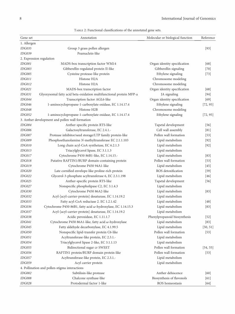
Table 2: Functional classifications of the annotated gene sets.
Gene set Annotation Molecular or biological function Reference
1. Allergen
IDG035 Group 3 grass pollen allergen [93]
IDG039 Peamaclein-like
2. Expression regulation
IDG001 MADS-box transcription factor WM14 Organ identity specification [68]
IDG003 Gibberellin-regulated protein II-like Gibberellin signaling [70]
IDG005 Cysteine protease-like protein Ethylene signaling [73]
IDG011 Histone H2A Chromosome modeling
IDG012 Histone H2A Chromosome modeling
IDG021 MADS-box transcription factor Organ identity specification [68]
IDG031 Glyoxysomal fatty acid beta-oxidation multifunctional protein MFP-a JA signaling [94]
IDG044 Transcription factor AGL6-like Organ identity specification [69]
IDG046 1-aminocyclopropane-1-carboxylate oxidase, EC 1.14.17.4 Ethylene signaling [72, 95]
IDG048 Histone H2B Chromosome modeling
IDG052 1-aminocyclopropane-1-carboxylate oxidase, EC 1.14.17.4 Ethylene signaling [72, 95]
3. Anther development and pollen wall formation
IDG004 Anther-specific protein RTS-like Tapetal development [56]
IDG006 Galactosyltransferase, EC 2.4.1.- Cell wall assembly [81]
IDG007 Protease inhibitor/seed storage/LTP family protein-like Pollen wall formation [53]
IDG009 Phosphoethanolamine N-methyltransferase EC 2.1.1.103 Lipid metabolism [92]
IDG010 Long chain acyl-CoA synthetase, EC 6.2.1.3 Lipid metabolism [92]
IDG013 Triacylglycerol lipase, EC 3.1.1.3 Lipid metabolism
IDG017 Cytochrome P450 86B1-like, EC 1.14.15.- Lipid metabolism [83]
IDG018 Putative RAFTIN1/BURP domain-containing protein Pollen wall formation [53]
IDG019 Cytochrome P450 94A1-like Lipid metabolism [45]
IDG020 Late cornified envelope-like proline-rich protein ROS detoxification [59]
IDG022 Glycerol-3-phosphate acyltransferase 6, EC 2.3.1.198 Lipid metabolism [46]
IDG025 Anther-specific protein RTS-like Tapetal development [56]
IDG027 Nonspecific phospholipase C2, EC 3.1.4.3 Lipid metabolism
IDG030 Cytochrome P450 86A2-like Lipid metabolism [83]
IDG032 Acyl-[acyl-carrier-protein] desaturase, EC 1.14.19.2 Lipid metabolism
IDG033 Fatty acyl-CoA reductase 2. EC 1.2.1.42 Lipid metabolism [48]
IDG036 Cytochrome P450 86B1, fatty acid ω-hydroxylase, EC 1.14.15.3 Lipid metabolism [83]
IDG037 Acyl-[acyl-carrier-protein] desaturase, EC 1.14.19.2 Lipid metabolism
IDG038 Acidic peroxidase, EC 1.11.1.7 Phenylpropanoid biosynthesis [52]
IDG041 Cytochrome P450 86A1-like, fatty acid ω-hydroxylase Lipid metabolism [83]
IDG045 Fatty aldehyde decarbonylase, EC 4.1.99.5 Lipid metabolism [50, 51]
IDG050 Nonspecific lipid-transfer protein C6-like Pollen wall formation [53]
IDG051 Acyltransferase-like protein, EC 2.3.1.- Lipid metabolism
IDG054 Triacylglycerol lipase 2-like, EC 3.1.1.13 Lipid metabolism
IDG055 Bidirectional sugar cr SWEET Pollen wall formation [54, 55]
IDG056 RAFTIN1 protein/BURP domain protein-like Pollen wall formation [53]
IDG057 Acyltransferase-like protein, EC 2.3.1.- Lipid metabolism
IDG059 Acyl carrier protein Lipid metabolism
4. Pollination and pollen-stigma interactions
IDG002 Subtilisin-like protease Anther dehiscence [60]
IDG008 Chalcone synthase-like Biosynthesis of flavonols [61]
IDG028 Protodermal factor 1-like ROS homeostasis [64]
8 International Journal of Genomics
senescing [73], pollen thermotolerance [74], and timing ofanther dehiscence [71]. Cheng et al. demonstrated that GAregulates stamen development through JA signaling [75].Mutations of genes encoding JA-biosynthetic enzymes resultin failure of filament elongation, delayed anther dehiscence,and unviable pollens [70, 76, 77].
A few identified genes were associated with pollen-stigma interactions. All but IDG028 had transcripts inspikes at meiosis and anthesis stages as well as in anthersbut the abundance varied considerably. The subtilase-encoding genes related to anther dehiscence (IDG002)were overwhelmingly expressed in anthers, especially atthe tetrad stage [60]. In male-sterile lines, their expressionwas downregulated [77]. Transcripts of pollen allergen-encoding IDG035 and ACO-encoding IDG046 were alsopresent in spikes at this stage. Accumulating evidenceindicates the involvement of ET signaling in fertilization[78, 79]. Valdiva et al. showed that disruption of a maizegroup-I allergen affected pollen-pollen competition for accessto the ovules [80]. In addition, the galactosyltransferase-encoding IDG006, predominantly expressed in stamen, wasrelated to pollen tube elongation [81].
Most of the genes showing anther-specific or predomi-nant expression are related to tapetal and pollen develop-ments (Table 2). Particularly worth mentioning are thegene set-encoding enzymes or proteins participating in lipidmetabolism. Lipid metabolism is important to pollen devel-opment because the distinct pollen wall structure is mainlymade of fatty (lipid) substances produced in the tapetum ofanthers [82]. Among the lipid metabolism-related gene sets,five (IDG017, IDG019, IDG030, IDG036, and IDG041)encode members of 86A, 86B, and 94A subfamilies of cyto-chrome P450 proteins that are related to fatty acid ω-hydrox-ylation in primary fatty alcohols and suberin monomerbiosynthesis for formation of anther cuticle and pollen spo-ropollenin in monocots and dicots [43–45, 47, 83–86]. Thesecytochrome P450 proteins might function in different subcel-lular locations, since IDG017, IDG019, and IDG030 havesecretory signal peptides, while IDG036 and IDG041 do not.
Pathways emerging from the lipid metabolism-relatedgenes included those for alkane and fatty alcohol produc-tion and glycerophospholipid metabolism. Genes encod-ing long-chain acyl-CoA synthetase (IDG010) and fattyaldehyde decarbonylase (CER1, IDG045), the two enzymesinvolved in the plant alkane-forming pathway [87], werecoexpressed in the wheat stamen. This CER1 gene was
downregulated in pistil or pistillody stamen [17], suggest-ing its specificity to stamen development. Very long-chain(VLC) alkanes are major components of the tryphinelayer covering pollen grains and are needed for properpollen-pistil signaling and fertility [50]. Mutation ofCER1 in both Arabidopsis and rice caused defective pol-lens [50, 51]. In Arabidopsis, the acyl-CoA synthetasegene ACOS5 was upregulated in the tapetal cells. Itsmutation led to failure in pollen production and pollenwall formation [88].
Fatty alcohols are components of surface lipid barrierssuch as anther cuticle and pollen wall [89]. Fatty acyl-CoAreductase, encoded by IDG033 in wheat and Ms2 in Arabi-dopsis, is the key enzyme for the production of fatty alcoholsin plastids [48]. Mutation ofMs2 led to abnormal pollen walldevelopment and reduced pollen fertility [90, 91]. Like Ms2,the IDG033-encoded proteins have plastidic localization sig-nal peptides. The encoded products of IDG059, IDG032, andIDG037, probably involved in the production of fatty acyl-CoA reductase substrates, all have plastidic localizationsignals. The diacylglycerol acyltransferase- (DGAT-) likeprotein encoded by IDG057 could also carry a plastidic pep-tide. The Arabidopsis DGAT1 contributes to triacylglycerolbiosynthesis and its function loss causes critical defects innormal pollen and embryo development [84]. However,information about its link to the plastidial fatty alcohol path-way is still lacking. The expression profiles of these geneswere different, although all expressed in stamen. IDG059,IDG032, and IDG037 appeared to be specifically expressedin this organ.
Among the lipid metabolism-related genes, five (IDG009,IDG013, IDG022, IDG027, and IDG054) encode proteinsputatively associated with glycerophospholipid metabolism,in agreement with the findings of Yang et al. [17]. TheIDG009-encoded phosphoethanolamine N-methyltransfer-ase (PEAMT) is the committing enzyme for choline biosyn-thesis. In Arabidopsis, silencing the PEAMT gene resultedin temperature-sensitive male sterility and salt hypersensitiv-ity [92]; knockdown of GPAT6, the homolog of IDG022,caused defective pollen grains [46]. PEAMT and GPAT6 alsoaffected pollen tube growth [46, 92]. Moreover, IDG051encode proteins predicted with lysophosphatidylethanola-mine acyltransferase activities, which probably participatesin the phospholipid metabolism in mitochondria.
Only 59 inflorescence development-related nonredun-dant gene sets were identified in this study. This could be
Table 2: Continued.
Gene set Annotation Molecular or biological function Reference
IDG040 Chemocyanin Pollen tube attraction [62]
5. Others
IDG014 Nitrate-induced NOI protein Plant defense [96]
IDG034 Nucleoside diphosphate kinase, EC 2.7.4.6 Nucleotide triphosphate generation
IDG053 Zinc transporter-like Early reproductive development
IDG058 Heat shock protein Thermotolerance
9International Journal of Genomics
much less than the actual number of genes differentiallyexpressed in inflorescence tissues. We reasoned that theEST libraries used in gene mining, which had limitations involume size and representation of tissue and developmentalstages, and the strict standard used in gene mining were themain causes. A sampling probability≤ 0.0001 has very likelyincrease type II error; however, it could minimize falsepositives, as shown in RNA-seq data analysis and RT-PCRvalidation, which is beneficial for correct data interpretation.
5. Conclusions
In this study, we identified 170 wheat genes for floralidentity determination, anther and pollen development,pollen-pistil interaction, and others using the comparativetranscriptomics approach. The potential importance ofthe identified genes to wheat inflorescence developmentwas manifested in the enhanced or specific expression inthe floral tissues. We noted that nearly one-third of thegene sets have undergone subgenome differentiation. Ofthe identified genes, those coding for enzymes or proteinsparticipating in lipid metabolic pathway accounted for thelargest category, implying the particularity and importantroles of lipid metabolism in wheat reproductive develop-ment. This study is useful for understanding the genenetwork underlying wheat inflorescence morphology andfertility, which eventually will allow us to purposelymanipulate fertility in breeding.
Conflicts of Interest
The authors declare that there are no conflicts of interestregarding the publication of this article.
Acknowledgments
This study was partially supported by Natural ScienceFoundation of China (31430064 and 30025030), Ministryof Science and Technology of China (2016YFD0101004and 2016ZX08002003), Jiangsu collaborative innovationinitiative for modern crop production (JCIC-MCP), “111”project B08025, and fund from the innovation team pro-gram for Jiangsu universities (2014).
Supplementary Materials
Supplementary 1. Figure S1: expression of a selected set ofidentified genes in different tissues, examined using qRT-PCR. The expression levels were estimated relative to thatin stamen. The tissues used included kernel 9th day post-anthesis, root, node, internode, flag leaf, glume, lemma, palea,lodicule, stamen, pistil, and rachis at the heading stage ofcommon wheat landrace “Wangshuibai.”
Supplementary 2. Table S1: cDNA libraries used in this study.
Supplementary 3. Table S2: primers used in RT-PCR.
Supplementary 4. Table S3: sequence, annotation, andexpression specificity of 170 identified genes. ∗ and ∗∗ signif-icantly different at P = 0 05 and 0.01, respectively.
References
[1] J. Kyozuka, “Grass inflorescence: basic structure and diver-sity,” Advances in Botanical Research, vol. 72, pp. 191–219,2014.
[2] H. T. Clifford, Spikelet and Floral Morphology, Washington,D.C, USA, Smithsonian, 1987.
[3] L. G. Clark and R.W. Pohl,Agnes Chase’s First Book of Grasses:The Structure of Grasses Explained for Beginners, WashingtonD.C, USA, Smithsonian, 1996.
[4] G. Theissen and H. Saedler, “Plant biology: floral quartets,”Nature, vol. 409, no. 6819, pp. 469–471, 2001.
[5] D. B. Zhang and Z. Yuan, “Molecular control of grass inflores-cence development,” Annual Review of Plant Biology, vol. 65,no. 1, pp. 553–578, 2014.
[6] J. Y. Lee and D. H. Lee, “Use of serial analysis of gene expres-sion technology to reveal changes in gene expression in Arabi-dopsis pollen undergoing cold stress,” Plant Physiology,vol. 132, no. 2, pp. 517–529, 2003.
[7] T. H. Lee, Y. K. Kim, T. T. Pham et al., “RiceArrayNet: a data-base for correlating gene expression from transcriptome pro-filing, and its application to the analysis of coexpressed genesin rice,” Plant Physiology, vol. 151, no. 1, pp. 16–33, 2009.
[8] H. P. Buermans and J. T. den Dunnen, “Next generationsequencing technology: advances and applications,” Biochi-mica et Biophysica Acta (BBA) - Molecular Basis of Disease,vol. 1842, no. 10, pp. 1932–1941, 2014.
[9] J. A. Schrauwen, P. F. de Groot, M. M. van Herpen et al.,“Stage-related expression of mRNAs during pollen develop-ment in lily and tobacco,” Planta, vol. 182, no. 2, pp. 298–304, 1990.
[10] J. Ma, D. S. Skibbe, J. Fernandes, and V. Walbot, “Male repro-ductive development: gene expression profiling of maizeanther and pollen ontogeny,” Genome Biology, vol. 9, no. 12,article R181, 2008.
[11] P. Deveshwar, W. D. Bovill, R. Sharma, J. A. Able, andS. Kapoor, “Analysis of anther transcriptomes to identifygenes contributing to meiosis and male gametophyte devel-opment in rice,” BMC Plant Biology, vol. 11, no. 1, pp. 78–78, 2011.
[12] N. Rutley and D. Twell, “A decade of pollen transcriptomics,”Plant Reproduction, vol. 28, no. 2, pp. 73–89, 2015.
[13] P. Chaturvedi, T. Ischebeck, V. Egelhofer, I. Lichtscheidl,and W. Weckwerth, “Cell-specific analysis of the tomatopollen proteome from pollen mother cell to mature pollenprovides evidence for developmental priming,” Journal ofProteome Research, vol. 12, no. 11, pp. 4892–4903, 2013.
[14] T. Marcussen, S. R. Sandve, L. Heier et al., “Ancient hybridiza-tions among the ancestral genomes of bread wheat,” Science,vol. 345, no. 6194, article 1250092, 2014.
[15] W. Crismani, U. Baumann, T. Sutton et al., “Microarrayexpression analysis of meiosis and microsporogenesis inhexaploid bread wheat,” BMC Genomics, vol. 7, no. 1,p. 267, 2006.
[16] S. McIntosh, L. Watson, P. Bundock et al., “SAGE of the devel-oping wheat caryopsis,” Plant Biotechnology Journal, vol. 5,no. 1, pp. 69–83, 2007.
[17] Z. J. Yang, Z. S. Peng, S. H. Wei, M. L. Liao, Y. Yu, and Z. Y.Jang, “Pistillody mutant reveals key insights into stamen andpistil development in wheat (Triticum aestivum L.),” BMCGenomics, vol. 16, no. 1, p. 211, 2015.
10 International Journal of Genomics

[18] M. Houde, M. Belcaid, F. Ouellet et al., “Wheat EST resourcesfor functional genomics of abiotic stress,” BMC Genomics,vol. 7, no. 1, p. 149, 2006.
[19] N. Z. Ergen and H. Budak, “Sequencing over 13,000 expressedsequence tags from six subtractive cDNA libraries of wild andmodern wheats following slow drought stress,” Plant, Cell &Environment, vol. 32, no. 3, pp. 220–236, 2009.
[20] A. Manickavelu, K. Kawaura, K. Oishi et al., “Comparativegene expression analysis of susceptible and resistant near-isogenic lines in commonwheat infected by Puccinia triticina,”DNA Research, vol. 17, no. 4, pp. 211–222, 2010.
[21] A. Manickavelu, K. Kawaura, K. Oishi et al., “Comprehensivefunctional analyses of expressed sequence tags in commonwheat (Triticum aestivum),” DNA Research, vol. 19, no. 2,pp. 165–177, 2012.
[22] M. Song, W. Xu, Y. Xiang, H. Jia, L. Zhang, and Z. Ma,“Association of jacalin-related lectins with wheat responsesto stresses revealed by transcriptional profiling,” PlantMolecular Biology, vol. 84, no. 1-2, pp. 95–110, 2014.
[23] O. D. Anderson, N. Huo, and Y. Q. Gu, “The gene space inwheat: the complete -gliadin gene family from the wheatcultivar Chinese Spring,” Functional & Integrative Geno-mics, vol. 13, no. 2, pp. 261–273, 2013.
[24] K. Rikiishi and M. Maekawa, “Seed maturation regulators arerelated to the control of seed dormancy in wheat (Triticum aes-tivum L.),” PLoS One, vol. 9, no. 9, article e107618, 2014.
[25] M. Domoki, A. Szucs, K. Jager, S. Bottka, B. Barnabas, andA. Feher, “Identification of genes preferentially expressed inwheat egg cells and zygotes,” Plant Cell Reports, vol. 32,no. 3, pp. 339–348, 2013.
[26] Z. Y. Chen, X. J. Guo, Z. X. Chen et al., “Genome-wide charac-terization of developmental stage- and tissue-specific tran-scription factors in wheat,” BMC Genomics, vol. 16, no. 1,p. 125, 2015.
[27] L. N. Ding, H. B. Xu, H. Y. Yi et al., “Resistance to hemi-biotrophic F.graminearum infection is associated with coordi-nated and ordered expression of diverse defense signalingpathways,” PLoS One, vol. 6, no. 4, article e19008, 2011.
[28] P. A. Wilkinson, M. O. Winfield, G. L. Barker et al.,“CerealsDB 3.0: expansion of resources and data integra-tion,” BMC Bioinformatics, vol. 17, no. 1, p. 256, 2016.
[29] V. Solovyev, P. Kosarev, I. Seledsov, and D. Vorobyev,“Automatic annotation of eukaryotic genes, pseudogenesand promoters,” Genome Biology, vol. 7, article S10, Supple-ment 1, 2006.
[30] R. Brenchley, M. Spannagl, M. Pfeifer et al., “Analysis of thebread wheat genome using whole-genome shotgun sequenc-ing,” Nature, vol. 491, no. 7426, pp. 705–710, 2012.
[31] F. Choulet, A. Alberti, S. Theil et al., “Structural and functionalpartitioning of bread wheat chromosome 3B,” Science,vol. 345, no. 6194, article 1249721, 2014.
[32] S. Lindgreen, “AdapterRemoval: easy cleaning of next-generation sequencing reads,” BMC Research Notes, vol. 5,no. 1, p. 337, 2012.
[33] D. Kim, B. Landmead, and S. L. Salzberg, “HISAT: a fastspliced aligner with low memory requirements,” NatureMethods, vol. 12, no. 4, pp. 357–360, 2015.
[34] Y. Liao, G. K. Smyth, and W. Shi, “featureCounts: an efficientgeneral purpose program for assigning sequence reads togenomic features,” Bioinformatics, vol. 30, no. 7, pp. 923–930, 2014.
[35] M. Song, W. Q. Xu, Y. Xiang, H. Y. Jia, L. X. Zhang, and Z. Q.Ma, “Association of jacalin-related lectins with wheatresponses to stresses revealed by transcriptional profiling,”Plant Molecular Biology, vol. 84, no. 1-2, pp. 95–110, 2014.
[36] K. J. Livak and T. D. Schmittgen, “Analysis of relative geneexpression data using real-time quantitative PCR and the2−ΔΔCT method,” Methods, vol. 25, no. 4, pp. 402–408, 2001.
[37] M. Kanehisa, Y. Sato, and K. Morishima, “BlastKOALA andGhostKOALA: KEGG tools for functional characterization ofgenome and metagenome sequences,” Journal of MolecularBiology, vol. 428, no. 4, pp. 726–731, 2016.
[38] R. D. Finn, P. Coggill, R. Y. Eberhardt et al., “The Pfam proteinfamilies database: towards a more sustainable future,” NucleicAcids Research, vol. 44, no. D1, pp. D279–D285, 2016.
[39] O. Emanuelsson, H. Nielsen, S. Brunak, and G. von Heijne,“Predicting subcellular localization of proteins based ontheir N-terminal amino acid sequence,” Journal of Molecu-lar Biology, vol. 300, no. 4, pp. 1005–1016, 2000.
[40] D. J. Cosgrove, P. Bedinger, and D. M. Durachko, “Group Iallergens of grass pollen as cell wall-loosening agents,” Pro-ceedings of the National Academy of Sciences of the UnitedStates of America, vol. 94, no. 12, pp. 6559–6564, 1997.
[41] L. M. Zahn, J. H. Leebens-Mack, J. M. Arrington et al., “Con-servation and divergence in the AGAMOUS subfamily ofMADS-box genes: evidence of independent sub- and neofunc-tionalization events,” Evolution & Development, vol. 8, no. 1,pp. 30–45, 2006.
[42] H. F. Li, W. Q. Liang, R. D. Jia et al., “The AGL6-like geneOsMADS6 regulates floral organ and meristem identities inrice,” Cell Research, vol. 20, no. 3, pp. 299–313, 2010.
[43] R. Hofer, I. Briesen, M. Beck, F. Pinot, L. Schreiber, andR. Franke, “The Arabidopsis cytochrome P450 CYP86A1encodes a fatty acid ω-hydroxylase involved in suberin mono-mer biosynthesis,” Journal of Experimental Botany, vol. 59,no. 9, pp. 2347–2360, 2008.
[44] V. Compagnon, P. Diehl, I. Benveniste et al., “CYP86B1 isrequired for very long chain ω-hydroxyacid and α,ω-dicarbox-ylic acid synthesis in root and seed suberin polyester,” PlantPhysiology, vol. 150, no. 4, pp. 1831–1843, 2009.
[45] N. Tijet, C. Helvig, F. Pinot et al., “Functional expression inyeast and characterization of a clofibrate-inducible plant cyto-chrome P-450 (CYP94A1) involved in cutin monomers syn-thesis,” Biochemical Journal, vol. 332, no. 2, pp. 583–589, 1998.
[46] X. C. Li, J. Zhu, J. Yang et al., “Glycerol-3-phosphate acyltrans-ferase 6 (GPAT6) is important for tapetum development inArabidopsis and plays multiple roles in plant fertility,” Molec-ular Plant, vol. 5, no. 1, pp. 131–142, 2012.
[47] S. G. Rupasinghe, H. Duan, andM. A. Schuler, “Molecular def-initions of fatty acid hydroxylases in Arabidopsis thaliana,”Proteins, vol. 68, no. 1, pp. 279–293, 2007.
[48] W. Chen, X. H. Yu, K. Zhang et al., “Male Sterile2 encodes aplastid-localized fatty acyl carrier protein reductase requiredfor pollen exine development in Arabidopsis,” Plant Physiol-ogy, vol. 157, no. 2, pp. 842–853, 2011.
[49] A. A. Dobritsa, Z. T. Lei, S. Nishikawa et al., “LAP5 andLAP6 encode anther-specific proteins with similarity tocalcone synthase essential for pollen exine development inArabidopsis thaliana,” Plant Physiology, vol. 153, no. 3,pp. 937–955, 2010.
[50] M. G. Aarts, C. J. Keijzer, W. J. Stiekema, and A. Pereira,“Molecular characterization of the CER1 gene of arabidopsis
11International Journal of Genomics

involved in epicuticular wax biosynthesis and pollen fertility,”The Plant Cell, vol. 7, no. 12, pp. 2115–2127, 1995.
[51] K. H. Jung, M. J. Han, D. Y. Lee et al., “Wax-deficient anther1 isinvolved in cuticle and wax production in rice anther walls andis required for pollen development,” The Plant Cell, vol. 18,no. 11, pp. 3015–3032, 2006.
[52] A. Skirycz, S. Jozefczuk, M. Stobiecki et al., “Transcriptionfactor AtDOF4;2 affects phenylpropanoid metabolism inArabidopsis thaliana,” New Phytologist, vol. 175, no. 3,pp. 425–438, 2007.
[53] A. M. Wang, Q. Xia, W. S. Xie, R. Datla, and G. Selvaraj, “Theclassical Ubisch bodies carry a sporophytically produced struc-tural protein (RAFTIN) that is essential for pollen develop-ment,” Proceedings of the National Academy of Sciences of theUnited States of America, vol. 100, no. 24, pp. 14487–14492,2003.
[54] B. Yang, A. Sugio, and F. F. White, “Os8N3 is a host disease-susceptibility gene for bacterial blight of rice,” Proceedings ofthe National Academy of Sciences of the United States of Amer-ica, vol. 103, no. 27, pp. 10503–10508, 2006.
[55] Y. F. Guan, X. Y. Huang, J. Zhu, J. F. Gao, H. X. Zhang, andZ. N. Yang, “RUPTURED POLLEN GRAIN1, a member ofthe MtN3/saliva gene family, is crucial for exine pattern for-mation and cell integrity of microspores in Arabidopsis,” PlantPhysiology, vol. 147, no. 2, pp. 852–863, 2008.
[56] H. Luo, J. Y. Lee, Q. Hu et al., “RTS, a rice anther-specific geneis required for male fertility and its promoter sequence directstissue-specific gene expression in different plant species,” PlantMolecular Biology, vol. 62, no. 3, pp. 397–408, 2006.
[57] L. P. Taylor and K. D. Miller, “The use of a photoactivatablekaempferol analogue to probe the role of flavonol 3-O-galacto-syltransferase in pollen germination,” Advances in Experimen-tal Medicine and Biology, vol. 505, pp. 41–50, 2002.
[58] T. Suzuki, J. O. Narciso, W. Zeng et al., “KNS4/UPEX1: a typeII arabinogalactan β-(1,3)-galactosyltransferase required forpollen exine development,” Plant Physiology, vol. 173, no. 1,pp. 183–205, 2017.
[59] W. P. Vermeij, A. Alia, and C. Backendorf, “ROS quenchingpotential of the epidermal cornified cell envelope,” Journal ofInvestigative Dermatology, vol. 131, no. 7, pp. 1435–1441,2011.
[60] A. A. Taylor, A. Horsch, A. Rzepczyk, C. A. Hasenkampf, andC. D. Riggs, “Maturation and secretion of a serine proteinase isassociated with events of late microsporogenesis,” The PlantJournal, vol. 12, no. 6, pp. 1261–1271, 1997.
[61] G. J. vanEldik, W. H. Reijnen, R. K. Ruiter, M. M. A.vanHerpen, J. A. M. Schrauwen, and G. J. Wullems, “Reg-ulation of flavonol biosynthesis during anther and pistildevelopment, and during pollen tube growth in Solanumtuberosum,” The Plant Journal, vol. 11, no. 1, pp. 105–113, 1997.
[62] J. Dong, S. T. Kim, and E. M. Lord, “Plantacyanin plays a rolein reproduction in Arabidopsis,” Plant Physiology, vol. 138,no. 2, pp. 778–789, 2005.
[63] S. Kim, J. C. Mollet, J. Dong, K. L. Zhang, S. Y. Park, andE. M. Lord, “Chemocyanin, a small basic protein from thelily stigma, induces pollen tube chemotropism,” Proceedingsof the National Academy of Sciences of the United States ofAmerica, vol. 100, no. 26, pp. 16125–16130, 2003.
[64] F. L. Deng, L. L. Tu, J. F. Tan, Y. Li, Y. C. Nie, and X. L. Zhang,“GbPDF1 is involved in cotton fiber initiation via the core cis-
element HDZIP2ATATHB2,” Plant Physiology, vol. 158, no. 2,pp. 890–904, 2012.
[65] M. Potocky, M. A. Jones, R. Bezvoda, N. Smirnoff, andV. Zarsky, “Reactive oxygen species produced by NADPHoxidase are involved in pollen tube growth,” New Phytolo-gist, vol. 174, no. 4, pp. 742–751, 2007.
[66] Q. H. Duan, D. Kita, E. A. Johnson et al., “Reactive oxygenspecies mediate pollen tube rupture to release sperm forfertilization in Arabidopsis,” Nature Communications,vol. 5, article 3129, 2014.
[67] L. F. Hu, W. Q. Liang, C. S. Yin et al., “Rice MADS3 regulatesROS homeostasis during late anther development,” The PlantCell, vol. 23, no. 2, pp. 515–533, 2011.
[68] E. Hama, S. Takumi, Y. Ogihara, and K. Murai, “Pistillody iscaused by alterations to the class-B MADS-box gene expres-sion pattern in alloplasmic wheats,” Planta, vol. 218, no. 5,pp. 712–720, 2004.
[69] T. Zhao, Z. F. Ni, Y. Dai, Y. Y. Yao, X. L. Nie, and Q. X. Sun,“Characterization and expression of 42 MADS-box genes inwheat (Triticum aestivum L.),” Molecular Genetics and Geno-mics, vol. 276, no. 4, pp. 334–350, 2006.
[70] Z. Y. Peng, X. Zhou, L. C. Li et al., “Arabidopsis hormone data-base: a comprehensive genetic and phenotypic informationdatabase for plant hormone research in Arabidopsis,” NucleicAcids Research, vol. 37, Supplement 1, pp. D975–D982, 2009.
[71] I. Rieu, M. Wolters-Arts, J. Derksen, C. Mariani, andK. Weterings, “Ethylene regulates the timing of anther dehis-cence in tobacco,” Planta, vol. 217, no. 1, pp. 131–137, 2003.
[72] X. Tang, A. Gomes, A. Bhatia, and W. R. Woodson, “Pistil-specific and ethylene-regulated expression of 1-aminocyclo-propane-1-carboxylate oxidase genes in petunia flowers,”The Plant Cell, vol. 6, no. 9, pp. 1227–1239, 1994.
[73] M. L. Jones, P. B. Larsen, and W. R. Woodson, “Ethylene-reg-ulated expression of a carnation cysteine proteinase duringflower petal senescence,” Plant Molecular Biology, vol. 28,no. 3, pp. 505–512, 1995.
[74] N. Firon, E. Pressman, S. Meir, R. Khoury, and L. Altahan,“Ethylene is involved in maintaining tomato (Solanum lyco-persicum) pollen quality under heat-stress conditions,” AobPlants, vol. 2012, no. 0, article pls024, 2012.
[75] H. Cheng, S. S. Song, L. T. Xiao et al., “Gibberellin acts throughjasmonate to control the expression of MYB21, MYB24, andMYB57 to promote stamen filament growth in Arabidopsis,”PLoS Genetics, vol. 5, no. 3, article e1000440, 2009.
[76] S. Ishiguro, A. Kawai-Oda, J. Ueda, I. Nishida, and K. Okada,“The defective in anther dehiscence1 gene encodes a novelphospholipase A1 catalyzing the initial step of jasmonic acidbiosynthesis, which synchronizes pollen maturation, antherdehiscence, and flower opening in Arabidopsis,” The PlantCell, vol. 13, no. 10, pp. 2191–2209, 2001.
[77] P. Lou, J. G. Kang, G. Y. Zhang, G. Bonnema, Z. Y. Fang, andX. W. Wang, “Transcript profiling of a dominant male sterilemutant (Ms-cd1) in cabbage during flower bud development,”Plant Science, vol. 172, no. 1, pp. 111–119, 2007.
[78] S. Bhattacharya and I. T. Baldwin, “The post-pollinationethylene burst and the continuation of floral advertisementare harbingers of non-random mate selection in Nicotianaattenuata,” The Plant Journal, vol. 71, no. 4, pp. 587–601,2012.
[79] R. Volz, J. Heydlauff, D. Ripper, L. von Lyncker, andR. Gross-Hardt, “Ethylene signaling is required for synergid
12 International Journal of Genomics

degeneration and the establishment of a pollen tube block,”Developmental Cell, vol. 25, no. 3, pp. 310–316, 2013.
[80] E. R. Valdivia, Y. Wu, L. C. Li, D. J. Cosgrove, and A. G.Stephenson, “A group-1 grass pollen allergen influencesthe outcome of pollen competition in maize,” PLoS One,vol. 2, no. 1, article e154, 2007.
[81] S. J. Roy, T. L. Holdaway-Clarke, G. R. Hackett, J. G. Kunkel,E. M. Lord, and P. K. Hepler, “Uncoupling secretion and tipgrowth in lily pollen tubes: evidence for the role of calciumin exocytosis,” The Plant Journal, vol. 19, no. 4, pp. 379–386,1999.
[82] S. Blackmore, A. H. Wortley, J. J. Skvarla, and J. R. Rowley,“Pollen wall development in flowering plants,” New Phytolo-gist, vol. 174, no. 3, pp. 483–498, 2007.
[83] D. S. Zhang, W. Q. Liang, Z. Yuan et al., “Tapetum degenera-tion retardation is critical for aliphatic metabolism and generegulation during rice pollen development,” Molecular Plant,vol. 1, no. 4, pp. 599–610, 2008.
[84] D. B. Zhang and Z. A. Wilson, “Stamen specification andanther development in rice,” Chinese Science Bulletin, vol. 54,no. 14, pp. 2342–2353, 2009.
[85] D. B. Zhang, X. Luo, and L. Zhu, “Cytological analysis andgenetic control of rice anther development,” Journal of Genet-ics and Genomics, vol. 38, no. 9, pp. 379–390, 2011.
[86] D. S. Zhang, W. Q. Liang, C. S. Yin, J. Zong, F. W. Gu, andD. B. Zhang, “OsC6, encoding a lipid transfer protein, isrequired for postmeiotic anther development in rice,” PlantPhysiology, vol. 154, no. 1, pp. 149–162, 2010.
[87] A. Bernard, F. Domergue, S. Pascal et al., “Reconstitution ofplant alkane biosynthesis in yeast demonstrates that Arabidop-sis eceriferum1 and eciriferum3are core components of a very-long-chain alkane synthesis complex,” The Plant Cell, vol. 24,no. 7, pp. 3106–3118, 2012.
[88] V. F. Souza, M. S. Pagliarini, C. B. Valle, N. C. Bione, M. U.Menon, and A. B. Mendes-Bonato, “Meiotic behavior of Bra-chiaria decumbens hybrids,” Genetics and Molecular Research,vol. 14, no. 4, pp. 12855–12865, 2015.
[89] O. Rowland and F. Domergue, “Plant fatty acyl reductases:enzymes generating fatty alcohols for protective layers withpotential for industrial applications,” Plant Science, vol. 193-194, pp. 28–38, 2012.
[90] M. G. Aarts, R. Hodge, K. Kalantidis et al., “The Arabidopsismale sterility 2 protein shares similarity with reductases inelongation/condensation complexes,” The Plant Journal,vol. 12, no. 3, pp. 615–623, 1997.
[91] A. A. Dobritsa, S. I. Nishikawa, D. Preuss et al., “LAP3, a novelplant protein required for pollen development, is essential forproper exine formation,” Sexual Plant Reproduction, vol. 22,no. 3, pp. 167–177, 2009.
[92] Z. L. Mou, X. Q. Wang, Z. M. Fu et al., “Silencing of phos-phoethanolamine N-methyltransferase results in temperature-sensitivemale sterility and salt hypersensitivity inArabidopsis,”The Plant Cell, vol. 14, no. 9, pp. 2031–2043, 2002.
[93] M. A. Zaidi, S. O'Leary, S. B. Wu et al., “A molecular and pro-teomic investigation of proteins rapidly released from triticalepollen upon hydration,” Plant Molecular Biology, vol. 79,no. 1-2, pp. 101–121, 2012.
[94] A. Baker, I. A. Graham, M. Holdsworth, S. M. Smith, and F. L.Theodoulou, “Chewing the fat: β-oxidation in signalling anddevelopment,” Trends in Plant Science, vol. 11, no. 3,pp. 124–132, 2006.
[95] S. F. Yang and N. E. Hoffman, “Ethylene biosynthesis and itsregulation in higher plants,” Annual Review of Plant Physiol-ogy, vol. 35, no. 1, pp. 155–189, 1984.
[96] A. J. Afzal, J. H. Kim, and D. Mackey, “The role of NOI-domain containing proteins in plant immune signaling,”BMC Genomics, vol. 14, no. 1, p. 327, 2013.
13International Journal of Genomics

Hindawiwww.hindawi.com
International Journal of
Volume 2018
Zoology
Hindawiwww.hindawi.com Volume 2018
Anatomy Research International
PeptidesInternational Journal of
Hindawiwww.hindawi.com Volume 2018
Hindawiwww.hindawi.com Volume 2018
Journal of Parasitology Research
GenomicsInternational Journal of
Hindawiwww.hindawi.com Volume 2018
Hindawi Publishing Corporation http://www.hindawi.com Volume 2013Hindawiwww.hindawi.com
The Scientific World Journal
Volume 2018
Hindawiwww.hindawi.com Volume 2018
BioinformaticsAdvances in
Marine BiologyJournal of
Hindawiwww.hindawi.com Volume 2018
Hindawiwww.hindawi.com Volume 2018
Neuroscience Journal
Hindawiwww.hindawi.com Volume 2018
BioMed Research International
Cell BiologyInternational Journal of
Hindawiwww.hindawi.com Volume 2018
Hindawiwww.hindawi.com Volume 2018
Biochemistry Research International
ArchaeaHindawiwww.hindawi.com Volume 2018
Hindawiwww.hindawi.com Volume 2018
Genetics Research International
Hindawiwww.hindawi.com Volume 2018
Advances in
Virolog y Stem Cells International
Hindawiwww.hindawi.com Volume 2018
Hindawiwww.hindawi.com Volume 2018
Enzyme Research
Hindawiwww.hindawi.com Volume 2018
International Journal of
MicrobiologyHindawiwww.hindawi.com
Nucleic AcidsJournal of
Volume 2018
Submit your manuscripts atwww.hindawi.com