identifying children at very low risk of clinically ... · pediatrics/original research identifying...
TRANSCRIPT

PEDIATRICS/ORIGINAL RESEARCH
Identifying Children at Very Low Risk of Clinically ImportantBlunt Abdominal Injuries
James F. Holmes, MD, MPH; Kathleen Lillis, MD; David Monroe, MD; Dominic Borgialli, DO, MPH; Benjamin T. Kerrey, MD;Prashant Mahajan, MD, MPH; Kathleen Adelgais, MD, MPH; Angela M. Ellison, MD, MSc; Kenneth Yen, MD, MS;Shireen Atabaki, MD, MPH; Jay Menaker, MD; Bema Bonsu, MD; Kimberly S. Quayle, MD; Madelyn Garcia, MD;Alexander Rogers, MD; Stephen Blumberg, MD; Lois Lee, MD, MPH; Michael Tunik, MD; Joshua Kooistra, DO;
Maria Kwok, MD; Lawrence J. Cook, PhD; J. Michael Dean, MD, MBA; Peter E. Sokolove, MD; David H. Wisner, MD;Peter Ehrlich, MD; Arthur Cooper, MD, MS; Peter S. Dayan, MD, MSc; Sandra Wootton-Gorges, MD;
Nathan Kuppermann, MD, MPH; for the Pediatric Emergency Care Applied Research Network (PECARN)*
Study objective: We derive a prediction rule to identify children at very low risk for intra-abdominal injuriesundergoing acute intervention and for whom computed tomography (CT) could be obviated.
Methods: We prospectively enrolled children with blunt torso trauma in 20 emergency departments. Weused binary recursive partitioning to create a prediction rule to identify children at very low risk of intra-abdominal injuries undergoing acute intervention (therapeutic laparotomy, angiographic embolization, bloodtransfusion for abdominal hemorrhage, or intravenous fluid for �2 nights for pancreatic/gastrointestinalinjuries). We considered only historical and physical examination variables with acceptable interraterreliability.
Results: We enrolled 12,044 children with a median age of 11.1 years (interquartile range 5.8, 15.1 years). Ofthe 761 (6.3%) children with intra-abdominal injuries, 203 (26.7%) received acute interventions. The predictionrule consisted of (in descending order of importance) no evidence of abdominal wall trauma or seat belt sign,Glasgow Coma Scale score greater than 13, no abdominal tenderness, no evidence of thoracic wall trauma, nocomplaints of abdominal pain, no decreased breath sounds, and no vomiting. The rule had a negative predictivevalue of 5,028 of 5,034 (99.9%; 95% confidence interval [CI] 99.7% to 100%), sensitivity of 197 of 203 (97%;95% CI 94% to 99%), specificity of 5,028 of 11,841 (42.5%; 95% CI 41.6% to 43.4%), and negative likelihoodratio of 0.07 (95% CI 0.03 to 0.15).
Conclusion: A prediction rule consisting of 7 patient history and physical examination findings, and withoutlaboratory or ultrasonographic information, identifies children with blunt torso trauma who are at very low risk forintra-abdominal injury undergoing acute intervention. These findings require external validation beforeimplementation. [Ann Emerg Med. 2012;xx:xxx.]
Please see page XX for the Editor’s Capsule Summary of this article.
0196-0644/$-see front matterCopyright © 2012 by the American College of Emergency Physicians.http://dx.doi.org/10.1016/j.annemergmed.2012.11.009
ma
I
idpic
*Participating centers and site investigators are listed in the Appendix.
SEE EDITORIAL, P.●●●.
INTRODUCTIONBackground
Intra-abdominal injury is a leading cause of morbidity inchildren,1 and early identification is imperative to minimizemorbidity and mortality from delayed or missed diagnosis.In the last 2 decades, computed tomography (CT) hasbecome the reference standard for diagnosing manytraumatic injuries,2-5 including intra-abdominal ones.6-10 CT
use in trauma and emergency care, however, has expanded dVolume xx, . x : Month
uch faster than the evidence for its appropriatepplication.2,3,11
mportanceAlthough CT provides detailed and useful information about
njuries and helps clinicians to make informed managementecisions, it has important drawbacks, primarily that it exposesatients to relatively large radiation dosages, placing them atncreased risk of radiation-induced malignancy. Unfortunately,hildren’s inherent radiosensitivity makes them
isproportionately at risk compared with adults.12-14 SeveralAnnals of Emergency Medicine 1

S
epwPiprap
S
dbc
FaS
Fv
Identifying Children at Very Low Risk of Clinically Important Blunt Abdominal Injuries Holmes et al
small, single-center studies suggest that children with blunttorso trauma can be risk stratified for intra-abdominal injurythrough a combination of readily accessible clinicalvariables.10,15-18 These studies are limited, however, by theirretrospective or single-center study designs but indicate that alarge, multicenter study may create a robust and preciseprediction rule.
Goals of This InvestigationThe objective of the current investigation was to derive a
prediction rule to identify children with blunt torso trauma whoare at very low risk for intra-abdominal injury undergoing acuteintervention. We hypothesized that a reliable prediction rulecould be created to identify a cohort of these children for whomCT would generally not be indicated.
MATERIALS AND METHODSStudy Design
We conducted a prospective, observational cohort study ofchildren with blunt torso trauma in the Pediatric EmergencyCare Applied Research Network (PECARN).19 The study wasapproved by the institutional review board at each participating
Editor’s Capsule Summary
What is already known on this topicThe radiation risks of computed tomography (CT)scanning in children are well recognized.
What question this study addressedAre there history and physical examination criteriathat might identify which children with blunt torsotrauma do not require abdominal CT scans?
What this study adds to our knowledgeA 7-point decision rule derived in this multicenterstudy of 12,044 children was 97% sensitive inidentifying children with intra-abdominal injuryrequiring acute intervention. Actual CT orderingcharacteristics were 99% sensitive. The rule,however, had a 99.9% negative predictive value inidentifying a population of children at very low riskfor intra-abdominal injury warranting acuteintervention.
How this is relevant to clinical practiceIf externally validated, this rule could aid cliniciansin lowering abdominal CT use in children byidentifying a low risk population based on simplehistory and examination variables.
site. t
2 Annals of Emergency Medicine
election of ParticipantsChildren with blunt torso (thorax and abdomen) trauma
valuated in the emergency department (ED) at any of 20articipating PECARN centers from May 2007 to January 2010ere eligible. Inclusion criteria are summarized in Figure 1.atients were excluded if they met any of the following criteria:
njury occurring greater than 24 hours before presentation,enetrating trauma, preexisting neurologic disorders impedingeliable examination, known pregnancy, or transfer fromnother hospital with previous abdominal CT or diagnosticeritoneal lavage.
tudy ProtocolThe ED faculty or fellow physician providing care
ocumented patient history and physical examination findingsefore CT scanning (if performed), using a standardized dataollection form. Data collected are listed in Figure 2.
Abdominal CT scans were performed at the discretion of the
Decreased level of consciousness (GCS score <15 or neurologic/behavioral status not age-appropriate) in association with blunt torso trauma (but not isolated head trauma). Blunt traumatic event with either of the following (regardless of the injury mechanism):
Paralysis Multiple nonadjacent long bone fractures (eg, tibia fracture, ulna fracture)
Blunt torso trauma due to any of the following mechanisms of injury:
mph)
Crush injury to the torso Physical assault involving the abdomen
Physician concern for abdominal trauma resulting in any of the following diagnostic or screening tests:
Abdominal CT or ultrasound (FAST) Laboratory testing to screen for intra-abdominal injury Chest or pelvic radiography
igure 1. Patient inclusion criteria. FAST, Focusedssessment sonography for trauma; GSC, Glasgow Comacale.
32
33
34
igure 2. Patient history and physical examinationariables collected.
reating physicians and according to the CT protocols at each
Volume xx, . x : Month

P
sedpaa
arCrdtsuTc1mict
rptTteeaicybscd(epopcvpba
pl
Holmes et al Identifying Children at Very Low Risk of Clinically Important Blunt Abdominal Injuries
institution. For study purposes of intra-abdominal injuryidentification, abdominal CT results were those from the finalinterpretation by the site’s faculty or board-certified radiologists.Those CT scans considered inconclusive for the determinationof intra-abdominal injury were initially reviewed at the studysite for definitive interpretation. Final interpretations of CTscans still considered inconclusive were adjudicated by a studyradiologist (S.W.-G.).
Hospitalization of study patients was at the discretion of thetreating physicians. Medical records of hospitalized studypatients were reviewed to identify those with intra-abdominalinjuries, particularly those undergoing acute intervention. Weconducted telephone follow-up at least 7 days after the originalED visit to identify any patients subsequently receiving adiagnosis of an intra-abdominal injury. If telephone follow-upwas unsuccessful, we mailed the guardians the same follow-upsurvey. For those not returning their mail surveys, we reviewedthe medical records, ED and trauma continuous qualityimprovement records, and local morgue records to identify anypatient who subsequently received a diagnosis of intra-abdominal injury or died.
Outcome MeasuresThe outcome of interest was intra-abdominal injury
undergoing acute intervention. Intra-abdominal injury wasdefined as any radiographically or surgically apparent injury tothe following structures: spleen, liver, urinary tract (from thekidney to the urinary bladder), gastrointestinal tract (includingthe bowel or associated mesentery from the stomach to thesigmoid colon), pancreas, gallbladder, adrenal gland, intra-abdominal vascular structure, or traumatic fascial defect(traumatic abdominal wall hernia). Acute intervention wasdefined by an intra-abdominal injury associated with any of thefollowing: death caused by the intra-abdominal injury, atherapeutic intervention at laparotomy, angiographicembolization to treat bleeding from the intra-abdominal injury,blood transfusion for anemia as a result of hemorrhage from theintra-abdominal injury, or administration of intravenous fluidsfor 2 or more nights in patients with pancreatic orgastrointestinal injuries. Therapeutic laparotomy was defined asany surgical intervention to treat an intra-abdominal injury.Blood transfusion for anemia as a result of hemorrhage from theintra-abdominal injury was based on predefined criteria anddetermined by the site investigator at each site after review ofthe medical records. Any case in which the site investigatorcould not make a definitive determination was adjudicated by a5-member study panel for final determination.
Each site identified eligible patients not enrolled andcollected data from each about patient age, mechanism ofinjury, and intra-abdominal injury status for comparison to theenrolled population. We compared the characteristics of theenrolled patients with those eligible but not enrolled to evaluate
for enrollment bias. aVolume xx, . x : Month
rimary Data AnalysisWe calculated simple descriptive statistics on the study
ample with 95% confidence intervals (CIs) around pointstimates. Bivariable comparisons are presented with rateifferences and 95% CIs to demonstrate associations betweenossible predictor variables and the outcomes of intra-bdominal injury undergoing acute intervention and any intra-bdominal injury.
We derived the prediction rule for patients with intra-bdominal injury undergoing acute intervention by using binaryecursive partitioning (CART software, version 6.0, San Diego,A), an analytic technique used to develop clinical decision
ules when rule sensitivity is most important.20 This techniqueivides the population into subpopulations (“nodes”) accordingo the risk of the outcome of interest. Each subpopulation isubsequently divided to minimize misclassification of patientsntil the final population meets predefined stopping criteria.21
he results are displayed in a treelike format, easy for thelinician to interpret. We used the Gini splitting technique and0-fold cross-validation to generate a conservative tree. Toinimize the risk for misclassifying a subject with intra-abdominal
njury undergoing acute intervention, we set the misclassificationosts of a type II error at 500:1 (misclassifying 500 subjects withouthe outcome of interest to identify 1 with the outcome of interest).
Variables were considered for inclusion into the predictionule according to previous literature and biological orhysiologic plausibility. In addition, we excluded any variablehat was missing on more than 5% of the data collection forms.his cutoff was chosen to ensure that we included only variables
hat are readily available in the ED and to limit bias fromxcessive missing data.22 Finally, all variables considered forntry into the final model had at least moderate interratergreement, with the lower bound of the 95% confidencenterval (CI) of the � measurements at least 0.4.23-27 Variablesonsidered for inclusion into the prediction rule included ageounger than 2 years, severe mechanism of injury (as definedelow), vomiting, hypotension, Glasgow Coma Scale (GCS)core, thoracic tenderness, evidence of thoracic wall trauma,ostal margin tenderness, decreased breath sounds, abdominalistention, complaints of abdominal pain, abdominal tendernessstratified by degree of tenderness as mild, moderate, or severe),vidence of abdominal wall trauma or seat belt sign, distractingainful injury, and femur fracture. We defined severe mechanismf injury a priori, according to previous literature and physiologiclausibility and included any of the following: motor vehiclerashes with ejection, rollover, or death in the same crash; motorehicle crashes with speed greater than 20 miles per hour andatient unrestrained; falls greater than 10 feet; pedestrians oricyclists struck by vehicles moving greater than 20 miles per hour;nd bicycle collision with handlebars striking the abdomen.
Finally, we calculated the sensitivity, specificity, positiveredictive value, negative predictive value, and negative
ikelihood ratios for the derived prediction rule. All statistical
nalyses were performed by the study statisticians (L.J.C. andAnnals of Emergency Medicine 3

T
C
AASEHNURA
ABN
WUOMMFFP
B
M
OOUH
tient enrollment.
Identifying Children at Very Low Risk of Clinically Important Blunt Abdominal Injuries Holmes et al
another) at the data coordinating center in conjunction with theprincipal investigators (J.H. and N.K.).
RESULTSCharacteristics of Study Subjects
Of the 14,882 eligible patients, we enrolled 12,044 (81%)(Figure 3). The median age was 11.1 years (interquartile range5.8, 15.1 years). Baseline characteristics of enrolled patients aredescribed in Table 1; 5,991 children (50%) were hospitalizedand 6,053 (50%) were discharged from the ED. Telephonefollow-up was successful for 4,459 (74%) patients dischargedfrom the ED, and the remainder had follow-up performed bymail (n�136) or by review of medical records, continuousquality improvement records, trauma registry records, and localmorgue records. A total of 2,838 patients were eligible but notenrolled. The median age (enrolled 11.1 years versus notenrolled 11.7 years), rate of abdominal CT in the ED (enrolled44.7% versus not enrolled 43.5%), and rate of intra-abdominalinjury (enrolled 6.3% versus not enrolled 5.8%) were similarbetween those enrolled and those eligible but not enrolled.
Main ResultsAbdominal CT scans were obtained for 5,514 (46%)
patients, including 5,380 in the ED, 232 during hospitalization,and 55 after discharge from the ED (some patients withmultiple CT scans performed). In total, 761 patients (6.3%;95% CI 5.9% to 6.8%) were diagnosed with intra-abdominalinjuries, including 204 with injuries to more than 1 organ.Specific organ injuries included the spleen 299 (39%), liver 282(37%), kidney 147 (19%), gastrointestinal tract 115 (15%),adrenal gland 89 (12%), pancreas 51 (7%), intra-abdominalvascular structure 16 (2%), urinary bladder 18 (2%), ureter 4(0.5%), and gallbladder 4 (0.5%), and a traumatic fascial defect
14,882 eli
Enrolled patients12,044 (81%)
Patients with IAI 761 (6.3%)
IAI undergoing acute intervention 203 (1.7%)
Figure 3. Pa
was identified in 4 patients (0.5%). Intraperitoneal fluidI
4 Annals of Emergency Medicine
able 1. Baseline characteristics of study population.
haracteristic
IAIUndergoingIntervention
(n�203)
No IAIUndergoingIntervention(n�11,841)
Total(n�12,044)
ge (SD), y 9.9 (5.3) 10.3 (5.4) 10.3 (5.4)ge �2 y (%) 10 (5) 1,157 (10) 1,167 (10)ex (% male) 125 (62) 7,259 (61) 7,384 (61)thnicity (%)ispanic 18 (9) 1,273 (11) 1,291 (11)on-Hispanic 119 (59) 7,537 (64) 7,656 (64)nknown 66 (33) 3,031 (26) 3,097 (26)ace (%)merican Indian or AlaskaNative
0 85 (1) 85 (1)
sian 4 (2) 218 (2) 222 (2)lack 45 (22) 3,699 (31) 3,744 (31)ative Hawaiian or otherPacific Islander
0 38 (0) 38 (0)
hite 123 (61) 6,366 (54) 6,489 (54)nknown 30 (15) 976 (8) 1,006 (8)ther 1 (0) 459 (4) 460 (4)echanism of injury (%)otor vehicle crash 91 (45) 3,739 (32) 3,830 (32)
all from an elevation 11 (5) 1,612 (14) 1,623 (13)all down stairs 4 (2) 277 (2) 281 (2)edestrian or bicycliststruck by moving vehicle
34 (17) 2,238 (19) 2,272 (19)
icycle collision or fallfrom bicycle while riding
19 (9) 739 (6) 758 (6)
otorcycle/ATV/motorized scootercollision
9 (4) 593 (5) 602 (5)
bject struck abdomen 10 (5) 783 (7) 793 (7)ther 18 (9) 1,673 (14) 1,691 (14)nknown 7 (3) 187 (2) 194 (2)igh-risk mechanism ofinjury (%)
72 (35) 2,646 (22) 2,718 (23)
gible patients
Eligible patients not enrolled2,838
Patients with IAI 164 (5.8%)
AI, Intra-abdominal injury; ATV, all-terrain vehicle.
Volume xx, . x : Month

nrtii1arIvpuwsaim
uTivuvicpwwlta
fawn74pa9
L
lavcltti
Holmes et al Identifying Children at Very Low Risk of Clinically Important Blunt Abdominal Injuries
(hemoperitoneum) was identified in 568 patients (75%; 95%CI 71.4% to 77.7%) with intra-abdominal injuries.
The primary outcome of interest (intra-abdominal injuryundergoing acute intervention) was identified in 203 of thepatients (26.7%; 95% CI 23.6% to 30.0%) with intra-abdominal injuries, including 103 who had more than 1 ofthese defining events. Events defining the outcomes in these 203patients included death as a result of the intra-abdominal injury9 (4.4%), therapeutic laparotomy 114 (56.6%), angiographicembolization of a bleeding abdominal organ 11 (5.4%), bloodtransfusion for abdominal hemorrhage 122 (60.1%), andadministration of intravenous fluids for greater than or equal to2 nights for patients with pancreatic or gastrointestinal injuries79 (38.9%). Information about missing data for the enrolledpatients is presented in Table E1 (available online athttp://www.annemergmed.com). Bivariable associationsbetween the clinical variables collected and both intra-abdominal injury and intra-abdominal injury undergoing acuteintervention are presented in Tables E2 and E3 (available onlineat http://www.annemergmed.com).
Sixteen of the 6,053 patients (0.3%; 95% CI 0.1% to 0.4 %)initially discharged from the ED were subsequently identifiedwith an intra-abdominal injury, including 2 (0.03%; 95% CI0% to 0.12%) with intra-abdominal injury in need of acuteintervention (gastrointestinal tract and spleen injuries). One ofthese 2 patients with intra-abdominal injury undergoing acuteintervention (laparotomy for gastrointestinal tract injury) hadan abdominal CT during the initial ED visit, and it wasinterpreted as normal. Abdominal CT scans were obtained fromthe ED for 191 of 203 patients (94%; 95% CI 90% to 97%)with intra-abdominal injury undergoing acute intervention.Eleven of the 12 patients who did not undergo abdominal CTscanning in the ED received laparotomy without imaging. Onepatient with intra-abdominal injury undergoing acuteintervention was discharged from the ED without imaging butreturned 1 day later and received a diagnosis of a spleniclaceration and underwent splenic artery embolization.Abdominal CT scans were obtained in the ED for 542 of 558patients (97%; 95% CI 95% to 98%) with intra-abdominalinjury never undergoing acute intervention and 4,647 of 11,283patients (41%; 95% CI 40% to 42%) without any intra-abdominal injury. Thus, test characteristics of actual abdominalCT ordering for intra-abdominal injury undergoing acuteintervention were sensitivity 191 of 192 (99%; 95% CI 97% to100%) and specificity 6,652 of 11,841 (56%; 95% CI 55% to57%).
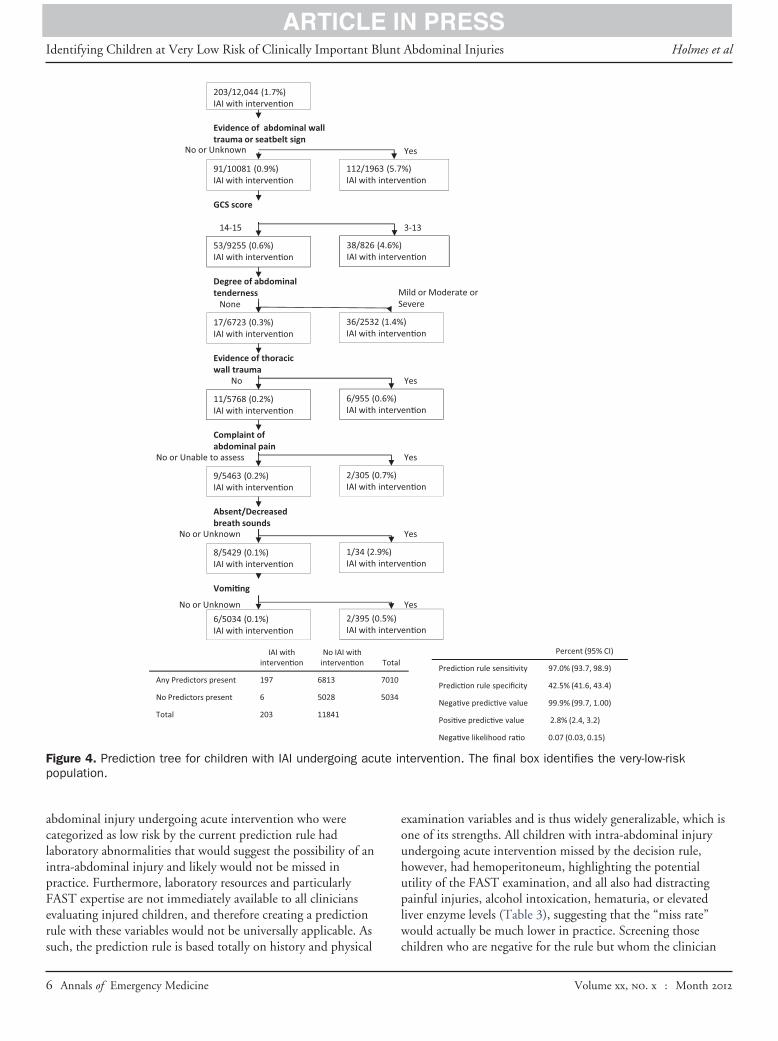
The derived prediction rule consisted of the following 7variables, in descending order of importance (Figure 4):evidence of abdominal wall trauma or seat belt sign, GCS scoreless than 14, abdominal tenderness, evidence of thoracic walltrauma, complaints of abdominal pain, decreased breath sounds,and vomiting. All 7 variables in the derived rule had substantialinterrater agreement, with � values all greater than 0.6 and
lower bounds of the 95% CI above 0.4.26,27 aVolume xx, . x : Month
The test characteristics (sensitivity, specificity, positive andegative predictive values, and positive and negative likelihoodatios) of the prediction rule are demonstrated in Figure 4. Fivehousand thirty-four patients (42%) were at very low risk forntra-abdominal injury undergoing acute intervention asdentified by the absence of any prediction rule variables, and,254 of these patients (25%) nevertheless underwentbdominal CT scanning during their ED evaluation. Thisepresents 23% of all the CTs performed on the study patients.f all patients positive for 1 or more of the prediction ruleariables underwent abdominal CT and patients negative for therediction rule did not, a total of 7,010 patients (58%) wouldndergo abdominal CT scanning. This type of strict applicationould result in a substantial increase in abdominal CT
canning. However, the intent of the rule is to identify patientst very low risk of important injuries who do not need CTmaging and is not meant to suggest that all patients with 1 or
ore variables undergo CT imaging.Figure 5 provides the risk of intra-abdominal injury
ndergoing acute intervention, stratified by specific rule criteria.able 2 details the number of patients with intra-abdominal
njury undergoing acute intervention stratified by the number ofariables present. The frequency of intra-abdominal injuryndergoing acute intervention increases as the number of ruleariables increases. Clinical characteristics of the 6 patients withntra-abdominal injury undergoing acute intervention who wereonsidered very low risk (no rule variables present) by therediction rule are presented in Table 3. Five of the 6 patientsith intra-abdominal injury undergoing acute intervention whoere categorized as very low risk by the prediction rule had
aboratory abnormalities (hematuria or elevated liverransaminase levels) suggestive of the presence of intra-bdominal injury, and all 6 had hemoperitoneum.
Although the rule was derived to identify patients at low riskor intra-abdominal injury undergoing acute intervention, welso assessed the performance of the rule for identifying childrenith any intra-abdominal injury (undergoing intervention orot), which yielded the following test characteristics: sensitivity04 of 761 (92.5%; 95% CI 90.4% to 94.3%), specificity,977 of 11,283 (44.1%; 95% CI 43.2% to 45.0%), positiveredictive value 704 of 7,010 (10.0%; 95% CI 9.3% to 10.8%),nd negative predictive value 4,977 of 5,034 (98.9%; 95% CI8.5% to 99.1%).
IMITATIONSThis study has some limitations. We did not include
aboratory testing or abdominal ultrasonography (focusedssessment sonography for trauma [FAST]) as possible predictorariables because of variability in the use of these tests amongenters and our inability to establish uniformity aroundaboratory and FAST use in all participating sites for purposes ofhis study. Previous studies, however, suggest that laboratoryesting and the FAST examination may play an important rolen risk stratifying children with blunt torso trauma for intra-
bdominal injury.10,15-18 In fact, 5 of the 6 patients with intra-Annals of Emergency Medicine 5
eouhuplw
Identifying Children at Very Low Risk of Clinically Important Blunt Abdominal Injuries Holmes et al
abdominal injury undergoing acute intervention who werecategorized as low risk by the current prediction rule hadlaboratory abnormalities that would suggest the possibility of anintra-abdominal injury and likely would not be missed inpractice. Furthermore, laboratory resources and particularlyFAST expertise are not immediately available to all cliniciansevaluating injured children, and therefore creating a predictionrule with these variables would not be universally applicable. As
203/12,044 (1.7%)IAI with interven�on
Evidence of abdominal walltrauma or seatbelt sign
91/10081 (0.9%)IAI with interven�on
GCS score
112/196IAI with
No or Unknown
14-15
53/9255 (0.6%)IAI with interven�on
Degree of abdominaltenderness
17/6723 (0.3%)IAI with interven�on
Evidence of thoracicwall trauma
11/5768 (0.2%)IAI with interven�on
38/826 (IAI with
36/2532IAI with
6/955 (0IAI with
None
No
IAI with interven�on
Complaint ofabdominal pain
9/5463 (0.2%)IAI with interven�on
Absent/Decreasedbreath sounds
8/5429 (0.1%)IAI with interven�on
IAI with
2/305 (0IAI with
1/34 (2.9IAI with
No or Unable to assess
No or Unknown
Vomi�ng
6/5034 (0.1%)IAI with interven�on
2/395 (0IAI with
No or Unknown
IAI withinterven�on
No IAI withinterven�on
Any Predictors present 197 6813
No Predictors present 6 5028
Total 203 11841
Figure 4. Prediction tree for children with IAI undergoing acupopulation.
such, the prediction rule is based totally on history and physical c
6 Annals of Emergency Medicine
xamination variables and is thus widely generalizable, which isne of its strengths. All children with intra-abdominal injuryndergoing acute intervention missed by the decision rule,owever, had hemoperitoneum, highlighting the potentialtility of the FAST examination, and all also had distractingainful injuries, alcohol intoxication, hematuria, or elevated
iver enzyme levels (Table 3), suggesting that the “miss rate”ould actually be much lower in practice. Screening those
)n�on
Yes
3-13
n�on
)n�on
n�on
ild or Moderate orevere
Yes
n�on
n�on
n�on
Yes
Yes
n�on
Yes
Percent (95% CI)
Predic�on rule sensi�vity 97.0% (93.7, 98.9)
Predic�on rule specificity 42.5% (41.6, 43.4)
Nega�ve predic�ve value 99.9% (99.7, 1.00)
Posi�ve predic�ve value 2.8% (2.4, 3.2)
Nega�ve likelihood ra�o 0.07 (0.03, 0.15)
tervention. The final box identifies the very-low-risk
3 (5.7%interve
4.6%)interve
(1.4%interve
.6%)interve
MS
interve
.7%)interve
%)interve
.5%)interve
Total
7010
5034
te in
hildren who are negative for the rule but whom the clinician
Volume xx, . x : Month

app
D
ciapaatosspcnbnoSsmia
bhsca
n of
Holmes et al Identifying Children at Very Low Risk of Clinically Important Blunt Abdominal Injuries
nevertheless considers at risk for intra-abdominal injury with theFAST or laboratory testing would further limit missed injuries.It is likely that a prediction rule with better test characteristicscould be derived if laboratory screening or the FASTexamination were included.16,17,28,29
We could not mandate uniform CT use in this study forethical reasons. As a result, some minor, clinically silent intra-abdominal injuries may have been missed. However, because wehad a clinical, patient-oriented outcome (intra-abdominal injuryundergoing acute intervention), missed minor intra-abdominalinjuries did not affect our primary endpoint. The importance ofthese “missed” intra-abdominal injuries is unclear, and manyclinicians would be willing to miss minor intra-abdominalinjuries that do not require specific therapy. In fact, a particularstrength of the study was that we derived the prediction rulewith a patient-oriented outcome (intra-abdominal injuryundergoing acute intervention) and not a disease-orientedoutcome (any intra-abdominal injury regardless of need forintervention). Using the patient-oriented outcome minimizesbias occurring with false-positive abdominal CT scan results.
Finally, the prediction rule was derived in highly specializedtrauma referral centers with pediatric trauma expertise and
Table 2. Risk of IAI undergoing acute intervention according tothe number of prediction rule variables present.
Number ofVariables Present Patients (%)
IAI AcuteIntervention % (95% CI)
0 5,040 (41.9) 6 0.1 (0.04–0.3)1 2,679 (22.2) 37 1.4 (1.0–1.9)2 2,576 (21.4) 47 1.8 (1.3–2.4)3 1,280 (10.6) 57 4.5 (3.4–5.7)
4 or more 469 (3.9) 56 11.9 (9.2–15.2)
Evidence of abdominal wall trauma/seatbelt signor GCS score < 14 with blunt abdominal trauma
No
Abdominal tenderness
Thoracic wall trauma, complaints of abdominal
No
Thoracic wall trauma, complaints of abdominal pain, decreased breath sounds, vomiting
No
Very Low Risk42% of population
0.1% risk of IAI-intervention
Figure 5. Clinical risk stratificatio
designation, and by general and pediatric emergency physicians s
Volume xx, . x : Month
ccustomed to pediatric trauma patients. It is likely that therediction rule would be of greater use in centers with lessediatric trauma experience.
ISCUSSIONWe derived a clinical prediction rule that risk stratifies
hildren for intra-abdominal injury undergoing acutentervention after blunt torso trauma. Those children withoutny of the historical or physical examination findings in therediction rule are at very low risk (6/5,028, or 0.1%) for intra-bdominal injury undergoing acute intervention, and thereforebdominal CT is generally not warranted for them. Twenty-hree percent of the abdominal CT scans performed werebtained in the very-low-risk patients. This suggests that there isubstantial potential for reducing unnecessary abdominal CTcanning in children after blunt torso trauma because theurpose of the current prediction rule was to identify low-riskhildren in whom CT could generally be obviated. For childrenot at low risk, the rule is meant to be assistive for the cliniciany providing evidence to aid clinical decisionmaking. The rule isot intended to suggest that all those who screen positive for 1r more rule variables must undergo abdominal CT scanning.uch a practice would increase the rate of abdominal CTcanning and is not recommended. Ultimately, the rule helpsatch the risk of radiation with the risk of intra-abdominal
njury in that CT scan use should be minimized in patients whore at very low risk by the prediction rule.
Previous research suggests that risk stratifying children withlunt abdominal trauma is possible.10,15-18 These studies,owever, are limited by small sample sizes, being performed atingle centers, or retrospective designs. In addition, they use aombination of patient history, physical examination variables,nd laboratory screening tests. In the current study, we relied
23% of population5.4% risk of IAI-intervention
Yes
YesAdditional 21% of the population
1.4% risk of IAI-intervention
Additional 14% of the population0 7% i k f IAI i t ti
Yes
0.7% risk of IAI-intervention
children with blunt torso trauma.
olely on clinical variables available during initial ED evaluation
Annals of Emergency Medicine 7

sntuijimaavaCtbpccf
civdtaFaGatbah
ed fie
Identifying Children at Very Low Risk of Clinically Important Blunt Abdominal Injuries Holmes et al
and did not include laboratory or radiography results. Patienthistory and physical examination variables in the prediction ruleare available to all clinicians evaluating injured children,regardless of location and resources, which enhances thegeneralizability of the results and the applicability to numerousclinical settings.
Clinicians identified almost all children with intra-abdominalinjury undergoing acute intervention because only 1 patient wasdischarged home and returned with a missed intra-abdominalinjury undergoing acute intervention. The remaining patientswere all identified during ED evaluation or at laparotomy.Clinicians, however, frequently obtained abdominal CT scanfor children at low risks of intra-abdominal injury undergoingacute intervention, exposing children with very low risk ofimportant injuries to unnecessary radiation risk. Those childrenwho have none of the variables in the prediction rule are at verylow risk for intra-abdominal injury undergoing acuteintervention and therefore abdominal CT is generallyunwarranted. In the cohort of children with no variables in theprediction rule, the risk of intra-abdominal injury undergoingacute intervention was just 0.1%, which is less than the risk ofradiation-induced malignancy from a single, current-generationabdominal CT scan.12-14 However, because the risk for intra-abdominal injury undergoing acute intervention is not zero inthe low-risk group, clinicians should carefully consider which ofthese children may benefit from screening laboratory tests orFAST evaluation and provide all patients being discharged fromthe ED with proper instructions about indications to return formedical care. Patients admitted for other injuries should also becarefully assessed for intra-abdominal injury to avoidcomplications associated with missed or delayed diagnosis ofintra-abdominal injury.
Furthermore, the data provide the evidence for furtherrisk stratification of those children who may have 1 or more
Table 3. Characteristics of the children with IAI undergoing acu
Age,Years Mechanism Additional Clinical Fi
2 Pedestrian/bicyclist struck byvehicle traveling 5–20 mph
Gross hematuria
2 Fell down �5 stairs(nonaccidental trauma)
Distracting painful injury, ASALT�368
16 Motorcycle/ATV/motorizedscooter collision
Distracting painful injury (ferequired nonabdominal suhematuria (5 RBC/hpf)
17 Rollover MVC, patientwearing seat belt
Alcohol intoxication, hematutoo numerous to count/h
17 MVC Distracting painful injury (ribrequired nonabdominal suhematuria (10 RBC/hpf)
17 Ejected in a MVC Alcohol intoxication, thoracirequired nonabdominal su
ALT, Alanine aminotransferase; AST, aspartate aminotransferase; hpf, high-power
variables in the prediction rule. However, patients who t
8 Annals of Emergency Medicine
creen positive for the clinical prediction rule do notecessarily require abdominal CT scanning. Management ofhese children, and specifically decisionmaking around CTse, can be based on the specific risks of intra-abdominal
njury identified in this large study, as well as on clinicianudgment and risk tolerance and guardian preference. Thats, the rule is meant to be assistive rather than directive in the
anagement of these patients. This can lead to moreppropriate, evidence-based, patient-centered ED evaluationsnd resource use. Depending on the number and type ofariables present, clinician and patient or parent preferences,nd other factors, these patients may be observed withoutT, further risk stratified with laboratory screening tests or
he FAST examination, or undergo abdominal CT. Weelieve that prediction rules aid and empower clinicians byroviding evidence with regard to risk but must be used inonjunction with sound clinical judgment to provide optimalare (ie, prediction rules are not meant to be blindlyollowed, but rather are assistive decision tools).
Both Figure 5 and Table 2 provide levels of risk forombinations of criteria. As is apparent, risk of intra-abdominalnjury undergoing acute intervention increases as the number ofariables increases. Patients with blunt abdominal trauma andecreased mental status or physical findings of abdominal wallrauma (abdominal ecchymosis, abrasion, seat belt sign, etc) aret highest risk of intra-abdominal injury, according to the rule.urther abdominal evaluation for these patients is indicated andbdominal CT is warranted in many. However, children withCS scores greater than or equal to 14 and without evidence of
bdominal wall trauma but with evidence of other variables inhe prediction rule are at lower risk and ED evaluation shoulde appropriately modified. Within this cohort, patients withbdominal tenderness (especially moderate to severe) are atighest risk, and additional evaluation (with laboratory tests or
ervention not identified by the prediction rule.
s Abdominal Injury Therapy Provided
Kidney, hemoperitoneum Blood transfusion
55, Liver, gastrointestinal,hemoperitoneum
Intravenous fluids �2 nights
racture),,
Spleen, gastrointestinal,hemoperitoneum
Angiographic embolization,blood transfusion,intravenous fluids �2nights
BCs Spleen, hemoperitoneum Angiographic embolization
ture),,
Spleen, kidney,hemoperitoneum
Angiographic embolization
derness, Spleen, hemoperitoneum Angiographic embolization
ld; MVC, motor vehicle crash; mph, miles per hour.
te int
nding
T�2
mur frgery
ria (Rpf)fracrgery
c tenrgery
he FAST examination) is warranted, along with consideration
Volume xx, . x : Month

DS(HPMStSo(CtCDCW(So(ACPDSERU(UY
ApssBPhrtafiKAaBMt
FtrawrtR
Holmes et al Identifying Children at Very Low Risk of Clinically Important Blunt Abdominal Injuries
of abdominal CT. In the remaining patients with lower-riskvariables in the prediction rule present, the risk of intra-abdominal injury undergoing acute intervention is lessthan 1%. In this group of patients, clinicians shouldindividualize evaluation strategies, which may includelaboratory screening tests, the FAST examination, or aperiod of observation.
Screening strategies in low-risk patients depends on manyissues, including but not limited to available local resources,physician comfort with caring for children with trauma, follow-up availability, clinician and patient or guardian preferencesabout tradeoffs between risk of missing injuries and radiationrisk, and clinician and parent or guardian willingness to toleratemissing some intra-abdominal injuries that do not require anyacute intervention. Certain laboratory tests that can be useful tofurther risk stratify patients include the hematocrit, urinalysisfor hematuria, and liver transaminases.10,15-18 The FASTexamination serves as a screening test to risk stratify patients forintra-abdominal injury and had a negative likelihood ratio of0.2 in a large meta-analysis of children with blunt abdominaltrauma.30 Such a negative likelihood ratio suggests that anormal FAST examination in patients with a pre-FAST risk forintra-abdominal injury of approximately 1% may indicate sucha low risk for intra-abdominal injury that CT is unlikely to benecessary.30 Some clinicians may wish to use a strategy ofobservation (instead of abdominal CT) for patients who have 1or more variables in the rule but nevertheless remain at low risk.A period of ED observation has been shown to decrease cranialCT use in children with minor blunt head trauma withoutincreasing the risk of missing injuries,31 although it is unclearwhether this can be generalized to abdominal CT in the settingof blunt abdominal trauma.
In summary, a prediction rule consisting of 7 patient historyand physical examination variables and without laboratory orultrasonographic information identifies a population of childrenwith blunt torso trauma at very low risk for intra-abdominalinjury undergoing acute intervention. These findings requireexternal validation before implementation.
The authors acknowledge the research coordinators in PECARN,without whose dedication and hard work this study would not havebeen possible; and all the clinicians around the PECARN whoenrolled children in this study.
Supervising editor: Steven M. Green, MD
Author affiliations: From the Department Emergency Medicine(Holmes, Sokolove, Kuppermann), the Department of Surgery(Wisner), the Department of Radiology (Wootton-Gorges), andthe Department of Pediatrics (Kuppermann), UC Davis Schoolof Medicine, Sacramento, CA; the Department of Pediatrics,State University of New York at Buffalo School of Medicine,Buffalo, NY (Lillis); the Department of Emergency Medicine,
Howard County General Hospital, Columbia, MD (Monroe); the RVolume xx, . x : Month
epartment of Emergency Medicine, University of Michiganchool of Medicine and Hurley Medical Center, Flint, MI
Borgialli); the Department of Pediatrics, Cincinnati Children’sospital, Cincinnati, OH (Kerrey); the Department ofediatrics, Wayne State University School of Medicine, Detroit,I (Mahajan); the Department of Pediatrics, University of Utahchool of Medicine, Salt Lake City, UT (Adelgais, Cook, Dean);he Department of Pediatrics, University of Pennsylvaniachool of Medicine, Philadelphia, PA (Ellison); the Departmentf Pediatrics, Medical College of Wisconsin, Milwaukee, WIYen); the Department of Pediatrics and Emergency Medicine,hildren’s National Medical Center, Washington, DC (Atabaki);he Department of Surgery, University of Maryland Medicalenter, Shock Trauma, Baltimore, MD (Menaker); theepartment of Pediatrics, Nationwide Children’s Hospital,olumbus, OH (Bonsu); the Department of Pediatrics,ashington University School of Medicine, St. Louis, MO
Quayle); the Department of Pediatrics, University of Rochesterchool of Medicine, Rochester, NY (Garcia); the Departmentf Emergency Medicine (Rogers) and Department of SurgeryEhrlich), University of Michigan School of Medicine, Annrbor, MI; the Department of Pediatrics, Albert Einsteinollege of Medicine, Bronx, NY (Blumberg); the Department ofediatrics, Harvard School of Medicine, Boston, MA (Lee); theepartment of Pediatrics and Emergency Medicine, NYUchool of Medicine, New York, NY (Tunik); the Department ofmergency Medicine, Helen DeVos Children’s Hospital, Grandapids, MI (Kooistra); the Department of Pediatrics, Columbianiversity College of Physicians and Surgeons, New York, NY
Kwok, Dayan); and the Department of Surgery, Columbianiversity Medical Center at Harlem Medical Center, Nework, NY (Cooper).
uthor contributions: JFH obtained grant funding for theroject and drafted the article. JFH and NK conceived thetudy. JFH, NK, DB, DM, AME, MD, DHW, and PE designed thetudy. JFH, NK, KL, DM, DB, BTK, PM, KA, AME, KY, SA, JM,B, KSQ, MG, AR, SB, LL, MT, JK, MK, PES, DHW, PE, AC,SD, and SW-G acquired data for the study. LJC, JFH, and NKad full access to the data in the study and takeesponsibility for the integrity of the data and the accuracy ofhe data analysis. JFH, NK, LJC, and MD participated in datanalysis and interpreted the data. JFH and LJC created thegures. JFH and NK performed the literature search. JFH, NK,L, DM, DB, BTK, PM, KA, AME, KY, SA, JM, BB, KSQ, MG,R, SB, LL, MT, JK, MK, LJC, MD, PES, DHW, PE, AC, PSD,nd SW-G critically revised the article. JFH, NK, KL, DM, DB,TK, PM, KA, AME, KY, SA, JM, BB, KSQ, MG, AR, SB, LL,T, JK, MK, LJC, and MD provided study supervision. JFH
akes responsibility for the paper as a whole.
unding and support: By Annals policy, all authors are requiredo disclose any and all commercial, financial, and otherelationships in any way related to the subject of this articles per ICMJE conflict of interest guidelines (seeww.icmje.org). The authors have stated that no such
elationships exist. This work was supported by a grant fromhe Centers for Disease Control and Injury Prevention (149CE00100201). The Pediatric Emergency Care Applied
esearch Network is supported by the Health Resources andAnnals of Emergency Medicine 9

1
1
1
1
1
2
2
2
2
2
2
2
2
2
2
3
3
3
3
3
Identifying Children at Very Low Risk of Clinically Important Blunt Abdominal Injuries Holmes et al
Services Administration, Maternal and Child Health Bureau,Emergency Medical Services for Children Program through thefollowing cooperative agreements: U03MC00001,U03MC00003, U03MC00006, U03MC00007, U03MC00008,U03MC22684, and U03MC22685.
Publication dates: Received for publication June 6, 2012.Revisions received August 21, 2012; October 4, 2012; andNovember 5, 2012. Accepted for publication November 13,2012.
Presented at the Pediatric Academic Societies annualmeeting, May 2011, Denver, CO; and the Society forAcademic Emergency Medicine annual meeting, June 2011,Boston, MA.
Address for correspondence: James F. Holmes, MD, MPH,E-mail [email protected].
REFERENCES1. Heron M. Deaths: leading causes for 2007. Natl Vital Stat Rep.
2011;59:1-95.2. Blackwell CD, Gorelick M, Holmes JF, et al. Pediatric head
trauma: changes in use of computed tomography in emergencydepartments in the United States over time. Ann Emerg Med.2007;49:320-324.
3. Broder J, Fordham LA, Warshauer DM. Increasing utilization ofcomputed tomography in the pediatric emergency department,2000-2006. Emerg Radiol. 2007;14:227-232.
4. Broder J, Warshauer DM. Increasing utilization of computedtomography in the adult emergency department, 2000-2005.Emerg Radiol. 2006;13:25-30.
5. Korley FK, Pham JC, Kirsch TD. Use of advanced radiology duringvisits to US emergency departments for injury-related conditions,1998-2007. JAMA. 2010;304:1465-1471.
6. Jhirad R, Boone D. Computed tomography for evaluating bluntabdominal trauma in the low-volume nondesignated traumacenter: the procedure of choice? J Trauma. 1998;45:64-68.
7. Jindal A, Velmahos GC, Rofougaran R. Computed tomography forevaluation of mild to moderate pediatric trauma: are we overusingit? World J Surg. 2002;26:13-16.
8. Neish AS, Taylor GA, Lund DP, et al. Effect of CT information onthe diagnosis and management of acute abdominal injury inchildren. Radiology. 1998;206:327-331.
9. Stuhlfaut JW, Anderson SW, Soto JA. Blunt abdominal trauma:current imaging techniques and CT findings in patients with solidorgan, bowel, and mesenteric injury. Semin Ultrasound CT MR.2007;28:115-129.
10. Taylor GA, O’Donnell R, Sivit CJ, et al. Abdominal injury score: aclinical score for the assignment of risk in children after blunttrauma. Radiology. 1994;190:689-694.
11. Larson DB, Johnson LW, Schnell BM, et al. Rising use of CT inchild visits to the emergency department in the United States,1995-2008. Radiology. 2011;259:793-801.
12. Brenner DJ, Hall EJ. Computed tomography—an increasing sourceof radiation exposure. N Engl J Med. 2007;357:2277-2284.
13. Frush DP, Frush KS. The ALARA concept in pediatric imaging:building bridges between radiology and emergency medicine:consensus conference on imaging safety and quality for childrenin the emergency setting, Feb. 23-24, 2008, Orlando,FL—executive summary. Pediatr Radiol. 2008;38(suppl 4):S629–632.
14. Committee to Assess Health Risks From Exposure to Low Levels
of Ionizing Radiation NRC. Health Risks From Exposure to Low10 Annals of Emergency Medicine
Levels of Ionizing Radiation: BEIR VII, Phase 2. Washington, DC:National Academies Press; 2006.
5. Cotton BA, Beckert BW, Smith MK, et al. The utility of clinical andlaboratory data for predicting intraabdominal injury amongchildren. J Trauma. 2004;56:1068-1074; discussion1074–1075.
6. Holmes JF, Mao A, Awasthi S, et al. Validation of a prediction rulefor the identification of children with intra-abdominal injuries afterblunt torso trauma. Ann Emerg Med. 2009;54:528-533.
7. Holmes JF, Sokolove PE, Brant WE, et al. Identification of childrenwith intra-abdominal injuries after blunt trauma. Ann Emerg Med.2002;39:500-509.
8. Isaacman DJ, Scarfone RJ, Kost SI, et al. Utility of routinelaboratory testing for detecting intra-abdominal injury in thepediatric trauma patient. Pediatrics. 1993;92:691-694.
9. The Pediatric Emergency Care Applied Research Network(PECARN): rationale, development, and first steps. Acad EmergMed. 2003;10:661-668.
0. Stiell IG, Wells GA. Methodologic standards for the developmentof clinical decision rules in emergency medicine. Ann Emerg Med.1999;33:437-447.
1. Brieman L, Friedman JH, Olshen RA, et al. Classification andRegression Trees. Washington, DC: Chapman & Hall; 1984.
2. Gorelick MH. Bias arising from missing data in predictive models.Acad Emerg Med. 2002;9:483-484.
3. Gorelick MH, Atabaki SM, Hoyle J, et al. Interobserver agreementin assessment of clinical variables in children with blunt headtrauma. Acad Emerg Med. 2008;15:812-818.
4. Holmes JF. Clinical prediction rules. In: Li G, Baker SP, eds. InjuryResearch: Theories, Methods, Approaches. New York, NY:Springer; 2012:317-336.
5. Kuppermann N, Holmes JF, Dayan PS, et al. Identification ofchildren at very low risk of clinically-important brain injuries afterhead trauma: a prospective cohort study. Lancet. 2009;374:1160-1170.
6. Yen K, Kuppermann N, Lillis K, et al. Inter-observer agreement inclinical assessment of children with blunt abdominal trauma.Acad Emerg Med. 2013. In press.
7. Landis JR, Koch GG. The measurement of observer agreement forcategorical data. Biometrics. 1977;33:159-174.
8. Holmes JF, Sokolove PE, Land C, et al. Identification of intra-abdominal injuries in children hospitalized following blunt torsotrauma. Acad Emerg Med. 1999;6:799-806.
9. Taylor GA, Eichelberger MR, O’Donnell R, et al. Indications forcomputed tomography in children with blunt abdominal trauma.Ann Surg. 1991;213:212-218.
0. Holmes JF, Gladman A, Chang CH. Performance of abdominalultrasonography in pediatric blunt trauma patients: a meta-analysis. J Pediatr Surg. 2007;42:1588-1594.
1. Nigrovic LE, Schunk JE, Foerster A, et al. The effect ofobservation on cranial computed tomography utilization forchildren after blunt head trauma. Pediatrics. 2011;127:1067-1073.
2. US Department of Health and Human Services NIH, NationalHeart, Lung, and Blood Institute, National High Blood PressureEducation Program. The Fourth Report on the Diagnosis,Evaluation, and Treatment of High Blood Pressure in Children andAdolescents. Bethesda, MD: National Heart, Lung, & BloodInstitute; 2005:8–15.
3. Teasdale G, Jennett B. Assessment of coma and impairedconsciousness. A practical scale. Lancet. 1974;2:81.
4. James HE. Neurologic evaluation and support in the child with an
acute brain insult. Pediatr Ann. 1986;15:16-22.Volume xx, . x : Month

DLRs
MZ
AH
Ca
(DM
EL
S
Holmes et al Identifying Children at Very Low Risk of Clinically Important Blunt Abdominal Injuries
APPENDIXParticipating centers and site investigators are listed below in
alphabetical order: Bellevue Hospital Center (M. Tunik); Chil-dren’s Hospital Boston (L. Lee); Children’s Hospital of Michigan(P. Mahajan); Children’s Hospital of New York–Presbyterian (M.Kwok); Children’s Hospital of Philadelphia (F. Nadel); Chil-dren’s National Medical Center (S. Atabaki); Cincinnati Chil-dren’s Hospital Medical Center (B. Kerrey); DeVos Children’sHospital (J. Kooistra); Howard County Medical Center (D.Monroe); Hurley Medical Center (D. Borgialli); Jacobi MedicalCenter (S. Blumberg); Medical College of Wisconsin/Children’sHospital of Wisconsin (K. Yen); Nationwide Children’s Hospital(B. Bonsu); University of California Davis Medical Center (N.Kuppermann, J. Holmes); University of Maryland (J. Menaker);University of Michigan (A. Rodgers); University of Rochester (M.Garcia); University of Utah/Primary Children’s Medical Center(K. Adelgais); Washington University/St. Louis Children’s Hos-pital (K. Quayle); Women and Children’s Hospital of Buffalo (K.Lillis).
We acknowledge the efforts of the following individuals partic-ipating in PECARN at the time this study was initiated:
PECARN Steering Committee: N. Kuppermann, Chair; E.
Alpern, D. Borgialli, J. Callahan, J. Chamberlain, P. Dayan, J. M. HVomit/retch None
Volume xx, . x : Month
ean, M. Gerardi, M. Gorelick, J. Hoyle, E. Jacobs, D. Jaffe, R.ichenstein, K. Lillis, P. Mahajan, R. Maio, D. Monroe, R.uddy, R. Stanley, M. Tunik, A. Walker. MCHB/EMSC liai-
ons: D. Kavanaugh, H. ParkCentral Data Management and Coordinating Center (CD-CC): M. Dean, R. Holubkov, S. Knight, A. Donaldson, S.
uspan, M. Miskin, J. Wade, A. Jones, M. FjelstadFeasibility and Budget Subcommittee (FABS): T. Singh, Chair;
. Drongowski, L. Fukushima, E. Kim, D. Monroe, G. O’Gara,. Rincon, M. Tunik, S. ZuspanGrants and Publications Subcommittee (GAPS): M. Gorelick,
hair; E. Alpern, D. Borgialli, K. Brown, L. Cimpello, A. Don-ldson, G. Foltin, F. Moler, S. Teach
Protocol Concept Review and Development SubcommitteePCRADS): D. Jaffe, Chair; J. Chamberlain, A. Cooper, P.ayan, J. M. Dean, R. Holubkov, P. Mahajan, R. Maio, N. C.ann, K. Shaw, A. WalkerQuality Assurance Subcommittee (QAS): R. Stanley, Chair; P.
hrlich, R. Enriquez, M. Gerardi, R. Holubkov, E. Jacobs, R.ichenstein, K. Lillis, B. Millar, R. Ruddy, M. ShultsSafety and Regulatory Affairs Subcommittee (SRAS): W.
chalick, J. Callahan, Cochairs; S. Atabaki, J. Burr, K. Call, J.
oyle, R. Ruddy, J. Suhajda, N. SchambanTable E1. Splitting variables used by the CART software and the number of times the variable of interest was missing.
Node Surrogate Missing (%)
Abdominal trauma/seat belt sign Abdominal trauma or seat belt sign 376 (3.1)GCS 14–15 None 4 (0.03)Abdominal tenderness degree Abdominal tenderness, abdominal pain, costal tenderness, left costal tenderness 205 (1.4)Thoracic trauma None 76 (0.6)Abdomen pain None 148 (1.2)Decreased breath sounds None 199 (1.7)
84 (0.7)
Annals of Emergency Medicine 10.e1

Identifying Children at Very Low Risk of Clinically Important Blunt Abdominal Injuries Holmes et al
Table E2. Significant associations on bivariable analysis of variables for the presence of any IAI (regardless for the need ofintervention).
Any IAI (n�761)* No IAI (n�11,283)* Difference, %
Complaint of abdominal pain 468/752, 62.2 (58.7 to 65.7) 3,152/11,144, 28.3 (27.4 to 29.1) 33.9 (30.4 to 37.5)Evidence of seat belt sign or
abdominal wall trauma294/731, 40.2 (36.6 to 43.9) 1,669/10,937, 15.3 (14.6 to 15.9) 25.0 (21.3 to 28.6)
GCS 3–13 149/761, 19.6 (16.8 to 22.6) 846/11,279, 7.5 (7.0 to 8.0) 12.1 (9.2 to 14.9)Degree of abdominal tenderness
No abdominal tenderness 270/752, 35.9 (32.5 to 39.4) 8,001/11,087, 72.2 (71.3 to 73.0) �36.3 (�39.8 to �32.7)Mild 88/752, 11.7 (9.5 to 14.2) 1,525/11,087, 13.8 (13.1 to 14.4) �2.1 (�4.4 to 0.3)Moderate 250/752, 33.2 (29.9 to 36.7) 1,286/11,087, 11.6 (11.0 to 12.2) 21.6 (18.2 to 25.1)Severe 144/752, 19.1 (16.4 to 22.1) 275/11,087, 2.5 (2.2 to 2.8) 16.7 (13.8 to 19.5)
Evidence of thoracic trauma 246/758, 32.5 (29.1 to 35.9) 1,796/11,210, 16.0 (15.3 to 16.7) 16.4 (13.0 to 19.8)Absent/decreased breath sounds 62/749, 8.3 (6.4 to 10.5) 194/11,096, 1.7 (1.5 to 2.0) 6.5 (4.5 to 8.5)Vomiting/retching 129/752, 17.2 (14.5 to 20.0) 1,024/11,208, 9.1 (8.6 to 9.7) 8.0 (5.3 to 10.8)Age �2 y 40/761, 5.3 (3.8 to 7.1) 1,127/11,283, 10.0 (9.4 to 10.6) �4.7 (�6.4 to �3.1)High-risk mechanism of injury 231/747, 30.9 (27.6 to 34.4) 2,487/11,090, 22.4 (21.7 to 23.2) 8.5 (5.1 to 11.9)Low initial systolic blood pressure
(age adjusted)40/755, 5.3 (3.8 to 7.1) 167/11,027, 1.5 (1.3 to 1.8) 3.8 (2.2 to 5.4)
Thoracic tenderness 184/746, 24.7 (21.6 to 27.9) 1,738/11,046, 15.7 (15.1 to 16.4) 8.9 (5.8 to 12.1)Left or right costal tenderness 206/742, 27.8 (24.6 to 31.1) 1,159/11,041, 10.5 (9.9 to 11.1) 17.3 (14.0 to 20.5)Abdominal distention 85/750, 11.3 (9.2 to 13.8) 192/11,001, 1.7 (1.5 to 2.0) 9.6 (7.3 to 11.9)Distracting painful injury 200/755, 26.5 (23.4 to 29.8) 2,605/11,206, 23.2 (22.5 to 24.0) 3.2 (0 to 6.5)Femur fracture 53/757, 7.0 (5.3 to 9.1) 509/11,031, 4.6 (4.2 to 5.0) 2.4 (0.5 to 4.2)
*Data are presented as n/N, percentage (95% CI).
Table E3. Significant associations on bivariable analysis of variables for IAI undergoing acute intervention.
IAI UndergoingIntervention (n�203)*
No IAI UndergoingIntervention (n�11,841)* Difference, %
Complaint of abdominal pain 105/201, 52 (45 to 59) 3,515/11,695, 30 (29 to 31) 22 (15 to 29)Evidence of seat belt sign or abdominal wall trauma 112/195, 57 (50 to 64) 1,851/11,473, 16 (15 to 17) 41 (34 to 48)GCS 3–13 67/203, 33 (27 to 40) 928/11,837, 8 (7 to 8) 25 (19 to 32)Degree of abdominal tenderness
No abdominal tenderness 87/200, 44 (37 to 51) 8,184/11,639, 70 (69 to 71) �27 (�34 to �20)Mild 12/200, 6 (3 to 10) 1,601/11,639, 14 (13 to 14) �8 (�11 to �4)Moderate 42/200, 21 (16 to 27) 1,494/11,639, 13 (12 to 13) 8 (2 to 14)Severe 59/200, 30 (23 to 36) 360/11,639, 3 (3 to 3) 26 (20 to 33)
Evidence of thoracic trauma 66/201, 33 (26 to 40) 1,976/11,767, 17 (16 to 17) 16 (10 to 23)Absent/decreased breath sounds 25/200, 13 (8 to 18) 231/11,645, 2 (2 to 2) 11 (6 to 15)Vomiting/retching 49/198, 25 (19 to 31) 1,104/11,762, 9 (9 to 10) 15 (9 to 21)Age �2 y 10/203, 5 (2 to 9) 1,157/11,841, 10 (9 to 10) �5 (�8 to �2)High-risk mechanism of injury 72/196, 37 (30 to 44) 2,646/11,641, 23 (22 to 24) 14 (7 to 21)Low initial systolic blood pressure (age adjusted) 24/200, 12 (8 to 17) 183/11,582, 2 (1 to 2) 10 (6 to 15)Thoracic tenderness 36/198, 18 (13 to 24) 1,886/11,594, 16 (16 to 17) 2 (�3 to 7)Left or right costal tenderness 41/199, 21 (15 to 27) 1,324/11,584, 11 (11 to 12) 9 (4 to 15)Abdominal distention 49/201, 24 (19 to 31) 228/11,550, 2 (2 to 2) 22 (16 to 28)Distracting painful injury 61/200, 31 (24 to 37) 2,744/11,761, 23 (23 to 24) 7 (1 to 14)Femur fracture 17/201, 8 (5 to 13) 545/11,587, 5 (4 to 5) 4 (�0 to 8)
*Data are presented as n/N, percentage (95% CI).
10.e2 Annals of Emergency Medicine Volume xx, . x : Month