idiopathic toe-walking in children;
TRANSCRIPT

Thesis for doctoral degree (Ph.D.)2006
Title of thesis
Name Surname
Thesis for doctoral degree (Ph.D.) 2006
Nam
e Surname
Title of th
esis
Idiopathic Toe-Walking in Children; Prevalence, Neuropsychiatric Symptoms and the Effect of Botulinum Toxin A Treatment
Thesis for doctoral degree (Ph.D.) 2012
Pähr Engström
Thesis fo
r do
ctoral d
egree (P
h.D.) 2012
Idiopathic Toe-Walking in C
hildren; Prevalence, Neuropsychiatric Sym
ptoms and the Effect of Botulinum
Toxin A Treatm
entPähr Engström
Thesis for doctoral degree (Ph.D.)2006
Title of thesis
Name Surname
Thesis for doctoral degree (Ph.D.) 2006
Nam
e Surname
Title of th
esis

From THE DEPARTMENT OF WOMEN'S AND CHILDREN'S HEALTH
Karolinska Institutet, Stockholm, Sweden
IDIOPATHIC TOE-WALKING IN CHILDREN PREVALENCE, NEUROPSYCHIATRIC SYMPTOMS AND
THE EFFECT OF BOTULINUM TOXIN A TREATMENT
Pähr Engström
Stockholm 2012
From THE DEPARTMENT OF WOMEN'S AND CHILDREN'S HEALTH
Karolinska Institutet, Stockholm, Sweden
IDIOPATHIC TOE-WALKING IN CHILDREN PREVALENCE, NEUROPSYCHIATRIC SYMPTOMS AND
THE EFFECT OF BOTULINUM TOXIN A TREATMENT
Pähr Engström
Stockholm 2012
From THE DEPARTMENT OF WOMEN'S AND CHILDREN'S HEALTH
Karolinska Institutet, Stockholm, Sweden
IDIOPATHIC TOE-WALKING IN CHILDREN PREVALENCE, NEUROPSYCHIATRIC SYMPTOMS AND
THE EFFECT OF BOTULINUM TOXIN A TREATMENT
Pähr Engström
Stockholm 2012
From THE DEPARTMENT OF WOMEN'S AND CHILDREN'S HEALTH
Karolinska Institutet, Stockholm, Sweden
IDIOPATHIC TOE-WALKING IN CHILDREN PREVALENCE, NEUROPSYCHIATRIC SYMPTOMS AND
THE EFFECT OF BOTULINUM TOXIN A TREATMENT
Pähr Engström
Stockholm 2012

II
Cover: Painting by my favourite artist, Jörgen Landehag, a man who is a master with the brush and in taking care of sick children. All previously published papers were reproduced with permission from the publisher. Published by Karolinska Institutet. Printed by Print Fabriken, Karlskrona © Pähr Engström, 2012 ISBN 978-91-7457-806-5

III
Don’t ask what medicine can do for you
Ask what you can do for medicine

IV

V
ABSTRACT
Idiopathic toe-walking (ITW) is a term used to describe a state in which a child, in the absence of a known cause, walks on his or her toes, as compared to the normal heel-toe gait. The diagnosis is thus used when other defined causes are excluded. Problems that may develop with untreated ITW are shortened calf muscles with limited mobility in the ankle, pain, balance and foot problems. It has been shown that impaired ankle mobility is common in patients seeking orthopaedic help for foot problems. It is not uncommon for children with ITW to have problems playing sports or to be bullied. It has thus far been unknown how common it is for children to walk on toes. The first study in this thesis evaluates the prevalence of ITW in children evaluated at their final check-up (aged 5.5 years) at their local Child Welfare Centre (CWC). The result shows that out of 1,436 examined children, 2.09% were still toe-walking and 2.79% had been toe-walking but stopped prior to the final check-up. Neuropsychiatric conditions include among others ADHD, tics and autism. It is known that toe-walking is a common phenomenon in children with autism. The general impression among clinicians working with children and young people with other neuropsychiatric conditions is that toe-walking is more common also among these children. This potential comorbidity had not previously been investigated. The second study in this thesis shows that out of 51 children referred to Astrid Lindgren Children’s Hospital for ITW and evaluated for neuropsychiatric symptoms with a validated screening tool, about 25% are likely to have some sort of neuropsychiatric problem. There are many treatment options for ITW ranging from observation and stretch exercises to cast treatment and surgical procedures. Treatment of ITW with botulinum toxin A (BTX) is increasingly being used in clinical practice despite little scientific evidence as to its effectiveness in children with ITW. Studies 3 & 4 examine whether BTX treatment can improve the walking pattern in children with ITW, wherein Study 3 cautiously suggests that it can. Study 4 is a randomised controlled study that compares two groups of children with one group being treated with casts for 4 weeks and the second group receiving the same type of cast treatment in addition to treatment with BTX injections in the calf muscles. However, the results show that BTX does not improve the treatment outcome compared to cast treatment only. Clinical implications: The prevalence and early spontaneous course of ITW in children aged 5.5 years has been established and will affect the accuracy of the information we can give parents and influence the choice of treatment strategy for these children. We have become aware that children with ITW can have a variety of neuropsychiatric problems and that ITW should not always be seen as an isolated phenomenon. It is furthermore advisable to stop BTX treatment for ITW, thus preventing children from being exposed to ineffective treatment.

VI
VII
LIST OF PUBLICATIONS
1. Engström P, Tedroff K. The Prevalence and Course of Idiopathic Toe-Walking in 5-year-old Children. Pediatrics 2012 Aug;130(2):279-284.
2. Engström P, Van´t Hooft I, Tedroff K. Neuropsychiatric symptoms and problems among children with idiopathic toe-walking. Journal of Pediatric Orthopedics, In Press
3. Engström P, Gutierrez-Farewik EM, Bartonek Å, Tedroff K, Orefelt C, Haglund-Åkerlind Y. Does botulinum toxin A improve the walking pattern in children with idiopathic toe-walking? Journal of Children’s Orthopaedics 2010 Aug;4(4):301-308.
4. Engström P, Bartonek Å, Tedroff K, Orefelt C, Haglund-Åkerlind Y, Gutierrez-Farewik EM. Botulinum toxin A does not improve cast treatment for idiopathic toe-walking- a randomized controlled trial. Journal of Bone and Joint Surgery-American, In Press

VIII

IX
TABLE OF CONTENTS
1 Introduction and background ...................................................................................... 1 1.1 General ............................................................................................................... 1 1.2 Nomenclature and definitions ............................................................................ 1 1.3 Epidemiology ..................................................................................................... 2 1.4 Natural history ................................................................................................... 2 1.5 Is toe-walking a problem? ................................................................................. 3 1.6 Normal gait development .................................................................................. 7 1.7 The toe-walking gait .......................................................................................... 8 1.8 Three-dimensional gait analysis ...................................................................... 10 1.9 Classification .................................................................................................... 11 1.10 Neuropsychiatry ............................................................................................. 13 1.11 Child welfare centres ..................................................................................... 14 1.12 Treatment ....................................................................................................... 14 1.13 General remarks on published Literature ...................................................... 24
2 Aim of the thesis .......................................................................................................... 25 3 Materials and methods ................................................................................................. 27
3.1 Study outlines .................................................................................................. 27 3.2 Subjects ............................................................................................................ 28 3.3 Methods ............................................................................................................ 28 3.4 Statistics ........................................................................................................... 31
4 Results .......................................................................................................................... 33 5 Discussion .................................................................................................................... 37 6 Conclusions .................................................................................................................. 43 7 Swedish summary ........................................................................................................ 45 8 Future directions .......................................................................................................... 47 9 Acknowledgements ..................................................................................................... 48 10 References ................................................................................................................... 51

X
XI
LIST OF ABBREVIATIONS
ADHD Attention-deficit/hyperactivity disorder
BTX Botulinum toxin A
CP Cerebral Palsy
CWC Child welfare centre (in Swedish BVC)
EMG Electromyography
FTF Five to Fifteen
ITW Idiopathic toe-walking
OCD Obsessive compulsive disorder
PROM Passive range of movement
RCT Randomised controlled trial
ROM Range of movement
Gait analysis vocabulary
Heel strike The instance at which the heel contacts the ground
Kinematics Joint or segment motion
Kinetics Joint moment and power
Stance phase The period in which the foot is in contact with the ground
Swing phase The period in which the foot is not in contact with the ground

XII

1 INTRODUCTION AND BACKGROUND
1.1 GENERAL
The decision to start a thesis project on the subject of idiopathic toe-walking sprung from our everyday work at the outpatient clinic at Astrid Lindgren Children’s Hospital. Our clinic receives a considerable number of referrals relating to children walking on their toes. We, however, had felt that we have a limited knowledge of this condition. Why do the children walk on their toes? How common is it? Which children should be treated and what kind of treatment should we offer them? Is toe-walking an isolated feature or should we adopt a broader perspective? As the author of this thesis started to study what is known about ITW, he realised that the general knowledge on this subject matter was limited and that there was a lack of high-quality studies that could guide the clinician in her/his clinical work. The decision was made to start a thesis project about idiopathic toe-walking (ITW). 1.2 NOMENCLATURE AND DEFINITIONS
Children with a seemingly typical development and who walked on their toes were first described by Hall in 1967.1 Hall's 20 patients with a mean age of 7.5 years all had a contracture of the Achilles tendon, the cause of which was thought to be congenital. Since Hall’s publication, many articles have been published on the different aspects of children who toe-walk. We have learnt that many of the children who toe-walk only have a mild contracture of the Achilles tendon or no contracture at all.2 Thus, the term idiopathic toe-walking or the synonymous ‘habitual toe-walking’ is mostly used today. The focus of the diagnosis was to exclude other defined medical conditions responsible for the toe-walking gait.2 Consequently, when a clinical assessment of a child does not display a medical cause for toe-walking, it may be labelled as ITW. ITW is therefore a diagnosis of exclusion in which the cause is unknown. There is no unanimous definition of ITW and it is therefore important for each author to define what is meant by ITW. Some considerations to be specified are:
- Duration of the toe-walk - Contracture of the Achilles tendon and calf muscles - Coexisting neuropsychiatric symptoms
Duration of the toe-walk As no official definition of ITW exists, there is no unanimous definition of how long time a child must have been walking on her/his toes to be labelled an idiopathic toe-walker. An arbitrary definition not uncommonly used is three months.3

Pähr Engström
2
Contracture of the Achilles tendon and calf muscles Several authors have only included children without a contracture of the Achilles tendon under the heading ITW.2 Others have divided the toe walkers into two main groups based on the presence or absence of an Achilles tendon contracture.4 We agree with this view, there are two clinically distinguished subtypes of toe-walkers, i.e. those who are usually referred at an age of between one to three years with a contracture of the Achilles tendon and those who are generally referred later in life with no contracture. As suggested by Hall et.al.1, the first entity should probably be called congenital short tendo calcaneous/Achilles tendon and the second called ITW. One common problem is that many children are first seen by an orthopaedic surgeon when they are between five and ten years of age, when it is often impossible to establish if a contracture of the Achilles tendon is congenital or secondary to prolonged toe-walking. Therefore, we feel it is justified, for practical reasons, to label a child who toe-walks as having ITW even though the cause might be a congenital short Achilles tendon. Coexisting neuropsychiatric symptoms It is well-known that children with autism and language difficulties have a high prevalence of toe-walking.5-7 Whether a neuropsychiatric diagnose excludes the diagnose ITW is a matter of preference. 1.3 EPIDEMIOLOGY
Prior to this doctoral work, the prevalence of ITW in a large population based cohort was unknown. In previous studies, a family history was reported in 30 - 40% of children with ITW.8-
10 1.4 NATURAL HISTORY
The natural history is not well evaluated. No published study has prospectively followed a cohort of ITW children from early age to adulthood. Three retrospective studies report on the outcome of ITW in children who have only been observed or received conservative treatment such as special shoes, orthotics, stretch exercises or failed cast treatment. (Eastwood et al.11, Stricker and Angulo8, Hirsch and Wagner12) The study by Eastwood et al.11 report a rather negative outcome, based on simple observation of children with ITW. They followed up 49 idiopathic toe-walkers who had at first presentation spent 90 - 100% of their time toe-walking. The median age of the children was initially four years (ranging from 1 to 10 years) and the follow-up was carried out 3 (2-13) years later. At the follow-up, the children spent a median 60% of their walking time on toes. Half of the patients reported that their walking pattern had improved but a normal gait was rare. The established outcome by the physician showed that only 12% of the children had a normal heel-toe gait.

Idiopathic Toe-Walking 5
3
Contrary to the results of Eastwood and colleagues, Stricker and Angulo8 concluded that prolonged toe-walking did not result in significant functional disturbance after having followed-up 48 children who had only been observed, received special shoes or instructed to do heel-cord exercises. (This group could therefore, based on strict criteria, not be labelled as ‘untreated’). The children’s median age at presentation was 3 years (range not stated) with 7 children walking 25% of the time on their toes, 19 children 50% of the time, 12 children 75% and 10 children 100%. The median ankle dorsiflexion was 10 degrees at first presentation and 10 degrees at the last follow-up, on average 36 months later. At final follow-up, 25% of the parents were satisfied with the outcome, 21% dissatisfied, and 54% neither satisfied nor dissatisfied. The amount of toe-walking was not stated at the follow-up. Apart from 11 children having abnormal shoe wear, 4 poor balance and 3 occasional foot pain, the authors regarded the outcome as good. Hirsch and Wagner12 also reported that the toe-walking often ceased spontaneously in their 14 patients, followed up over a period of 7 - 21 years after first presentation. At that time, 2 children had been operated with Achilles tendon lengthening and were excluded from the follow up, 3 patients still walked on their toes when oblivious to being observed, and the remaining children did not toe-walk or have any problems related to toe-walking. Clearly, the retrospective design, the limited number of participants, the absence of standardised follow-up criteria and the variance in children with ITW receiving conservative treatments vs. simply being observed, makes it difficult to draw any firm conclusions about the natural history. Much work remains in this field. 1.5 IS TOE-WALKING A PROBLEM?
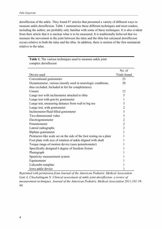
When discussing ITW with colleagues, in particular orthopaedic surgeons working with the adult population, they quite often comment, ‘Why bother? We never see adults who toe-walk. Leave the kids alone and they will be just fine!’ Is it so? To be able to answer this question, it is important to understand that dealing with children who toe-walk is not the same as dealing with just a peculiar way of walking. The toe-walking might have much wider implementations for the child in question, affecting his/her well being for a long time and from several perspectives. A good starting point for discussing potential problems caused by ITW is to look at the range of movement (ROM) of the ankle. Whether persistent toe-walking results in shortening of the calf muscles and Achilles tendon (commonly referred to as the triceps surae) has been extensively discussed. At present, no convincing high-quality study has been published which can conclusively answers that question; that would require a large cohort of children with follow-ups from birth to adulthood. Lacking such a study, one is left to draw as good conclusions as possible from what has been published so far. First of all, ankle dorsiflexion is not as easy to measure clinically as one might think. Gatt and Chockalingam13 reviewed different techniques used for measuring the
Pähr Engström
4
dorsiflexion of the ankle. They found 87 articles that presented a variety of different ways to measure ankle dorsiflexion. Table 1 summarises these different techniques and most readers, including the author, are probably only familiar with some of these techniques. It is also evident from their article that it is unclear what is to be measured. It is traditionally believed that we measure the movement in the joint between the talus and the tibia but calcaneal dorsiflexion occurs relative to both the talus and the tibia. In addition, there is motion of the first metatarsal relative to the talus.
Table 1. The various techniques used to measure ankle joint complex dorsiflexion
Device used
No. of Trials found
Conventional goniometer 23 Dynamometer, various (mostly used in neurologic conditions, thus excluded. Included in list for completeness)
20
Unsure 12 Lunge test with inclinometer attached to tibia 5 Lunge test with gravity goniometer 1 Lunge test, measuring distance from wall to big toe 3 Lunge test, with goniometer 2 Inclinometer/fluid-filled goniometer 3 Two-dimensional video 3 Electrogoniometer 2 Potentiometer 2 Lateral radiographs 2 Biplane goniometer 1 Protractor-like scale set on the side of the foot resting on a plate 1 Foot plate with axis of rotation of ankle aligned with shaft 1 Torque range-of-motion device (uses potentiometer) 1 Specifically designed 6 degree of freedom fixture 1 Photograph 1 Spasticity measurement system 1 Equinometer 1 Lidcombe template 1 Iowa ankle device 1
Reprinted with permission from Journal of the American Podiatric Medical Association Gatt A, Chockalingam N. Clinical assessment of ankle joint dorsiflexion: a review of measurement techniques. Journal of the American Podiatric Medical Association 2011;101:59-69.

Idiopathic Toe-Walking 5
5
There is no consensus as to what dorsiflexion limit defines a motion restriction. Some authors propose 5 degrees14, others 10 degrees15 or even 016 and 20 degrees17. The orthopaedic definition of ankle equinus is a foot in plantarflexion but the podiatric definition of equinus is less than 10 degrees of dorsiflexion13 (Podiatry or podiatric medicine is a branch of medicine devoted to the study, diagnosis and medical treatment of disorders of the foot, ankle and lower extremity). A cross-sectional study of 348 healthy children, adolescents and young adults by Engelbert et al.3 queried the presence of toe-walking in the past. The results showed that 9% of participants reported ITW and had a decreased dorsiflexion of the ankle. In 4% of participants, a decreased dorsiflexion was present without a history of ITW, and in 3%, ITW was present without decreased dorsiflexion. In 84% of participants, no dorsiflexion restriction or history of ITW was present. This means that participants with ITW had a 3 times greater chance of having a decreased ankle dorsiflexion. When reviewing other studies on children with ITW and restricted dorsiflexion, one can find many studies that describe their study population as having less than normal ankle dorsiflexion1
,18-34. Even if the studies are numerous and varied, they all agree that children (seen as a group) commonly labelled as idiopathic toe-walkers have a restricted ankle dorsiflexion or equinus contracture. Having said that, many clinicians have met children with ITW who have a normal dorsiflexion of the ankle. There are also published studies in which a connection between ITW and restricted dorsiflexion has not been found8 ,27. Stricker et al8. reported that ‘in absence of treatment, mild progression of heel-cord contractures occurred about as frequently as did mild regression’. These conflicting findings are most likely the result of the toe-walking population being a heterogeneous group amassed under one diagnosis. After a thorough examination of 28 toe-walkers, Furrer and Deonna4 were able to divide the toe-walking children into three main diagnostic groups: (1) Children with pyramidal tract signs, with or without motor delay and a limited dorsiflexion of the ankle. This group comprised all children with minimal cerebral palsy. (2) No pyramidal tract signs, no motor delay and no limited ankle dorsiflexion. These children were labelled habitual toe-walkers, which can be confusing as habitual toe-walking is also used synonymous with ITW. These two groups of children were early walkers, walking intermittently on toes. Most of them were very active and exploring children who had a normal development. (3) The final group had no pyramidal tract signs, no motor retardation and always limited ankle dorsiflexion. These were the children that sometimes are labelled as having a ‘congenital short Achilles tendon’. Some children in this group did not toe-walk at commencement of independent walking, indicating that the restricted dorsiflexion was not congenital but evolved later. From the author’s point of view, this grouping seems reasonable at present, until we know more about the different types of toe-walking. It is also a useful conceptual model to consider ITW and restricted ankle dorsiflexion. If this model reflects the clinical reality, then it is understandable why clinicians continue to argue about whether or not ITW leads to ankle contractures, as it would depend on which type of toe-walking one means. Further academic questions left to solve are whether Group 2 toe-walkers eventually develop contractures or not, and whether the contractures found in Group 3 toe-walkers are congenital or acquired. As mentioned above, further high-quality studies are needed to answer these questions.

Pähr Engström
6
If children with ITW, seen as a group, have a restricted ankle dorsiflexion, and a substantial number of these children also develop ankle contractures, what are the implications for the future? What is known about ankle contractures/equinus and its link to orthopaedic problems? A recently published article by DiGiovanni et al.35 in the American Journal of Bone and Joint Surgery stated that ‘except for a few still controversial examples of plantar fasciitis, forefoot ulceration in diabetics or progressive hallux valgus or flatfoot, the relationship between tightness of the superficial posterior compartment and progressive pathological changes in the foot in non-spastic individuals has been over-looked entirely by the orthopaedic community’. In contrast, the podiatric community has been more interested in these issues. Hill361995 published a study about ankle equinus and its link to common foot pathology. Hill linked equinus deformity to a variety of foot problems, such as plantar fasciitis, Achilles tendonitis, retrocalcaneal exostosis, calcaneal apophysitis, posterior or anterior tibial tendonitis, painful bunions, hallux limitus, collapsing arches, medial ankle capsulitis, painful heloma dura or heloma molle, peroneal tendonitis and Mortons neuroma. In addition Subotnic37 stated that ‘gastrocnemius or soleus equinus is the greatest symptom producer in the human foot’. These studies have been criticised for the methodology used but they indicate nonetheless that podiatrists showed an early interest in the effects of restricted dorsiflexion of the ankle. In later years, further articles have been published in orthopaedic literature about restricted ankle dorsiflexion and its relation to various pathological foot conditions, for example posterior tibial tendon dysfunction35, ankle sprains and fractures38, diabetic foot ulcers39, Charcot neuroarthropathy40 and metatarsalgia35. DiGiovanni et al.35 made a prospective comparison of two groups: one group of 34 consecutive patients with the diagnosis of metatarsalgia and a second control group of 34 individuals without any foot or ankle symptoms. With the knee fully extended, the average maximal ankle dorsiflexion was about 5 degrees in the patient group and 13 degrees in the control group. When the limit for gastrocnemius contracture was defined as 5 degrees of ankle dorsiflexion, it was identified in 65% of the patients compared to 24% of the controls. The authors concluded that ‘the findings support the existence of isolated gastrocnemius contracture in the development of forefoot and/or midfoot pathology in otherwise healthy people. These data may have implications for preventive and therapeutic care of patients with chronic foot problems’. Aronow41 makes an interesting description of how a limited ankle dorsiflexion may lead to foot and ankle problems:
‘An individual may compensate for limited ankle dorsiflexion caused by contracture of the gastrocnemius, soleus or Achilles tendon in several ways. One compensatory mechanism is an early heel off, which in its extreme would lead to a toe-walking gait. Alternatively, the centre of body mass can move forward relative to the foot by increased lumbar lordosis, hip flexion or knee recurvatum. Most commonly, however, increased dorsiflexion of the leg relative to the ground occurs through the joints of the hindfoot and midfoot. The subtalar joint goes into pronation, unlocking the talonavicular and

Idiopathic Toe-Walking 5
7
calcaneocuboid joints and allowing them to undergo increased dorsiflexion. Over time the spring ligament may stretch out, as well as the plantar ligaments of the naviculocuneiform and tarsometatarsal joints, leading to a progressive flatfoot deformity. The associated increased biomechanical strain on the posterior tibial tendon may lead to tendinitis, elongation or rupture. Increased Achilles tendon tension increases plantar fascia strain, likely secondary to the increased planovalgus deformity. In an attempt to overcome the triceps surae contracture, the relatively weaker anterior tibial may recruit the extensor digitorum longus and extensor hallucis longus muscles to help dorsiflex the ankle. This may lead to hyperextension of the metatarsophalangeal joints and resultant hammertoes. The triceps surae contracture may shift plantar weightbearing pressure from the hindfoot to the forefoot. This pressure shift, along with the distal plantar fat pad migration accompanying hammertoes, may lead to metatarsalgia and metatarsophalangeal joint synovitis.
Referring back to the question at the beginning of this section. ‘Why bother? We never see adults who toe-work.’ No, perhaps not, but the adult presentation of a childhood ITW may not manifest itself as toe-walking but instead as any of the problems described above. It is also important to remember that a decreased ankle dorsiflexion could be masked with compensatory mechanisms42 such as external rotation of the leg to shorten the lever arm of the foot, dorsiflexion through the talonavicular and calcanoeocuboid joints, shifting the centre of mass anterior in relation to the foot by hyperextension of knees, flexing the hip and increased lumbar lordosis or early heel off. A decreased ankle dorsiflexion could therefore easily be overlooked if not specifically checked for. 1.6 NORMAL GAIT DEVELOPMENT
Stepping-like movements can be seen in the fetus at 10‐12 weeks of gestation. These stepping movements can also be elicited in the neonate if he/she is held over a surface. These stepping like movements have been referred to as infant stepping. The child can usually walk with support at 7‐9 months of age and the movements are then voluntary and probably goal‐oriented and not as in infant stepping, induced by weight-bearing and stretching of the hips.43 Unsupported walking typically develops between 9‐15 months of age and is probably closely correlated to the development of postural control. The first pattern of locomotion is immature with co-activation of flexor and extensor muscles. At the end of the swing phase (foot off the ground) the plantarflexors activate, resulting in a digitigrade (toes first) gait pattern. At two years of age, the majority of children have developed a prominent heel strike, which includes active dorsiflexion of the forefoot. The transformation of the digitigrade to plantigrade gait is dependent on supraspinal circuits. Experience and activity‐dependent neural plasticity achieved during the first year of walking probably contribute too. Up to the age of 12 years, energy expenditure for walking is higher in children than in adults, indicating that the fine tuning and final maturation of locomotion is a lengthy process.43 Perry44 ,45 described the normal ankle kinematics (joint movement) pattern during walking. It can be divided into three rockers. The first rocker starts at heel strike with subsequent ankle

Pähr Engström
8
plantarflexion to lower the foot to the surface via eccentric contraction of the tibialis anterior muscle. At the second rocker, the ankle is relatively dorsiflexed as the tibia moves forward over the foot through eccentric contraction of the calf muscles. At the third rocker, the ankle plantarflexes as the gastrocnemius and soleus muscles contract concentrically. The third rocker is responsible for push-off at the end of the stance phase. During the swing phase, the dorsiflexion of the ankle is achieved through contraction of the tibialis anterior muscle. It is now, however, more common to acknowledge 4 ankle rockers, wherein the first ankle rocker as described above is called the ‘heel rocker’, the third ankle rocker described above is called the ‘toe rocker’, and the second ankle rocker is divided into the ‘ankle rocker’ (tibial advancement over the foot during stance) and the ‘forefoot rocker’, during which the ankle continues to dorsiflex, but the load is supported by the forefoot.46 Sutherland et al.47 studied normal gait development in 186 children aged 1 to 7 years from which they concluded that the heel-strike, knee flexion pattern, reciprocal arm-swing and an adult pattern of joint angles throughout the walking cycle are all acquired at an early age, before the development of mature cadence, step length and walking velocity. Close to 100% of the 18-month-old children walked with a heel-strike. The sagittal-plane angular rotations found in children from two years on are very similar to those of normal adults. Sutherland et al. also concluded that the five most typical determinants for a development to a mature gait are increased single-limb stance, increased walking velocity, decreased cadence, increased step length and an increased ratio of pelvic span to ankle spread. A mature gait pattern according to these determinants is usually well established at the age of three years. In their study carried out in 1971, Burnett and Johnson48 followed 28 children approximately every four weeks, filming their walking development. Seven children were followed and filmed for varying periods of up to 12 months and 21 children were followed from 12 to 22 months. Eighteen of the children were observed before they had developed an independent walk. Burnett and Johnson concluded that a consistent heel strike was present on an average of 22 weeks (range 3 - 50 weeks) after the commencement of independent walking. In all cases, the children initially made contact with the floor with their feet flat. Only two children were subsequently toe-walkers but progressed to a normal heel-toe walk in the follow-up periods. 1.7 THE TOE-WALKING GAIT
Several authors have examined children with ITW with electromyography (EMG),24-26 and a typical finding is a premature activation of the gastrocnemius muscle in the swing phase (the phase during which the foot is not in contact with the ground), indicating a premature plantarflexion of the ankle in swing. A gait cycle can be presented as percentage, wherein 0% represents the instance that one heel contacts the ground and 100%, the instance when the same heel contacts the ground again, i.e. completing a full gait cycle. Kalen et al.25 studied 18 children with ITW, comparing them with ‘known normal values’ and concluding that normal timing of gastrocnemius activity during the stance phase (foot in contact with the ground) is 15 - 50% of the gait cycle. The toe-walkers’ mean timing for the gastrocnemius activity was 92 - 52% of the gait cycle (i.e., starting during one gait cycle and continuing on to the next). During

Idiopathic Toe-Walking 5
9
the swing phase, normal tibialis anterior activity is 55 to 15% of the gait cycle. The mean timing for toe-walkers was reported as 53 - 3% of the gait cycle. This strongly indicates that the activity of both the gastrocnemius muscle, which is an ankle plantarflexor, and the tibialis anterior muscle, which is an ankle dorsiflexor, is out of phase. In other words, if the child’s ankle is observed in the sagittal plane, it first moves toward dorsiflexion, followed by a sudden plantar flexion midway through the swing phase, prepositioning the foot to land in equinus. With the development of quantitative 3-dimensional (3D) gait analysis, the walking pattern of idiopathic toe-walkers can be more closely studied in all three planes. This has meant that the joint movements and power generation can be measured much more accurately, and that comparisons can be made before and after an intervention. This has also helped physicians and surgeons tremendously in the evaluation of gait deviations before deciding on a specific treatment. As mentioned above, ankle kinematics (joint or segment motion) are often described as different ‘rockers’45 ,49. Westberry el al.20 investigated 51 idiopathic toe-walkers (mean age 9 years, range 6-18 years) with gait analysis and the three ankle rocker definitions. When the toe-walkers walked at a self-selected speed and in a self-selected fashion, the most striking ankle characteristics were:
- Absence of first rocker - Inverted second rocker - Swing phase disruptions with increased plantar flexion
This implies the absence of the normal plantarflexion of the ankle seen just after the foot contacts the ground, followed by a reversed movement of the ankle in relation to the tibia (plantarflexion) and an increased plantarflexion during the swing phase. Absence of dorsiflexor moment at initial contact This is a consequence of landing on the sole or forefoot instead of the heel. The gentle lowering down of the forefoot generally seen in normal gait is absent.
- Elevated midstance plantarflexor moment This is a consequence of supporting the ground reaction force under the forefoot during the single support phase instead of the normal progression of support from under the heel to the midfoot to the forefoot as seen in normal gait.
- Diminished plantarflexor moment in terminal stance The forefoot has supported the ground reaction force throughout the stance, causing high loading on the ankle plantarflexors. However, when these should be most active, i.e. during what is commonly known as 'push-off', the supported load under the forefoot and toes is abnormally small. Both of these observations (early plantarflexor moment at midstance and diminished plantarflexor moment in terminal stance) will lead to a less efficient gait.
Contrary to that in children with cerebral palsy, several authors have described a normal kinematic profile of the knee in ITW20 ,27

Pähr Engström
10
It is well-known that children with ITW can walk with a heel-toe pattern when asked to do so and that they have a varying walking pattern, sometimes walking quite normally.20 ,23-25 ,27 ,50 Consequently, Westberry at al. investigated all 51 of the children in their study after they were asked to make every effort to walk as normally as possible. They found that the toe-walkers could only normalize all characteristic parameters of the gait analysis in in 17% of these trials. In 70% of the trials, the children with ITW normalise either stance or swing phase parameters, but not both. The ability to normalise the gait parameters was not related to the ages of the children or the passive range of movement (PROM) of the ankle. 1.8 THREE-DIMENSIONAL GAIT ANALYSIS
Before three-dimensional (3D) gait analysis was available one was left to carefully observe a patient’s gait pattern and try to make appropriate conclusions. Naturally, it is impossible, using only vision, to record the complex interaction of movements from various joints to make a reliable description of a person’s walking pattern. Instrumented gait analysis has made quantification and documentation of gait data much easier. As expressed by Jacquelin Perry in her classic book: Gait Analysis, Normal and Pathological Function:45
The asynchronous series of changes occurring at each joint of the two limbs presents such a maze of data that few persons can assimilate it all. The result may be premature conclusions. An alternate approach is quantitated documentation of the person’s performance with reliable instrumentation that provides a permanent record of fact. The indecisions of subjective observation are avoided. A printed record of the patient’s motion pattern provides a reference base for interpreting EMG, stride and force data.
In 3D gait analysis, the collection of movement data from various body segments is facilitated by placing reflective markers on various anatomical landmarks on the patient. The markers reflect light from infrared cameras to sensors mounted on the cameras. Information from multiple cameras reconstructs the position of the markers in three dimensions. Kinematics is the term used to describe joint or segment motion. Force plates are placed on the floor of the gait laboratory. These plates register the position of ground reaction forces as the patient walks. By combining segmental data from the kinematic analysis with ground reaction force, joint forces and moments can be calculated. The term kinetics is used to describe joint moments and forces. The gait cycle is divided into different phases. The stance phase starts when the foot contacts the ground, which is called initial contact or heel strike, as it in normal gait is the heel that first contacts the ground. When the foot leaves the ground, which is called toe-off, as the toes are the last part of the foot to lift off the ground, the swing phase begins. One gait cycle represents the movements performed from initial contact with for example the right foot to the next initial

Idiopathic Toe-Walking 5
11
contact with the right foot. The gait cycle is converted into percentage were the stance phase makes up approximately 60% (from initial contact to toe-off). The remaining 40% represented by the swing phase starts with toe-off and ends with initial contact. Double support occurs twice during the gait cycle when both feet have contact with the ground. This makes up about 20% of the gait cycle. In addition, data about walking velocity, cadence (steps per minute) and step length are collected. 1.9 CLASSIFICATION
No generally accepted classification of ITW exists but Alvarez et al.30 developed a useful classification based on gait analysis parameters and the three ankle rocker definition. The classification identifies three severity types of ITW (mild, moderate, severe) based on the presence of a first ankle rocker (heel vs. forefoot contact), an early third ankle rocker (early heel rise) and a predominant ankle plantarflexion moment during loading response (forefoot weight acceptance). The classification is outlined in Table 2 and graphical illustration of ankle kinematic curves of normal and ITW gait cycles are outlined in Figures 1a and1b. Table 2. Idiopathic toe walking severity classification Toe walking severity group
Primary criteria and definitions Presence of first ankle rockera
Presence of early third ankle rockerb
Predominant first ankle momentc
Type 1 Yes No No Type 2 Yes or No Yes or No No Type 3 No Yes Yes
a Present if ankle angle at initial contact was greater than 5° of plantarflexion and there is a downgoing ankle excursion. b Present if third ankle rocker occur at or below 30% of the gait cycle. c Present if the ratio of the peak plantar-flexor moment at initial stance (AM1) to the peak plantar-flexor moment at late stance (AM2) is greater than 1 (Figure 1b). Reprinted with permission from Elsevier from Alvarez C, De Vera M, Beauchamp R, et al. Classification of idiopathic toe walking based on gait analysis: development and application of the ITW severity classification. Gait Posture 2007;26:428-35.

Pähr Engström
12
Figure 1a. Graphical illustration of first ankle rocker and early heel rise using ankle kinematics. The ankle kinematics for a normal gait cycle represented by the solid curve (-) is superimposed on ankle kinematics for toe walking represented by the dashed curve (- -).
Figure 1b. Graphical illustration of early predominant first ankle moment (AM1) using ankle kinetics. The ankle kinetics for a normal gait cycle represented by the solid curve (-) is superimposed on ankle kinetics for toe walking represented by the dashed curve (- -). Figure 1a & 1b reprinted with permission from Elsevier from Alvarez C, De Vera M, Beauchamp R, et al. Classification of idiopathic toe walking based on gait analysis: development and application of the ITW severity classification. Gait Posture 2007;26:428-35.

Idiopathic Toe-Walking 5
13
1.10 NEUROPSYCHIATRY
‘Neuropsychiatry is a field of scientific medicine that concerns itself with the complex relationship between human behaviour and brain function and endeavours to understand abnormal behaviour and behavioural disorders on the basis of an interaction of neurobiological and psychological–social factors. It is rooted in clinical neuroscience and provides a bridge between the disciplines of Psychiatry, Neurology and Neuropsychology’.51 Common neuropsychiatric diagnoses in children are attention deficit hyperactivity disorder (ADHD), autism spectrum disorders (ASD), developmental coordination disorder (DCD), language disorders, tics and obsessive-compulsive disorder (OCD). Reported prevalence for ADHD, DCD and language disorders are 5 - 7%.52-57 ADHD is three times more likely in boys than in girls and six to nine times more likely in boys in clinic-referred children.58 ,59 ADHD, probably the most common diagnosis, has been extensively discussed and debated in the media in the last few years. ADHD is characterised by different levels of inattention, hyperactivity and impulsivity and gives rise to academic, social, and emotional problems.58-60 Socially, ADHD children have more often poor relationships and emotionally, they often have poor self-esteem with an increased risk for depression and anxiety58-60. Individuals with ADHD present in childhood may continue to show symptoms in adolescence and adulthood.61 ,62. The extent of comorbidity is high, as ADHD symptoms often occur with e.g. learning disabilities, anxiety disorders and depression.63 Among clinicians working with children with pediatric neuropsychiatric disorders, there is a general impression that children with ADHD are more often toe-walkers than children with age-appropriate behaviours. However, to the best of our knowledge, there are no scientific studies exploring this correlation. The prevalence of ASD is about 1%64. Children with ASD can have various symptoms, which can be divided into the following three categories: Social impairment, communication impairment and repetitive behaviour.64 Comorbidity with other conditions is common, including intellectual disability, hyperactivity and anxiety.65 ,66 For children with an autism spectrum disorder or a communication/language disorder, the prevalence of ITW has been reported to be as high as 19 - 63%.5 ,6 ,67 Children with ITW have been reported to have an increased prevalence of various developmental delays. Following a study on 13 children with ITW, Shulman et al. reported that 10 out of 13 children had speech/language delays, 4 out of 12 fine motor delays, 4 out of 10 ‘visuomotor’ delays, and 3 out of 11 gross motor delays.6 In another study involving 163 children seen at ‘well-child visits’ and in which ITW was defined as “toe-walking during at least one month or at the examination”, the observed prevalence of ITW was 24%.67 The children with ITW (n=39) had a consistently lower mean language quotient compared to children without toe-walking (n=127). The specificity of toe-walking for low language scores was 85% but the sensitivity only 32%.

Pähr Engström
14
At a tertiary-level clinic, 799 children with developmental delays were studied, 224 of whom were toe-walkers.5 In this group the prevalence of toe-walking was 63% in children with autism, 40% in children with a ‘communication disorder’, 36% in children with mental retardation and 20% in children with learning disabilities.5 The development of a child’s abilities are often divided into domains such as motor skills, executive functions, perception, memory, language, learning, social skills and emotions/behaviours.68 In conclusion, it is well documented that children with ASD and language impairment have an increased prevalence of ITW but the prevalence of ITW in other neuropsychiatric disorders such as ADHD is unclear. 1.11 CHILD WELFARE CENTRES
Child Welfare Centres (CWC, in Swedish Barnavårds Central) provide the primary health care for children in Sweden.69-74 Their purpose is to promote children’s health and development from infancy to six years of age. Specially trained nurses and doctors (general practitioners or paediatricians) provide this service and nearly 100% of children attend follow-up checks.13-15 The instructions given to all CWCs in every county are set forth in a ‘method-book’ based on recommendations from the National Board of Health and Welfare. However, the details of these may differ from county to county but the same standardised CWC record is used all over the country69 The ‘5.5 years follow-up’ is the last visit to the CWC before the responsibility of health monitoring is transferred to the school health services. At the ‘5.5 year visit’, several different aspects of the child’s development are assessed such as gross and fine motor development, perception, executive functions, hearing, language and general development. 1.12 TREATMENT
A wide variety of treatment recommendations have been suggested for ITW. Physiotherapy Physiotherapy is often recommended to children with ITW but there is almost nothing written about the effects of physiotherapy as the sole treatment for ITW. Physiotherapy is of course not a defined treatment; in order to evaluate treatment results, one must first of all define exactly what kind of physiotherapy treatment that has been given. As this is not the case in the literature, treatment results are difficult to assess. In articles about ITW treatment published by Stricker el al.8 and Hirsch and Wagner12 and referred to in the ‘natural history section (1.4)’, physiotherapy formed one part of the treatment. Stricker et al. mention heel-cord stretching exercises under the supervision of a parent or

Idiopathic Toe-Walking 5
15
physical therapist and Hirsch and Wagner stated that the children performed passive stretching exercises to increase the length of the calf muscles and some children performed exercises to increase the active dorsiflexion of the ankle. Both studies concluded that this treatment probably did not alter the natural history. Whether any type of physiotherapeutic treatment can alter the spontaneous course remains to be evaluated. Casting Several studies have reported on the use of cast treatment for ITW (see below). The cast treatment generally consists of a 2 to10 week period of below knee walking casts, even if the type of casts varies between studies. The effect of the cast has been postulated to result from elongation of non-contractile elements 28 ,75 and increasing numbers of sarcomeres in series in the calf muscles 76. It could also be hypothesized that the casts function as a constant remainder of the walking pattern and that this may alter the walk. The casts also make it difficult to walk on one’s toes, further helping the child to adopt a different walking pattern. The reported outcome varies from very positive to no effect at all. A positive outcome was reported by Griffin et al.24, Katz and Mubarak77, Brouwer et al.28, Fox et al.9 and Stott et al18. The designs of the studies differ but it is notable that no randomised controlled studies have been performed, which obviously weakens the argument for cast treatment. Four of five studies involved less than 15 patients9 ,24 ,28 ,77. EMG was used as a outcome measure in the study by Griffin el al.24, who treated six ITW children (age range 5 - 9 years) with below knee walking casts for 6 - 8 weeks. All children attained a normal walking pattern after cast treatment and the pathological electromyography present before treatment normalised. The PROM of the ankle improved substantially. Follow-up time is only stated as post treatment and seems only to have been for a short period of time. Gait analysis was used to evaluate the gait only in the study carried out by Stott et al18. They reported on thirteen patients that had been toe-walkers as children and who were followed-up for an average period of 11 years following treatment. All children had first received serial casting for six weeks. Six children needed no further intervention and seven children went on to Achilles tendon lengthening (n=5) or Baker’s gastroc-soleus lengthening (n=2). At follow-up, all 13 patients were satisfied with the treatment. Only one patient reported minor limitations in sporting activities. Two subjects stated that they still, occasionally, walked on their toes. One of these patients had been treated by casting only and one had undergone surgery. At final follow-up, all but three patients had a visually normal gait. Equinus foot positioning was observed in one patient, at initial contact, who had only had cast treatment. Two patients had and early heel rise and they were both treated with surgery following their cast treatment. At 3D gait analysis, 12 out of 13 patients had deviations from normal gait. The most common deviation was restriction in ankle dorsiflexion in stance. The authors concluded that patient satisfaction was high after either cast treatment only or cast treatment plus surgical intervention. Restriction of ankle dorsiflexion in gait was difficult to detect visually but was commonly seen at gait analysis. The study suffers from its retrospective design, which makes it difficult to interpret.

Pähr Engström
16
There was no speculation from the authors whether they considered the good results attributable to the treatment only or whether it was possibly also an effect of natural history. The studies by Katz and Mubarak77, Brouwer el al.28 and Fox are prospective, single intervention studies without randomisation. The eight children in the study by Katz and Mubarak77 were followed clinically. The children were diagnosed as having hereditary tendo Achilles contractures. Based on the article, it is understood that these children would today probably have been diagnosed as having ITW. The average pre-treatment passive ankle dorsiflexion with an extended knee ranged from -10 to +5 degrees. For six of the children, treatment consisted of serial casting with dorsiflexion cutout casts, permitting active dorsiflexion exercises, and for two of the children, it involved Achilles tendon exercises under a physiotherapist’s management. The casting period lasted on average 7 weeks (range 2 - 16 weeks). After treatment, the average dorsiflexion of the ankle was 12 degrees (range 5 - 20 degrees). At the final follow-up, around 25 months after treatment (range 16 - 38 months), five children walked without toe-walking and two walked predominantly with a heel-toe gait with only occasional episodes of toe-walk. One patient was lost to follow-up. The study by Brouwer el al.28 reports on eight idiopathic toe-walkers (mean age 8 years, range 5 - 10 years) treated over a period of 3 - 6 weeks with a short-leg fibre glass walking cast. The mean dorsiflexion angle of the ankle increased substantially. The conclusion was that except for one child who preferred foot flat at initial contact, all the other children walked in a normal heel-toe fashion six weeks after treatment. The study by Fox et al.9 is the only study on cast treatment for ITW that involves more than 15 patients. The study followed 44 ITW children treated with walking casts for 3 - 10 weeks. The age of the children was between 24 months and 14 years and 4 months with a median age of 60 months. Also in this study, the dorsiflexion of the ankle evidently improved. The cast were set at neutral ankle position and changed every 2 weeks until there was a reduced resistance to passive ankle dorsiflexion. The mean follow-up was 14 months (range not stated). If parents were satisfied with the treatment and plantar grade walking was present, the child was discharged. However if problems returned, the children were recommended to return to the clinic. The outcome showed that 29 children (66%) either stopped toe-walking or improved to a extent that satisfied their parents. The authors also concluded that younger children appeared more likely to improve their gait. The results in different age groups are outlined in Table 4. A less favourable outcome was reported in the two retrospective studies carried out by Stricker el al.8 and Eastwood et al.11 Stricker el al.8 evaluated 80 children, of which 17 children (mean age 4 years, range 2 - 13 years) had been treated with short-leg casts (eight patients) or solid plastic ankle-foot orthoses (nine patients). Before treatment, seven of the children walked 100% of the time on their toes, eight children 75% of the time and two children 50% of the time. Median pre-treatment ankle dorsiflexion was 5 degrees. At follow-up 2-8 years later (mean 34 months), the median ankle dorsiflexion was still 5 degrees. Among the parents, 24% were satisfied with the treatment, 65% were neither satisfied nor dissatisfied and 12% were dissatisfied. The authors concluded that cast and brace treatment appear to offer little long-term improvement compared with no treatment at all.
Idiopathic Toe-Walking 5
17
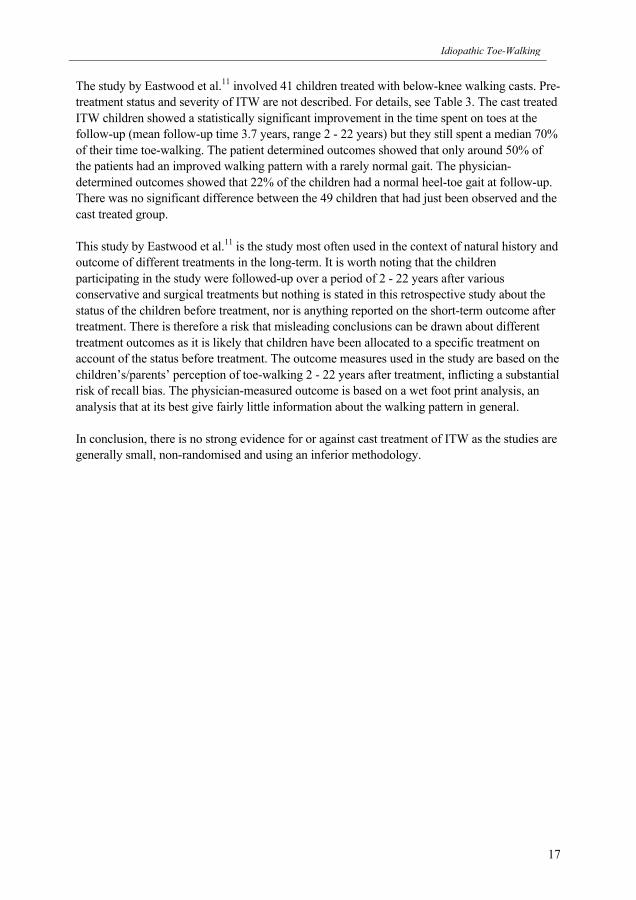
The study by Eastwood et al.11 involved 41 children treated with below-knee walking casts. Pre-treatment status and severity of ITW are not described. For details, see Table 3. The cast treated ITW children showed a statistically significant improvement in the time spent on toes at the follow-up (mean follow-up time 3.7 years, range 2 - 22 years) but they still spent a median 70% of their time toe-walking. The patient determined outcomes showed that only around 50% of the patients had an improved walking pattern with a rarely normal gait. The physician-determined outcomes showed that 22% of the children had a normal heel-toe gait at follow-up. There was no significant difference between the 49 children that had just been observed and the cast treated group. This study by Eastwood et al.11 is the study most often used in the context of natural history and outcome of different treatments in the long-term. It is worth noting that the children participating in the study were followed-up over a period of 2 - 22 years after various conservative and surgical treatments but nothing is stated in this retrospective study about the status of the children before treatment, nor is anything reported on the short-term outcome after treatment. There is therefore a risk that misleading conclusions can be drawn about different treatment outcomes as it is likely that children have been allocated to a specific treatment on account of the status before treatment. The outcome measures used in the study are based on the children’s/parents’ perception of toe-walking 2 - 22 years after treatment, inflicting a substantial risk of recall bias. The physician-measured outcome is based on a wet foot print analysis, an analysis that at its best give fairly little information about the walking pattern in general. In conclusion, there is no strong evidence for or against cast treatment of ITW as the studies are generally small, non-randomised and using an inferior methodology.

Pähr Engström
18
Table 3. Patient details and results Patient details Observation Cast Surgery No. of patients 49 41 46 Male-Female ratio 27:22 25:16 26:20 Age at presentation: Median (range) (y) 4 (1.5-1 0) 3.3 (1.5-1 0) 6.5 (2.5-1 3)* Age at treatment (y) 3.5 (1.5-1 0.3) 6.9 (2.5-14.5)* Duration of follow-up (y) 3.2 (2-12.8) 3.7 (2-21.5) 7.9 (2-22)* % Time toe-walking at presentation: Median (Range)
90 (90-100)
100 (80-100)1 100 (75-100)1
% Time toe-walking at review 60 (0-100)1 70 (0-100)1 25 (0-100)1
Patient-determined outcome Normal gait, n (%) 3 (6) 4 (10)2 10 (22) Improved, n (%) 22 (45) 17 (41) 23 (50) Unchanged, n (%) 24 (49) 20 (49) 12 (26) Worse, n (%) 0 0 1 (2) Physician-determined outcome Normal Gait, n (%) 6 (12) 9 (22)3 17 (37) Toe-walking, n (%) 43 (88) 32 (78) 29 (63) * The surgical group was significantly older at presentation and at treatment and had a longer follow-up. Kruskal-Wallis test, P = 0.0001. 1 For each of the groups, there was a significant reduction in the time spent toe-walking at review. Mann- Whitney test, P = 0.0001. 2 The differences between the observation and the cast groups were not significant when the normal and improved results were combined and compared to the unchanged results. Fisher's exact test, P =1. 3 The differences between the observation and cast groups were not significant. Fisher's exact test, P = 0.26.Reprinted from Eastwood DM, Menelaus MB, Dickens DR, et al. Idiopathic toe-walking: does treatment alter the natural history? Journal of Pediatric Orthopedics B 2000;9:47-9. With permission from Lippincott Williams & Wilkins
Table 4. Outcome following casting, results stratified by age
Age at treatment n= Successful Unsuccess- ful
%
All ages < 3 years 3-5 years 5-8 years 8-12 years > 12 years
44 11 9 12 10 2
29 9 6 6 6 2
15 2 3 6 4 0
66 82 67 50 60 100
Reprinted from Fox A, Deakin S, Pettigrew G, et al. Serial casting in the treatment of idiopathic toe-walkers and review of the literature. Acta Orthopaedica Belgica 2006;72:722-30. With permission from Acta Orthopaedica Belgica.
Idiopathic Toe-Walking 5
19
Botulinum toxin A Botulinum toxin (BTX) has been known to cause lethal poisoning for centuries. In medieval times, guild regulations were used to control sausage production, which was a major source of BTX poisoning78-82. The word botulinum is sprung from the Latin word botulus, which means sausage. The molecular action of BTX includes extracellular binding to glycoprotein structures on cholinergic nerve terminals and intracellular blockade of the acetylcholine secretion, causing a chemical denervation83. When BTX is injected in striate muscles, a paresis occurs that lasts for 2 - 12 months in children with CP.84 ,85 The reason for termination of the effect is a restoration of the protein complex (SNARE) in the cholinergic nerve terminals, which was inactivated by BTX86. In vivo studies in mice have shown extensive nerve sprouting at the affected nerves but it is believed to be a transient phenomenon not responsible for the termination of the BTX effect.86 There are seven different serotypes (A-G) of which type A has been most widely studied and used in medicine. There is a correlation between the amount of BTX injected and the extent of the paresis provoked87. BTX-A is quantified in units (U). Dr. Alan Scott, an American Ophthalmologist, was in 1980 the first to report on the therapeutic use of BTX in humans.88 Dr. Andrew Koman and colleagues published the first study using BTX to reduce increased muscle tone in children with cerebral palsy. Since then, the indications for BTX have expanded 89. Gradually its use became more commonplace, also in conditions were there was a lack of studies of its efficacy and safety. The first study (retrospective case series) of BTX for ITW was published in 2004, in which 10 children (2 - 17 years) with ITW were treated with 10U/kg BTX in the calf muscles, immediately followed by 1 - 3 weeks in a below knee walking cast and thereafter bracing and physical therapy. All children had ceased toe-walking at the 3 month follow-up but two children received repeated injections at 3 and 12 month follow-ups 90. Another study by Brunt et al.91 involving five children with ITW (mean age 4 years, range 3 - 6 years) and treated with 12U/kg BTX and physiotherapy also showed an improved walking pattern. Two of the children in that study, however, received repeated BTX injections and splints after 3 months. The ROM of the ankle before treatment is not stated but three of the children were reported to have a dorsiflexion ‘within normal limits’ while two had resistance to passive dorsiflexion due to contracture. The authors speculated about BTX being a possible treatment for ITW before contractures had developed and that improvement of clinical results may be possible through cast treatment post-BTX injection, particularly when an ankle plantarflexion contracture is present. Why would BTX affect the walking pattern in children with ITW? There are several reports24 ,25
,91 about EMG changes in ITW with out-of-phase gastrocnemius activity during the swing phase. Brunt 91 showed that swing phase gastrocnemius activity and an early offset of tibialis anterior activity may lead to a toe-strike. BTX was reported to change the gastrocnemius activity, resulting in heel strike or nearly heel strike, enabling extended tibialis anterior activity to be present at terminal swing and possibly at loading response. Another hypothesised reason for the effect may be the induced weakness in the calf muscles, making it difficult for the child to walk on the toes and thus resulting in a new walking pattern.
Pähr Engström
20
Other non-surgical treatments One study by Conrad and Bleck31 reports on treatment with ‘augmented auditory feedback’. Eight children (6 with CP and 2 with ITW) with a dynamic equinus were treated with a pressure-sensitive device in the shoe that produced an auditory signal when the heel contacted ground. The children used the shoes in practice sessions while walking for one hour every day. The two ITW children used the device for three and six months respectively. As regards the eight children, the authors report that the passive ankle dorsiflexion improved 8 degrees with the knee extended and 4 degrees with the knee flexed. After treatment, with the device off, there was a 38% improvement in the total accumulated number of heel strikes in a three-minute period. A ‘motor control intervention programme’ was used for five children (age 30 - 72 months) in a study by Clark et al.33 The intervention emphasised ‘activities and habits to influence muscle activation and posture deficiencies, with the objective of expanding the child’s ability to manage the body centre of mass over the feet’. The intervention consisted of two one-hour sessions per week over a period of nine weeks. Each child was provided with an individual ‘play’ programme, adapted to certain therapy objectives. The results did not show less toe-walking at follow-up. Surgery The aim of surgery for ITW is to lengthen the triceps surae muscle-tendon complex when a decreased dorsiflexion of the ankle is present92-100. Many different surgical techniques have been described to lengthening the triceps surae. Figure 2 outlines the different levels of the muscle-tendon complex were these various techniques are performed. To identify where the contracture is present, the Silfverskiöld test can be utilised. This test measures the ankle dorsiflexion with the knee in both extension and flexion. The measurement of ankle dorsiflexion with knee in extension is mainly a test of the gastrocnemius muscles’ length as these muscles cross the knee joint. Measuring ankle dorsiflexion with the knee in flexion, which slackens the gastrocnemius muscles, mainly tests the soleus muscle length.

Idiopathic Toe-Walking 5
21
Figure 2. The posterior leg can be divided into five anatomical levels. Based on anatomical level and clinical assessment, specific surgical procedures are indicated. The location for superficial gastrocnemius soleus recession is highlighted. GT, gastrocnemius tenotomy; GSR, gastrocnemius soleus recession; TAL, tendo Achillis lengthening. Printed with permission from Journal of the American Podiatric Medical Association. Lamm BM, Paley D, Herzenberg JE. Gastrocnemius soleus recession: a simpler, more limited approach. Journal of the American Podiatric Medical Association 2005 Jan-Feb;95(1):18-25. Two studies have compared the outcome after Achilles tendon lengthening and the Valpius procedure (McMulkin et al.34, Jahn et al.32). In a retrospective study by McMulkin et al.34, 14 children with ITW were treated with either Achilles tendon lengthening (six percutaneously and one open z-lengthening) or a Valpius gastrocnemius recession (seven children). A 3D gait analysis was performed before and after surgery. The average age of the children at surgery was in the Achilles lengthening group 8.5 years and was not statistically different from the Valpius group. The study was not randomised and the decision to perform an Achilles tendon lengthening was based on a limited ankle dorsiflexion with the knee in both extension and flexion. The Valpius procedure was used when there was only a limited ankle dorsiflexion with the knee in flexion. Post-operative gait analysis was performed at a mean of 13 months (range 10 - 17 months) after surgery. The Valpius group had a significantly greater dorsiflexion of the ankle before surgery compared with the Achilles tendon group. Postoperatively, all ITW children seen as one group showed an improvement in several gait parameters. The Achilles tendon group had a significantly better maximum knee extension in stance. The increased external foot progression noted pre-operatively in both groups had not changed following surgery. According to the authors, this was due to an external rotation of the lower leg and external rotation of the hips that was not corrected during surgery.

Pähr Engström
22
When examined physically after surgery, both groups showed significantly increased ankle dorsiflexion. The authors concluded that both surgical techniques produced a good outcome and they recommended the use of the Valpius technique for gastrocnemius tightness and Achilles tendon lengthening for a more severe contracture. Jahn et al. found a similar outcome in their study.32 These authors saw two main differences between the groups after surgery. Firstly, the gastrocnemius lengths in stance increased significantly after Achilles tendon lengthening but not after the Valpius procedure, whereas the soleus muscle lengths increased significantly after both procedures. Secondly, the muscle-tendon lengths were shorter in the Achilles tendon group before surgery but ended up longer compared to the Valpius group. The authors did not see this difference in lengthening as mainly an effect of the different surgical procedures but rather felt it was more related to the muscle-tendon length before surgery. The children with the most severe contractures gained much length while the children with minor contracture did not gain much in length. The kinematic outcome was good in both groups. No surgical procedure was recommended in favour of another. Let us now change focus from different types of surgery to what is known about the different results of surgery. The first description of surgery for children with a contracture of the triceps surae and a toe-walking gait was given by Hall et al.1 in 1967. Hall's 20 patients with a mean age of 8 years had all a contracture of the Achilles tendon and a dorsal extension of the ankle ranging from -30 to -60 degrees. The patients were first observed for 6 - 24 months but eventually all 20 patients underwent surgery for the lengthening of the Achilles tendon. Details of the surgeries are not stated. A cast was worn after surgery for six weeks without weight bearing and for three weeks with weight bearing. Follow-up after surgery ranged from 1.5 years to seven years (mean about three years). At follow-up, all children walked with a normal heel-toe gait with some of the children occasionally walking on their toes. A favourable outcome has also been reported by Kogan and Smith101 who, in a retrospective study, contacted 10 out of 15 children in a telephone survey 3 months to 6 years after a percutaneous Achilles tendon lengthening. Age of the children is not stated, nor the ROM of the ankle before surgery but all children could put their feet flat on the ground when asked before surgery. After the surgery, the children were placed in a below-knee walking cast for one month and were allowed full weight bearing immediately. At follow-up, according to the parents, no child toe-walked and no parent/child reported any loss of strength. All parents were satisfied with the procedure. Likewise, in a retrospective review of 15 ITW children treated by open (n=12) or percutaneous (n=3) Achilles tendon lengthening, Hemo et al.19 reported an encouraging outcome. The mean clinical follow-up was 3 years. At final follow-up, 12 out of the 15 children were, according to their parents, walking with a heel-to gait all the time. Three children had a heel-to gait most of the time and occasionally walked on their toes. One interesting follow-up parameter in this study was the strength of ankle dorsiflexion. The study concluded that ankle ‘push-off’ power generation improved after surgery but did not reach the level of the reference values for norms. All children could do 10 heel rises, which is considered the threshold for normal strength. Three patients had a significantly decreased power generation. Two of these patients did not have a equinus contracture before treatment and the

Idiopathic Toe-Walking 5
23
authors warn about performing Achilles tendon lengthening surgery on children without a true contracture as there seems to be a risk of excessive lengthening. One patient had a recurrence of toe-walking three years after surgery that was successfully treated with casts. Under the ‘cast section’ above, a study by Stricker et al8 is mentioned. In that retrospective study comparing different treatments for ITW, a group of 15 surgically treated ITW children was included. The overall number of children studied was 80, divided into three groups based on treatment. Group 1 (n=48) had been observed or prescribed special shoes and/or stretching exercises. Group 2 (n=17) was treated with casts (see above under cast treatment) and group 3 (n=15) was treated with open Achilles Z-plasty (n=11) or with bilateral, Baker-type, gastrocnemius recession (n=4). The dorsiflexion of the ankle before treatment and at final follow-up, 34 months after initial presentation (range 2 - 8 years), are outlined in Table 5 together with parental satisfaction with the outcome. All surgical treated children had an equinus contracture (0 to -20 degrees) of the ankle before surgery. Overall, only about one fourth of the parents were satisfied with the outcome in Groups 1 and 2. In contrast, two thirds of the parents were satisfied in the surgical group. Only one parent was dissatisfied. However, one third of the surgically-treated children were still, to some extent, walking on their toes. No cases of excessive lengthening, hypertrophic scars or other postoperative complications were noted. The authors concluded that surgical treatment showed significantly better results than conservative measures with respect to restoration of ankle dorsiflexion and parental satisfaction.
Table 5. Idiopathic toe-walking treatment results
Group 1 Group 2 Group 3
Median pre-treatment ankle DF 10° 5° -10° Median ankle DF at final follow-up
10° 5° 10°
Mean follow-up (mo) 36.0 34.6 32.0
Outcome
Satisfied (%) 25.0 23.5 66.7
Neutral (%) 54.2 64.7 26.6
Dissatisfied (%) 20.8 11.8 6.7
Reprinted from Stricker SJ, Angulo JC. Idiopathic toe walking: a comparison of treatment methods. Journal of Pediatric Orthopedics 1998;18:289-93. With permission from Wolters Kluwer Health Less convincing results were found in the other long term retrospective review by Eastwood et al.11, involving 136 children with ITW that had been treated by either observation (n=49), casting (n=46) or a Baker type of aponeurotic lengthening of the Achilles tendon, followed by six weeks in a below-knee walking cast. Patient details and results are shown in Table 3. The follow-up was done in a research clinic 2 to 22 years after first presentation. The results were

Pähr Engström
24
based on the parents’ perception of toe-walking frequency in addition to which the parents were asked to classify the severity of the toe-walking. The classification used three different grades of toe-walk. Walking on the tips of the toes (Grade 1), toe-walking on the metatarsal heads (Grade 2) or a toe-walking with the heel being off the ground during gait (Grade 3). Physician determined outcome to decide if a toe-walk was present was based on wet footprint analysis. It is worth noting in Table 3 that the parent/patient determined outcome revealed that only 22% considered their child to have a normal walk after surgery and that the physician determined outcome showed a normal walk in only 37% after surgery. Twenty-nine (63%) of the children were still, according to the physician, walking on their toes (extent not stated) at the follow-up. The authors concluded that direct comparisons between the different treatment groups cold not be made due to the differences in age at presentation and duration of follow-ups. It was likely that the outcome of a surgical intervention was an improvement in the natural history of ITW. To conclude, a favourable outcome was generally reported for ITW following lengthening of the Achilles tendon/calf muscles. No specific type of surgery can be recommended based on the current literature. Complications after surgery have generally been a minor problem but the risk of excessive lengthening, as described in one study, must be taken into account. No RCT studies comparing surgery to other types of treatment are available, nor are there large prospective studies, all in all reducing the scientific evidence for surgical treatment from the perspective of evidence-based medicine. However, when equinus contracture is present, surgical treatment seems to be the best option available on the basis of current evidence. 1.13 GENERAL REMARKS ON PUBLISHED LITERATURE
Even though a number of articles have been published about ITW, there is a complete absence of studies trying to define the prevalence of ITW in a large and well-defined cohort of children. Unfortunately, the same applies to randomised controlled trials (RCT) for the treatment of ITW. Treatment studies generally involve a fairly low number of participants evaluated after one type of treatment with comparisons to other treatments usually set forth in the discussion section. When two or more treatments have been evaluated, it has been done without randomisation. To be able to more accurately conclude anything about the natural history of ITW, a large and well-defined cohort of children must be followed from birth to adulthood. Different treatments must be assessed in RCT studies.

Idiopathic Toe-Walking 5
25
2 AIM OF THE THESIS
The overall aim of the thesis is to better understand different aspects of idiopathic toe-walking, a common and yet largely unexplored condition in childhood. The specific aims of the thesis are to elucidate:
The prevalence and early course of ITW in children aged 5.5 years in a well-defined cohort. Study 1
The occurrence of neuropsychiatric problems and symptoms in children with ITW. Study 2
Whether BTX treatment can improve the walking pattern in children with ITW. Study 3
Whether a combination of BTX and casting is more effective than casting alone in reducing toe-walking in 5 to15-year-old children. Study 4
Whether treatment outcome is correlated to co-existing neuropsychiatric problems. Study 4

Pähr Engström
26

Idiopathic Toe-Walking 5
27
3 MATERIALS AND METHODS
3.1 STUDY OUTLINES
Study 1 A cross-sectional prevalence study of children aged 5.5 years (n=1436) living in Blekinge County, Sweden, was performed during the regular ‘5.5 year visit’ at the local Child Welfare Centre (CWC). Children were assessed for a history of toe-walking or whether they still walked on their toes. Additionally, all children aged 5.5 years (n=35) admitted to the Clinic for Children with Special Needs in the county were assessed with regard to toe-walking. Study 2 Fifty-one consecutive children (31 boys, 20 girls) with a mean age of 9 years and 1 month and referred to Astrid Lindgren Children’s Hospital for ITW were enrolled in the study. Evaluations included assessments by a pediatric orthopaedic surgeon and a pediatric neurologist, and the parents were asked to complete the 5-15 (Five to Fifteen) questionnaire, a validated screening tool for neuropsychiatric problems. The study cohort was compared with an age-matched normative group. Study 3 A consecutive series of 15 children (aged 5 – 13 years) were enrolled in the study. The children underwent a 3-D gait analysis prior to treatment with a total of 6 units/kg bodyweight Botox® (BTX) in the calf muscles and an exercise programme. The gait analysis was repeated 3 weeks as well as 3, 6 and 12 months after treatment. A classification of toe-walking severity was made prior to treatment and after 12 months. The parents rated the perceived amount of toe-walking prior to treatment and 6 and 12 months after treatment. Study 4 All ITW patients referred to the Pediatric Orthopedics Department at Astrid Lindgren Children’s Hospital between November 2005 and April 2010 were considered for inclusion. Forty-seven children constituted the study population. Children were randomized to a 4-week treatment with below knee circular casts, either as sole treatment or 1 - 2 weeks after undergoing injections with 12 units/kg bodyweight BTX (Botox®) in the calves. Before treatment and 3 and 12 months following cast removal, all children underwent 3-D gait analysis. Classification of ITW severity based on the gait analysis was performed and parents were asked to assess how much time their child spent on toes while walking barefoot. The passive range of motion in hip, knee and ankle joints was measured with a goniometer and ankle dorsiflexor strength was measured with a hand-held dynamometer. Prior to treatment, all children were evaluated with a screening questionnaire for neuropsychiatric problems.

Pähr Engström
28
3.2 SUBJECTS
The thesis cover four studies (1, 2, 3 and 4)
Table 6. Subjects Study n Age
(Years) Category Main effect studied
1 1436 5-6 General population Prevalence and early course
2 51 5-14 Referred for ITW Neuropsychiatric symptoms
3 15 4-13 Referred for ITW Effect of BTX treatment on walking pattern
4 47 5-l4 Referred for ITW Treatment outcome after cast or cast + BTX treatment
Overlapping subjects: All children included in Study 4 are also included in Study 2. Inclusion / Exclusion criteria Study 1: Inclusion of all children attending the regular ‘5.5 year visit’ at local CWCs in Blekinge County from 3 May 2010 to 15 June 2011 and all children of the same age, listed at the Clinic for Children with Special Needs in Blekinge County. There were no exclusion criteria. Studies 2 and 4: All children with ITW, consecutively referred to the Pediatric Orthopaedics Department at Astrid Lindgren Children’s Hospital between 15 November 2005 and 15 April 2010 were considered for inclusion. Each child should, as perceived by the parents, have walked at least 25% of the time on his/her toes for a minimum period of three months. Other inclusion criteria were no other pathology that could explain the toe-walking and no known congenital Achilles tendon contracture. Exclusion criterions were previous ITW treatment such as Achilles tendon surgery, casting, orthotics or BTX treatment. Study 3: All children with ITW, consecutively referred to the Pediatric Orthopaedics Department at Astrid Lindgren Children’s Hospital between November 2003 and May 2005, were considered for inclusion. Otherwise, all inclusion and exclusion criteria were the same as in Studies 2 and 4. 3.3 METHODS
Study 1 General Before starting the study, one of the authors (PE) visited all CWCs informing the staff about ITW and the study. The staff at the CWCs was instructed to classify a child as a toe-walker regardless of the amount or duration of toe-walking. One registered nurse at each CWC was

Idiopathic Toe-Walking 5
29
assigned as the local study contact. At the ‘5.5 year visit’, the staff at the CWC questioned all parents whether their child still, to any extent, walked on his or her toes (active toe-walking) or whether their child had a history of previous toe-walking (inactive toe-walking). Question forms were sent to one of the authors (PE) at the Orthopaedic Clinic in Blekinge County. The parent(s) of the inactive toe-walkers were interviewed over the telephone and asked to specify during which time in the child’s life the toe-walking had been evident and estimate the maximum percentage of time (25, 50, 75, or 100%) that the child had spent on his/her toes when walking barefoot. In addition, questions were asked about the child’s age when starting to walk independently, when toe walking was first noticed and whether there was any family history of toe-walking. Lastly, any medical conditions that could be considered as a cause of toe walking were elicited. All children classified as active toe-walkers at the time of the ‘5.5 year visit’ were clinically examined at the Orthopaedic Outpatient Clinic and parents were given the same questions as the inactive toe-walkers. The clinical assessment focused on detecting a possible cause of toe-walking and included an orthopaedic examination with emphasis on the lower extremities as well as a neurological examination. The range of movement in the hip, knee and ankle joints was measured with a goniometer (ankle range of motion was assessed with the knee both flexed and extended). Gait, deep tendon reflexes, muscle tone and presence of possible Babinski signs were evaluated. The same paediatric orthopaedic surgeon (PE) conducted all assessments. Parents of children in the appropriate age range and listed at the Clinic for Children with Special Needs were interviewed over the telephone and questioned about their child’s medical/developmental diagnosis and toe-walking history. The same set of questions used for the other groups of children was asked. To check for method validity, 10% (n=140) of the parents of the 5 – 6-year-old children who were assessed at different CWCs were randomly chosen and contacted after the study. It was verified that the parents had understood the question about ITW at the ‘5.5 year visit’ and that no child with ITW had been missed. Outcomes The ‘Five to Fifteen’ questionnaire (Study 2) The parent questionnaire ‘Five to Fifteen’ (FTF) was developed in order to screen for neuropsychiatric symptoms in children five to fifteen years of age.102 The questionnaire was designed to tap into the problems and strengths most commonly encountered in children and young adolescents presented with ADHD, tics and/or high-functioning autism spectrum symptoms. The questionnaire has been validated in several studies.102-104 The FTF is comprised of 181 statements related to behavioural or developmental problems that can be endorsed as ‘does not apply’ (0), ‘applies sometimes or to some extent’ (1) and ‘definitely applies’ (2). The statements are arranged into eight different domains, which can be further split into 22 subdomains. For statement examples, see Table 1 in Study 2. For each

Pähr Engström
30
domain/subdomain, a mean score ranging from 0 - 2 can be calculated, with higher scores implying more problems. The studied child/children can be compared with normative data, based on a study of 769 five-year-old children 68 and on one study of 854 children aged 6 - 15 years .102 The latter group was divided into three age groups, i.e. 6 - 8 years, 9 - 12 years and 13 - 15 years. The mean, median, 90th percentile and 98th percentile for the domains/subdomains of the norm group are stated in the manual for the questionnaire. As it is much more common that the parents score 0 than 1 or 2 in the different domains, the results do not represent a normal distribution. Thus, it is more appropriate to compare medians than means in the statistical analysis. Gait analysis (Studies 3 & 4) An 8-camera system (Vicon® MX40, UK) with 15 retro-reflective markers placed on anatomical landmarks was used.105 Children walked barefoot at a self-selected speed and were instructed to walk in their usual manner. Twenty-seven discrete kinematic and kinetic values from each gait cycle were obtained. Three trials were analysed, each containing one right and left gait cycle. Average values were calculated for each side of every child. Walking speed and step length were normalized to leg length. Classification of ITW severity as per Alvarez et al. was performed, using the gait analysis data, before treatment and at 3 and 12months follow-ups 30. Parents’ perception of toe-walking frequency and side effects (Studies 3 & 4) The parents rated the time their child spent on his/her toes during barefoot walking to the nearest quartile (0%, 25%, 50%, 75% or 100%) before treatment and at 3 and 12 month follow-ups. They were also asked whether they had noticed any side effects of the treatment. Joint range of motion measurements (Studies 3 & 4) An experienced physiotherapist used a goniometer to measure the PROM in hip, knee and ankle joints.106 Ankle PROM was measured with knees in both extended and 90° flexed positions. Strength of ankle dorsiflexors (Study 4) Ankle dorsiflexion strength was measured with a handheld dynamometer (MicroFET2®, Hoggan Health Industries, USA). Each child lay supine on an examination bed with calves overhanging the bed. The moment arm was measured from the lateral malleolus to mid-foot dorsally. The physiotherapist held the dynamometer to the child’s mid-foot dorsally (while stabilising the heel) and resisted motion while the child pressed the dynamometer for 5 seconds 107. The highest force of three measurements was used for analysis. Strength measurements were normalized to the weight of each child108. The same physiotherapist performed all measurements.

Idiopathic Toe-Walking 5
31
Interventions Casting (Studies 3 &4) All children were casted with below-knee walking circular casts in Softcast® reinforced with Scotchcast® (3M, Saint Paul, Minnesota, USA) posteriorly from toes to mid-calf. A nurse with special education in casting techniques applied all casts. The aim was to cast the ankle in a neutral position. Children wore the casts for four weeks. Children in the CA+BX group (Study 4) received their casts one to two weeks after BTX injections.
Botulinum toxin A treatment Study 3 The children underwent bilateral treatment with a total of 6 units/kg bodyweight Botox® (Allergan, California, USA) with a maximum of 400 units. One hour before the injection, all children were given oral paracetamol (40 mg/kg) and a topical anaesthetic cream (EMLA®, AstraZeneca, Sweden) was applied at the injection sites. For patients/parents who requested sedation, 0.3 - 0.5 mg/kg midazolam was given 15 minutes prior to injection. Four injection sites in each calf, two in the proximal third of the lateral and medial gastrocnemius bellies and two distally in the gastrocnemius-soleus complex were administered. All injections were given with electromyogram amplifier guidance to ensure intramuscular position. Study 4 Children in the CA+BX group underwent bilateral treatment with 12 units/kg bodyweight Botox® (Allergan, California, USA). One hour before the injection, all children were given oral paracetamol (40 mg/kg) and a topical anaesthetic cream (EMLA®, AstraZeneca, Sweden) was applied at the injection sites. Just prior to and during injections, all children inhaled nitrous oxide for pain relief and sedation. Injection technique was the same as in Study 3. Stretching program After cast removal, all children/parents were given verbal and written instructions by a physiotherapist to perform calf muscle stretches five times a week and walk at least 50 steps on their heels every day. No ‘training diary’ was used.
3.4 STATISTICS
Study 1 The effect of gender was analysed with χ2 test with one degree of freedom: p ≤ 0.05 for statistical significance. Study 2 Medians of the domains and sub domains were computed. The approximate 90% and 98% confidence intervals for the median in the study group were calculated. A significant difference

Pähr Engström
32
of the median in the study and norm group was considered when the median for the norm group was not within the confidence interval of the median for the study group. Study 3 All statistical analyses were based on the Intention To Treat principle (ITT), i.e. patients participating in the study being included in the statistical analysis even if they withdrew from the follow-up. Gait data were analysed with a mixed model using commercially available statistical software (SPSS, Chicago IL, USA). Measurements were performed before treatment at intervals of 3 weeks, 3 months, 6 months and 12 months prior to treatment. The within-subject factors in the mixed model were Time (5 testing occasions) and Side (left and right). Patterns over time were evaluated by Helmert contrast 109, which compares the mean of the dependent variable at time j with the average of the subsequent means, thereby indicating the time point of the appearance of the treatment effect. P<0.05 was considered statistically significant. Passive joint ranges of movement, before treatment and after the follow-up at 12 months, were analysed with Wilcoxon signed ranks test. Study 4 Statistical analysis was based on the Intention To Treat (ITT) principle. Power analysis was performed to perceive a difference of 5° in ankle angle with SD=9.5° (based on pilot data), i.e. an effect size of 0.5 at the alpha=0.05 significance level. A study population of n=50 is equal to a sample power of 0.940 and with n=40, the sample power is 0.881. In this study; n=47. Gait data and ankle PROM were analysed with a mixed model. Measurements were performed before treatment and at intervals of 3 and 12 months after treatment. The within-subject factors in the mixed model were Time (3 testing occasions) and Side (left and right) and the between-subject factor was Group (CA or CA+BX). In order to analyse hip and knee PROMs, a Generalized Estimating Equations (GEE) model was combined with the Genmod procedure (SAS®, System 9.1, USA), as the limited variability in measured angles is not suitable for a mixed model. Gender distribution between groups was analysed with a χ2 test. Age distribution between groups was analysed with the independent samples t-test. The within-group time effect of parents’ perception of toe-walking and classification of toe-walking severity was analysed using Friedman’s two-way analysis of variance by ranks. The difference between groups at all testing occasions was analysed with Mann-Whitney U-tests. In order to evaluate whether the presence of neuropsychiatric problems had any effect on the treatment results, three parameters were used to express the overall treatment effect: Ankle angle at initial contact, ITW classification and parents’ perception of toe-walking frequency. The difference between pre-treatment and 12-month post-treatment evaluations for these three parameters was computed. Correlations between the overall treatment effect and number of domains in which the child scored above the 90th percentile were computed using Spearman rank tests.

Idiopathic Toe-Walking 5
33
4 RESULTS
Study 1 Out of all 1,677 eligible children living in the County of Blekinge and reaching an age of 5.5 years during the study period, 1,436 children or 86 % constituted the study population. Out of the participating 1,436 children, 35 were listed at the regional Clinic for Children with Special Needs. Eighteen of these children had a motor disorder such as cerebral palsy, spina bifida or tethered cord, excluding them as ITW children. However, these 35 children were included in the prevalence calculation for the whole study cohort (n=1,436). Calculation of prevalence was also performed on the 1,401 children without a motor or neuropsychiatric disability. These children are referred to as ‘healthy children’. Out of the 1,436 children (750 boys, 686 girls), 30 children (2.09%) still walked on their toes and were considered active toe-walkers. Forty children (2.79%) had previously walked on their toes but stopped before the ‘5.5 year visit’ and were considered inactive toe-walkers. At an age of 5.5 years, the total prevalence for children ever having been toe walkers was 70 out of 1,436 = 4.87% (CI 95%: 3.76 - 5.99%). Details about the prevalence and different subgroups (active, inactive) are outlined in Table 1, Study 1. There was no statistical difference in the prevalence between boys and girls in the total study population, nor in the active or inactive subgroups. Study 2 All parents completed the FTF form. The study population constituted nine 5-year-old, fifteen 6 – 8-year-old, twenty-one 9 – 12-year-old and six 13 – 15-year-old children. In the study group, a higher percentage of children scored above the 90th percentile in all eight domains compared to the norm group, representing difficulties in the whole spectrum of abilities (motor skills, executive functions, perception, memory, language, learning, social skills and emotions/behaviours). A higher percentage of children in the study group than in the norm group scored above the 98th percentile, indicating considerable difficulties in five domains (motor skills, executive functions, memory, language and learning). The percentage and numbers of children scoring at or above the 90th and 98th percentile for each domain are outlined in Table 2, Study 2. Study 3 Eleven of 15 children completed the 12-month study period. Three participants were evaluated for six months and one patient was evaluated for three months. All children had a normal range of motion in hip and knee joints before and after treatment. One patient had a 25º equinus contracture at baseline and three children achieved a neutral ankle joint position but no dorsiflexion range. There was no significant difference in passive range of motion of the ankle between the 12-month follow-up and baseline values before treatment. Maximal dorsiflexion of ankle and parents’ ratings of toe-walking is shown in Table 1, Study 3.

Pähr Engström
34
Gait analysis There were no significant differences in pelvis and hip parameters during the follow-up period. The results for the entire study group at the 12 month follow-up compared to pre-treatment indicate a decreased plantarflexion angle at initial contact (p<0.001) and during swing phase (p<0.001) and an increased dorsiflexion angle during stance (p<0.001). Maximum plantarflexion during the gait cycle decreased (p<0.001). The timing of maximum dorsiflexion and transition from dorsiflexion to plantarflexion ankle position occurred later in the gait cycle at the 12-month follow-up compared to pre-treatment (p<0.001, p<0.001 respectively). Analysis with Helmert contrast showed that the observed significant changes in gait analysis variables occurred between the pre-treatment gait evaluation and later follow-ups (p<0.001), i.e. between the pre-treatment value and the mean of all post-treatment values. There was no significant change between any post-treatment follow-up evaluations. According to the classification of ITW severity at the initial assessment and the 12-month follow-up (6-month follow-up for 4 children) in the 14 children for whom all relevant data was available, 9 out of 14 children displayed improvement. Four children went from severe to moderate and 3 children from severe to mild. Study 4 Fifty-two families agreed to participate in the study and 47 children were allocated to treatment with casts plus BTX (CA+BX group) or only casts (CA group). For flow of subjects through the study, see Figure 3. There were no differences between the groups in distribution of age and gender. There were no significant differences in any gait analysis parameters between the CA and CA+BX groups before treatment or at the 3 and 12-month follow-ups. In both groups, several gait analysis parameters had improved significantly at the 3 and 12-month follow-ups, see Table 2, Study 4. Classification of ITW severity showed no difference between the groups at any time. Both groups improved significantly from before treatment to follow-ups with no change between the 3 and 12-month follow-ups. Thus, the most significant improvement occurred between treatment and the follow-up at 3 months. Corresponding to parental perception, there was no difference between CA and CA+BX groups in toe-walking frequency before treatment. At the 3-month follow-up, parents in the CA+BX group perceived a significantly lower toe-walking frequency than parents in the CA group (p=0.033), though the improvement (i.e. change) seen between the pre-treatment and post-treatment at 3 months was not quite significantly different from one group to another (p=0.053). No between-group difference was observed at the 12-month follow-up. Ankle dorsiflexion PROM with knees both flexed and extended was shown to have increased in both groups at the follow-ups. No difference was observed between groups at any testing

Idiopathic Toe-Walking 5
35
occasion. This also applied to differences between groups in ankle dorsiflexion strength before treatment or at follow-ups.
Figure 3.
BX=Botulinum toxin A
Assessed for eligibility (n=103) Excluded (n=51) ♦ Flexion contracture in ankle
beyond 10 degrees (n=19)
♦ Neurologic symptoms (n=3) ♦ Treatment before study
(n=3) ♦ Declined to participate
(n=26)
Analysed (n=26) ♦ Excluded from analysis (n=0)
Lost to follow-up (n=0)
Allocated to casting treatment (n=26): CA Group ♦ Received allocated intervention (n=26) ♦ Did not receive allocated intervention
(n=0 )
Lost to follow-up (n=0 at 3 months, n=1 at 12 months).
Allocated to casting+botulinum toxin A treatment (n=26): CA+BX Group ♦ Received allocated intervention (n=21) ♦ Did not receive allocated intervention
(n= 5) Reason for no intervention; Patients /children afraid of BX (n=4) Child moved away from area (n=1)
Analysed (n=21) ♦ Excluded from analysis (n=0)
ALLOCATION
ANALYSIS
FOLLOW-‐UP
Randomized (n=52)

Pähr Engström
36

Idiopathic Toe-Walking 5
37
5 DISCUSSION
Study 1 The prevalence of ITW has previously only been conjectured. We are the first to report reliable figures of its prevalence. At 5.5 years of age, 2.09% of children are ITWs and an additional 2.79% of children have previously been toe-walkers but stopped at an age of 5.5 years. For children with an autism spectrum disorder or a communication/language disorder, the prevalence of ITW has been reported as much higher, i.e. 19 - 63% in several studies.16, 18, 19 Thus, the prevalence of ITW appears to be lower among children with a typical development while it is a common symptom among children with a cognitive or neuropsychiatric disorder. Overall, most children who are idiopathic toe-walkers will not have any developmental problems or neuropsychiatric symptoms. Hopefully, it will help in the decision-making process of clinicians to know that 50% of children who start toe-walking early in life will have ceased toe-walking when they reach an age of 5.5 years. As long as no ankle contractures develop, it seams advisable to simply observe these young children and await the spontaneous course. On the other hand, for those children who continue toe-walking, continued follow-ups are advisable. As outlined in the introduction, ITW children have, as a group, a decreased dorsiflexion range of the ankle. As further outlined in the introduction, this may cause a variety of problems in the long term. From a social perspective, it is not uncommon to hear these children state that they have attracted nasty comments from peers and been the victim of bullying owing to their toe-walking. The size of this study gives it a high level of validity. In addition, the validity of the study is further enhanced by the interviews performed after the study completion with a randomly-chosen 10% of parents. This was done to evaluate whether any toe-walkers had been missed and whether the parents had understood the question about ITW asked by the CWC. For some aspects of this study there is a risk of recall bias; it is sometimes difficult for parents to remember when the child started to walk independently, when the toe-walk was first noticed and when it ended. We believe this recall bias could be more frequent in inactive rather than active toe-walkers. Another area of this study where parental answers could be less precise is perhaps the perceived percentage of toe-walking frequency. Lastly, the family history of ITW might be underestimated, as parents do not usually question all their relatives about toe-walking. About 40% of the children had a first or second degree relative who had been a toe-walker. This is in line with previous studies, which have reported a family history in 30 - 40% of children with ITW.5, 8, 17
The cohort of children with a neuropsychiatric disorder in this study is too small from which to draw any conclusions. Nevertheless, the prevalence of 41.2% ITW is in line with larger studies, which establishes this relationship. 16, 18, 19

Pähr Engström
38
Study 2 The study population had more neuropsychiatric symptoms compared to a normative group of age-matched children. This is the most important finding of this study. Almost 25% of the children had problems in all aspects of behavioural or developmental issues, assessed through the FTF. Although boys were overrepresented in this study, with boys more often than girls having neuropsychiatric problems102, it may only marginally contribute to our results as no effect of gender on subdomain scores could be seen. It is important to stress that a neuropsychiatric diagnosis cannot be established after an evaluation based only on a questionnaire. A thorough step-by-step investigation carried out by a multi-professional team and which includes the assessments of a number of professionals such as a child psychologist, a pediatric neurologist or a child psychiatrist as well as a speech therapist, would be required. The FTF is merely a screening tool that could help to identify children at a higher risk for neuropsychiatric problems than the general population. Our clinical impression and hypothesis was that children with ITW are more likely to have neuropsychiatric problems than age-matched controls, a hypothesis supported by the results of this study. As the cause of ITW is unknown, one can only speculate about the reason for this. As Forssberg described108, the first pattern of locomotion is immature with coactivation of flexor and extensor muscles resulting in a digitigrade walking pattern. At two years of age, the majority of children have developed a prominent heel strike, which includes active dorsiflexion of the forefoot. The transformation of the digitigrade to plantigrade gait is dependent on supraspinal circuits. Experience and activity‐dependent neural plasticity achieved during the first year of walking is probably a contributing factor. A hypothesis is that this modulation of the immature walking pattern is affected due to a central nervous system dysfunction, possibly also causing or contributing to the neuropsychiatric symptoms. Another theory that has been put forward is that toe-walking is due to a sensory processing dysfunction. This complex conceptual model is explained as ‘the neurological process that organises sensation from one's own body and from the environment and makes it possible to use the body effectively within the environment’.110 Williams et al. recently examined the theory of sensory processing dysfunction as a cause for ITW in a review article.111 The review could not confirm a relationship between ITW and sensory processing dysfunction but concluded that no high-quality studies have been performed confirming or excluding such a relationship. The theory is interesting since in ASD, the neuropsychiatric disorder with the highest reported degree of toe-walking, it has been proposed that the varied symptoms expressed may partially be due to an atypical use of sensory input to generate movements and interact with others112 ,113. Overall, ITW has been reported in 19 - 63% of children with ASD 5 ,7 and in 40 - 77% of children with a communication/language disorder.5 ,6. We believe that this can be taken as an indicator of some common pathology, possibly affecting sensory input processing and motor feedback among children with ITW only or with ITW plus a neuropsychiatric disorder.

Idiopathic Toe-Walking 5
39
In our opinion, parents of children with ASD seldom ask medical advice for their child’s toe-walking unless the child displays severe problems or contractures. Thus, none of the children in this study had ASD, possibly resulting in an underreporting of the number of children with neuropsychiatric symptoms. The most important conclusion that can be drawn from this study is that with a validated screening protocol, children with ITW display an increased prevalence of neuropsychiatric problems over the whole spectrum of neuropsychiatric symptoms. These findings warrant larger studies in the future, which ideally ought to include a more thorough neuropsychiatric and cognitive assessment. Study 3 As measured with 3-D gait analysis, several gait parameters improved after BTX treatment, e.g. decreased plantarflexion angle at initial contact and during swing, increased dorsiflexion angle during stance, decreased maximum plantarflexion, timing of maximum dorsiflexion and transition from dorsiflexion to plantarflexion. A possible explanation for this improved gait pattern could be a change in timing of muscle activity in the muscles affecting the ankle joint as described by Brunt 91 and outlined in the introduction. The improvement of reduced plantarflexion as seen in the present study may be attributed to a similar effect. The prolonged effect of BTX on the walking pattern after only one BTX injection is considered to be caused by a modified walking pattern adopted by some children under the influence of BTX. This study did not find any relationship between passive ankle dorsiflexion and the amount of toe-walking, which is in line with other studies 18 ,23. Passive range of motion of the ankle was unchanged after BTX treatment and thus, the treatment effect on the walking pattern cannot be attributed to an improved passive range of motion of the ankle. It is reported that idiopathic toe walkers can spontaneously adopt both a toe/forefoot and a heel contact pattern during the same gait analysis 23. This is very likely the reason why in this study less consistency between individual gait cycles was seen compared to other patient populations examined with 3-D gait analysis. Hence, it could be questioned whether a gait analysis represents the child’s most common gait pattern. It might be possible that a child with ITW walks less on his/her toes when being observed due to activation of higher cerebral functions to monitor the walking pattern, which is normally automatically regulated on spinal cord level 114. As the gait analyses were undertaken in the same environment with the same staff member giving identical instructions, these possible bias effects on the outcome both before and after treatment were hopefully minimized. Westberry et al20 have shown that when children with ITW are asked to concentrate on walking as normal as possible, only 17% of the children can normalise all gait analysis parameters while 70% can normalise either stance or swing phase parameters but not both. This indicates that even if the children walk with ‘an observed gait’, they still walk with the characteristics of a toe-walker most of the time.

Pähr Engström
40
According to the parents’ perception of their children’s toe-walking frequency, 6 children reduced their toe-walking whereas only 3 out of 15 children ceased to toe-walk. Even if the parent questionnaire is not a validated tool, we still regard the parents’ opinion as very important, as it gives us a good indication of their child’s walking pattern in an unobserved environment. Nine out of 14 children improved their walking pattern according to Alvarez classification, meaning that either presence of first ankle rocker, early third ankle rocker and predominant ankle moment during loading response shifted towards normalisation at the gait analysis, even though this does not mean they stopped toe-walking. Comparing our result with BTX for ITW versus other treatment options would of course have been easier if a general accepted classification for ITW existed and had been used in previous studies. The children who responded to the treatment and ceased toe-walking also had a favourable outcome in the long-term as confirmed through telephone contacts 3 to 5 years following completion of the study. Overall, the 12-month follow-up was a good indicator of the outcome over a 3 – 5-year perspective. This study, being a pilot study before a main RCT study of BTX treatment for ITW, has several limitations. Even if it is the largest study exploring the effects of BTX on walking patterns in children with ITW, the number of children studied is still quite small and only 11 out of 15 participating children could be followed for the entire study period of 12 months. One obvious limitation is the lack of a control group receiving no treatment. The reason for not having a control group was that this was a pilot study. We did not know if BTX would have any effect at all on the walking pattern or if our follow-up logistics would be manageable. The other reason for not having a control group is that the parents of the referred children who we met before the start of the study were generally tired of health workers telling them that toe-walking was self-limiting. The parents ‘requested action’ as they had seen no change in the toe-walking behaviour. Therefore, when planning this study, we found it difficult and possible unethical to include a group of children who were not to receive any treatment. Due to these limitations, the results should be interpreted with caution and seen as a basis for further studies. Study 4 We have found that administering BTX injections prior to a four-week casting treatment for ITW does not improve the treatment outcome. No differences could be seen between the two treatment groups in improvement of gait parameters, gait classification, parents’ perceived frequency of toe-walking, passive joint range of movements or strength of ankle dorsal extensors at any evaluation instance. Both groups displayed a marked improvement in all these parameters after their respective treatments, both at 3 and 12-month follow-ups, though the majority of children still, to a varying extent, walked on their toes after treatment. As no other study has investigated this question previously, we cannot compare our findings with others.

Idiopathic Toe-Walking 5
41
There are reasons to believe that it is not uncommon among clinicians to treat ITW with BTX and perhaps this study will lead to a more critical reflection of its use in clinical practise. At present, there is insufficient evidence in favour of treating ITW with BTX. More RCTs are needed and prior to the results of such studies being published, BTX cannot be recommended for treating ITW. The primary research question in this study was to evaluate whether BTX adds a favourable effect to the treatment of ITW with below-knee walking casts. Another interesting question is if cast treatment for ITW is advisable or not. As outlined in the introduction, previous studies have produced contradicting results and the studies have generally been of a low quality. The design of this study was not meant to answer this question but some features may be of interest for discussion. As many follow-up parameters improved in the CA group (and the CA+BX), one can say that the treatment was successful. However, if the obvious objective is to cease toe-walking completely then the results are rather disappointing as only three children in the CA group had achieved that objective at the 12-month follow-up. In clinical practice, this information may be used to provide parents with information about the mixed results of cast treatment together with information about other treatments such as surgery. It is our experience that some parents prefer to test cast treatment first despite its outcome being uncertain. If cast treatment fails then other treatment options, such as surgery, are still available. In a clinical setting, the fear that children with ITW and a neuropsychiatric diagnosis may have a less favourable outcome if treated for their toe-walking problem is often expressed. However, no previously published study comparing the effects of neuropsychiatric problems on treatment outcome for ITW has been found. We found that greater incidence of neuropsychiatric symptoms had no influence on the results of treatment. Even though our study population is too small to draw any firm conclusions on this question, the general belief of many clinicians that children with ITW and neuropsychiatric problems are more difficult to treat successfully could not be confirmed. A limitation of this study is that five of the children originally assigned to the CA+BX group withdrew from the study after randomisation but before treatment. There is, however, no reason to believe that these children would have changed the overall results, as their reasons for withdrawal were not related to their toe-walking severity or frequency. As discussed under Study 3, the inclusion of a group receiving no treatment would in this study have further facilitated the interpretation of the overall treatment results, not a comparison between the CA and CA+BX groups. In general, high-quality studies exploring a wider variety and combination of treatments are required to find an optimal treatment strategy. Until such evidence is obtained, we cannot recommend clinicians to complement casting treatment with botulinum toxin A injections in children with idiopathic toe-walking.

Pähr Engström
42

Idiopathic Toe-Walking 5
43
6 CONCLUSIONS
Study 1 This study establishes the prevalence of ITW in 5.5-year-old-children in Sweden. At 5.5 years of age, 2.09% are idiopathic toe-walkers and an additional 2.79% of the children have previously been toe-walkers but ceased to toe-walk at an age of around 5.5 years. Study 2 Children with idiopathic toe-walking as a group display more neuropsychiatric problems than a normative group of age-matched children. These findings will merit larger studies in the future. Furthermore, when children with idiopathic toe-walking are referred for an orthopaedic or neurologic assessment, a structured neuropsychiatric history is advisable and additional neuropsychiatric investigations should be considered. Study 3 A single injection of botulinum toxin A in combination with an exercise program can improve the walking pattern in children with idiopathic toe-walking, as seen in connection with gait analysis. However, the obvious objective of ceasing the toe-walking is only occasionally achieved. Study 4 Adding botulinum toxin A injections prior to cast treatment of idiopathic toe-walking does not improve the treatment outcome of cast-only treatment.

Pähr Engström
44
Idiopathic Toe-Walking 5
45
7 SWEDISH SUMMARY
Idiopatisk tågång (ITW) är en term som används för att beskriva ett tillstånd där barn går på tå-tå istället för som normalt häl-tå, i frånvaro av känd orsak. Diagnosen används alltså när man kan utesluta andra definierade bakomliggande orsaker. Problem som kan utvecklas vid obehandlad tågång är förkortad vadmuskulatur med inskränkt rörlighet i fotleden, smärta, balansproblem och fotproblem. Det har visats att nedsatt rörelseförmåga är vanligt hos patienter som söker på en ortopedisk fotmottagning. Det är inte ovanligt att barn och unga som går på tårna har problem med att utöva sporter och att de blir retade. Det har hittills varit okänt hur vanligt det är att barn går på tå. Första studien i denna doktorsavhandling har undersökt hur vanligt det är att barn som kommer till sin sista kontroll, vid 5,5 år, på barnavårdscentralen fortfarande går på tå eller har gått på tå men slutat. Resultatet visar att hos 1436 undersökta barn är det 2,09 % av barnen som fortfarande är tågångare och 2,79 % som har varit tågångare men slutat gå på tå när de är 5,5 år. Neuropsykiatriska tillstånd inkluderar bl.a. ADHD, tics och autism. Det är känt att tågång är ett vanligt fenomen hos barn med en autism. En allmän uppfattning bland de som utreder och arbetar med barn och unga med övriga neuropsykiatriska tillstånd är att tågång är vanligt också bland dessa. Ett sådan eventuellt samband har dock tidigare inte undersökts. Andra studien i avhandlingen visar att av de 51 barn som remitterats för ITW till Astrid Lindgrens Barnsjukhus så är det ca 25 % av barnen som kan misstänkas ha någon typ av neuropsykiatrisk problematik. Behandlingsalternativen vid ITW har historiskt varit många, från observation ev. i kombination med töjningsövningar, gipsbehandling under några veckor till kirurgisk hälseneförlängning. Behandling av ITW med botulinum toxin A (BTX) har spridits i klinisk praxis trots liten vetenskaplig dokumentation. Studie 3 & 4 undersöker om BTX behandling kan förbättra gångmönstret hos tågångare, vilket studie 3 tyder på att det kan. Studie 4 som jämför två grupper av barn, där den ena gruppen behandlades med 4 veckors gipsbehandling och där den andra gruppen fick samma typ av gipsbehandling men som tillägg även behandling med BTX, visar dock att BTX inte tillför något i behandlingen av ITW. Slutsatser/klinisk betydelse av avhandling: Att man nu har kunskap om hur vanligt det är med ITW och hur många barn som spontant slutat gå på tå vid 5,5 ålder innebär att vi kan ge säker information till föräldrar samt underlätta val av behandlingsstrategi för dessa barn. Man har blivit medveten om att barn med ITW kan ha en varierande neuropsykiatrisk problematik och att tågång inte bara ska ses som ett isolerat fenomen. Vi kan avråda från behandling med BTX vid ITW, vilket förhindrar att barn utsätts för overksam behandling.

Pähr Engström
46
Idiopathic Toe-Walking 5
47
8 FUTURE DIRECTIONS
As always, answers create new questions. The fundamental question of the spontaneous course (natural history) of ITW is highest on the list of issues to be solved. No clinical handling of ITW patients can be trustworthy before we know the spontaneous course long-term. The studies currently available do not provide an answer. So far, Study 1 in this thesis provides the best information we have about the early spontaneous course. The plan is to follow these children well into adulthood to see if they continue to toe-walk or acquire any other problems related to toe-walking such as decreased ankle mobility, and to establish what implications these problems might have for the well-being of these persons. It would be desirable if other research groups interested in ITW could also prospectively follow a large cohort of children with ITW from early age to late adulthood. The cohort being monitored would then probably have to be passed on from one ‘generation’ of researchers to the next. However, this is a difficulty that must be overcome, as the alternative is to proceed by trial and error. An issue closely linked to the above relates to the implications of an equinus contracture or decreased dorsiflexion for the lower extremity problems outlined in the introduction. The orthopaedic community needs to show a greater interest in this issue than before. A diagnosis that contains the word idiopathic strongly suggests that more research about its cause is needed, as no condition is truly idiopathic. Why do these children seem to have more neuropsychiatric problems than other children? Perhaps the cause can be found within the central nervous system? Almost certainly and I for one believe that we must involve skilled neuroscientists in the research on ITW. Toe-walking may be a minor problem to humanity but its cause may unveil answers of a much greater significance than is generally believed at present.

Pähr Engström
48
9 ACKNOWLEDGEMENTS
No other section of this thesis has caused me to worry as much as this one. For all other parts I may be openly scrutinised and criticised but failing here would mean that I make someone unhappy and that causes my fingers to tremble when I write. A good friend and fellow scientist advised me to keep the acknowledgment section short as otherwise, people look more for who is not mentioned rather than who is. This persons advice has never let me down so I intend to stick to it. I hope in my everyday life that I show how much I appreciate all of you who are not mentioned here. If not, then please do not tell me as I will be forever regretful. Kristina Tedroff, truly the gold standard of a main supervisor. I have many times asked what I have done to deserve a supervisor like you but contrary to the ITW questions, I know I cannot expect an answer. Yvonne Haglund-Åkerlind, my former main supervisor and present co-supervisor, who invested a lot of her precious time in me before all her other commitments made it impossible to continue as a main supervisor. Without all your support and knowledge at the beginning of this work, nothing would have materialised. Elena Gutierrez-Farewik, my co-supervisor and solid rock, who were always there when trouble was looming. You are younger than me but your academic skills always make me feel hundred years younger than you. Åsa Bartonek, my co-supervisor. Thank you for never giving up trying to teach me how to write an academic article. All the mistakes in this book are my own and all that is written correctly have somehow come from your patience with Study 3. I realise that you have spent much more time with toe-walkers in the gait lab than you ever wished for but not a single word of complaints have I heard. Christina Orefelt, for all the help with the ROM and strength measurements in Studies 3 and 4. You too must have been tired to death of all these toe-walkers but all I have seen is a big smile on your face. Marie Ljungqvist, for all your work with Study 4. I know it was much more work than you had ever dreamt of and that is the reason why I chose you. I had to have someone I could rely on. Lars-Olof Olsson, for coordinating follow-ups and gait analyses and being a handy man. I owe you truckloads of bottles of wine. Anne-Charlott Dufke, for all your help with Study 3. Your laughter and good mood saved me a lot of times.

Idiopathic Toe-Walking 5
49
Miyabi Watanabe, for processing of the ‘Five to Fifteen’ questionnaires. A tedious job, which you performed with perfection. Ingrid Van´t Hooft, for your valuable input on Study 2. Anna Åkesson, for all help with preparing the manuscript for printing. Lillemor Svensson and Anna Olsson, for your help with interviewing parents in Study 1. Mikael Reimeringer, for your technical support with the gait analyses. None-Marie Kemp and Astrid Häggblad, without you I would probably still struggle with all the paperwork. All the staff at the various Child Welfare Centres in Blekinge County, for their enduring work during Study 1. All my friends, colleagues and fellow PhD students at the Motion Analysis Laboratory at Astrid Lindgren Children’s Hospital, it has been a pleasure working in an environment always looking to gain further knowledge. All my colleagues and staff at the Paediatric Orthopaedic Department at Astrid Lindgren Children’s Hospital, for always making me feel welcome even when you knew I would soon be on the run again. A special thank you to my room mates Henrik and Göran for allowing me space in a room without space and for being such fun companions. All my colleagues and staff at the orthopaedic department at Blekinge Hospital, for being my extended family and never complaining even if I were always on the run from you too. A special thank you to my paediatric orthopaedic colleague Peter Munksgaard, who have looked after our small friends while I have been away doing research. Per-Mats Janarv, without you, I would never have entered the field of pediatric orthopaedics. Your positive way of handling difficult cases and performing surgery convinced me that this must be one of the most rewarding professions one can have. Georg Hirsch, for introducing me to the ITW enigma and all interesting discussions. Bertil Romanus, making me understand late one night in London, both of us having drunk the right amount of beer, how to find the right perspective for my thesis work. It was there and then that I decided to carry on to the end. Lars Rehnberg, for sharing his vast pediatric orthopaedic knowledge and always being so helpful and kind. Henrik Lauge-Pedersen, for sharing all your knowledge and saving the clinic at times.

Pähr Engström
50
Leif and Gunnel Engström, my father and mother who have always put my wellbeing before their own and always brought me up in the spirit of seeking knowledge. My sister, Mari Alexandersson, for always being there when I need you. My dearest Pernilla, Moa, and Moltas. Without you, there would just be a book but no life. My love for you is boundless.
Idiopathic Toe-Walking 5
51
10 REFERENCES
1. Hall JE, Salter RB, Bhalla SK. Congenital short tendo calcaneus. J Bone Joint Surg Br
1967;49:695-7. 2. Sala DA, Shulman LH, Kennedy RF, et al. Idiopathic toe-walking: a review. Dev Med Child
Neurol 1999;41:846-8.
3. Engelbert R, Gorter JW, Uiterwaal C, et al. Idiopathic toe-walking in children, adolescents and young adults: a matter of local or generalised stiffness? BMC Musculoskelet Disord;12:61.
4. Furrer F, Deonna T. Persistent toe-walking in children. A comprehensive clinical study of 28 cases. Helv Paediatr Acta 1982;37:301-16.
5. Accardo P, Whitman B. Toe walking. A marker for language disorders in the developmentally disabled. Clin Pediatr (Phila) 1989;28:347-50.
6. Shulman LH, Sala DA, Chu ML, et al. Developmental implications of idiopathic toe walking. J Pediatr 1997;130:541-6.
7. Ming X, Brimacombe M, Wagner GC. Prevalence of motor impairment in autism spectrum disorders. Brain Dev 2007;29:565-70.
8. Stricker SJ, Angulo JC. Idiopathic toe walking: a comparison of treatment methods. J Pediatr Orthop 1998;18:289-93.
9. Fox A, Deakin S, Pettigrew G, et al. Serial casting in the treatment of idiopathic toe-walkers and review of the literature. Acta Orthop Belg 2006;72:722-30.
10. Engstrom P, Gutierrez-Farewik EM, Bartonek A, et al. Does botulinum toxin A improve the walking pattern in children with idiopathic toe-walking? J Child Orthop 2010;4:301-8.
11. Eastwood DM, Menelaus MB, Dickens DR, et al. Idiopathic toe-walking: does treatment alter the natural history? J Pediatr Orthop B 2000;9:47-9.
12. Hirsch G, Wagner B. The natural history of idiopathic toe-walking: a long-term follow-up of fourteen conservatively treated children. Acta Paediatr 2004;93:196-9.
13. Gatt A, Chockalingam N. Clinical assessment of ankle joint dorsiflexion: a review of measurement techniques. J Am Podiatr Med Assoc 2011;101:59-69.
14. Johnson CH, Christensen JC. Biomechanics of the first ray part V: The effect of equinus deformity. A 3-dimensional kinematic study on a cadaver model. J Foot Ankle Surg 2005;44:114-20.
15. Rome K. Ankle joint dorsiflexion measurement studies. A review of the literature. J Am Podiatr Med Assoc 1996;86:205-11.

Pähr Engström
52
16. Lavery LA, Armstrong DG, Boulton AJ. Ankle equinus deformity and its relationship to
high plantar pressure in a large population with diabetes mellitus. J Am Podiatr Med Assoc 2002;92:479-82.
17. Brantingham JW, Lee Gilbert J, Shaik J, et al. Sagittal plane blockage of the foot, ankle and hallux and foot alignment-prevalence and association with low back pain. J Chiropr Med 2006;5:123-7.
18. Stott NS, Walt SE, Lobb GA, et al. Treatment for idiopathic toe-walking: results at skeletal maturity. J Pediatr Orthop 2004;24:63-9.
19. Hemo Y, Macdessi SJ, Pierce RA, et al. Outcome of patients after Achilles tendon lengthening for treatment of idiopathic toe walking. J Pediatr Orthop 2006;26:336-40.
20. Westberry DE, Davids JR, Davis RB, et al. Idiopathic toe walking: a kinematic and kinetic
profile. J Pediatr Orthop 2008;28:352-8.
21. Kelly IP, Jenkinson A, Stephens M, et al. The kinematic patterns of toe-walkers. J Pediatr Orthop 1997;17:478-80.
22. Sobel E, Caselli MA, Velez Z. Effect of persistent toe walking on ankle equinus. Analysis of 60 idiopathic toe walkers. J Am Podiatr Med Assoc 1997;87:17-22.
23. Crenna P, Fedrizzi E, Andreucci E, et al. The heel-contact gait pattern of habitual toe walkers. Gait Posture 2005;21:311-7.
24. Griffin PP, Wheelhouse WW, Shiavi R, et al. Habitual toe-walkers. A clinical and electromyographic gait analysis. J Bone Joint Surg Am 1977;59:97-101.
25. Kalen V, Adler N, Bleck EE. Electromyography of idiopathic toe walking. J Pediatr Orthop 1986;6:31-3.
26. Policy JF, Torburn L, Rinsky LA, et al. Electromyographic test to differentiate mild diplegic cerebral palsy and idiopathic toe-walking. J Pediatr Orthop 2001;21:784-9.
27. Hicks R, Durinick N, Gage JR. Differentiation of idiopathic toe-walking and cerebral palsy. J Pediatr Orthop 1988;8:160-3.
28. Brouwer B, Davidson LK, Olney SJ. Serial casting in idiopathic toe-walkers and children with spastic cerebral palsy. J Pediatr Orthop 2000;20:221-5.
29. Levine MS. Congenital short tendo calcaneus. Report of a family. Am J Dis Child 1973;125:858-9.
30. Alvarez C, De Vera M, Beauchamp R, et al. Classification of idiopathic toe walking based on gait analysis: development and application of the ITW severity classification. Gait Posture 2007;26:428-35.

Idiopathic Toe-Walking 5
53
31. Conrad L, Bleck EE. Augmented auditory feed back in the treatment of equinus gait in children. Dev Med Child Neurol 1980;22:713-8.
32. Jahn J, Vasavada AN, McMulkin ML. Calf muscle-tendon lengths before and after Tendo-
Achilles lengthenings and gastrocnemius lengthenings for equinus in cerebral palsy and idiopathic toe walking. Gait Posture 2009;29:612-7.
33. Clark E, Sweeney JK, Yocum A, et al. Effects of motor control intervention for children with idiopathic toe walking: a 5-case series. Pediatr Phys Ther 2010;22:417-26.
34. McMulkin ML, Baird GO, Caskey PM, et al. Comprehensive outcomes of surgically treated idiopathic toe walkers. J Pediatr Orthop 2006;26:606-11.
35. DiGiovanni CW, Kuo R, Tejwani N, et al. Isolated gastrocnemius tightness. J Bone Joint Surg Am 2002;84-A:962-70.
36. Hill RS. Ankle equinus. Prevalence and linkage to common foot pathology. J Am Podiatr Med Assoc 1995;85:295-300.
37. Subotnick SI. Equinus deformity as it affects the forefoot. J Am Podiatry Assoc 1971;61:423-7.
38. Tabrizi P, McIntyre WM, Quesnel MB, et al. Limited dorsiflexion predisposes to injuries of the ankle in children. J Bone Joint Surg Br 2000;82:1103-6.
39. Armstrong DG, Stacpoole-Shea S, Nguyen H, et al. Lengthening of the Achilles tendon in diabetic patients who are at high risk for ulceration of the foot. J Bone Joint Surg Am 1999;81:535-8.
40. Schon LC, Easley ME, Weinfeld SB. Charcot neuroarthropathy of the foot and ankle. Clin Orthop Relat Res 1998:116-31.
41. Aronow MS, Diaz-Doran V, Sullivan RJ, et al. The effect of triceps surae contracture force on plantar foot pressure distribution. Foot Ankle Int 2006;27:43-52.
42. Solan MC, Kohls-Gatzoulis J, Stephens MM. Idiopathic toe walking and contractures of the triceps surae. Foot Ankle Clin 2010;15:297-307.
43. Forssberg H, Hirschfeld H. Movement disorders in children. Basel: Karger, 1992.
44. Perry J. Kinesiology of lower extremity bracing. Clin Orthop Relat. Res 1974:18-31.
45. Perry J. Gait analysis. Normal and pathological function. Thorofare: Slack incorporated, 1992.
46. Perry J, Burnfield JM. Gait analysis : normal and pathological function. 2. ed. Thorofare, N.J.: SLACK, 2010.

Pähr Engström
54
47. Sutherland DH, Olshen R, Cooper L, et al. The development of mature gait. J Bone Joint Surg Am 1980;62:336-53.
48. Burnett CN, Johnson EW. Development of gait in childhood. II. Dev Med Child Neurol 1971;13:207-15.
49. Perry J. Gait analysis : normal and pathological function. Thorofare, N.J.: SLACK, 1992.
50. Papariello SG, Skinner SR. Dynamic electromyography analysis of habitual toe-walkers. J Pediatr Orthop 1985;5:171-5.
51. Sachdev P. International neuropsychiatric association. Neuropsychiatr Dis Treat 2005;1:191-2.
52. Kadesjo B, Gillberg C. Attention deficits and clumsiness in Swedish 7-year-old children. Dev Med Child Neurol 1998;40:796-804.
53. Kadesjo B, Gillberg C. Developmental coordination disorder in Swedish 7-year-old children. J Am Acad Child Adolesc Psychiatry 1999;38:820-8.
54. Johnson CJ, Beitchman JH, Young A, et al. Fourteen-year follow-up of children with and without speech/language impairments: speech/language stability and outcomes. J Speech Lang Hear Res 1999;42:744-60.
55. Tomblin JB, Records NL, Buckwalter P, et al. Prevalence of specific language impairment in kindergarten children. J Speech Lang Hear Res 1997;40:1245-60.
56. Polanczyk G, de Lima MS, Horta BL, et al. The worldwide prevalence of ADHD: a systematic review and metaregression analysis. Am J Psychiatry 2007;164:942-8.
57. Frances A, American Psychiatric Association. Diagnostic and statistical manual of mental disorders : DSM-IV : prepared by the Task Force on DSM-IV. 4. ed. Washington, D.C.: American Psychiatric Assoc., 1994.
58. Barkley RA. Attention-deficit hyperactivity disorder : a handbook for diagnosis and treatment. 2. ed. New York: Guilford Press, 1998.
59. Klassen AF, Miller A, Fine S. Health-related quality of life in children and adolescents who have a diagnosis of attention-deficit/hyperactivity disorder. Pediatrics 2004;114:e541-7.
60. Clinical practice guideline: diagnosis and evaluation of the child with attention-deficit/hyperactivity disorder. American Academy of Pediatrics. Pediatrics 2000;105:1158-70.
61. Biederman J, Faraone S, Milberger S, et al. Predictors of persistence and remission of ADHD into adolescence: results from a four-year prospective follow-up study. J Am Acad Child Adolesc Psychiatry 1996;35:343-51.

Idiopathic Toe-Walking 5
55
62. Biederman J, Faraone SV, Spencer T, et al. Patterns of psychiatric comorbidity, cognition, and psychosocial functioning in adults with attention deficit hyperactivity disorder. The Am J Psychiatry 1993;150:1792-8.
63. Biederman J, Newcorn J, Sprich S. Comorbidity of attention deficit hyperactivity disorder with conduct, depressive, anxiety, and other disorders. Am J Psychiatry 1991;148:564-77.
64. McPheeters ML, Warren Z, Sathe N, et al. A systematic review of medical treatments for children with autism spectrum disorders. Pediatrics 2011;127:e1312-21.
65. Myers SM, Johnson CP. Management of children with autism spectrum disorders. Pediatrics 2007;120:1162-82.
66. Myers SM. Management of autism spectrum disorders in primary care. Pediatr Ann 2009;38:42-9.
67. Accardo P, Morrow J, Heaney MS, et al. Toe walking and language development. Clin Pediatr (Phila) 1992;31:158-60.
68. Korkman M, Jaakkola M, Ahlroth A, et al. Screening of developmental disorders in five-year-olds using the FTF (Five to Fifteen) questionnaire: a validation study. Eur Child Adolesc Psychiatry 2004;13 Suppl 3:31-8.
69. Larsson J-O. Aspects of health surveillance at child welfare centres. PhD thesis, Karolinska Institutet, Stockholm, 1996.
70. Nilsson C, Sundelin C, Vuille JC. General health screening of four-year-olds in a Swedish
county. IV. An analysis of the effectiveness of the psychological examination program. Acta Paediatr Scand 1976;65:663-8.
71. Sundelin C, Vuille JC. Health screening of four-year-olds in a Swedish county. III. Variation of effectiveness among examining teams. Acta Paediatr Scand 1976;65:193-200.
72. Sundelin C, Vuille JC. Health screening of four-year-olds in a Swedish county. II. Effectiveness in detecting health problems. Acta Paediatr Scand 1975;64:801-6.
73. Sundelin C, Vuille JC. Health screening of four-year-olds in a Swedish county. I. Organization, methods and participation. Acta Paediatr Scand 1975;64:795-800.
74. Broberg A, Svensson A, Borres MP, et al. Atopic dermatitis in 5-6-year-old Swedish children: cumulative incidence, point prevalence, and severity scoring. Allergy 2000;55:1025-9.
75. Gossman MR, Sahrmann SA, Rose SJ. Review of length-associated changes in muscle.
Experimental evidence and clinical implications. Physical therapy 1982;62:1799-808.

Pähr Engström
56
76. Gajdosik RL. Passive extensibility of skeletal muscle: review of the literature with clinical implications. Clin Biomech (Bristol, Avon) 2001;16:87-101.
77. Katz MM, Mubarak SJ. Hereditary tendo Achillis contractures. J Pediatr Orthop 1984;4:711-4.
78. Kreyden OP, Geiges ML, Boni R, et al. Botulinum toxin: from poison to drug. A historical review. Hautarzt 2000;51:733-7.
79. Dressler D, Saberi FA. Botulinum toxin: from drug to poison. Fortschr Neurol Psychiatr 2009;77 Suppl 1:S49-54.
80. Erbguth FJ. From poison to remedy: the chequered history of botulinum toxin. J Neural Transm 2008;115:559-65.
81. Erbguth FJ. Historical notes on botulism, Clostridium botulinum, botulinum toxin, and the idea of the therapeutic use of the toxin. Mov Disord : official journal of the Movement Disorder Society 2004;19 Suppl 8:S2-6.
82. Aoki KR. Botulinum toxin: a successful therapeutic protein. Curr Med Chem 2004;11:3085-92.
83. Dressler D, Adib Saberi F. Botulinum toxin: mechanisms of action. Eur Neurol 2005;53:3-9.
84. Heinen F, Desloovere K, Schroeder AS, et al. The updated European Consensus 2009 on the use of Botulinum toxin for children with cerebral palsy. Eur J Paediatr Neurol 2010;14:45-66.
85. Fattal-Valevski A, Sagi L, Domenievitz D. Botulinum toxin a injections to the upper limbs in children with cerebral palsy: duration of effect. J Child Neurol 2011;26:166-70.
86. de Paiva A, Meunier FA, Molgo J, et al. Functional repair of motor endplates after botulinum neurotoxin type A poisoning: biphasic switch of synaptic activity between nerve sprouts and their parent terminals. Proc Natl Acad Sci U. S. A. 1999;96:3200-5.
87. Dressler D, Rothwell JC. Electromyographic quantification of the paralysing effect of botulinum toxin in the sternocleidomastoid muscle. Eur Neurol 2000;43:13-6.
88. Scott AB. Botulinum toxin injection into extraocular muscles as an alternative to strabismus surgery. J Pediatr Ophthalmol Strabismus 1980;17:21-5.
89. Brashear A. Botulinum toxin type A: Exploring new indications. Drugs Today (Barc) 2010;46:671-82.
90. Jacks LK, Michels DM, Smith BP, et al. Clinical usefulness of botulinum toxin in the lower extremity. Foot Ankle Clin 2004;9:339-48.

Idiopathic Toe-Walking 5
57
91. Brunt D, Woo R, Kim HD, et al. Effect of botulinum toxin type A on gait of children who are idiopathic toe-walkers. J Surg Orthop Adv 2004;13:149-55.
92. Bleck EE. Management of the lower extremities in children who have cerebral palsy. J Bone Joint Surg Am 1990;72:140-4.
93. Rose SA, DeLuca PA, Davis RB, 3rd, et al. Kinematic and kinetic evaluation of the ankle after lengthening of the gastrocnemius fascia in children with cerebral palsy. J Pediatr Orthop 1993;13:727-32.
94. Damron TA, Greenwald TA, Breed AL. Chronologic outcome of surgical tendoachilles lengthening and natural history of gastroc-soleus contracture in cerebral palsy. A two-part study. Clin Orthop Relat Res 1994:249-55.
95. Olney BW, Williams PF, Menelaus MB. Treatment of spastic equinus by aponeurosis lengthening. J Pediatr Orthop 1988;8:422-5.
96. Greene WB. Achilles tendon lengthening in cerebral palsy: comparison of inpatient versus
ambulatory surgery. J Pediatr Orthop 1987;7:256-8.
97. Moreau MJ, Lake DM. Outpatient percutaneous heel cord lengthening in children. J Pediatr Orthop 1987;7:253-5.
98. Banks HH. Equinus and cerebral palsy--its management. Foot Ankle 1983;4:149-59.
99. Banks HH, Green WT. The correction of equinus deformity in cerebral palsy. J Bone Joint Surg Am 1958;40-A:1359-79.
100. Gaines RW, Ford TB. A systematic approach to the amount of Achilles tendon lengthening in cerebral palsy. J Pediatr Orthop 1984;4:448-51.
101. Kogan M, Smith J. Simplified approach to idiopathic toe-walking. J Pediatr Orthop 2001;21:790-1.
102. Kadesjo B, Janols LO, Korkman M, et al. The FTF (Five to Fifteen): the development of a parent questionnaire for the assessment of ADHD and comorbid conditions. Eur Child Adolesc Psychiatry 2004;13 Suppl 3:3-13.
103. Trillingsgaard A, Damm D, Sommer S, et al. Developmental profiles on the basis of the FTF (Five to Fifteen) questionnaire-clinical validity and utility of the FTF in a child psychiatric sample. Eur Child Adolesc Psychiatry 2004;13 Suppl 3:39-63.
104. Bohlin G, Janols LO. Behavioural problems and psychiatric symptoms in 5-13 year-old Swedish children-a comparison of parent ratings on the FTF (Five to Fifteen) with the ratings on CBCL (Child Behavior Checklist). Eur Child Adolesc Psychiatry 2004;13 Suppl 3:14-22.

Pähr Engström
58
105. Davis RB, Onunpuu S, Tyberski D. A gait analysis data collection and reduction technique. Hum Mov Sci 1991;10:575-87.
106. Gajdosik RL, Bohannon RW. Clinical measurement of range of motion. Review of goniometry emphasizing reliability and validity. Phys Ther 1987;67:1867-72.
107. Eek MN, Kroksmark AK, Beckung E. Isometric muscle torque in children 5 to 15 years of age: normative data. Arch Phys Med Rehabil 2006;87:1091-9.
108. Hof AL. Scaling gait data to body size. Letter to editor. Gait Posture 1996;4:222-23.
109. Crowder MJ HD. Analysis of repeated measures. London: Chapman & Hall, 1990.
110. Ayres AJ. Improving Academic Scoresthrough Sensory Integration. J Learn Disabil 1972;5:338-43.
111. Williams CM, Tinley P, Curtin M. Idiopathic toe walking and sensory processing dysfunction. J Foot Ankle Res 2010;3:16.
112. Klintwall L, Holm A, Eriksson M, et al. Sensory abnormalities in autism. A brief report. Res Dev Disabil 2011;32:795-800.
113. Fuentes CT, Mostofsky SH, Bastian AJ. No proprioceptive deficits in autism despite movement-related sensory and execution impairments. J Autism Dev Disord 2011;41:1352-61.
114. Kandel ER, Scwarz JH. Principles of neural science. New York: Elsevier, 1985.

Thesis for doctoral degree (Ph.D.)2006
Title of thesis
Name Surname
Thesis for doctoral degree (Ph.D.) 2006
Nam
e Surname
Title of th
esis
Idiopathic Toe-Walking in Children; Prevalence, Neuropsychiatric Symptoms and the Effect of Botulinum Toxin A Treatment
Thesis for doctoral degree (Ph.D.) 2012
Pähr Engström
Thesis fo
r do
ctoral d
egree (P
h.D.) 2012
Idiopathic Toe-Walking in C
hildren; Prevalence, Neuropsychiatric Sym
ptoms and the Effect of Botulinum
Toxin A Treatm
entPähr Engström
Thesis for doctoral degree (Ph.D.)2006
Title of thesis
Name Surname
Thesis for doctoral degree (Ph.D.) 2006
Nam
e Surname
Title of th
esis