ieee transa ctions on biomedical engineering, v ol. 54, no. 11
TRANSCRIPT
IEEE TRANSACTIONS ON BIOMEDICAL ENGINEERING, VOL. 54, NO. 11, NOVEMBER 2007 2037
HermesB: A Continuous Neural Recording Systemfor Freely Behaving Primates
Gopal Santhanam, Michael D. Linderman, Student Member, IEEE, Vikash Gilja,Afsheen Afshar, Student Member, IEEE, Stephen I. Ryu, Teresa H. Meng, Fellow, IEEE, and
Krishna V. Shenoy*, Senior Member, IEEE
Abstract—Chronically implanted electrode arrays have en-abled a broad range of advances in basic electrophysiology andneural prosthetics. Those successes motivate new experiments,particularly, the development of prototype implantable pros-thetic processors for continuous use in freely behaving subjects,both monkeys and humans. However, traditional experimentaltechniques require the subject to be restrained, limiting boththe types and duration of experiments. In this paper, we presenta dual-channel, battery-powered neural recording system withan integrated three-axis accelerometer for use with chronicallyimplanted electrode arrays in freely behaving primates. Therecording system called HermesB, is self-contained, autonomous,programmable, and capable of recording broadband neural (sam-pled at 30 kS/s) and acceleration data to a removable compactflash card for up to 48 h. We have collected long-duration datasets with HermesB from an adult macaque monkey which provideinsight into time scales and free behaviors inaccessible undertraditional experiments. Variations in action potential shape androot-mean square (RMS) noise are observed across a range of timescales. The peak-to-peak voltage of action potentials varied by upto 30% over a 24-h period including step changes in waveformamplitude (up to 25%) coincident with high acceleration move-ments of the head. These initial results suggest that spike-sortingalgorithms can no longer assume stable neural signals and willneed to transition to adaptive signal processing methodologies tomaximize performance. During physically active periods (defined
Manuscript received October 24, 2006; revised February 4, 2007.G. Santhanam and M. D. Linderman contributed equally to this work. Thework of M. D. Linderman and T. H. Meng was supported by the Focus CenterResearch Program Center for Circuit and System Solutions (www.c2s2.org)under Contract 2003-CT-888. The work of V. Gilja, M. D. Linderman, andG. Santhanam was supported by the National Defense Science and EngineeringGraduate (NDSEG) fellowship. The work of G. Santhanam was also supportedby the National Science Foundation (NSF). The work of A. Afshar was sup-ported by the Medical Scientist Training Program and the Bio-X fellowships.The work of S. I. Ryu and K. V. Shenoy was supported by the ChristopherReeve Paralysis Foundation. The work of K. V. Shenoy was also supported bythe following awards from NSF Center for Neuromorphic Systems Engineeringat Caltech, the ONR Adaptive Neural Systems, the Whitaker Foundation,the Center for Integrated Systems at Stanford, the Sloan Foundation, and theBurroughs Wellcome Fund Career Award in the Biomedical Sciences. Asteriskindicated the corresponding author.
G. Santhanam, M. D. Linderman, and T. H. Meng are with the Department ofElectrical Engineering, Stanford University, Stanford, CA 94305 USA (e-mail:[email protected]; [email protected]; [email protected]).
V. Gilja is with the Department of Computer Science, Stanford University,Stanford, CA 94305 USA (e-mail: [email protected]).
A. Afshar is with the Department of Electrical Engineering and the Med-ical Scientist Training Program, Stanford University, Stanford, CA 94305 USA(e-mail: [email protected]).
S. I. Ryu is with the Department of Electrical Engineering and the Depart-ment of Neurosurgery, Stanford University, Stanford, CA 94305 USA (e-mail:[email protected]).
*K. V. Shenoy is with the Department of Electrical Engineering and theNeurosciences Program, 319 CISX, 330 Serra Mall, Stanford University, Stan-ford, CA 94305 USA (e-mail: [email protected]).
Digital Object Identifier 10.1109/TBME.2007.895753
by head-mounted accelerometer), significantly reduced 5–25-Hzlocal field potential (LFP) power and increased firing rate vari-ability were observed. Using a threshold fit to LFP power, 93%of 403 5-min recording blocks were correctly classified as activeor inactive, potentially providing an efficient tool for identifyingdifferent behavioral contexts in prosthetic applications. Theseresults demonstrate the utility of the HermesB system and moti-vate using this type of system to advance neural prosthetics andelectrophysiological experiments.
Index Terms—Electrode recording stability, freely behavingelectrophysiology, neural prostheses, neural recordings.
I. INTRODUCTION
THE development of chronically implantable electrodearrays for in vivo neural recording in primates (both mon-
keys and humans) has enabled a range of advances, in neuralprostheses [1]–[6] and basic electrophysiology experiments[7]–[9]. However, most current state-of-the-art experimentalsystems require the animal to be restrained, restricting both thetypes and duration of experiments. As a result there is limiteddata available with which to characterize both the nature andcontent of neural recordings over the broader range of timescales and free behaviors relevant to future prosthetic andelectrophysiology experiments. To make the transition to newexperimental paradigms possible, continuous, long-duration,broadband [sampled as 30 kS/s (kilosamples per second)] neuralrecordings from freely behaving subjects are needed. Thesedata sets will enable validation of spike discrimination anddecoding algorithm performance in freely behaving subjects,multiday plasticity and learning experiments, determinationof neural correlates of free behaviors, and direct measurementof the stability of neural recordings. In this paper, we presentresults from the hours of electrophysiological recordings inmonkey with an extensible system, version B (the whole systemwas nicknamed HermesB), addressing the latter two questionsto demonstrate the utility of long-duration recording fromfreely behaving subjects.
Recording stability is a critical issue for neural prostheticsystems. Here, we define recording stability, or more specifi-cally, recording instability, as change in the gross presence orabsence of neural signals off an electrode, time-varying fluctu-ations of the observed action potential shape, and time-varyingfluctuations in the background noise process on an electrode.Neural recordings during any given session are considered to bequasi-stable; there is usually very little change in the numbers of
0018-9294/$25.00 © 2007 IEEE
2038 IEEE TRANSACTIONS ON BIOMEDICAL ENGINEERING, VOL. 54, NO. 11, NOVEMBER 2007
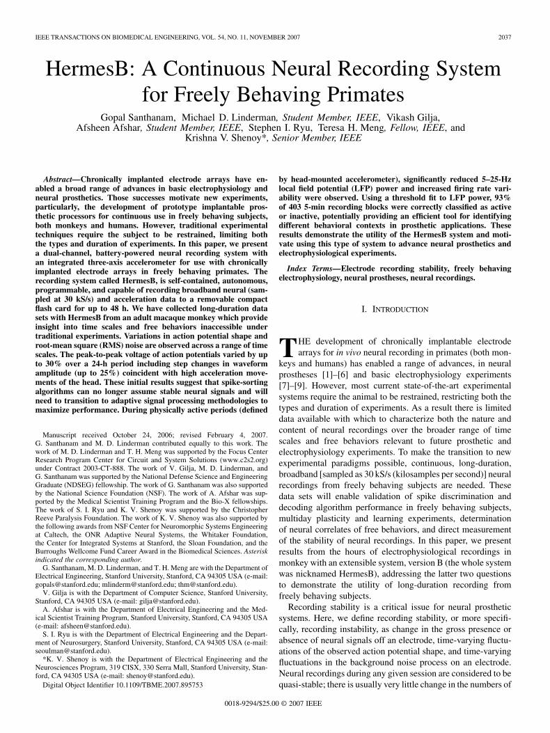
Fig. 1. Summary of array lifetime and available data for recording from indi-vidual, identifiable neurons using a chronically implanted electrode array.
neurons recorded and their action potential shapes during a sev-eral hour recording session. However, recording instability hasbeen observed between sessions, likely resulting from the sub-jects freely behaving in the housing room between sessions [10].Long-durations data sets will enable us to reconcile the currentassumptions of quasi-stable neural signals during a highly con-trolled experimental session with the variation in the neural sig-nals observed between sessions.
Fig. 1 summarizes the significant time scales in the life ofa chronically implanted electrode array. In this paper, we areonly concerned with neural recording stability in the high-yieldrecording period during which most experiments are conducted[11]. Within this window, neural interface systems are poten-tially affected by recording instability at all three time scales(short, intermediate, and long). However, current experiments,with their discrete daily recording periods, are only able tocharacterize variations on time scales less than a few hours oracross days. Thus, past studies have only characterized neuralrecording stability on short (e.g., seconds or minutes [10],[12]) and long time scales (e.g., days [10], [13], [14]). Oververy short time scales, observed variations in action potentialwaveform shape are a function of the short-term spiking fre-quency of a neuron [12]; at high frequencies, the waveformis typically broader (in time) and decreased in amplitude dueto depletion of ion gradients in and around a highly activeneuron. At longer time scales, the variation in spike waveformis not as systematic, potentially arising from a number ofmechanisms such as neural plasticity, physical movement ofthe electrode relative to nearby neurons, chemical degradationof the electrode tip, or immunological reactions to the implant[11], [15]. Studying neural stability at intermediate time scaleswill enable characterization (along with existing short and longtime scale data) of the full range of time scales relevant to aneural interface system and may also provide insight into longtime scale phenomena.
Experimental protocols in which the subject is retrained limitthe types of behaviors that can be observed. Long-duration datasets recorded during free behavior provide neural data associ-ated with a broader range of behaviors than traditionally pos-sible. To maximize system performance, prostheses must besensitive to behavioral and neural changes across the day andmust react robustly in the face of variable background condi-tions. For example, such systems should reliably detect differentbehavioral contexts such as whether the user is awake or asleep,or intends to be active or not. If a neural prosthetic attempts
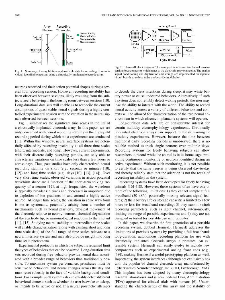
Fig. 2. HermesB block diagram. The neuroport is a custom 96 channel zero in-sertion force connector which mates to the electrode array connector. The analogsignal conditioning and digitization and storage are implemented on separatecircuit boards to reduce noise and provide modularity.
to decode the users intentions during sleep, it may waste bat-tery power or cause undesired behaviors. Alternatively, if sucha system does not reliably detect waking periods, the user maylose the ability to interact with the world. The ability to recordneural activity across a variety of different behaviors and con-texts will be allowed for characterization of the true neural en-vironment in which chronic implantable systems will operate.
Long-duration data sets are of considerable interest forcertain multiday electrophysiology experiments. Chronicallyimplanted electrode arrays can support multiday learning orplasticity experiments. However, because the time betweentraditional daily recording periods is unobserved, there is noreliable method to track single neurons over multiple days.Recording systems for freely behaving subjects can allowresearchers to record while the animal is in its home cage, pro-viding continuous monitoring of neurons identified during anactive experiment. Without such monitoring, it is not possibleto certify that the same neuron is being observed day-to-dayand thereby reliably state that the adaption is not the result ofrecording instability in the system.
Recording systems have been developed for freely behavinganimals [16]–[18]. However, these systems often have one ormore of the following limitations: 1) they cannot sample at fullbroadband (30 kS/s), potentially missing relevant signal fea-tures; 2) their battery life or storage capacity is limited to a fewhours or less for broadband recording; 3) they cannot switchrecording parameters, such as input channel, autonomously,limiting the range of possible experiments; and 4) they are notdesigned or tested for portable use with primates.
In this paper, we describe the first generation of a portablerecording system, dubbed HermesB. HermesB addresses thelimitations of previous systems by providing a full broadband,long-duration, autonomous recording platform for use withchronically implanted electrode arrays in primates. An ex-tensible system, HermesB can easily evolve to include newcomponents such as experimental analog front ends (e.g.,[19]), making HermesB a useful prototyping platform as well.Importantly, the system interfaces (although not exclusively so)with the popular 96 channel electrode array manufactured byCyberkinetics Neurotechnology, Inc. (CKI, Foxborough, MA).This implant has been adopted by many electrophysiologyresearch laboratories and is now Federal Drug Administration(FDA) approved for clinical trials with humans [6]. Under-standing the characteristics of this array and the stability of
SANTHANAM et al.: HERMESB: A CONTINUOUS NEURAL RECORDING SYSTEM FOR FREELY BEHAVING PRIMATES 2039
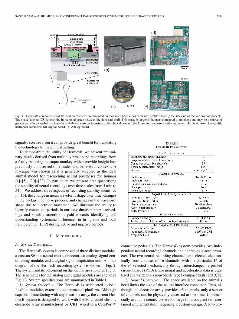
Fig. 3. HermesB components. (a) Illustration of enclosure mounted on monkey’s head along with side profile showing the stack up of the various components.The space labeled ICS denotes the intracranial space between the dura and skull. This space is larger in humans compared to monkeys and may be a source ofgreater recording variability when electrode-based systems transition to the clinical domain. (b) Aluminum enclosure with centimeter ruler. (c) Custom low-profileneuroport connector. (d) Digital board. (e) Analog board.
signals recorded from it can provide great benefit for translatingthe technology to the clinical setting.
To demonstrate the utility of HermesB, we present prelimi-nary results derived from multiday broadband recordings froma freely behaving macaque monkey which provide insight intopreviously unobserved time scales and behavioral contexts. Amacaque was chosen as it is generally accepted as the idealanimal model for researching neural prostheses for humans[1]–[5], [20]–[22]. In particular, we present data quantifyingthe stability of neural recordings over time scales from 5 min to54 h. We address three aspects of recording stability identifiedin [15]: the change in mean waveform shape over time, changesin the background noise process, and changes in the waveformshape due to electrode movement. We illustrate the ability toidentify contextual periods in our long-duration neural record-ings and specific attention is paid towards identifying andunderstanding systematic differences in firing rate and localfield potential (LFP) during active and inactive periods.
II. METHODOLOGY
A. System Description
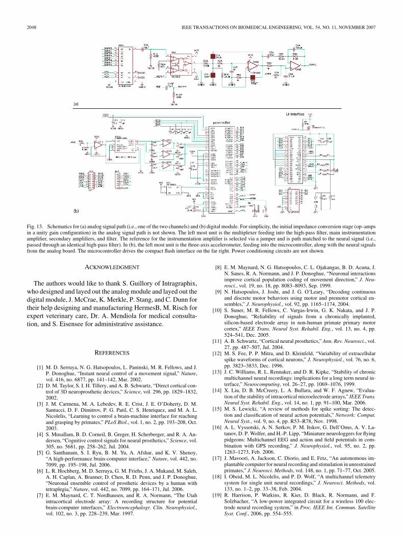
The HermesB system is composed of three distinct modules,a custom 96-pin neural microconnector, an analog signal con-ditioning module, and a digital signal acquisition unit. A blockdiagram of the HermesB recording system is shown in Fig. 2.The system and its placement on the animal are shown in Fig. 3.The schematics for the analog and digital modules are shown inFig. 13. System specifications are summarized in Table I.
1) System Overview: The HermesB is architected to be aflexible, modular, extensible experimental platform. Althoughcapable of interfacing with any electrode array, the current Her-mesB system is designed to work with the 96-channel chronicelectrode array manufactured by CKI (wired to a CerePort™
TABLE IHERMESB PARAMETERS
connector pedestal). The HermesB system provides two inde-pendent neural recording channels and a three-axis accelerom-eter. The two neural recording channels are selected electron-ically from a subset of 16 channels, with the particular 16 ofthe 96 selected mechanically through interchangeable printedcircuit boards (PCBs). The neural and acceleration data is digi-tized and written to a nonvolatile type I compact flash card (CF).
2) Neural Connector: The space available on the animal’shead limits the size of the neural interface connector. Thus, al-though the electrode array provides 96 channels, only a subsetof channels can be physically accessed at one time. Commer-cially available connectors are too large for a compact self-con-tained implementation, requiring a custom design. A low-pro-

2040 IEEE TRANSACTIONS ON BIOMEDICAL ENGINEERING, VOL. 54, NO. 11, NOVEMBER 2007
file zero-insertion-force (ZIF) connector was developed, shownin Fig. 3(c). The new connector is comprised of a mechanicalcomponent which allows access to all 96 electrodes and a set ofthree PCBs interface that provide access to 32 channel subsets(PCBs are interchanged manually). Of those 32, a subset of 16is selected with a second PCB for input to the analog module.
3) Analog Module: The analog signal conditioning path isshown in the upper dashed box of Figs. 2 and 3(e) [schematicsare shown in Fig. 13(a)]. The necessary amplification andfiltering is determined by the characteristics of neural signalsrecorded from chronic silicon electrode arrays. Neural signalshave strong spectral power content up to 8 kHz, thus a minimumsampling rate of 15 kS/s is required. In the HermesB system,as in many commercial systems, we chose to oversample(30 kS/s) to relax the specifications for the antialias filter.1 All16 accessible electrode channels undergo immediate impedanceconversion using a unity gain source follower complimentarymetal–oxide–semiconductor (CMOS) op–amp (TLC2254,Texas Instruments, Dallax TX). The electrodes have a highimpedance (100–300 k ), which must be reduced to controlnoise. The desired channels are digitally selected using 8 : 1analog multiplexers (ADG658, Analog Devices, Norwood,MA). After the multiplexers, the selected signals are passivelyhigh-pass filtered with a cutoff of 0.5 Hz to remove electrodedc bias [which would otherwise exceed the analog-to-digitalconverter (ADC) dynamic range]. The main amplification stageuses differential instrumentation amplifiers (Texas InstrumentsINA121) with gain of 98.8. Three path-matched referencesare provided—two reference signals and analog ground—se-lectable via a jumper. The two references are platinum–iridiumreference wires that accompany the electrode array and providean electrical reference local to the implantation site. Afterward,the signals were actively low-pass filtered with a cutoff 7.5 kHzand amplified with a gain of 6.2 (using Texas InstrumentsOPA2344). The 7.5-kHz cutoff ensures all of the importantspectral content is preserved, while providing excellent sup-pression at the Nyquist frequency. Resistor and capacitor valuesand filter topologies are shown in the schematics. The totalgain is determined by the ADC dynamic range (2.5 V) and themaximum amplitude of the local field potential (estimated at3000 V).
4) Digital Module: The digital module is shown in thelower dashed box of Figs. 2 and 3(d) [schematics are shown inFig. 13(b)]. Neural signals are digitized at 30 kS/s by a 12-bitsuccessive approximation ADC integrated in the ARM mi-crocontroller, which is clocked at 22.5 MHz (Analog DevicesADUC2106). The dynamic range for the ADC is 2.5 V, whichwith the total gain of 610 provides an input referred resolu-tion of 1 V/lsb for the neural signals. The 6 g three-axisaccelerometer (ST Microsystems STM9321) is mounted onthe digital board. Each accelerometer channel is also digitizedby the microcontroller at 1 kS/s and has a nominal resolution
1Rack-mounted commercial systems (which are not power limited) typicallyuse sampling rates in 30–40-kS/s range. In addition to relaxing the requiredorder of the antialias filter, the higher sampling rate reduces the need to inter-polate and align spike snippets during postprocessing. To ensure maximum per-formance, however, we continue to interpolate and align, even with the highersampling rate.
Fig. 4. Sample program for autonomous execution. The initial standby periodis added to allow the experimenters sufficient time to close up the protectiveenclosure before recording commences.
of 0.003 g/lsb. The HermesB system is controlled by customfirmware written in C.
5) Software Control: The firmware includes a simple com-mand interpreter which allows the user to interact with thesystem in real time when tethered to a portable computer (viaRS-232 serial port), as well as write simple sequencing pro-grams for autonomous execution. A sample program is shownin Fig. 4. The system is highly configurable. Parameters suchas neural sampling rate and accelerometer sampling rate can beset to balance sampling precision against data storage capacity.The experimenter can then specify a sequence of epochs, eacheither a data sampling period or quiescent standby period, tobalance data set continuity against battery lifetime.
6) Power Supply: The positive and negative power suppliesare provided by separate lithium ion batteries (positive: LGChem ICP633450A1; 49.0 33.6 6.8 mm ; 24.3 g; 4.2 V;1120 mAh; negative: Varta EasyPack; 43.5 35.4 5.8 mm ;14 g; 4.2 V; 570 mAh). The 3.3-V digital supply voltage and2.5-V analog supply voltage are provided by linear voltageregulators (LTC1844 series, Linear Technology, Milpitas, CA).The 2.5-V analog supply voltage is provided by a third linearregulator (Linear Technology LT1961). HermesB draws aconstant 5.3 mA from the negative battery and 38.8, 11.4, and71 mA from the positive battery during idle upon reset, standby,and active sampling periods, respectively.
7) Physical Construction: The entire system is housedin a lightweight, protective aluminum case, shown inFig. 3(a) and (b), secured with methyl methacrylate, whichwas in turn secured to the skull. The enclosure was sealedwith a watertight gasket and grounded to the monkey to pro-vide electromagnetic (EM) shielding for the electronics. Theelectronics are tightly packed in the case with nonconductivefoam to prevent vibration, shown in Fig. 3(a).2 The entiresystem weighs 200 g, which is light enough that no behavioraldifferences were observed (ensuring collected data representsnatural behavior).
8) Limitations and Future Work: The tight space, weight,and power constraints force a number of design limitations onthe HermesB system. However, many of these limitations can
2Thus, we can safely state that our accelerometer records head motion andnot any residual board vibrations.
SANTHANAM et al.: HERMESB: A CONTINUOUS NEURAL RECORDING SYSTEM FOR FREELY BEHAVING PRIMATES 2041
be addressed in future designs by leveraging improving com-mercial-off-the-shelf (COTS) technology, or recently developedneural recording specific integrated circuits.
The number of neural channels is determined by the foot-print of an analog signal path. In the present design, only twochannels could fit within the available space (the size of whichis set by the animal’s head). Future versions of the HermesBsystem can replace the current discrete analog signal paths withsoon to be available custom integrated circuits (such as [19])enabling 16 or more channels to be recorded simultaneouslywithin the same overall footprint. Although not needed for thecurrent system (since only two channels can be recorded simul-taneously), more channels than the current 16 can be made avail-able by replacing the current connector PCB and headers with amore dense design.
The ability to digitally select among the 16 channels enablestime multiplexing of the neural recording paths. However, theswitching speed is limited by the slow dynamics of the largecapacitor in the 0.5-Hz high-pass filter. More complex circuitsare under development to increase switching speed for the cur-rent analog signal path. Switching speed is not a problem forthe integrated analog front end approach which uses indepen-dent per-channel amplifiers and filters.
The system is very sensitive to the write latency of the com-pact flash card. “Fast” cards intended for use in high-end digitalcameras are required to supply the necessary write bandwidth.However, the capacity and performance of CF cards is con-stantly improving allowing the capabilities of HermesB to scaleas well without redesign or remanufacturing. In future designs,the CF recording will be supplanted by wireless transmission toeliminate storage limitations and enable real-time interaction.
By utilizing modular construction (separate analog and dig-ital PCBs) and standard interfaces like CF, the HermesB systemcan be easily upgraded. The future work would only requirereplacement of the analog module or the substitution of a CFform factor wireless transceiver for the current flash card. Ad-ditional ADC channels are available to support new analog datasources, such as chronically implanted electromyogram (EMG)electrodes.
B. Recordings and Analyses
Primary data for this report was collected from an adultfemale macaque monkey (monkey D) freely moving in ahome cage. All experiments and procedures were approvedby the Stanford University Institutional Animal Care and UseCommittee (IACUC, Stanford, CA). We performed a sterilesurgery to implant a head restraint system. At this time, wealso implanted a silicon 96-electrode array. The electrodearray (Cyberkinetics, Foxborough, MA) was implanted in aregion spanning the arm representation of the dorsal aspectof premotor cortex (PMd) and primary motor cortex (M1), asestimated visually from local anatomical landmarks. Surgicalmethods are very similar to that described in [9].
HermesB was used to record starting in August 2005. Anumber of recording profiles were used. One profile con-sisted of recording at a 67% duty cycle (5 min of recordingfollowed by 2.5 min of standby). Total experiment duration
was approximately 54 h, broken up into three 18-h sessions.The duty cycling is a compromise between memory capacityand battery life constraints. When recording continuously, thecurrent memory capacity can be quickly exhausted. At verylow duty cycling, the battery is discharged by the static powerconsumption before the CF card is full, despite putting themicrocontroller in standby between recording blocks. Betweeneach session, the monkey was transferred from the home cageto the training chair to replace the battery and download the
4 GB of recorded data. During these “pit stops,” recordingwas continued with a second smaller CF card and new battery tomaintain data set continuity. Other profiles include round-robinrecording of 4–8 channels over a 24-h schedule. Two neuralchannels were recorded per data set in full broadband (0.5 Hzto 7.5 kHz at 30 kS/s with 12-bit resolution) and a three-axisaccelerometer fixed to the monkey’s head was sampled (1 kS/swith 12-bit resolution) and stored to compact flash.
Accelerometer data was used from each 5-min data blockto label the blocks as either “active,” “inactive,” or “mixed.”Blocks in which the maximum accelerometer magnitude(MAM) was greater than 1.25 g were labeled active, blocks inwhich the MAM was less than 1.15 g were labeled inactive,and blocks that were within these bounds were labeled mixed.These thresholds were selected to roughly balance the numberof active and inactive blocks with the ratio of day (lights on)versus night (lights off) blocks (as we expect low activity whenthe lights are off), while retaining a 0.1-g margin betweenclassifications.
The recorded neural signals from each 5-min block werepostprocessed with the Sahani spike-sorting algorithm, whichis an unsupervised clustering algorithm [23], [24]. Spike timeswere identified using a threshold determined from data acrossthe block ( with respect to the RMS noise estimate fromfiltered data). A spike waveform, or snippet, comprised ofa 32-sample window around the threshold event, and wasextracted and aligned to its center of mass (COM). Snippetswere projected into a 4-D robust, noise-whitened principalcomponents space (NWrPCA) and clustered using a maximuma posteriori (MAP) clustering technique. Well-isolated unitswere identified and cross referenced across blocks by hand.For LFP analyses, broadband data was filtered by applyingChebyshev type I low-pass and bandpass filters with a passbandripple of 1 dB. Power spectral density estimates were calculatedusing the Welch periodgram method.
C. Recording Stability Analyses
To quantify the stability and consistency of waveformsrecorded from our electrode array, we analyzed data from ourlong-duration recordings in several ways. First, snippets fromthe entire session were extracted using a threshold andprojected into a single 2-D principal components subspace.By graphing a 2-D histogram of the snippets in this subspace,snippets with similar waveform shapes are grouped into distinctclusters. Movement of these clusters across the session indi-cates drift in the waveforms. The magnitude of the shifts werethen assessed by examining the actual waveform shapes overthese periods of interest. Second, to observe more continuous

2042 IEEE TRANSACTIONS ON BIOMEDICAL ENGINEERING, VOL. 54, NO. 11, NOVEMBER 2007
shifts in waveform shape, we chose a feature of the averagewaveform shape, the peak-to-peak voltage , and plottedthis quantity over the course of the recording session. Thewas determined on a block-by-block basis by using the Sahanialgorithm per block, providing local estimates of the averagewaveform shapes.
Last, to search for potentially abrupt changes in waveformshape, the neural recordings were analyzed in conjunction withthe accelerometer data. An abrupt change in electrode arrayposition in the cortex would presumably manifest itself as anabrupt change in waveform amplitude, as the neuron–electrodedistance would change. If such changes do occur, we addition-ally presume they are correlated with high acceleration eventssuch as vigorous head movement. Therefore, we examined theneural recordings straddling high acceleration events ( 3 gthreshold) and examined the metric around these events.To help search for events of interest, we computed the localchange of the metric , constructed from200 snippets before and 200 snippets after the accelerationevent. This allowed us to narrow in on high acceleration eventsthat coincided with large shifts in action potential waveformshape.
Single neural units were used for these analyses to observethe recording stability from our chronic implant. One importantconcern is that if a unit is automatically identified by the spikesorter, large changes in the unit’s waveform shape could causethe unit to no longer be classified correctly, thereby obscuringthe analyses. Thus, the NWrPCA projections of the selectedunits were examined separately by the experimenters to ensurethat snippets were not ignored or improperly included. Thiswas accomplished by ensuring all units included in the afore-mentioned stability analyses were well-isolated, high-firing-ratesingle neurons, and sufficiently distinct from other signals ontheir respective electrodes such that reasonably large variationswould not result in a high rate of misclassification.
III. RESULTS
A. System Verification
Fig. 5 shows example data recorded from our animal subjectfreely moving in her home cage. The top traces [Fig. 5(a)] showthe three-axis acceleration of the monkey’s head over a 10-speriod. This data segment was recorded in the early eveningduring a period in which the monkey was quite active. Fig. 5(b)shows 100 ms of broadband neural data recorded from a singlechannel on the electrode array. The LFP is easily visible as isa number of spikes “riding” on top of the LFP. Fig. 5(c) showsthe same data segment filtered with a 250-Hz high-pass infiniteimpulse response (IIR) filter, which is the same filter used forspike sorting.
Data sets, like that shown in Fig. 5, were used as part ofa three step verification process to ensure the accuracy ofHermesB recordings. The steps were as follows: 1) measureHermesB circuit parameters, 2) compare recordings of the CKINeural Simulator made with HermesB and our standard labora-tory recording system (CKI Cerebus System), and 3) compareHermesB recordings of neural activity in a rhesus monkey torecordings made by the fixed laboratory system.
Fig. 5. Sample neural and accelerometer data recorded from a freely behavingmonkey. (a) Accelerometer channels: (blue), (green), and (red). The directcurrent (dc) levels on the channels are due to the particular orientation of theaccelerometer with respect to Earth’s gravity vector. (b) Unfiltered broadbandneural data taken from the middle of the recording period. (c) Filtered broadbandneural data.
Fig. 6. Comparison of snippets recorded with CKI Cerebus system (left) andHermesB (right). (a) Snippets recorded from CKI Neural Simulator. (b) Snip-pets from four neurons recorded from a single electrode channel in a monkeycomfortably in a chair with head restrained. Snippets have been sorted andthe 10th–90th percentile in amplitude indicated by the colored region for eachwaveform.
The measured circuit parameters are summarized in Table I.The input referred noise, measured with grounded inputs, iscomparable to or better than current state-of-the-art commer-cial (CKI Cerebus System) and research systems [19]. The CKINeural Simulator is a neural recording playback device thatprovides 128 channels of simulated neural signals at typicalamplitudes for array recordings (e.g., maximum of 500 Vpeak-to-peak for spikes) and similar output impedance to a stan-dard electrode array. Fig. 6(a) shows a side-by-side compar-ison of Neural Simulator recordings made with the CKI Cerebussystem (left) and with HermesB (right). The three spike wave-forms are clearly visible, with comparable levels of noise (mea-sured as the spread of the curves) between the two systems.Fig. 6(b) shows a similar comparison for a channel from theelectrode array, recorded from a monkey sitting quietly in a pri-mate chair. The figure shows the 10th–90th percentile in am-plitude of action potential waveforms recorded from a single
SANTHANAM et al.: HERMESB: A CONTINUOUS NEURAL RECORDING SYSTEM FOR FREELY BEHAVING PRIMATES 2043
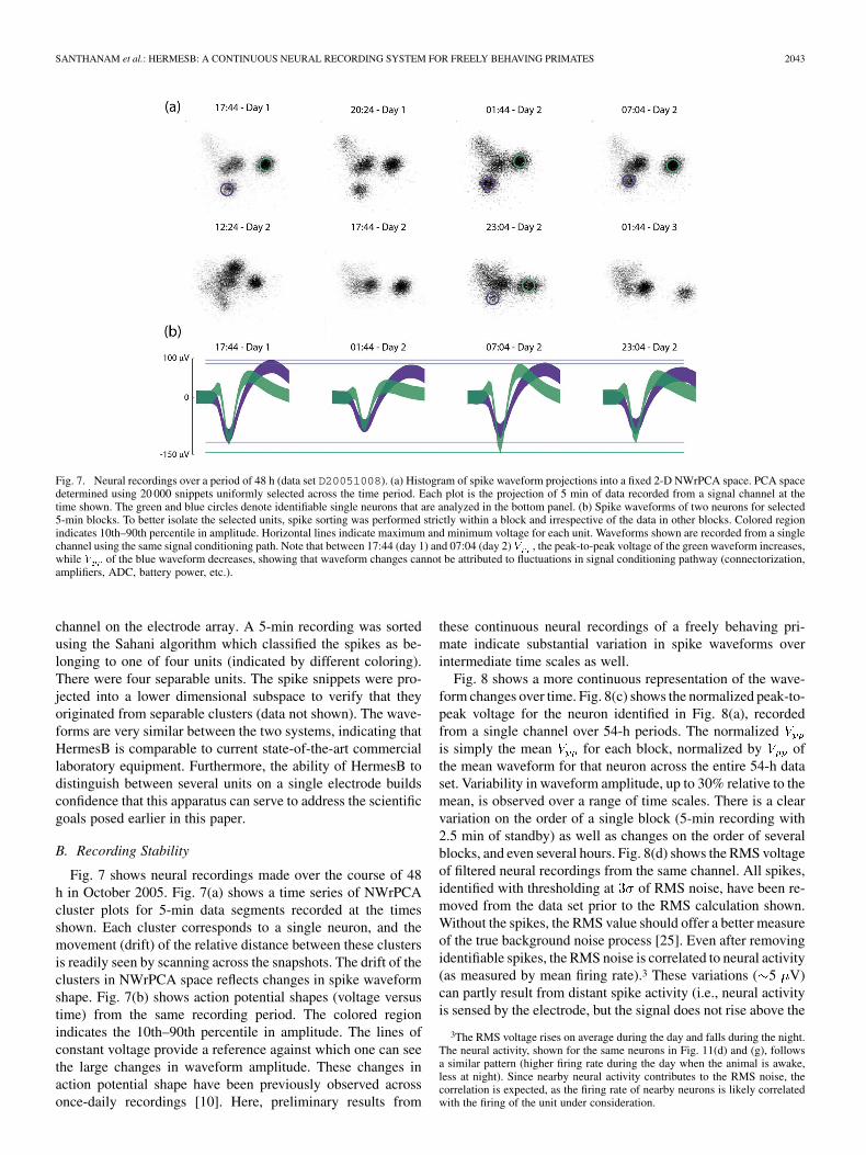
Fig. 7. Neural recordings over a period of 48 h (data set D20051008). (a) Histogram of spike waveform projections into a fixed 2-D NWrPCA space. PCA spacedetermined using 20 000 snippets uniformly selected across the time period. Each plot is the projection of 5 min of data recorded from a signal channel at thetime shown. The green and blue circles denote identifiable single neurons that are analyzed in the bottom panel. (b) Spike waveforms of two neurons for selected5-min blocks. To better isolate the selected units, spike sorting was performed strictly within a block and irrespective of the data in other blocks. Colored regionindicates 10th–90th percentile in amplitude. Horizontal lines indicate maximum and minimum voltage for each unit. Waveforms shown are recorded from a singlechannel using the same signal conditioning path. Note that between 17:44 (day 1) and 07:04 (day 2) , the peak-to-peak voltage of the green waveform increases,while of the blue waveform decreases, showing that waveform changes cannot be attributed to fluctuations in signal conditioning pathway (connectorization,amplifiers, ADC, battery power, etc.).
channel on the electrode array. A 5-min recording was sortedusing the Sahani algorithm which classified the spikes as be-longing to one of four units (indicated by different coloring).There were four separable units. The spike snippets were pro-jected into a lower dimensional subspace to verify that theyoriginated from separable clusters (data not shown). The wave-forms are very similar between the two systems, indicating thatHermesB is comparable to current state-of-the-art commerciallaboratory equipment. Furthermore, the ability of HermesB todistinguish between several units on a single electrode buildsconfidence that this apparatus can serve to address the scientificgoals posed earlier in this paper.
B. Recording Stability
Fig. 7 shows neural recordings made over the course of 48h in October 2005. Fig. 7(a) shows a time series of NWrPCAcluster plots for 5-min data segments recorded at the timesshown. Each cluster corresponds to a single neuron, and themovement (drift) of the relative distance between these clustersis readily seen by scanning across the snapshots. The drift of theclusters in NWrPCA space reflects changes in spike waveformshape. Fig. 7(b) shows action potential shapes (voltage versustime) from the same recording period. The colored regionindicates the 10th–90th percentile in amplitude. The lines ofconstant voltage provide a reference against which one can seethe large changes in waveform amplitude. These changes inaction potential shape have been previously observed acrossonce-daily recordings [10]. Here, preliminary results from
these continuous neural recordings of a freely behaving pri-mate indicate substantial variation in spike waveforms overintermediate time scales as well.
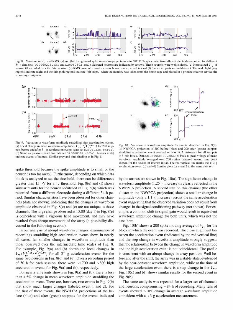
Fig. 8 shows a more continuous representation of the wave-form changes over time. Fig. 8(c) shows the normalized peak-to-peak voltage for the neuron identified in Fig. 8(a), recordedfrom a single channel over 54-h periods. The normalizedis simply the mean for each block, normalized by ofthe mean waveform for that neuron across the entire 54-h dataset. Variability in waveform amplitude, up to 30% relative to themean, is observed over a range of time scales. There is a clearvariation on the order of a single block (5-min recording with2.5 min of standby) as well as changes on the order of severalblocks, and even several hours. Fig. 8(d) shows the RMS voltageof filtered neural recordings from the same channel. All spikes,identified with thresholding at of RMS noise, have been re-moved from the data set prior to the RMS calculation shown.Without the spikes, the RMS value should offer a better measureof the true background noise process [25]. Even after removingidentifiable spikes, the RMS noise is correlated to neural activity(as measured by mean firing rate).3 These variations ( 5 V)can partly result from distant spike activity (i.e., neural activityis sensed by the electrode, but the signal does not rise above the
3The RMS voltage rises on average during the day and falls during the night.The neural activity, shown for the same neurons in Fig. 11(d) and (g), followsa similar pattern (higher firing rate during the day when the animal is awake,less at night). Since nearby neural activity contributes to the RMS noise, thecorrelation is expected, as the firing rate of nearby neurons is likely correlatedwith the firing of the unit under consideration.
2044 IEEE TRANSACTIONS ON BIOMEDICAL ENGINEERING, VOL. 54, NO. 11, NOVEMBER 2007
Fig. 8. Variation in and RMS. (a) and (b) Histogram of spike waveform projections into NWrPCA space from two different electrodes recorded for different54-h data sets (D20060225.ch1 and D20060302.ch2). Selected neurons are indicated by arrows. These neurons were well isolated. (c) Normalized ofneuron #1 recorded over the 54-h session. (d) RMS noise of recorded channels over same period. (e) and (f) Same two plots second data set. The wide light grayregions indicate night and the thin pink regions indicate “pit stops,” when the monkey was taken from the home cage and placed in a primate chair to service therecording equipment.
Fig. 9. Variation in waveform amplitude straddling high acceleration events.(a) Local change in mean waveform amplitude for 200 snip-pets before and after 3 g acceleration events (data set D20060225.ch1u1).(b) Same as previous panel for data set D20060302.ch2u1. Arrows in (b)indicate events of interest. Similar gray and pink shading as in Fig. 8.
spike threshold because the spike amplitude is to small or theneuron is too far away). Furthermore, depending on which datablock is analyzed to set the threshold, there can be differencesgreater than 15 V for a threshold. Fig. 8(e) and (f) showssimilar results for the neuron identified in Fig. 8(b) which wasrecorded from a different electrode during a different 54-h pe-riod. Similar characteristics have been observed for other chan-nels (data not shown), indicating that the changes in waveformamplitude observed in Fig. 8(c) and (e) are not unique to thosechannels. The large change observed at 13:00 (day 1) in Fig. 8(e)is coincident with a vigorous head movement, and may haveresulted from abrupt movement of the array (a possibility dis-cussed in the following section).
In our analysis of abrupt waveform changes, examination ofrecordings straddling high acceleration events show, in nearlyall cases, far smaller changes in waveform amplitude thanthose observed over the intermediate time scales of Fig. 8.For example, Fig. 9(a) and (b) shows the local changes in
for all 3 g acceleration events for thesame two neurons in Fig. 8(c) and (e). Over a recording periodof 50 h for each session, there were 1700 and 800 highacceleration events for Fig. 9(a) and (b), respectively.
For nearly all events shown in Fig. 9(a) and (b), there is lessthan a 5% change in mean waveform amplitude straddling theacceleration event. There are, however, two events in Fig. 9(b)that show much larger changes (labeled event 1 and 2). Forthe first of these events, the NWrPCA projections of the be-fore (blue) and after (green) snippets for the events indicated
Fig. 10. Variation in waveform amplitude for events identified in Fig. 9(b).(a) NWrPCA projection of 200 before (blue) and 200 after (green) snippetsstraddling acceleration event overlaid on NWrPCA histogram for all snippetsin 5-min block. Data set D20060302.ch2. (b) Peak-to-peak voltage of meanwaveform amplitude averaged over 200 spikes centered around time pointshown, for the neuron of interest in (a). The red vertical line marks the 3 gacceleration event. (c) and (d) Similar plots for event 2 in the same data set.
by the arrows are shown in Fig. 10(a). The significant change inwaveform amplitude (1.25 increase) is clearly reflected in theNWrPCA projection. A second unit on this channel (the othercluster in the NWrPCA projection) shows a smaller change inamplitude (only a 1.1 increase) across the same accelerationevent suggesting that the observed variation does not result fromchanges in the signal conditioning pathway (not shown). For ex-ample, a common shift in signal gain would result in equivalentwaveform amplitude change for both units, which was not thecase here.
Fig. 10(b) shows a 200 spike moving average of for theblock in which the event was recorded. The close alignment be-tween the acceleration event (indicated by the red vertical line)and the step change in waveform amplitude strongly suggeststhat the relationship between the change in waveform amplitudeand the high acceleration event is not coincidental. The profileis consistent with an abrupt change in array position. Well be-fore and after the shift, the array was in a stable state, evidencedby the near-constant waveform amplitude, while at the time ofthe large acceleration event there is a step change in the .Fig. 10(c) and (d) shows similar results for the second event inFig. 9(b).
The same analysis was repeated for a larger set of channelsand neurons, compromising 84 h of recording. Many tens ofevents showed 10% change in average waveform amplitudecoincident with a 3-g acceleration measurement.

SANTHANAM et al.: HERMESB: A CONTINUOUS NEURAL RECORDING SYSTEM FOR FREELY BEHAVING PRIMATES 2045
Fig. 11. Neural and accelerometer data recorded from a freely behaving monkey. (a) and (b) Histogram of spike waveform projections into NWrPCA space fromtwo different electrodes recorded for different 54-h data sets (D20060225.ch1 and D20060302.ch2). Selected neurons are indicated by arrows. These neuronswere well isolated. (c) Firing rate of the neuron shown in (a) calculated over a 1-s interval using a Hamming window. Red and blue data points were recordedin time periods labeled as “active” and “inactive,” respectively. Green data points were recorded during unlabeled periods. (d) Accelerometer magnitude overthe recording period downsampled to 100 Hz. (e) LFP power per block, recorded from the same electrode, calculated by integrating the power over the 5–25-Hzfrequency band. (f)–(h) Same plots for second data set. Similar gray and pink shading as in Fig. 8.
Fig. 12. LFP analyses for data set D20060302.ch2. (a) Power spectral density (PSD) recorded during “active” (red) and “inactive” (blue) periods. The thinlines are the mean PSDs and the standard error of the mean is represented by their thickness. The thickness of the wider translucent lines are the standard deviations.Each PSD is calculated over 5 min of data and their distributions were taken from data across the 54-h data set for neuron 1. (b) Spectral power recorded during“active” (red) and “inactive” (blue) periods for the 5–25-Hz frequency band. The dotted line represents the learned classification threshold between “active” and“inactive” blocks.
C. Neural Correlates of Behavioral Contexts
Fig. 11 shows data from two 54-h recordings. For the neuron1 data set [presented in Fig. 11(a) and (c)–(e)] there were 438data blocks. Active blocks, in which the monkey was putativelymoving in its home cage, constituted 40% of the blocks while52% of the blocks were inactive. From the accelerometer data[Fig. 11(d)], it is clear that the monkey is more physically ac-tive during the day, and as expected, firing rates tend to be higherduring these periods. Note that LFP power was generally lowerduring these periods as shown in Fig. 11(e). During the “pitstops” (battery swap periods) the monkey’s head was comfort-ably restrained in a fixed position (the time duration indicated bythe pink bands); therefore, accelerometer magnitude remainedflat at 1 g. Likewise, few movements were made, and conse-quently, firing rates were suppressed. These trends were con-sistent across two data sets collected from different electrodesand at different times. Neural activity recorded from a secondchannel show similar patterns [Fig. 11(b) and (f)–(h)].
As shown in Fig. 12(a), the mean LFP power differed between“active” and “inactive” periods in the 2–30-Hz and 50–100-Hzfrequency bands. For the majority of this range, the standarddeviations are large relative to the difference in the mean; thisrelationship makes power modulation in these bands an unreli-able classifier for per-block behavior (i.e., “active” versus “in-
active”). However, the 5–25-Hz band was well separated, so thepower in this range can be used to develop a reliable classifier.This differentiation in LFP power was observed across multiplechannels and multiple days, and is consistent with previous re-sults showing that 10–100-Hz LFP activity diminished duringmovement [26], [27] and increased during sleep [28].
Fig. 12(b) plots 5–25-Hz LFP power versus MAM for each5-min block. When we classified the activity level of blocksby thresholding LFP power at 56.5 dB, 93% (131/141) of“active” blocks and 92% (175/191) of “inactive” blocks werecorrectly classified. Results were similar for a second channelfor a different session: 89% (150/169) of “active” blocks and88% (81/92) of “inactive” blocks were correctly classified witha threshold of 57.1 dB. These results were obtained by pickingthe optimal linear classification boundary using the first 40 ac-tive and first 40 inactive blocks and testing on the remainingblocks. Head posting during “pit stops” can create confoundsince the accelerometer was held in a fixed position even if themonkey was otherwise active during these periods. Hence, theseperiods were removed prior to the aforementioned analysis.
A similar classification was not as successful when using theaverage firing rate over a 5-min block (data not shown). Themean and variance of the MAM increased as the firing rate in-creased, but the likelihood of a small MAM (i.e., an inactiveperiod) remained relatively high even for high firing rates (data
2046 IEEE TRANSACTIONS ON BIOMEDICAL ENGINEERING, VOL. 54, NO. 11, NOVEMBER 2007
not shown). Recall that the electrode was implanted in a re-gion spanning PMd and M1, which is strongly believed to beinvolved in motor planning and execution of arm movements[29]–[34]. If arm movements are made while the head positionremains fixed, firing rates could increase without large accel-eration events. Also, motor plans can be generated and subse-quently canceled. Thus, absolute firing rate may not be the bestproxy for activity level.
Given that “active” and “inactive” periods tended to occurduring day and night, respectively, the variations in firing rateand LFP might be explained, in part, by circadian rhythms(or direct modulation by light level). One might hypothesizethat 5–25-Hz LFP power is increased and firing rates aredepressed in association with day–night cycles. However, forblocks within a single activity condition (either “active” or“inactive”), the differences between day and night for both LFPand firing rate were at least an order of magnitude smaller thanthe difference between “active” and “inactive” blocks duringeither time period. This suggests that circadian rhythms do notheavily influence these effects.
IV. DISCUSSION
In this paper, we report HermesB, a new, self-contained, long-duration, neural recording system for use with freely behavingprimates. HermesB records dual channel broadband and three-axis head acceleration data to a high density compact flash card.Controlled by simple sequencing programs written by the exper-imenter, HermesB can autonomously change recording channeland pause recording during the experiment. With a single bat-tery charge, HermesB can record for up to 48 h (at a low dutycycle). With short breaks to replace the batteries and compactflash card, HermesB can record nearly continuously for an in-definite period.
The high quality of the broadband recordings, despite beingin the electrically noisy environment of the home cage room(e.g., florescent lights), enables results from HermesB to be in-tegrated into experiments using the traditional laboratory rig.There are varieties of applications for such a platform. For ex-ample, the long-duration recordings, in concert with traditionalexperiments, enable important multiday learning and plasticityexperiments, an application not explored in detail in this paper.Researchers can use HermesB to record during periods whenthe animal is outside the rig to provide continuous monitoringof significant neurons identified during active experiments.
To demonstrate the utility of HermesB, we analyzed the firstset of multiday broadband neural and accelerometer data fromarea PMd in a freely behaving primate. In one set of analyses,we showed how HermesB can be useful in exploring the dif-ferent neural contexts of a freely behaving subject. Practicalneural prosthetic systems need to work well under varied con-ditions. We can begin to understand these conditions as con-texts, defined here as a set of behavioral states and/or goals(such as active versus inactive or keyboard typing versus free-hand drawing). A switch in context may change the dynamicsof observed neural signals and a neural prosthetic must be sen-sitive to these changes.
We examined a very simple pair of contexts—physically ac-tive and inactive. The monkey moved freely, so we could register
these periods with an accelerometer. For an immobile patient,however, we would have to determine the intent to be physi-cally active or inactive. As shown in Fig. 12, LFP is a promisingproxy for activity level. Firing rate can also be used for this pur-pose. However, LFP power measurement consumes less batterypower than firing rate measurement (a low-power LFP powermeasurement circuit is described in [35]), potentially enablinga power efficient implant “standby” mode when the user is in-active. When LFP power falls below a defined threshold, indi-cating that the user is, or intends to be, active, the prostheticcan switch out of this “standby” mode. Furthermore, using LFPthresholding could help prevent undesired movements from theprosthetic system during “inactive” periods.
In future studies, we plan to examine subtler context changes.Some contexts may require fewer neurons for acceptable perfor-mance; under these conditions, we can conserve power by dis-abling a subset of the neural channels. Under different contexts,users may require different sets of behavioral responses (such asdiscrete target selection versus continuous motion) or the under-lying dynamics of the observed cortical area may change drasti-cally; we would like to respond to these concerns by switchingthe decoding model according to context. By identifying con-texts and adjusting hardware configuration accordingly, it maybe possible to boost performance in terms of power consump-tion and decoding accuracy.
We were able to identify natural behavior across multipledays using accelerometer measurements and correlating theseto neural recordings. Such an ability coupled with more ad-vanced behavioral monitoring, such as chronically implantedEMG electrodes [36], [37] or motion tracking, can enable theexploration of questions that have been unapproachable untilnow. Mining large data sets of free behavior to find neural cor-relates may help us develop new controlled experiments; thesedata sets are also necessary for testing and developing neuralprosthetics systems with the ability to operate autonomouslyover extended periods of time. Similar investigations are alreadyunderway by other researchers and HermesB can serve as an-other tool in these types of experiments [38], [39].
Our most novel investigation were our analyses of neuralrecording stability. It is in this particular class of experi-ments that HermesB is most differentiated from other portablerecording systems currently in use. In particular, we addressedthree aspects of recording stability identified in [15] and listedearlier: the change in mean waveform shape over time, changesin the background noise process, and changes in waveformshape due to electrode array movement. Initial analysis showssignificant nonabrupt variation in waveform amplitude andRMS noise at intermediate time scales, along with step changesin waveform amplitude coincident with high accelerationmovements of the head. Although some of these instabilitieshave been observed previously (or speculated), they havenot been specifically, quantitatively measured. The resultspresented here provide preliminary characterization of neuralrecording stability, providing a meaningful starting point forboth electrophysiology work and prosthetics oriented postpro-cessing algorithm development. In the following paragraphs,we describe some of the potential causes of these variationsand their potential implications for prosthetic systems.
SANTHANAM et al.: HERMESB: A CONTINUOUS NEURAL RECORDING SYSTEM FOR FREELY BEHAVING PRIMATES 2047
What might be the cause for these variations in waveformamplitude? The step changes in waveform amplitude appear,in some cases, to result from abrupt shifts in electrode posi-tion caused by head movement. For the nonabrupt variation inwaveform shape and RMS noise, we believe there could be anumber of factors that may play a significant role, includingchanges in the cortical environment in response to subject ac-tivity, including “brain bounce,” changes in intracranial pres-sure (ICP), and other homeostatic factors. At short-to-interme-diate timescales (i.e., longer than bursting periods), Lewicki[15] suggests that array movement, or more specifically changesin the neuron–electrode distance, might play a role in waveformshape change. Fluctuations in the ICP could potentially movethe cortex tissue relative to the array (or vice versa). Confirmingsuch a relationship is beyond the scope of this work, though maybe of interest in future studies.
Since so few of the high acceleration events were coincidentwith large changes in waveform amplitude, there is the tempta-tion to dismiss these events as rare and unimportant. However,it is important to note that a practical neural prosthesis will haveto operate 24 h a day and 7 days a week. As such, the prostheticsystem must be able to recognize the 3–4 abrupt changes thatmight occur in a week, especially as such systems are used formore ambulatory patients. In fact, in one stretch of 84 h ofrecording, we found that there were many tens of events thatshowed abrupt waveform change coincident with high acceler-ation movements of the head. Our results are only preliminary,however, and will require more data sets and more animals forcomprehensive characterization.
Traditional experimental protocols that utilize discrete, dailyrecording periods have provided limited information regardingneural recording stability. The daily sampling limits the po-tential characterization of variations to time scales of eitherminutes or days. It is important to note that similar variationswere not observed, however, in the hour long broadbandrecordings described in [10]. However, those recordings weremade under a more traditional experimental protocol in whicha restrained monkey performed a repetitive reaching task. It ispossible that the more controlled and consistent environmentof those recordings, in contrast to the animal freely behavingin the home cage, produces a more consistent cortical environ-ment (e.g., less “brain bounce,” smaller changes in intracranialpressure, etc.) and thus reduced variation in waveform shape.
We have shown examples from preliminary data sets of sig-nificant waveform shape and RMS noise variation at all threetime scales. Both types of variation can have adverse effects onspike-sorting performance, either through the use of an inap-propriate threshold or outright misclassification. The improvedstatistical characterization of the stability of neural recordingsenabled by these new long-duration data sets will allow the prin-cipled design and evaluation of sorting algorithms. Tolerance tosome instabilities in neural recordings has already been incor-porated into sorting algorithms. The short-time-scale variationsin spike shape can be addressed by incorporating firing statis-tics into the spike-sorting algorithm [40] and changes in RMSvoltage (from which the threshold is typically derived) can be
addressed through adaptive thresholding [25]. Long-term vari-ation, however, may require periodic retraining of the spike-sorting parameters. With such readjustments, experimenters re-port the ability to track single neurons across months or evenyears (although experimenters cannot be sure the same neuronsare being observed without constant tracking, a capability nowavailable with HermesB). There does not appear to be a con-sensus on exactly what retraining period is required. Currentexperiments that use discrete daily recording periods naturallyupdate once per day.
The quality of the trained spike-sorting parameters is para-mount. Poor classification parameters, and thus poor classifi-cation performance, will affect all aspects of neural prostheticsystem performance. This does not imply that systems shouldretrain arbitrarily often. Frequent retraining can have significantcosts. For advanced spike-sorting algorithms [23], the trainingalgorithm is computationally expensive. Although our recentpower feasibility study has shown that the power consumptionof the algorithm in [23] is small relative to real-time classifi-cation, it was assumed that retraining would be required onlyevery 12 h [24]. If a much shorter training period is required,the power consumption of training could quickly become signif-icant, making these advanced algorithms inappropriate for fullyintegrated and implantable prosthetic systems.
Sorting algorithms with an adaptive training approach thatcontinuously integrates over an extended period, similar to themethod proposed in [41], as opposed to discrete retraining,might be the best approach in light of the instability of neuralrecordings. A suitable adaptive algorithm would have an ef-fective training interval short enough to track variations inwaveform shape and background process, without the cost oftraditional discrete retraining. The apparent sparsity of abruptchanges in waveform shape due to rapid array movement mayreduce the occurrence of a potential problem scenario in whichabrupt retraining is required. Nonetheless, the presence ofthese abrupt changes in waveform shape does suggest that, tomaximize spike classification accuracy, any algorithm wouldbenefit from the ability to initiate discrete retraining whenstep changes in the waveform shape are observed. As thesechronic electrode arrays are implanted in amputees (rather thantetraplegics), the head will move substantially. It is worthwhileto note that the space between the brain and the dura is largerin humans than monkeys. Therefore “brain bounce” and othernonstationarities may be a larger issue.
At present, HermesB is in active use supporting a numberof experiments, as well as ongoing development to increaserecording capabilities. As CF technology and battery energydensity improve, recording duration will be expanded. Futuregenerations of HermesB will also incorporate wireless telemetryand more simultaneous recording channels.
APPENDIX
The schematics for the analog signal path and the digitalmodule are shown in Fig. 13(a) and (b). For simplicity, theinitial impedance conversion op–amps, reference circuitry, andpower conditioning circuits are not shown.
2048 IEEE TRANSACTIONS ON BIOMEDICAL ENGINEERING, VOL. 54, NO. 11, NOVEMBER 2007
Fig. 13. Schematics for (a) analog signal path (i.e., one of the two channels) and (b) digital module. For simplicity, the initial impedance conversion stage (op–ampsin a unity gain configuration) in the analog signal path is not shown. The left most unit is the multiplexer feeding into the high-pass filter, main instrumentationamplifier, secondary amplifiers, and filter. The reference for the instrumentation amplifier is selected via a jumper and is path matched to the neural signal (i.e.,passed through an identical high-pass filter). In (b), the left most unit is the three-axis accelerometer, feeding into the microcontroller, along with the neural signalsfrom the analog board. The microcontroller drives the compact flash interface on the far right. Power conditioning circuits are not shown.
ACKNOWLEDGMENT
The authors would like to thank S. Guillory of Intragraphix,who designed and layed out the analog module and layed out thedigital module, J. McCrae, K. Merkle, P. Stang, and C. Dunn fortheir help designing and manufacturing HermesB, M. Risch forexpert veterinary care, Dr. A. Mendiola for medical consulta-tion, and S. Eisensee for administrative assistance.
REFERENCES
[1] M. D. Serruya, N. G. Hatsopoulos, L. Paninski, M. R. Fellows, and J.P. Donoghue, “Instant neural control of a movement signal,” Nature,vol. 416, no. 6877, pp. 141–142, Mar. 2002.
[2] D. M. Taylor, S. I. H. Tillery, and A. B. Schwartz, “Direct cortical con-trol of 3D neuroprosthetic devices,” Science, vol. 296, pp. 1829–1832,2002.
[3] J. M. Carmena, M. A. Lebedev, R. E. Crist, J. E. O’Doherty, D. M.Santucci, D. F. Dimitrov, P. G. Patil, C. S. Henriquez, and M. A. L.Nicolelis, “Learning to control a brain-machine interface for reachingand grasping by primates,” PLoS Biol., vol. 1, no. 2, pp. 193–208, Oct.2003.
[4] S. Musallam, B. D. Corneil, B. Greger, H. Scherberger, and R. A. An-dersen, “Cognitive control signals for neural prosthetics,” Science, vol.305, no. 5681, pp. 258–262, Jul. 2004.
[5] G. Santhanam, S. I. Ryu, B. M. Yu, A. Afshar, and K. V. Shenoy,“A high-performance brain-computer interface,” Nature, vol. 442, no.7099, pp. 195–198, Jul. 2006.
[6] L. R. Hochberg, M. D. Serruya, G. M. Friehs, J. A. Mukand, M. Saleh,A. H. Caplan, A. Branner, D. Chen, R. D. Penn, and J. P. Donoghue,“Neuronal ensemble control of prosthetic devices by a human withtetraplegia,” Nature, vol. 442, no. 7099, pp. 164–171, Jul. 2006.
[7] E. M. Maynard, C. T. Nordhausen, and R. A. Normann, “The Utahintracortical electrode array: A recording structure for potentialbrain-computer interfaces,” Electroencephalogr. Clin. Neurophysiol.,vol. 102, no. 3, pp. 228–239, Mar. 1997.
[8] E. M. Maynard, N. G. Hatsopoulos, C. L. Ojakangas, B. D. Acuna, J.N. Sanes, R. A. Normann, and J. P. Donoghue, “Neuronal interactionsimprove cortical population coding of movement direction,” J. Neu-rosci., vol. 19, no. 18, pp. 8083–8093, Sep. 1999.
[9] N. Hatsopoulos, J. Joshi, and J. G. O’Leary, “Decoding continuousand discrete motor behaviors using motor and premotor cortical en-sembles,” J. Neurophysiol., vol. 92, pp. 1165–1174, 2004.
[10] S. Suner, M. R. Fellows, C. Vargas-Irwin, G. K. Nakata, and J. P.Donoghue, “Reliability of signals from a chronically implanted,silicon-based electrode array in non-human primate primary motorcortex,” IEEE Trans. Neural Syst. Rehabil. Eng., vol. 13, no. 4, pp.524–541, Dec. 2005.
[11] A. B. Schwartz, “Cortical neural prosthetics,” Ann. Rev. Neurosci., vol.27, pp. 487–507, Jul. 2004.
[12] M. S. Fee, P. P. Mitra, and D. Kleinfeld, “Variability of extracellularspike waveforms of cortical neurons,” J. Neurophysiol., vol. 76, no. 6,pp. 3823–3833, Dec. 1996.
[13] J. C. Williams, R. L. Rennaker, and D. R. Kipke, “Stability of chronicmultichannel neural recordings: implications for a long term neural in-terface,” Neurocomputing, vol. 26–27, pp. 1069–1076, 1999.
[14] X. Liu, D. B. McCreery, L. A. Bullara, and W. F. Agnew, “Evalua-tion of the stability of intracortical microelectrode arrays,” IEEE Trans.Neural Syst. Rehabil. Eng., vol. 14, no. 1, pp. 91–100, Mar. 2006.
[15] M. S. Lewicki, “A review of methods for spike sorting: The detec-tion and classification of neural action potentials,” Network: Comput.Neural Syst., vol. 9, no. 4, pp. R53–R78, Nov. 1998.
[16] A. L. Vyssotski, A. N. Serkov, P. M. Itskov, G. Dell’Omo, A. V. La-tanov, D. P. Wolfer, and H.-P. Lipp, “Miniature neurologgers for flyingpidgeons: Multichannel EEG and action and field potentials in com-bination with GPS recording,” J. Neurophysiol., vol. 95, no. 2, pp.1263–1273, Feb. 2006.
[17] J. Mavoori, A. Jackson, C. Diorio, and E. Fetz, “An autonomous im-plantable computer for neural recording and stimulation in unrestrainedprimates,” J. Neurosci. Methods, vol. 148, no. 1, pp. 71–77, Oct. 2005.
[18] I. Obeid, M. L. Nicolelis, and P. D. Wolf, “A multichannel telemetrysystem for single unit neural recordings,” J. Neurosci. Methods, vol.133, no. 1–2, pp. 33–38, Feb. 2004.
[19] R. Harrison, P. Watkins, R. Kier, D. Black, R. Normann, and F.Solzbacher, “A low-power integrated circuit for a wireless 100 elec-trode neural recording system,” in Proc. IEEE Int. Commun. SatelliteSyst. Conf., 2006, pp. 554–555.
SANTHANAM et al.: HERMESB: A CONTINUOUS NEURAL RECORDING SYSTEM FOR FREELY BEHAVING PRIMATES 2049
[20] R. E. Isaacs, D. J. Weber, and A. B. Schwartz, “Work toward real-timecontrol of a cortical neural prosthesis,” IEEE Trans. Rehabil Eng., vol.8, no. 2, pp. 196–198, Jun. 2000.
[21] J. Wessberg, C. R. Stambaugh, J. D. Kralik, P. D. Beck, M. Laubach,J. K. Chapin, J. Kim, S. J. Biggs, M. A. Srinivasan, and M. A.L. Nicolelis, “Real-time prediction of hand trajectory by ensemblesof cortical neurons in primates,” Nature, vol. 408, no. 6810, pp.361–365, Nov. 2000.
[22] K. V. Shenoy, D. Meeker, S. Cao, S. A. Kureshi, B. Pesaran, P. Mitra,C. A. Buneo, A. P. Batista, J. W. Burdick, and R. A. Andersen, “Neuralprosthetic control signals from plan activity,” NeuroReport, vol. 14, no.4, pp. 591–596, Mar. 2003.
[23] M. Sahani, “Latent variable models for neural data analysis,” Ph.D.dissertation, Comput. Neural Syst., California Inst. Technol., Pasadena,CA, May 1999.
[24] Z. S. Zumsteg, C. Kemere, S. O’Driscoll, G. Santhanam, R. E. Ahmed,K. V. Shenoy, and T. H. Meng, “Power feasibility of implantable dig-ital spike sorting circuits for neural prosthetic systems,” IEEE Trans.Neural Syst. Rehabil. Eng., vol. 13, no. 3, pp. 272–279, Sep. 2005.
[25] P. T. Watkins, G. Santhanam, K. V. Shenoy, and R. R. Harrison, “Val-idation of adaptive threshold spike detector for neural recording,” inProc. 26th Annu. Conf. IEEE Eng. Med. Biol. Soc., San Francisco, CA,Sep. 2004, vol. 6, pp. 4079–4082.
[26] J. P. Donoghue, J. N. Sanes, N. G. Hatsopoulos, and G. Gál, “Neuraldischarge and local field potential oscillations in primate motor cortexduring voluntary movements,” J. Neurophysiol., vol. 79, no. 1, pp.159–173, Jan. 1998.
[27] G. Santhanam, M. M. Churchland, M. Sahani, and K. V. Shenoy,“Local field potential activity varies with reach distance, direction,and speed in monkey pre-motor cortex,” Soc. Neurosci. Abstracts, no.918.1, Nov. 2003, poster presentation. New Orleans, LA.
[28] A. Destexhe, D. Contreras, and M. Steriade, “Spatiotemporal anal-ysis of local field potentials and unit discharges in cat cerebral cortexduring natural wake and sleep states,” J. Neurosci., vol. 19, no. 11, pp.4595–4608, Jun. 1999.
[29] J. Tanji and E. V. Evarts, “Anticipatory activity of motor cortex neuronsin relation to direction of an intended movement,” J. Neurophysiol., vol.39, no. 5, pp. 1062–1068, Sep. 1976.
[30] M. Weinrich and S. P. Wise, “The premotor cortex of the monkey,” J.Neurosci., vol. 2, no. 9, pp. 1329–1345, Sep. 1982.
[31] M. Weinrich, S. P. Wise, and K. H. Mauritz, “A neurophysiologicalstudy of the premotor cortex in the rhesus monkey,” Brain, vol. 107,no. 2, pp. 385–414, Jun. 1984.
[32] M. Godschalk, R. N. Lemon, H. G. Kuypers, and J. van der Steen,“The involvement of monkey premotor cortex neurons in preparationof visually cued arm movements,” Behav. Brain Res., vol. 18, no. 2, pp.143–157, Nov.–Dec. 1985.
[33] K. Kurata, “Distribution of neurons with set- and movement-related ac-tivity before hand and foot movements in the premotor cortex of rhesusmonkeys,” Exp. Brain Res., vol. 77, no. 2, pp. 245–256, Sep. 1989.
[34] M. M. Churchland, B. M. Yu, S. I. Ryu, G. Santhanam, and K. V.Shenoy, “Neural variability in premotor cortex provides a signature ofmotor preparation,” J. Neurosci., vol. 26, no. 14, pp. 3697–3712, Apr.2006.
[35] R. R. Harrison, G. Santhanam, and K. V. Shenoy, “Local field potentialmeasurement with low-power analog integrated circuit,” in Proc. 26thAnnu. Conf. IEEE Eng. Med. Biol. Soc., San Francisco, CA, Sep. 2004,vol. 6, pp. 4067–4070.
[36] R. N. Holdefer and L. E. Miller, “Primary motor cortical neurons en-code functional muscle synergies,” Exp. Brain Res., vol. 146, no. 2, pp.233–243, Sep. 2002.
[37] M. M. Morrow and L. E. Miller, “Prediction of muscle activity by popu-lations of sequentially recorded primary motor cortex neurons,” J. Neu-rophysiol., vol. 89, no. 4, pp. 2279–2288, Apr. 2003.
[38] A. Jackson, J. Mavoori, and E. E. Fetz, “Correlations between the samemotor cortex cells and arm muscles during a trained task, free behaviorand natural sleep in the macaque monkey,” J. Neurophysiol., vol. 97,pp. 360–374, 2007.
[39] A. Jackson, C. T. Moritz, J. Mavoori, T. H. Lucas, and E. E. Fetz, “Theneurochip BCI: Towards a neural prosthesis for upper limb function,”IEEE Trans. Neural Syst. Rehabil. Eng., vol. 14, no. 2, pp. 187–190,Jun. 2006.
[40] C. Pouzat, M. Delescluse, P. Viot, and J. Diebolt, “Improved spike-sorting by modeling firing statistics and burst-dependent spike ampli-tude attenuation: A Markov chain Monte Carlo approach,” J. Neuro-physiol., vol. 91, no. 6, pp. 2910–2928, Jan. 2004.
[41] A. Bar-Hillel, A. Spiro, and E. Stark, , L. K. Saul, Y. Weiss, andL. Bottou, Eds., “Spike sorting: Bayesian clustering of non-sta-tionary data,” in Advances in Neural Information Processing Systems17. Cambridge, MA: MIT Press, Jan. 2004, pp. 105–112.
Gopal Santhanam received the B.S. degree in elec-trical engineering and computer science and the B.A.degree in physics from the University of California,Berkeley, in 1999 and the M.S. and Ph.D. degreesin electrical engineering from Stanford University,Stanford, CA, in 2002 and 2006, respectively.
His research involved neural prosthetics systemdesign, neural signal processing, and embeddedneural recording systems. He also has extensiveindustry experience through various consultingprojects involving embedded systems.
Dr. Santhanam is the recipient of notable awards including the National De-fense Science and Engineering Graduate fellowship and the National ScienceFoundation graduate fellowship.
Michael D. Linderman (S’04) received the B.S.degree in engineering from Harvey Mudd College,Claremont, CA, in 2003 and the M.S. degree inelectrical engineering from Stanford University,Stanford, CA, where he is currently working towardthe Ph.D. degree in electrical engineering.
His research interests are multiprocessor architec-tures and programming models for neural prostheticsystems and other challenging information pro-cessing problems.
Mr. Linderman is the recipient of the of theNational Defense Science and Engineering Graduate Fellowship and StanfordGraduate Fellowship.
Vikash Gilja received the S.B. degrees in electricalengineering and computer science and brain andcognitive sciences and the M.Eng. degree in elec-trical engineering and computer science from theMassachusetts Institute of Technology, Cambridge,MA, in 2003 and 2004, respectively. Currently, heis working towards the Ph.D. degree in computerscience at Stanford University, Stanford, CA.
At Stanford, he joined the Neural ProstheticsLaboratory. His research interests center aroundthe design of practical and robust neural prosthetics
systems.Mr. Gilja is the recipient of awards and honors including the National De-
fense Science and Engineering Graduate Fellowship and the National ScienceFoundation Graduate Fellowship.
Afsheen Afshar (S’99) received the B.S.E. de-gree in electrical engineering and a Certificate inengineering biology from Princeton University,Princeton, NJ, in 2002 and the M.S. in electricalengineering from Stanford University, Stanford, CA,in 2005, where he is currently working towards theM.D. and Ph.D. degrees in electrical engineering.
His research involves improving neural prosthesisperformance, developing better neural processing al-gorithms, and helping further the understanding ofthe Parkinsonian brain.
Mr. Afshar received the Medical Scientist Training Program (MSTP) Fellow-ship and the Bio-X Fellowship.

2050 IEEE TRANSACTIONS ON BIOMEDICAL ENGINEERING, VOL. 54, NO. 11, NOVEMBER 2007
Stephen I. Ryu received the B.S. and M.S. degreein electrical engineering from Stanford University,Stanford, CA, in 1994 and 1995, respectively, andthe M.D. degree from the University of Californiaat San Diego, La Jolla, in 1999. He completedneurosurgical residency and fellowship training atStanford University in 2006.
Currently, he is an Assistant Professor at theDepartment of Neurosurgery, Stanford Univer-sity. His research interests include brain-machineinterfaces, neural prosthetics, spine biomechanics,
and stereotactic radiosurgery.
Teresa H. Meng (S’82–M’83–SM’93–F’99) re-ceived the Ph.D. degree in electrical engineering andcomputer science from the University of California,Berkeley, in 1988.
She is the Reid Weaver Dennis Professor of Elec-trical Engineering at Stanford University, Stanford,CA. Her research activities during the first ten years atStanford included low-power circuit and system de-sign, video signal processing, and wireless communi-cations. In 1999, she took leave from Stanford Uni-versity and founded Atheros Communications, which
delivers the core technology for high-performance wireless communication sys-tems. She returned to Stanford University in 2000 to continue her research andteach. Her current research interests focus on neural signal processing and com-putation architectures for future scaled CMOS technology. She is the author ofone book, several book chapters, and over 200 technical articles in journals andconferences.
Dr. Meng has received many awards and honors for her research work at Stan-ford including an National Science Foundation Presidential Young InvestigatorAward, an Office of Naval Research Young Investigator Award, an IBM Fac-ulty Development Award, a Best Paper Award from the IEEE Signal ProcessingSociety, the Eli Jury Award from the University of California, Berkeley, andawards from AT&T, Okawa Foundation, and other industry and academic or-ganizations. As a result of founding Atheros Communications, she was namedone of the Top 10 Entrepreneurs in 2001 by Red Herring, Innovator of the Yearin 2002 by MIT Sloan School eBA, and the CIO 20/20 Vision Award in 2002.
Krishna V. Shenoy (S’87–M’01–SM’06) receivedthe B.S. degree in electrical engineering from theUniversity of California, Irvine, in 1990, and theS.M. and Ph.D. degrees in electrical engineering andcomputer science from the Massachusetts Instituteof Technology, Cambridge, in 1992 and 1995,respectively.
He was a Systems Neuroscience PostdoctoralFellow in the Division of Biology, California Insti-tute of Technology, Pasadena, from 1995 to 2001. Hejoined the Faculty of Stanford University, Stanford,
CA, in 2001, where he is currently an Assistant Professor at the Departmentof Electrical Engineering and Neurosciences Program. His current researchactivities include neurophysiological investigations of sensorimotor integrationand coordination, neural prosthetic system design, and neural signal processingand electronics.
Dr. Shenoy is the recipient of awards and honors including the 1996 HertzFoundation Doctoral Thesis Prize, a Burroughs Wellcome Fund Career Awardin the Biomedical Sciences, the William George Hoover Faculty Scholar in Elec-trical Engineering at Stanford University, the Robert N. Noyce Family Scholarin the Stanford University School of Engineering, an Alfred P. Sloan ResearchFellow and a Defense Science Research Council Fellow. He is also an AmericanPhysical Society (APS) member.