iic;11ai ouai 1 1-osy l 6, br 4 rntd th (s.a. srl pn t...
TRANSCRIPT

INTLIC,;,\110XAI. JOURNAI. 01' 41-,PROSY^
Volume 67, Number 4Printed is the (LS.A.
(ISSN 0148-916X)
Serologic Response to Mycobacterial Proteins inHansen's Patients During Multidrug Treatment'
Elsa Rada, Nacarid Aranzazu, Marian Uirich, and Jacinto Convitr'
Serologic studies have been widely donein leprosy using the bacterial cell wall com-ponent phenolic glycolipid (PGL-1) for pur-poses of diagnosis, classitication in the dis-case spectrum, chemotherapy mon itoring,detection of subclinical infection, prognosisand response to vaccines (1.4. I". '3). Therole which might be played by other struc-tural components, individual Mrcobac-terium leprae proteins among them, couldbe very interesting for follow up of the be-havior of antibodies directed to those pro-teins, both in serologic reactions and incell-mediated immunity.
In previous studies we have reported ona group of 150 patients followed over 5-10years through repeated determinations ofanti-M. lepme PGL-I IgM antibody leveisand studies of the lymphoproliferative re-sponses toward various M. Ieprae antigens(whole bacilli, complete protein extract).Multidrug therapy (MDT) plus im-munotherapy (IT) or MDT alone resulted ina statistically significant decrease in anti-body leveis in the multibacillary (MB)group ztt the end of 2 years of treatment, andthose values continued decreasing in laterevaluations (15.1").
With the introduction of molecular biol-ogy technology and with monoclonal anti-bodies, it has been possible to obtain indi-vidual proteins from mycobacterial anti-gens "•2('23). A good knowledge of thehumoral response of the host directed to
' Received for publication on 22 April 1999. Ac-ccpted for publication in revised form on 5 August1999.
= E. Rada, M.Sc.: N. Aranzazu, M.D.; M. Ulrich,Ph.D.; J. Convit, M.D., Instituto de Biomedicina,Apartado 4043, Caracas 1010A, Venezuela.
Reprint request to Elsa Rada, M.Sc., at the aboveaddress or FAX: 58-2-861-1258: e-mail:[email protected]
specific M. leprae mycobacterial antigens,especially heat shock proteins, could beuseful in vaccine preparation and epidemio-logical studies (5)•
In the present study, we monitored I,gGantibodies directed to mycobacterial pro-teins from M. tuberculosis (Ml 70), M. bo-ris (Mb 65), M. leprae (M1 36, MI 28, MI18, MI 10) and the complete protein M. lep-me antigen (MISA) before starting MDT(year 0) and during and after completingtreatment (years 2-3). The recombinantantigens used in these assays were obtainedfrom the Recombinant Protein Bank of theWorld Health Organization.
MATERIAIS AND METHODSPatients. Patients were examined at the
Clinicai Section of the institute of Biomed-icine, Caracas, Venezuela. The forni of thedisease in each patient was classitied ac-cording to clinicai, histopathologicztl andbacteriological criteria as defined in theRidley-Jopling scale Originally, a sam-ple of 15 multibacillary (MB) patients and17 paucibacillary (PB) patients were evalu-ated. The follow-up process included 12MB patients and 12 PB patients who weresampled before, during and after MDT.Only the data from the groups of 12 with alisamples are reported in the results. Ali MBpatients (80% men and 20% women) andPB patients (30% men and 70% women)were ztdults.
Six Mitsuda-positive and 10 Mitsuda-negative contacts were also evaluated. Thepositive contacts were personnel of the In-stitute of Biomedicine in frequent contactwith patients over a period of 10 years ormore. Negative contacts were householdcontacts of both MB and PB patients.
Antigens. The antigens used for thisstudy were: soluble M. leprae extractMLSA obtained by rupturing bacilli with a
414
67, 4^ Rada, et al.: Serology During MDT^ 415
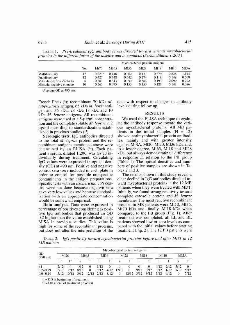
Tini 1. P/-e-treta/nem igG antibods lereis directed toward rarious inrcobacterialproteins in the different forms of the disease and in emacieis. (Sentiu i dilate(' 1:200.)
No.
N1yeobacterial protein antigens
Mt70 NIb65 MI36 MI28 MI18 N1110 NI1SA
Multibacillary 12 0.629' 0.446 0.662 0.431 0.279 0.828 1.114Ritleihaeillary 1") 0.427 0.448 0.642 0.274 0.118 0.149 0.508NI itsuda-positive com aels 6 0.883 0.343 0.052 0.384 0.193 0.099 0.202NI itsuda-negative contaets 10 0.265 0.095 0.155 0.153 0.181 0.141 0.086
'Average OD at 490 mu.
French Press (`'); recombinant 70 kDa M.tuberculosis antigen, 65 kDa AI. boris anti-gen and 36 kDa, 28 kDa 18 kDa and 10kDa M. leprae antigens. Ali recombinantantigens were used at a 5-pg/m1 concentra-tion and the complete soluble leprae at 2pg/m1 according to standardization estab-lished in previous studies (").
Serologic tests. IgG antibodies directedto the total M. leprae protein and the re-combinam antigens mentioned above weredetermined by an ELISA ("). Each pa-tient's sentiu, diluted 1:200, was tested in-dividually during treatment. CirculatingIgG values were expressed in optical den-sity (OD) at 490 nm. Positive and negativecontrol sera were included in each plate inorder to control for possible nonspecificcontaminants in the antigen preparations.Specitic tests with an Escherichia coli con-trol were not done because negative semgave very low values and because standard-ization with arl appropriate concentrationwould be somewhat empirical.
Data analysis. Data were expressed inpercentage of positives considering as posi-tive IgG antibodies that produced an OD0.2 higher than the value established usingM1SA in previous studies. This value ishigh for some of the recombinant proteins,but does not alter the interpretation of the
data with respect to changes in antibodyleveis during follow up.
RESULTSWe used the ELISA technique to evalu-
ate the antibody response toward the vari-ous mycobacterial proteins. All MB pa-tients in the initial samples (N = 12)showed antimycobacterial protein antibod-ies, mainly ind with greater intensityagainst MISA, M120, Mt70, MI36 kDa and,to a lesser degree, MI65, MI18 and M128kDa, but always demonstrating a differencein response in relation to the PB group(Table 1). The optical densities and num-bers of positive samples are shown in Ta-bles 2 and 3.
The results shown in this study reveal aclear decline in IgG antibodies directed to-ward mycobacterial proteins in the 12 MBpatients when they were treated with MDT.Initially, we found strong reactivity towardcomplete cytosolic protein and Al. lepraemembrane. The most reactive recombinantproteins in MB patients were M110, M136,Mt70 kDa and, finally, M118 kDa whencompared to the PB group (Fig. 1). Aftertreatment was completed, ali LL and BLpatients showed low or zero leveis as com-pared with the initial values before startingtreatment (Fig. 2). The 12 PB patients were
TABLI: 2. IgG positivits toward mycobacterial protejas before and afiei- MD7' ia 12M13 patients.
Myeobacterial protein antigensOD(490 uni) Mt70 Mh65 M136 N1128 NI118 N1110 NI1SA
f t-
>1 2/12 O 1/12 O 1/12 O O O 0 0 4/12 2/12 5/12 O0.2-0.99 5/12 2/12 8/12 O 9/12 4/12 12/12 O 9/12 3/12 3/12 1/12 7/12 5/120.0-0.19 5/12 10/12 3/12 12/12 2/12 8/12 O 12/12 3/12 9/12 5/12 9/12 O 7/12
"i = OD at heginning oU treatinent.= OD ai end of treatment (2 years).

416^ International Journal of Leprosy^ 1999
TABU 3.patients.
IgG positivity toward nircobacterial proteins before and atter MDT in 12 PB
Mycobacterial protelo antigensOD(490 nm) M128^M118^M110^M1SAMt70^Mb65^M136
f
>1 2/12 0 0 0 0 0 0 0 0 0 0 0 2/121/120.2-0.99 8/12 6/12 9/12 9 11/12 8/12 7/12 2/12 2/12 6/12 2/12 3/12 4/12 6/120-0.19 2/12 6/12 3/12 3/12 1/12 4/12 5/12 10/12 10/12 6/12 10/12 9/12 6/12 5/12
"i = OD at beginning of treatment.= OD at end of treatment (1 year).
followed up for only 1 year since the dura-tion of their treatment is shorter than that ofpatients with a high bacterial load (Fig. 3).
DISCUSSIONAccording to WHO treatment guide-
lines, MB patients were followed up for a2- to 3-year treatment period and PB pa-tients were evaluated for a 1-year treatmentperiod. Ali 12 patients from each groupwere followed individually until comple-tion of treatment, contrary to a previousstudy (14) in which we evaluated a pool of
sera from patients belonging to each type ofthe disease. IgG antibodies directed towardheat shock protein (hsp) MI10, Mt70, Mb65and MI28 showed high leveis. Other work-ers have reported that two of these hsp,Mt70 and M165, are not good specificmarkers in the disease spectrum (o). We alsodetected IgG antibodies directed towardthese proteins in MB and PB patients and inhealthy Mitsuda-positive contacts, but notin Mitsuda-negative contacts.
Heat or stress shock proteins are inducedby a variety of stimuli and one of them is
0,4
0.2
MI70 MD65 mi28 Mitél M I1 0 MISA
Mytobacterlal proteln antigens
FiG. 1. Comparison of the initizd serologie responses (year 0) in multibacillary (0) and patieibacillary (74 )patients.

0,8
;-;o
; 0,8
0.4
67, 4^ Rada, et ai.: Serology During MDT^ 417
1,2
M170^
M065^
MI36^
MI28^
MI18^
MI10^
MISA
Mylcobact.hal &AN..
FIG. 2. Serologic follow tip^ patients treated with titultidrug therapy.^= year O; R = year 1;= year 2.
mycobacterial infection They also havegreat homology with gene sequences thatcode for these proteins in other phylogenet-ically separated species such as E. co/i andHome sapiens (7). These proteins are con-stitutively expressed, and one of the func-tions in which they are involved is proteintranslocation and folding at an intracellularlevei. The average decrease in antibody lev-eis against heat shock proteins during ther-apy is almost twice as accelerated in MBpatients as compared with the leveis of an-tibodies directed toward other proteins(MI10, MI36 and M128 kDa).
Only the M110 kDa protein establishesdifferences in the group of patients studied,on the basis of the difference in bacillaryload between the two groups (MB and PB).It is considered a promising antigen for skintesting and the most adequate for measuringleprosy incidence. Quantitatively, it is themost abundant, representing around 1% ofthe bacterial mass (1')• The M110 protein,together with the M136 and MI28 proteins,are the ones against which IgG antibodies
persist for longer periods in MB patientsduring MDT.
It is notable that the only serologic re-sponse directed toward recombinant pro-teins which increased during treatment wasthe one directed toward Mb65, and this wasobserved in PB patients with low bacillaryloads. This protein could be involved in theinununopathological processes which char-acterize this group of patients, amono themperipheral nerve damage, which is the mostimportant cause of deformities in leprosy.
MI36 protein is a disease marker; it is aspecific immunodominant protein withproline-rich regions where, according toother authors ( ), 100% of MB and 91%of PB patients present positive serology. Inour study, MB patients showed a high per-centage of positives (83%). Among PB pa-tients, 91.6% were positive. This proteincould be used to detect high-risk persons,which could be very useful for leprosy con-trol programs. The antibody decreaseagainst MI36 is much faster in the PBgroup compared with the high bacillary

418^ Inlernational .10111-11(11 oJ LeprOSN'
0.8
0.7
1999
Mt70 M665 MI36 MI28 MI18 MI10 7.4.SA
0.6
0.5
•0,4
0,3 •
02
MycobacterIal antlgens
FiG. 3. Comparison oi. serologie responses after 1 year of Ireannent in multibaeillary (El) and paucibacillary
() panents.
load in MB patients. This is the only proteinwhich does not show crossreactivity \valiautoimmune processes; whereas the otherproteins tested in this study reflect the pres-ence of antibodies against kDa 70 (meanOD = 0.873), 65 (0.823), 36 (0.060), 28(0.387), 18 (0.519) and 1() (0.782) myco-bacterial proteins in sem a from patients withautoimmune diseases (collagen type clis-cases, lupus erythematosus, scleroderma).
The MI28 kDa protein is an enzyme, su-peroxide clismutase (SOD), secreted in cul-ture medium by growing bacilli. and thereis preliminary evidence that the 28 IcDii pro-tein can be secreted in infected tissues(2.21) in which the bacterium occupies anenvironment in vhich it is StIbIllinCd to se-vere oxidative stress. The importzince ofthis enzyme as a cytoprotective inummo-dominant antigen lies in maintaining myco-bacterial survival ‘vithin the macrophage.Antibodies against this protein were net de-tected in lupus erythematosus but werepresent in other autoimmune processes,such as scleroderma. rvi itsu ci a-positive con-
tacts showed 0.384 OD IgG antibody leveistoward MI28 as compareci w'ith Mitsucia-ne;ative contacts who only showed 0.153OD. 11 is interesting to note that detectableexcretion of. this protein by the presence ofantibodies was zero in MB patients aftercompleting 2 years of MDT. Nlitsuda-posi-tive contacts also showed high leveis of an-tibodies directed toward the 28 kDa proteincompareci with Mitsucia-negative contacts.
Once more we would stress the high ho-mology with human heat shock proteinswhere stress proteins protect cells from thetoxic effects of oxidative intermediates andmitochondria are their selective target (")•SOD is a bacterial defense componentagainst the intracellular attack of phagocytecells against mycobacteria (2")• The amino-acid sequence of this protein shows 67%homology with human SOD.
Regarding M118 kDa protein, the zuni-body response is much higher in MB ascompared \vith PB patientst it does not dis-appear completely but, ralhem, maintainslow leveis. This protein lias been consid-

67, 4^ Rada, et al.: Serology During MDT^ 419
ered as an important antigen for T lympho-cyte presentation ().
Total proteins represent the combinationof ali the individual proteins and, therefore,retlect a high value in antibody detection,even though used at a lower (21..tg/m1) con-centration. Thus, the variability of the re-sponses may be due to the role that each ofthe individual proteins plays.
In summary, we muni! that IgG antibodyleve Is toward mycobacterial proteins(MISA, MI36, M128, MI10, MI18) in mostpatients are expressei! in relation to the sizeof the bacillary load, and the response to the70 and 65 kDa proteins reflects the high ho-mology they show with Mimai! 70 and 65kDa proteins in patients previously sensi-tized to mycobacterial antigens. Themarked decrease in antibody leveis oncemore demonstrates the efficacy of MDT ad-ministered to these patients.We cannot overlook certain other mycobac-terial proteins, such as those belonging to thegroup of proteins secreted by M. bons,among them 30, 31 and 32 kDa proteins,which could be modulating the host responsesince MB patients presented high levels ofIgG antibodies directed to this group of pro-teins compared with PB patients and con-trols using the Western blot technique (17)•
SUMMARYHumoral immune responses were stud-
ied in 24 leprosy patients treated with mul-tidrug therapy (MDT) and 16 contacts. Thepatients were monitored for 2 to 3 yearswith repeated determination of IgG anti-body leveis directed to different mycobac-terial proteins (Mvcobacterium fuberculo-sis, Mt70; M. bovis, Mb65; M. leprae,M136, 28, 18, 10 kDa, and the completeprotein M. leprae extract, MLSA). Ali re-combinam antigens were usei! at 5 pg/m1concentration and the complete soluble M.leprae extract at 2 tly./inl. The results shownin this study reveal a clear decline in IgGantibodies directed toward mycobacterialproteins in the 12 multibacillary (MB) pa-tients when they were submitted to MDT.Initially we found strong reactivity towardcomplete cytosolic protein and M. lepraemembrane protein. The most reactive re-combinam proteins in MB patients wereM110, MI36, Mt70 kDa and, finally, MI18
kDa when compared to the paucibacillary(PB) group. After treatment was completedall lepromatous and borderline lepromatouspatients showed low or undetectable leveisas compared with their initial values beforestarting treatment.
RESUMENSe estudiaron Ias respuestas humorales en 24 pa-
cientes con lepra tratados con poliquimioterapia (1)QT)y en 16 contactos saltos. Los pacientes fueron estudia-dos durante 2 6 3 afios con relación a Ia presencia deanticuerpos IgG contra diferentes proteinas microbac-terianzts (Mycobacterhun luberculosis,Mt70: 41. bovis,M065; 51. lepme, MI36, 28, 18, 10 kDa, y un extractototal de M. Ieprae , MISA). Todos los antigenos re-combinantes fueron usados a Ia concentración de 5 pgpor ml y cl extracto soluble MLSA a 2 pg por ml. Losresultados de este estudio mostraron una clara dismi-nución CII los anticuerpos IgG contra Ias proteinas mi-cobacterianas em los 12 pacientes multibacilares (MB)sujetos aI tratamiento con PQT. Inicialmente se encon-tró una fuerte reactividad bacia las proteinascas y de membrana de Al. leproe. Las proteinas recom-binantes más reactivas en los pacientes multibacilaresfueron M110, N1136, Mt70 y finalmente M118. En com-paración con los niveles de anticuerpos encontrados aiinicio, los niveles de anticuerpos encontrados después decompletar cl tratamiento fueron baios o no detectables.
RÉSUNIELa réponse immunitaire à médiation humorale fut
étudiée chez 24 patients traités par la polychintio-thérapie (I'CT) et chez 16 personnes en contact avecces derniers. Les patients furent suivis pendam aumoins 2 à 3 années, période au cours de laquelle lesniveaux d'anticorps de type IgG diriges curare des pro-téi nesvariées d'origine mycobactérienne (hlsrobac-teriam taberculosis. Nit 70; 41. bovis, Mb 65; 41. lep-rae, MI 36. 28, 18, 10 kDa, et l'extrait complet solu-ble de M. !clave MLSA) furem déterminés de façonrépétée. Tons les antigènes recombinants furent utilisésà une concentration de 5 (pg/mI et l'extrait com pletsoluble de 41. lepra(' à 2 (p/ml. Les résultats présen-tés dans cette étude ont montré une nette diminutioneles anticorps de type IgG dirigés cultue les protéi nesd'origine mycobactérienne chez les 12 patients multi-bacillaires (MO) lorsqu'ils débutèrent la PCT. Audébut, nous avons trouvé une forte réactivité contre Iaprotéine cytosolique complete et Ia protéine mem-branaire de M. Itpme. Les protéines recombinantes lesplus reactives chez les patients MB étaient MI 10. MI36, Mt 70 kDa et, tinalement N11 18 kDa, comparé aligroupe des patients paucibacillaires (PB). Après Ia lindu traitement. tous les patients lépromateux et lépro-mateux borderline ont montré des niveattx faibles ounon-détectables, comparés aux valeurs initialesmesurées ;Avant le ft:atentem.

420^ International Journal of Lepro.sy^ 1999
MITIRA, V., BLOONI, B. R., BAJARDI, A. C., Gitisso,C. L., SIELING, P. A., ALLANi), D., CoNvrt, J., FAN,X. D., IltiNTut, S. W., BRENNAN, P. J., REA, T. H.and MooLIN, R. L. A major T-cell antigen of My-cobacterium leprae is a 10 kD heat-shock cognateprotein. J. Exp. Med. 175 (1992) 275-284.MISTRY,Y., Yotimi, D. and MUKIIERJEE, R. I Isp70synthesis in Schwann cells in response to heatshock anil infection with Mycobacterium leprae.Inject. Inimun. 60 (1992) 3105-3110.
13. Poi.i.A, B. S., KAIENowA, S., FRANCOIS, D., SAINI-OH, S., FRANCESCIII, C., MARSAC, C. and COS-SARIZZA, A. Mitochondria are selective targets forthe protective effects of heat shock against oxida-tive injury. Proc. Nau. Acad. Sei. U.S.A. 93(1996) 6458-6463.RADA, F., ARANZAZU, N. and CONVET, J. Immuno-logical reactions to mycobacterial proteins in thespectrum of leprosy. Int. J. Lcpr. 65 (1997)497-500.
15. RADA, E., Ui.ticii, M., ARANZAZU, N., RooRicitiEz,V., CENTEN(), M., GoNzÁi.Ez, I., SANTALI.I.A, C.,
RODRiGUEZ, M. and CoNviT, J. A follow-up studyof multibacillary Ilansen's disease patients treatedwith multidrug therapy (MDT) or MDT + im-munotherapy (IMT). Int. J. Lepr. 65 (1997)320-327.
16. RADA, E., ULRicii, M., ARANzAzu, N., SANTAELLA,C., GALLINOTO, M. E., CENTENO, M., RODRÍGUEZ,
V. and CONVIT, J. A longitudinal study of im-munologic reactivity in leprosy patients treatedwith immunotherapy. Int. J. Lepr. 62 (1994)552-558.
Acknowledgment. We wish to thank the WII0^11.Recombinant Protein Bank for supplying the proteinsused in this study.
REFERENCES1. BAC'', M.-A., WALLACII, D., FLAGRA., B., Ilor-
FENI3ACI I , A. and CorrENar, F. Antibodies to pite-noite glycolipid-I and to whole Alycobacteriumleprae in leprosy patients: evolution during ther-apy. Int. J. Lepr. 54 (1986) 256-267.
2. Boo) ti, R. J., HARRIS, D. P., LOVE, J. M. and WAT-SON, J. D. Antigenic properties of Mycobacteritonleprae, complete sequence of the gene for the 18kDa protein. J. Immunol. 140 (1998) 597-601.
3. BUCHANAN, T. M., Nommitvin, H., ANDERsoN, D.C., Yourvo, R. A., GII.0s, T. P., BiarroN, W. J.,^14.IvANyt, J., Koi.K, A. ii. , Ci.oss, O., BLoont, B. R.ilild MEIIRA, V. Characterization of antibody-reac-tive epitopes on the 65-kilodalton protein of My-cobacterium leprae. Infect. Immun. 55 (1987)1000-1003.
4. ClIANTEAU, S., CARTEL, I. L., CEnimit, P.,PLICIIART, R., DEsuoRoEs, S. and Roux, J. PGL-Iantigen and antibodies detection in leprosy pa-tients: evolution under chemotherapy. Int. J. Lepr.57 (1989) 735-743.
5. Clioutmium, K. The immunology of leprosy; un-raveling an enigma. Int. J. Lepr. 63 (1995)430-447.
6. CONVIT, J., ARANZAZU, N., UTRICH, M., PINARDI,M. E., RI:YES, O. and Ai.vARADo, J. Inuntinother-apy with a mixture of Mycobacterium lepra( andBCG in different forms of leprosy and Mitsuda-^17.negative contacts. Int. J. Lepr. 50 (1982) 415-424.
7. GARSIA, R., FIELLQvisT, L., Bo(yol, R., RADR)Ro,A., BRITTON, W., ASTI3URY, L., TRENT, R. and BAS-TEN, A. Homology of the 70-kilodalton antigensfrom Mycobacterium leprae and illycobacterbon^18.bovis with the Mycobacterium tuberculosis 71-kilodalton antigen and with the conserved heatshock protein 70 of eucaryotes. Infect. Inuntin. 57(1989) 204-212.
8. KLAUSER, P. R., tt Wrn , M. Y. and Koi.K, A. H. J.An ELISA-inhibition test using monoclonal anti-body for the serology of leprosy. Clin. Exp. Im-munol. 62 (1985) 468-473.
9. LAUNOIS, P., NiANG, M. N., DRowART, A., VANVOOREN, J. P., SARTimu, J. L., LAIA), T., MILLAN,J. and HUYGEN, K. IgG response to purified 65-and 70-kDa mycobacterial heat shock proteins andto antigen 85 in leprosy. Int. J. Lepr. 62 (1994)^21.48-54.
10. MEEKER, 1-1. C., Situ0.0z-LEvis, G., FUSCO, F.,GIARDINA-BECKET, M.-A., SERSEN, E. and LEvis,W. R. Sequential monitoring of leprosy patientswith serum antibody leveis to phenolic glycolipid-1, a synthetic analog of phenolic glycolipid-I, andmycobacterial lipoarabinomannan. Int. J. Lepr. 58(1990) 503-511.
HUNTER, S. W., HEETA, S. A., MEIIRA, V. andBRENNAN, P. Chemical delinition, cloning, andexpression of the major protein of the leprosybacillus. Infect. Immun. 62 (1994) 2417-2425.TIIANGARAJ, 1-1. S., LANIII, F. I., DAVIS, E. O., JEN-NER, P. J., JEYAKUNIAR, L. H. and Coi.sToN, M. J.Identitication, sequencing, and expression of My-cobacterium leprae superoxide dismutase, a majorantigen. Infect. 'mutua. 58 (1990) 1937-1942.Toou:, J. E. R., JANSON, A. A. M., KIRA:, A.,Howh, R. C., MCLEAN, K., NURILYGN, A., FILLEY,E., SHANNON, E. J., BULLA, G. J., HERNIANS, J., DEVRIES, R. R. P., FRommui., D. and RINKE DE WIT,T. Analysis of T-cell and B-cell responses to re-combinant M. leprae antigens ia leprosy patientsand healthy contacts: signiticant T-cell responsesto antigens in M. leprae nonresponders. Int. J.Lepr. 63 (1995) 369-380.
RADA-SC/II.AEFLI, E., SANTAELLA, C., ARANZAZU,N. and CoNviT, J. Detection of antibodies towardsecreted mycobacterial antigen 85 ia untreatedleprosy patients' sera. (Letter) Int. J. Lepr. 67(1999) 167-170.Riot.Ev, D. S. and JOPLING,W. H. Classification ofleprosy according to immunity; a tive-grottp sys-tem. Int. J. Lepr. 34 (1966) 255-273.
19. Rivolity, B., PEssol.ANi, M. C., Bom', C. M.,

67, 4^ Rada, et al.: ,S'erology During MD7'^ 421
Titot.[, J. E. R., STABlit., E. E. M., StlYKIII21111YK,
M. E. G., ia: WriT, M. Y. L., KLATSIIR, P. R., KOJAK,A. 1-1. J. and IIAIUSKIIERL, R. A. A major int-munogenic 36,000-molecular-weight antigenfrom Afreobacierium lepra(' contatos anmunoreactive region of proline-rich repeats. In-fect. Immun. 58 (1990) 80-87.Utioct I, M., SMITH, P., SANIPSON, C., ZUI■IIGA, M.,CENTENO, M., GARCÍA, V., MANRIQUII, X., SAI.-(;AIX), A. and CotiviT, J. IgM antibodies to native
phenolic g,lycolipid-1 in contztets of leprosy pa-tients in Venezuela; epidemiological observationsand a prospective study of the risk of leprosy. Int.J. Lepr. 59 (1991) 405-415.
24. %MON, J. D. Leprosy: understanding protectiveimmunity. lmmunol. Today 10 (1989) 218-221.
25. YouNc;, D. B., KAUFNIANN, S. 1-I. E., IINIZMANS, P.W. M. and Ttioi.F., J. E. R. Myeobacterial proteinantigens. Moi. Microbiol. 6 (1992) 133-145.