il8 (human) elisa kit - abnova · principle of the assay ... the il8 (human) elisa kit is for...
TRANSCRIPT

www.abnova.com
IL8 (Human) ELISA Kit
Catalog Number KA0115
96 assays
Version: 05
Intended for research use only
KA0115 2 / 22
Table of Contents
Introduction ...................................................................................................... 3
Intended Use ................................................................................................................. 3
Background ................................................................................................................... 3
Principle of the Assay .................................................................................................... 5
General Information ......................................................................................... 7
Materials Supplied ......................................................................................................... 7
Storage Instruction ........................................................................................................ 7
Materials Required but Not Supplied ............................................................................. 7
Precautions for Use ....................................................................................................... 8
Assay Protocol ............................................................................................... 10
Reagent Preparation ................................................................................................... 10
Sample Preparation ..................................................................................................... 12
Assay Procedure ......................................................................................................... 13
Data Analysis.................................................................................................. 15
Calculation of Results .................................................................................................. 15
Performance Characteristics ....................................................................................... 18
Resources ....................................................................................................... 22
Plate Layout ................................................................................................................ 22

KA0115 3 / 22
Introduction
Intended Use
The IL8 (Human) ELISA Kit is an enzyme-linked immunosorbent assay for the quantitative detection of human
IL-8/NAP-1. The IL8 (Human) ELISA Kit is for research use only. Not for diagnostic or therapeutic procedures.
Background
Interleukin-8/Neutrophil-Activating Peptide-1 selectively stimulates the ability of neutrophils and T-lymphocytes
to invade injured or inflamed tissue. IL-8 purified from peripheral blood monocytes is a 10 kDa protein showing
at least 4 variant forms (77, 72, 70, and 69 amino acids, respectively) which differ in the length of the
N-terminal and readily form dimers in solution. Two cystin-bridges are essential for the biological activity of IL-8.
Exogenous stimuli like LPS, but also IL-1, TNFα and TNFβ induce the secretion of IL-8 in a variety of different
cell types including monocytes, endothelial and epithelial cells, peripheral blood mononuclear cells, dermal
fibroblasts, keratinocytes, neutrophils, hepatocytes, synovial cells, and T-lymphocytes. When IL-8 was
subcutaneously injected into rats, both lymphocytes and neutrophils migrated to the site of injection within 3
hours. At lower dosages, only lymphocytes migrated towards the site of injection, while at higher dosages
primarily neutrophils were attracted. It was found that T-lymphocytes are 10 times more sensitive to IL-8 than
neutrophils. IL-8 exerts its effects via specific cell membrane receptors (chemotactic agonist receptor-family)
with homogeneous high-affinity activity and two binding sites for its ligand. The receptor density is determined
by the cell type and ranges from 300 on T-lymphocytes up to 20.000 on neutrophils. After binding of IL-8, the
receptor expression is downregulated >90 % within 10 minutes at 37°C, together with the internalization of the
ligand. IL-8 is proteolytically degraded in the cytoplasm and released into the culture medium as soluble
fragments. The IL-8 receptors are probably recycled. Besides its chemotactic influence, IL-8 exerts other
distinct characteristics. In neutrophils it triggers the secretion of superoxide anions and lysosomal enzymes,
thereby indirectly augmenting the permeability of blood vessels, and IL-8 enhances the fungicidal activity
against Candida albicans. Neutrophils are more readily liberated from the bone marrow reservoir under the
influence of this cytokine. In vitro, IL-8 stimulates a rapid Mac-1 as well as CR1, p150,95 and LFA-1 expression
on neutrophils which enables the adherence to activated vascular endothelial cells expressing e.g. ICAM-1.
This may account for the accumulation of neutrophils at IL-8 injection sites. Other findings suggest that
endothelial-derived IL-8 may function to attenuate inflammatory events at the interface between vessel wall
and blood, via inhibiting neutrophil adhesion to cytokine-activated endothelial monolayers. Therefore these
cells seem to be protected against neutrophil-mediated damage. In basophils, besides its chemotactic effects,
IL-8 stimulates the histamin liberation in atopic as well as healthy persons.
The property of IL-8 to stimulate movement of neutrophils across endothelial monolayers in vitro supports the
concept of a central role for this molecule in the accumulation of neutrophils at inflammatory lesions in vivo.
Data indicate that IL-8 may participate in the pathogenesis of rheumatoid arthritis via the induction of
neutrophil-mediated cartilage damage, and psoriasis. A causative involvement of IL-8 is found within a broad

KA0115 4 / 22
range of clinico-pathological conditions: adult respiratory distress syndrome, asthma, bacterial infections,
bladder cancer, blood incomptability, contact dermatitis, empyema, graft rejection, glomerulonephritis, gout,
hemolytic uremic syndrome, immune vasculitis, inflammatory bowel disease, influenza virus infection,
Jarisch-Herxheimer reaction, malignancies of the central nervous system, myocardial infarction, placental
infection, sepsis, uveitis, among others.
Due to the nowadays known biological properties of IL-8, this cytokine, especially in combinations with other
neutrophil activating agents, may prove helpful in the treatment of patients suffering from granulocytopenia,
severe infections against which antibiotics are not effective, and immunodeficiency caused by HI-virus.

KA0115 5 / 22
Principle of the Assay
An anti-human IL-8/NAP-1 coating antibody is adsorbed onto
microwells.
Human IL-8/NAP-1 present in the sample or standard binds to
antibodies adsorbed to the microwells. A biotin-conjugated
anti-human IL-8/NAP-1 antibody is added and binds to human
IL-8/NAP-1 captured by the first antibody.
Following incubation unbound biotin-conjugated anti-human
IL-8/NAP-1 antibody is removed during a wash step.
Streptavidin-HRP is added and binds to the biotin-conjugated
anti-human IL-8/NAP-1 antibody.
Following incubation unbound Streptavidin-HRP is removed
during a wash step, and substrate solution reactive with HRP is
added to the wells.
KA0115 6 / 22
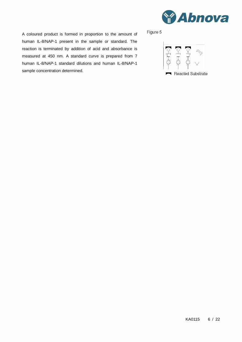
A coloured product is formed in proportion to the amount of
human IL-8/NAP-1 present in the sample or standard. The
reaction is terminated by addition of acid and absorbance is
measured at 450 nm. A standard curve is prepared from 7
human IL-8/NAP-1 standard dilutions and human IL-8/NAP-1
sample concentration determined.

KA0115 7 / 22
General Information
Materials Supplied
List of component
Component Amount
Microwell Plate coated with monoclonal antibody to human IL-8/NAP-1 1 aluminium pouch
Biotin-Conjugate anti-human IL-8/NAP-1 polyclonal antibody 70 µL
Streptavidin-HRP 150 µL
human IL-8/NAP-1 Standard, 100 ng/mL 100 µL x 2
Control, high, lyophilized 1 vial
Control, low, lyophilized 1 vial
Sample Diluent 12 mL
Assay Buffer Concentrate 20x (PBS with 1% Tween 20 and 10% BSA) 5 mL
Wash Buffer Concentrate 20x (PBS with 1% Tween 20) 50 mL
Substrate Solution (tetramethyl-benzidine) 15 mL
Stop Solution (1M Phosphoric acid) 15 mL
Adhesive Films 4 slices
Note: In some, very rare cases, an insoluble precipitate of stabilizing protein has been seen in the standard
and Biotin-Conjugate vials. This precipitate does not interfere in any way with the performance of the test and
can thus be ignored.
Storage Instruction
Store kit reagents between 2° and 8°C except controls. Store lyophilized control at -20°C.
Immediately after use remaining reagents should be returned to cold storage (2° to 8°C), controls to -20°C,
respectively. Expiry of the kit and reagents is stated on labels.
Expiry of the kit components can only be guaranteed if the components are stored properly, and if, in case of
repeated use of one component, this reagent is not contaminated by the first handling.
Materials Required but Not Supplied
5 mL and 10 mL graduated pipettes
5 µL to 1000 µL adjustable single channel micropipettes with disposable tips
50 µL to 300 µL adjustable multichannel micropipette with disposable tips
Multichannel micropipette reservoir
Beakers, flasks, cylinders necessary for preparation of reagents
Device for delivery of wash solution (multichannel wash bottle or automatic wash system)
Microwell strip reader capable of reading at 450 nm (620 nm as optional reference wave length)
KA0115 8 / 22
Glass-distilled or deionized water
Statistical calculator with program to perform regression analysis
Precautions for Use
Precautions
All chemicals should be considered as potentially hazardous. We therefore recommend that this product
is handled only by those persons who have been trained in laboratory techniques and that it is used in
accordance with the principles of good laboratory practice. Wear suitable protective clothing such as
laboratory overalls, safety glasses and gloves. Care should be taken to avoid contact with skin or eyes. In
the case of contact with skin or eyes wash immediately with water. See material safety data sheet(s)
and/or safety statement(s) for specific advice.
Reagents are intended for research use only and are not for use in diagnostic or therapeutic procedures.
Do not mix or substitute reagents with those from other lots or other sources.
Do not use kit reagents beyond expiration date on label.
Do not expose kit reagents to strong light during storage or incubation.
Do not pipette by mouth.
Do not eat or smoke in areas where kit reagents or samples are handled.
Avoid contact of skin or mucous membranes with kit reagents or specimens.
Rubber or disposable latex gloves should be worn while handling kit reagents or specimens.
Avoid contact of substrate solution with oxidizing agents and metal.
Avoid splashing or generation of aerosols.
In order to avoid microbial contamination or cross-contamination of reagents or specimens which may
invalidate the test use disposable pipette tips and/or pipettes.
Use clean, dedicated reagent trays for dispensing the conjugate and substrate reagent.
Exposure to acid inactivates the conjugate.
Glass-distilled water or deionized water must be used for reagent preparation.
Substrate solution must be at room temperature prior to use.
Decontaminate and dispose specimens and all potentially contaminated materials as they could contain
infectious agents. The preferred method of decontamination is autoclaving for a minimum of 1 hour at
121.5°C.
Liquid wastes not containing acid and neutralized waste may be mixed with sodium hypochlorite in
volumes such that the final mixture contains 1.0% sodium hypochlorite. Allow 30 minutes for effective
decontamination. Liquid waste containing acid must be neutralized prior to the addition of sodium
hypochlorite.

KA0115 9 / 22
Limitations
Since exact conditions may vary from assay to assay, a standard curve must be established for every
run.
Bacterial or fungal contamination of either screen samples or reagents or cross-contamination between
reagents may cause erroneous results.
Disposable pipette tips, flasks or glassware are preferred, reusable glassware must be washed and
thoroughly rinsed of all detergents before use.
Improper or insufficient washing at any stage of the procedure will result in either false positive or false
negative results. Empty wells completely before dispensing fresh wash solution, fill with Wash Buffer as
indicated for each wash cycle and do not allow wells to sit uncovered or dry for extended periods.
The use of radioimmunotherapy has significantly increased the number of patients with human
anti-mouse IgG antibodies (HAMA). HAMA may interfere with assays utilizing murine monoclonal
antibodies leading to both false positive and false negative results. Serum samples containing antibodies
to murine immunoglobulins can still be analysed in such assays when murine immunoglobulins (serum,
ascitic fluid, or monoclonal antibodies of irrelevant specificity) are added to the sample.

KA0115 10 / 22
Assay Protocol
Reagent Preparation
Buffer concentrates should be brought to room temperature and should be diluted before starting the test
procedure. If crystals have formed in the Buffer Concentrates, warm them gently until they have completely
dissolved.
Wash Buffer (1x)
Pour entire contents (50 mL) of the Wash Buffer Concentrate (1x) into a clean 1000 mL graduated cylinder.
Bring to final volume of 1000 mL with glass-distilled or deionized water.
Mix gently to avoid foaming.
Transfer to a clean wash bottle and store at 2° to 25°C. Please note that Wash Buffer (1x) is stable for 30 days.
Wash Buffer (1x) may also be prepared as needed according to the following table:
Number of Strips Wash Buffer Concentrate (20x)
(mL)
Distilled Water
(mL)
1-6 25 475
1-12 50 950
Assay Buffer (1x)
Pour the entire contents (5 mL) of the Assay Buffer Concentrate (20x) into a clean 100 mL graduated cylinder.
Bring to final volume of 100 mL with distilled water. Mix gently to avoid foaming.
Store at 2° to 8°C. Please note that the Assay Buffer (1x) is stable for 30 days.
Assay Buffer (1x) may also be prepared as needed according to the following table:
Number of Strips Assay Buffer Concentrate (20x)
(mL)
Distilled Water
(mL)
1-6 2.5 47.5
1-12 5.0 95.0
Biotin-Conjugate
Please note that the Biotin-Conjugate should be used within 30 minutes after dilution.
Make a 1:100 dilution of the concentrated Biotin-Conjugate solution with Assay Buffer (1x) in a clean plastic
tube as needed according to the following table:
KA0115 11 / 22
Number of Strips Biotin-Conjugate
(mL)
Assay Buffer (1x)
(mL)
1-6 0.03 2.97
1-12 0.06 5.94
Please note: In some, very rare cases, an insoluble precipitate of stabilizing protein has been seen in the
standard and Biotin-Conjugate vials. This precipitate does not interfere in any way with the performance of the
test and can thus be ignored.
Streptavidin-HRP
Please note that the Streptavidin-HRP should be used within 30 minutes after dilution.
Make a 1:200 dilution of the concentrated Streptavidin-HRP solution with Assay Buffer (1x) in a clean plastic
tube as needed according to the following table:
Number of Strips Streptavidin-HRP
(mL)
Assay Buffer (1x)
(mL)
1-6 0.03 5.97
1-12 0.06 11.94
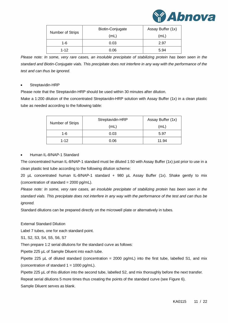
Human IL-8/NAP-1 Standard
The concentrated human IL-8/NAP-1 standard must be diluted 1:50 with Assay Buffer (1x) just prior to use in a
clean plastic test tube according to the following dilution scheme:
20 µL concentrated human IL-8/NAP-1 standard + 980 µL Assay Buffer (1x). Shake gently to mix
(concentration of standard = 2000 pg/mL).
Please note: In some, very rare cases, an insoluble precipitate of stabilizing protein has been seen in the
standard vials. This precipitate does not interfere in any way with the performance of the test and can thus be
ignored.
Standard dilutions can be prepared directly on the microwell plate or alternatively in tubes.
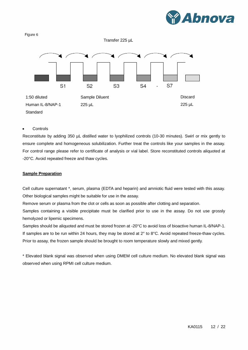
External Standard Dilution
Label 7 tubes, one for each standard point.
S1, S2, S3, S4, S5, S6, S7
Then prepare 1:2 serial dilutions for the standard curve as follows:
Pipette 225 µL of Sample Diluent into each tube.
Pipette 225 µL of diluted standard (concentration = 2000 pg/mL) into the first tube, labelled S1, and mix
(concentration of standard 1 = 1000 pg/mL).
Pipette 225 µL of this dilution into the second tube, labelled S2, and mix thoroughly before the next transfer.
Repeat serial dilutions 5 more times thus creating the points of the standard curve (see Figure 6).
Sample Diluent serves as blank.
KA0115 12 / 22
Controls
Reconstitute by adding 350 µL distilled water to lyophilized controls (10-30 minutes). Swirl or mix gently to
ensure complete and homogeneous solubilization. Further treat the controls like your samples in the assay.
For control range please refer to certificate of analysis or vial label. Store reconstituted controls aliquoted at
-20°C. Avoid repeated freeze and thaw cycles.
Sample Preparation
Cell culture supernatant *, serum, plasma (EDTA and heparin) and amniotic fluid were tested with this assay.
Other biological samples might be suitable for use in the assay.
Remove serum or plasma from the clot or cells as soon as possible after clotting and separation.
Samples containing a visible precipitate must be clarified prior to use in the assay. Do not use grossly
hemolyzed or lipemic specimens.
Samples should be aliquoted and must be stored frozen at -20°C to avoid loss of bioactive human IL-8/NAP-1.
If samples are to be run within 24 hours, they may be stored at 2° to 8°C. Avoid repeated freeze-thaw cycles.
Prior to assay, the frozen sample should be brought to room temperature slowly and mixed gently.
* Elevated blank signal was observed when using DMEM cell culture medium. No elevated blank signal was
observed when using RPMI cell culture medium.
Transfer 225 µL
1:50 diluted
Human IL-8/NAP-1
Standard
Sample Diluent
225 µL
Discard
225 µL
KA0115 13 / 22
Assay Procedure
1. Determine the number of microwell strips required to test the desired number of samples plus appropriate
number of wells needed for running blanks and standards. Each sample, standard, blank and optional
control sample should be assayed in duplicate. Remove extra microwell strips from holder and store in
foil bag with the desiccant provided at 2°-8°C sealed tightly.
2. Wash the microwell strips twice with approximately 400 µL Wash Buffer per well with thorough aspiration
of microwell contents between washes. Allow the Wash Buffer to sit in the wells for about 10 – 15
seconds before aspiration. Take care not to scratch the surface of the microwells.
After the last wash step, empty wells and tap microwell strips on absorbent pad or paper towel to remove
excess Wash Buffer. Use the microwell strips immediately after washing. Alternatively microwell strips
can be placed upside down on a wet absorbent paper for not longer than 15 minutes. Do not allow wells
to dry.
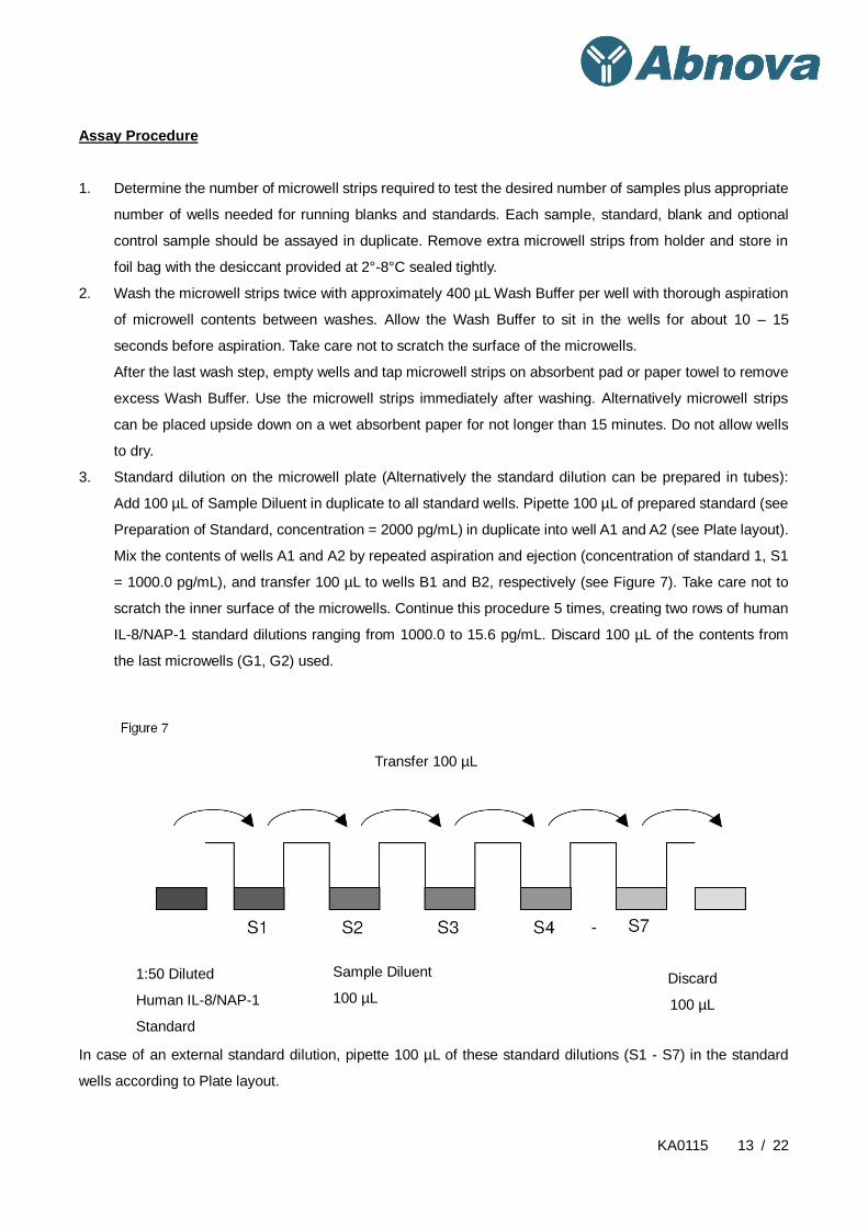
3. Standard dilution on the microwell plate (Alternatively the standard dilution can be prepared in tubes):
Add 100 µL of Sample Diluent in duplicate to all standard wells. Pipette 100 µL of prepared standard (see
Preparation of Standard, concentration = 2000 pg/mL) in duplicate into well A1 and A2 (see Plate layout).
Mix the contents of wells A1 and A2 by repeated aspiration and ejection (concentration of standard 1, S1
= 1000.0 pg/mL), and transfer 100 µL to wells B1 and B2, respectively (see Figure 7). Take care not to
scratch the inner surface of the microwells. Continue this procedure 5 times, creating two rows of human
IL-8/NAP-1 standard dilutions ranging from 1000.0 to 15.6 pg/mL. Discard 100 µL of the contents from
the last microwells (G1, G2) used.
In case of an external standard dilution, pipette 100 µL of these standard dilutions (S1 - S7) in the standard
wells according to Plate layout.
Transfer 100 µL
Discard
100 µL
Sample Diluent
100 µL
1:50 Diluted
Human IL-8/NAP-1
Standard

KA0115 14 / 22
4. Add 100 µL of Sample Diluent in duplicate to the blank wells.
5. Add 50 µL of Sample Diluent to the sample wells.
6. Add 50 µL of each sample in duplicate to the sample wells.
7. Prepare Biotin-Conjugate.
8. Add 50 µL of Biotin-Conjugate to all wells.
9. Cover with an adhesive film and incubate at room temperature (18 to 25°C) for 2 hours, if available on a
microplate shaker set at 400 rpm.
10. Prepare Streptavidin-HRP.
11. Remove adhesive film and empty wells. Wash microwell strips 3 times according to point 2. of the test
protocol. Proceed immediately to the next step.
12. Add 100 µL of diluted Streptavidin-HRP to all wells, including the blank wells.
13. Cover with an adhesive film and incubate at room temperature (18° to 25°C) for 1 hour, if available on a
microplate shaker set at 400 rpm.
14. Remove adhesive film and empty wells. Wash microwell strips 3 times according to point 2. of the test
protocol. Proceed immediately to the next step.
15. Pipette 100 µL of TMB Substrate Solution to all wells.
16. Incubate the microwell strips at room temperature (18° to 25°C) for about 10 min. Avoid direct exposure
to intense light.
The colour development on the plate should be monitored and the substrate reaction stopped (see next
point of this protocol) before positive wells are no longer properly recordable. Determination of the ideal
time period for colour development has to be done individually for each assay.
It is recommended to add the stop solution when the highest standard has developed a dark blue colour.
Alternatively the colour development can be monitored by the ELISA reader at 620 nm. The substrate
reaction should be stopped as soon as Standard 1 has reached an OD of 0.9 – 0.95.
17. Stop the enzyme reaction by quickly pipetting 100 µL of Stop Solution into each well. It is important that
the Stop Solution is spread quickly and uniformly throughout the microwells to completely inactivate the
enzyme. Results must be read immediately after the Stop Solution is added or within one hour if the
microwell strips are stored at 2 - 8°C in the dark.
18. Read absorbance of each microwell on a spectro-photometer using 450 nm as the primary wave length
(optionally 620 nm as the reference wave length; 610 nm to 650 nm is acceptable). Blank the plate reader
according to the manufacturer's instructions by using the blank wells. Determine the absorbance of both
the samples and the standards.
Note: In case of incubation without shaking the obtained O.D. values may be lower than indicated below.
Nevertheless the results are still valid.

KA0115 15 / 22
Data Analysis
Calculation of Results
Calculate the average absorbance values for each set of duplicate standards and samples. Duplicates
should be within 20 per cent of the mean value.
Create a standard curve by plotting the mean absorbance for each standard concentration on the
ordinate against the human IL-8/NAP-1 concentration on the abscissa. Draw a best fit curve through the
points of the graph (a 5-parameter curve fit is recommended).
To determine the concentration of circulating human IL-8/NAP-1 for each sample, first find the mean
absorbance value on the ordinate and extend a horizontal line to the standard curve. At the point of
intersection, extend a vertical line to the abscissa and read the corresponding human IL-8/NAP-1
concentration.
If instructions in this protocol have been followed samples have been diluted 1:2 (50 µL sample + 50 µL
Sample Diluent) and the concentration read from the standard curve must be multiplied by the dilution
factor (x 2).
Calculation of samples with a concentration exceeding standard 1 may result in incorrect, low human
IL-8/NAP-1 levels. Such samples require further external predilution according to expected human
IL-8/NAP-1 values with Sample Diluent in order to precisely quantitate the actual human IL-8/NAP-1
level.
It is suggested that each testing facility establishes a control sample of known human IL-8/NAP-1
concentration and runs this additional control with each assay. If the values obtained are not within the
expected range of the control, the assay results may be invalid.
A representative standard curve is shown in Figure 8. This curve cannot be used to derive test results.
Each laboratory must prepare a standard curve for each group of microwell strips assayed.

KA0115 16 / 22
Figure 8
Representative standard curve for IL8 (Human) ELISA Kit. Human IL-8/NAP-1 was diluted in serial 2-fold steps
in Sample Diluent. Do not use this standard curve to derive test results. A standard curve must be run for each
group of microwell strips assayed.
KA0115 17 / 22
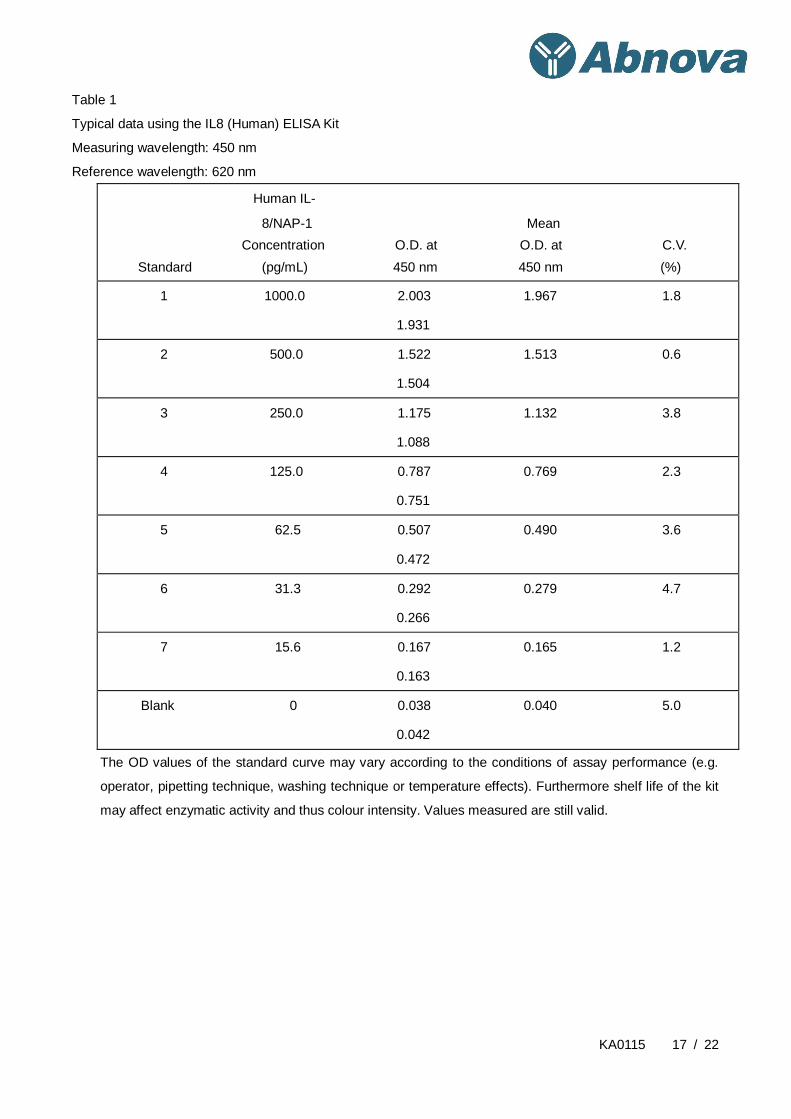
Table 1
Typical data using the IL8 (Human) ELISA Kit
Measuring wavelength: 450 nm
Reference wavelength: 620 nm
Human IL-
8/NAP-1 Mean
Concentration O.D. at O.D. at C.V.
Standard (pg/mL) 450 nm 450 nm (%)
1 1000.0 2.003 1.967 1.8
1.931
2 500.0 1.522 1.513 0.6
1.504
3 250.0 1.175 1.132 3.8
1.088
4 125.0 0.787 0.769 2.3
0.751
5 62.5 0.507 0.490 3.6
0.472
6 31.3 0.292 0.279 4.7
0.266
7 15.6 0.167 0.165 1.2
0.163
Blank 0 0.038 0.040 5.0
0.042
The OD values of the standard curve may vary according to the conditions of assay performance (e.g.
operator, pipetting technique, washing technique or temperature effects). Furthermore shelf life of the kit
may affect enzymatic activity and thus colour intensity. Values measured are still valid.
KA0115 18 / 22
Performance Characteristics
Sensitivity
The limit of detection of human IL-8/NAP-1 defined as the analyte concentration resulting in an absorbance
significantly higher than that of the dilution medium (mean plus 2 standard deviations) was determined to be
2.0 pg/mL (mean of 6 independent assays).
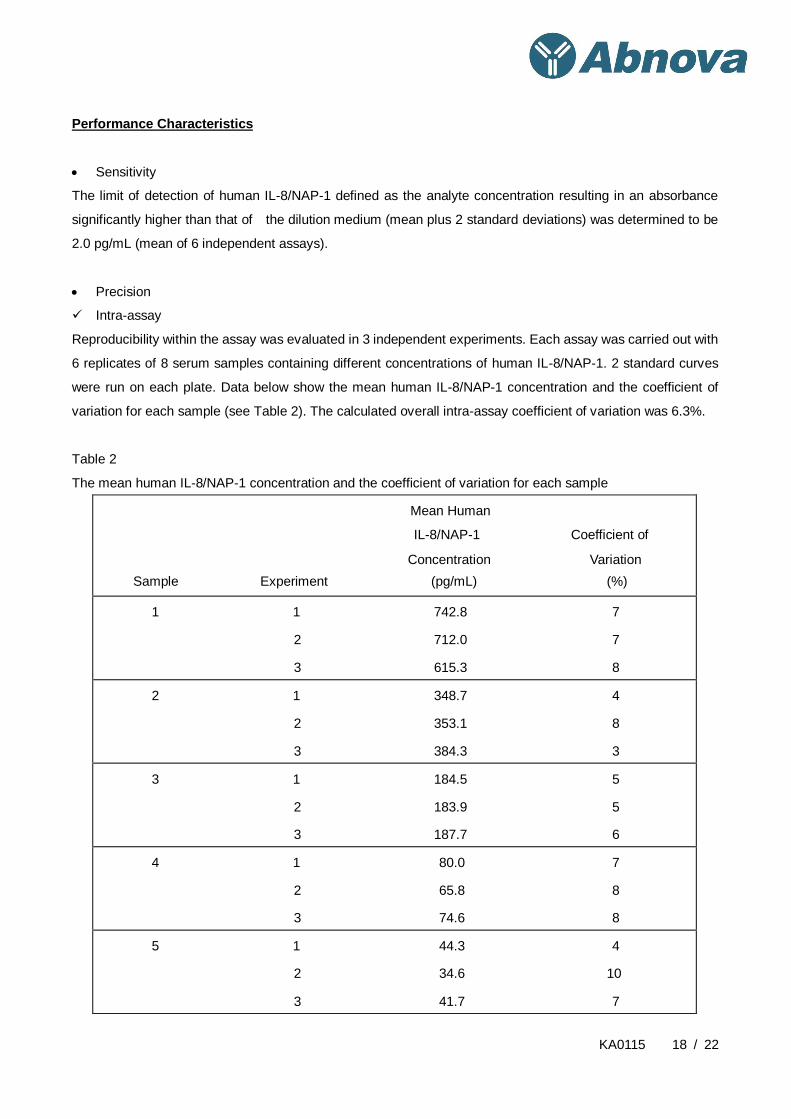
Precision
Intra-assay
Reproducibility within the assay was evaluated in 3 independent experiments. Each assay was carried out with
6 replicates of 8 serum samples containing different concentrations of human IL-8/NAP-1. 2 standard curves
were run on each plate. Data below show the mean human IL-8/NAP-1 concentration and the coefficient of
variation for each sample (see Table 2). The calculated overall intra-assay coefficient of variation was 6.3%.
Table 2
The mean human IL-8/NAP-1 concentration and the coefficient of variation for each sample
Mean Human
IL-8/NAP-1 Coefficient of
Concentration Variation
Sample Experiment (pg/mL) (%)
1 1 742.8 7
2 712.0 7
3 615.3 8
2 1 348.7 4
2 353.1 8
3 384.3 3
3 1 184.5 5
2 183.9 5
3 187.7 6
4 1 80.0 7
2 65.8 8
3 74.6 8
5 1 44.3 4
2 34.6 10
3 41.7 7

KA0115 19 / 22
6 1 520.4 4
2 477.9 6
3 615.1 5
7 1 273.0 4
2 251.2 3
3 291.8 11
8 1 316.0 9
2 313.3 6
3 260.0 9
Inter-assay
Assay to assay reproducibility within one laboratory was evaluated in 3 independent experiments. Each assay
was carried out with 6 replicates of 8 serum samples containing different concentrations of human IL-8/NAP-1.
2 standard curves were run on each plate. Data below show the mean human IL-8/NAP-1 concentration and
the coefficient of variation calculated on 18 determinations of each sample (see Table 3).The calculated overall
inter-assay coefficient of variation was 8.7%.
Table 3
The mean human IL-8/NAP-1 concentration and the coefficient of variation of each sample
Mean Human IL-8/NAP-1 Coefficient of
Concentration Variation
Sample (pg/mL) (%)
1 690.0 9.6
2 362.0 5.4
3 185.4 1.1
4 73.5 9.7
5 40.2 12.5
6 537.8 13.1
7 272.0 7.5
8 296.4 10.7
Spiking Recovery
The spike recovery was evaluated by spiking 4 levels of human IL-8/NAP-1 into serum samples. Recoveries
were determined in 3 independent experiments with 6 replicates each. The amount of endogenous human
IL-8/NAP-1 in unspiked serum was subtracted from the spike values. The recovery ranged from 72% to 125%
with an overall mean recovery of 88%.

KA0115 20 / 22
Dilution Linearity
4 serum samples with different levels of human IL-8/NAP-1 were analysed at serial 2 fold dilutions with 4
replicates each. The recovery ranged from 90% to 119% with an overall recovery of 107% (see Table 4).
Table 4
Recovery
Expected Observed of Expected
Human Human Human
IL-8/NAP-1 IL-8/NAP-1 IL-8/NAP-1
Concentration Concentration Concentration
Sample Dilution (pg/mL) (pg/mL) (%)
1 1:2 --- 771.7 --
1:4 385.9 402.4 104
1:8 201.2 217.8 108
1:16 108.9 104.3 96
2 1:2 --- 369.9 --
1:4 185.0 194.0 105
1:8 97.0 102.6 106
1:16 51.3 61.1 119
3 1:2 --- 49.7 --
1:4 24.8 22.5 90
1:8 11.2 11.6 104
1:16 5.8 6.6 114
4 1:2 --- 315.9 --
1:4 158.0 175.6 111
1:8 87.8 99.5 113
1:16 49.8 59.0 119
Sample Stability
Freeze-Thaw Stability
Aliquots of serum samples (spiked or unspiked) were stored at -20°C and thawed 5 times, and the human
IL-8/NAP-1 levels determined. There was no significant loss of human IL-8/NAP-1 immunoreactivity detected
by freezing and thawing.

KA0115 21 / 22
Storage Stability
Aliquots of serum samples (spiked or unspiked) were stored at -20°C, 2-8°C, room temperature (RT) and at
37°C, and the human IL-8/NAP-1 level determined after 24 h. There was no significant loss of human
IL-8/NAP-1 immunoreactivity detected during storage under above conditions.
Specificity
The assay detects both natural and recombinant human IL-8/NAP-1. The interference of circulating factors of
the immune system with high homology to IL-8/NAP-1 was evaluated by spiking these proteins at
physiologically relevant concentrations (up to 1000 ng/mL) into a human IL-8/NAP-1 positive serum.
There was no crossreactivity detected with connective tissue activating peptide 3, platelet factor 4 and
neutrophil activating peptide 2 as well as IL-6, IL-2R, rhTNF-β and CD8.
Expected Values
Panels of 40 serum as well as EDTA and heparin plasma samples from randomly selected apparently healthy
donors (males and females) were tested for human IL-8/NAP-1.
Elevated human IL-8/NAP-1levels depend on the type of immunological disorder. The levels measured may
vary with the sample collection used. For detected human IL-8/NAP-1levels see Table 5.
Table 5
Sample
Matrix
Number of Samples
Evaluated
Range
(pg/mL) % Detectable
Mean of Detectable
(pg/mL)
Serum 40 34.8 - 666.4 22.5 114.0
Plasma
(EDTA) 40 nd *- 97.4 2.5 --
Plasma
(Heparin) 40 nd *- 34.8 2.5 --
* n.d. = non-detectable, samples measured below the lowest standard point are considered to be
non-detectable.
KA0115 22 / 22
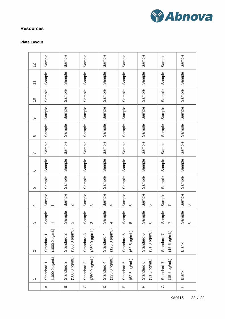
Resources
Plate Layout
12
Sam
ple
Sam
ple
Sam
ple
Sam
ple
Sam
ple
Sam
ple
Sam
ple
Sam
ple
11
Sam
ple
Sam
ple
Sam
ple
Sam
ple
Sam
ple
Sam
ple
Sam
ple
Sam
ple
10
Sam
ple
Sam
ple
Sam
ple
Sam
ple
Sam
ple
Sam
ple
Sam
ple
Sam
ple
9
Sam
ple
Sam
ple
Sam
ple
Sam
ple
Sam
ple
Sam
ple
Sam
ple
Sam
ple
8
Sam
ple
Sam
ple
Sam
ple
Sam
ple
Sam
ple
Sam
ple
Sam
ple
Sam
ple
7
Sam
ple
Sam
ple
Sam
ple
Sam
ple
Sam
ple
Sam
ple
Sam
ple
Sam
ple
6
Sam
ple
Sam
ple
Sam
ple
Sam
ple
Sam
ple
Sam
ple
Sam
ple
Sam
ple
5
Sam
ple
Sam
ple
Sam
ple
Sam
ple
Sam
ple
Sam
ple
Sam
ple
Sam
ple
4
Sam
ple
1
Sam
ple
2
Sam
ple
3
Sam
ple
4
Sam
ple
5
Sam
ple
6
Sam
ple
7
Sam
ple
8
3
Sam
ple
1
Sam
ple
2
Sam
ple
3
Sam
ple
4
Sam
ple
5
Sam
ple
6
Sam
ple
7
Sam
ple
8
2
Sta
ndard
1
(1000.0
pg/m
L)
Sta
ndard
2
(500.0
pg/m
L)
Sta
ndard
3
(250.0
pg/m
L)
Sta
ndard
4
(125.0
pg/m
L)
Sta
ndard
5
(62.5
pg/m
L)
Sta
ndard
6
(31.3
pg/m
L)
Sta
ndard
7
(15.6
pg/m
L)
Bla
nk
1
Sta
ndard
1
(1000.0
pg/m
L)
Sta
ndard
2
(500.0
pg/m
L)
Sta
ndard
3
(250.0
pg/m
L)
Sta
ndard
4
(125.0
pg/m
L)
Sta
ndard
5
(62.5
pg/m
L)
Sta
ndard
6
(31.3
pg/m
L)
Sta
ndard
7
(15.6
pg/m
L)
Bla
nk
A
B
C
D
E
F
G
H