illinois institute of technology physics 561
DESCRIPTION
Illinois Institute of Technology PHYSICS 561. Radiation Biophysics Lecture 5: Survival Curves and Modifiers of Response Andrew Howard 17 June 2014. Looking forward. Be alert for changes in the posted assignments: I may add a few things - PowerPoint PPT PresentationTRANSCRIPT
04/19/23 Survival Curves; Modifiers p. 1 of 96
Illinois Institute of TechnologyPHYSICS 561
Radiation BiophysicsLecture 5:
Survival Curves and Modifiers of Response
Andrew Howard17 June 2014

04/19/23 Survival Curves; Modifiers p. 2 of 96
Looking forward Be alert for changes in the posted
assignments: I may add a few things Midterm will cover chapters 1-7 and the bit of
extra material on free radicals that we discussed last week; therefore, it will not include the material from today’s lecture
04/19/23 Survival Curves; Modifiers p. 3 of 96
Survival Curves
We discussed models for cell survival last time We looked at various ln(S/S0) vs. dose models
and the logic behind them Today we’ll focus on the graphical implications
and how we can look at the numbers Then we’ll talk about cell cycles and other good
solid cell-biology topics. (Warning: I’m more of a biochemist than a cell
biologist, so don’t expect high expertise in this later section!)
04/19/23 Survival Curves; Modifiers p. 4 of 96
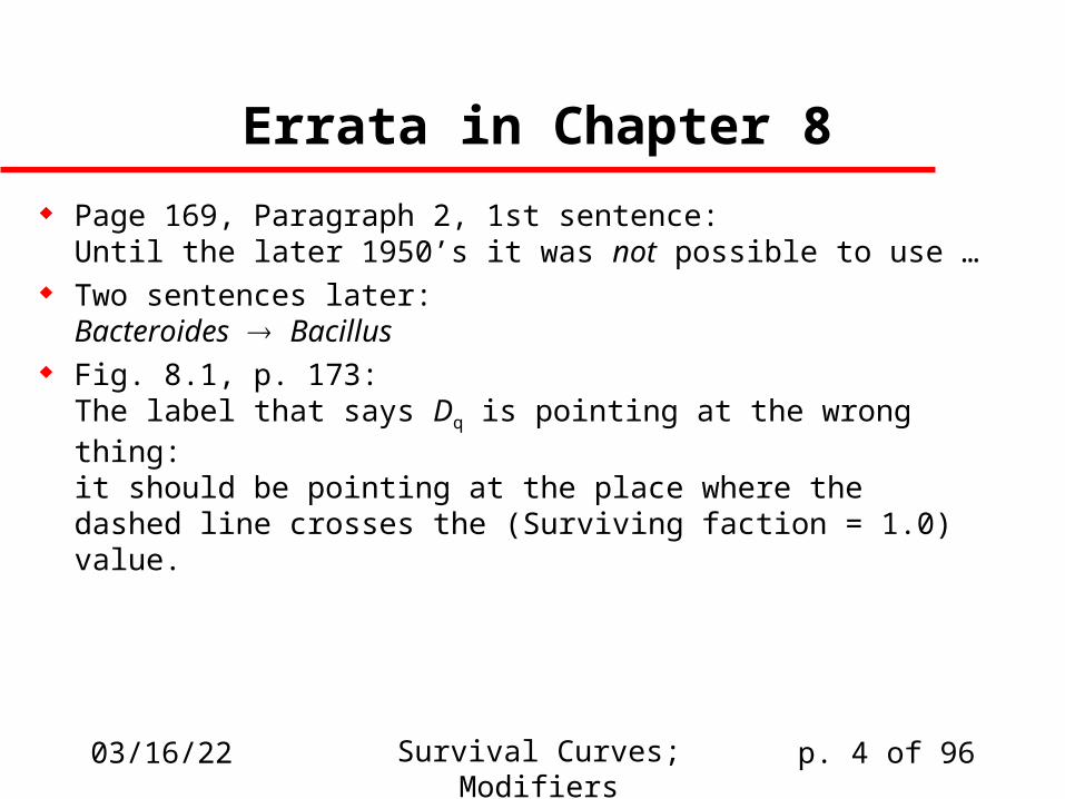
Errata in Chapter 8
Page 169, Paragraph 2, 1st sentence:Until the later 1950’s it was not possible to use …
Two sentences later:Bacteroides Bacillus
Fig. 8.1, p. 173:The label that says Dq is pointing at the wrong thing:it should be pointing at the place where the dashed line crosses the (Surviving faction = 1.0) value.
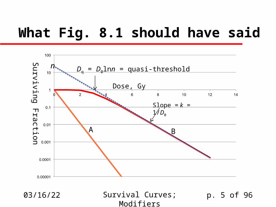
What Fig. 8.1 should have said
04/19/23 Survival Curves; Modifiers p. 5 of 96
Dose, Gy
Su
rviving
Fra
ction A B
Slope = k = 1/D0
Dq = D0lnn = quasi-thresholdn
04/19/23 Survival Curves; Modifiers p. 6 of 96
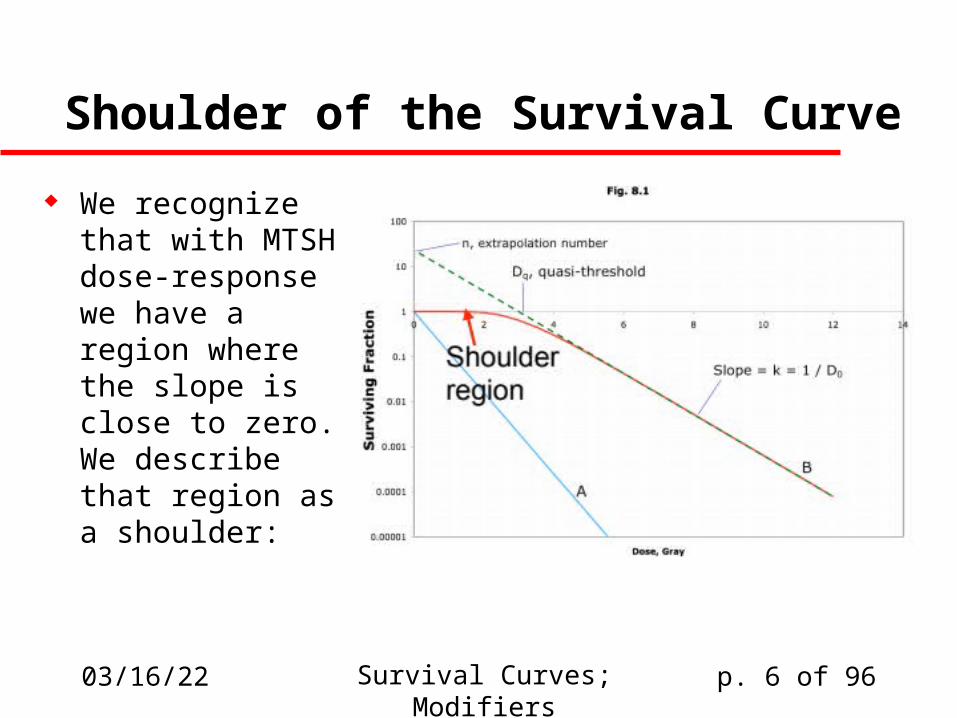
Shoulder of the Survival Curve
We recognize that with MTSH dose-response we have a region where the slope is close to zero. We describe that region as a shoulder:
04/19/23 Survival Curves; Modifiers p. 7 of 96
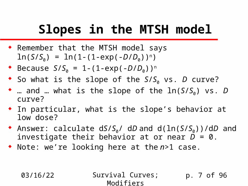
Slopes in the MTSH model Remember that the MTSH model says
ln(S/S0) = ln(1-(1-exp(-D/D0))n) Because S/S0 = 1-(1-exp(-D/D0))n
So what is the slope of the S/S0 vs. D curve? … and … what is the slope of the ln(S/S0) vs. D curve? In particular, what is the slope’s behavior at low dose? Answer: calculate dS/S0/ dD and d(ln(S/S0))/dD and
investigate their behavior at or near D = 0. Note: we’re looking here at the n>1 case.
04/19/23 Survival Curves; Modifiers p. 8 of 96
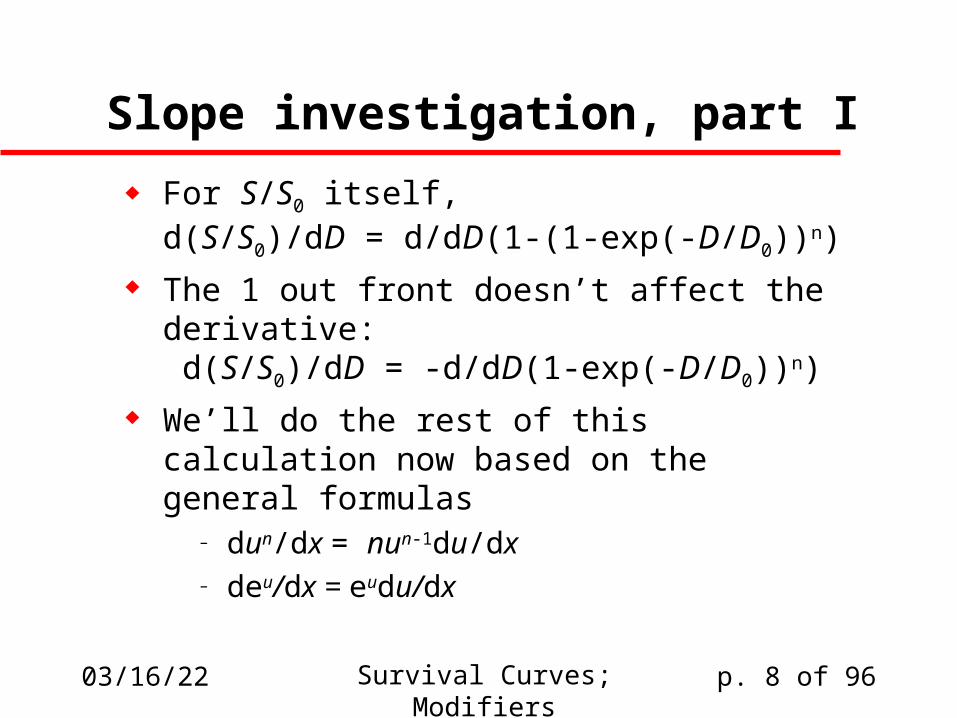
Slope investigation, part I
For S/S0 itself,d(S/S0)/dD = d/dD(1-(1-exp(-D/D0))n)
The 1 out front doesn’t affect the derivative: d(S/S0)/dD = -d/dD(1-exp(-D/D0))n)
We’ll do the rest of this calculation now based on the general formulas
– dun/dx = nun-1du/dx– deu/dx = eudu/dx
04/19/23 Survival Curves; Modifiers p. 9 of 96
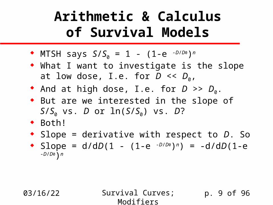
Arithmetic & Calculusof Survival Models
MTSH says S/S0 = 1 - (1-e -D/D0)n
What I want to investigate is the slope at low dose, I.e. for D << D0,
And at high dose, I.e. for D >> D0. But are we interested in the slope of S/S0 vs. D
or ln(S/S0) vs. D? Both! Slope = derivative with respect to D. So Slope = d/dD(1 - (1-e -D/D0)n) = -d/dD(1-e -D/D0)n
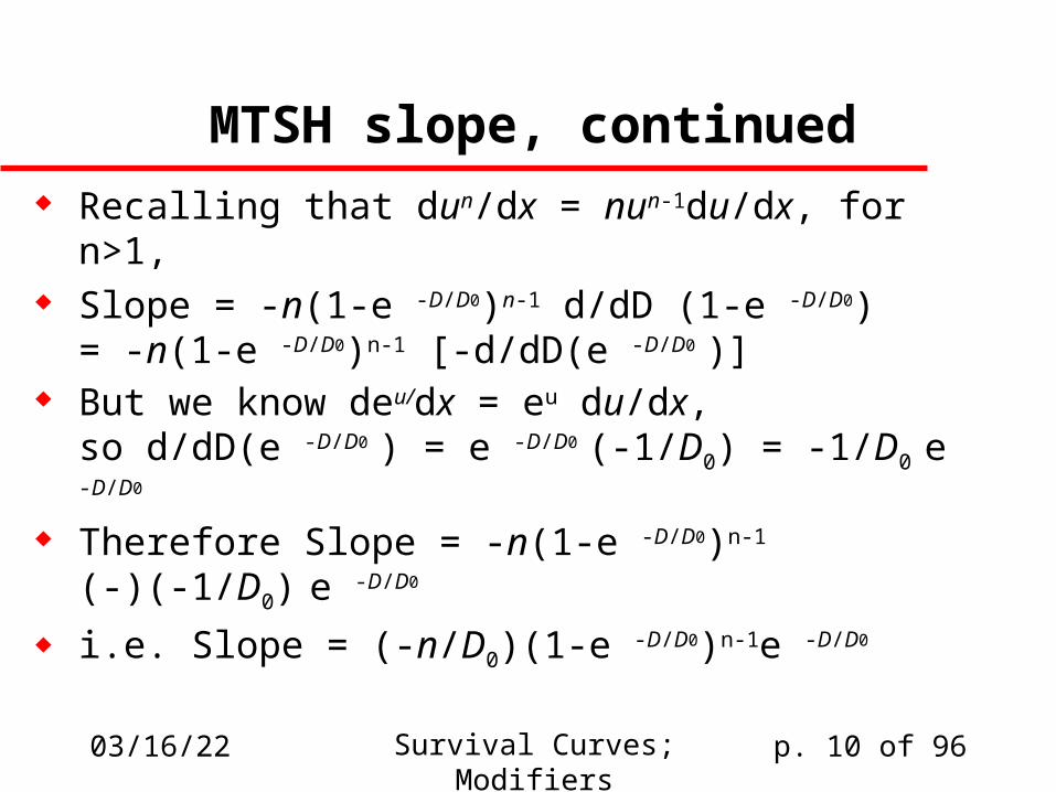
04/19/23 Survival Curves; Modifiers p. 10 of 96
MTSH slope, continued Recalling that dun/dx = nun-1du/dx, for n>1, Slope = -n(1-e -D/D0)n-1 d/dD (1-e -D/D0)
= -n(1-e -D/D0)n-1 [-d/dD(e -D/D0 )] But we know deu/dx = eu du/dx,
so d/dD(e -D/D0 ) = e -D/D0 (-1/D0) = -1/D0 e -D/D0
Therefore Slope = -n(1-e -D/D0)n-1 (-)(-1/D0) e -D/D0
i.e. Slope = (-n/D0)(1-e -D/D0)n-1e -D/D0
04/19/23 Survival Curves; Modifiers p. 11 of 96
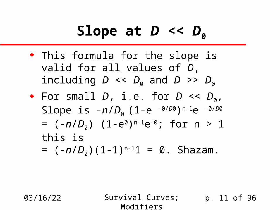
Slope at D << D0
This formula for the slope is valid for all values of D,including D << D0 and D >> D0
For small D, i.e. for D << D0,Slope is -n/D0 (1-e -0/D0)n-1e -0/D0
= (-n/D0) (1-e0)n-1e-0; for n > 1 this is= (-n/D0)(1-1)n-11 = 0. Shazam.
04/19/23 Survival Curves; Modifiers p. 12 of 96
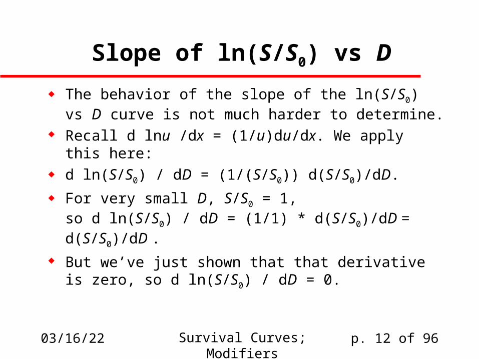
Slope of ln(S/S0) vs D
The behavior of the slope of the ln(S/S0) vs D curve is not much harder to determine.
Recall d lnu /dx = (1/u)du/dx. We apply this here: d ln(S/S0) / dD = (1/(S/S0)) d(S/S0)/dD.
For very small D, S/S0 = 1,so d ln(S/S0) / dD = (1/1) * d(S/S0)/dD = d(S/S0)/dD .
But we’ve just shown that that derivative is zero, so d ln(S/S0) / dD = 0.
04/19/23 Survival Curves; Modifiers p. 13 of 96
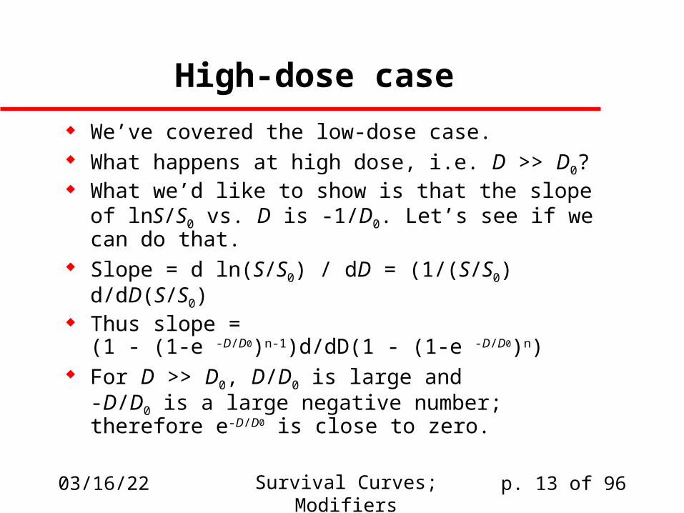
High-dose case
We’ve covered the low-dose case. What happens at high dose, i.e. D >> D0? What we’d like to show is that the slope of
lnS/S0 vs. D is -1/D0. Let’s see if we can do that. Slope = d ln(S/S0) / dD = (1/(S/S0) d/dD(S/S0) Thus slope =
(1 - (1-e -D/D0)n-1)d/dD(1 - (1-e -D/D0)n) For D >> D0, D/D0 is large and
-D/D0 is a large negative number;therefore e-D/D0 is close to zero.
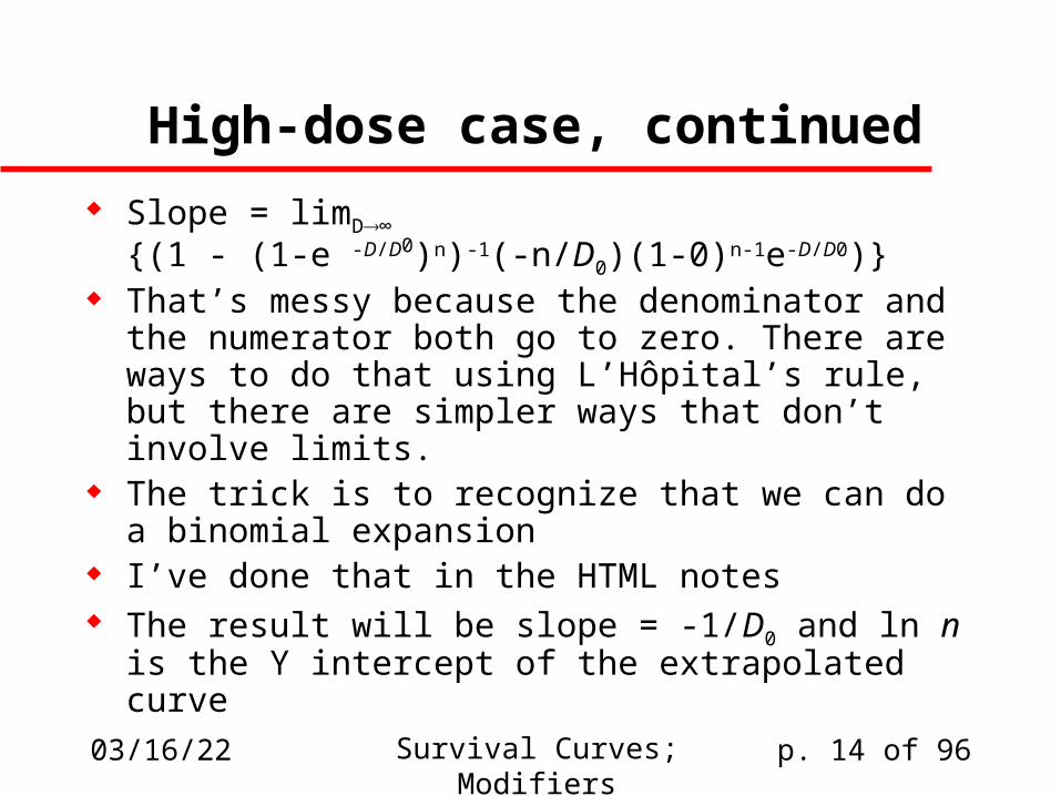
04/19/23 Survival Curves; Modifiers p. 14 of 96
High-dose case, continued
Slope = limD∞
{(1 - (1-e -D/D0)n)-1(-n/D0)(1-0)n-1e-D/D0)} That’s messy because the denominator and the
numerator both go to zero. There are ways to do that using L’Hôpital’s rule, but there are simpler ways that don’t involve limits.
The trick is to recognize that we can do a binomial expansion
I’ve done that in the HTML notes The result will be slope = -1/D0 and ln n is the Y
intercept of the extrapolated curve
04/19/23 Survival Curves; Modifiers p. 15 of 96
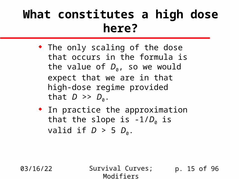
What constitutes a high dose here?
The only scaling of the dose that occurs in the formula is the value of D0, so we would expect that we are in that high-dose regime provided that D >> D0.
In practice the approximation that the slope is -1/D0 is valid if D > 5 D0.
04/19/23 Survival Curves; Modifiers p. 16 of 96
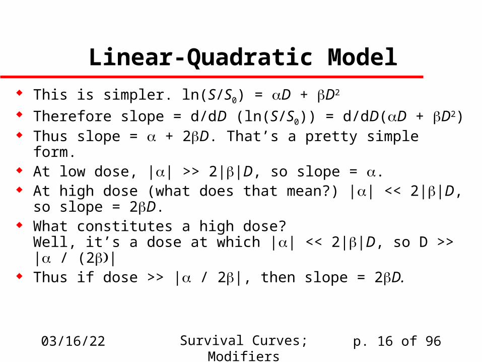
Linear-Quadratic Model This is simpler. ln(S/S0) = D + D2
Therefore slope = d/dD (ln(S/S0)) = d/dD(D + D2) Thus slope = + 2D. That’s a pretty simple form. At low dose, || >> 2||D, so slope = . At high dose (what does that mean?) || << 2||D,
so slope = 2D. What constitutes a high dose?
Well, it’s a dose at which || << 2||D, so D >> | / (2| Thus if dose >> | / 2|, then slope = 2D.
04/19/23 Survival Curves; Modifiers p. 17 of 96
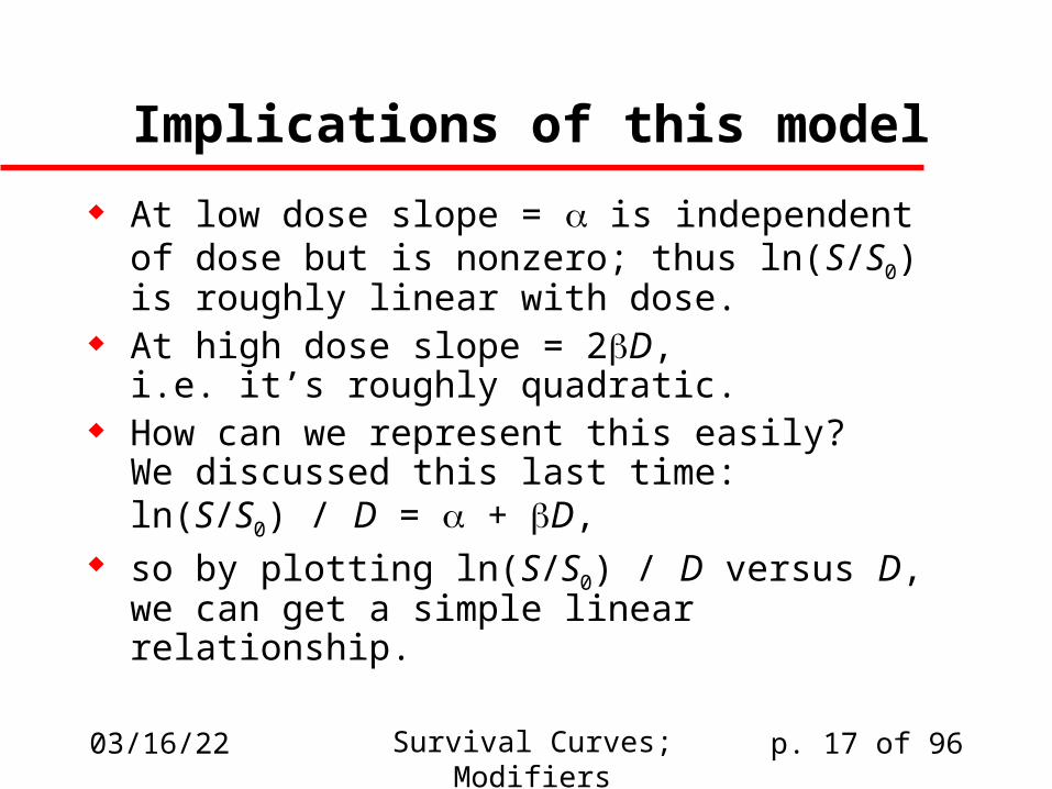
Implications of this model
At low dose slope = is independent of dose but is nonzero; thus ln(S/S0) is roughly linear with dose.
At high dose slope = 2D,i.e. it’s roughly quadratic.
How can we represent this easily?We discussed this last time:ln(S/S0) / D = + D,
so by plotting ln(S/S0) / D versus D, we can get a simple linear relationship.
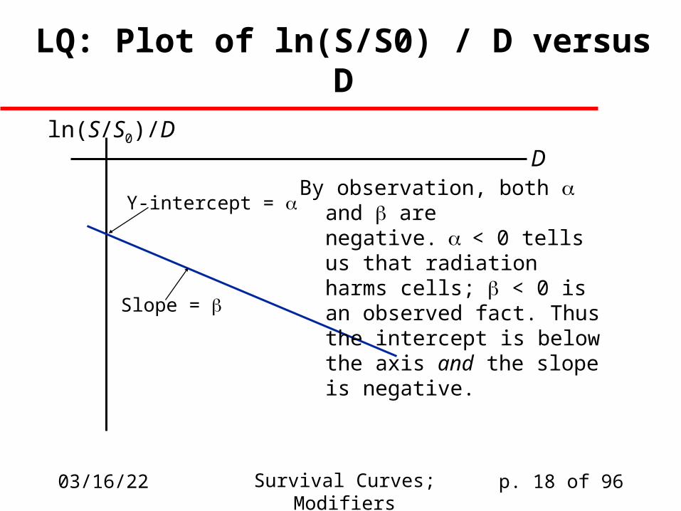
04/19/23 Survival Curves; Modifiers p. 18 of 96
Y-intercept =
Slope =
ln(S/S0)/DD
LQ: Plot of ln(S/S0) / D versus D
By observation, both and are negative.< 0 tells us that radiation harms cells; < 0 is an observed fact. Thus the intercept is below the axis and the slope is negative.
04/19/23 Survival Curves; Modifiers p. 19 of 96
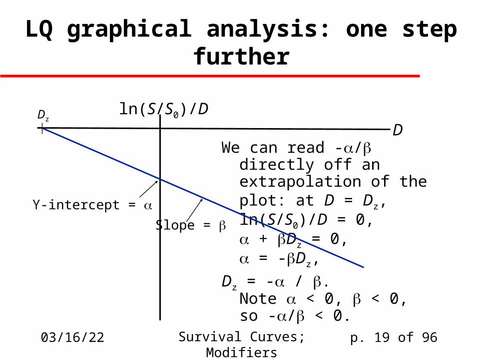
LQ graphical analysis: one step further
We can read -/ directly off an extrapolation of the plot: at D = Dz,ln(S/S0)/D = 0, + Dz = 0, = -Dz,
Dz = - / .Note < 0, < 0,so -/ < 0.
Y-intercept = Slope =
ln(S/S0)/DD
Dz
04/19/23 Survival Curves; Modifiers p. 20 of 96
Where do linear and quadratic responses become equal?
At what dose does the linear response equal the quadratic response?
At that dose, D = D2, D = / So the value we read off the X-intercept of
the previous curve is simply the opposite of the dose value at which the two influences are equal.
We mentioned this last time, but we’re reminding you now
04/19/23 Survival Curves; Modifiers p. 21 of 96
How plausible is all this?
Model studies suggest reasons to think that ln(S/S0) = D + D2 is a good approach.
Much experimental data are consistent with the model
Some of these LQ approaches allow for time-dependence to be built in.
04/19/23 Survival Curves; Modifiers p. 22 of 96
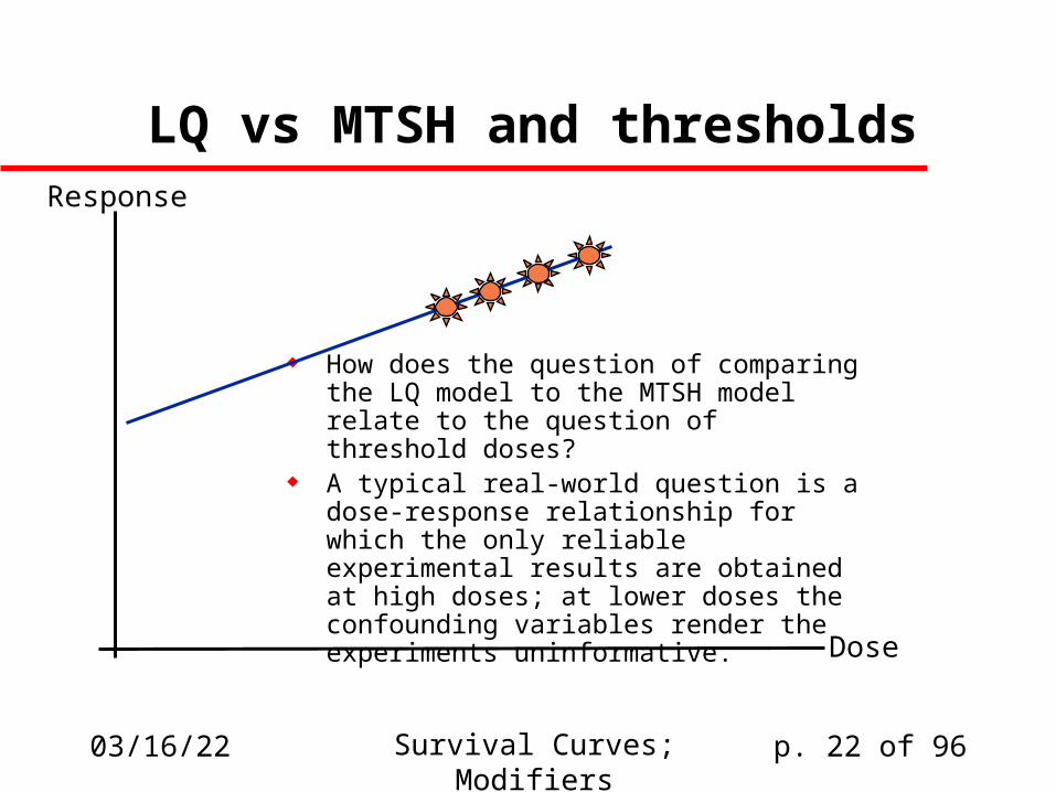
LQ vs MTSH and thresholds
How does the question of comparing the LQ model to the MTSH model relate to the question of threshold doses?
A typical real-world question is a dose-response relationship for which the only reliable experimental results are obtained at high doses; at lower doses the confounding variables render the experiments uninformative.
Dose
Response
04/19/23 Survival Curves; Modifiers p. 23 of 96
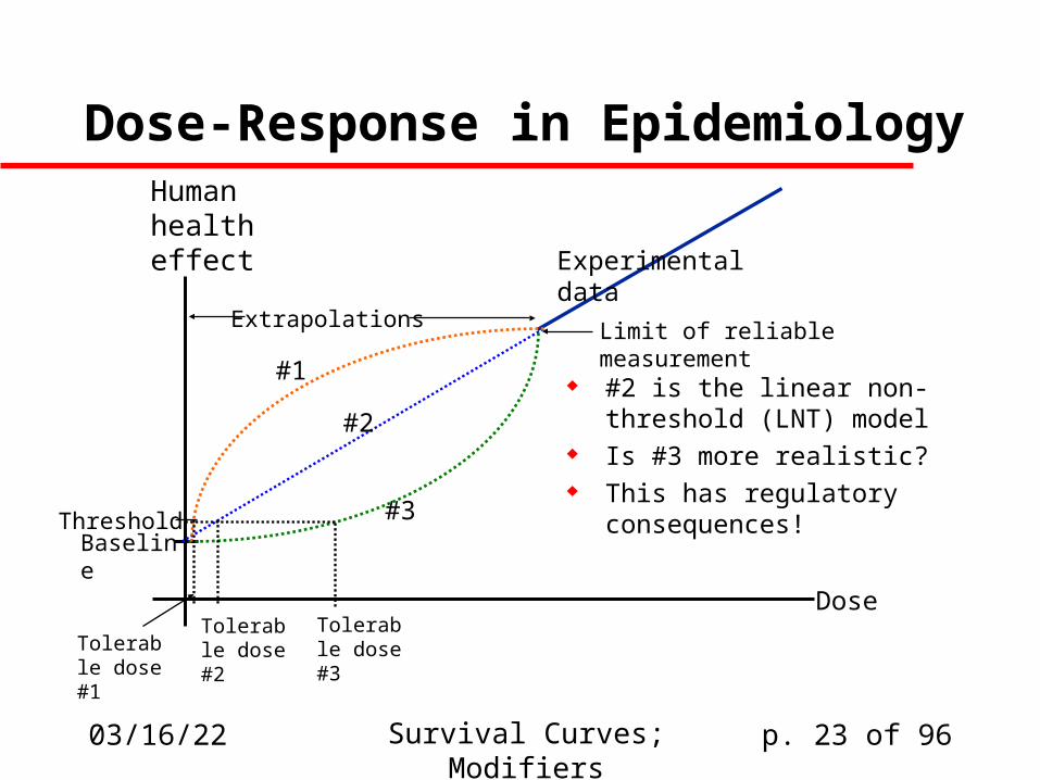
Dose-Response in Epidemiology
#2 is the linear non-threshold (LNT) model
Is #3 more realistic? This has regulatory
consequences!
Human health effect
Baseline
Dose
Experimental data
#3
#2
#1
Threshold
Tolerable dose #3
Tolerable dose #2Tolerable
dose #1
Limit of reliable measurementExtrapolations
04/19/23 Survival Curves; Modifiers p. 24 of 96
Studying repair
We’ve been suggesting that LQ models and even some MTSH models are dependent on the idea that some DNA damage can be repaired accurately.
Let’s look for approaches to studying DNA damage that might provide a fuller understanding of the effects of repair.
04/19/23 Survival Curves; Modifiers p. 25 of 96
The Elkind-Sutton Experiment
Provides a way of probing repair functions in cells Procedure:
– Irradiate and establish survival curve(“conditioning dose”)
– Take cells surviving at S=0.1 and subject them to further irradiation at varying time intervals after reaching S=0.1
If repair is taking place, then the appearance of a curve similar to the original shoulder is indicative of full recovery
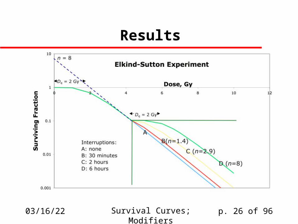
04/19/23 Survival Curves; Modifiers p. 26 of 96
Results
04/19/23 Survival Curves; Modifiers p. 27 of 96
Interpretation
If slope and implied n value are equivalent to the original curve, then repair is complete
Smaller n values indicate insufficient time has elapsed
n=1 implies repair has not begun

04/19/23 Survival Curves; Modifiers p. 28 of 96
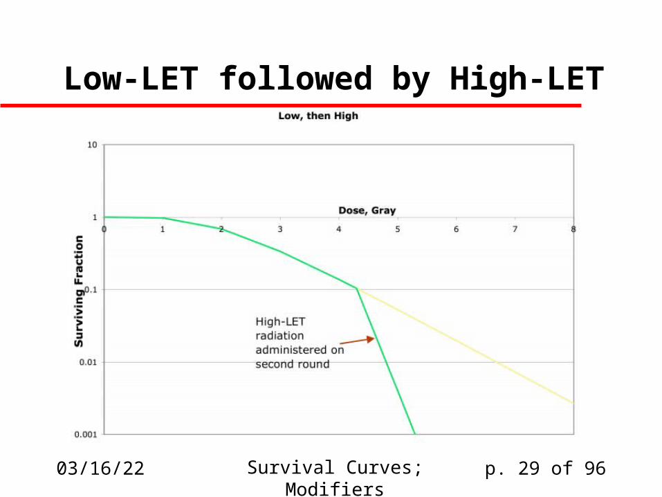
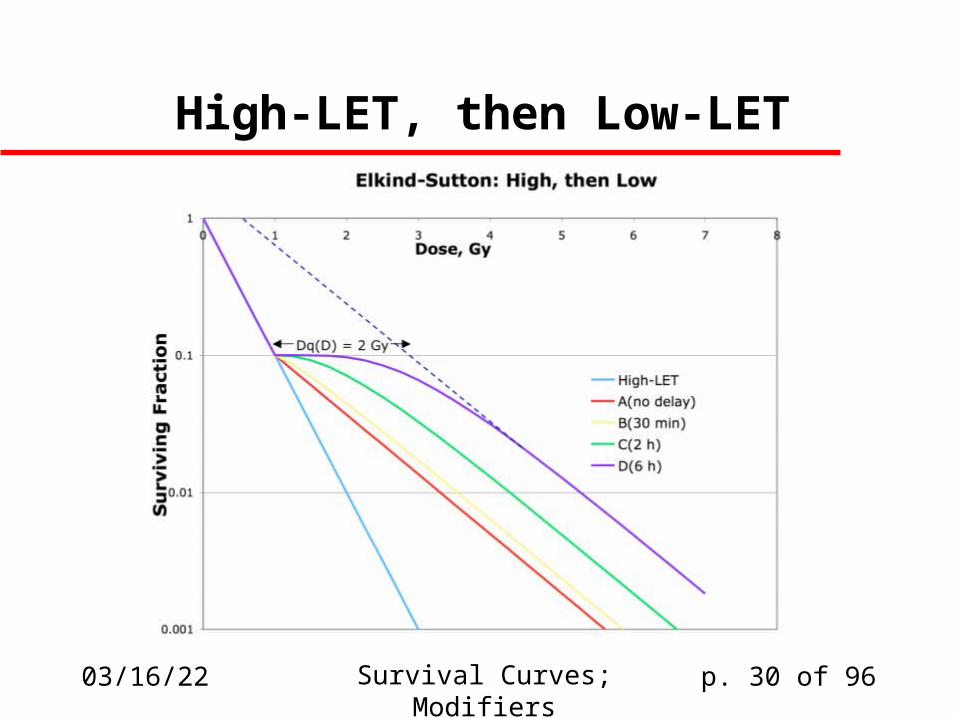
Elkind-Sutton and LET
We might expect more complicated results if we vary the LET for the two dosing regimens
Low-LET first, high-LET second gives two lines of different slope, independent of the time interval
High-LET first, low-LET second gives line followed by usual Elkind-Sutton distribution
04/19/23 Survival Curves; Modifiers p. 29 of 96
Low-LET followed by High-LET
04/19/23 Survival Curves; Modifiers p. 30 of 96
High-LET, then Low-LET

04/19/23 Survival Curves; Modifiers p. 31 of 96
The Cell Cycle Cells have a definite cycle over which specific activities occur. Particular activities are limited to specific parts of the cycle Howard and Pelc (1953) characterized four specific phases:
– M (mitosis, i.e cell division)– G1 (growth prior to DNA replication)– S (synthesis, i.e DNA replication)– G2 (preparation for mitosis)
Mitosis(M)
Presynthetic(G1)
Synthesis(S)
Post-synthetic(G2)
04/19/23 Survival Curves; Modifiers p. 32 of 96
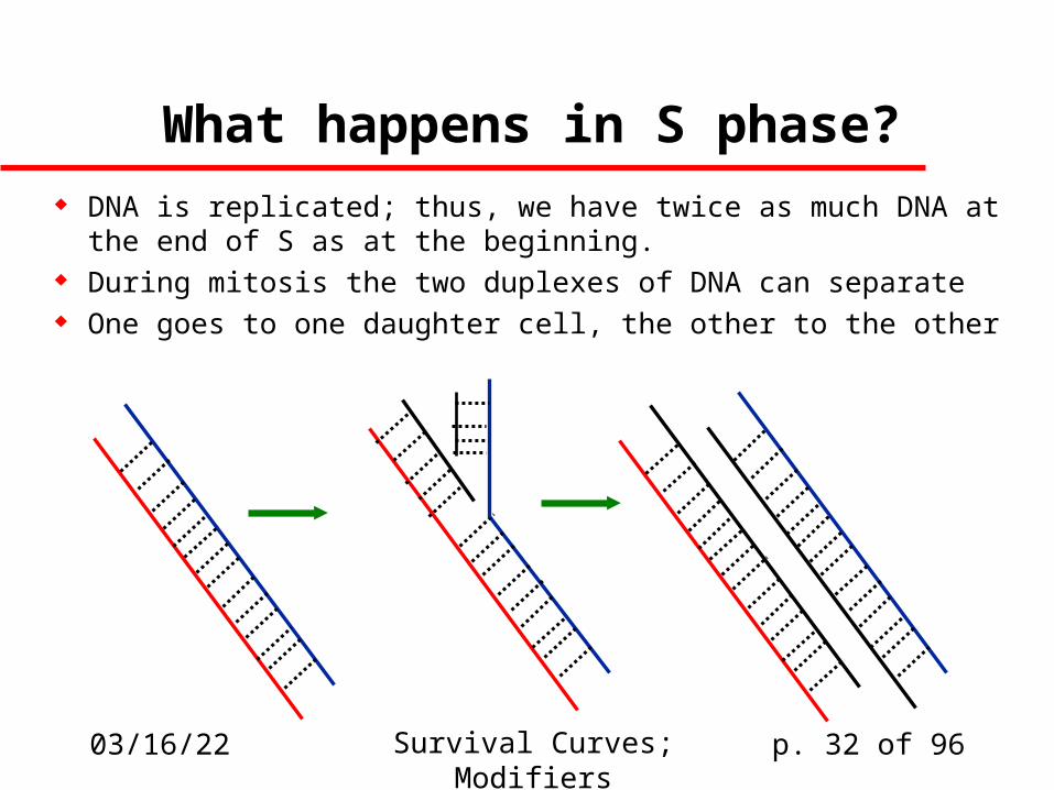
What happens in S phase?
DNA is replicated; thus, we have twice as much DNA at the end of S as at the beginning.
During mitosis the two duplexes of DNA can separate One goes to one daughter cell, the other to the other
04/19/23 Survival Curves; Modifiers p. 33 of 96
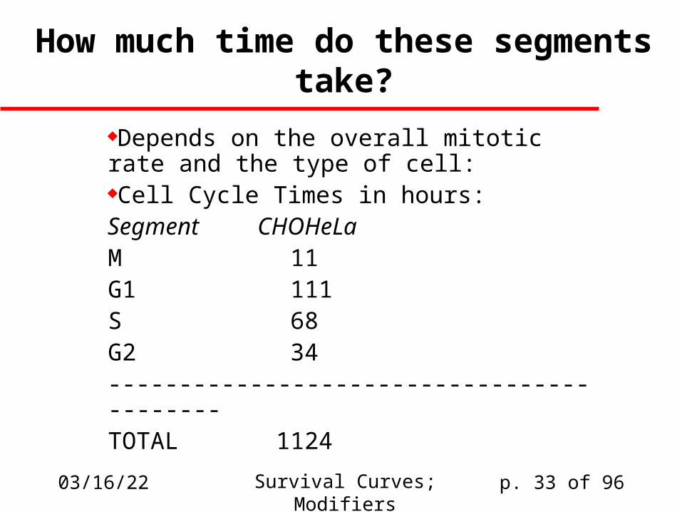
How much time do these segments take?
Depends on the overall mitotic rate and the type of cell:Cell Cycle Times in hours:Segment CHO HeLaM 1 1G1 1 11S 6 8G2 3 4------------------------------------------TOTAL 11 24
04/19/23 Survival Curves; Modifiers p. 34 of 96
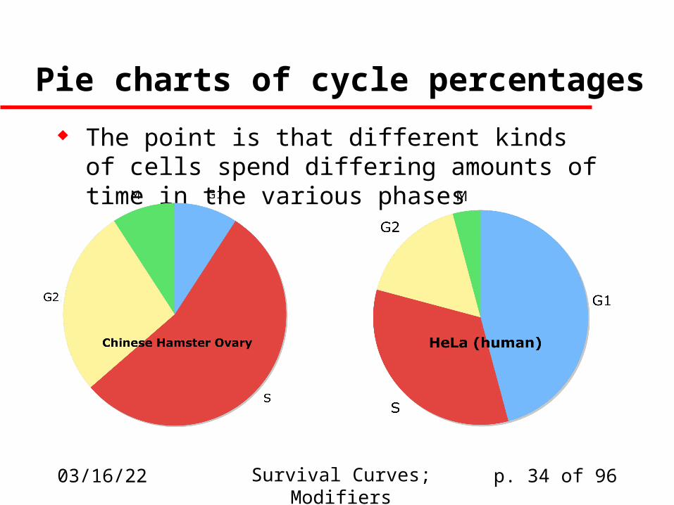
Pie charts of cycle percentages
The point is that different kinds of cells spend differing amounts of time in the various phases
04/19/23 Survival Curves; Modifiers p. 35 of 96
Phase Sensitivity
Many cells are much more sensitive to radiation in some parts of the cell cycle than they are in others.
Why?– Repair is more vigorous in some stages– Unrepaired damage has more opportunity to
manifest itself as clonal alteration close to mitosis– Access of repair enzymes to damaged DNA is
sometimes influenced by how organized the DNA is.
04/19/23 Survival Curves; Modifiers p. 36 of 96
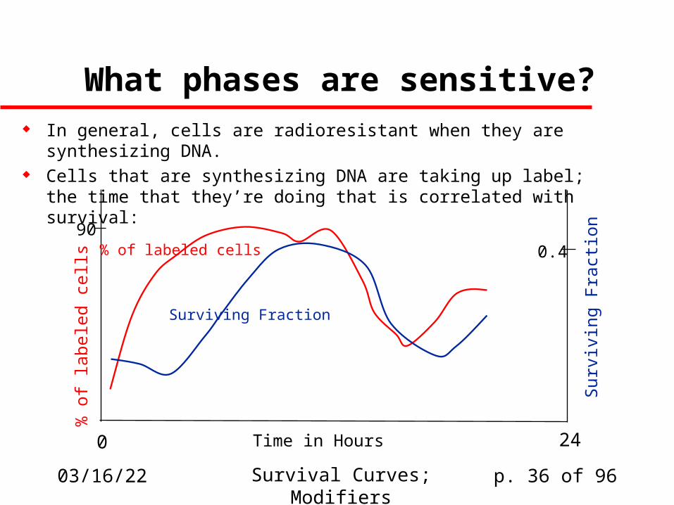
What phases are sensitive? In general, cells are radioresistant when they are
synthesizing DNA. Cells that are synthesizing DNA are taking up label;
the time that they’re doing that is correlated with survival:
% o
f la
bele
d ce
lls
Sur
vivi
ng F
ract
ion% of labeled cells
Surviving Fraction
0.490
Time in Hours0 24
04/19/23 Survival Curves; Modifiers p. 37 of 96
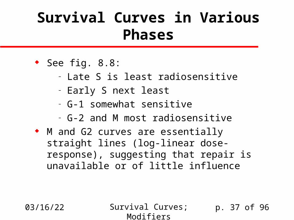
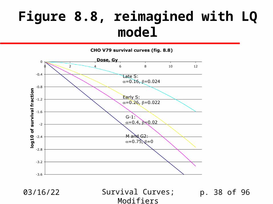
Survival Curves in Various Phases
See fig. 8.8:– Late S is least radiosensitive– Early S next least– G-1 somewhat sensitive– G-2 and M most radiosensitive
M and G2 curves are essentially straight lines (log-linear dose-response), suggesting that repair is unavailable or of little influence
04/19/23 Survival Curves; Modifiers p. 38 of 96
Figure 8.8, reimagined with LQ model

04/19/23 Survival Curves; Modifiers p. 39 of 96
Radiation-InducedCell Progression Delay
Note that various biochemical signals regulate progression from one phase of the cycle to another.
To study this, you need synchronized cells . . . Sample study (Leeper, 1973):
– CHO cells exposed to 1.5 Gy in mid-G1 experienced a delay of 0.5 h in cell division
– 1.5 Gy in late S or early G2 caused a delay of 2-3 h– Dose-dependent: (4h for 3Gy, 6-7h for 6 Gy)
04/19/23 Survival Curves; Modifiers p. 40 of 96
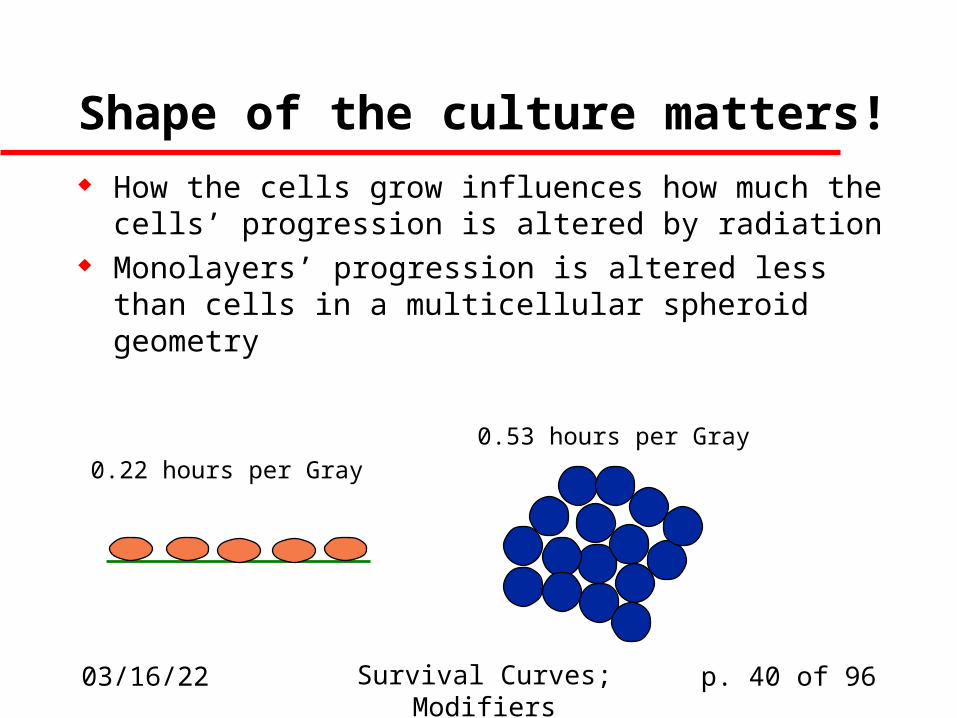
Shape of the culture matters! How the cells grow influences how much the cells’
progression is altered by radiation Monolayers’ progression is altered less than cells
in a multicellular spheroid geometry
0.22 hours per Gray
0.53 hours per Gray
04/19/23 Survival Curves; Modifiers p. 41 of 96
Is that such a big deal?
Probably not: The cells in the spheroidal mass divide
half as fast even in the absence of radiation, possibly due to contact inhibition.
Therefore it may simply be that the whole mitotic clock has been slowed down, including the clock as it’s been influenced by radiation.

04/19/23 Survival Curves; Modifiers p. 42 of 96
Causes for these effects
Why are cells more radiosensitive in M and G2?– Reduced availability of repair enzymes– Repackaged DNA is hard to repair
How is cell progression influenced by radiation?– Damage to protein kinases and cyclins involved
in cellular checkpoints– Premature degradation of p21, maybe…– Sample 1994 study:
Edgar et al, Genes Dev. 440: 52 (1994)
04/19/23 Survival Curves; Modifiers p. 43 of 96
Effectors of Radiation Sensitivity
Biological– Cells go through life cycles & are
much more sensitive to radiation damage at some stages than at others
Chemical Physical
04/19/23 Survival Curves; Modifiers p. 44 of 96
Assignment related to amino acids
1. There are exactly twenty amino acids that serve as the building blocks for proteins in almost all organisms. The general formula for 19 of these 20 amino acids is +NH3-CHR-COO-, where R is any of 19 different side-chain groups. The simplest of these R groups is H, for which the amino acid is called glycine; the most complicated is a moiety known as indole, for which the amino acid is called tryptophan.
04/19/23 Survival Curves; Modifiers p. 45 of 96
Upcoming Problem 1, cont’d
Much of the chemistry that these R groups participate in in proteins is ionic in nature, involving charges or partial charges; but usually these ionic interactions involve pairs of electrons rather than unpaired electrons. An exception is the amino acid tyrosine, which can participate in free-radical (unpaired-electron) interactions. Draw a structure of the tyrosyl free radical and explain why it might have a reasonably long lifetime, as compared to a hydroxyl radical or some other short-lived free radical.

04/19/23 Survival Curves; Modifiers p. 46 of 96
Second upcoming problem 2. Most biochemical oxidation-reduction reactions involve
transfers of pairs of electrons and therefore do not involve free radical mechanisms. A sizeable minority, however, do involve free radicals. Which of the following biochemical oxidizing or reducing agents are capable of participating in single-electron (free-radical) reactions, and which are not? Explain briefly. You may need to look up the structures of some of these compounds.
(a) ferric iron, Fe3+
(b) nicotinamide adenine dinucleotide, oxidized form (NAD) (c) flavonamide mononucleotide (FMN)
(also spelled flavin amide mononucleotide; it’s an instance of what are generally known as flavin prosthetic groups)
04/19/23 Survival Curves; Modifiers p. 47 of 96
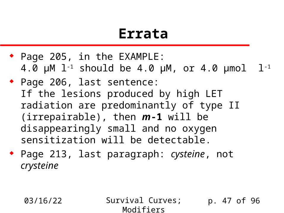
Errata
Page 205, in the EXAMPLE:4.0 µM l-1 should be 4.0 µM, or 4.0 µmol l-1
Page 206, last sentence:If the lesions produced by high LET radiation are predominantly of type II (irrepairable), then m-1 will be disappearingly small and no oxygen sensitization will be detectable.
Page 213, last paragraph: cysteine, not crysteine
04/19/23 Survival Curves; Modifiers p. 48 of 96
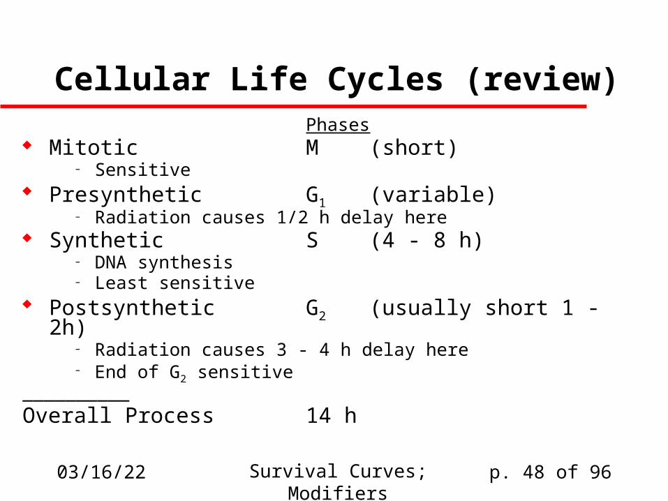
Cellular Life Cycles (review)Phases
Mitotic M (short)– Sensitive
Presynthetic G1 (variable)– Radiation causes 1/2 h delay here
Synthetic S (4 - 8 h)– DNA synthesis– Least sensitive
Postsynthetic G2 (usually short 1 - 2h)– Radiation causes 3 - 4 h delay here– End of G2 sensitive
__________Overall Process 14 h
04/19/23 Survival Curves; Modifiers p. 49 of 96
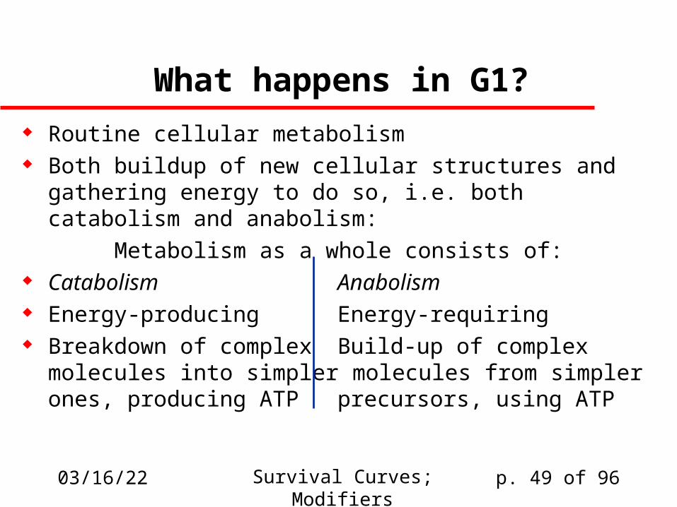
What happens in G1?
Routine cellular metabolism Both buildup of new cellular structures and gathering
energy to do so, i.e. both catabolism and anabolism:
Metabolism as a whole consists of: Catabolism Anabolism Energy-producing Energy-requiring Breakdown of complex Build-up of complex
molecules into simpler molecules from simplerones, producing ATP precursors, using ATP

04/19/23 Survival Curves; Modifiers p. 50 of 96
A mitotic cell
Nucleus
(DNA location;
cellular organization)
Mitochondrion(energy metabolism)
Ribosomes(protein synthesis)
Mitotic Spindle
Other organelles
04/19/23 Survival Curves; Modifiers p. 51 of 96
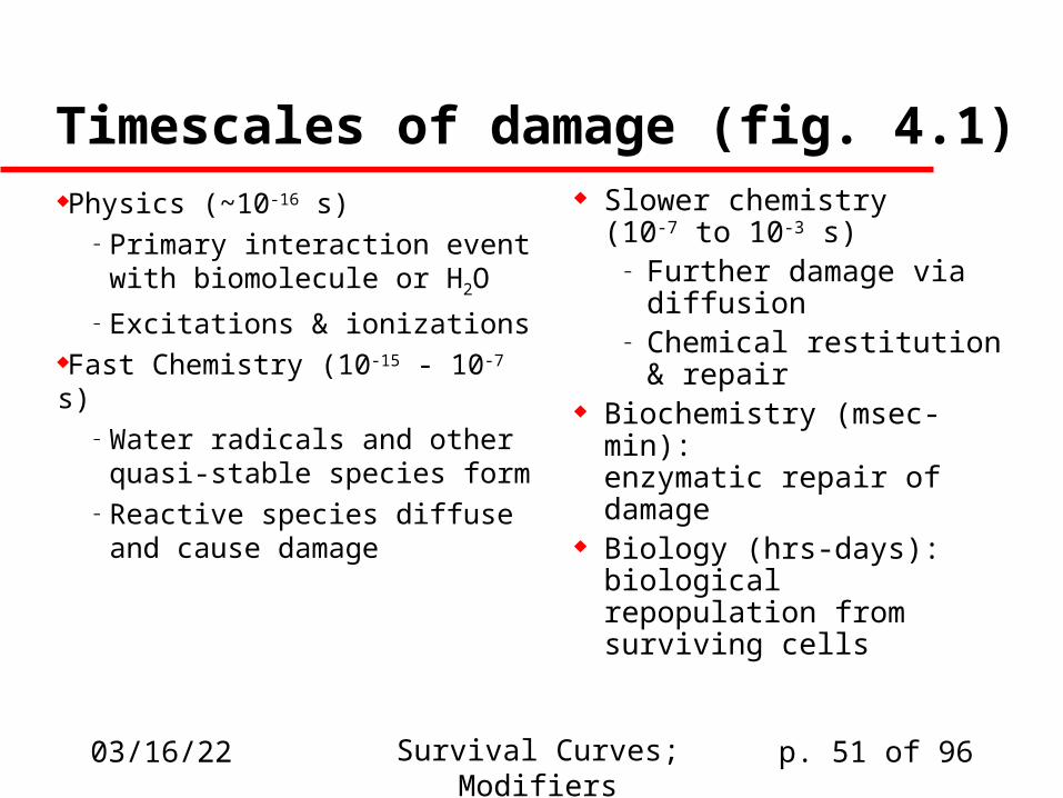
Timescales of damage (fig. 4.1)Physics (~10-16 s)
– Primary interaction event with biomolecule or H2O
– Excitations & ionizationsFast Chemistry (10-15 - 10-7 s)
– Water radicals and other quasi-stable species form
– Reactive species diffuse and cause damage
Slower chemistry(10-7 to 10-3 s)
– Further damage via diffusion
– Chemical restitution & repair
Biochemistry (msec-min):enzymatic repair of damage
Biology (hrs-days):biological repopulation from surviving cells
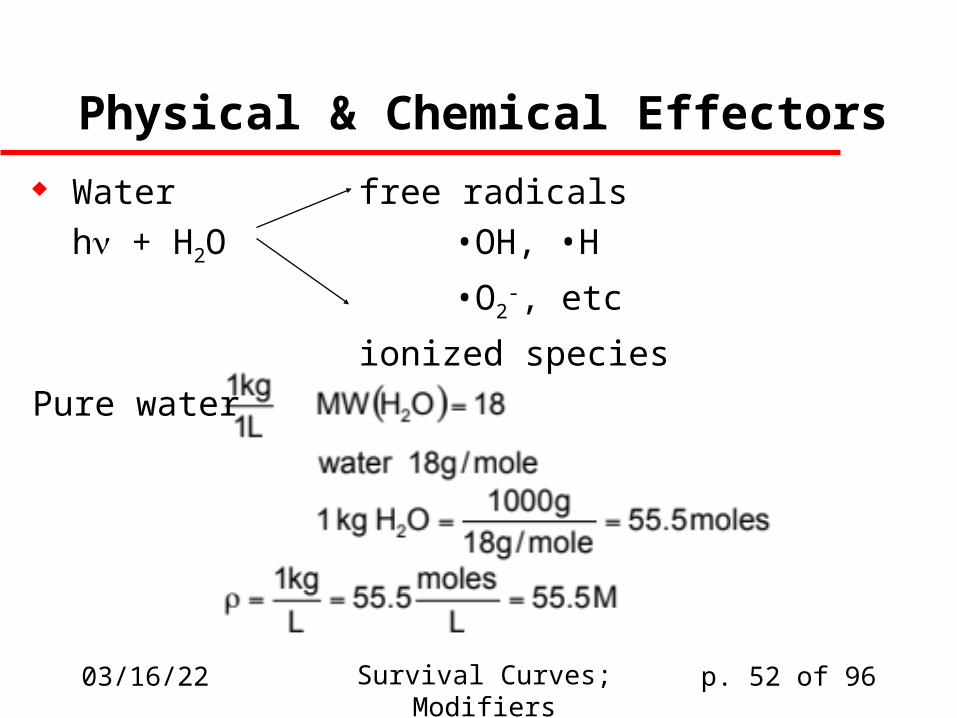
04/19/23 Survival Curves; Modifiers p. 52 of 96
Physical & Chemical Effectors
Water free radicals
h + H2O •OH, •H
•O2-, etc
ionized species
Pure water
04/19/23 Survival Curves; Modifiers p. 53 of 96
How does water matter?
In a dry setting, the damage must be direct In a wet environment, water-derived free
radicals and ions are the source of much of the chemical damage
Experiments that can eliminate secondary (radical-mediated) damage show much reduced radiosensitivity
04/19/23 Survival Curves; Modifiers p. 54 of 96
An example from my field
Protein crystals must be irradiated wet because they fall apart when they’re dry.
Protein crystals irradiated at 300K are destroyed:
– In days on a conventional X-ray source– In minutes on a 2nd-generation synchrotron– In seconds on a 3rd-generation synchrotron
They’re essentially immortal at 110K except on a 3rd-generation synchrotron source, where they live for 5-100 minutes
04/19/23 Survival Curves; Modifiers p. 55 of 96
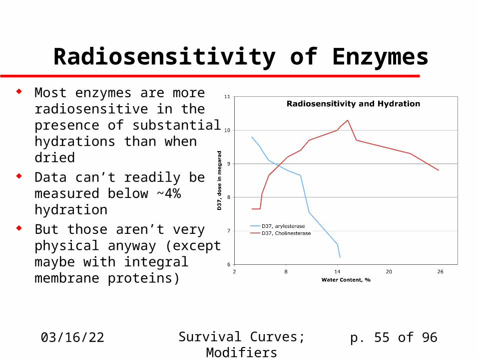
Radiosensitivity of Enzymes Most enzymes are more
radiosensitive in the presence of substantial hydrations than when dried
Data can’t readily be measured below ~4% hydration
But those aren’t very physical anyway (except maybe with integral membrane proteins)
04/19/23 Survival Curves; Modifiers p. 56 of 96
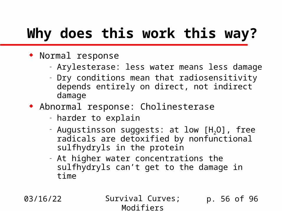
Why does this work this way?
Normal response– Arylesterase: less water means less damage– Dry conditions mean that radiosensitivity
depends entirely on direct, not indirect damage Abnormal response: Cholinesterase
– harder to explain– Augustinsson suggests: at low [H2O], free
radicals are detoxified by nonfunctional sulfhydryls in the protein
– At higher water concentrations the sulfhydryls can’t get to the damage in time

04/19/23 Survival Curves; Modifiers p. 57 of 96
Do we care?
Not much:– We can’t really alter the hydration of most cell
systems without destroying them– We definitely can’t influence the hydration states
of whole organisms without doing damage that is probably much more significant than that of the radiation
But these studies may help us understand mechanisms of damage, and that could be relevant
04/19/23 Survival Curves; Modifiers p. 58 of 96
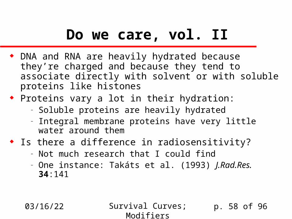
Do we care, vol. II DNA and RNA are heavily hydrated because they’re
charged and because they tend to associate directly with solvent or with soluble proteins like histones
Proteins vary a lot in their hydration:– Soluble proteins are heavily hydrated– Integral membrane proteins have very little water
around them Is there a difference in radiosensitivity?
– Not much research that I could find– One instance: Takáts et al. (1993) J.Rad.Res. 34:141
04/19/23 Survival Curves; Modifiers p. 59 of 96
Temperature-sensitivity
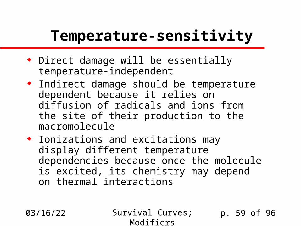
Direct damage will be essentially temperature-independent
Indirect damage should be temperature dependent because it relies on diffusion of radicals and ions from the site of their production to the macromolecule
Ionizations and excitations may display different temperature dependencies because once the molecule is excited, its chemistry may depend on thermal interactions

04/19/23 Survival Curves; Modifiers p. 60 of 96
Arrhenius plots
We can examine temperature dependence via the Arrhenius plot, wherein we expect k = Qexp(-G‡/RT)
Thus ln k = ln[Qexp(-G‡/RT)] = lnQ - G‡/RT, so if we plot ln(k) as a function of 1/T then the relationship should be linear, and the slope will be -G‡/R, i.e it will be proportional to the activation energy G‡.
If multiple processes are involved we may get a non-log-linear response.
Over temperature ranges typical of the internals of homeothermic organisms, we’re not going to see much effect!

04/19/23 Survival Curves; Modifiers p. 61 of 96
Physical meaning of G‡
Chemical reactions are characterized by an activation energy barrier, i.e., an energy that must be put into the system in order to get from reactant to product or vice versa.
G‡ is the height of that activation barrier.
Energy
Reaction coordinate
ReactantProduct
G‡

04/19/23 Survival Curves; Modifiers p. 62 of 96
Radiation & Temperature
Kinetics
37oC = 310K27oC = 300K
p199 T < 100K 100 < T < 170 K 170 < T < 420 K
04/19/23 Survival Curves; Modifiers p. 63 of 96
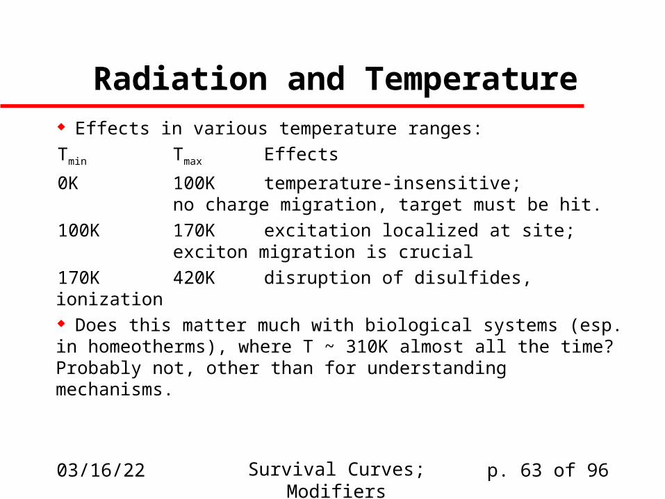
Radiation and Temperature Effects in various temperature ranges:
Tmin Tmax Effects
0K 100K temperature-insensitive;no charge migration, target must be
hit.
100K 170K excitation localized at site;exciton migration is crucial
170K 420K disruption of disulfides, ionization Does this matter much with biological systems (esp. in homeotherms), where T ~ 310K almost all the time?Probably not, other than for understanding mechanisms.
04/19/23 Survival Curves; Modifiers p. 64 of 96
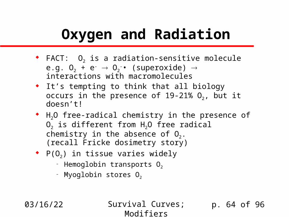
Oxygen and Radiation
FACT: O2 is a radiation-sensitive moleculee.g. O2 + e- O2
-• (superoxide) interactions with macromolecules
It’s tempting to think that all biology occurs in the presence of 19-21% O2, but it doesn’t!
H2O free-radical chemistry in the presence of O2 is different from H2O free radical chemistry in the absence of O2.(recall Fricke dosimetry story)
P(O2) in tissue varies widely– Hemoglobin transports O2
– Myoglobin stores O2
04/19/23 Survival Curves; Modifiers p. 65 of 96
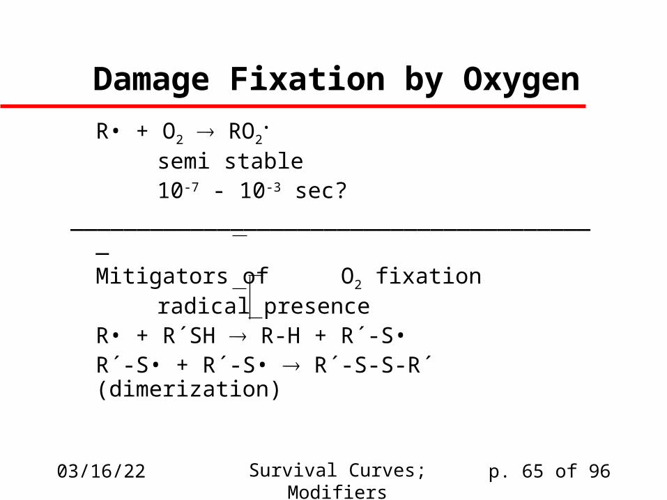
Damage Fixation by Oxygen
R• + O2 RO2•
semi stable10-7 - 10-3 sec?
________________________________________
Mitigators of O2 fixationradical presence
R• + R´SH R-H + R´-S•R´-S• + R´-S• R´-S-S-R´ (dimerization)
04/19/23 Survival Curves; Modifiers p. 66 of 96
Minor syntactic point
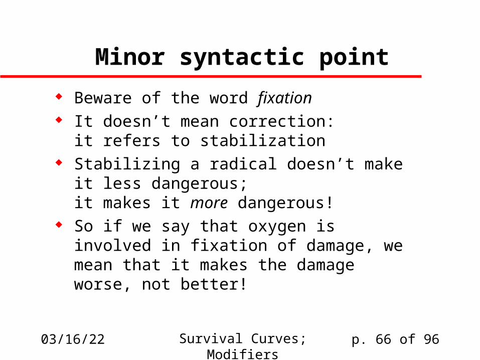
Beware of the word fixation It doesn’t mean correction:
it refers to stabilization Stabilizing a radical doesn’t make it less
dangerous;it makes it more dangerous!
So if we say that oxygen is involved in fixation of damage, we mean that it makes the damage worse, not better!
04/19/23 Survival Curves; Modifiers p. 67 of 96
Experiments on Oxygen Sensitivity
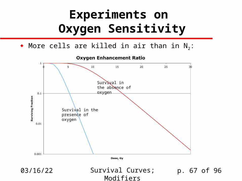
More cells are killed in air than in N2:
Survival in the absence of oxygen
Survival in the presence of oxygen
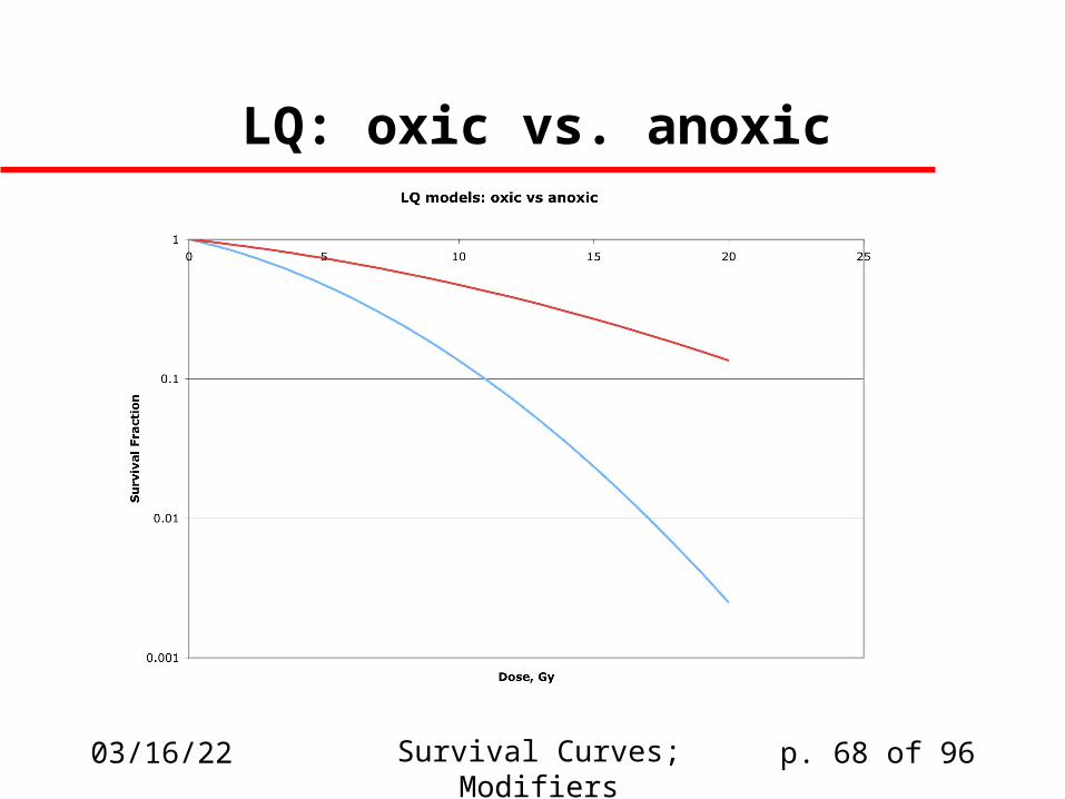
04/19/23 Survival Curves; Modifiers p. 68 of 96
LQ: oxic vs. anoxic
04/19/23 Survival Curves; Modifiers p. 69 of 96
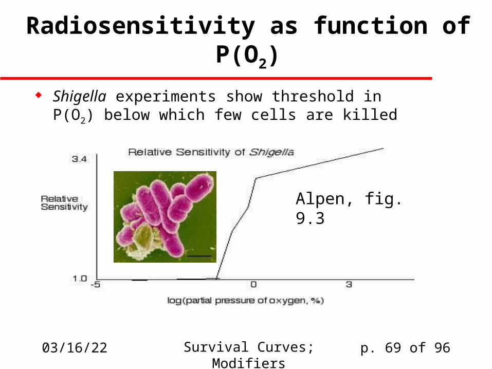
Radiosensitivity as function of P(O2)
Shigella experiments show threshold in P(O2) below which few cells are killed
Alpen, fig. 9.3
04/19/23 Survival Curves; Modifiers p. 70 of 96
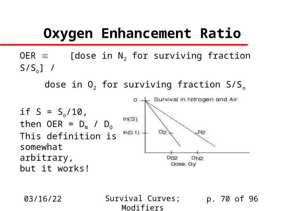
Oxygen Enhancement Ratio
OER [dose in N2 for surviving fraction S/So] /
_____________________________
dose in O2 for surviving fraction S/So
if S = So/10,then OER = DN / DO
This definition issomewhatarbitrary,but it works!
04/19/23 Survival Curves; Modifiers p. 71 of 96
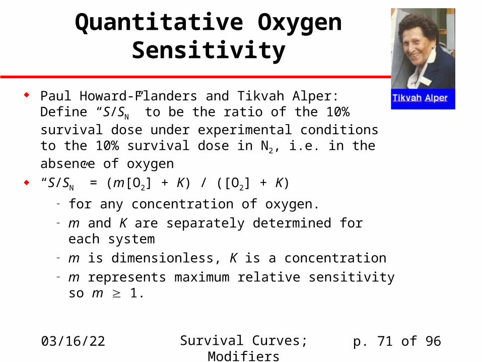
Quantitative Oxygen Sensitivity
Paul Howard-Flanders and Tikvah Alper:Define “S/SN” to be the ratio of the 10% survival dose under experimental conditions to the 10% survival dose in N2, i.e. in the absence of oxygen
“S/SN” = (m[O2] + K) / ([O2] + K)
– for any concentration of oxygen.– m and K are separately determined for each
system– m is dimensionless, K is a concentration– m represents maximum relative sensitivity
so m 1.

04/19/23 Response Modifiers; Tumors p. 72 of 96
Extrema of “S/SN”
At [O2] = 0, S/SN = (m*0 + K) / (0 + K) = 1
For [O2] >> K,S/SN = (m[O2] + K) / ([O2] + K) = m[O2] / [O2] = m
Thus justifying description of m as maximum relative sensitivity
04/19/23 Response Modifiers; Tumors p. 73 of 96
How to compute m and K
m is available from asymptotic behavior– It’s equal to S/SN for [O2] >> K.
– In practice most systems are equally radiosensitive from about 1% [O2] on up
To compute K, note that if [O2] = K, thenSK/SN = (mK + K) / (K+K) = (m+1)/2
Therefore if we know m from asymptotic behavior,we can examine a curve like 9.3 to find the point where S/SN = (m+1)/2 and read off K from the abscissa
04/19/23 Response Modifiers; Tumors p. 74 of 96
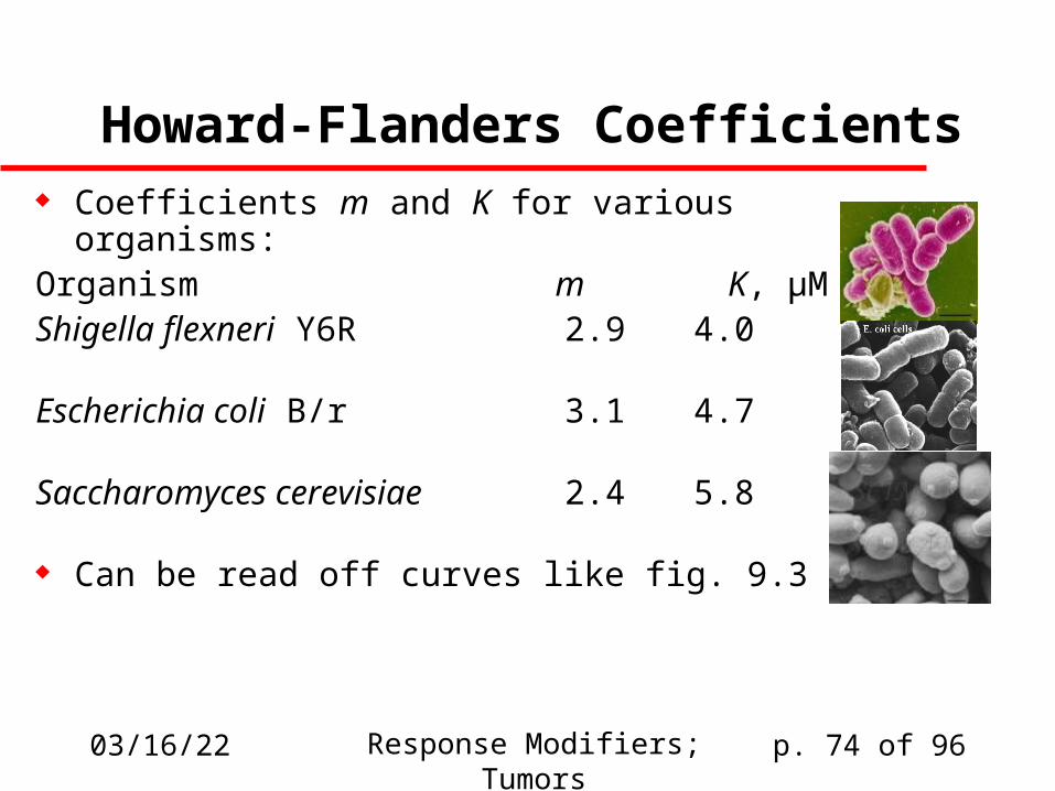
Howard-Flanders Coefficients Coefficients m and K for various organisms:Organism m K, µMShigella flexneri Y6R 2.9 4.0
Escherichia coli B/r 3.1 4.7
Saccharomyces cerevisiae 2.4 5.8
Can be read off curves like fig. 9.3
04/19/23 Response Modifiers; Tumors p. 75 of 96
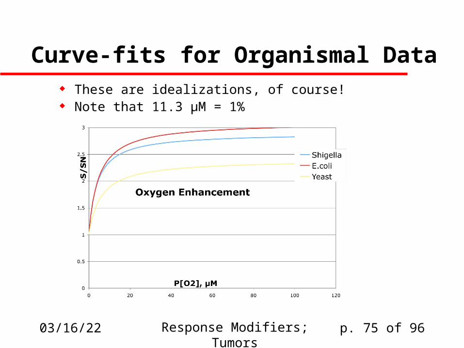
Curve-fits for Organismal Data These are idealizations, of course! Note that 11.3 µM = 1%
04/19/23 Response Modifiers; Tumors p. 76 of 96
Why does this happen?
Alper’s model:2 types of damage from primary radiation event
– Type I: lesion requires oxygen for lethality– Type II: always lethal, independent of oxygen
Thus: type I can be chemically restituted Restitution competes with oxygen fixation Posit: n1 type I lesions, n2 type II lesions,
krep = rate of repair, kfix = rate of O2 fixation, then:
m-1 = n1/n2 and K = krep/kfix

04/19/23 Response Modifiers; Tumors p. 77 of 96
What happens with high-LET?
Essentially all damage is type 2 Nothing depends on restitution Therefore n1/n2 << 1, m-1=0, m=1
Thus S/SN = ([O2] + K) / ([O2] + K) = 1,independent of K.
The fact that high-LET produces this behavior is mildly supportive of the appropriateness of Alper’s model, although it’s far from conclusive
04/19/23 Response Modifiers; Tumors p. 78 of 96
Time-Dependence Study the kinetics via short bursts of radiation We look to see whether providing O2 at a certain
time-point before or after irradiation sensitizes cells– Oxygen sensitizes the cell if present before
irradiation– If available till ~3 msec after irradiation, it still matters– If oxygen is made available later than a few msec
post-irradiation, it doesn’t sensitize the cell This suggests that the oxygen-dependent free
radicals have lifetimes shorter than 3 msec.
04/19/23 Response Modifiers; Tumors p. 79 of 96
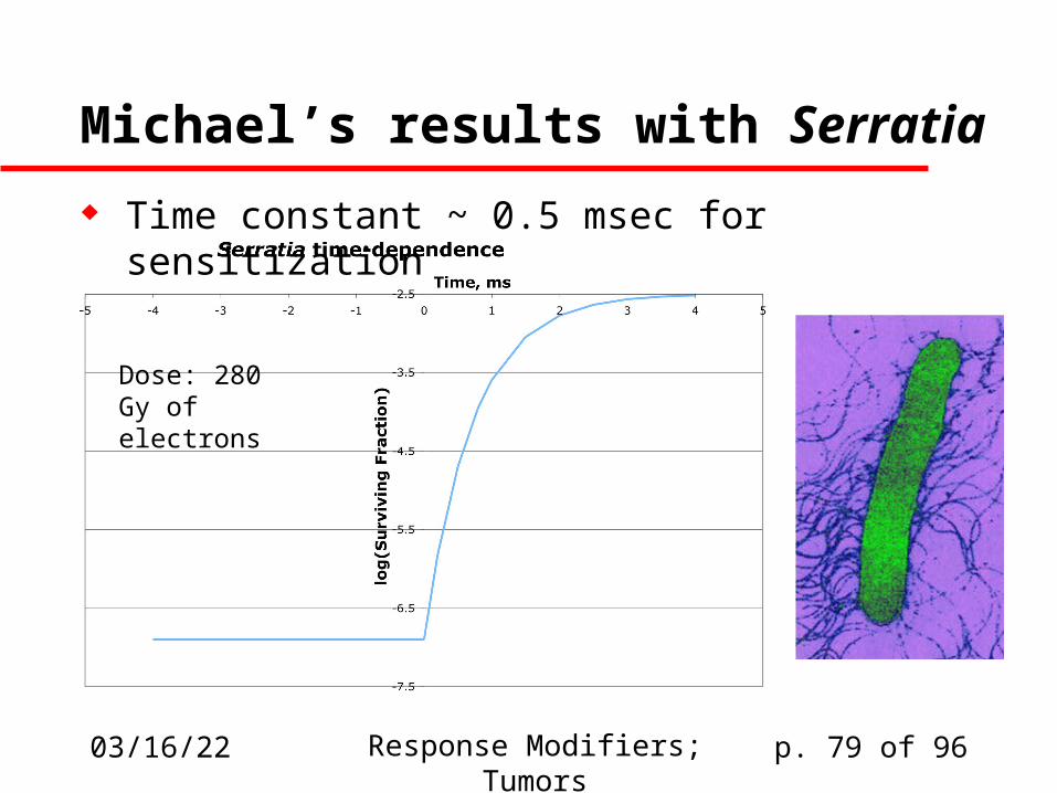
Michael’s results with Serratia
Time constant ~ 0.5 msec for sensitization
Dose: 280 Gy of electrons
04/19/23 Response Modifiers; Tumors p. 80 of 96
What radicals are involved? The short time-span shown in this experiment suggests
that free radicals are involved:but which ones?
Michael suggests that superoxide (O2-•), hydroperoxyl
(H-O=O•), and eaq- have shorter lifetimes than is
consistent with 0.5 msec timing Other researchers continue to plug for superoxide and
hydroperoxyl as candidates OH• doesn’t interact with O2 that much except via CO2•-:
OH• + HCO2- -> CO2•- + H2O
CO2•- + O2 -> CO2 + O2-•

04/19/23 Response Modifiers; Tumors p. 81 of 96
Superoxide We’re still not sure if superoxide is
a major actor in oxygen-mediated damage But this is how superoxide is detoxified
by superoxide dismutase (SOD):– 2 O2•- + 2H+ H2O2 + O2 (catalyzed by SOD)
– H2O2 H2O + (1/2)O2 (catalyzed by catalase)
Exogenous SOD doesn’t help alleviate damage(but maybe that’s because it isn’t tied to catalase, so peroxide can build up to toxic levels?)
Manipulating constitutive levels of SOD by genetic means gives muddy results
Human Cu-Zn SOD
04/19/23 Response Modifiers; Tumors p. 82 of 96
Thiol Mitigators
We expect that oxygen competes with thiols, such that the more thiol mitigation is involved, the less oxygen-dependent fixation of damage can occur:
1. [macro]-R• + R´SH [macro]-R-H + R´S•
2R´S• R´-S-S-R´
2. [small]• + R´SH [small]-H + R´S•
2R´S• R´-S-S-R´

04/19/23 Response Modifiers; Tumors p. 83 of 96
Glutathione as mediator
Glutathione can readily dimerize under oxidizing conditions:R-SH + HS-R R-S-S-H
Reasonably prevalent in cells(millimolar: [glut] >> [cys])
In principle cysteine could also operate this way, but its cellular concentration is too low
Cysteine
glutathione
04/19/23 Response Modifiers; Tumors p. 84 of 96
Are thiols really important?
Some argument about that Revesz’s results support the competition model Maybe only exogenous thiols really influence
radiosensitivity Introducing cysteine or synthetic thiols does definitely
mitigate damage:RSH + OH• RS• + H2ORS- + OH• RS• + OH-
. . . And then these RS• radicals recombine as disulfides
04/19/23 Response Modifiers; Tumors p. 85 of 96
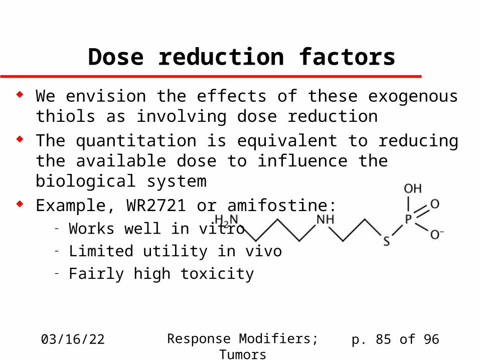
Dose reduction factors
We envision the effects of these exogenous thiols as involving dose reduction
The quantitation is equivalent to reducing the available dose to influence the biological system
Example, WR2721 or amifostine:– Works well in vitro– Limited utility in vivo– Fairly high toxicity
04/19/23 Response Modifiers; Tumors p. 86 of 96
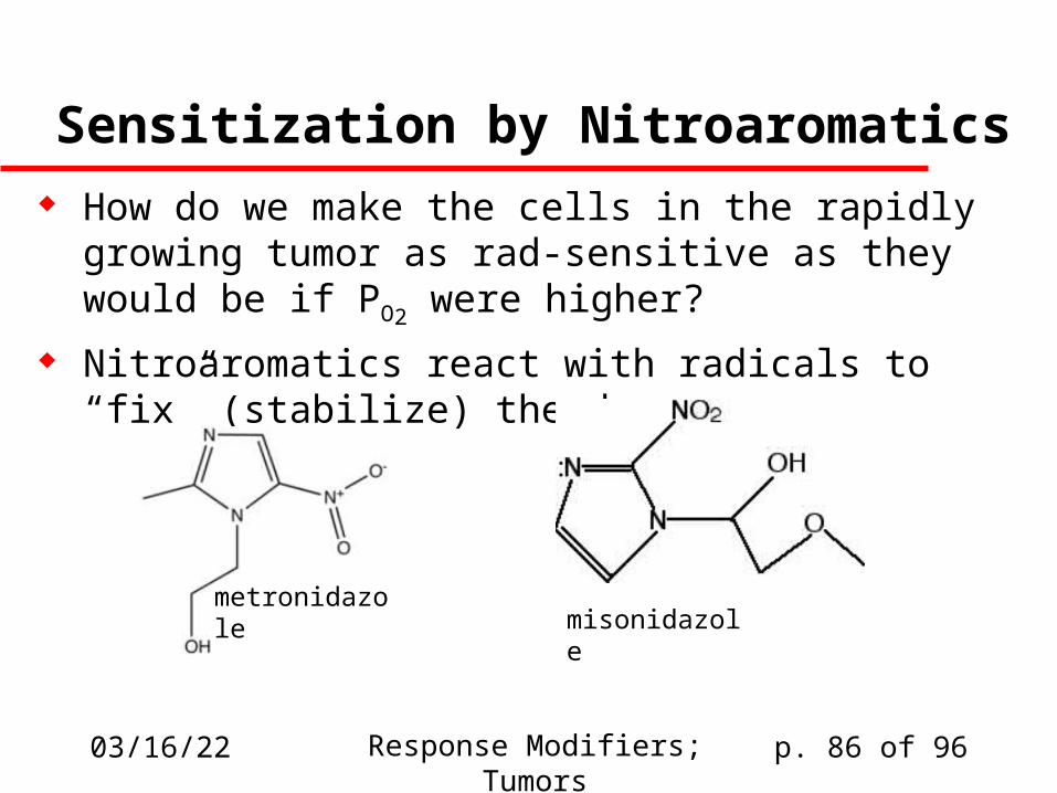
Sensitization by Nitroaromatics How do we make the cells in the rapidly growing tumor
as rad-sensitive as they would be if PO2 were higher?
Nitroaromatics react with radicals to “fix” (stabilize) the damage
metronidazole
misonidazole
04/19/23 Response Modifiers; Tumors p. 87 of 96
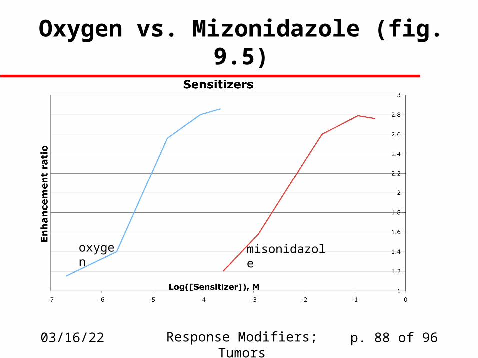
Nitroaromatic action
Like oxygen, nitroaromatics react with short-lived radicals to produce longer-lived and therefore more reactive radicals
Concentrations required to enhance radiosensitivity are much higher than with O2
These compounds have other applications, but they can be used therapeutically as potentiators of radiation damage
04/19/23 Response Modifiers; Tumors p. 88 of 96
Oxygen vs. Mizonidazole (fig. 9.5)
oxygen misonidazole
04/19/23 Response Modifiers; Tumors p. 89 of 96
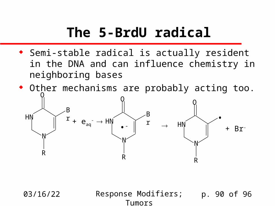
5-Halogen-substituted pyrimidines
These are molecules that resemble thymine The halogen at the 5- position looks like the
methyl group in thymine and can be incorporated in place of thymine in DNA
Most common: 5-bromodeoxyuridine Sensitization produced by ready reaction with
the aqueous electron
04/19/23 Response Modifiers; Tumors p. 90 of 96
The 5-BrdU radical Semi-stable radical is actually resident in the DNA
and can influence chemistry in neighboring bases Other mechanisms are probably acting too.
N
HN
R
O
Br
+ eaq-
N
HN
R
O
Br
•-
N
HN
R
O
+ Br-
•
04/19/23 Response Modifiers; Tumors p. 91 of 96
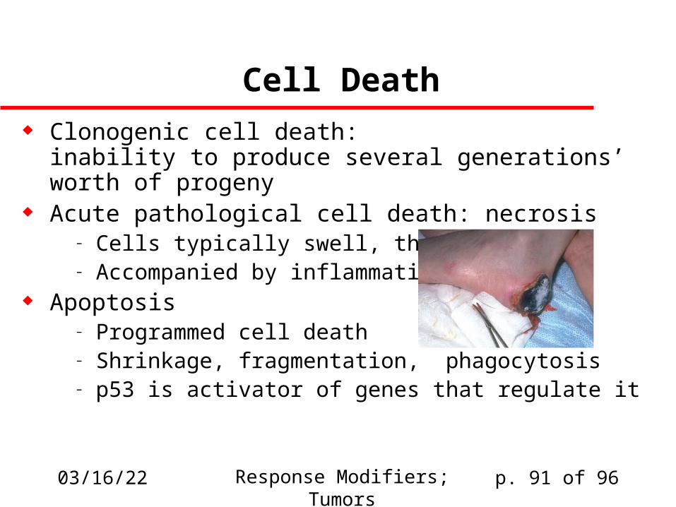
Cell Death Clonogenic cell death:
inability to produce several generations’ worth of progeny
Acute pathological cell death: necrosis– Cells typically swell, then lyse– Accompanied by inflammation
Apoptosis– Programmed cell death– Shrinkage, fragmentation, phagocytosis– p53 is activator of genes that regulate it
04/19/23 Response Modifiers; Tumors p. 92 of 96
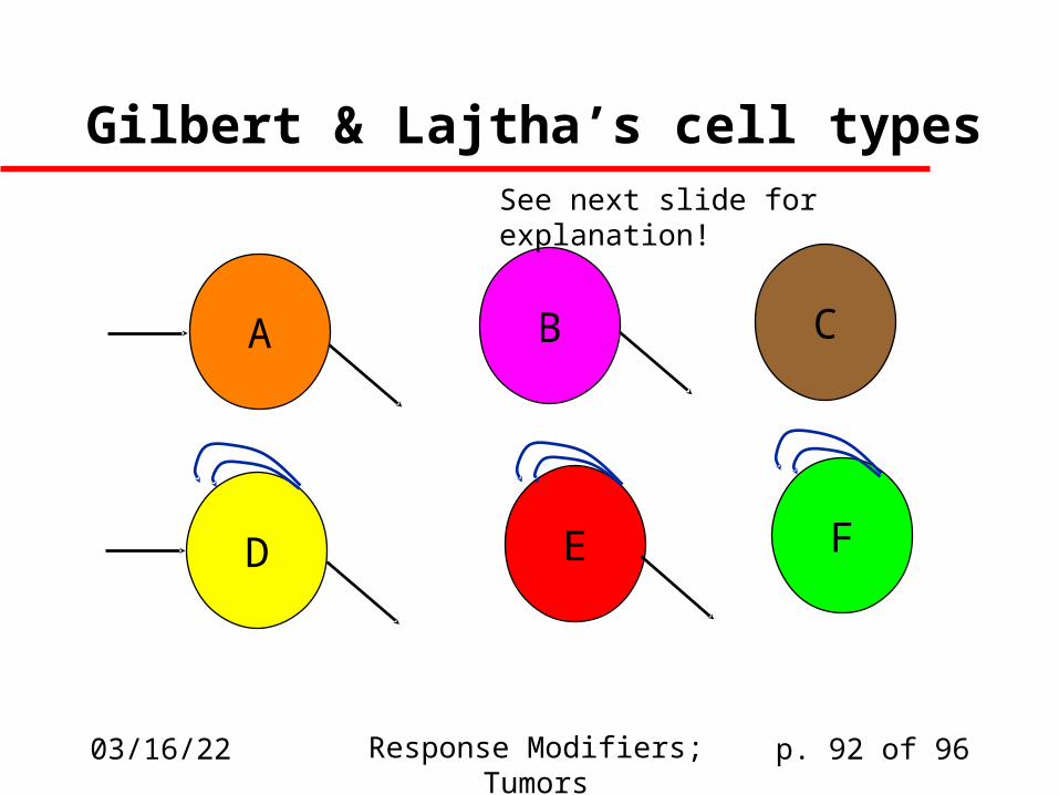
Gilbert & Lajtha’s cell types
A B C
D E F
See next slide for explanation!
04/19/23 Response Modifiers; Tumors p. 93 of 96
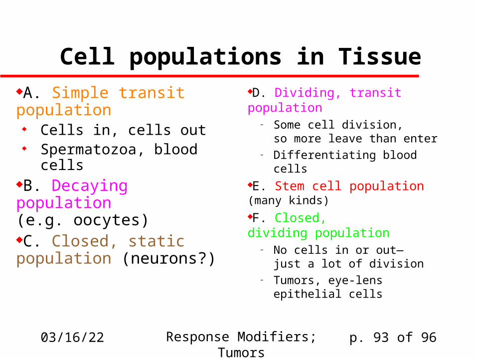
Cell populations in TissueA. Simple transit population
Cells in, cells out Spermatozoa, blood cells
B. Decaying population(e.g. oocytes)C. Closed, static population (neurons?)
D. Dividing, transit population– Some cell division,
so more leave than enter– Differentiating blood cells
E. Stem cell population(many kinds)F. Closed,dividing population
– No cells in or out—just a lot of division
– Tumors, eye-lensepithelial cells
04/19/23 Response Modifiers; Tumors p. 94 of 96
Cell population kinetics
Cell types that divide are the most sensitive. Cells are most sensitive during G2 and M,
so cells that spend a lot of timein G2 and M are more sensitive
If a cell population is exposed to radiation, the outcome depends on there being an adequate number of (clonogenically) surviving cells.
04/19/23 Response Modifiers; Tumors p. 95 of 96
Growth Fraction Lajtha (1963): described “G0” phase in cell cycle:
cell is not engaged in proliferation but could later re-enter proliferative stage
Growth fraction is defined as fraction of total cellular population that is clonogenically competent and actually in the process of DNA replication and cell division.
Measurement: uses 3H-thymidine uptake Significance: cells in G0 have time to repair DNA damage
– Works even if [repair enzymes] is low during G0– This is suspected but not proven
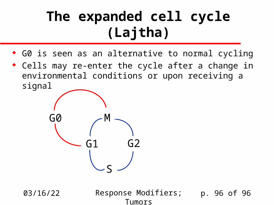
04/19/23 Response Modifiers; Tumors p. 96 of 96
The expanded cell cycle (Lajtha)
G0 is seen as an alternative to normal cycling Cells may re-enter the cycle after a change in
environmental conditions or upon receiving a signal
M
G1
S
G2
G0