image processing and informatics lab – usc | rsna 2008
TRANSCRIPT

Image Processing and Informatics Lab – USC | RSNA 2008 | Page 1
Image Processing and Informatics Lab – USC | RSNA 2008 | Page 2
WELCOME Dear Colleagues and Friends of IPILab, Welcome to another exciting year of IPILab’s presentations and exhibits at RSNA 2008.
This year we are presenting:
Two large screen presentation:
Tuesday December 02, 2008 - Scientific Poster 12:15PM - 1:15PM ROOM Lakeside Learning Center 07
An Online Real-time DICOM Web-based Computer-aided Dagnosis (CAD) System for Bone Age Assessment (BAA) of Children in a PACS Environment: Clinical Validation Cross-Racial Discrepancies of Growth Patterns in Bone Age Assessment Kevin C Ma, BS, Los Angeles, CA; A Zhang, PhD; M Fleshman; L A Vachon, MD; B J Liu, PhD; H K Huang, DSc; et al.
Wednesday, December 03, 2008 - Scientific Poster 12:15PM - 1:15PM ROOM Lakeside Learning Center 07
RadSearch: A RIS/PACS Integrated Retrospective Medical Image Retrieval Tool Sinchai Tsao, MS, Los Angeles, CA; J Documet, MS; P Moin, MD; K Wang; B J Liu, PhD
Education Exhibits (Standalone Presentation) 7 Education Exhibits (Electronics Presentation) 2 Education Exhibits (Poster) 1 Scientific Posters (Electronics Presentation) 2
Image Processing and Informatics Lab – USC | RSNA 2008 | Page 3
Contents WELCOME ....................................................................................................................................... 2 STAFF AND COLLABORATORS .................................................................................................... 4 IPILAB NEW LOCATION @ HEALTH SCIENCE CAMPUS ............................................................ 5 IPILAB NETWORK LAYOUT & COLLABORATIONS ...................................................................... 6
EDUCATION EXHIBITS – STANDALONE PRESENTATIONS ...................................................... 7 LL- IN1062: Extending Imaging Informatics beyond Radiology: The Development of an Image-intensive ePR for Image-guided Minimally Invasive Surgery Applications Including Real-time Intraoperative Image Acquisition, Archival, and Display .............................................................. 7 LL-IN1068: A Viewbox.com: A Tool for Radiology Web-based Content Management and Personalization ............................................................................................................................. 8 LL-IN1105: A Timely Computer-aided Detection System for Acute Ischemic and Hemorrhagic Stroke on CT in an Emergency Environment ............................................................................... 9 LL-IN1106: Automated Quantitative Analysis of MR Images of the Pelvic Hindgut and its Correlation with Fecal Incontinence and Interventional Outcomes ............................................ 10 LL-IN1116: A Radiology Dashboard Integrated with RFID Location System ............................ 11 LL-IN1123: Integration of Content-based DICOM-SR for CAD in the Medical Imaging Informatics Data Grid with Examples in CT Chest, Mammography, and Bone-Age Assessment11 LL-IN1130: Spine Assistant: A Tool to Efficiently Generate Cervical and Lumbar Spine Reports12
EDUCATION EXHIBITS – ELECTRONICS PRESENTATIONS ................................................... 14 LL-MK5234: Review of Hemipelvectomy Endoprostheses: What the Orthopedic Oncologist Wants to Know ........................................................................................................................... 14 LL-IN2060-B06: Optical Imaging of Melanoma Mimics: An Imaging Informatics Tool Kit Using Discriminant Analysis, Optical Properties, and Histological Characteristics .............................. 15
EDUCATION EXHIBITS – POSTER .............................................................................................. 16 LL-IN1052: Optimization and Management of User Registration and HIPAA-Compliance in a Clinical PACS Environment by Integration with a HIPAA-Compliant Auditing Toolkit ............... 16
SCIENTIFIC POSTERS – ELECTRONICS ................................................................................... 17 LL-IN2087-L07: RadSearch: A RIS/PACS Integrated Retrospective Medical Image Retrieval Tool ............................................................................................................................................ 17 LL-PD4093-H07: An Online Real-time DICOM Web-based Computer-aided Dagnosis (CAD) System for Bone Age Assessment (BAA) of Children in a PACS Environment: Clinical Validation .................................................................................................................................... 18
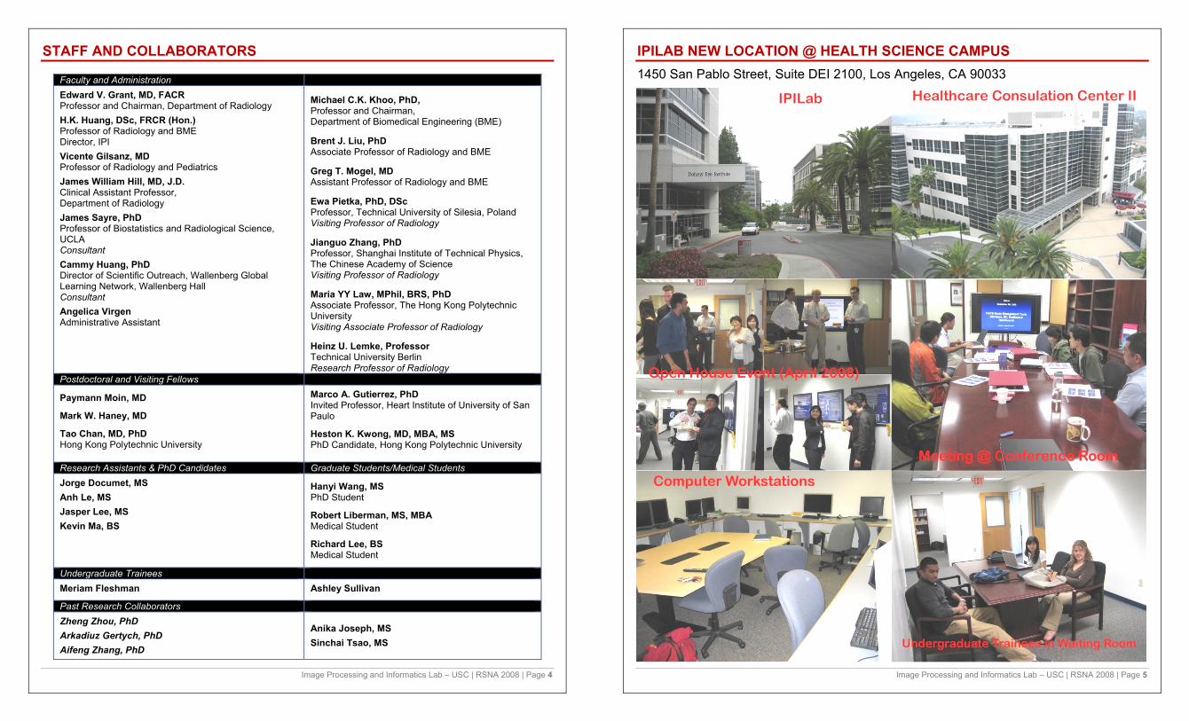
Image Processing and Informatics Lab – USC | RSNA 2008 | Page 4
STAFF AND COLLABORATORS
Faculty and Administration Edward V. Grant, MD, FACR Professor and Chairman, Department of Radiology H.K. Huang, DSc, FRCR (Hon.) Professor of Radiology and BME Director, IPI Vicente Gilsanz, MD Professor of Radiology and Pediatrics James William Hill, MD, J.D. Clinical Assistant Professor, Department of Radiology James Sayre, PhD Professor of Biostatistics and Radiological Science, UCLA Consultant Cammy Huang, PhD Director of Scientific Outreach, Wallenberg Global Learning Network, Wallenberg Hall Consultant Angelica Virgen Administrative Assistant
Michael C.K. Khoo, PhD, Professor and Chairman, Department of Biomedical Engineering (BME)
Brent J. Liu, PhD Associate Professor of Radiology and BME
Greg T. Mogel, MD Assistant Professor of Radiology and BME
Ewa Pietka, PhD, DSc Professor, Technical University of Silesia, Poland Visiting Professor of Radiology
Jianguo Zhang, PhD Professor, Shanghai Institute of Technical Physics, The Chinese Academy of Science Visiting Professor of Radiology
Maria YY Law, MPhil, BRS, PhD Associate Professor, The Hong Kong Polytechnic University Visiting Associate Professor of Radiology
Heinz U. Lemke, Professor Technical University Berlin Research Professor of Radiology
Postdoctoral and Visiting Fellows
Paymann Moin, MD
Mark W. Haney, MD
Tao Chan, MD, PhD Hong Kong Polytechnic University
Marco A. Gutierrez, PhD Invited Professor, Heart Institute of University of San Paulo
Heston K. Kwong, MD, MBA, MS PhD Candidate, Hong Kong Polytechnic University
Research Assistants & PhD Candidates Graduate Students/Medical Students Jorge Documet, MS Anh Le, MS Jasper Lee, MS Kevin Ma, BS
Hanyi Wang, MS PhD Student
Robert Liberman, MS, MBA Medical Student
Richard Lee, BS Medical Student
Undergraduate Trainees Meriam Fleshman Ashley Sullivan
Past Research Collaborators Zheng Zhou, PhD Arkadiuz Gertych, PhD Aifeng Zhang, PhD
Anika Joseph, MS Sinchai Tsao, MS
Image Processing and Informatics Lab – USC | RSNA 2008 | Page 5
IPILAB NEW LOCATION @ HEALTH SCIENCE CAMPUS 1450 San Pablo Street, Suite DEI 2100, Los Angeles, CA 90033
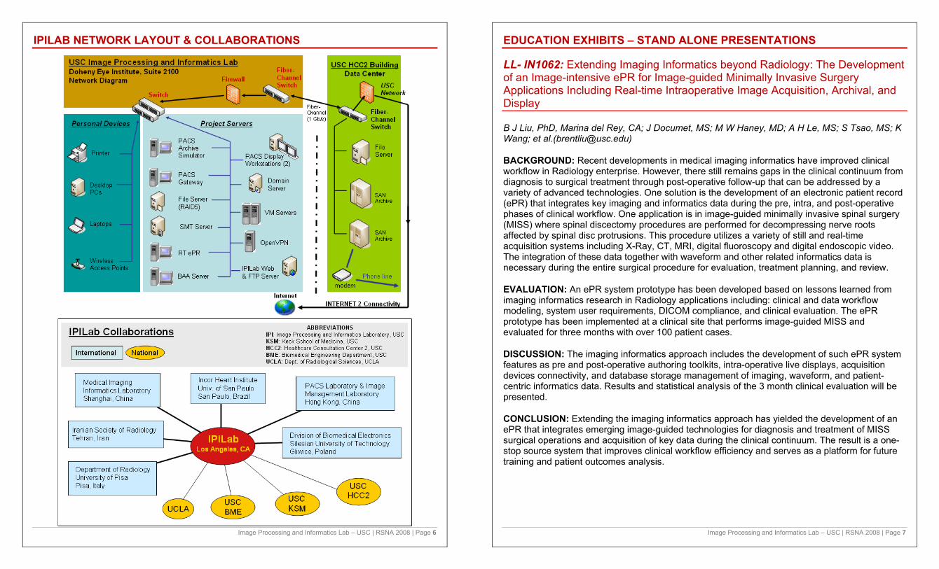
Image Processing and Informatics Lab – USC | RSNA 2008 | Page 6
IPILAB NETWORK LAYOUT & COLLABORATIONS
Image Processing and Informatics Lab – USC | RSNA 2008 | Page 7
EDUCATION EXHIBITS – STAND ALONE PRESENTATIONS
LL- IN1062: Extending Imaging Informatics beyond Radiology: The Development of an Image-intensive ePR for Image-guided Minimally Invasive Surgery Applications Including Real-time Intraoperative Image Acquisition, Archival, and Display
B J Liu, PhD, Marina del Rey, CA; J Documet, MS; M W Haney, MD; A H Le, MS; S Tsao, MS; K Wang; et al.([email protected])
BACKGROUND: Recent developments in medical imaging informatics have improved clinical workflow in Radiology enterprise. However, there still remains gaps in the clinical continuum from diagnosis to surgical treatment through post-operative follow-up that can be addressed by a variety of advanced technologies. One solution is the development of an electronic patient record (ePR) that integrates key imaging and informatics data during the pre, intra, and post-operative phases of clinical workflow. One application is in image-guided minimally invasive spinal surgery (MISS) where spinal discectomy procedures are performed for decompressing nerve roots affected by spinal disc protrusions. This procedure utilizes a variety of still and real-time acquisition systems including X-Ray, CT, MRI, digital fluoroscopy and digital endoscopic video. The integration of these data together with waveform and other related informatics data is necessary during the entire surgical procedure for evaluation, treatment planning, and review.
EVALUATION: An ePR system prototype has been developed based on lessons learned from imaging informatics research in Radiology applications including: clinical and data workflow modeling, system user requirements, DICOM compliance, and clinical evaluation. The ePR prototype has been implemented at a clinical site that performs image-guided MISS and evaluated for three months with over 100 patient cases.
DISCUSSION: The imaging informatics approach includes the development of such ePR system features as pre and post-operative authoring toolkits, intra-operative live displays, acquisition devices connectivity, and database storage management of imaging, waveform, and patient-centric informatics data. Results and statistical analysis of the 3 month clinical evaluation will be presented.
CONCLUSION: Extending the imaging informatics approach has yielded the development of an ePR that integrates emerging image-guided technologies for diagnosis and treatment of MISS surgical operations and acquisition of key data during the clinical continuum. The result is a one-stop source system that improves clinical workflow efficiency and serves as a platform for future training and patient outcomes analysis.
Image Processing and Informatics Lab – USC | RSNA 2008 | Page 8
LL-IN1068: A Viewbox.com: A Tool for Radiology Web-based Content Management and Personalization
P Moin, MD, Los Angeles, CA; J Documet, MS; J F Fernandez, MS; B J Liu, PhD ([email protected])
BACKGROUND: There has been a rapid proliferation of medically related websites, especially those with a focus upon radiology. Many of these contain content designed for use by radiologists. Educational materials, reference resources, articles, images, practice cases, and radiology-centric search engines have made for a rich, but dizzying volume of resources. We introduce a web-based program to manage and personalize the continually growing number of informational resources.
EVALUATION: Web resources are organized by modality and body region. Within each subheading, applets contain descriptive bookmarks for several content types provided in an organized format for review including sites that highlight relevant anatomy, key articles, reference resources, unknown (practice) cases, reference cases, and search tools. A standard template is provided for each user with the ability to add, remove, or change any listing within their profile to better fit their needs. Radiology-centric sites as well as relevant sites from related specialties (eg. Wheeless' Textbook of Orthopeadics for the musculoskeletal radiology section) will be included.
DISCUSSION: The internet and the methods of organizing and communicating information which it allows are a valuable tool to learn or reference when learning more about a topic or reading a difficult case on call. As these resources proliferate and become more complex, a management tool is needed in order to organize and effectively use them. Radiologists in training and practice are increasingly incorporating web-based resources for learning and decision-support. TheViewbox.com is developed with the intent of being a tool to facilitate the management of these resources and to allow the continued expansion and complexity of these resources while still maintaining quick access to promote their use.
CONCLUSSION: We introduce an organized, customizable web-based program to allow radiology residents and those in private practice to easily manage and reference the growing number of radiology internet resources. To our knowledge, this is the first attempt to allow individual tailoring of the management and organization of access to desired web-based radiology resources.
Image Processing and Informatics Lab – USC | RSNA 2008 | Page 9
LL-IN1105: A Timely Computer-aided Detection System for Acute Ischemic and Hemorrhagic Stroke on CT in an Emergency Environment K C Ma, BS, Los Angeles, CA; A H Le, MS; J F Fernandez, MS; T Chan, MBChB; B J Liu, PhD; H K Huang, DSc; et al. ([email protected]) BACKGROUND: When a patient is accepted in the emergency room suspected of stroke, time is of the most importance. The infarct brain area suffers irreparable damage as soon as three hours after the onset of stroke symptoms. Non-contrast CT scan is the standard first line of investigation used to identify hemorrhagic stroke cases. However, CT brain images do not show hyperacute ischemia and small hemorrhage clearly and thus may be missed by emergency physicians. We reported a timely computer-aided detection (CAD) system for small hemorrhages on CT that has been successfully developed as an aid to ER physicians to help improve detection for Acute Intracranial Hemorrhage (AIH). This CAD system has been enhanced for diagnosis of acute ischemic stroke in addition to hemorrhagic stroke, which becomes a more complete and clinically useful tool for assisting emergency physicians and radiologists. In the detection algorithm, brain matter is first segmented, realigned, and left-right brain symmetry is evaluated. As in the AIH system, the system confirms hemorrhagic stroke by detecting blood presence with anatomical and medical knowledge-based criteria. For detecting ischemia, signs such as regional hypodensity, blurring of grey and white matter differentiation, effacement of cerebral sulci, and hyperdensity in middle cerebral artery, are evaluated.
EVALUATION: 25 confirmed cases with acute ischemic or/and hemorrhagic stroke with match normals have been collected for this study. For each case, the CAD results are matched with the initial diagnoses.
DISCUSSION: The CAD system is able to successfully detect signs of acute ischemia and hemorrhage in brain CT images. Potentially, the CAD system increases stroke detection speed and improves sensitivity and specificity in detecting acute ischemic and hemorrhagic stroke. The system provides opportunities for better timely stroke detection for patients in the emergency environment when a neuroradiologist may not be readily available.
CONCLUSION: The CAD system for acute stroke detection is can increase the accuracy of stroke detection by emergency physicians on CT. It can be useful for both clinical applications and educational purposes.

Image Processing and Informatics Lab – USC | RSNA 2008 | Page 10
LL-IN1106: Automated Quantitative Analysis of MR Images of the Pelvic Hindgut and its Correlation with Fecal Incontinence and Interventional Outcomes Mark W. Haney, MD, Anika Joseph, MS, Haig Dudukgian, MD, Brent J. Liu, PhD ([email protected]) BACKGROUND: The second leading cause of elderly admission to long-term care facilities is fecal incontinence which has a prevalence of 0.3 to 5%. Addressing the pathology of the fecal incontinence, an anoplasty may be performed but patient outcomes are highly variable. Prior studies have evaluated imaging techniques including 3D ultrasound and computed tomography to define gross anatomic defects in the wall but thus far have been unable to better delineate patients who will have resolution of their incontinence. Magnetic resonance imaging (MRI) techniques appear to be able to delineate soft tissue more specifically than US and CT. We discovered that in some cases, combining MRI with computer aided image analysis of the tissue components (adipose, fibrosis and specifically, the anal canal muscle and sphincters) has shown an indication of patients who were more likely to have a successful outcome after anoplasty. This paper describes this preliminary study.
METHODS: Thirty patients were retrospectively collected who underwent MR imaging of the pelvis as part of an evaluation of pelvic floor disorders. An image analysis system was developed which identifies the anal canal via local anatomy and anatomic characteristics of the canal itself. The program segmented the muscle from adipose external to the canal and feces internal to the canal. Radial projects from the canal’s center are used to calculate a mean and variance of the thickness of the muscular wall. A second set of projects then identify regions which exist outside two standard deviations of the mean as muscularly weak regions. Finally, a 3-D plot combines the canal’s muscle thickness along with the fluid stress exerted on the wall to provide the clinician with a visual means of identifying regions of muscle which are less likely to contribute to continence.
DISCUSSION: Wall thickness and fluid stress were analyzed with respect to the presence of confirmed anal canal muscle pathology and known patient outcome after anoplasty.
CONCLUSION: MRI, with its greater soft tissue specificity, in conjunction with computer analysis of the anal canal’s muscular features can help predict between successful and non successful outcomes.
Acknowledgements: Mark W. Haney, MD is supported by the USC Provost Fellowship, and was a former trainee of the NIH T32 EB00438 “Biomedical Imaging Informatics Training”. Anika Joseph, MS is supported by the NIH T32 EB00438: “Biomedical Imaging Informatics Training”
Image Processing and Informatics Lab – USC | RSNA 2008 | Page 11
LL-IN1116: A Radiology Dashboard Integrated with RFID Location System
J Documet, MS, Los Angeles, CA; B J Liu, PhD; K Wang; R J Lee, BS ([email protected])
PURPOSE/AIM:
To develop a Physician's dashboard that will help improve the workflow processes in a radiology department, monitoring every stage of the clinical care process within radiology. An integrated location tracking interface using RFID (Radio Frequency Identification) will permit to know location of patients and staff inside the clinical facility.
CONTENT ORGANIZATION:
The development of an extensible, scalable and secure web-based Physician's Dashboard; that allows access to display:
• real-time clinical and workflow metrics, • related reports from the RIS, • the location of patients with the waiting times that were collected in the different areas
inside a clinical facility while a procedure was performed, • the location of staff to more quickly assess who is the most appropriate person to contact
in the clinical facility, ie. tech support.
SUMMARY:
This exhibit implements a Physician's Dashboard for the Radiology Department that monitors ongoing clinical and workflow metrics. The real-time monitoring of the flow of the patient in the radiology department will use data entered in the RIS. Combining the above with the location of patients will add more powerful spatial information to better measure true waiting times for examinations. The primary users of the application will be administrative staff, technologists and PACS administrators.

Image Processing and Informatics Lab – USC | RSNA 2008 | Page 12
LL-IN1123: Integration of Content-based DICOM-SR for CAD in the Medical Imaging Informatics Data Grid with Examples in CT Chest, Mammography, and Bone-Age Assessment
J Lee, MS, Marina del Rey, CA; A H Le, MS; B J Liu, PhD ([email protected])
BACKGROUND: The Medical Imaging Informatics Data Grid started at the IPILab in 2005 as a secure and DICOM-compliant Data Grid for backing up medical imaging exams in multi-site research and radiological environments. The Data Grid includes DICOM-compliant services and SQL databases to handle storage, query and retrieve requests from the radiological environment. It also utilizes middleware from the Globus Toolkit 4.0 for file transmission, replica location, and grid security. Additional features include HIPAA-compliant events auditing, data resource failover and recovery, requests buffering, and a web-based GUI for user access and Data Grid management. The Data Grid currently extends its application to support DICOM Structured Reports (SR) generated by select CAD methods.
EVALUATION: The Medical Imaging Informatics Data Grid has been deployed at the USC Health Science campus and also among international medical imaging research laboratories and hospitals. The DICOM-SR for CAD is being evaluated using examples in CT chest, mammography, and bone-age assessment within the lab environment for comprehensiveness of its content extraction algorithm and database, as well as the user GUI.
DISCUSSION: The integration of DICOM-SR into the Data Grid involves new database schemas, a DICOM-SR receiver, content extraction algorithms, and updated DICOM services. Storing DICOM-SR in the same Data Grid repository as the original DICOM images allows the Data Grid to extend its benefits of fault-tolerant replication and federated storage towards CAD development and potentially complex radiological reports.
CONCLUSION: Due to the more complex CAD and radiological reporting, the Medical Imaging Informatics Data Grid seeks to incorporate CAD findings and DICOM key image referencing into its DICOM-compliant databases and services in the form of DICOM-SR data objects. By supporting DICOM-SR of CAD algorithms, content-based query/retrieval of DICOM imaging studies can be performed based on quantitative findings rather than patient identification and/or disease category. The advantages of query/retrieving anonymized yet content-based imaging data can be a great benefit for medical imaging research and imaging-based clinical trials.
Image Processing and Informatics Lab – USC | RSNA 2008 | Page 13
LL-IN1130: Spine Assistant: A Tool to Efficiently Generate Cervical and Lumbar Spine Reports
P Moin, MD, Los Angeles, CA; A H Le, MS; J Documet, MS; S Antani; R Long; B J Liu, PhD ([email protected])
BACKGROUND: Evaluation of the anatomy and pathology of cervical and lumbar spines is a tedious process with specific reporting requirements for each level. In practice, accepted descriptive terminology is not always followed in reporting. This creates the potential for miscommunication and errors both in reporting and subsequent clinical management.
EVALUATION: In 2001, the Combined Task Force of the North American Spine Society, American Society of Spine Radiology, and American Society of Neuroradiology released a consensus document entitled “Nomenclature and Classification of Lumbar Disc Pathology” in an effort to standardize the language of reporting spinal imaging findings. We use the definitions and guidelines set forth in this paper to construct a web-based program in a graphical format to quickly generate reports compliant with Task Force descriptions. Analogous terminology is then used in the generation of cervical spine reports.
DISCUSSION: Aims: 1) To increase awareness and correct use of accepted descriptions of disc pathology through
use as an educational tool in the training of radiology residents and as a refresher for those already in practice.
2) To quickly and efficiently generate well-organized reports that clearly communicate pathology. Simple measurement and classification tools in graphical format with easily referenced schematics to teach or clarify preferred correct disc nomenclature and pathology are provided. Pertinent descriptions of discs at each level including selections for normal, developmental variants, degenerative lesions, herniations, etc. are generated.
3) To gather data for computer aided diagnosis (CAD). Imaging studies and the reports generated from them is being collected to develop a CAD function to identify and characterizedisc pathology to facilitate the quick and efficient generation of reports. The second step will be to put the report in DICOM-SR data format for integrating CAD results into a PACS-based clinical workflow.
CONCLUSION: We introduce a web-based report generation tool to quickly, systematically, and efficiently produce cervical and lumbar spine reports that are compliant with accepted nomenclature and classification of disc pathology.

Image Processing and Informatics Lab – USC | RSNA 2008 | Page 14
EDUCATION EXHIBITS – ELECTRONIC PRESENTATIONS
LL-MK5234: Review of Hemipelvectomy Endoprostheses: What the Orthopedic Oncologist Wants to Know
P Moin, MD, Los Angeles, CA; C Allison, MD; E Ahlmann, MD; L R Menendez, MD; T J Learch, MD; E White, MD ([email protected])
PURPOSE/AIM:
The purposes of this exhibit are: 1) To understand the indications, various treatment methods, contraindications, and complications of endoprostheses used for pelvic reconstruction after tumor resection, with orthopedic oncologist input. 2) To highlight the Mark II saddle prosthesis, a new type of peri-acetabular reconstruction endoprosthesis. CONTENT ORGANIZATION:
1) Anatomy, including surgically important structures. 2) Indications and contraindications for hemipelvectomy endoprostheses. 3) Review of endoprosthesis types with emphasis upon the Mark II saddle prosthesis 4) Radiologically important outcomes, including complications such as dislocation, loosening, and recurrence of tumor. Examples will be presented using multimodality images and intraoperative photos SUMMARY:
The major teaching points of this exhibit are: 1) To provide the radiologist with a detailed understanding of pelvic reconstruction after tumor resection. 2) To highlight different types of pelvic reconstruction endoprostheses with particular attention to a new type of peri-acetabular reconstruction endoprosthesis. 3) The accurate recognition of post-operative complications is crucial to preventing long-term patient morbidity.
Image Processing and Informatics Lab – USC | RSNA 2008 | Page 15
LL-IN2060-B06: Optical Imaging of Melanoma Mimics: An Imaging Informatics Tool Kit Using Discriminant Analysis, Optical Properties, and Histological Characteristics A Joseph, MS, Los Angeles, CA; G Galliano, MD; S Bose, MD; D Farkas, PhD ([email protected])
PURPOSE/AIM:
The long term objective of this study is to evaluate optical imaging methods that could be applied clinically to distinguish between aggressive and benign forms of cutaneous melanoma in humans. In this study, we would like to apply an optical imaging method, specifically, spectral imaging, to image mimics, of human melanoma to determine if distinct imaging markers can be found that are not present in human melanoma with metastatic potential.
CONTENT ORGANIZATION:
These research efforts have focused on both the detection of melanoma mimics and the in-depth evaluation of pigmented lesions for propensity to develop melanoma. We have developed an imaging informatics tool kit with vendor supplied software packages and in-house customized software applied on reflectance imaging spectroscopy in the range of 400–1000 nm. The tool kit has been applied to discriminate and characterize melanoma mimics. Images were acquired for 20 primary biopsies of suspected melanomas and 20 re-excisions of tissues suspected to be reoccurrences of melanoma in patients, later confirmed by histology to be negative for melanoma. The images of melanomas mimics were processed semi-automatically through two software packages in the tool kit that spectrally separate and characterize tissue features from hematoxylin and eosin stained and unstained tissue. A Least Mean Square and Mahalanobis method based in-house customized algorithm in the tool kit was used to classify the lesions. The "gold standard" for training and testing these classifiers were verified by a dermatopathologist.
SUMMARY:
From each spectral image, corresponding to a selected wavelength, four parameters were derived to quantify color, pigment distribution, dimension and boundary of each lesion. A statistical analysis performed on the selected parameters shows that the mean effective reflectance, pigment distribution and lesion area are significantly different with the different pathological imitators of melanoma.
Image Processing and Informatics Lab – USC | RSNA 2008 | Page 16
EDUCATION EXHIBITS – POSTER
LL-IN1052: Optimization and Management of User Registration and HIPAA-Compliance in a Clinical PACS Environment by Integration with a HIPAA-Compliant Auditing Toolkit
J Lee, MS, Marina del Rey, CA; B Guo, MD; B J Liu, PhD; K Wang ([email protected])
BACKGROUND: USC Health Science Campus is supported by an enterprise PACS with four hospital sites. To access PACS, USC radiology staff and referring physicians must fax a written HIPAA Form to the PACS support team and wait for an email confirmation. However with the myriad of user types and hundreds of users to manage across the department's multiple healthcare systems, not all forms are processed in a timely fashion and not all user accounts are kept up-to-date. An online registration GUI will be integrated into a HIPAA-Compliant Auditing Toolkit which extracts pertinent auditing information from the logs of various PACS components into an auditing database. The challenges of user registration efficiency, HIPAA authorization and auditing are presented in an electronic workflow solution.
EVALUATION: A Web-based Radiology ID Authorization System will be evaluated for users in the Internal Medicine and Orthopedics departments within HCC2 because they currently have the largest number of new referring physician requests per month. Evaluation will be based on average user registration turn-around time and analysis of user activities by integrating the user authorization list with the HIPAA-Compliant Auditing System.
DISCUSSION: A web-based HIPAA authorization and auditing system allows the PACS support staff in a large radiology environment to manage new user requests online rather than via faxed sheets of paper. Furthermore, the system database allows the staff to monitor account privileges and user activity across multiple healthcare information systems. Automatic email notification is also done for users with expiring HIPAA authorization to make sure annual renewals of HIPAA rules are actually met.
CONCLUSION: An electronic HIPAA authorization and auditing system allows the PACS support team to better communicate and manage its healthcare information system users by allowing them to submit their HIPAA statements of confidentiality forms online. Furthermore, the user accounts system can be linked to a HIPAA-Compliant Auditing System for user-based events monitoring. The system hopefully reduces turn-around time while improving HIPAA-compliance of users in the PACS and healthcare information systems.
Image Processing and Informatics Lab – USC | RSNA 2008 | Page 17
SCIENTIFIC POSTERS – ELECTRONICS
LL-IN2087-L07: RadSearch: A RIS/PACS Integrated Retrospective Medical Image Retrieval Tool
Wed Dec 03 2008 12:15PM - 1:15PM ROOM Lakeside Learning Center 07
S Tsao, MS, Los Angeles, CA; J Documet, MS; P Moin, MD; K Wang; B J Liu, PhD ([email protected])
BACKGROUND: Radiology Information Systems (RIS) hold text reports that contain a wealth of information that can be used for research, education, and practice management. However, there are currently very few tools available that query specific data from these reports from an existing RIS database and also allow for query/retrieval of relevant image studies from the Picture Archival and Communications System (PACS). This project aims to create a software tool that integrates a Radiology text report query tool with PACS’ abilities to view and move images within the clinical workplace. RadSearch allows users to seamlessly data mine the radiology reports whilst previewing and moving related medical images.
EVALUATION: As a means of evaluation, RadSearch is integrated into University of Southern California’s Health Consultation Center’s Two’s PACS and RIS system to allow clinical user feedback on the tool. Two versions of the tool is available, one that uses only mysql database’s built in full text searching tool and another hybrid version that searches based on all related terms defined within the RadLex lexicon. Feedback is presented in the form of suggestions in improving ease of use as well as requests for future features.
DISCUSSION: RadSearch relates data fields in the RIS such as Current Procedure Terminology 4 (CPT-4) codes and the Radiology Report text back to the PACS study. This allows RadSearch to implement the new paradigm of performing retrospective studies or generating teaching files by leveraging existing data within the clinical practice. This project will also discuss the utility of the different strategies in integrating RadLex as well as CPT-4 codes as a means to provide synonymity and structure into the searching algorithm. The potential problems and pitfalls in adoption as well as implementation of such a system will also be discussed.
CONCLUSION: RadSearch illustrates a development of a data mining tool built using open source components such as CPT-4, RadLex, mySQL and PHP that allows research institution to maximally utilize and data mine existing clinical data for knowledge discovery and scientific research.
Image Processing and Informatics Lab – USC | RSNA 2008 | Page 18
LL-PD4093-H07: An Online Real-time DICOM Web-based Computer-aided Dagnosis (CAD) System for Bone Age Assessment (BAA) of Children in a PACS Environment: Clinical Validation
Tue Dec 02 2008 12:15PM - 1:15PM ROOM Lakeside Learning Center 07 K C Ma, BS, Los Angeles, CA; A Zhang, PhD; M Fleshman; L A Vachon, MD; B J Liu, PhD; H K Huang, DSc; et al. ([email protected])
PURPOSE: During the past 10 years, 1,390 hand images of normal children, both male and female from Asian, African American, Caucasian and Hispanic descends were collected and a digital hand atlas (DHA) was formed. Based on the DHA, a fully automatic, objective, racial, and gender specific computer-aided-diagnosis (CAD) method has been developed within the Image Processing and Informatics Lab (IPILAB), USC. To bring the DHA and CAD method to clinical environment for daily use in assisting radiologist to achieve higher accuracy in BAA, a web-based client-server system is designed as a novel clinical implementation approach for online and real-time BAA. This paper presents the clinical integration of the CAD system with the PACS at the Los Angeles County Women’s and Children’s Hospital (LAC WCH) for clinical validation.
METHOD AND MATERIALS: In the CAD system clinical implementation workflow, a second copy of the hand image from CR modality is sent to the CAD server with secure intranet connection. The CAD server includes the following major components: DICOM receiver, BAA CAD engine, web service and clinical database. The DICOM receiver listens and receives the hand image from CR modality. The CAD engine is then triggered to segment the hand images, extract bony features for automatic bone age assessment by fuzzy logic. The CAD report, including assessed bone age, best match image from DHA and patient displayed on the normal development graph for the specific racial group, was then generated automatically at CAD server as a DICOM SR (Structure Report). The radiologist at the clinical site can log into the website and review CAD results from a PACS WS and the assessment will be uploaded back to the server and stored in the final SR file of the clinical database.
RESULTS: The CAD server and PACS integration has been successfully implemented in LAC WCH. A fully automatic workflow is achieved without manual intervention. More than 60 prospective clinical cases have been evaluated.
CONCLUSION: This paper presents a novel clinical implementation approach for a CAD system using web technology for online and real time BAA. The success of the prototype can be easily extended to multiple clinical sites and will provide the foundation for broader use of the CAD system for BAA.
CLINICAL RELEVANCE/APPLICATION: To bring the DHA and CAD method to the clinical environment as a useful tool in assisting radiologist to achieve higher accuracy in BAA.