imaging diagnosis of interstitial pneumonia with emphysema
TRANSCRIPT

Hindawi Publishing CorporationPulmonary MedicineVolume 2012, Article ID 816541, 9 pagesdoi:10.1155/2012/816541
Review Article
Imaging Diagnosis of Interstitial Pneumonia with Emphysema(Combined Pulmonary Fibrosis and Emphysema)
Fumikazu Sakai,1 Junya Tominaga,1 Akiko Kaga,2 Yutaka Usui,2 Minoru Kanazawa,2
Takashi Ogura,3 Noriyo Yanagawa,4 and Tamiko Takemura5
1 Department of Diagnostic Radiology, Saitama International Medical Center, Saitama Medical University, Hidaka,Saitama 350-1298, Japan
2 Department of Respiratory Medicine, Saitama Medical University, Moroyama-Machi, Saitama 350-0040, Japan3 Department of Respiratory Medicine, Kanagawa Prefectural Cardiovascular and Respiratory Disease Center,Yokohama 236-0051, Japan
4 Department of Radiology, Tsukuba Memorial Hospital, Tsukuba 300-2622, Japan5 Department of Pathology, Japanese Red Cross Medical Center, Tokyo 150-8935, Japan
Correspondence should be addressed to Fumikazu Sakai, [email protected]
Received 23 June 2011; Accepted 23 October 2011
Academic Editor: Masahito Ebina
Copyright © 2012 Fumikazu Sakai et al. This is an open access article distributed under the Creative Commons AttributionLicense, which permits unrestricted use, distribution, and reproduction in any medium, provided the original work is properlycited.
Based on clinical and radiological findings, Cottin defined combined pulmonary fibrosis and emphysema (CPFE) as pulmonaryemphysema in the upper lungs and interstitial pneumonia in the lower lungs with various radiological patterns. Pathologic findingsof CPFE probably corresponded with diffuse interstitial pneumonia with pulmonary emphysema, emphysema with fibrosis, andthe combination of both. We described reported radiological findings of CPFE.
1. Introduction
Interstitial pneumonia (IP) with pulmonary emphysema haslong been a topic of controversy because we have not decidedif the disease is simple coincidence of IP and emphysema orIP and emphysema may be caused by common etiology. In1990, Wiggins et al. [1] presented 8 cases in the first reportin the English literature. In 2005, Cottin et al. [2] describedcombined pulmonary fibrosis and emphysema (CPFE) basedon clinical and radiological findings and noted some char-acteristic clinical features. Description of the pathologicalmechanisms in such cases is very limited.
Defined by clinical and imaging findings, CPFE is one ofclinicoradiological syndromes and probably includes severalkinds of pulmonary fibrosis, so its clinical course and prog-nosis various [3]. It is unclear how CPFE should be cate-gorized under interstitial pneumonia to ensure the integrityof idiopathic interstitial pneumonia (IIP) classification. Weherein review and summarize the imaging features of CPFE.
2. Smoking and Interstitial Pneumonia
Many pulmonary diseases are strongly associated with smok-ing-pulmonary emphysema, lung cancer, and some specificinterstitial diseases, such as pulmonary Langerhans cellhistiocytosis (LCH), desquamative interstitial pneumonia(DIP), and respiratory bronchiolitis interstitial lung disease(RBILD) [4–7].
Coexistence of pathologic findings of LCH, DIP, orRBILD in the same patient with smoking habit has been re-ported, and these three disease entities were sometimes cat-egorized as smoking-related interstitial lung disease (SRILD)in narrow definition [7, 8]. However, in a broad sense ofterm, SRILD may include pulmonary emphysema, IPF, andCPFE.
Smoking and/or dust inhalation are also important fac-tors in the development of idiopathic interstitial pneumonias[9], and most patients with idiopathic pulmonary fibrosis(IPF) have a history of smoking [10]. Overt pulmonary

2 Pulmonary Medicine
(a) (b)
(c) (d)
Figure 1: Diffuse interstitial pneumonia (nonspecific interstitial pneumonia (NSIP) pattern) with emphysema. (a) Chest X-ray showsbibasilar ground glass and reticular opacity. Volumes of bilateral lungs are almost normal. (b) High-resolution computed tomography(HRCT) of upper lung shows centrilobular pulmonary emphysema. (c), (d) HRCT of lower lung shows ground glass opacity along thebronchovascular bundle including cysts with varying size. Distribution of abnormal opacity mimics NSIP.
emphysema complicates IPF in approximately 30% ofpatients [11]. Iwai et al. calculated an odds ratio of 3.4relating IPF to smoking or dust inhalation [12]. A greaterprevalence of nonspecific interstitial pneumonia (NSIP) isalso reported in smokers [13], with DIP and RBILD generallyconfined to current smokers [8, 14–16].
Smoking has been closely related to DIP, RBILD, andLCH [8, 17], and features of all three are reported in path-ologic specimens of one individual [16, 17]. These 3 diseasesand IPF are also generalized as smoking-related interstitiallung disease (SRILD) [4, 5, 18].
Based on clinical and imaging findings, Cottin et al. alsorediscussed CPFE as pulmonary emphysema, defined as low-attenuation areas surrounded by normal lung with thin or nowall and/or multiple bullae predominantly in upper lungs,and interstitial pneumonia in the lower lungs with imagingpatterns of various IPs, including usual interstitial pneu-monia (UIP), NSIP, RBILD, cryptogenic organizing pneu-monia (OP), and others [8].
Cottin’s article reported characteristic clinical featuresincluding poor prognosis, frequently complicated by pul-monary hypertension [19, 20], preserved forced expiratoryvolume in one second (FEV 1.0), and severely impaired dif-fusion capacity [2, 21]. Mejıa et al. also reported decreasedsurvival of patients with IPF and emphysema with pulmon-ary hypertension [10]. However, we believe that the progno-sis of interstitial pneumonia is better than that of IPF/UIP[22], despite a high prevalence of lung cancer reported incases of CPFE [23, 24] and even IPF/UIP [25].
In a mass screening of smokers in the United States, theCOPD Gene Study Group reported interstitial shadow orradiologically overt IP on high-resolution computed tomog-raphy (HRCT) in approximately 8% of 2500 subjects withchronic obstructive pulmonary disease (COPD) [26, 27].They categorized interstitial changes on HRCT into follow-ing four types: centrilobular, subpleural, mixed abnormalopacity, or radiologically overt IP. On pulmonary functiontests, total lung capacity was increased in subjects with
Pulmonary Medicine 3
(a) (b) (c)
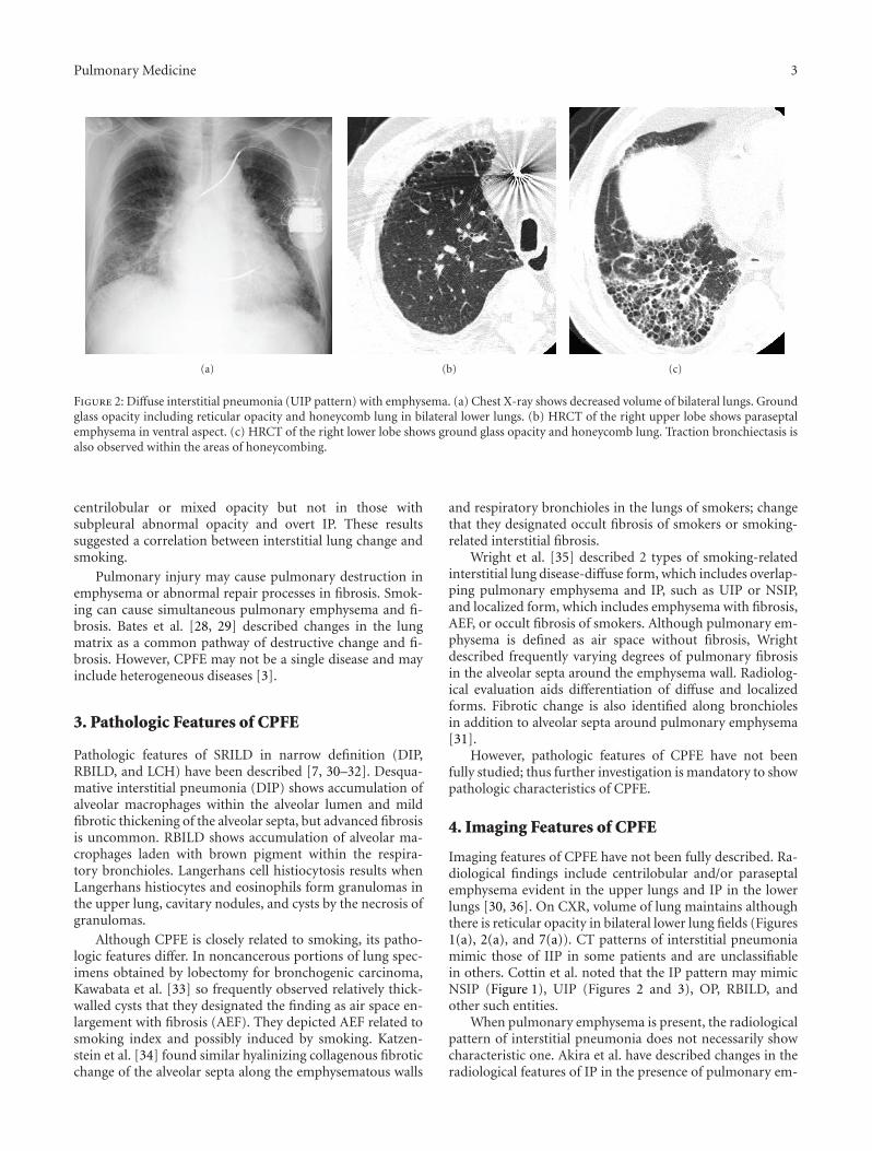
Figure 2: Diffuse interstitial pneumonia (UIP pattern) with emphysema. (a) Chest X-ray shows decreased volume of bilateral lungs. Groundglass opacity including reticular opacity and honeycomb lung in bilateral lower lungs. (b) HRCT of the right upper lobe shows paraseptalemphysema in ventral aspect. (c) HRCT of the right lower lobe shows ground glass opacity and honeycomb lung. Traction bronchiectasis isalso observed within the areas of honeycombing.
centrilobular or mixed opacity but not in those withsubpleural abnormal opacity and overt IP. These resultssuggested a correlation between interstitial lung change andsmoking.
Pulmonary injury may cause pulmonary destruction inemphysema or abnormal repair processes in fibrosis. Smok-ing can cause simultaneous pulmonary emphysema and fi-brosis. Bates et al. [28, 29] described changes in the lungmatrix as a common pathway of destructive change and fi-brosis. However, CPFE may not be a single disease and mayinclude heterogeneous diseases [3].
3. Pathologic Features of CPFE
Pathologic features of SRILD in narrow definition (DIP,RBILD, and LCH) have been described [7, 30–32]. Desqua-mative interstitial pneumonia (DIP) shows accumulation ofalveolar macrophages within the alveolar lumen and mildfibrotic thickening of the alveolar septa, but advanced fibrosisis uncommon. RBILD shows accumulation of alveolar ma-crophages laden with brown pigment within the respira-tory bronchioles. Langerhans cell histiocytosis results whenLangerhans histiocytes and eosinophils form granulomas inthe upper lung, cavitary nodules, and cysts by the necrosis ofgranulomas.
Although CPFE is closely related to smoking, its patho-logic features differ. In noncancerous portions of lung spec-imens obtained by lobectomy for bronchogenic carcinoma,Kawabata et al. [33] so frequently observed relatively thick-walled cysts that they designated the finding as air space en-largement with fibrosis (AEF). They depicted AEF related tosmoking index and possibly induced by smoking. Katzen-stein et al. [34] found similar hyalinizing collagenous fibroticchange of the alveolar septa along the emphysematous walls
and respiratory bronchioles in the lungs of smokers; changethat they designated occult fibrosis of smokers or smoking-related interstitial fibrosis.
Wright et al. [35] described 2 types of smoking-relatedinterstitial lung disease-diffuse form, which includes overlap-ping pulmonary emphysema and IP, such as UIP or NSIP,and localized form, which includes emphysema with fibrosis,AEF, or occult fibrosis of smokers. Although pulmonary em-physema is defined as air space without fibrosis, Wrightdescribed frequently varying degrees of pulmonary fibrosisin the alveolar septa around the emphysema wall. Radiolog-ical evaluation aids differentiation of diffuse and localizedforms. Fibrotic change is also identified along bronchiolesin addition to alveolar septa around pulmonary emphysema[31].
However, pathologic features of CPFE have not beenfully studied; thus further investigation is mandatory to showpathologic characteristics of CPFE.
4. Imaging Features of CPFE
Imaging features of CPFE have not been fully described. Ra-diological findings include centrilobular and/or paraseptalemphysema evident in the upper lungs and IP in the lowerlungs [30, 36]. On CXR, volume of lung maintains althoughthere is reticular opacity in bilateral lower lung fields (Figures1(a), 2(a), and 7(a)). CT patterns of interstitial pneumoniamimic those of IIP in some patients and are unclassifiablein others. Cottin et al. noted that the IP pattern may mimicNSIP (Figure 1), UIP (Figures 2 and 3), OP, RBILD, andother such entities.
When pulmonary emphysema is present, the radiologicalpattern of interstitial pneumonia does not necessarily showcharacteristic one. Akira et al. have described changes in theradiological features of IP in the presence of pulmonary em-
4 Pulmonary Medicine
(a) (b) (c)
(d) (e) (f)
(g)
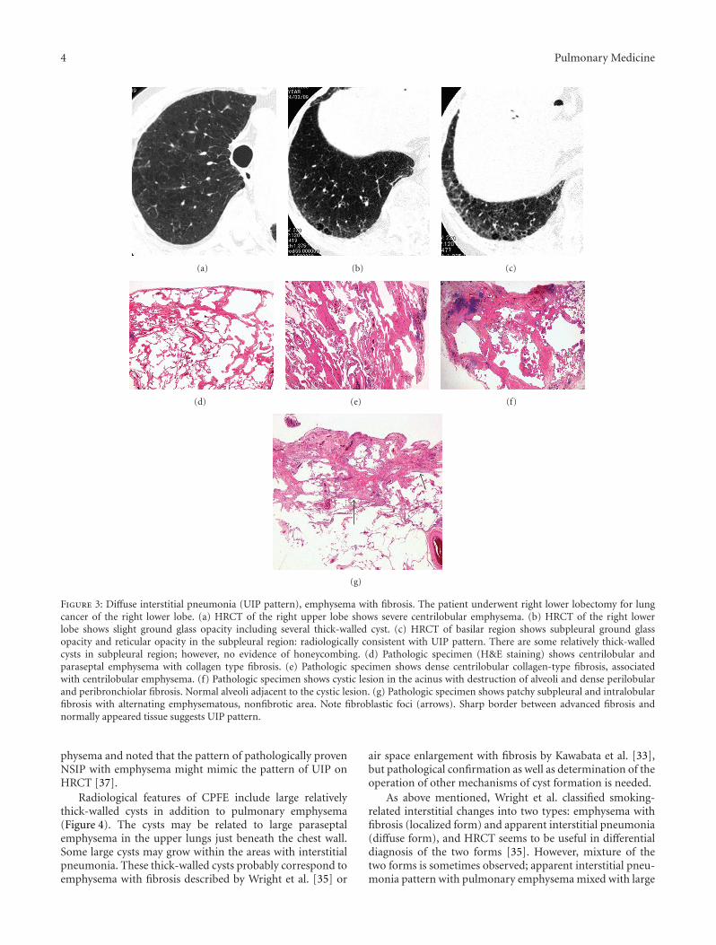
Figure 3: Diffuse interstitial pneumonia (UIP pattern), emphysema with fibrosis. The patient underwent right lower lobectomy for lungcancer of the right lower lobe. (a) HRCT of the right upper lobe shows severe centrilobular emphysema. (b) HRCT of the right lowerlobe shows slight ground glass opacity including several thick-walled cyst. (c) HRCT of basilar region shows subpleural ground glassopacity and reticular opacity in the subpleural region: radiologically consistent with UIP pattern. There are some relatively thick-walledcysts in subpleural region; however, no evidence of honeycombing. (d) Pathologic specimen (H&E staining) shows centrilobular andparaseptal emphysema with collagen type fibrosis. (e) Pathologic specimen shows dense centrilobular collagen-type fibrosis, associatedwith centrilobular emphysema. (f) Pathologic specimen shows cystic lesion in the acinus with destruction of alveoli and dense perilobularand peribronchiolar fibrosis. Normal alveoli adjacent to the cystic lesion. (g) Pathologic specimen shows patchy subpleural and intralobularfibrosis with alternating emphysematous, nonfibrotic area. Note fibroblastic foci (arrows). Sharp border between advanced fibrosis andnormally appeared tissue suggests UIP pattern.
physema and noted that the pattern of pathologically provenNSIP with emphysema might mimic the pattern of UIP onHRCT [37].
Radiological features of CPFE include large relativelythick-walled cysts in addition to pulmonary emphysema(Figure 4). The cysts may be related to large paraseptalemphysema in the upper lungs just beneath the chest wall.Some large cysts may grow within the areas with interstitialpneumonia. These thick-walled cysts probably correspond toemphysema with fibrosis described by Wright et al. [35] or
air space enlargement with fibrosis by Kawabata et al. [33],but pathological confirmation as well as determination of theoperation of other mechanisms of cyst formation is needed.
As above mentioned, Wright et al. classified smoking-related interstitial changes into two types: emphysema withfibrosis (localized form) and apparent interstitial pneumonia(diffuse form), and HRCT seems to be useful in differentialdiagnosis of the two forms [35]. However, mixture of thetwo forms is sometimes observed; apparent interstitial pneu-monia pattern with pulmonary emphysema mixed with large

Pulmonary Medicine 5
(a) (b) (c)
5000 µm
(d)
1000 µm
(e)
Figure 4: Emphysema with fibrosis, thick-walled large cyst. (a) HRCT of upper lungs shows severe centrilobular emphysema. A relativelythick-walled cyst in the superior basal segment of the left lower lobe. Lung cancer is noted in the lingual division of the left upper lobe. (b)HRCT of left lower lung shows large thick-walled cysts surrounded by ground glass opacity in the regions apart form pleural surface. (c)HRCT of left lower lobe shows multiple bizarre-shaped cysts aggregated in the central portion of the left lower lobe. (d) Pathologic specimensof the resected left upper lobe shows centrilobular cystic lesion involving the membranous and respiratory bronchioles with fibrosis. Noteperipheral lung parenchyma spared. (e) Mucus filling in the alveoli with slight septal thickening adjacent to the cyst (circle of Figure 5(e)).Mucus filling and septal thickening correspond to ground glass opacity surrounding large cysts.
(a) (b)
Figure 5: Airway-centered fibrosis with cysts. (a) Chest X-ray shows increased volume of bilateral lungs. (b) High-resolution computedtomography (HRCT) of lower lung shows ground glass opacity and multiple cysts in subpleural region, but these abnormal opacities arepredominant in centrilobular region (centrilobular accentuation).

6 Pulmonary Medicine
(a) (b)
(c) (d)
Figure 6: Emphysema with fibrosis, pulmonary hypertension. Estimated right ventricular pressure by cardiac ultrasound is 80 mmHg. (a)Chest X-ray shows cardiomegaly with enlargement of central pulmonary arteries. (b) Contrast-enhanced CT shows dilatation of centralpulmonary arteries. There is no evidence of pulmonary thromboembolism. (c) High-resolution computed tomography (HRCT) of upperlung shows severe pulmonary emphysema; some cystic lesions have thick wall. (d) HRCT of the lower lung shows marked destructive changein the lung. There are several large cysts with thick wall.
thick-wall cysts is sometimes observed (Figures 1, 4, and 6)on HRCT images. Clinical, radiological differences betweentwo forms classified by Wright et al. must be investigated infurther study.
Other findings include centrilobular relatively thick-walled cyst accompanying interstitial change or centrilobularnodules/subpleural curvilinear opacity. Centrilobular lesionscorrespond with airway-centered fibrosis, that is, fibroticchanges along the respiratory bronchioles (Figure 5).
CPFE may show very high prevalence of lung cancer; thepossibility of lung cancer must be remained in the radio-logical differential diagnosis of nodules/mass coinciding withCPFE (Figure 4).
Cottin et al. frequently observed pulmonary hyperten-sion with poor prognosis, but our clinical experience suggestsless frequent pulmonary hypertension and relatively goodprognosis of interstitial pneumonia [23]. The causes of the
difference are unclear but may represent difference in race ordisease severity.
CT features of pulmonary hypertension are dilatation ofthe central pulmonary arteries, enlargement of the right-sided heart chambers, reduced number of peripheral pul-monary artery branches, and mosaic attenuation of the lungparenchyma [38] (Figure 6). However, changes in periph-eral pulmonary vasculature and parenchyma are unclearwhen destructive and fibrotic changes in the pulmonaryparenchyma are prominent.
Acute exacerbation is less frequent for CPFE than idi-opathic pulmonary fibrosis, but its precise frequency has notbeen determined (Figure 7). Neither is the precise risk ofdrug-induced lung injury known. Because preexisting IP is arisk factor for drug-induced lung injury, CPFE may affect in-dication for anticancer drug treatment [39]. In patients withacute lung injury immediately following surgery for lung

Pulmonary Medicine 7
(a) (b) (c)
(d) (e) (f)
Figure 7: Emphysema with fibrosis, acute exacerbation by anticancer drug. (a)–(e) Imaging findings before acute exacerbation. (a) ChestX-ray shows normal volume of bilateral lungs. There is reticular opacity in bilateral lower lung fields. (b), (c) High-resolution CT (HRCT).In upper lung, centrilobular emphysema, subpleural cyst, and profuse centrilobular small nodular opacities are identified. In lower lung,subpleural cysts and reticular opacity are noted abutting chest wall, most compatible with UIP pattern. (d)–(f) Imaging findings at acuteexacerbation. (d) Chest X-ray shows diffuse ground glass opacity in the left entire lung. Pneumothorax is noted on right side. (e), (f) HRCTshows diffuse ground glass opacity overlapping preexisting interstitial shadow.
cancer, acute exacerbation of occult minimal IP might besuspected because this minimal fibrosis is frequently iden-tified in postoperative patients [40].
The radiological features of CPFE correspond to patho-logic features of destructive pulmonary changes in smokers,such as emphysema with fibrosis and overt interstitial pneu-monia with emphysema. Further investigation is needed toclarify clinical characteristics, complications, and prognosisof CPFE.
Abbreviations
COP: Cryptogenic organizing pneumoniaCOPD: Chronic obstructive pulmonary diseaseDIP: Desquamative interstitial pneumoniaGGO: Ground glass opacityHRCT: High-resolution computed tomographyIIP: Idiopathic interstitial pneumoniaIP: Interstitial pneumoniaIPF: Idiopathic pulmonary fibrosisLCH: Langerhans cell histiocytosisNSIP: Nonspecific interstitial pneumonia
RBILD: Respiratory bronchiolitis interstitial lungdisease
SRILD: Smoking-related interstitial lung diseaseUIP: Usual interstitial pneumonia.
Acknowledgment
This paper is partly supported by a grant to the Diffuse LungDiseases Research Group from the Ministry of Labor, Healthand Welfare, Japan.
References
[1] J. Wiggins, B. Strickland, and M. Turner-Warwick, “Combinedcryptogenic fibrosing alveolitis and emphysema: the value ofhigh resolution computed tomography in assessment,” Respi-ratory Medicine, vol. 84, no. 5, pp. 365–369, 1990.
[2] V. Cottin, H. Nunes, P. Y. Brillet et al., “Combined pulmonaryfibrosis and emphysema: a distinct underrecognised entity,”European Respiratory Journal, vol. 26, no. 4, pp. 586–593, 2005.
[3] M. D. Jankowich, M. Polsky, M. Klein, and S. Rounds, “Het-erogeneity in combined pulmonary fibrosis and emphysema,”Respiration, vol. 75, no. 4, pp. 411–417, 2008.
8 Pulmonary Medicine
[4] S. Nagai, Y. Hoshino, M. Hayashi, and I. Ito, “Smoking-relatedinterstitial lung diseases,” Current Opinion in PulmonaryMedicine, vol. 6, no. 5, pp. 415–419, 2000.
[5] J. H. Ryu, T. V. Colby, T. E. Hartman, and R. Vassallo,“Smoking-related interstitial lung diseases: a concise review,”European Respiratory Journal, vol. 17, no. 1, pp. 122–132, 2001.
[6] A. U. Wells, A. G. Nicholson, and D. M. Hansell, “Challengesin pulmonary fibrosis—4: smoking-induced diffuse interstitiallung diseases,” Thorax, vol. 62, no. 10, pp. 904–910, 2007.
[7] S. R. Desai, S. M. Ryan, and T. V. Colby, “Smoking-relatedinterstitial lung diseases: histopathological and imaging per-spectives,” Clinical Radiology, vol. 58, no. 4, pp. 259–268, 2003.
[8] R. Vassallo, E. A. Jensen, T. V. Colby et al., “The overlap be-tween respiratory bronchiolitis and desquamative interstitialpneumonia in pulmonary langerhans cell histiocytosis: high-resolution CT, histologic, and functional correlations,” Chest,vol. 124, no. 4, pp. 1199–1205, 2003.
[9] W. D. Travis, T. E. King, E. D. Bateman et al., “American tho-racic society/European respiratory society international multi-disciplinary consensus classification of the idiopathic intersti-tial pneumonias,” American Journal of Respiratory and CriticalCare Medicine, vol. 165, no. 2, pp. 277–304, 2002.
[10] K. B. Baumgartner, J. M. Samet, C. A. Stidley, T. V. Colby, andJ. A. Waldron, “Cigarette smoking: a risk factor for idiopathicpulmonary fibrosis,” American Journal of Respiratory and Crit-ical Care Medicine, vol. 155, no. 1, pp. 242–248, 1997.
[11] M. Mejıa, G. Carrillo, J. Rojas-Serrano et al., “Idiopathicpulmonary fibrosis and emphysema: decreased survival asso-ciated with severe pulmonary arterial hypertension,” Chest,vol. 136, no. 1, pp. 10–15, 2009.
[12] K. Iwai, T. Mori, N. Yamada, M. Yamaguchi, and Y. Hosoda,“Idiopathic pulmonary fibrosis: epidemiologic approaches tooccupational exposure,” American Journal of Respiratory andCritical Care Medicine, vol. 150, no. 3, pp. 670–675, 1994.
[13] K. Marten, D. Milne, K. M. Antoniou et al., “Non-specificinterstitial pneumonia in cigarette smokers: a CT study,”European Radiology, vol. 19, no. 7, pp. 1679–1685, 2009.
[14] J. H. Ryu, J. L. Myers, S. A. Capizzi, W. W. Douglas, R. Vassallo,and P. A. Decker, “Desquamative interstitial pneumonia andrespiratory bronchiolitis- associated interstitial lung disease,”Chest, vol. 127, no. 1, pp. 178–184, 2005.
[15] A. Churg, N. L. Muller, and J. L. Wright, “Respiratory bron-chiolitis/interstitial lung disease: fibrosis, pulmonary function,and evolving concepts,” Archives of Pathology & LaboratoryMedicine, vol. 134, no. 1, pp. 27–32, 2010.
[16] J. Moon, R. M. Du Bois, T. V. Colby, D. M. Hansell, and A. G.Nicholson, “Clinical significance of respiratory bronchiolitison open lung biopsy and its relationship to smoking relatedinter-stitial lung disease,” Thorax, vol. 54, no. 11, pp. 1009–1014, 1999.
[17] A. Caminati and S. Harari, “Smoking-related interstitial pneu-monias and pulmonary Langerhans cell histiocytosis,” Pro-ceedings of the American Thoracic Society, vol. 3, no. 4, pp. 299–306, 2006.
[18] M. Selman, “The spectrum of smoking-related interstitialLung disorders: the never-ending story of smoke and disease,”Chest, vol. 124, no. 4, pp. 1185–1187, 2003.
[19] K. M. Antoniou, D. M. Hansell, M. B. Rubens et al., “Idio-pathic pulmonary fibrosis: outcome in relation to smokingstatus,” American Journal of Respiratory and Critical CareMedicine, vol. 177, no. 2, pp. 190–194, 2008. 0pt
[20] T. E. King, J. A. Tooze, M. I. Schwarz, K. R. Brown, and R.M. Cherniack, “Predicting survival in idiopathic pulmonary
fibrosis: scoring system and survival model,” American Journalof Respiratory and Critical Care Medicine, vol. 164, no. 7, pp.1171–1181, 2001.
[21] M. Mura, M. Zompatori, A. M. G. Pacilli, L. Fasano, M.Schiavina, and M. Fabbri, “The presence of emphysema fur-ther impairs physiologic function in patients with idiopathicpulmonary fibrosis,” Respiratory Care, vol. 51, no. 3, pp. 257–265, 2006.
[22] K. Kurashima, N. Takayanagi, N. Tsuchiya et al., “The effectof emphysema on lung function and survival in patients withidiopathic pulmonary fibrosis,” Respirology, vol. 15, no. 5, pp.843–848, 2010.
[23] Y. Kitaguchi, K. Fujimoto, M. Hanaoka, S. Kawakami, T.Honda, and K. Kubo, “Clinical characteristics of combinedpulmonary fibrosis and emphysema,” Respirology, vol. 15, no.2, pp. 265–271, 2010.
[24] S. Mizuno, Y. Takiguchi, A. Fujikawa et al., “Chronic obstruc-tive pulmonary disease and interstitial lung disease in patientswith lung cancer,” Respirology, vol. 14, no. 3, pp. 377–383,2009.
[25] R. Hubbard, A. Venn, S. Lewis, and J. Britton, “Lung cancerand cryptogenic fibrosing alveolitis: a population-based co-hort study,” American Journal of Respiratory and Critical CareMedicine, vol. 161, no. 1, pp. 5–8, 2000.
[26] G. R. Washko, D. A. Lynch, S. Matsuoka et al., “Identifi-cation of early interstitial lung disease in smokers from theCOPDGene study,” Academic Radiology, vol. 17, no. 1, pp. 48–53, 2010.
[27] G. R. Washko, G. M. Hunninghake, I. E. Fernandez et al.,“Lung volumes and emphysema in smokers with interstitiallung abnormalities,” The New England Journal of Medicine, vol.364, no. 10, pp. 897–906, 2011.
[28] J. H. T. Bates, G. S. Davis, A. Majumdar, K. J. Butnor, and B.Suki, “Linking parenchymal disease progression to changes inlung mechanical function by percolation,” American Journalof Respiratory and Critical Care Medicine, vol. 176, no. 6, pp.617–623, 2007.
[29] B. Suki and J. H. T. Bates, “Extracellular matrix mechanicsin lung parenchymal diseases,” Respiratory Physiology andNeurobiology, vol. 163, no. 1–3, pp. 33–43, 2008.
[30] P. Rogliani, M. Mura, P. Mattia et al., “HRCT and histopatho-logical evaluation of fibrosis and tissue destruction in IPFassociated with pulmonary emphysema,” Respiratory Med-icine, vol. 102, no. 12, pp. 1753–1761, 2008.
[31] S. A. Yousem, “Respiratory bronchiolitis-associated interstitiallung disease with fibrosis is a lesion distinct from fibrotic non-specific interstitial pneumonia: a proposal,” Modern Pathology,vol. 19, no. 11, pp. 1474–1479, 2006.
[32] D. E. Niewoehner, J. Kleinerman, and D. B. Rice, “Pathologicchanges in the peripheral airways of young cigarette smokers,”The New England Journal of Medicine, vol. 291, no. 15, pp. 755–758, 1974.
[33] Y. Kawabata, E. Hoshi, K. Murai et al., “Smoking-related chan-ges in the background lung of specimens resected for lung can-cer: a semiquantitative study with correlation to postoperativecourse,” Histopathology, vol. 53, no. 6, pp. 707–714, 2008.
[34] A. L. A. Katzenstein, S. Mukhopadhyay, C. Zanardi, and E.Dexter, “Clinically occult interstitial fibrosis in smokers: clas-sification and significance of a surprisingly common findingin lobectomy specimens,” Human Pathology, vol. 41, no. 3, pp.316–325, 2010.
[35] J. L. Wright, H. D. Tazelaar, and A. Churg, “Fibrosis withemphysema,” Histopathology, vol. 58, no. 4, pp. 517–524, 2011.

Pulmonary Medicine 9
[36] P. Y. Brillet, V. Cottin, P. Letoumelin et al., “Combined apicalemphysema and basal fibrosis syndrome (emphysema/fibrosissyndrome): CT imaging features and pulmonary functiontests,” Journal de Radiologie, vol. 90, no. 1, pp. 43–51, 2009.
[37] M. Akira, Y. Inoue, M. Kitaichi, S. Yamamoto, T. Arai, andK. Toyokawa, “Usual interstitial pneumonia and nonspecificinterstitial pneumonia with and without concurrent emphy-sema: thin-section CT findings,” Radiology, vol. 251, no. 1, pp.271–279, 2009.
[38] A. Devaraj, A. U. Wells, M. G. Meister, T. J. Corte, and D.M. Hansell, “The effect of diffuse pulmonary fibrosis on thereliability of CT signs of pulmonary hypertension,” Radiology,vol. 249, no. 3, pp. 1042–1049, 2008.
[39] S. Kudoh, H. Kato, Y. Nishiwaki et al., “Interstitial lung diseasein Japanese patients with lung cancer: a cohort and nestedcase-control study,” American Journal of Respiratory andCritical Care Medicine, vol. 177, no. 12, pp. 1348–1357, 2008.
[40] H. Saito, Y. Minamiya, H. Nanjo et al., “Pathological findingof subclinical interstitial pneumonia as a predictor of postop-erative acute respiratory distress syndrome after pulmonaryresection,” European Journal of Cardio-Thoracic Surgery, vol.39, no. 2, pp. 190–194, 2011.

Submit your manuscripts athttp://www.hindawi.com
Stem CellsInternational
Hindawi Publishing Corporationhttp://www.hindawi.com Volume 2014
Hindawi Publishing Corporationhttp://www.hindawi.com Volume 2014
MEDIATORSINFLAMMATION
of
Hindawi Publishing Corporationhttp://www.hindawi.com Volume 2014
Behavioural Neurology
EndocrinologyInternational Journal of
Hindawi Publishing Corporationhttp://www.hindawi.com Volume 2014
Hindawi Publishing Corporationhttp://www.hindawi.com Volume 2014
Disease Markers
Hindawi Publishing Corporationhttp://www.hindawi.com Volume 2014
BioMed Research International
OncologyJournal of
Hindawi Publishing Corporationhttp://www.hindawi.com Volume 2014
Hindawi Publishing Corporationhttp://www.hindawi.com Volume 2014
Oxidative Medicine and Cellular Longevity
Hindawi Publishing Corporationhttp://www.hindawi.com Volume 2014
PPAR Research
The Scientific World JournalHindawi Publishing Corporation http://www.hindawi.com Volume 2014
Immunology ResearchHindawi Publishing Corporationhttp://www.hindawi.com Volume 2014
Journal of
ObesityJournal of
Hindawi Publishing Corporationhttp://www.hindawi.com Volume 2014
Hindawi Publishing Corporationhttp://www.hindawi.com Volume 2014
Computational and Mathematical Methods in Medicine
OphthalmologyJournal of
Hindawi Publishing Corporationhttp://www.hindawi.com Volume 2014
Diabetes ResearchJournal of
Hindawi Publishing Corporationhttp://www.hindawi.com Volume 2014
Hindawi Publishing Corporationhttp://www.hindawi.com Volume 2014
Research and TreatmentAIDS
Hindawi Publishing Corporationhttp://www.hindawi.com Volume 2014
Gastroenterology Research and Practice
Hindawi Publishing Corporationhttp://www.hindawi.com Volume 2014
Parkinson’s Disease
Evidence-Based Complementary and Alternative Medicine
Volume 2014Hindawi Publishing Corporationhttp://www.hindawi.com