imaging findings of adipocytic tumors - intech -...
TRANSCRIPT
5
Imaging Findings of Adipocytic Tumors
Jun Nishida, Shigeru Ehara and Tadashi Shimamura Departments of Orthopaedic Surgery and Radiology,
School of Medicine Iwate Medical University, Morioka City, Japan
1. Introduction
Adipose tissue tumors are the most common soft tissue tumors in both benign and
malignant categories. A presumptive diagnosis of the adipose tissue tumors usually can be
made based on the imaging findings [5]. However, there are some exceptions. Making
differential diagnosis of hibernoma from lipoma-like well-differentiated liposarcoma may
be difficult by imaging findings, while the making histological diagnosis is not difficult.
Lipoma-like well-differentiated liposarcomas can mimic intramuscular or intermuscular
lipomas in radiological as well as histological findings, and they may occasionally cause
problems in establishing diagnosis and treatment planning. Although making differential
diagnosis of these benign lesions from lipoma-like well-differentiated liposarcomas may be
important, it may also be important to make a differential diagnosis between intramuscular
lipoma, intermuscular lipoma and hibernoma for appropriate surgical treatment and follow-
up, because differences in recurrence rates among these benign tumors exist.
The purpose of this section is to elucidate the differences in imaging features among these
adipocytic neoplasms for appropriate treatment planning. The imaging findings of other
adipocytic sarcomas are also included.
2. Intramuscular lipoma
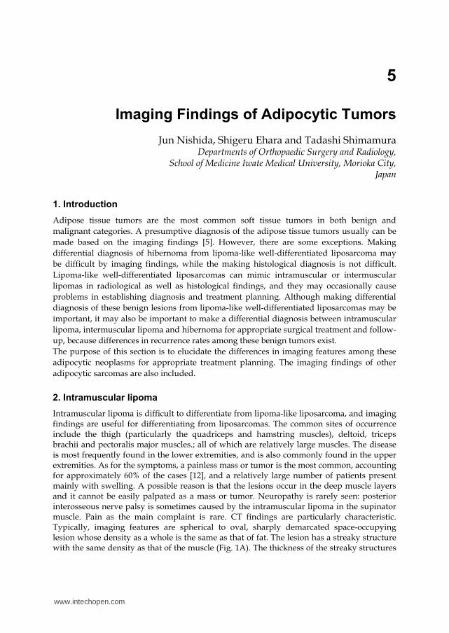
Intramuscular lipoma is difficult to differentiate from lipoma-like liposarcoma, and imaging findings are useful for differentiating from liposarcomas. The common sites of occurrence include the thigh (particularly the quadriceps and hamstring muscles), deltoid, triceps brachii and pectoralis major muscles.; all of which are relatively large muscles. The disease is most frequently found in the lower extremities, and is also commonly found in the upper extremities. As for the symptoms, a painless mass or tumor is the most common, accounting for approximately 60% of the cases [12], and a relatively large number of patients present mainly with swelling. A possible reason is that the lesions occur in the deep muscle layers and it cannot be easily palpated as a mass or tumor. Neuropathy is rarely seen: posterior interosseous nerve palsy is sometimes caused by the intramuscular lipoma in the supinator muscle. Pain as the main complaint is rare. CT findings are particularly characteristic. Typically, imaging features are spherical to oval, sharply demarcated space-occupying lesion whose density as a whole is the same as that of fat. The lesion has a streaky structure with the same density as that of the muscle (Fig. 1A). The thickness of the streaky structures
www.intechopen.com
Soft Tissue Tumors
92
varies, and the streaks may be discontinuous. At surgery, intramuscular lipomas are often removed with a part of the surrounding muscle to ensure an adequate surgical margin, because it is difficult to separate the lesion from the surrounding muscle. The reported local recurrence rates vary from 3.0 to 62.5% when intramuscular lipomas are simply removed without attempting wide resection [2, 3, 6, 9]. Therefore, wide resection is recommended to avoid local recurrence. However, such recommendation was made partly to prevent recurrence or dedifferentiation as a result of misdiagnosis of lipoma-like liposarcoma in an era when the accurate imaging diagnosis was not made, and the differences between intramuscular lipomas and well-differentiated liposarcomas were not fully recognized. The streaky structures observed on CT macroscopically and histologically represent muscle fibers involved in the lesion, and are often difficult to separate from the lesion. Although capsule or pseudocapsule is not generally obvious macroscopically, intramuscular lipomas are multilocular elastic soft lesions, similar to normal subcutaneous lipomas. However, in approximately 15% of the cases, no streaks are observed on CT and the lesions have a capsule that allows separating easily from the surrounding muscle tissue at surgery [12]. Macroscopically, intramuscular lipomas are yellowish multilocular lesions with a smooth surface similar to normal lipomas, and have a capsule in some cases. The streaky structures are visualized as low signal intensities on T2-weighted MR images, however, it is believed that these tumors cannot be as clearly detected by MR as by CT, because of the strong high signal intensities in the lesion, representing fat (Fig. 1B).
A B
Fig. 1. A. Computed tomography reveals a fat density mass containing thick streaks with occasional interruption. B. Magnetic resonance imaging (axial T2 weighted image) demonstrates a fat signal mass with streaky structures are in the tibialis anterior muscle.
3. Intermuscular lipoma
Intermuscular lipoma is a benign lesion that literally occurs between deep muscles. The thigh is the most common site of occurrence, and the chest wall, buttock, forearm, upper arm and neck are also commonly involved. A painless mass is often observed as a presenting symptom, but only a few patients present with the chief complaint of swelling,
www.intechopen.com
Imaging Findings of Adipocytic Tumors
93
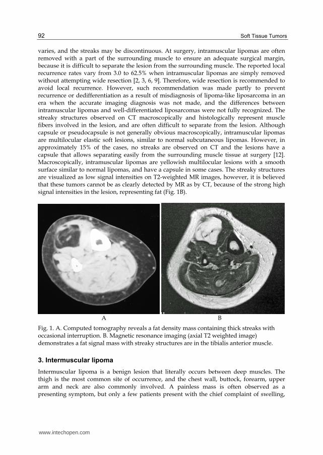
since the lesion arises between muscles and extend into regions of low tissue pressure while growing; it is often recognized as mass on the body surface. Patients rarely complain of pain. On CT, intermuscular lipomas are most often visualized as space-occupying lesions with the same density as that of fat, a dumb-bell or gourd shaped, with a narrow part, and the streakiness inside the lesions are characteristic (Fig. 2A). However, such streaky structures are thinner and more continuous and have smoother curves than those in intramuscular lipomas in many cases. Similar findings are also seen on MR imaging, but the streaky structures visualized as low signal intensities on T2-weighted images may not be so prominent due to the high signal intensity of the whole lesion, similar to intramuscular lipomas (Fig. 2B). At surgery, intermuscular lipomas can be easily manually separated from the surrounding tissues. The streaky structure on CT is fibrous tissue in the fascia and inter-tissue spaces, and can be easily detached by lifting from the margin of the lesion. Intermuscular lipomas are encapsulated and marginal resection is easily performed. Both the macroscopic and histological appearances are similar to those of normal lipomas and no muscle tissue is observed. Basically recurrence or complications are not common.
A B
Fig. 2. A. Computed tomography reveals a dumb-bell-shaped fat density tumor containing
thin streaky densities in intermuscular lipoma of the forearm. B. Magnetic resonance
imaging of the intermuscular lipoma (axial T2 weighted image) reveals a high signal
intensity mass. The streaky structures are seen again but are less distinctive than on
computed tomography.
4. Hibernoma
Hibernoma is a lesion showing proliferation of adipocytes similar to those in brown fat in
hibernators. In humans, brown fat usually exists in the neck, upper back, retroperitoneum,
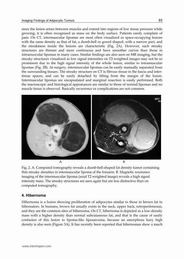
and they are the common sites of hibernoma. On CT, hibernoma is depicted as a low-density
mass with a higher density than normal subcutaneous fat, and that is the cause of easily
confusion of this lesion to lipoma-like liposarcoma, because an amorphous hazy high
density is also seen (Figure 3A). It has recently been reported that hibernomas show a much
www.intechopen.com
Soft Tissue Tumors
94
higher degree of accumulation than liposarcomas on PET images (Figure 3B), and PET is
currently considered as a major modality for differentiation between benignity and
malignancy [13]. Histologically, brown adipocytes are characteristic.
A B
Fig. 3. A. Axial slice of computed tomography of a case with hibernoma demonstrated a fat density lesion with areas of hazy amorphous density in the medial compartment of the pelvis. B. Coronal FDG PET showed the extremely intense uptake (SUV : 98.1). Used with permission from Med Sci Monit [13].
5. Well-differentiated liposarcoma
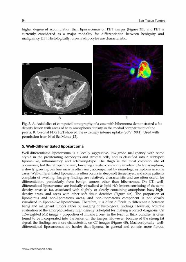
Well-differentiated liposarcoma is a locally aggressive, low-grade malignancy with some atypia in the proliferating adipocytes and stromal cells, and is classified into 3 subtypes: lipoma-like, inflammatory and sclerosing-type. The thigh is the most common site of occurrence, but the retroperitoneum, lower leg are also commonly involved. As for symptoms, a slowly growing painless mass is often seen, accompanied by neurologic symptoms in some cases. Well-differentiated liposarcoma often occurs in deep soft tissue layer, and some patients complain of swelling. Imaging findings are relatively characteristic and are often useful for differentiation, particularly from benign tumors other than hibernomas. On CT, well-differentiated liposarcomas are basically visualized as lipid-rich lesions consisting of the same density areas as fat, associated with slightly or clearly containing amorphous hazy high-density areas, and areas with other soft tissue densities (Figure 4A). The proportion of lipomatous and non-lipomatous areas, and non-lipomatous component is not clearly visualized in lipoma-like liposarcoma. Therefore, it is often difficult to differentiate between benig and malignant tumors either by imaging or histologocal findings. However, accurate evaluation of the amorphous-hazy high density is helpful for making a correct diagnosis. On T2-weighted MR image a proportion of muscle fibers, in the form of thick bundles, is often found to be incorporated into the lesion on the images. However, because of the strong fat signal, the findings are more characteristic on CT images (Figure 4B). Macroscopically, well-differentiated liposarcomas are harder than lipomas in general and contain more fibrous
www.intechopen.com
Imaging Findings of Adipocytic Tumors
95
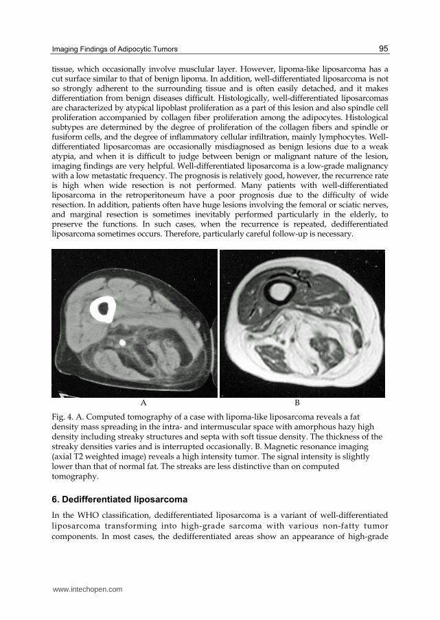
tissue, which occasionally involve musclular layer. However, lipoma-like liposarcoma has a cut surface similar to that of benign lipoma. In addition, well-differentiated liposarcoma is not so strongly adherent to the surrounding tissue and is often easily detached, and it makes differentiation from benign diseases difficult. Histologically, well-differentiated liposarcomas are characterized by atypical lipoblast proliferation as a part of this lesion and also spindle cell proliferation accompanied by collagen fiber proliferation among the adipocytes. Histological subtypes are determined by the degree of proliferation of the collagen fibers and spindle or fusiform cells, and the degree of inflammatory cellular infiltration, mainly lymphocytes. Well-differentiated liposarcomas are occasionally misdiagnosed as benign lesions due to a weak atypia, and when it is difficult to judge between benign or malignant nature of the lesion, imaging findings are very helpful. Well-differentiated liposarcoma is a low-grade malignancy with a low metastatic frequency. The prognosis is relatively good, however, the recurrence rate is high when wide resection is not performed. Many patients with well-differentiated liposarcoma in the retroperitoneum have a poor prognosis due to the difficulty of wide resection. In addition, patients often have huge lesions involving the femoral or sciatic nerves, and marginal resection is sometimes inevitably performed particularly in the elderly, to preserve the functions. In such cases, when the recurrence is repeated, dedifferentiated liposarcoma sometimes occurs. Therefore, particularly careful follow-up is necessary.
A B
Fig. 4. A. Computed tomography of a case with lipoma-like liposarcoma reveals a fat density mass spreading in the intra- and intermuscular space with amorphous hazy high density including streaky structures and septa with soft tissue density. The thickness of the streaky densities varies and is interrupted occasionally. B. Magnetic resonance imaging (axial T2 weighted image) reveals a high intensity tumor. The signal intensity is slightly lower than that of normal fat. The streaks are less distinctive than on computed tomography.
6. Dedifferentiated liposarcoma
In the WHO classification, dedifferentiated liposarcoma is a variant of well-differentiated
liposarcoma transforming into high-grade sarcoma with various non-fatty tumor
components. In most cases, the dedifferentiated areas show an appearance of high-grade
www.intechopen.com
Soft Tissue Tumors
96
malignant fibrous histiocytoma or fibrosarcoma. However, it has been reported that when
the primary lesions were reassessed after development of the dedifferentiated liposarcoma,
some patients had higher-grade malignant components than those initially seen. Thus, the
precise pathology cannot be established initially in some cases with dedifferentiated
liposarcoma. It is generally considered that the occurrence of dedifferentiation liposarcoma
is time-dependent and that approximately 10% of well-differentiated liposarcomas
dedifferentiate after at least a few years of progression. Dedifferentiated liposarcoma is
often found in the retroperitoneum, and the reason is considered to be that well-
differentiated liposarcomas in the retroperitoneum cannot be easily detected and cannot be
radically resected, and lesions in the retroperitoneum are often found incidentally. In many
cases of dedifferentiated liposarcoma in the extremities, a painless mass or tumor present for
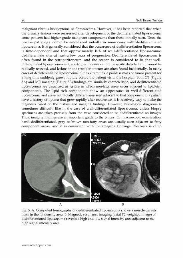
a long time suddenly grows rapidly before the patient visits the hospital. Both CT (Figure
5A) and MR imaging (Figure 5B) findings are similarly characteristic, and dedifferentiated
liposarcomas are visualized as lesions in which non-fatty areas occur adjacent to lipid-rich
components. The lipid-rich components show an appearance of well-differentiated
liposarcoma, and areas with totally different area seen adjacent to that component. If a patient
have a history of lipoma that grew rapidly after recurrence, it is relatively easy to make the
diagnosis based on the history and imaging findings. However, histological diagnosis is
sometimes difficult, like in the case of well-differentiated liposarcoma, unless biopsy
specimens are taken precisely from the areas considered to be dedifferentiated on images.
Thus, imaging findings are an important guide to the biopsy. On macroscopic examination,
hard, dedifferentiated, gray to brown non-fatty areas are usually seen adjacent to fatty
component areas, and it is consistent with the imaging findings. Necrosis is often
A B
Fig. 5. A. Computed tomography of dedifferentiated liposarcoma shows a muscle density mass in the fat density area. B. Magnetic resonance imaging (axial T2 weighted image) of dedifferentiated liposarcoma reveals a high and low signal intensity area adjacent to the high signal intensity area.
www.intechopen.com
Imaging Findings of Adipocytic Tumors
97
observed in dedifferentiated areas. Histologically, high-grade malignant component with clearly distinct histology are observed adjacent to areas of well-differentiated liposarcoma. The border between these areas is usually clear. On histological evaluation, major part of the dedifferentiated areas shows the appearances of malignant fibrous histiocytoma and fibrosarcoma, and various other features, including those of rhabdomyosarcoma and osteosarcoma, can also be observed depending on the case. Dedifferentiated liposarcoma is a high-grade malignancy and requires wide resection. Many patients with dedifferentiated liposarcoma of the retroperitoneum, a common site of occurrence, have a poor prognosis, because wide resection is not possible. However, despite the high-grade histological malignancy in the dedifferentiated areas, the probability of metastasis is not extremely high. The 5-year survival rate is approximately 70%, which is perhaps higher than the supposed rate, considering the histological malignancy and common sites of occurrence [7, 10, 16]. Nevertheless, the 10-year survival is considered to be poor.
7. Myxoid/round cell liposarcoma
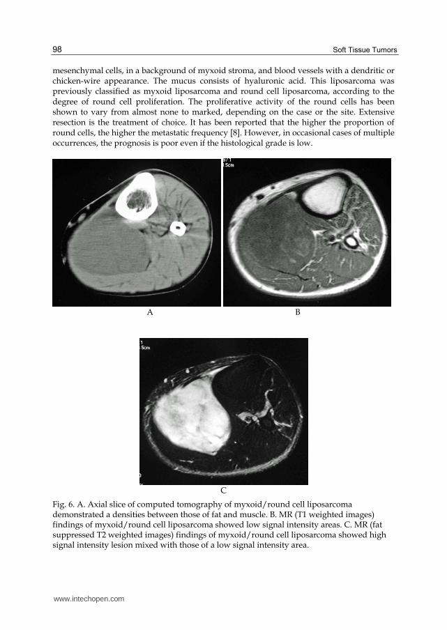
According to the WHO definition, myxoid/round cell liposarcoma is a disease in which round to oval malignant mesenchymal cells proliferate in a background of proliferating signet ring-like lipoblasts and myxoid stroma. In this 2002 WHO classification, myxoid liposarcoma and round cell liposarcoma are dealt with as the same category. It has been estimated that distant metastases are seen in approximately one-third of the cases. The prognosis varies significantly depending on the histological findings: the percentage of the total area occupied by round cells of greater than 5%, the presence of a necrotic area, and overexpression of p53: all are considered to be poor-prognostic factors [1, 8, 15]. Myxoid/round cell liposarcoma often occurs in the deep layer of the extremities, and the thigh is the most commonly involved site, accounting for approximately two-thirds of all cases. Unlike other liposarcomas, myxoid/round cell liposarcoma rarely occurs in the retroperitoneum. A slowly growing, painless mass is the most common presenting complaint. In addition, as compared to other liposarcomas, myxoid/round cell liposarcoma occurs more often in younger age groups, particularly those in their 30s to 40s, and it is the most common liposarcoma seen in patients younger than 20 years. In imaging findings, myxoid/round cell liposarcoma is basically characterized by a strongly represented mucous stroma, but imaging modalities show the appearance of non-specific soft tissue tumor when the proliferation of the round cells is predominant. On CT, myxoid/round cell liposarcomas show densities between those of fat and muscle, with lipid-rich areas having a density similar to that of fat and areas with densely proliferating cells having a density similar to that of muscle (Fig. 6A). Myxoid/round cell liposarcomas are visualized as low signal intensities on T1-weighted MR images (Fig. 6B) and as high signal intensities on T2-weighted images, and it also shows areas of high T2-weighted signal intensity mixed with those of a low signal intensity, reflecting the degree of proliferation of the fatty component (Fig. 6C). These findings make differenitation difficult from other mucous tumors, including myxoma, extraskeletal myxoid chondrosarcoma and myxofibrosarcoma. Macroscopically, myxoid/round cell liposarcomas are solid multilocular tumors with a relatively clear border. Myxoid/round cell liposarcomas are characterized by a myxoid cut surface, however, the myxoid stroma is sometimes not macroscopically visible in cases with predominant round cell proliferation. Histologically, myxoid/round cell liposarcomas are characterized by the proliferation of signet ring-like lipoblasts, round to oval malignant
www.intechopen.com
Soft Tissue Tumors
98
mesenchymal cells, in a background of myxoid stroma, and blood vessels with a dendritic or chicken-wire appearance. The mucus consists of hyaluronic acid. This liposarcoma was previously classified as myxoid liposarcoma and round cell liposarcoma, according to the degree of round cell proliferation. The proliferative activity of the round cells has been shown to vary from almost none to marked, depending on the case or the site. Extensive resection is the treatment of choice. It has been reported that the higher the proportion of round cells, the higher the metastatic frequency [8]. However, in occasional cases of multiple occurrences, the prognosis is poor even if the histological grade is low.
A B
C
Fig. 6. A. Axial slice of computed tomography of myxoid/round cell liposarcoma demonstrated a densities between those of fat and muscle. B. MR (T1 weighted images) findings of myxoid/round cell liposarcoma showed low signal intensity areas. C. MR (fat suppressed T2 weighted images) findings of myxoid/round cell liposarcoma showed high signal intensity lesion mixed with those of a low signal intensity area.
www.intechopen.com
Imaging Findings of Adipocytic Tumors
99
8. Pleomorphic liposarcoma
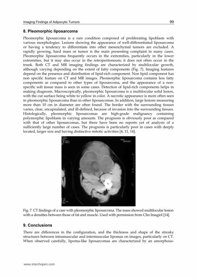
Pleomorphic liposarcoma is a rare condition composed of proliferating lipoblasts with various morphologies. Lesions showing the appearance of well-differentiated liposarcoma or having a tendency to differentiate into other mesenchymal tumors are excluded. A rapidly growing, hard mass or tumor is the main presenting complaint in many cases. Pleomorphic liposarcoma frequently occurs in the extremities, particularly in the lower extremities, but it may also occur in the retroperitoneum; it does not often occur in the trunk. Both CT and MR imaging findings are characterized by multilocular growth, although varying depending on the extent of fatty components (Fig. 7). Imaging features depend on the presence and distribution of lipid-rich component. Non lipid component has non specific feature on CT and MR images. Pleomorphic liposarcoma contains less fatty components as compared to other types of liposarcoma, and the appearance of a non-specific soft tissue mass is seen in some cases. Detection of lipid-rich components helps in making diagnosis. Macroscopically, pleomorphic liposarcoma is a multilocular solid lesion, with the cut surface being white to yellow in color. A necrotic appearance is more often seen in pleomorphic liposarcoma than in other liposarcomas. In addition, large lesions measuring more than 10 cm in diameter are often found. The border with the surrounding tissues varies, clear, encapsulated, and ill-defined, because of invasion into the surrounding tissues. Histologically, pleomorphic liposarcomas are high-grade malignancy containing polymorphic lipoblasts in varying amounts. The prognosis is obviously poor as compared with that of other liposarcomas, but there have been no reports yet of analysis of a sufficiently large number of cases. The prognosis is particularly poor in cases with deeply located, larger size and having distinctive mitotic activities [4, 11, 14].
Fig. 7. CT findings of a case with pleomorphic liposarcoma. The mass showed multilocular lesion with a densities between those of fat and muscle. Used with permission from Clin Imagiol [14].
9. Conclusions
There are differences in the configuration, and the thickness and shape of the streaky structures between intramuscular and intermuscular lipomas on images, particularly on CT. When observed carefully, lipoma-like liposarcomas are characterized by an amorphous-
www.intechopen.com
Soft Tissue Tumors
100
hazy high density and show invasive, spherical expansion into the surrounding tissues in many cases. Imaging findings of well-differentiated liposarcomas are clearly different from those of benign lesions, and differentiation is usually possible on imaging features. Clinical or imaging differentiation of high-grade malignancy, including dedifferentiated liposarcoma, from benign lesions is often possible. When performing biopsy, the imaging guidance for establishing a precise diagnosis is important.
10. References
[1] Antonescu CR, et al. Prognostic impact of P53 status, TLS-CHOP fusion transcript structure, and histological grade in myxoid liposarcoma: a molecular and clinicopathologic stud of 82 cases. Clin Cancer Res 7: 3977-3987, 2001.
[2] Bjerregaard P, et al. Intramuscular lipoma of the lower limb. J Bone Joint Surg 71-B: 812-5, 1989.
[3] Dionne G P, et al. Infiltrating lipomas and angiolipomas revisited. Cancer 33: 732-8, 1974. [4] Downes KA, et al. Pleomorphic liposarcoma. Clinicopathologic study of 19 cases. Mod
Pathol 14. 179-184, 2001. [5] Ehara S, et al. Atypical lipomas, liposarcomas, and other fat-containing sarcomas. CT
analysis of fat element. Clin Imaging 19: 50-3, 1995. [6] Fletcher C D, et al. Martin-Bates E. Intra-muscular and intermuscular lipoma: neglected
diagnoses. Histopathology 12: 275-87, 1988. [7] Henricks WH, et al. Dedifferentiated liposarcoma: a clinicopathological analysis of 155
cases with proposal for an expanded definition of differentiation. Am J Surg Pathol 21, 271-281, 1997.
[8] Kilpatrick SE, et al. The clinicopathologic spectrum of myxoid and round cell liposarcoma. A study of 95 cases. Cancer 77 1450-1458 1996.
[9] Kindblom L G, et al. Intermuscular and inter-muscular lipomas and hibernomas. A clinical, roentgenologic, histologic, and prognostic study of 46 cases. Cancer 33: 754-62, 1974.
[10] McCormic D, et al. Dedifferentiated liposarcoma. A clinico-pathologic analysis of 32 cases suggesting a better prognostic subgroup among pleomorphic sarcomas. Am J Surg Pathol 18, 1213-2123, 1997.
[11] Miettinen M, et al. Epithelioid variant of pleomorphic liposarcoma. Study of 12 cases of distinctive variant of high-grade liposaecoma. Mod Pathol 12: 722-728, 1999.
[12] Nishida J, et al. Imaging characteritics of the deep-seated lipomatous tumors: Intramuscular lipoma, intermuscular lipoma and lipoma-like liposarcoma. J.Orthop. Sci 12: 533-541, 2007.
[13] Nishida J, et al. Clinical findings of Hibernoma of the buttock and thigh: Rare involvements and extremely high uptake of FDG-PET. Med Sci Monit 15. CS117 –122, 2009.
[14] Nishida J, et al. Transition of the concept of adipose tissue tumors and imaging diagnosis. Clinical Imagiol. 25. 54 –61, 2009.
[15] Smith TA, et al. Myxoid /round cell liposarcoma of the extremities. A clinicopathologic study of 29 cases with particular attention to extent of round cell liposarcoma. Am J Surg Pathol 20, 171-180, 1996.
[16] Weiss SW, et al. Well-differentiated liposarcoma (atypical lipoma) of deep soft tissue of the extremities, retroperitoneum, and miscellaneous sites. A follow-up study of 92 cases with analysis of incidence of “dedifferentiation”. Am J Surg Pathol 16, 1051-1058, 1997.
www.intechopen.com

Soft Tissue TumorsEdited by Prof. Fethi Derbel
ISBN 978-953-307-862-5Hard cover, 270 pagesPublisher InTechPublished online 16, November, 2011Published in print edition November, 2011
InTech EuropeUniversity Campus STeP Ri Slavka Krautzeka 83/A 51000 Rijeka, Croatia Phone: +385 (51) 770 447 Fax: +385 (51) 686 166www.intechopen.com
InTech ChinaUnit 405, Office Block, Hotel Equatorial Shanghai No.65, Yan An Road (West), Shanghai, 200040, China
Phone: +86-21-62489820 Fax: +86-21-62489821
Soft tissue tumors include a heterogeneous group of diagnostic entities, most of them benign in nature andbehavior. Malignant entities, soft tissue sarcomas, are rare tumors that account for1% of all malignancies.These are predominantly tumors of adults, but 15% arise in children and adolescents. The wide biologicaldiversity of soft tissue tumors, combined with their high incidence and potential morbidity and mortalityrepresent challenges to contemporary researches, both at the level of basic and clinical science. Determiningwhether a soft tissue mass is benign or malignant is vital for appropriate management. This book is the resultof collaboration between several authors, experts in their fields; they succeeded in translating the complexity ofsoft tissue tumors and the diversity in the diagnosis and management of these tumors.
How to referenceIn order to correctly reference this scholarly work, feel free to copy and paste the following:
Jun Nishida, Shigeru Ehara and Tadashi Shimamura (2011). Imaging Findings of Adipocytic Tumors, SoftTissue Tumors, Prof. Fethi Derbel (Ed.), ISBN: 978-953-307-862-5, InTech, Available from:http://www.intechopen.com/books/soft-tissue-tumors/imaging-findings-of-adipocytic-tumors

© 2011 The Author(s). Licensee IntechOpen. This is an open access articledistributed under the terms of the Creative Commons Attribution 3.0License, which permits unrestricted use, distribution, and reproduction inany medium, provided the original work is properly cited.