imaging findings of focal and multiple cystic and cavitary ... · fokal ve multipl kistik ve...
TRANSCRIPT
Imaging Findings of Focal and Multiple Cystic and Cavitary Lung Lesions
Abstract
Cystic and cavitary lung lesions constitute a spectrum of pulmonary diseases diagnosed in both children and adults. Cysts and cavities are commonly encountered abnormalies on chest radiography and chest computed tomography. High-resolution computed tomography (HRCT) of the chest frequently helps define morphologic features that may serve as important clues regarding the nature of cystic and cavitary lesions of the lung. Occasionally, the underlying nature of the lesions can be readily apparent as in bullae associated with emphysema. Cystic and cavitary lung lesions can be diagnostic challange. Although many patients with cystic and cavitary lung lesions have a known un-derlying disease, in many cases the considerable overlap in morpho-logic features of these lesions tenders transthoracic needle biopsy necessary to establish the correct diagnosis.
Key words: Radiography, CT scan, cyst, lung, cavitary
Fokal ve Multipl Kistik ve Kaviteli Akciğer Lezyonlarında Radyolojik Bulgular
Özet:
Kistik ve kaviteli akciğer lezyonları çocuklarda ve yetişkinlerde görülen akciğer hastalıklarının bir spektrumunu oluşturur. Kistler ve kaviteler akciğer radyografisinde ve bilgisayarlı toraks tomografisinde yaygın olarak görülen lezyonlardır. Yüksek çözünürlüklü bilgisayarlı tomografi (YÇBT) sıklıkla akciğerin kistik ve kaviteli lezyonları hakkında önemli ipuçları vererek morfolojik özelliklerini tanımlamaya yardım eder. Bazen, amfizem ile birlikte bül gibi, lezyonların altta yatan nedeni açıkça görülebilmektedir. Kistik ve kaviteli akciğer lezyonlarında tanısal zorluklar halen devam etmektedir. Kistik ve kaviteli akciğer lezyonları bulunan birçok olguda altta yatan bilinen bir sebep olmakla birlikte, birçoğunda bu lezyonların morfolojik özelliklerinde belirgin örtüşme olması nedeniyle doğru tanıyı koymak için transtorasik iğne biyopisisi gerekmektedir.
Anahtar sözcükler: Radyografi, BT, kist, akciğer, kavite,
Konya University, Meram Medical Faculty, Department of Radiology, Konya, Turkey
Eur J Gen Med 2012;9 (Suppl 1):3-14
Received: 02.12.2011
Accepted: 17.01.2012
Correspondence: Dr. Kemal ÖdevKonya University, Meram Faculty of Medi-cine, Department of Radiology, Konya.TurkeyE-mail: [email protected]
Kemal Ödev, Hüseyin Özbiner
European Journal of General Medicine
Review Article

Eur J Gen Med 2012;9(Suppl 1):3-14
Imaging findings of cystic lung lesions
4
INTRODUCTION
The cysts and cavities are seen as foci of decreased lung density with definable walls (1). In contrast, emphysema-tous airspaces usually lack such perceptible walls (bullae and blebs are expection). The presence or absence of a wall around a radiolucent area can be accurately depict-ed by high resolution computed tomography (HRCT), if it is not apparent on chest radiography. The term cyst, cys-tic air space and cavity have overlapping meanings and sometimes used interchangeably. The terms cyst and cav-ity convey different meanings and ranges of diagnostic possibilities to clinicians (1). In the literature, the term cyst is used to mean a clearly defined air-containing space surrounded by a relatively thin (≤4 mm) wall. In contrast, the term cavity is used to refer to an air-containing le-sions with a relatively thick (≥4 mm) wall or within an area of a surrounding infiltrate or mass. Cystic lesions in the lungs are rarely malignant. However, malignancy is commonly the first diagnosis to consider for a cavitary lesion, particularly in a middle-aged or oder adult with a history of cigarette smoking. Some cystic and cavitary le-sions may be filled with fluid or solid contents. For exam-ple, a bronchogenic cyst may be filled with fluid and ap-pear as a mass lesion on chest radiography. The presence of an air-fluid level does not correlate well with benignity or malignancy (1). Solid contents within a cavity may be seen in infectious processes (e.g. invasive aspergillosis), and in necrotic tumors. The location of focal lesions may be of help in narrowing the differental diagnosis (e.g. propensity of tuberculosis to affect the upper lobes of
the lung) (1). Chest radiograph remains the first imaging technique in the evaluation of cystic and cavitary lung lesions. However, computed tomography (CT) and high resolution CT (HRCT) can show the size, shape and pre-cise location of cysts and cavities when these lesions are not apparent on chest radiography. The purpose of this review is to describe the characteristic radiologic findings of focal and multiple lung cystic and cavitary lesions.
Congenital abnormalities
Congenital lobar emphysema
Lobar emphysema can be either acquired, or secondary or congenital. CLE refers to progressive overinflation of a pulmonary lobe secondary to air trapping clinically, most infants with CLE present within the first 6 months of life, with symptoms and signs of respiratory distress. Bilateral or multifocal involvement is rare. With severe overdis-tention of a lobe, contralateral lober compression results and there is cardiomediastinal shift (Figure 1). CT scan demonstrates which lobes or segments are involved. The affected lobe is overdistended and hypodense, with at-tenuated vascular markings (Figure 1) (1,2).
Bronchopulmonary dysplasia and Wilson-Mikity syn-drome
In the neonatal period, several disease entities, such as bronchopulmonary dysplasia and Wilson-Mikity syndrome are associated with respiratory distress. Bronchopulmonary dysplasia results infantile respiratory distress syndrome and respiratory therapy with high oxy-
Figure 1. Congenital lobar emphysema. CT scan shows a largehyperlusent area in the left hemithorax. There is marked overinflation in the left hemitoraks and the mediastinal structures are shifted to the right side and the lower lobe is compressed
Figure 2. Type I congenital cystic adenomatoid malfor-mation a.chest radiograph shows thin walled cysts with multiple internal septa expand left lung and displace diaphragm and mediastinum b.CT scan shows replace-ment of the left lung by cystic lesions of varying sizes.
a b
Ödev and Özbiner
Eur J Gen Med 2012;9(Suppl 1):3-14 5
gen concentrations. Multiple lung cysts reflect air trap-ping due to broncholitis or areas of destroyed lung sur-rounded by fibrous tissue. Lesions are typically bilateral. Wilson-Mikity syndrome or pulmonary dysmaturity , has a radiologic appearance similar to that of bronchopulmo-nary dysplasia, but is not associated with infantile respi-ratory distress syndrome (3).
Congenital cystic adenomatoid malformation of the lung
Congenital cystic adenomatoid malformation of the lung (CCAM) is an uncommon developmental abnormal-
ity. CCAM is usually discovered in neonates, because of respiratory distress and may occasionally be discovered in older children or adults who have recurrent infec-tion (2-4). Stocker et al. (5) classified into three types based on clinical, gross and microscopic criteria. In type 1 CCAM, lesions consist of multipl large cysts (2-10 cm in diameter). Type 2 lesions have numerous smaller, more uniform-sized cysts (0.5-2 cm in diameter, rarely larger). Type 3 lesions are bulky solid lesions that usually involve an entire lobe or lung (2,4,6). Typical radiographic find-ing in patients with CCAM is a large air-filled multicys-tic lesion. However, variable radiographic features can
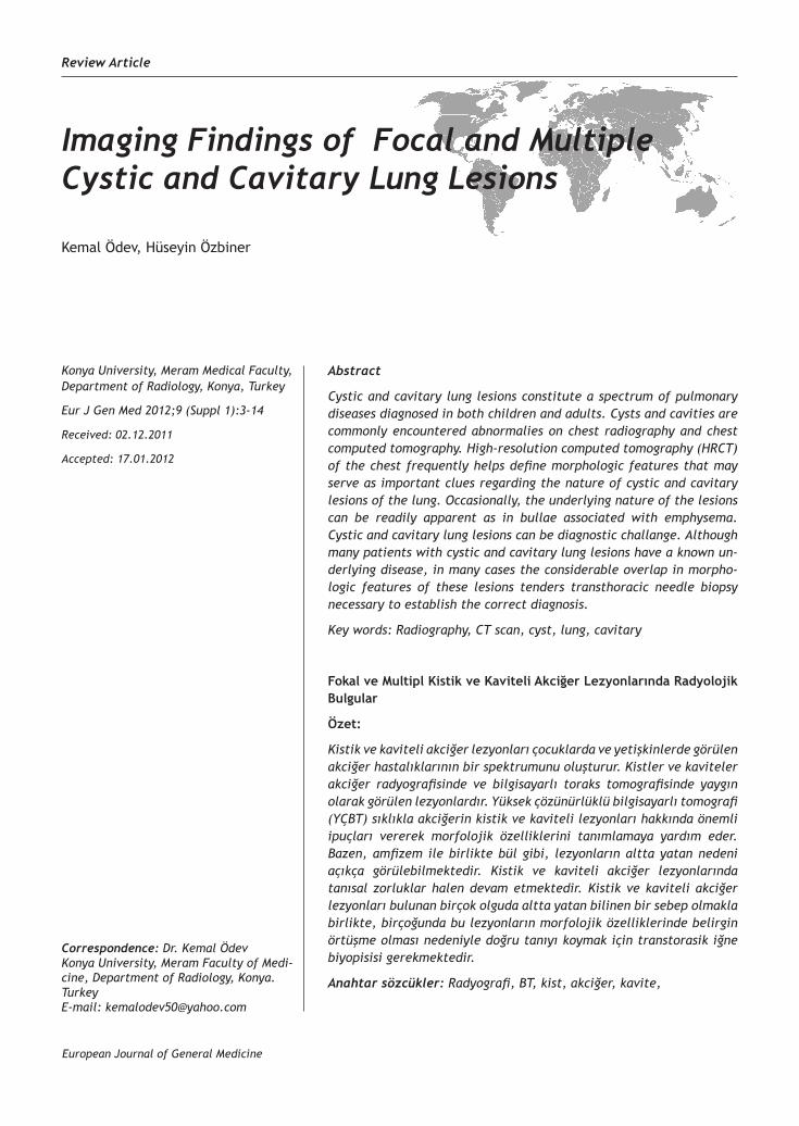
Figure 3. Bronchogenic cyst a.Chest radiograph shows a bronchogenic cyst in theleft lung with an air filled level b.CT scan (lungwindow) shows a unilocular air-filled bronchogenic cyst on the left lung
Figure 4. İntralobar sequestration in a child with recur-rent pneumonia in the lower lobe. a.CT scan shows a large multicystic mass b. The arterial supply to the sequastered segment (arrow) arises from the descend-ing aorta as demonstrated on the surface rendered 3-D image
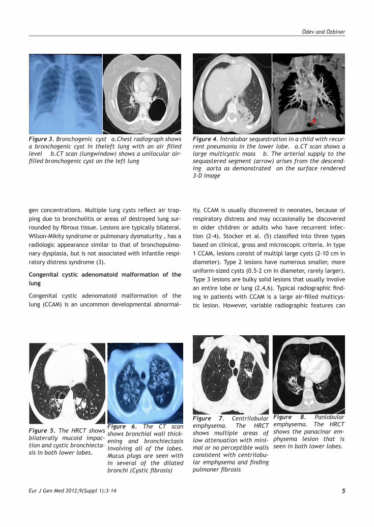
Figure 5. The HRCT shows bilaterally mucoid impac-tion and cystic bronchiecta-sis in both lower lobes.
Figure 6. The CT scan shows bronchial wall thick-ening and bronchiectasis involving all of the lobes. Mucus plugs are seen with in several of the dilated bronchi (Cystic fibrosis)
Figure 7. Centrilobular emphysema. The HRCT shows multiple areas of low attenuation with mini-mal or no perceptible walls consistent with centrilobu-lar emphysema and finding pulmoner fibrosis
Figure 8. Panlobular emphysema. The HRCT shows the panacinar em-physema lesion that is seen in both lower lobes.

Eur J Gen Med 2012;9(Suppl 1):3-146
Imaging findings of cystic lung lesions
make diagnosis difficult (Figure 2). CT demonstrates the air-filled or fluid-filled cystic lesions with thin walls and air-fluid levelsin cysts or a combination of these findings (Figure 2).
Bronchogenic cysts
Bronchogenic cysts are found most frequently in the me-diastinum or lungs, but they can develop in extrathoracic locations, such as neck, pericardium or abdominal cavity (2,4). On chest radiography, an intrapulmonary broncho-genic cyst may appear as well-defined, noncalcified lung mass with water density or air-space cysts with air-fluid levels (Figure 3). CT often demonstrates a cyst not visible by conventional radiographic techniques. CT density of bronchogenic cysts can vary from typical water density (0 to 20 HU) to high density (89 to 99 HU). CT provides
optimal demonstration of cyst location, morphology and contents (Figure 3) (7,8).
Pulmonary sequestration
Classically, two forms of pulmonary sequestration (PS) have been described: intralober (ILPS) and extralober (ELPS). ILPS is a segment of pulmonary tissue that shares the visceral pleural covering of normal adjacent lung tis-sue and are usually located in the posterobasal portion of the lower lobes. The arterial supply is typically from tho-racic or upper abdominal aorta and most drain by way of the pulmonary venous system to the left atrium. ELPS is entirely seperate segment of pulmonary tissue that is in-vested in its own pleural layers. ELPS may also be located in the mediastinum, pericardium and within or below the hemidiaphragm (2,9). The abdominal or thoracic aorta
Figure 9. Distal acinar em-physema. TheHRCT shows distal acinar and central acinar emphysema in both lungs.
Figure 10. Bullous emphy-sema. The CT scan shows a large bullae in the left lung and absence of em-physema in the lung.
Figure 12. The honeycomb-ing in idiopathic pulmonary fibrosis. The HRCT shows honeycomb cysts with a dis-tinct predominance in the-peripheral and subpleural regions.
Figure 11. The HRCT shows a large bullae on the right and presence of of distal acinar emphy-sema in both lungs.
Figure 13. Wegener granulamatosis. a;The CT shows a large cavitating mass associated with consolidation in the right upper lobe. b; Cavitating nodules in the left subpleural region.
Figure 14. Staphylococcal pneumonia. The CT scan shows multiple cavitation in the right and left lobes
a b

Ödev and Özbiner
Eur J Gen Med 2012;9(Suppl 1):3-14 7
usually supplies the arterial vessels. In 80 % of cases, ve-nous drainage is to the systemic circulation , usually by way of azygous system or IVC. It is difficult to distinguish between an ILPS and ELPSs on plain raidograph alone. CT or MRI provide a more gobal assessment of sequestration and are in general, recommended for full evaluation. CT images shows an ILPS as a more irregular outline and also a heterogenous appearance. Cysts and cavities with air-filled levels may be seen (Figure 4). CT images show an ELPS as a homogenous, well-circumscribed areas of soft tissue attenuation (2). With its multiplanar capability, MR imaging can successfully demonstrate the systemic feed-ing vessels of an intralober sequestration (Figure 4). MR imaging can also depict the pulmonary venous return of the lesion and the relationship of the draining vein to the cardiac chambers. MR imaging can also reveal the cystic nature of many intralober sequestrations, as well as the
variable solid, fluid, hemorrhagic and mucus-containing components (9).
Airway abnormalities
Bronchiectasis
Bronchiectasis is a disease process characterized by ab-normally dilated bronchi with thickened bronchial walls, and which has a number of potential underlying causes. Ninety percent of bronchiectatic patients have some ab-normalities on chest radiograph as follows: focal opacifi-cations, scattered irregular opacities, linear or plate-like atelectasis or dilated and thickened airways that appear as ring-like shadows (10) (Figure 5). HRCT has now be-come the best tool for diagnosis. Typical findings on chest CT scan tend to correlate with type of bronchiectasis present (e.g. cylindrical, saccular or varicose), although
Figure 15. Pneumatocele formation in staphylococcal pneumonia a-b .The CT scan show multiple pneumoto-celes with thin walled in right lower lobe pneumonia.
Figure 16. Pulmonary abscess. The CT scan shows con-solidation with cavitation and air- fluid level in the right lobe and cavitating lesion on the left.
Figure 17. Active cavitary pulmonary tuberculosis.a. The HRCT shows an irregular small and large thick-walled cavity in the left upper lobe.b. CT scan obtained at level of carina shows thicked walled cavities associ-ated with segmental consolidation and scattered nod-ules and clusters of nodules that are typical of endo-broncial spread of infection (three-in-bud appearence)
Figure 18. Cystic pneumocystis carinii pneumonia.The HRCT shows poorly marginated air-space nodules and foci of graund glass opasification scattered through out the upper lobes and small thin-walled cavities.
a b
a b

Eur J Gen Med 2012;9(Suppl 1):3-14
Imaging findings of cystic lung lesions
8
patterns may be mixed. HRCT findings of bronchiectasis include increased bronchoarterial ratio, lack of appropri-ate airway tapering, bronchial wall thickening and irregu-larity, mucoid impaction and mosaic perfusion with air trapping (Figure 5). Bronchial dilatation is considered to be present when the bronchus is larger than the adjacent pulmonary artery (signet ring sign) or when bronchi are visible within 1 cm of the pleura (10-12).
Cystic fibrosis
Cystic fibrosis is transmitted as an autosomal recessive trait. The CT findings of cystic fibrosis include bronchial wall thickening, bronchiectasis and mucus plugs within the bronchi (Figure 6). The abnormalities are usually most
severe in the upper lobes. The bronchiectasis is most of-ten cylindrical, but varicose and cystic bronchiectasis can be seen in advanced cases (12).
Emphysema
Emphysema is defined as a condition of the lung character-ised by permanent, abnormal enlargement of the airspac-es distal to the terminatal bronchiole, accompanied by destruction of the airspace walls (11,13). The HRCT find-ings of emphysema include centrilobular(centriacinar), panlobular(panacinar) and distal acinar (paraseptal). Centrilobular emphysema (Figure 7) is found most com-monly in the upper lobes and manifests as multiple small areas of low attenuation without a perceptible wall,
Figure 19. Pulmonary aspergilloma (mycetoma) a. Chest radiograph shows pulmonary cavity in the right upper lung surrounded by circumferential pleural thick-ening and containing aspergilloma.b. CT scan shows a right intracavitary aspergilloma with associated pleural thickening.
Figure 20. İnvasive pulmonary aspergillosis with acute leukemia.a.active early lesion of invasive pulmonary aspergillosis with CT halo sign.b. Four months later, le-sion are healed. CT shows residual thin-walled cyst.
Figure 21. Allergic bronchopulmonary aspergillosis in a 50- year -old asthmatic patient a.The HRCT shows fungus ball with in multiple dilated bronchi in the left upper lobe and b. lower lobe.
Figure 22. Hydatid cyst. CT scan shows a hypoatenuat-ing crescent sign (meniscus sign)
a b a b
a b
Ödev and Özbiner
Eur J Gen Med 2012;9(Suppl 1):3-14 9
producing a punched-out appearance. In contrast to centrilobular emphysema, panlobular emphysema is his-topathollogically characterized by complete destruction of the entire pulmonary lobule. On HRCT panlobular em-physema appears as extensive low attenuation that mani-fests as diffuse simplification of pulmonary architecture (Figure 8) and the pulmonary vessels appear stretched and attenuated in the presence of panlobular emphysema (11,13). Distal acinar or paraseptal emphysema appears on HRCT imaging as multiple areas of low attenuation with thin, definable, uniform walls distributed in the sub-plevral regions of lung (Figure 9).
Bullous emphysema
Bullosu emphysema is the most common cause of multi-
ple lung cysts. Bullae can occur in otherwise normal lung (Figure 10), but are often associated with emphysema (Figure 11). Pathologically, bullae are filled spaces lined by pleura, connective tissue septa or emphysematous lung. The walls of bullae appear hair-thin (Figure 11). CT scan may be necessary to demonstrate the walls and es-tablish the presence of bullae (13).
Pulmonary fibrosis and honeycomb lung
In patients with chronic interstitial fibrosis, fibrosis as-sociated with areas of lung destruction and disorganisa-tion of lung architecture (honeycomb) results in a cystic appearance on HRCT scans. That can be characterized and localized precisely (Figure 12). Honeycombing results in cystic spaces several milimeters to several centime-
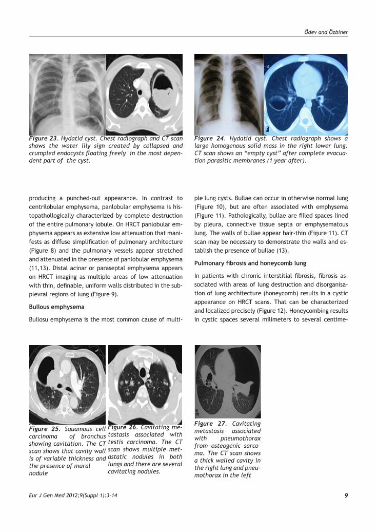
Figure 23. Hydatid cyst. Chest radiograph and CT scan shows the water lily sign created by collapsed and crumpled endocysts floating freely in the most depen-dent part of the cyst.
Figure 24. Hydatid cyst. Chest radiograph shows a large homogenous solid mass in the right lower lung. CT scan shows an “empty cyst” after complete evacua-tion parasitic membranes (1 year after).
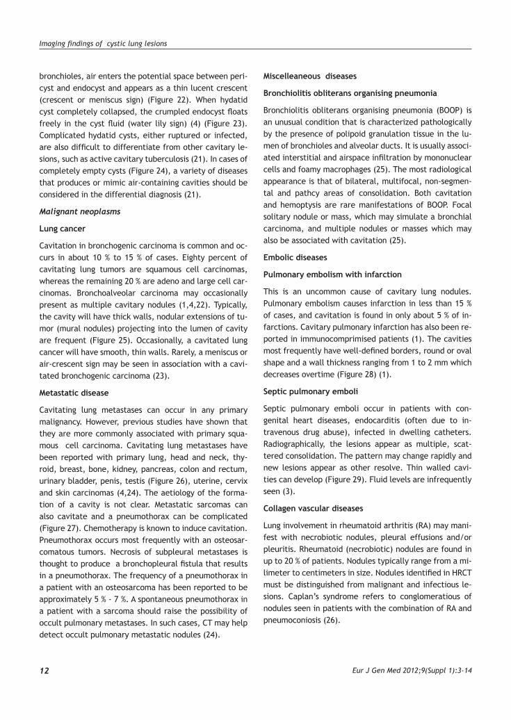
Figure 26. Cavitating me-tastasis associated with testis carcinoma. The CT scan shows multiple met-astatic nodules in both lungs and there are several cavitating nodules.
Figure 25. Squamous cell carcinoma of bronchus showing cavitation. The CT scan shows that cavity wall is of variable thickness and the presence of mural nodule
Figure 27. Cavitating metastasis associated with pneumothorax from osteogenic sarco-ma. The CT scan shows a thick walled cavity in the right lung and pneu-mothorax in the left

Imaging findings of cystic lung lesions
Eur J Gen Med 2012;9(Suppl 1):3-1410
ters in diameter, which are often peripheral in location and are characterized by thick clearly definable walls. Honeycombing often is associated with other findings of fibrosis, such as septal thickening, intralobular lines and irregular pleural thickening (14). Traction bronchiectasis; In patients who have severe lung fibrosis and distortion of lung architecture, so-called traction bronchiectasis may be present. In the presence of severe fibrosis and par-ticularly honeycombing, traction by fibrous tissue on the bronchial walls can result in areas of irregular bronchial dilatation (e.g. bronchiectasis) (14).
Granulomatous diseases
Sarcoidosis
The CT lesions of pulmonary sarcoidosis include nodular
areas of attenuation, linear areas of attenuation, alveo-lar or pseudoalveolar consolidations, irregular interfaces, peribronchovascular thickening, lung distortion, honey-comb cysts, cavitary nodules and traction bronchiectasis (15). CT provides a precise assessment of the pattern and distribution of the disease and it depicts cavitary lesions not shown with chest radiography (4,15).
Wegener’s granulomatosis
Wegener’s granulomatosis (WG) is an idiopathic disease characterized by a granulomatous and necrotizing vascu-litis (16). The CT lesions of pulmonary WG include nod-ules, masses, pulmonary consolidations and ground glass opacities. Nodules are the most frequent parenchymal abnormality. In untreated disease, nodules tend to in-
Figure 29. Septic pulmonary emboli in 30 years old who had bacterial endocarditis. The CT scan shows scat-tered mostly peripheral poorly defined foci of air-space consolidation and multiple round pulmonary cavities with thin walls. Note that a number of these cavities appear to be associated with feeding vessels.
Figure 28. Acute pulmonary embolism with infarction a.Coronal reformation obtained along the main axis of the left pulmonary artery shows the presence of en-doluminal clots within the left pulmonary artery b. Coronal reformat CT reveals the cavitating infarct and residual scarring of pleura in the left lung and mosaic perfusion in both lungs.
Figure 32. Traumatic pseudocyst. The CT scan shows cystic lesions with an air-filled level and some sur-rounding lung contusion in the left lower lobe.
Figure 31. Lymphangi-oleimyomatosis in a 40- year- old woman. The HRCT shows numerous discrete round, thin walled lung cysts.
Figure 30. Langerhans cell histiocytosis. The CT scan shows diffuse thick walled cavitary nodules and small nodular opacities.
a b

Ödev and Özbiner
Eur J Gen Med 2012;9(Suppl 1):3-14 11
crease both in size and number and may cavitate (Figure 13). The cavitary nodules may occasionally become in-fected to give an air-filled level (4,16).
Infections diseases
Infectious diseases due to bacteria, fungi or parasites can result in cystic lesions in the lung. Staphylococcus aureus, Klebsiella and anaerobic bacteria commonly re-sult in the development of thick or thin-walled, air-filled cystic lesions (Figure 14). Pneumatocele are thin-walled, cystic lesions commonly seen in children (Figure 15) and infrequently in adults as a sequelea of Staphylococcal pneumonia (1,4). Gram-negative, anaerobic bacteria and occasionally Streptococcus pneumonia are responsible for the development of lung abscesses. A lung abscess is a thick-walled cavity that contains purulent material re-sulting from a pulmonary infection. It is believed to be less common in children than adults. Secondary lung ab-scesses may be seen in children at increased risk of pul-monary aspiration, immunocompromised hosts and those with underlying localized structural lung abnormalities or generalized suppurative lung disease (17). The character-istic appearance of a lung abscess on a CT image is thick-walled cavity. It contains mobile, central fluid occuring in the middle of an area of consolidated lung. An air-filled level is often appearent on the CT, even when it is not evident on the chest radiograph (Figure 16).
Mycobacterial infection
Two forms of pulmonary tuberculosis (Tb) is known: Primary and post-primary or reactivated tuberculosis which is more frequent in the adult population. The high resolution CT findings of active post primary Tb are cen-trilobular lesions appearing as a nodule or a branching linear structure (tree-in-bud appearance), bronchial wall thickening, a poorly defined nodule, a cavity and lobular consolidation (18). Cavities result when areas of caesa-tion necrosis erode into the bronchial tree, expelling liq-uefied debris. CT is more sensitive than plain radiograph in the detection of small cavities, particularly ones in the apices, lung bases and paramediastinal and retrocardiac locations. On CT scans, cavities due to mycobacterial dis-ease can be thick or thin walled and smooth or irregular with or without air-filled levels (Figure 17) (18). Those caused by atypical mycobacteria are indistinguishable from thosed by caused by M.tuberculosis. Tuberculosis may manifest with different patterns and distribution in patients with diabetes or acquired immunodeficiency syndrome (AIDS) (4).
Pneumocystis carinii pneumonia
Pneumocystis carinii is the commonest cause of pneumo-nia in patients with AIDS, occuring in 60 % to 80 % pa-tients. Radiographic and CT manifestations of P.carinii pneumonia is bilateral, central or basal, reticular or ground glass opacification, which may progress over a few days to diffuse air-space consolidation. Cavitation is rare and has been reported in 10 % of cases. Thin-walled, air-filled lung cysts occur frequently (38 % of cases) (Figure 18). They often persist after treatment. Such cavities which are often apical and subpleural may cause pneuo-mothorax (19 ).
Fungal infections
In the liteature, several forms of pulmonary aspergillosis have been described, including saprophytic (aspergillo-ma), invasive, semi-invasive and allergic bronchopulmo-nary aspergillosis (ABPA) (4,20). The radiographic presen-tations of pulmonary aspergillosis vary depending on the form of the disease and the patient’s clinical setting. At CT aspergillomas are characterized by the presence of a solid, round or oval mass with soft tissue opacity within a lung cavity. The mass is typicially seperated from the cavity wall by an air-space (“air cresent sign”) and is of-ten associated with thickening of the wall and adjacent pleura (Figure 19) Characteristic CT findings in invasive aspergillosis consist of nodules surrounded by a halo of groung glass attenuation (“halo sign”) or pleura-based, wedge-shaped areas of consolidation (Figure 20). CT find-ings in allergic bronchopulmonary aspergillosis (ABPA) consist primarly of mucoid impaction and bronchiectasis involving predominantly the segmental and subsegmen-tal bronchi of the upper lobes (Figure 21). Semi-invasive (chronic necrotizing) aspergillosis may be seen at CT as an endobronchial mass, obstructive pneumonitis or col-lapse or hilar mass. Only a few reports have described CT findings in aspergillus necrotising bronchitis involving the central airways (20).
Hydatid disease
The localization of hydatid cysts in humans is mostly he-patic (55 to 75 percent), with the lungs being the sec-ond most frequent location in adults (10 to 30 percent). Radiographically, the cysts are commonly seen as spheri-cal, homogenous masses with smooth borders surrounded by normal lung tissue. An intact cysts is filled with clear fluid (21). Cysts may rupture spontaneously (Figure 22) or due to trauma. As the cysts enlarges and erodes into the
Imaging findings of cystic lung lesions
Eur J Gen Med 2012;9(Suppl 1):3-1412
bronchioles, air enters the potential space between peri-cyst and endocyst and appears as a thin lucent crescent (crescent or meniscus sign) (Figure 22). When hydatid cyst completely collapsed, the crumpled endocyst floats freely in the cyst fluid (water lily sign) (4) (Figure 23). Complicated hydatid cysts, either ruptured or infected, are also difficult to differentiate from other cavitary le-sions, such as active cavitary tuberculosis (21). In cases of completely empty cysts (Figure 24), a variety of diseases that produces or mimic air-containing cavities should be considered in the differential diagnosis (21).
Malignant neoplasms
Lung cancer
Cavitation in bronchogenic carcinoma is common and oc-curs in about 10 % to 15 % of cases. Eighty percent of cavitating lung tumors are squamous cell carcinomas, whereas the remaining 20 % are adeno and large cell car-cinomas. Bronchoalveolar carcinoma may occasionally present as multiple cavitary nodules (1,4,22). Typically, the cavity will have thick walls, nodular extensions of tu-mor (mural nodules) projecting into the lumen of cavity are frequent (Figure 25). Occasionally, a cavitated lung cancer will have smooth, thin walls. Rarely, a meniscus or air-crescent sign may be seen in association with a cavi-tated bronchogenic carcinoma (23).
Metastatic disease
Cavitating lung metastases can occur in any primary malignancy. However, previous studies have shown that they are more commonly associated with primary squa-mous cell carcinoma. Cavitating lung metastases have been reported with primary lung, head and neck, thy-roid, breast, bone, kidney, pancreas, colon and rectum, urinary bladder, penis, testis (Figure 26), uterine, cervix and skin carcinomas (4,24). The aetiology of the forma-tion of a cavity is not clear. Metastatic sarcomas can also cavitate and a pneumothorax can be complicated (Figure 27). Chemotherapy is known to induce cavitation. Pneumothorax occurs most frequently with an osteosar-comatous tumors. Necrosis of subpleural metastases is thought to produce a bronchopleural fistula that results in a pneumothorax. The frequency of a pneumothorax in a patient with an osteosarcoma has been reported to be approximately 5 % - 7 %. A spontaneous pneumothorax in a patient with a sarcoma should raise the possibility of occult pulmonary metastases. In such cases, CT may help detect occult pulmonary metastatic nodules (24).
Miscelleaneous diseases
Bronchiolitis obliterans organising pneumonia
Bronchiolitis obliterans organising pneumonia (BOOP) is an unusual condition that is characterized pathologically by the presence of polipoid granulation tissue in the lu-men of bronchioles and alveolar ducts. It is usually associ-ated interstitial and airspace infiltration by mononuclear cells and foamy macrophages (25). The most radiological appearance is that of bilateral, multifocal, non-segmen-tal and pathcy areas of consolidation. Both cavitation and hemoptysis are rare manifestations of BOOP. Focal solitary nodule or mass, which may simulate a bronchial carcinoma, and multiple nodules or masses which may also be associated with cavitation (25).
Embolic diseases
Pulmonary embolism with infarction
This is an uncommon cause of cavitary lung nodules. Pulmonary embolism causes infarction in less than 15 % of cases, and cavitation is found in only about 5 % of in-farctions. Cavitary pulmonary infarction has also been re-ported in immunocomprimised patients (1). The cavities most frequently have well-defined borders, round or oval shape and a wall thickness ranging from 1 to 2 mm which decreases overtime (Figure 28) (1).
Septic pulmonary emboli
Septic pulmonary emboli occur in patients with con-genital heart diseases, endocarditis (often due to in-travenous drug abuse), infected in dwelling catheters. Radiographically, the lesions appear as multiple, scat-tered consolidation. The pattern may change rapidly and new lesions appear as other resolve. Thin walled cavi-ties can develop (Figure 29). Fluid levels are infrequently seen (3).
Collagen vascular diseases
Lung involvement in rheumatoid arthritis (RA) may mani-fest with necrobiotic nodules, pleural effusions and/or pleuritis. Rheumatoid (necrobiotic) nodules are found in up to 20 % of patients. Nodules typically range from a mi-limeter to centimeters in size. Nodules identified in HRCT must be distinguished from malignant and infectious le-sions. Caplan’s syndrome refers to conglomeratious of nodules seen in patients with the combination of RA and pneumoconiosis (26).

Ödev and Özbiner
Eur J Gen Med 2012;9(Suppl 1):3-14 13
Diffuse lung diseases
Langerhans cell histiocytosis
Langerhans cell histiocytosis is a proliferative disorder of unknown aetiology, that may involve many organs and tis-sue. Pulmonary involvement consists of small nodules and cystic air space. Nodules usually measuring less than 5 cm in diameter, are seen in most patients. Nodules are char-acteristic of the early stage, whereas cystic air spaces present late stage of the disease (Figure 30). The cystic air spaces usually measure less than 10 mm in diameter. CT is superior to the chest radiograph (27).
Lymphangioleiomyomatosis
Lymphangioleiomyomatosis (LAM) is a rare hamartoma-tosis characterized by smooth cell hyperplasia along the terminal bronchioles, lymphatic vessels and blood ves-sels. It was suggested that oestrogens play a role in the patho-mechanism since LAM occurs almost exclusively in woman of child-bearing age (1,28). On CT scans LAM is characterized by the presence of diffuse distributed cysts. Cystic changes in LAM are described as multiple thin-walled cysts uniformly distributed throught the lung (Figure 31).
Chest trauma
Blunt chest trauma frequently produces pulmonary con-tusions, hematomas or effusions, but rarely leads to ap-pearance of a cystic lesion. Cystic lesions are either a direct result of the injury itself (primary pseudocyst) or can develop after resolution of a pulmonary hematoma (secondary pseudocyst). On chest radiography an air-fluid level usually seen and the surrounding lung often shows consolidation due to pulmonary contusion. On CT posttraumatic pneumatoceles appear as round, well-circumscribed, single or multiple cavitary lesions with air and thin wall (Figure 32). They may contain blood. Radiological resolution usually occurs within 2-3 months (29).
REFERENCES
1. Ryu JH, Swensen SJ. Cystic and cavitary lung diseases: fo-cal and diffuse. Mayo Clin Proc 2003; 78:744–52
2. Paterson A.. Imaging evaluation of congenital lung ab-normalities in infants and children, Radiol Clin North Am 2005;43:303-23
3. Godwin JD, Webb WR, Savoca CJ, et. al. Multiple, thin-walled cystic lesions of the lung. AJR 1980;135:593-604.
4. Vourtsi A, Gouliamos A, Moulopoulos L, et. al. CT appear-ance of solitary and multiple cystic and cavitary lung le-sions. Eur Radiol 2001;11:612-22.
5. Stocker JT, Madewell JE, Drake RM. Congenital cystic ad-enomatoid malformation of the lung. Classification and morphologic spectrum. Hum Pathol 1977;8:155-71.
6. Kim WS, Lee KS, Kim IO, et. al. Congenital cystic adeno-matoid malformation of the lung: CT-pathologic correla-tion. AJR1997;168:47-53.
7. Mendelson DS, Rose JS, Efremidis SC, et. al. Bronchogenic cysts with high CT numbers. AJR 1983;140:463-5.
8. McAdams HP, Kirejczyk WM, Rosado-de-Christenson ML, et al. Bronchogenic cyst: imaging features with clinical and histopathologic correlation. Radiology 2000;217:441-6.
9. Frazier AA, Rosado de Christenson ML, Stocker JT, et. al. Intralobar sequestration: radiologic-pathologic correla-tion. Radiographics 1997;17:725-45.
10. Quast TM, Self AR, Browning RF. Diagnostic evaluation of bronchiectasis. Dis Mon 2008;54:527-39.
11. Gotway MB, Reddy GP, Webb WR, et. al. High-resolution CT of the lung: patterns of disease and differential diag-noses. Radiol Clin North Am 2005;43:513-42
12. Hartman TE, Primack SL, Lee KS, et. al. CT of bronchial and bronchiolar diseases. Radiographics 1994;14:991-1003.
13. Sanders C. The radiographic diagnosis of emphysema. Radiol Clin North Am 1991;29(5):1019-30.
14. Webb WR. High resolution lung computed tomography. Normal anatomic and pathologic findings. Radiol Clin North Am 1991;1051-63.
15. Brauner MW, Lenoir S, Grenier P et. al. Pulmonary sar-coidosis: CT assessment of lesion reversibility. Radiology 1992;182:349-54.
16. Lee KS, Kim TS, Fujimoto K, et. al Thoracic manifestation of Wegener's granulomatosis: CT findings in 30 patients. Eur Radiol 2003;13:43-51.
17. Patradoon-Ho P, Fitzgerald DA. Lung abscess in children. Paediatr Respir Rev 2007;8:77-84.
18. Lee KS, Im JG. CT in adults with tuberculosis of the chest: characteristic findings and role in management. AJR 1995;164:1361-7.
19. Naidich DP, McGuinness G. Pulmonary manifestations of AIDS. CT and radiographic correlations. Radiol Clin North Am 1991;29:999-1017.
20. Franquet T, Müller NL, Giménez A, et. al. Spectrum of pulmonary aspergillosis: histologic, clinical, and radio-logic findings. Radiographics 2001;21:825-37.
21. Gouliamos AD, Kalovidouris A, Papailiou J, et. al. CT appearance of pulmonary hydatid disease. Chest 1991;100:1578-81.
22. Weisbrod GL, Towers MJ, Chamberlain DW, et. al. Thin-walled cystic lesions in bronchioalveolar carcinoma. Radiology 1992;185:401-5
23. Woodring JH. Unusual radiographic manifestations of
Imaging findings of cystic lung lesions
Eur J Gen Med 2012;9(Suppl 1):3-1414
lung cancer. Radiol Clin North Am 1990;28:599-618.
24. Seo JB, Im JG, Goo JM, et. al. Atypical pulmonary me-tastases: spectrum of radiologic findings. Radiographics 2001;21:403-17.
25. Murphy JM, Schnyder P, Verschakelen J, et. al. Linear opacities on HRCT in bronchiolitis obliterans organising pneumonia. Eur Radiol 1999;9:1813-7.
26. Brown KK. Rheumatoid lung disease. Proc Am Thorac Soc 2007;4:443-8.
27. Brauner MW, Grenier P, Tijani K, et. al. Pulmonary Langerhans cell histiocytosis: evolution of lesions on CT scans. Radiology 1997;204:497-502.
28. Kirchner J, Stein A, Viel K, Dietrich CF, et. al. Pulmonary lymphangioleiomyomatosis: high-resolution CT findings. Eur Radiol 1999;9:49-54.
29. Athanassiadi K, Gerazounis M, Kalantzi N, et. al. Primary traumatic pulmonary pseudocysts: a rare entity. Eur J Cardiothorac Surg 2003;23:43-5.