imaging of postoperative avascular necrosis of the ankle ... · avn is a well-recognized...
TRANSCRIPT

Imaging of Postoperative Avascular Necrosisof the Ankle and FootCraig A. Buchan, B.Com., M.B.B.S., F.R.A.N.Z.C.R. 1, 2, 3 Dawn H. Pearce, M.D., F.R.C.P.C. 4
Johnny Lau, M.D., F.R.C.S.C. 5 Lawrence M. White, M.D., F.R.C.P.C. 1, 2
1 Joint Department of Medical Imaging,Mount Sinai Hospital, Toronto,Ontario, Canada
2University Health Network and Women’s College Hospital, Universityof Toronto, Toronto, Ontario, Canada
3Department of Medical Imaging, Gold Coast Hospital, Southport,Australia
4Department of Radiology, St. Michael’s Hospital, University ofToronto, Toronto, Ontario, Canada
5Department of Orthopedic Surgery, Toronto Western Hospital,University Health Network and Women’s College Hospital, Universityof Toronto, Toronto, Ontario, Canada
Semin Musculoskelet Radiol 2012;16:192–204.
Address for correspondence and reprint requests Dawn H. Pearce,Department of Radiology, St. Michael’s Hospital, 30 Bond St., Toronto,Ontario, Canada M5B 1W8 (e-mail: [email protected]).
The terms avascular necrosis (AVN) and osteonecrosis are usedinterchangeably in the medical literature and imply bonedeath secondary to circulatory disturbance.1 Disturbance ofthe vascular supply and subsequent deprivation of oxygen canresult from traumatic or compressive arterial inflow disrup-tion, intraluminal vascular occlusion, or venous outflowobstruction.
The most common sites of AVN in the body after traumaticdisruption to the intramedullary blood supply are the femoralhead, humeral head, scaphoid, and talus.1–3 PostoperativeAVN is a well-recognized complication in the hip and alsooccurs rarely in the knee after arthroscopy; contributorytechnical factors include surgical disruption of extraosseous
blood supply, hardware placement injury to nutrient arteries,and thermal necrosis.4–6 Subchondral and intramedullaryhemorrhage at the time of initial injury or surgerymay induceintravascular coagulation mediated by embolic lipid, hypofi-brinolysis, proteolytic enzymes, and tissue factor release.Furthermore, deposition of fibroadipose granulation tissueadjacent to necrotic bone and persistent mechanical stressmay impair the ingrowth of new vessels, thereby impairinghealing. The common denominator in AVN is compromisedblood flow to the affected area.
AVN has been described in almost every bone of the ankleand foot, with trauma the leading cause.3 The talus is mostcommonly affected due to its tenuous blood supply.3,5
Keywords
► avascular necrosis► osteonecrosis► foot and ankle► postoperative► magnetic resonance
imaging
Abstract Avascular necrosis (AVN) of the ankle and foot is an uncommon and often unexpectedpostoperative complication in patients with persistent pain and disability postproce-dure. Artifacts frommetallic implants may obscure characteristic imaging signs of AVN,and radiography and computer tomography are the mainstay imaging modalities of thepostoperative ankle and foot. MRI and nuclear medicine imaging play an importantcomplementary role in problem solving and excluding differential diagnostic consider-ations including infection, nonunion, occult fracture, and secondary osteoarthritis.
This review article evaluates different imaging modalities and discusses characteris-tic sites of AVN of the ankle and foot in the postoperative setting including the distaltibia, talus, navicular, and first and lesser metatarsals. Radiologists play a key role in theinitial diagnosis of postoperative AVN and the surveillance of temporal evolution andcomplications including articular collapse and fragmentation, thus influencing ortho-pedic management.
Issue Theme Imaging of thePostoperative Ankle and Foot; GuestEditor, James M. Linklater, F.R.A.N.Z.C.R.
Copyright © 2012 by Thieme MedicalPublishers, Inc., 333 Seventh Avenue,New York, NY 10001, USA.Tel: +1(212) 584-4662.
DOI http://dx.doi.org/10.1055/s-0032-1320060.ISSN 1089-7860.
192
Thi
s do
cum
ent w
as d
ownl
oade
d fo
r pe
rson
al u
se o
nly.
Una
utho
rized
dis
trib
utio
n is
str
ictly
pro
hibi
ted.

Nontraumatic etiologies of AVN include corticosteroids, alco-holism, hyperlipidemia, hemoglobinopathies, inherited throm-bophilias, renal transplantation, diabetes, systemic lupuserythematosus (SLE), and irradiation.2,3,7 Systemic causes in-cluding sickle cell disease, SLE, corticosteroids, and ischemiadueto diabetes may cause multifocal infarctions of the foot andankle andhave a predilection for the talus and calcaneus.8 Someauthors limit the definition of AVN to include only systemiccauses, arguing that bone necrosis is a histological end point ofmany disease processes including severe osteoarthritis, frac-tures, infections, and tumors.9 In posttraumatic cases involvingfractures of the distal tibia and talar neck, quantifying the effectof the initial traumatic disruption of blood supply as opposed tosubsequent orthopedic intervention is difficult. A further com-plicating factor in assessing pathogenesis and determining theclinical prevalence is the observation that although most casesof posttraumatic AVN of the foot manifest clinically within thefirst 10months after injury, cases in the distal tibial metaphysismay remain clinically asymptomatic.3 Our discussion is limitedto include the characteristic patterns of bone necrosis observedin the postoperative setting. Purported so-called idiopathic orstress-related AVN of the foot bones including the sesamoidbones, Mueller-Weiss disease of the lateral navicular bone, andFreiberg’s disease of the metatarsal heads are beyond the scopeof this review article.
Imaging Techniques
RadiographsThe goal of imaging following open reduction and internalfixation or reconstructive surgery is the assessment of heal-ing. In the first 4 months postsurgery, clinical distinctionbetween AVN and normal postoperative findings is difficult,and serial radiographs are performed in the orthopedicoutpatient setting in conjunction with clinical assessment.Routine radiographic evaluation of the ankle and foot shouldinclude as a minimum anteroposterior (AP) and oblique“mortise” views of the ankle, weightbearing AP, and obliqueviews of the foot and lateral weightbearing views of the ankleand foot. Coned radiographs centered on the head of the firstmetatarsal are useful if clinical suspicion of pathology. Com-parison with the prior radiographs and the contralateral sideis often helpful to observe early signs of AVN.
Detecting early stages of AVN is difficult radiographically andmust be inferred by a lack of bone resorption relative to disuseosteopenia in the adjacent bones secondary to immobilization.8
The “Hawkins sign” in the talardome, seen onAP radiographs asa thin subchondral radiolucent line 4 to 8weeks after talar neckfracture/surgery, represents resorption of subchondral boneand is predictive of the absence of AVN (►Fig. 1).10 Absenceof the Hawkins sign, however, does not predict the occurrenceof AVN.11 Increased radiodensity, mild osseous resorption, andsmall subchondral cyst formation may be seen as part of thenormal healing process after successful osteotomy. Subtle earlysigns of AVN may be obscured by soft tissue swelling, overlyingplaster casts, skin dressings, and skin closure devices.
Later in the process, increased sclerosis of infarcted bonebecomes more apparent as new bone is laid down over
necrotic trabeculae. The infarcted region becomes sharplymarginated due to revascularization and resorption of theedges, leaving a radiolucent rim around the devitalizedbone.12 At this stage the “crescent sign,” representing a linearsubchondral fracture,may be seen in the talus and distal tibialplafond but is less reliably detectable in the small bones of thefoot. End-stage radiographic findings of AVN, including col-lapse of the articular surface and the development of second-ary osteoarthritis, may be the first observable radiographicabnormality in the metatarsals.
Computed TomographyMultidetector computed tomography (CT) is a useful tool inthe postoperative ankle and foot to evaluate the position oforthopedic hardware, presence or absence of bone bridging,differential density of infarcted bone, and assessment ofarticular surfaces for subtle depression, collapse, or fragmen-tation. CT is more sensitive than radiographs in detectingsigns of AVN including marginated sclerosis, the Hawkinssign, and the crescent sign (►Fig. 1). Preoperative CT is oftenobtained in the polytrauma setting and in cases of complicat-ed foot and ankle fractures, andwe find temporal comparisonuseful in assessment for AVN.
Technique factors may be used to reduce beam hardeningand subsequent reconstruction artifacts associated with CT inthe presence of metallic hardware, thus improving diagnosticsensitivity. Traditional means include altering image acquisi-tion parameters such as the peak voltage (increased kVp), tubecharge (increasing mAs or utilizing automatic dose controloption), and use of narrow collimation settings. Image distor-tion at the bone/hardware interface (“splay artifact”) previous-ly seen with the imaging of large joint prostheses is lesspronounced with the greater number of detector rows andlower pitch settings of modern machines.13 Use of smoothreconstruction filters and slices slightly thicker than theminimum slice thickness available on the scanner also reducessplay artifact and image noise.13,14 Modern scanners suppressstreak artifacts caused by metal implants without increasingdose by applying inbuilt reconstruction techniques to smoothareaswith deficient photon counts in the rawdata. In the smalljoints of the ankle and foot whereminimal hardware is usuallypresent, artifacts are usually less pronounced (►Fig. 2). Weroutinely perform helical or volumetric CT examinations witha 120-kVp/200mAs technique with automatic dose controlmodulation using 0.5-mm-thick slices, reconstructed usingbone sharp and soft tissue standard filters at 2 mm thicknessby 2-mm intervals in three planes.
MRIMRI is the most sensitive imaging technique for detectingAVN in the early stages, with the initial finding of bonemarrow edema (ill-defined hypointensity on T1-weightedimages and patchy increased signal on T2-weighted/fluid-sensitive sequences). Bone marrow edema correlates withhistological findings of ischemic death of hematopoietic cells,capillary endothelial cells, and lipocytes with subsequentincrease in extracellular fluid in the bone, present by thesecond week after infarction.3 In the traumatic or
Seminars in Musculoskeletal Radiology Vol. 16 No. 3/2012
Imaging of Postoperative Avascular Necrosis of the Ankle and Foot Buchan et al. 193
Thi
s do
cum
ent w
as d
ownl
oade
d fo
r pe
rson
al u
se o
nly.
Una
utho
rized
dis
trib
utio
n is
str
ictly
pro
hibi
ted.
postoperative setting, marrow edema and hemorrhage alsooccurs due to disruption of the trabeculae and leakage of fluidand blood products into the extracellular space.15 Congestivemarrow edema, proposed as a mechanism of AVN in patientswith marrow packing disorders or on corticosteroids andpredominantly involving the femoral head and proximalhumerus, is likely secondary to vascular occlusion or throm-bosis and is probably less important in the postoperativeankle and foot.
Marrow edema-like signal is a nonspecific MR finding thatmay be secondary to multiple causes such as hematopoieticmarrow reconversion, infection, trauma, and tumor.16 Eliasand colleagues described a bone marrow edema pattern inthe foot and ankle seen within the first 12 weeks of immobi-lization for treatment of lower limb injury predominating insubchondral, subcortical, and subenthesial locations. Thissubsequently stabilized or resolved completely by 18 weeksand did not correlate with new pain or the clinical syndromeof reflex sympathetic dystrophy.17 In the postoperative set-ting, early bone marrow edema may be multifactorial and
alone should be interpreted with caution or further charac-terized with follow-up imaging.
As radiographic sclerosis develops in the infarcted bone,there is corresponding low signal intensity on both T1- andT2-weighted images. The reparative interface between ne-crotic tissue and viable granulation tissue manifests as the“double-line sign,” a low signal intensity rim in which theinner aspect becomes high signal on T2-weighted images.This sign is characteristic of AVN of the femur and humeralhead but less frequently observed in the ankle and foot.Weishaupt et al suggested that the double-line sign is morecommonly seen in systemic disorders that have a predilectionfor the talus and calcaneus.18 We have observed this sign inmultiple cases of AVN of the distal tibia (►Fig. 3).
In addition to suggesting the diagnosis of AVN,MR imagingoffers important information to the orthopedic surgeonincluding the site and size of the involved segment, presenceof associated fractures, and the integrity of overlying articularcartilage and subchondral bone. These factors affect subse-quent management decisions including whether to perform
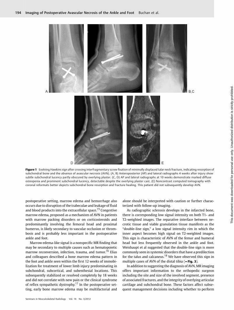
Figure 1 Evolving Hawkins sign after crossing interfragmentary screw fixation of minimally displaced talar neck fracture, indicating resorption ofsubchondral bone and the absence of avascular necrosis (AVN). (A, B) Anteroposterior (AP) and lateral radiographs 4 weeks after injury showsubtle subchondral lucency partly obscured by overlying plaster. (C, D) AP and lateral radiographs at 10 weeks demonstrate marked diffuseosteopenia and prominent subchondral lucency, detectable despite the overlying plaster cast. (E) Noncontrast computed tomography withcoronal reformats better depicts subchondral bone resorption and fracture healing. This patient did not subsequently develop AVN.
Seminars in Musculoskeletal Radiology Vol. 16 No. 3/2012
Imaging of Postoperative Avascular Necrosis of the Ankle and Foot Buchan et al.194
Thi
s do
cum
ent w
as d
ownl
oade
d fo
r pe
rson
al u
se o
nly.
Una
utho
rized
dis
trib
utio
n is
str
ictly
pro
hibi
ted.

core decompression, bone grafting, arthrodesis, or jointreplacement.19
The postsurgicalMR protocols for imaging the foot and ankleat our institution are listed in ►Tables 1A and 1B. We do notroutinely administer intravenous gadolinium unless there is ahigh index of suspicion for infection complicating orthopedichardwareplacement. Although intravenousgadoliniummayaidin the assessment of the extent of nonenhancing infarcted bonefor surgical planning, we do not usually find this necessary.
Artifacts caused by ferromagnetic screws and implants andmetal shavings from the use of surgical instruments may beproblematic in MR image acquisition and interpretation andare more severe if cobalt-chrome or stainless-steel alloy im-plants are used compared with titanium (►Fig. 2). The use offast spin-echo and short tau inversion recovery sequencesreduces metal-related artifacts compared with gradient-echoand frequency-selective fat saturation sequences.13,14Wehaveobserved an increase in susceptibility and misregistrationartifacts increase with greater field strength (1.5 T to 3.0 T),although broader receiver bandwidth and the higher gradientfield strengths used partially offsets the effect. Imaging pa-
rameter optimization at higher field strength may includeusing a small field of view, increasing the resolution (matrixsize), use of thin slices, increasing the echo train length, andincreasing the bandwidth.14
Nuclear MedicineThree phase Tc-99m-diphosphonate (Tc-99m-MDP) bonescintigraphy is not commonly used in clinical practice toconfirm the diagnosis of AVN of the ankle and foot. Recentstudies have demonstrated that bone scintigraphy is lesssensitive than MRI in diagnosing symptomatic AVN, withMRI detecting 100% of lesions, whereas bone scanningdetected 72% of oligofocal cases (two or fewer joints) and45% of multifocal cases.20 Sensitivity is reduced in AVN casesin the ankle compared with the hip and knee. Three-phasebone scintigraphyfindings in acute AVN include decreased ornormal flow activity with decreased activity on delayedphases. The “bull’s-eye sign” of a central region of photopeniawith a surrounding rim of hyperemia is infrequently ob-served in the femoral head and rarely seen in the foot andankle.21
Figure 2 Comparison of computed tomography (CT) and MRI artifacts from fractured stainless-steel screw fragment in talar dome aftermicrodrilling of osteochondral defect of talar dome. (A, B) Anteroposterior radiograph of the ankle and noncontrast CT with sagittal reformatsprovide high-resolution detail of the screw fragment and the sclerotic osteochondral lesion. (C, D) Sagittal fat saturation fast-spin echoT1-weighted and short tau inversion recovery MRI images are nondiagnostic due to extensive susceptibility and misregistration artifact.
Seminars in Musculoskeletal Radiology Vol. 16 No. 3/2012
Imaging of Postoperative Avascular Necrosis of the Ankle and Foot Buchan et al. 195
Thi
s do
cum
ent w
as d
ownl
oade
d fo
r pe
rson
al u
se o
nly.
Una
utho
rized
dis
trib
utio
n is
str
ictly
pro
hibi
ted.

Tc-99m-MDP bone scintigraphy in combination with gal-lium scan or labeled white blood cell scan aids in excludinginfection that may mimic AVN on other imaging modalities.Acute osteomyelitis shows tracer enhancement within 48
hours in all three phases on Tc-99m-MDP scans, although inchronic infection no increased flow is usually seen. Three-phase bone scan accuracy is only 50 to 70% because postop-erative and degenerative changes are difficult to distinguish.
Figure 3 “Double-line sign” characteristic of avascular necrosis (AVN) in distal tibial metaphysis. Subsequent collapse of the articular surface necessitatedtibio-talar arthrodesis in this patient. (A, B) Sagittal T1 fast spin-echo and fat saturation T2-weighted MRI, demonstrating serpiginous region of AVN/boneinfarction with low signal intensity rim on T1 images. The inner aspect of the rim becomes high signal on T2-weighted images.
Table 1A Imaging Parameters Ankle and Foot MRI with Metal Reduction – 1.5T
Ankle (Supine Position) T1 FSE STIR PD T2 FS FSE PD
Plane Sagittal Sagittal Coronal Axial Axial
TR (ms) 480 2000 2000 5600 4500
TE (ms) 12 30 30 40 38
TI (ms) 150 150
FOV (mm) 160 � 160 160 � 160 140 � 140 140 � 140 140 � 140
Matrix size (1.5 T) 192 � 256 256 � 256 448 � 328 140 � 320 288 � 512
Thickness (mm)/Skip 3/0 3/0 3/0 3/0 3/0
NEX 1 2 1 2 1
Turbo factor/ETL 3 5 6 7 9
Bandwidth (Hz/pixel) 250–395 401 250 401 391
Forefoot (Supine Position) T1 FSE T2 FS PD T1 FSE T2 FS
Plane Sagittal Sagittal Coronal Axial Axial
TR (ms) 480 3000 4400 470 5000
TE (ms) 12 70–80 40 10–14 80–90
FOV (mm) 160 � 160 260 � 260 140 � 140 140 � 140 140 � 140
Matrix size (1.5 T) 192 � 256 256 � 256 512 � 256 256 � 256 256 � 192
Thickness (mm) 4/0 4/0 4/0 3/0 3/0
NEX 1 2 1 2 1
Turbo factor/ETL 3 8 6 7 8
Bandwidth (Hz/pixel) 250–395 150–250 250 250–395 150–250
FSE, fast spin echo; STIR, short tau inversion recovery; FS, frequency-selective fat saturated; PD, proton density; FOV, field of view; NEX, number ofexcitations; ETL, echo train length; BW, bandwidth.T2 FS is usually used when imaging the forefoot rather than STIR unless significant artifacts result.
Seminars in Musculoskeletal Radiology Vol. 16 No. 3/2012
Imaging of Postoperative Avascular Necrosis of the Ankle and Foot Buchan et al.196
Thi
s do
cum
ent w
as d
ownl
oade
d fo
r pe
rson
al u
se o
nly.
Una
utho
rized
dis
trib
utio
n is
str
ictly
pro
hibi
ted.
Although the simultaneous addition of a gallium scan mayproduce a modest increase in accuracy, the addition oflabeled white blood cell scan (e.g., Indium-111-labeled leu-cocytes) to detect neutrophil accumulation increases thesensitivity, specificity, and accuracy to >90%.22 At our insti-tutionwe routinely add a sulfur colloid scan as amarrowmapin patients with prior orthopedic instrumentation.
Although 18F-fluorodeoxyglcose positron emission imag-ing (FDG-PET) has emerged as a sensitive modality in thedetection and management of multiple inflammatory andinfectious conditions, no role has been described in thediagnosis of AVN. Presently FDG-PET lacks sufficient specific-ity to be clinically useful in the imaging of postoperativecomplications of joint prostheses.23
Ankle and Hindfoot AVN
Distal TibiaAVN of the distal tibia post ankle fracture in the adult isuncommon. An isolated case report after Maisonneuve-typefracture of the proximal third of the fibula with syndesmoticinjury treated with lateral K-wire fixation of the syndesmosisand ligament reconstruction developed symptomatic andradiographic evidence of AVN of the tibial plafond 7 monthsafter injury.24 One published clinical series of nine patients,each of whom sustained a Weber C-type fracture-dislocation
of the tibio-talar joint, developed plain radiographic evidenceof AVN of the lateral tibial plafond between 4 and 8 monthspostinjury.25 An unpublished series of nine patients by Link-later (personal correspondence) demonstrated the coinci-dence of syndesmotic injury and distal tibial metaphysealor plafond bone infarction. All nine cases had MR features ofsyndesmotic injury, and five of these cases were initiallytreated with lateral syndesmotic screw fixation.
In our recent experience, we have observed AVN of thedistal tibia following Maisonneuve-type proximal fibularfracture with transsyndesmotic screw fixation (►Fig. 4),Weber C fibular fracture with associated posterior malleolarfracture, and Weber C trimalleolar fractures treated withinternal fixation without syndesmotic screw fixation. A com-mon observation in our cases and in those from the literatureis AVN consistently involving the posterolateral tibial plafondafter injuries to the distal tibio-fibular syndesmosis withconcomitant suprasyndesmotic fibula fracture.
Posttraumatic AVN of the distal tibia is probably multi-factorial and related to the relatively tenuous blood supplyof the lateral tibial plafond, the mechanism of injury, andthe treatment methods used. The distal tibial blood supplyarises from nutrient arteries from the posterior tibialartery (PTA) and extraosseous metaphyseal and periostealarteries from branches of the PTA, anterior tibial artery(ATA), and peroneal artery (PA) with a prominent medial
Table 1B Imaging Parameters Ankle and Foot MRI with Metal Reduction – 3T
Ankle (Supine Position) T1 FSE STIR PD T2 SPAIR PD
Plane Sagittal Sagittal Coronal Axial Axial
TR (ms) 700–1000 2000–4000 2000–4000 2000–4000 2000–4000
TE (ms) 10 30 30 60 38
TI (ms) 190 190
FOV (mm) 160 � 160 160 � 160 140 � 140 140 � 140 140 � 140
Matrix size (1.5T) 389 � 456 272 � 400 432 � 333 363 � 400 368 � 400
Thickness (mm)/Skip 3/0 3/0 2.5/0 3/0 3/0
NEX 1 2 1 2 1
Turbo factor/ETL 3 5 6 7 9
Bandwidth (Hz/pixel) 440 401 440 401 440
Forefoot (Supine Position) T1 FSE T2 SPAIR PD T1 FSE T2 SPAIR
Plane Sagittal Sagittal Coronal Axial Axial
TR (ms) 700–1000 2000–4000 2000–4000 500–1000 2000–4000
TE (ms) 10 60 37 10 60
FOV (mm) 150 � 150 260 � 260 140 � 140 140 � 140 140 � 140
Matrix size (1.5T) 418 � 500 323 � 376 396 � 468 367 � 400 352 � 308
Thickness (mm) 2.5/0 2.5/0 2.5/0 2.5/0 2.5/0
NEX 1 2 1 2 1
Turbo factor/ETL 3 8 6 7 8
Bandwidth (Hz/pixel) 440 400 440 440 400
FSE, fast spin echo; STIR, short tau inversion recovery; FS, frequency-selective fat saturated; PD, proton density; FOV, field of view; NEX, number ofexcitations; ETL, echo train length; BW, bandwidth.T2 FS is usually used when imaging the forefoot rather than STIR unless significant artifacts result.
Seminars in Musculoskeletal Radiology Vol. 16 No. 3/2012
Imaging of Postoperative Avascular Necrosis of the Ankle and Foot Buchan et al. 197
Thi
s do
cum
ent w
as d
ownl
oade
d fo
r pe
rson
al u
se o
nly.
Una
utho
rized
dis
trib
utio
n is
str
ictly
pro
hibi
ted.
perimalleolar anastomotic ring.25–27 The distal lateraltibial epiphysis is relatively less vascularized comparedwith the medial and is primarily supplied by the ATA andPA. ATA branches alone nourish this region in approximate-ly a third of cases, possibly further predisposing toischemia.25
The association in case reports with syndesmotic injury,bimalleolar and trimalleolar fractures, and tibio-talar jointdislocation suggests patients at risk of AVN have an unstableinjury. Initial radiographic findings suggestive of such inju-ries include reduced tibiofibular overlap (syndesmosis wid-ening), concomitant posterior malleolar fracture, andWeber C fracture or Maisonneuve fracture, tibio-talar jointdislocation, marked soft tissue swelling, and subcutaneousgas associated with open fracture. CT, MRI, and stressradiographs are more sensitive than plain radiographsfor syndesmosis diastasis. MRI determines the extent ofinterosseous membrane tearing more accurately than esti-mation from the level of fibula fracture.28 In cases likelypredisposed to AVN, advanced imaging is not routinelyperformed preoperatively because the presence of
posterior malleolar or suprasyndesmotic fibula fracturesnecessitates intraoperative surgical assessment of syndes-motic stability.
Surgical techniques aim for the anatomical restoration ofthe disrupted distal tibiofibular syndesmosis andmay requirefibular fracture internal fixation, repair of medial malleolarfracture/ligamentous injury, posterior malleolar fracture fix-ation (especially if large and posterior inferior tibiofibularligament is attached), and transsyndesmotic screw fixa-tion.28,29 The perforating branch of the PA has been shownto be at risk with syndesmotic screw fixation where ittraverses the interosseous membrane from posterior to ante-rior.30Open plating of fractures of the distal tibial metaphysishas also been demonstrated to cause greater disruption ofextraosseous blood supply than percutaneously appliedplates.27 The hypothesis that operative disruption of theextraosseous blood supply of the distal tibia may contributeto AVN seems reasonable but has not been confirmed.
Distinguishing early radiographic signs of AVN frompatchy sclerosis associated with fracture healing and hard-ware fixation can be challenging; however, temporal changes
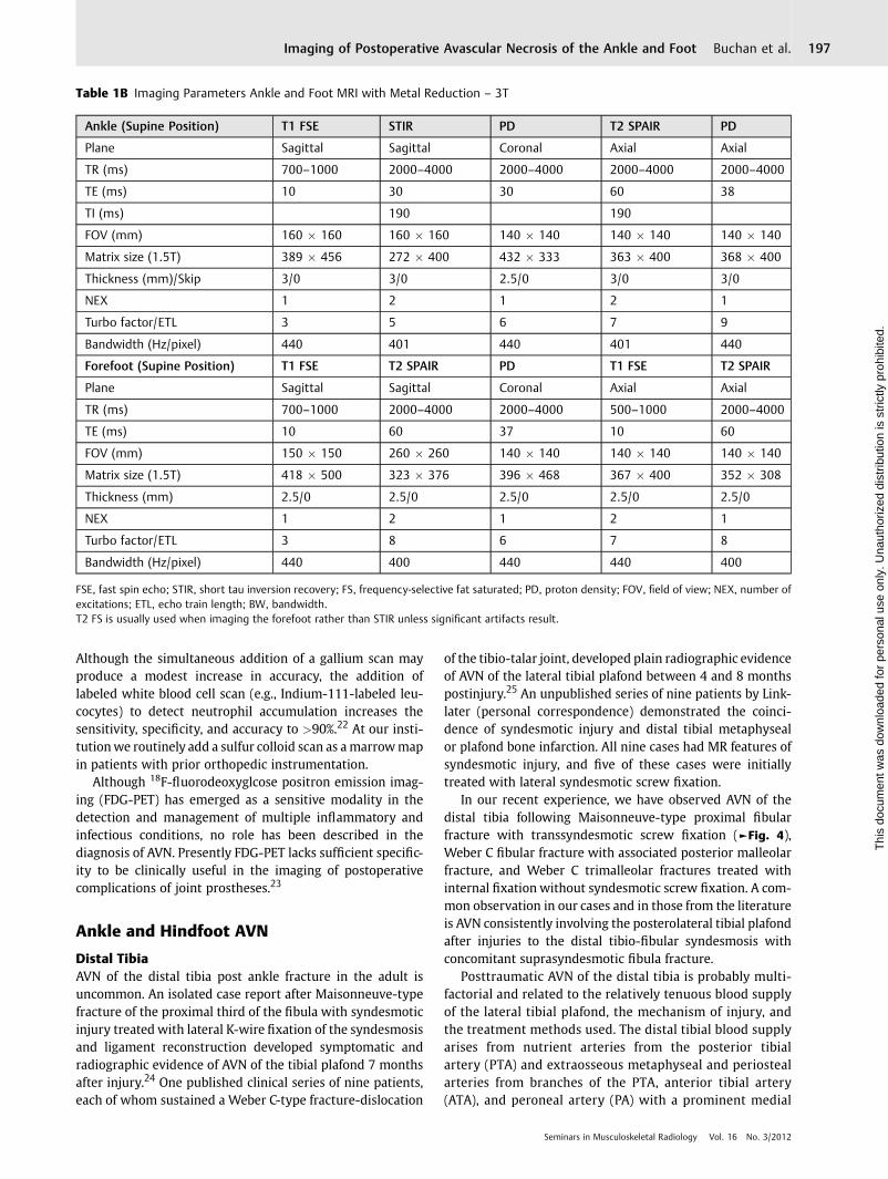
Figure 4 Avascular necrosis (AVN) of the distal tibial metaphysis and plafond in elite basketball player after Maisonneuve-type fibula fracture withsyndesmotic injury. (A, B) Anteroposterior (AP) radiograph of the ankle shows widening of the medial and lateral clear space and loss of congruityof the distal tibiofibular syndesmosis. AP radiograph of the proximal fibula confirms Maisonneuve-type fracture. (C) Initial postoperative APradiograph demonstrates reduction of the distal syndesmosis with two transsyndesmotic screws. (D, E) Coronal T1 fast spin-echo and short tauinversion recovery MRI, demonstrating AVN in the posterolateral distal tibia with disruption of the articular surface. Note the susceptibility artifactassociated with transsyndesmotic screws.
Seminars in Musculoskeletal Radiology Vol. 16 No. 3/2012
Imaging of Postoperative Avascular Necrosis of the Ankle and Foot Buchan et al.198
Thi
s do
cum
ent w
as d
ownl
oade
d fo
r pe
rson
al u
se o
nly.
Una
utho
rized
dis
trib
utio
n is
str
ictly
pro
hibi
ted.

in the density of the distalmetaphysis and plafond are usuallypresent. With later stages of AVN progressive collapse of thetibial plafond, joint space narrowing and ankle joint deformi-ty are seen with secondary osteoarthritis. Failure of previoussurgery may predispose to subsequent AVN, and signs ofhardware failure or residual instability of the syndesmosisshould be sought (►Fig. 5). In such cases, CT is sensitive todetect plate and screw fracture and widening of the syndes-mosis. Residual thickening and edema in the inferior syndes-motic ligaments and subtle marginated regions of AVN,depicted as the double-line sign, may be seen on MRI. CTand MRI also provide important complementary informationon the integrity of the articular surface and extent of boneinfarction for planning subsequent surgical salvage proce-dures including tibiotalar joint arthrodesis or total anklearthroplasty with or without bone grafting.
TalusAVN of the talus is common, in part due to its susceptibility totrauma with high-energy dorsiflexion, and partly due to itsreliance on direct vasculature for its blood supply. There areno tendinous attachments or muscle origins and �60% of thetalar surface is covered by articular cartilage, leaving littlearea for vascular perforation.3,7
Major extraosseous arterial supply comes from branchesof the posterior tibial artery, anterior tibial artery, dorsalispedis artery, and perforating peroneal artery with a keyanastomosis in the sinus tarsi between the artery of the tarsalcanal and tarsal sinus artery.
There is a significant retrograde component of the intra-osseous blood supply of the talus that is important in thetrauma setting because patients with a fracture of the talarneck usually develop AVN in the talar body. The Hawkinsclassification of talar neck fractures, later revised by Canaleand Kelly, recognizes the increased likelihood of developingAVN with increased fracture displacement and the presence
of associated dislocations. Type IV fractures, involving dislo-cation or subluxation of the subtalar, tibiotalar, and talona-vicular joints, almost universally cause AVN.10,31 The risk ofAVN increases in talar neck fractures that extend to involvethe body, increasingly comminuted and displaced talar bodyfractures, and with talar head fractures.3,32,33 A commonalityof higher grade injuries is the increased disruption to theextraosseous blood supply and soft tissue attachments thatoccurs with dislocations.
AVN of the talus has been described in cases reports ofsurgical procedures around the ankle and foot includingmedial and lateral release of congenital clubfoot, tibio-talar-calcaneal arthrodesis, triple arthrodesis, and talonaviculararthrodesis (►Fig. 6).34,35 A triple arthrodesis, often per-formed via a lateral approach with anterior to posteriortechnique, may disrupt the tarsal sinus artery when thechondral surfaces are removed. This necessitates reliance onintraosseous anastomoses with the artery of the tarsal canaland fused osseous surfaces to supply the lateral aspect of thetalar dome.34 Themore extensively the talar body and neck areresected, the more likely is subsequent AVN. The medialapproach used for talonavicular arthrodesis may disrupt theartery of the tarsal canal or intraosseous anastomoses andmayalso result in AVN of the lateral talar body and dome.35
Detection of the Hawkins sign 4 to 8 weeks after fractureor surgical intervention is suggestive of revascularization ofthe relevant portion of the talar body, and it usually pro-gresses from medial to lateral. A partial Hawkins sign indic-ative of incomplete AVN is more commonly observed in themedial talus, indicating susceptibility to AVN of the lateraltalar dome or inferior articular surface of the body.7 Com-plete revascularization after surgery may take between6 months and 3 years. During this time fractures may healas progressive sclerosis and cystic changes of AVN eitherresolve or lead to osseous collapse. MRI performed >3 weeksafter surgery is more sensitive than plain radiographs in the
Figure 5 Avascular necrosis (AVN) of the distal tibial metaphysis detected 6 months after trimalleolar fracture in patient with ongoing pain afterremoval of orthopedic hardware. (A) Oblique radiograph of the right ankle with patchy sclerosis in the distal tibial metaphysis and plafond withsubchondral “crescent sign.” (B) Noncontrast computed tomography of the ankle sagittal reformat confirms healed posterior malleolar fracture,mixed hypoattenuation, and sclerosis in the distal tibial metaphysis, subchondral lucency, and partial collapse of the articular surface.
Seminars in Musculoskeletal Radiology Vol. 16 No. 3/2012
Imaging of Postoperative Avascular Necrosis of the Ankle and Foot Buchan et al. 199
Thi
s do
cum
ent w
as d
ownl
oade
d fo
r pe
rson
al u
se o
nly.
Una
utho
rized
dis
trib
utio
n is
str
ictly
pro
hibi
ted.
assessment of AVN in Hawkins type II and type III fractures,and it provides important prognostic information regardingthe volume of avascular bone and risk of subsequent collapse(►Fig. 7).36 CT is particularly useful in the assessment ofhealing or evolution of AVN after triple arthrodesis (►Fig. 8).
Imaging findings are key determinants in the suitability forconservative management, core decompression, or bonegraft procedures. Salvage arthrodesis or resection is usuallyrequired after significant collapse and secondary osteoar-thritis supervenes.
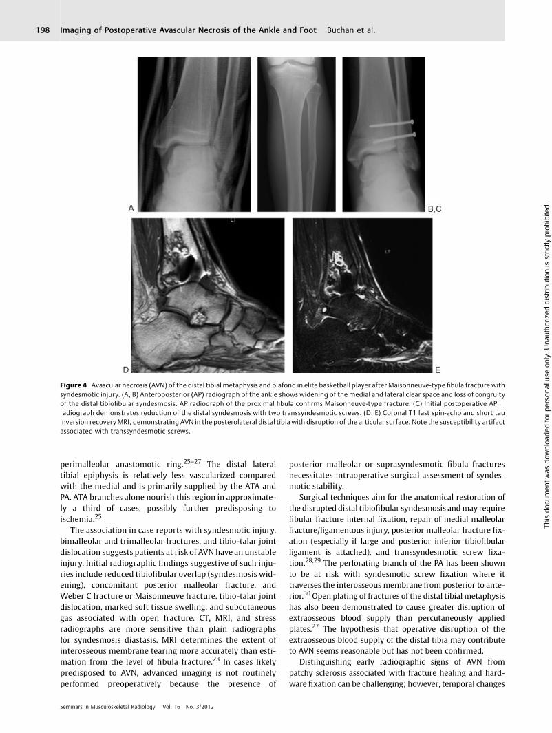
Figure 6 Sclerosis of tripartite talus secondary to medial clubfoot release, congenital talar dysplasia, subchondral avascular necrosis, and chronicnonunion. (A, B) Anteroposterior weightbearing radiographs of both ankles and lateral radiograph left ankle. (C, D) Noncontrast computedtomography with sagittal and coronal reformats demonstrates dense sclerosis and corticated chronic nonunion of talar fragments.
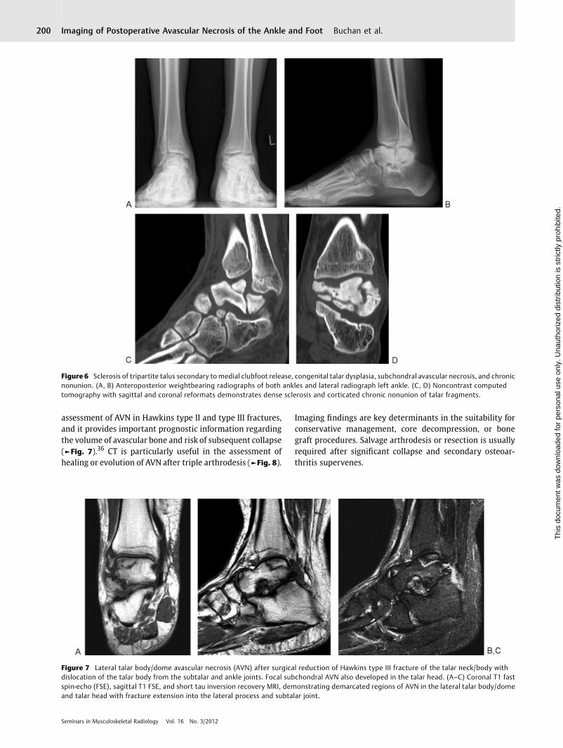
Figure 7 Lateral talar body/dome avascular necrosis (AVN) after surgical reduction of Hawkins type III fracture of the talar neck/body withdislocation of the talar body from the subtalar and ankle joints. Focal subchondral AVN also developed in the talar head. (A–C) Coronal T1 fastspin-echo (FSE), sagittal T1 FSE, and short tau inversion recovery MRI, demonstrating demarcated regions of AVN in the lateral talar body/domeand talar head with fracture extension into the lateral process and subtalar joint.
Seminars in Musculoskeletal Radiology Vol. 16 No. 3/2012
Imaging of Postoperative Avascular Necrosis of the Ankle and Foot Buchan et al.200
Thi
s do
cum
ent w
as d
ownl
oade
d fo
r pe
rson
al u
se o
nly.
Una
utho
rized
dis
trib
utio
n is
str
ictly
pro
hibi
ted.
Midfoot and Forefoot
Navicular
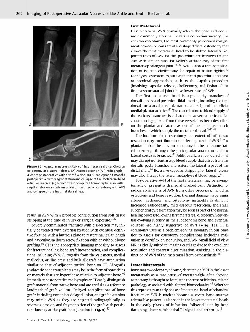
The navicular is vascularized dorsally from a branch of thedorsalis pedis artery with the plantar surface supplied by themedial plantar artery, entering through the nonarticularsurface. The intraosseous flow is centripetal with the centerof the bone susceptible to AVN.3,37 Trauma is the mostcommon etiology with injuries ranging from avulsion frac-
tures to comminuted fracture-dislocations. Fractures of thebody, usually due to high energy trauma including motorvehicle accidents, are the most likely to develop AVN and areclassified according to the Sangeorzan system.38 Type 1fractures involve the primary fracture line transverse in thecoronal plane, and type 3 fractures produce comminution inthe middle and lateral navicular with dislocation of thenaviculocuneiform joint. These two fracture types cause thegreatest disruption to the radial blood supply and commonly
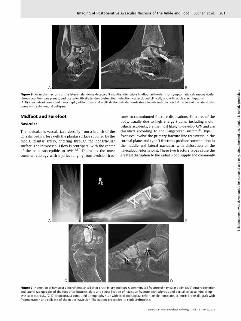
Figure 8 Avascular necrosis of the lateral talar dome detected 8 months after triple hindfoot arthrodesis for symptomatic calcaneonavicularfibrous coalition, pes planus, and posterior tibialis tendon dysfunction. Infection was excluded clinically and with nuclear scintigraphy.(A, B) Noncontrast computed tomography with coronal and sagittal reformats demonstrates sclerosis and subchondral fracture of the lateral talardome with subchondral collapse.
Figure 9 Nonunion of navicular allograft implanted after crush injury and type C comminuted fracture of navicular body. (A, B) Anteroposteriorand lateral radiographs of the foot after buttress plate and screw fixation of navicular fracture with sclerosis and partial collapse mimickingavascular necrosis. (C, D) Noncontrast computed tomography scan with axial and sagittal reformats demonstrates sclerosis in the allograft withfragmentation and collapse of the native navicular. The patient proceeded to triple arthrodesis.
Seminars in Musculoskeletal Radiology Vol. 16 No. 3/2012
Imaging of Postoperative Avascular Necrosis of the Ankle and Foot Buchan et al. 201
Thi
s do
cum
ent w
as d
ownl
oade
d fo
r pe
rson
al u
se o
nly.
Una
utho
rized
dis
trib
utio
n is
str
ictly
pro
hibi
ted.
result in AVN with a probable contribution from soft tissuestripping at the time of injury or surgical exposure.3,37
Severely comminuted fractures with dislocation may ini-tially be treated with external fixation with eventual defini-tive fixation with a buttress plate to restore navicular lengthand naviculocuneiform screw fixation with or without bonegrafting.39 CT is the appropriate imaging modality to assessfor fracture healing, bone graft incorporation, and complica-tions including AVN. Autografts from the calcaneus, medialmalleolus, or iliac crest and bulk allograft have attenuationsimilar to that of adjacent cortical bone on CT. Allografts(cadaveric bone transplants) may be in the form of bone chipsor morsels that are hyperdense relative to adjacent bone.40
Immediate postoperative radiographs or CT easily distinguishgraft material from native bone and are useful as a referencelandmark of graft volume. Delayed complications of bonegrafts including nonunion, delayed union, and graft extrusionmay mimic AVN as they are depicted radiographically assclerosis, erosion, and fragmentation of the graft with persis-tent lucency at the graft–host junction (►Fig. 9).40
First MetatarsalFirst metatarsal AVN primarily affects the head and occursmost commonly after hallux valgus correction surgery. Thechevron osteotomy, the most commonly performed realign-ment procedure, consists of a V-shaped distal osteotomy thatallows the first metatarsal head to be shifted laterally. Re-ported rates of AVN for this procedure are between 0% and20% with similar rates for Keller’s arthroplasty of the firstmetatarsophalangeal joint.41,42 AVN is also a rare complica-tion of isolated cheilectomy for repair of hallux rigidus.43
Diaphyseal osteotomies, such as the Scarf procedure, and baseor proximal approaches, such as the Lapidus procedure(involving capsular release, cheilectomy, and fusion of thefirst tarsometatarsal joint), have lower rates of AVN.
The first metatarsal head is supplied by branches ofdorsalis pedis and posterior tibial arteries, including the firstdorsal metatarsal, first plantar metatarsal, and superficialmedial plantar arteries.41 The contribution to blood supply ofthe various branches is debated; however, a pericapsularanastomosing plexus from these vessels has been describedon the plantar and lateral aspect of the metatarsal neck,branches of which supply the metatarsal head.3,41,42
The location of the osteotomy and extent of soft tissueresection may contribute to the development of AVN.3 Theplantar limb of the chevron osteotomy has been demonstrat-ed to emerge through the pericapsular anastomosis if thelateral cortex is breached.41 Additionally, a short dorsal limbmay disrupt nutrient artery blood supply that arises from thedorsalis pedis branches and enters the lateral aspect of thedistal shaft.44 Excessive capsular stripping for lateral releasemay also disrupt the lateral metaphyseal blood supply.45
Postoperative AVN of the first metatarsal may be asymp-tomatic or present with medial forefoot pain. Distinction ofradiographic signs of AVN from other processes, includingosteotomy and bone resection, thermal damage, hyperemia,altered mechanics, and osteotomy instability is difficult.Increased radiodensity, mild osseous resorption, and smallsubchondral cyst formationmay be seen as part of the normalhealing process followingfirst metatarsal osteotomy. Sequen-tial evolving lucency in the subchondral bone and eventualcollapse are highly suggestive of AVN (►Fig. 10). CT iscommonly used as a problem-solving modality in our prac-tice to assess for osteotomy complications including mal-union in dorsiflexion, nonunion, and AVN. Small field of viewMRI is ideally suited to imaging cartilage due to the excellentresolution and contrast discrimination, assisting in the dis-tinction of AVN of the metatarsal from osteoarthritis.46
Lesser MetatarsalsBonemarrow edema syndrome, detected onMRI in the lessermetatarsals as a rare cause of metatarsalgia after chevronosteotomy, is thought to be related to stress or friction-relatedpathology associated with altered biomechanics.47 Whetherthis represents an early phase ofmetatarsal head subchondralfracture or AVN is unclear because a severe bone marrowedema-like pattern is also seen in the lesser metatarsal headsin the early phases of infraction, followed later by headflattening, linear subchondral T1 signal, and arthrosis.48
Figure 10 Avascular necrosis (AVN) of first metatarsal after Chevronosteotomy and lateral release. (A) Anteroposterior (AP) radiograph4 weeks postoperative with K-wire fixation. (B) AP radiograph 8monthspostoperative with fragmentation and collapse of the metatarsal headarticular surface. (C) Noncontrast computed tomography scan withsagittal reformats confirms union of the Chevron osteotomy with AVNand collapse of the first metatarsal head.
Seminars in Musculoskeletal Radiology Vol. 16 No. 3/2012
Imaging of Postoperative Avascular Necrosis of the Ankle and Foot Buchan et al.202
Thi
s do
cum
ent w
as d
ownl
oade
d fo
r pe
rson
al u
se o
nly.
Una
utho
rized
dis
trib
utio
n is
str
ictly
pro
hibi
ted.

Theblood supply of the lessermetatarsal heads arises fromthe dorsal metatarsal arteries (branches of the dorsalis pedisartery) and plantar metatarsal arteries (from the posteriortibial artery).3 A nutrient artery traverses the cortex of thedistal metaphysis laterally (in the second to fourth metatar-sals) and medially (in the fifth) close to capsular and liga-mentous attachments, with terminal distal branchescontributing to the blood supply of the metatarsal head.49
An extraosseous arterial anastomotic ring from branches ofthe dorsal and plantar metatarsal arteries forms around themetatarsal heads, which may be affected by metatarsal headosteotomy or extensive capsular stripping.
Chevron, Helal, or Weil osteotomies of the lesser meta-tarsals may be performed to correct chronic metatarsopha-langeal joint dislocation, metatarsalgia due to an excessivelylong metatarsal, or intractable callosities. The most common-ly performed procedure, the Weil osteotomy, consisting of anoblique osteotomy through the metatarsal head and shaftparallel to the ground with detachment of lateral capsularligaments, provides controlled shortening of the metatarsalwithout increasing the load in the adjacentmetatarsals.50 Thedirection of the osteotomy cuts places the intraosseous bloodsupply at risk in addition to the extraosseous capsular supplyif the plantar cortex is breached. Recognized complications ofthe procedure include “floating toe” (a toe that does notcontact the ground), nonunion, malunion, and transfer meta-tarsalgia. Although AVN is mentioned in the surgical litera-ture as a potential complication, no case reports have beenpublished in the English literature, and this is likely a very rareoutcome. We have observed one case of probable AVN of thesecond metatarsal head after first metatarsal bone blockfusion and Weil osteotomy of the second metatarsal, withthe patient experiencing intractable second metatarsalgia5 years after the original procedure (►Fig. 11).
Conclusion
The imaging modalities and major causes of postoperativeAVN in the ankle and foot briefly discussed in this reviewrepresent those that radiologists are most likely to encounterin clinical practice.
Although rare, detection of AVN in the postoperativepatient is critical because the diagnosis is often not suspectedclinically, and appropriate management of these patientsmay be instituted to achieve satisfactory functionaloutcomes.
References1 McCarthy I. The physiology of bone blood flow: a review. J Bone
Joint Surg Am 2006;88(Suppl 3):4–92 Assouline-Dayan Y, Chang C, Greenspan A, Shoenfeld Y, Gershwin
ME. Pathogenesis and natural history of osteonecrosis. SeminArthritis Rheum 2002;32(2):94–124
3 DiGiovanni CW, Patel A, Calfee R, Nickisch F. Osteonecrosis in thefoot. J Am Acad Orthop Surg 2007;15(4):208–217
4 Steffen RT, Athanasou NA, Gill HS, Murray DW. Avascular necrosisassociated with fracture of femoral head after hip resurfacing.J Bone Joint Surg Br 2010;92(6):787–793
5 Bartonícek J, Fric V, Skála-Rosenbaum J, Dousa P. Avascular necro-sis of the femoral head in pertrochanteric fractures: a report of 8cases and a review of the literature. J Orthop Trauma 2007;21(4):229–236
6 Pape D, Seil R, Anagnostakos K, Kohn D. Postarthroscopic osteo-necrosis of the knee. Arthroscopy 2007;23(4):428–438
7 Pearce DH, Mongiardi CN, Fornasier VL, Daniels TR. Avascularnecrosis of the talus: a pictorial essay. Radiographics 2005;25(2):399–410
8 Koulouris G, MorrisonWB. Foot and ankle disorders: radiographicsigns. Semin Roentgenol 2005;40(4):358–379
9 Lafforgue P. Pathophysiology and natural history of avascularnecrosis of bone. Joint Bone Spine 2006;73(5):500–507
10 Hawkins LG. Fractures of the neckof the talus. J Bone Joint Surg Am1970;52(5):991–1002
11 Rammelt S, Zwipp H. Talar neck and body fractures. Injury 2009;40(2):120–135
12 Resnick D, Sweet DE, Madewell JE. Osteonecrosis: pathogenesis,diagnostic techniques, specific situations, and complications. In:Diagnosis of bone and joint disorders. 4th ed. Philadelphia, PA:Saunders; 2002:3599–3685
13 Buckwalter KA, Parr JA, Choplin RH, Capello WN. Multichannel CTimaging of orthopedic hardware and implants. Semin Musculos-kelet Radiol 2006;10(1):86–97
14 Lee MJ, Kim S, Lee SA, et al. Overcoming artifacts from metallicorthopedic implants at high-field-strengthMR imaging andmulti-detector CT. Radiographics 2007;27(3):791–803
15 O’Hare A, Shortt C, Napier N, Eustace SJ. Bone marrow edema:patterns and clinical implications. Semin Musculoskelet Radiol2006;10(4):249–257
16 Gyftopoulos S, Bencardino JT. Normal variants and pitfalls in MRimaging of the ankle and foot. Magn Reson Imaging Clin N Am2010;18(4):691–705
17 Elias I, Zoga AC, Schweitzer ME, Ballehr L, Morrison WB, RaikinSM. A specific bone marrow edema around the foot and anklefollowing trauma and immobilization therapy: pattern descrip-tion and potential clinical relevance. Foot Ankle Int 2007;28(4):463–471
18 Weishaupt D, Schweitzer ME. MR imaging of the foot and ankle:patterns of bone marrow signal abnormalities. Eur Radiol 2002;12(2):416–426
Figure 11 Avascular necrosis (AVN) of second metatarsal head afterbone block fusion of the first metatarsal and Weil osteotomy secondmetatarsal. (A) Noncontrast computed tomography (CT) scan withsagittal reformats 4 years after initial operation demonstrates sclerosisand partial collapse of the dorsal second metatarsal head with thedistal screw sitting proud of the metatarsal head in an intra-articularlocation. (B) Anteroposterior (AP) radiograph 1 year after the CT afterremoval of the distal screw with residual flattening and collapse of thesecond metatarsal head, consistent with previous AVN.
Seminars in Musculoskeletal Radiology Vol. 16 No. 3/2012
Imaging of Postoperative Avascular Necrosis of the Ankle and Foot Buchan et al. 203
Thi
s do
cum
ent w
as d
ownl
oade
d fo
r pe
rson
al u
se o
nly.
Una
utho
rized
dis
trib
utio
n is
str
ictly
pro
hibi
ted.

19 Hintermann B. What the orthopaedic foot and ankle surgeonwants to know from MR imaging. Semin Musculoskelet Radiol2005;9(3):260–271
20 Mont MA, Ulrich SD, Seyler TM, et al. Bone scanning of limitedvalue for diagnosis of symptomatic oligofocal and multifocalosteonecrosis. J Rheumatol 2008;35(8):1629–1634
21 Hain SF, Fogelman I. Nuclear medicine studies in metabolic bonedisease. Semin Musculoskelet Radiol 2002;6(4):323–329
22 Palestro CJ, Love C, Bhargava KK. Labeled leukocyte imaging:current status and future directions. Q J Nucl Med Mol Imaging2009;53(1):105–123
23 Love C, TomasMB, Tronco GG, Palestro CJ. FDG PETof infection andinflammation. Radiographics 2005;25(5):1357–1368
24 Lagier R. Case report 552: Post-traumatic remodelling of the distaltibial epiphysis: a form of aseptic osteonecrosis. Skeletal Radiol1989;18(4):331–333
25 Assal M, Sangeorzan BJ, Hansen ST. Post-traumatic osteonecrosisof the lateral tibial plafond. Foot Ankle Surg 2007;13:24–29
26 Nelson GE Jr, Kelly PJ, Peterson LF, Janes JM. Blood supply of thehuman tibia. J Bone Joint Surg Am 1960;42-A:625–636
27 Borrelli J Jr, Prickett W, Song E, Becker D, Ricci W. Extraosseousblood supply of the tibia and the effects of different platingtechniques: a human cadaveric study. J Orthop Trauma 2002;16(10):691–695
28 Zalavras C, Thordarson D. Ankle syndesmotic injury. J Am AcadOrthop Surg 2007;15(6):330–339
29 van den Bekerom MP, Hogervorst M, Bolhuis HW, van Dijk CN.Operative aspects of the syndesmotic screw: review of currentconcepts. Injury 2008;39(4):491–498
30 Fanter NJ, Inouye SE, McBryde AM Jr. Safety of ankle trans-syndesmotic fixation. Foot Ankle Int 2010;31(5):433–440
31 Canale ST, Kelly FB Jr. Fractures of the neck of the talus. Long-termevaluation of seventy-one cases. J Bone Joint Surg Am 1978;60(2):143–156
32 Inokuchi S, Ogawa K, Usami N. Classification of fractures of thetalus: clear differentiation between neck and body fractures. FootAnkle Int 1996;17(12):748–750
33 Rammelt S, ZwippH. Talar neck and body fractures. Injury 2009;40(2):120–135
34 Jones CK, Nunley JA. Osteonecrosis of the lateral aspect of the talardome after triple arthrodesis. A report of three cases. J Bone JointSurg Am 1999;81(8):1165–1169
35 Hermus JPS. Osteonecrosis of the talus after talonavicular arthrod-esis: a case report and review of the literature. J Foot Ankle Surg2011;50(3):343–346
36 Thordarson DB, Triffon MJ, Terk MR. Magnetic resonance imagingto detect avascular necrosis after open reduction and internalfixation of talar neck fractures. Foot Ankle Int 1996;17(12):742–747
37 Sizensky JA, Marks RM. Imaging of the navicular. Foot Ankle Clin2004;9(1):181–209
38 Sangeorzan BJ, Benirschke SK, Mosca V, Mayo KA, Hansen ST Jr.Displaced intra-articular fractures of the tarsal navicular. J BoneJoint Surg Am 1989;71(10):1504–1510
39 DiGiovanni CW. Fractures of the navicular. Foot Ankle Clin 2004;9(1):25–63
40 Beaman FD, Bancroft LW, Peterson JJ, Kransdorf MJ, Menke DM,DeOrio JK. Imaging characteristics of bone graft materials. Radio-graphics 2006;26(2):373–388
41 Malal JJG, Shaw-Dunn J, Kumar CS. Blood supply to the firstmetatarsal head and vessels at risk with a chevron osteotomy.J Bone Joint Surg Am 2007;89(9):2018–2022
42 Prasad MG, Shankar NS. Clinical results of Keller’s arthroplasty.Foot 1998;8:223–225
43 Brosky TA II, Menke CRD, Xenos D. Reconstruction of the firstmetatarsophalangeal joint following post-cheilectomy avascularnecrosis of the first metatarsal head: a case report. J Foot AnkleSurg 2009;48(1):61–69
44 Weinraub GM, Meberg R, Steinberg JS. Vascular perfusion of thelong dorsal arm versus chevron osteotomy: a cadaveric injectionstudy. J Foot Ankle Surg 2004;43(4):221–224
45 Jones KJ, Feiwell LA, Freedman EL, Cracchiolo A III. The effect ofchevron osteotomy with lateral capsular release on the bloodsupply to the first metatarsal head. J Bone Joint Surg Am 1995;77(2):197–204
46 Shortt CP. Magnetic resonance imaging of the midfoot and fore-foot: normal variants and pitfalls. Magn Reson Imaging Clin N Am2010;18(4):707–715
47 AignerN, Petje G, SteinboeckG, SchneiderW, KrasnyC, Landsiedl F.Bone marrow edema of the forefoot after chevron osteotomy—arare cause of metatarsalgia: a case report. Foot Ankle Int 2002;23(5):447–451
48 Torriani M, Thomas BJ, Bredella MA, Ouellette H. MRI of metatarsalhead subchondral fractures in patientswith forefoot pain. AJR Am JRoentgenol 2008;190(3):570–575
49 Petersen WJ, Lankes JM, Paulsen F, Hassenpflug J. The arterialsupply of the lesser metatarsal heads: a vascular injection study inhuman cadavers. Foot Ankle Int 2002;23(6):491–495
50 Espinosa N, Maceira E, Myerson MS. Current concept review:metatarsalgia. Foot Ankle Int 2008;29(8):871–879
Seminars in Musculoskeletal Radiology Vol. 16 No. 3/2012
Imaging of Postoperative Avascular Necrosis of the Ankle and Foot Buchan et al.204
Thi
s do
cum
ent w
as d
ownl
oade
d fo
r pe
rson
al u
se o
nly.
Una
utho
rized
dis
trib
utio
n is
str
ictly
pro
hibi
ted.