imaging of postpartum encephalopathy: a pictorial …jmscr.igmpublication.org/v5-i3/55...
TRANSCRIPT

Dr Shashibala J Yadav et al JMSCR Volume 05 Issue 03 March 2017 Page 18599
JMSCR Vol||05||Issue||03||Page 18599-18608||March 2017
Imaging of Postpartum Encephalopathy: A Pictorial Essay
Authors
Dr Shashibala J Yadav1, Dr Abhishek Jain
2, Dr Rajesh Mahavir Shanklesha
3,
Dr Nikhil Ihare4
1Fellow in Diagnostic Neuroradiology,
2Resident,
3Fellow in Body Imaging, T.N.M.C & B.Y.L.Nair
Charitable Hospital Mumbai, 4Consultant Radiologist
IQRAA International Hospital & Research Centre Calicut, Kerala
Corresponding Author
Dr Shashibala J Yadav
Fellow in Diagnostic Neuroradiology, T.N.M.C & B.Y.L.Nair Charitable Hospital Mumbai
Abstract
Cerebrovascular events are relatively rare in young patients. They may be seen in some patients at a
younger age if there is history of predisposing medical conditions like sickle cell disease, protein c, protein
s or antithrombin deficiency. The incident of stroke in non-pregnant females of reproductive age group is
reported to be around 10 per 100000 women-years. There is 3 fold rise in the risk of cerebrovascular
accidents in pregnant women of same age group. The common causes of such events in young women
include, oral contraceptive pills use, Systemic lupus erythromatosus (SLE), antiphospholipid antibody
syndrome (APLAS), reversible cerebral vasoconstriction syndrome (RCVS), vasculitis syndromes like
takayasu’s arteritis, moyamoya disease and susac’s syndrome. Adverse cerebrovascular events are more
common in pregnancy, postpartum and puerperal period as compared to non-pregnant women of same
age. The predisposing factors for postpartum acute cerebrovascular disease include pre-eclampsia,
eclampsia, pre-existing chronic kidney disease, diabetes mellitus, congenital or acquired hypercolagulable
states and puerperal sepsis. Some of the common cerebrovascular events seen in postpartum period
include cerebral venous thrombosis, reversible cerebral vasoconstriction syndrome, posterior reversible
encephalopathy syndrome and postpartum angiopathy. The diagnosis of these conditions is usually by
neuroimaging. This paper is a pictorial essay on the neuroimaging of some of the common cerebrovascular
events seen in postpartum period. Familiarity with these neuroimaging findings is important to
differentiate these conditions from other causes of altered sensorium in postpartum period like postpartum
psychoses, electrolyte imbalance and eclampsia.
Keywords: Postpartum encephalopathy, stroke, Magnetic resonance imaging, MR Angiography.
Introduction
Acute neurological conditions requiring critical
care is uncommon in women of childbearing age
unless they have conditions predisposing them for
developing neurological problems such as
Systemic lupus erythromatosus (SLE), antiphos-
pholipid antibody syndrome (APLAS), protein c,
and Protein-S and antithrombin deficiency [1]
.
Young woman even without any of these
condition are prone to develop acute neurological
condition during pregnancy and postpartum
period. One of the important and common causes
of altered sensorium and seizures encountered in
routine obstetric practice is eclampsia.
www.jmscr.igmpublication.org
Impact Factor 5.84
Index Copernicus Value: 83.27
ISSN (e)-2347-176x ISSN (p) 2455-0450
DOI: https://dx.doi.org/10.18535/jmscr/v5i3.55

Dr Shashibala J Yadav et al JMSCR Volume 05 Issue 03 March 2017 Page 18600
JMSCR Vol||05||Issue||03||Page 18599-18608||March 2017
Nonetheless there are other conditions which are
indirectly related to pregnancy and can cause post
partum encephalopathy [2]
. The predisposing
factors for postpartum acute cerebrovascular
disease include pre-eclampsia, eclampsia, pre-
existing chronic kidney disease, diabetes mellitus,
congenital or acquired hypercolagulable states and
puerperal sepsis [3]
. Some of the common
cerebrovascular events seen in postpartum period
include cerebral venous thrombosis, reversible
cerebral vasoconstriction syndrome, posterior
reversible encephalopathy syndrome and
postpartum angiopathy. These disorders carry
considerable morbidity and mortality unless
recognized and treated early. It’s important to
differentiate post partum psychoses from
electrolyte imbalance, eclmpsia and postpartum
psychoses because of obvious implication such a
diagnosis will have [4]
.
This pictorial essay focuses mainly on imaging
findings of post partum encephalopathy with an
emphasis on its early diagnosis. Such an early
diagnosis will definitely have a positive impact on
outcome of patients having such acute postpartum
neurological condition.
Pictorial Review:
Case 1: Postpartum Cerebral Sinus Thrombosis
A 29 years old female, 2nd
gravida was admitted
10 days after LSCS with a history of severe
headache since 6 hours. LSCS was uneventful,
patient was apparently alright for a week post
delivery when she developed above complaints
and was advice for MR brain imaging. Headache
was felt predominantly in occipital region. There
was also a history of altered sensorium, nausea
and vomiting. There was no history of seizures.
On examination air entry was bilaterally equal.
There were mild wheeze present bilaterally. Blood
pressure was 110/70 and pulse was regular
82/min. Auscultation of cardiovascular system
didn’t reveal any abnormality. Pupils were
bilaterally equal and reacting to light. There was
no e/o any rash or any localizing sign of infection.
There was no neck rigidity. Patient was admitted
and IV fluids and antibiotics were started. Patient
was kept nill by mouth and was sent for
neuroimaging. MRI was done which showed left
transverse sinus thrombosis causing hemorrhagic
infarct in temporal lobe. On T1 weighted sagittal
images it was seen as mixed signal intensity area
predominantly hyperintense in left temporal lobe.
Loss of flow void was noted in left transverse
sinus (Figure 1). On T2 weighted images there
was showing haematoma in left temporal lobe
which is predominantly hypointense with adjacent
perilesional edema (Figure 2). Further imaging by
fast field echo images reveals blooming area in
left temporal lobe consistent with hemorrhage.
This blooming effect was also present in the area
of left transverse sinus s/o thrombosis (Figure 3,
4). Finally MR angiography could conclusively
demonstrate the thrombosis of left transverse
sinus (Figure 5).
Figure 1: MRI brain showing left transverse sinus thrombosis leading to temporal lobe hemorrhagic infarct.
T1 weighted sagittal images reveals mixed signal intensity predominatly hyperintense signal in left temporal
lobe. Also there is loss of flow void in left transverse sinus (white arrowhead).
Dr Shashibala J Yadav et al JMSCR Volume 05 Issue 03 March 2017 Page 18601
JMSCR Vol||05||Issue||03||Page 18599-18608||March 2017
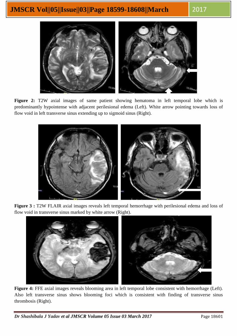
Figure 2: T2W axial images of same patient showing hematoma in left temporal lobe which is
predominantly hypointense with adjacent perilesional edema (Left). White arrow pointing towards loss of
flow void in left transverse sinus extending up to sigmoid sinus (Right).
Figure 3 : T2W FLAIR axial images reveals left temporal hemorrhage with perilesional edema and loss of
flow void in transverse sinus marked by white arrow (Right).
Figure 4: FFE axial images reveals blooming area in left temporal lobe consistent with hemorrhage (Left).
Also left transverse sinus shows blooming foci which is consistent with finding of transverse sinus
thrombosis (Right).

Dr Shashibala J Yadav et al JMSCR Volume 05 Issue 03 March 2017 Page 18602
JMSCR Vol||05||Issue||03||Page 18599-18608||March 2017
Figure 5: MR angiography reformatted images showing left transverse sinus thrombosis
Case2:Reversible Cerebral Vasoconstriction
Syndrome
35 years old female, one week post normal
delivery presented with thunderclap headache
which was diffuse in nature associated with
nausea, vomiting. There was no history of any
medical illness in past. On examination her blood
pressure was normal. MR brain was advice for
further evaluation which showed altered signal
intensity in right frontal lobe which was
hypointense on T1 and hyperintense on T2 (Figure
6). Axial images on T2 FLAIR showed hyperint-
ense signal in right frontal lobe (Figure 7). DWI
images revealed gyriform pattern hyperintensity in
right frontal lobe with signal drop on ADC
suggestive of restricted diffusion (Figure 8).
Finally MR angiography showed gross attenuation
of luminal caliber of bilateral middle cerebral
artery these narrowing was predominantly seen on
right side (Figure 9). Findings were demonstrated
on reformatted axial and coronal images (Fig. 10).
Figure 6: Altered signal intensity area hyperintense on T2 and hypointense on T1 in right frontal region
Figure 7: T2 FLAIR axial images reveals hyperintense signal in right frontal lobe

Dr Shashibala J Yadav et al JMSCR Volume 05 Issue 03 March 2017 Page 18603
JMSCR Vol||05||Issue||03||Page 18599-18608||March 2017
Figure 8: DWI reveals gyriform pattern hyperintensity in right frontal lobe (white arrow) which show signal
drop on ADC suggestive of restricted diffusion (black arrow)
Figure 9: MR angiography source images reveal gross attenuation of luminal caliber of bilateral middle
cerebral artery (right>left)
Figure 10: MR Angiography reformatted axial and coronal images reveals gross attenuation of luminal
caliber of bilateral middle cerebral artery (right>left)

Dr Shashibala J Yadav et al JMSCR Volume 05 Issue 03 March 2017 Page 18604
JMSCR Vol||05||Issue||03||Page 18599-18608||March 2017
Case 3: Posterior Reversible Encephalopathy
Syndrome.
A 31 years old female presented with severe
occipital headache, nausea, vomiting and two
episodes of seizure and photophobia. There was a
history of having delivered a full term male by
LSCS 10 days back. The indication of LSCS was
oligohydramnios. Immediate post LSCS period
was uneventful. But since 2 days she had a history
of nausea vomiting and headache. Today she got 2
episodes of generalized tonic clonic seizures. Each
episode lasted for about 2-3 minutes followed by
loss of consciousness for 10-15 minutes. Patient
remained drowsy in between the episodes of
seizure and the time of admission in hospital also
the patient had altered sensorium. MRI was done
which showed altered signal intensity areas in
bilateral parietal lobes which appeared
hyperintense on T2 weighted images and on T2
FLAIR (Figure 11). T2 FLAIR images of same
patient showing hyperintense signal in bilateral
parietal lobe MR Angiography reformatted, DWI
and ADC images were unremarkable (Figure
12,13,14 ).
Figure 11 : T2WI reveals hyperintense signal in bilateral parietal lobe (white arrow).
Figure 12: T2 FLAIR images of same patient showing hyperintense signal in bilateral parietal lobe (white
arrows).
Dr Shashibala J Yadav et al JMSCR Volume 05 Issue 03 March 2017 Page 18605
JMSCR Vol||05||Issue||03||Page 18599-18608||March 2017
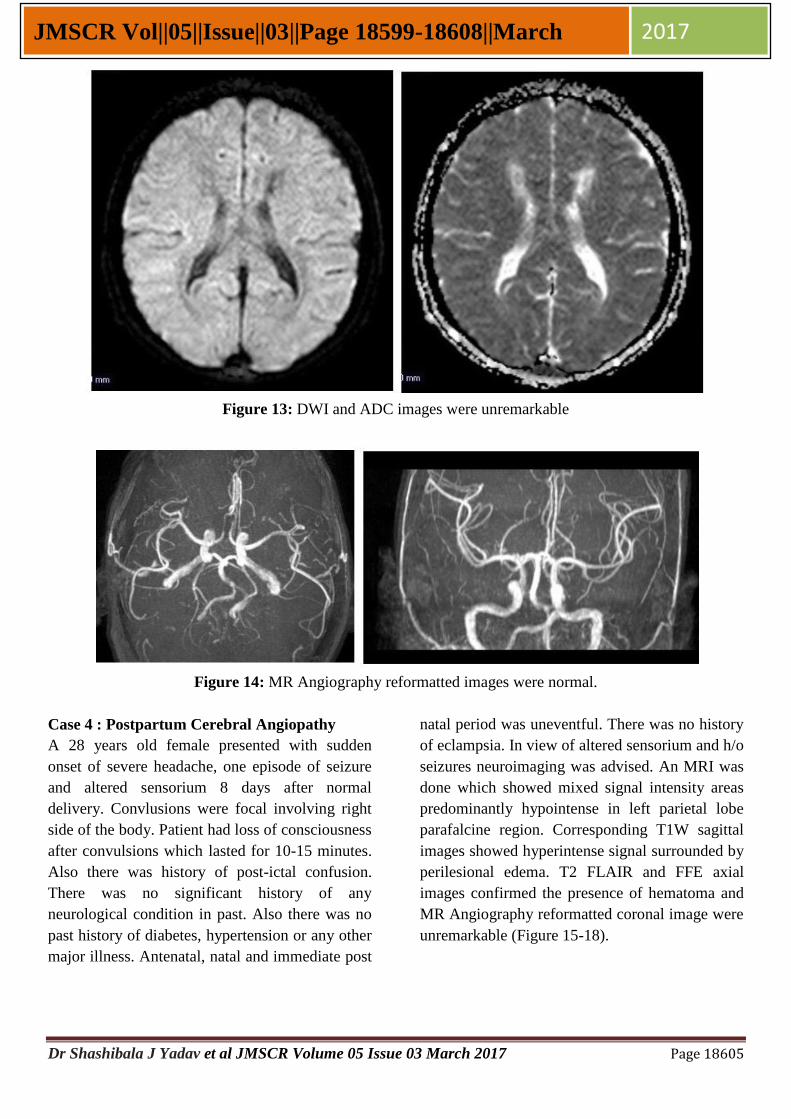
Figure 13: DWI and ADC images were unremarkable
Figure 14: MR Angiography reformatted images were normal.
Case 4 : Postpartum Cerebral Angiopathy
A 28 years old female presented with sudden
onset of severe headache, one episode of seizure
and altered sensorium 8 days after normal
delivery. Convlusions were focal involving right
side of the body. Patient had loss of consciousness
after convulsions which lasted for 10-15 minutes.
Also there was history of post-ictal confusion.
There was no significant history of any
neurological condition in past. Also there was no
past history of diabetes, hypertension or any other
major illness. Antenatal, natal and immediate post
natal period was uneventful. There was no history
of eclampsia. In view of altered sensorium and h/o
seizures neuroimaging was advised. An MRI was
done which showed mixed signal intensity areas
predominantly hypointense in left parietal lobe
parafalcine region. Corresponding T1W sagittal
images showed hyperintense signal surrounded by
perilesional edema. T2 FLAIR and FFE axial
images confirmed the presence of hematoma and
MR Angiography reformatted coronal image were
unremarkable (Figure 15-18).

Dr Shashibala J Yadav et al JMSCR Volume 05 Issue 03 March 2017 Page 18606
JMSCR Vol||05||Issue||03||Page 18599-18608||March 2017
Figure 15 : T2W axial images reveals hematoma in left parietal lobe parafalcine region showing mixed
signal intensity predominantly hypointense, corresponding T1W sagittal image show hyperintense signal
surronded by perilesional edema.
Figure 16: T2 FLAIR images of same patient showing hematoma.
Figure 17: FFE axial images showing hypointense signal supporting the diagnosis of hematoma.

Dr Shashibala J Yadav et al JMSCR Volume 05 Issue 03 March 2017 Page 18607
JMSCR Vol||05||Issue||03||Page 18599-18608||March 2017
Figure 18: MR Angiography reformatted coronal
image is unremarkable.
Discussion
Acute neurological conditions in post partum
period are usually related to eclampsia. Other
conditions which may be the cause of neurological
manifestations like seizures, focal deficit and
altered sensorium are dural sinus thrombosis,
cerebral vasoconstriction syndrome, posterior
reversible encephalopathy syndrome and
postpartum angiopathy. Eclampsia is usually well
recognized by obstetricians and usually there is
history of hypertension and albuminuria during
pregnancy. Other causes of acute neurological
manifestations like dural sinus thrombosis, stroke
or angiopathy should be diagnosed in early phase
and treated promptly as any can lead to
devastating complications [5]
. Since all these
conditions may initially present as headache there
is usually a history of initial treatment by oral
anaelgesics until serious neurological features
develop. Due to non-specific symptoms in initial
stages neuroimaging is of utmost importance for
early diagnosis [6]
.
Dural sinus thrombosis may manifest as
hemorrhagic or ischemic complications. It is more
common in females and it is of utmost importance
that obstetrician and those involved in care of
pregnant women should know that postpartum
period is associated with a significant increase in
incidence of cerebral venous thrombosis. Many
factors predispose women for development of
sinus thrombosis including hypercolagulable state
seen in pregnancy, infection, and antiphospholipid
antibody syndrome and prothrombin gene
mutation. Patient usually present with severe
headache, convulsions, and papilledema if severe
enough to cause raised intracranial pressure.
Though CT scan can be done MRI and MR
venography are the modalities of choice for the
diagnosis of dural sinus thrombosis [7]
.
Reversible vasoconstriction syndrome is another
cause of headache and neurological manifestation
in postpartum period. Patient usually present with
acute-onset severe (thunderclap) headaches which
may be accompanied by convulsions, features
simiar to or suggestive of stroke, and
encephalopathy. The condition is usually
reversible and prognosis is good. However, some
patients may experience severe spasm and
massive infarct. Rarely may it prove to be
fatal. The hallmark finding is the finding of
reversible vasoconstriction of the cerebral
vasculature mainly arteries. Convulsions caused
by reversible vasoconstriction syndrome should be
differentiated from seizures caused by eclampsia,
dural sinus thrombosis and angiopathy. The
diagnosis is usually by CT, MRI and MR
angiography. MR angiography may show attenu-
ation of luminal caliber of cerebral arteries [8]
.
Posterior reversible encephalopathy syndrome
(PRES) is characterized by headache, altered
sensorium, seizures, and visual disturbances.
Additionally, there are characteristic imaging
features associated with the syndrome which often
include focal regions of symmetric edema in the
parenchyma of posterior portion of brain. As the
name suggests It is usually reversible but some
patient may develop complications leading to
stroke and variable intensity of neurological
deficits. MRI may show altered signal intensity
areas in bilateral parietal lobes [9]
.
Postpartum angiopathy (PPA) is a
vasoconstriction syndrome of uncertain etiology
that affects medium and large sized cerebral
arteries. Postpartum angiopathy may cause
ischemic stroke. 2/3 of the cases of postpartum
angiopathy present within 1 week after delivery.
Dr Shashibala J Yadav et al JMSCR Volume 05 Issue 03 March 2017 Page 18608
JMSCR Vol||05||Issue||03||Page 18599-18608||March 2017
The diagnosis of postpartum angiopathy is made
by angiography. It may show segmental
narrowing and dilatation in large and medium-
sized cerebral vessels .MRI may show areas of
T2/FLAIR hyperintensities at any location,
especially in watershed areas of brain. It is
generally self limiting and the signs and
symptoms usually subside in 1- 3 months but
some patients may have residual neurological
manifestations for longer period of time. It is
important to know that angiography may be
normal in postpartum angiopathy in initial days
and if there is strong suspicion of angiopathy
repeat angiography should be done a few days
later [10]
.
Conclusion
Cerebrovascular events in postpartum period are
relatively common. These events may initially
present with non-specific symptoms like
headache, giddiness and vomiting. The familiarity
with the neuroimaging features of postpartum
encephalopathy may help in early diagnosis and
prompt treatment thereby preventing further
complications.
Conflict of interest: None
References
1. Anderson JA, Weitz JI. Hypercoagulable
states. Clin Chest Med. 2010 Dec;31
(4):659-73.
2. Maggi G, Lombana VA, Marcos EA, Ruiz
Huerta AD, Arévalo EG, Rodríguez FG.
Posterior leukoencephalopathy syndrome:
Postpartum focal neurologic deficits: A
report of three cases and review of the
literature. Saudi Journal of Anaesthesia.
2013;7(2):205-209.
3. SJ A, A B, Hussein OM, RA A. Stroke in
the Postpartum Period: A Case
Study. Journal of Clinical and Diagnostic
Research : JCDR. 2013;7(6):1183-1185.
4. Wagner SJ, Acquah LA, Lindell EP, et al.
Posterior Reversible Encephalopathy
Syndrome and Eclampsia: Pressing the
Case for More Aggressive Blood Pressure
Control. Mayo Clinic Proceedings.
2011;86(9):851-856.
5. Siddiqui FM, Kamal AK. Complications
associated with cerebral venous throm-
bosis. J Pak Med Assoc. 2006 Nov;56(11):
547-51.
6. Rijal JP, Giri S, Dawadi S, Dahal KV.
Posterior reversible encephalopathy synd-
rome (PRES) in a patient with late postp-
artum eclampsia. BMJ Case Reports. 2014
7. Prognosis of cerebral vein and dural sinus
thrombosis: results of the International
Study on Cerebral Vein and Dural Sinus
Thrombosis (ISCVT). Ferro JM, Canhão
P, Stam J, Bousser MG, Barinagar-
rementeria F, ISCVT Investigators.Stroke.
2004 Mar; 35(3):664-70.
8. Reversible cerebral vasoconstriction synd-
rome. Ducros A, Bousser MGPract
Neurol. 2009 Oct; 9(5):256-67.
9. Posterior reversible encephalopathy synd-
rome, part 1: fundamental imaging and
clinical features. Bartynski WSAJNR Am
J Neuroradiol. 2008 Jun; 29(6):1036-42.
10. Zunker P, Golombeck K, Brossmann J,
Georgiadis D, Deuschl G. Post-partum
cerebral angiopathy: repetitive TCD, MRI,
MRA, and EEG examinations. Neurol Res.
2002 Sep;24(6):570-2.