imaging of pulmonary vasculature - pulmonary... · introduction • many disease can affect the...
TRANSCRIPT

Imaging of Pulmonary Vasculature
Tan-Lucien H. Mohammed, M.D., FACR
Section of Thoracic Imaging
University of Florida

Introduction• Many disease can affect the pulmonary
vasculature ranging from large vessel disease such as aneurysm to capilliritis
• CT is the main diagnostic tool for evaluating the pulmonary vasculature
• Pulmonary angiography has limited use
• V/Q scanning remains an option for evaluating pulmonary thromboembolic disease

Objectives
• Recognize common imaging appearances of pulmonary vascular disease
• Learn the advantages and disadvantages of available imaging tools
• Use a case-based approach to pulmonary vascular disease

Case #1


Findings
• Axial CT images show filling defects in multiple subsegmental pulmonary artery branches
• Lung window image shows peripheral ground glass attenuation in RLL

Differential Dx
• Acute PE
• Chronic PE
• Tumor Thrombus (RCC, Head & Neck Tumors, Melanoma, etc.)

Diagnosis
• Acute Pulmonary Embolism with RLL infarction (Westermark Sign)

Case #2


Findings
• Axial CT image shows single filling defect in LLL subsegmental pulmonary artery branch
• Bilateral Pleural Effusions & Atelectasis (R>L)

Differential Dx
• Acute PE
• Chronic PE
• Tumor Thrombus (RCC, Head & Neck Tumors, Melanoma, etc.)
• Lymphadenopathy
• Artifact

Diagnosis
• Acute LLL Pulmonary Embolism in the setting of Congestive Heart Failure

Case #3


Findings
• Axial CT images show filling defects in multiple subsegmental pulmonary artery branches
• Enlargement of Right Heart

Differential Dx
• Acute PE
• Chronic PE
• PA Angiosarcoma

Diagnosis
• Acute Pulmonary Embolism (Shower Pattern) with Right Ventricular Strain

Case #4


Findings
• Selective Right PA Angiogram depicts filling defects in the right central and upper lobe pulmonary artery branches

Differential Dx
• Acute PE
• Chronic PE
• PA Angiosarcoma

Diagnosis
• Acute Pulmonary Embolism

Case #5


Findings
• V/Q scan showing multiple ventilation/perfusion mismatches

Differential Dx
• Acute PE
• Chronic PE
• Atelectasis
• Multifocal Pneumonia

Diagnosis
• High Probability V/Q Scan suggesting Acute Pulmonary Embolism

Case #6

Findings
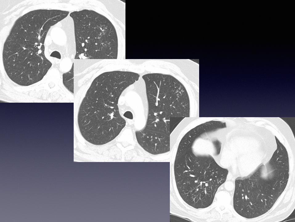
• Axial CT images reveal multiple eccentric filling defects in subsegmental pulmonary artery branches
• Cardiomegaly and enlarged Main Pulmonary Artery

Differential Dx
• Acute PE
• Chronic PE
• Tumor Thrombus (RCC, Head & Neck Tumors, Melanoma, etc.)

Diagnosis
• Chronic Pulmonary Embolism

Case #7


Findings
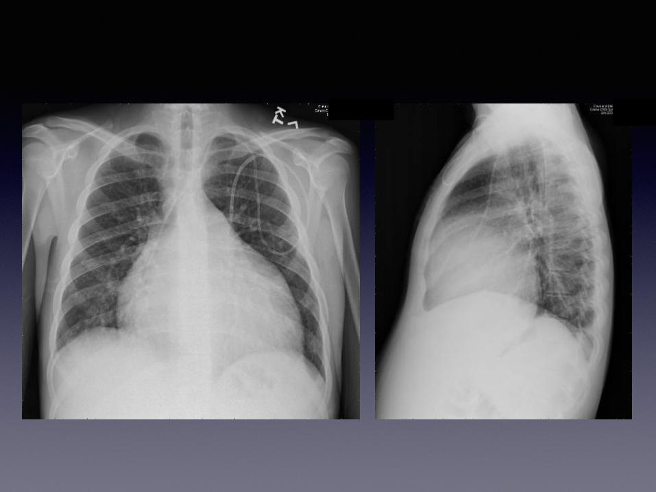
• Chest radiograph shows symmetrically enlarged central pulmonary arteries
• Axial CT images depict eccentric filling defects in multiple subsegmental pulmonary artery branches
• Markedly Enlarged Pulmonary Arteries

Differential Dx
• Acute PE
• Chronic PE
• Tumor Thrombus (RCC, Head & Neck Tumors, Melanoma, etc.)

Diagnosis
• Chronic Pulmonary Embolism

Case #8


Findings
• Axial CT images show filling defects in multiple subsegmental pulmonary artery branches
• Corresponding PET images depict increased metabolic activity in regions of filling defects

Differential Dx
• Acute PE
• Tumor Thrombus (RCC, Head & Neck Tumors, Melanoma, etc.)
• PA Angiosarcoma

Diagnosis
• Pulmonary Artery Angiosarcoma

Case #9


Findings
• Axial CT images show filling defects in RUL pulmonary artery branches
• Lung window images depict peripheral tree-in-bud nodules in LUL and RLL
• Single CT image through upper abdomen shows exophytic mass extending from right kidney

Differential Dx
• Tumor Thrombus (RCC, Head & Neck Tumors, Melanoma, etc.)
• Acute PE
• Multifocal Infection (Fungal Pneumonia, etc.)

Diagnosis
• Hematogenous Tumor Spread from RCC

Case #10


Findings
• Lung window image shows RLL clustered nodules
• Sequential soft tissue windows reveal RLL systemic arterial supply

Differential Dx
• Pulmonary Varix
• Pulmonary Sequestration
• RLL Pneumonia

Diagnosis
• RLL Sequestration (Intralobar)

Case #11


Findings
• CXR shows subtle left basilar nodular opacity
• CT images demonstrate LLL air trapping and serpiginous vessels

Differential Dx
• Lobar Sequestration
• Congenital lobar emphysema
• LLL Pneumonia

Diagnosis
• Congenital Absence of Right Pulmonary Artery (Hypoplastic right lung)

Case #11


Findings
• CXR shows cardiomegaly and vague LLL nodule
• Lung window images depict LLL serpiginous vessels

Differential Dx
• Pulmonary Varix
• Pulmonary AVM
• Neoplasm

Diagnosis
• LLL Pulmonary AVM

Case #12


Findings
• Coronal Reconstructed CT image shows serpiginous vessels in LLL

Differential Dx
• Pulmonary Varix
• Pulmonary AVM
• Neoplasm

Diagnosis
• LLL Pulmonary Varix

Case #13


Findings
• CXR Demonstrates cardiomegaly and prominent central pulmonary arteries (LUL embolization coils)
• CT image shows numerous corkscrew vessels in both lungs as well as LLL patchy ground glass

Differential Dx
• LLL Pneumonia
• Pulmonary AVMs
• LLL Infarct (from acute PE)

Diagnosis
• Osler-Weber-Rendu (Hereditary Hemorrhagic Telangectasia)

Case #14


Findings
• CXR shows rounded retrocardiac opacities
• Axial CT images depict numerous serpiginous paraesophageal vessels

Differential Dx
• Varices
• Vascular neoplasm (sarcoma, etc.) extending from upper abdomen
• LLL Sequestration

Diagnosis
• Paraesophageal Varices (from portal hypertension secondary to Cirrhosis)

Case #15


Findings
• CT images show prominent central pulmonary arteries as well as scattered, mild interlobular septal thickening

Differential Dx
• Congestive Heart Failure
• Pulmonary Hypertension
• Capillary Hemangiomatosis (PCH)
• Veno-Occlusive Disease (PVOD)

Diagnosis
• Pulmonary Veno-Occlusive Disease (PVOD)

Case #16


Findings
• CXR demonstrates cardiomegaly, prominent central pulmonary arteries and diffuse nodular opacities
• CT images show numerous centrilobular nodules as well as scattered, patchy ground glass

Differential Dx
• Multifocal Pneumonia
• Pulmonary Hypertension
• Capillary Hemangiomatosis (PCH)
• Chronic PE

Diagnosis
• Pulmonary Capillary Hemangiomatosis (PCH)

Case #17


Findings
• CXR demonstrates asymetric left hilar prominence (? central pulmonary arteries)
• CT image depicts focal stenotic narrowing of the proximal left pulmonary artery with associated post stenotic dilitation

Differential Dx
• Pulmonary Hypertension
• Pulmonary Valve Stenosis
• Behcet’s Disease
• Pulmonary Artery Aneurysm

Diagnosis
• Behcet’s Disease

Case #18


Findings
• CT images reveal fenestrated filling defect involving RLL pulmonary artery

Differential Dx
• Acute PE
• Chronic PE
• Pulmonary artery aneurysm
• Pulmonary artery dissection

Diagnosis
• Pulmonary artery dissection (Iatrogenic - after difficult Swan-Ganz placement)

Case #19


Findings
• CT image reveals abnormal vessel wrapping around left mainstem bronchus

Diagnosis
• Pulmonary Sling

Case #20


Findings
• Single CT images reveal reflux of contrast into the azygous vein

Differential Dx
• Venous fistula (Iatrongenic)
• PAPVR

Diagnosis
• PAPVR

Case #21


Findings
• CXR shows right hemithorax volume loss and paucity of right lung vessels
• CT images demonstrate no right pulmonary artery

Differential Dx
• Normal variant
• Congenital Absence of Right Pulmonary Artery (Hypoplastic right lung)

Diagnosis
• Congenital Absence of Right Pulmonary Artery (Hypoplastic right lung)

Case #22


Case #23


Findings
• CXR shows vertical vessel in right lung base with associated hemithorax volume loss
• CT images reveal anomolous vasculature in RLL

Differential Dx
• Scimitar Lung (Partial Anomolous Pulmonary Venous Return- PAPVR)
• Pulmonary Varix
• Hypoplastic Right Lung

Diagnosis
• Scimitar Lung (Partial Anomolous Pulmonary Venous Return- PAPVR)

Case #24


Findings
• Corona l Recon s t r u c t ed CT image demonstrates anomolous vasculature in hemithorax

Differential Dx
• Scimitar Lung (Partial Anomolous Pulmonary Venous Return- PAPVR)
• Pulmonary Varix
• Hypoplastic Right Lung

Diagnosis
• Scimitar Lung (Congenital Venolobar Syndrome)

Case #25


Findings
• Multiple CT images reveal multifocal ground glass and scattered foci of subpleural reticulation

Differential Dx
• Fungal Pneumonia (Low-grade)
• Vasculitis
• Early fibrosis

Diagnosis
• Churg-Strauss (Vasculitis)

Case #26


Case #27


Findings
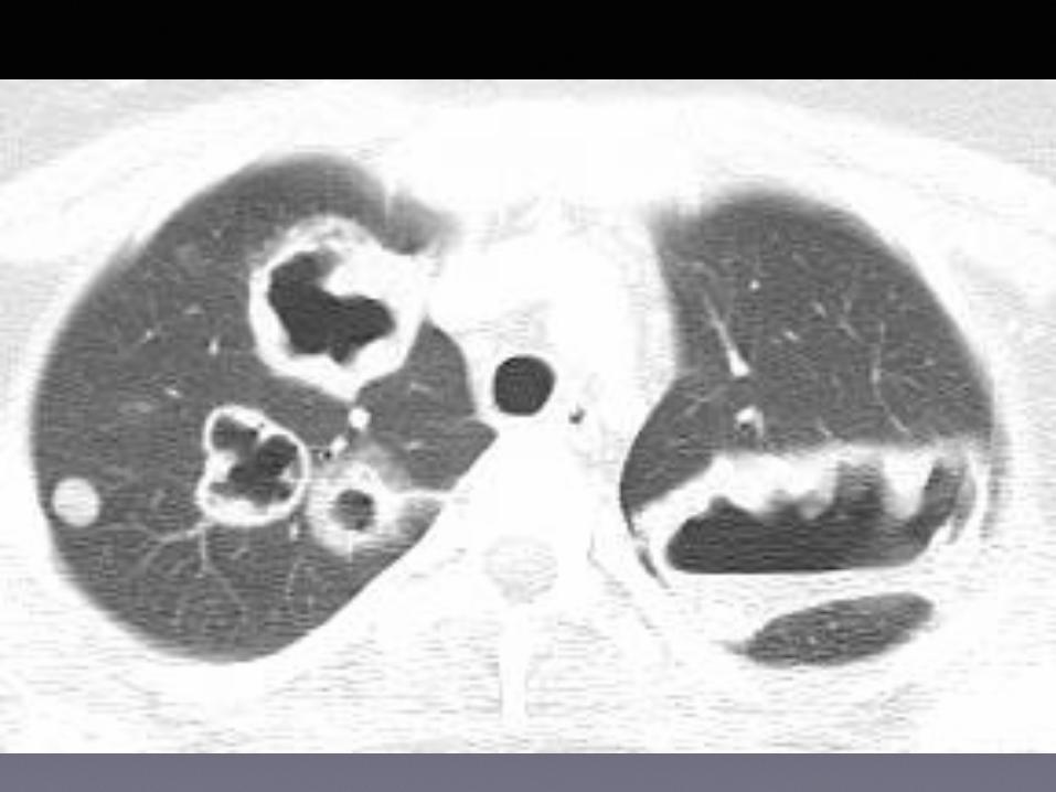
• CT images reveal multiple thin-walled cavities in both lungs

Differential Dx
• Pulmonary metastasis (RCC, Sarcoma, etc.)
• Cavitary pneumonia (fungal or bacterial)
• Vasculitis (Wegener’s)
• Septic Emboli

Diagnosis
• Wegener’s Granulomatosis

Case #28

Findings
• CT image shows multiple thick-walled cavities in both upper lungs

Differential Dx
• Pulmonary metastasis (RCC, Sarcoma, etc.)
• Cavitary pneumonia (fungal or bacterial)
• Vasculitis (Wegener’s)
• Septic Emboli

Diagnosis
• Wegener’s Granulomatosis

Case #29


Findings
• CXR depicts near complete right hemithorax opacification and left perihilar opacity
• CT image shows diffuse right lung ground glass attenuation & LUL non-dependent consolidation

Differential Dx
• Pulmonary lymphoma/leukemia
• Multifocal pneumonia (fungal or bacterial)
• Vasculitis (Wegener’s)
• COP (Cryptogenic Organizing Pneumonia)

Diagnosis
• Wegener’s Granulomatosis

Case #30


Findings
• CXR demonstrates patchy bibasilar opacities
• CT images depict multifocal ground glass primarily in the lung bases

Differential Dx
• Pulmonary hemorrhage
• Pulmonary edema
• Pulmonary contusion
• Multifocal Pneumonia

Diagnosis
• Pulmonary hemorrhage due to Goodpasture’s Disease (renal-pulmonary disease)

Summary
• Imaging manifestations of pulmonary vascular disease are numerous
• CT is the imaging tool of choice for evaluating the pulmonary vasculature
• CT findings of vasculitis are non-specific and can mimic infection and neoplasm