imaging of the abdomen and pelvis anne goldschmidt, md radiological associates of duluth
TRANSCRIPT

Imaging of the Abdomen and Pelvis
Anne Goldschmidt, MD
Radiological Associates of Duluth

Purpose
• Basic introduction to abdominal imaging modalities
• Introduction to common abdominal pathologies and how imaging can help in differential diagnosis
• Correlation with gross anatomy

Imaging Modalities
• Plain film (X-ray, KUB)
• Ultrasound (US)
• Computed Tomography (CT)
• Magnetic Resonance Imaging (MRI)– Problem solving, not primary
• Barium Studies
• Nuclear Medicine

Plain Films
• Electromagnetic energy passed through the body results in an image on the film
• Based on the density of the structures it passes through

Ultrasound
• Medical imaging technique uses high frequency sound waves
• Sound waves leave probe, travel into body and are reflected back to the machine to be analyzed

Ultrasound
• Location of structures is based on the time required for sound waves to return to the probe
• Intensity of the echoes on the image is dependent on composition of structures

Computed Tomography
• Electromagnetic energy (ionizing radiation)
• Gantry moves around patient, scanning from many angles
• Computer generates a 3D image

Magnetic Resonance Imaging
• No ionizing radiation
• Utilizes high field strength magnet to align hydrogen ions
• Radio frequency pulse specific to hydrogen ions causes them to precess
• With time hydrogen returns to alignment with magnetic field—different tissues, different time

Abdomen MRI

Nuclear Medicine
• Radiotracers are injected into body
• More functional than many tests
• Less detailed anatomic information
• GB disease

Barium Studies
• Utilizes x-rays• Contrast material
placed into GI tract• Antegrade for upper
GI tract• Retrograde for colon

Plain Films
• Dense=white– Bone or metal
• Lucent=black– Stomach bubble– Lung bases– Air in bowel

Two Views
• For CXR get PA and Lateral
• For abdominal imaging typically get supine and upright views– Lateral decubitus view if unable to stand

Abdominal Films

Approach to Interpretation
• Free air
• Bowel gas pattern
• Calcifications
• Soft tissues
• Bones
• Lines and tubes: NG tubes, drains, stents, surgical clips or hardware

Pneumoperitoneum(Free Air)
• Extraluminal air
• Post op finding
• Perforation of a hollow viscus
• Supine film—difficult to diagnose unless large volume
• Upright—look for air between liver and diaphragm

Free Air

Bowel Gas Pattern
• It is normal to see gas and stool within the colon
• Normal colon caliber less than 6cm
• Worry about cecum larger than 10cm

Plain film vs BE

Small Bowel Air
• Normal caliber less than 3cm
• Small bowel is located centrally in abdomen, compared with more peripheral colon

Dilated Small Bowel
• Primary question is whether or not there is a mechanical obstruction or if there is abnormal motility of the bowel
• Is there a SBO or ileus?

Signs and Symptoms of Bowel Obstruction
• Depends on level—higher, less bowel distention and earlier symptoms
• Vomiting—leads to loss of electrolyte rich fluid which can lead to shock
• Crampy pain
• Constipation
• Can be partial—symptoms less severe– May have diarrhea

Small Bowel Obstruction vs. Ileus
• SBO• Mechanical blockage
– Fibrous band or adhesion
– Hernia– Neoplasm– Stricture– Volvulus
• Abnormal peristalsis or motility– Post op, due to
manipulation– Intraabdominal or
retroperitoneal infection
– Ischemia

SBO
• Obstruction leads to distention proximal to level of block with collapse distally
• See differential caliber, with air/fluid levels on upright view

Complications
• Accumulation of ingested fluid and food, plus digestive secretions and gas leads to distention
• Strangulation—Abnormal blood flow– Initially see venous occlusion, then arterial
occlusion and finally ischemia and infarction– Usually seen with hernia, Volvulus, or
intussusception

Calcifications
• GB
• Kidneys
• Pancreas
• Vascular
• Pelvic—fibroids, ovary

Nephrolithiasis
• Pathogenesis related to dehydration (decreased urine volume) or increased excretion of stone constituents
• 90% calcium, 65% oxylate
• S/S:– May be silent– Renal colic—excruciating pain in flank—
genitalia, thigh

Urinary Tract Obstruction
• Excretory Urogram (IVP)– Inject x-ray dye and take serial films as it is
excreted by kidneys– Stone blocks the flow of contrast, leading to
delayed excretion and dilatation of the collecting system and ureter
– Depending on degree of obstruction can take a long time to complete

Obstruction on IVP

CT for Urinary Tract Obstruction
• CT has replaced IVP for most patients
• Requires no IV contrast material
• Very quick
• Can show other causes of pain if there is no stone

CT of Urinary Tract Obstruction

Calcifications

Calcifications

Calcifications

Calcifications

Calcifications

Common Abdominal Problems
• Appendicitis
• Cholelithiasis and Cholecystitis
• Pancreatitis
• Diverticulitis
• Abscess

Appendicitis
• Bacterial infection of appendix
• Adolescents and young adults– Peak age 15-24
• Inflammation – edema and ischemia—gangrene and perforation

Signs/Symptoms
• Midepigastrium pain—RLQ
• N/V
• +/- mild fever, elevated WBC
• Tenderness/guarding at McBurney’s point
• Rebound implies peritoneal inflammation
• Atypical location—atypical S/S

Diagnosis
• Physical exam• US—especially
women and children• CT

Ultrasound
• Normal appendix less than 6mm
• Normal appendix is compressible with transducer pressure
• DX—Tubular, dilated, non compressible structure in RLQ

CT of Appendicitis
• Normal appendix thin walled, non dilated
• Normal fat is black• DX—Dilated appendix
with thickening of the wall and dirty fat

Gall Bladder Disease
• Cholelithiasis—stones in the GB– 10% in US– 20% of patients over 40
Formation related to cholesterol biosynthesis
Often mixed—cholesterol, calcium, bilirubin
Medication may dissolve

Gall Stones

Acute Cholecystitis
• Inflammation of GB, usually due to obstruction by stone
• S/S: RUQ pain, NV, Flatulence
• Murphey’s sign—localized tenderness

Cholecystitis
• Diagnosis– Plain films– US– CT– HIDA Scan—a more functional test

Cholecystitis
• Ultrasound finding• Stones with
shadowing• Thickening of wall• Pericholecystic fluid• + Sonographic
Murphey’s

Cholecystitis
• CT diagnosis• Stones• Thick wall• Dirty fat• Abscess

HIDA Scan
• Functional study of liver uptake and excretion into biliary tree.
• Normal to see excretion into GB and then into SB

Pancreatitis
• Inflammation of pancreas
• Etiologies: Biliary disease, alcoholism, surgery, trauma, drugs—1/3 unknown
• Gross path: edema, hemorrhage, necrosis
• Complications related to enzymes: pseudocysts, abscess, fat necrosis
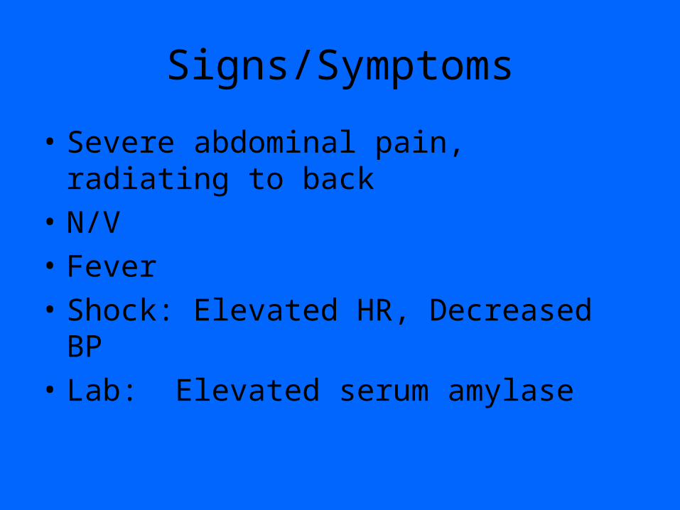
Signs/Symptoms
• Severe abdominal pain, radiating to back
• N/V
• Fever
• Shock: Elevated HR, Decreased BP
• Lab: Elevated serum amylase

Pancreatitis Diagnosis
• Plain Films– Calcification in chronic– GS
• CT– May be normal– Use CT to look for complications

Pancreatitis

Diverticular Disease
• Diverticula—small, sacular mucosal herniation through muscular wall of colon– Most common in sigmoid– 30-40% over age 50– Increased incidence with age– Related to refined, low fiber diet

Diverticula

Diverticulitis
• Inflammation of the Diverticula, leads to perforation with inflammation of pericolonic tissues
• Complications:– Abscess– Wall thickening with obstruction– Perforation

Diverticulitis
• S/S
• Pain with localized tenderness, esp. LLQ
• Crampy pain if obstruction
• Diagnosis– BE– CT

Diverticulitis
• Barium enema• Diverticula• Thickening of wall
with stricture• Extraluminal contrast• Mass effect from
adjacent inflammation or abscess
• CT• Diverticula• Thick wall with
stricture• Dirty fat• Abscess, Phlegmon• CAN MIMIC CANCER

BE of Diverticulitis

CT of Diverticulitis

ABCESS
• Inflammation—walled off collection
• Appendicitis, diverticulitis, IBD, PID, etc
• S/S: fever, pain with local tenderness, elevated WBC
• Diagnosis: CT– Need oral contrast material– IV helpful

Abscess

Summary
• Because of overlap in S/S of various abdominal processes, imaging is critical in evaluating patients presenting with acute abdominal complaints
• Plain films are helpful to assess bowel gas patterns
• US is primary modality for evaluating the GB
• CT is primary modality for most other DDx