immune checkpoint receptors: homeostatic regulators of ... · ctla-4 cytotoxic t-lymphocyte...
TRANSCRIPT

REVIEW ARTICLE
Immune checkpoint receptors: homeostatic regulators of immunity
Antonio Riva1,2 • Shilpa Chokshi1,2
Received: 5 February 2018 / Accepted: 23 April 2018 / Published online: 8 May 2018� The Author(s) 2018
AbstractAlcoholic liver disease (ALD) is an escalating global problem accounting for more than 3 million deaths annually. Bacterial
infections are diagnosed in 25–47% of hospitalized patients with cirrhosis and represent the most important trigger for acute
decompensation, multi-organ failure, septic shock and death. Current guidelines recommend intensive antibiotic therapy,
but this has led to the emergence of multi-drug resistant bacteria, which are associated with increased morbidity and
mortality rates. As such, there is a pressing need to explore new paradigms for anti-infective therapy and host-directed
immunomodulatory therapies are a promising approach. Paradoxically, cirrhotic patients are characterised by heightened
immune activity and exacerbated inflammatory processes but are unable to contend with bacterial infection, demonstrating
that whilst immune effector cells are primed, their antibacterial effector functions are switched-off, reflecting a skewed
homeostatic balance between anti-pathogen immunity and host-induced immunopathology. Preservation of this equilibrium
physiologically is maintained by multiple immune-regulatory checkpoints and these feedback receptors serve as pivotal
regulators of the host immunity. Checkpoint receptor blockade is proving to be effective at rescuing deranged/exhausted
immunity in pre-clinical studies for chronic viral infection and sepsis. This approach has also obtained FDA approval for
restoring anti-tumor immunity, with improved response rates and good safety profiles. To date, no clinical studies have
investigated checkpoint blockade in ALD, highlighting an area for development of host-targeted immunotherapeutic
strategies in ALD, for which there are no current specific treatment options. This review aims at framing current knowledge
on immune checkpoints and the possibility of their therapeutic utility in ALD-associated immune dysfunctions.
Keywords Checkpoint � Immunotherapy � ALD
AbbreviationsARC Alcohol-related cirrhosis
APC Antigen-presenting cell
CD Cluster of differentiation
CTLA-4 Cytotoxic T-lymphocyte associated protein 4
IFN Interferon
IL Interleukin
LAG-3 Lymphocyte-activation gene 3
MHC Major histocompatibility complex
MAIT Mucosa-associated invariant T cells
NK Natural killer cells
NKT Natural killer T cells
PBMC Peripheral blood mononuclear cells
PD-1 Programmed death 1
PD-L1/2 Programmed death ligands 1 and 2
ROS Reactive oxygen species
SAH Severe alcoholic hepatitis
TIM-3 T cell immunoglobulin and mucin domain 3
HAVCR2 Hepatitis A virus cellular receptor 2
TNF Tumor necrosis factor
Alcoholic liver disease
Alcoholic liver disease (ALD) is an escalating problem
worldwide and is responsible for more than 3 million
deaths annually, representing 5.9% of all deaths globally
[1]. Alcohol-attributable deaths vary by continent and
This review is based on a presentation by Dr Shilpa Chokshi at the
‘Frontiers in Hepatology’ meeting held at the Institute of Hepatology,
Foundation for Liver Research on 10th October 2017 in London, UK.
& Shilpa Chokshi
Antonio Riva
1 Institute of Hepatology London, Foundation for Liver
Research, 111 Coldharbour Lane, London SE5 9NT, UK
2 Faculty of Life Sciences and Medicine, King’s College
London, London, UK
123
Hepatology International (2018) 12:223–236https://doi.org/10.1007/s12072-018-9867-9(0123456789().,-volV)(0123456789().,-volV)
country, but Europe maintains the global record in terms of
prevalence of alcohol consumption [2], with alcohol being
responsible for 1 in 7 male deaths and 1 in 13 female
deaths in the 15–64 year age group. In the UK alone, liver
disease is the third biggest cause of premature mortality in
the 18–64 year age group after ischemic heart disease and
self-harm, with standardised mortality rates 4–5 times
higher since the 1970s [3, 4]. Alcohol abuse also represents
one of the strongest risk factors for the development of
liver cancer [5], which is 16% more likely in those who
drink heavily ([ 5 units/day). Cancer Research UK esti-
mates that liver cancer killed 4500 people in the UK in
2012, 3% of all cancer deaths [5]. Clearly, the potential
societal impact of the development of effective therapeutic
agents for ALD is far reaching.
There are currently no specific and efficacious thera-
peutic options for ALD patients, with abstinence being the
cornerstone of treatment together with supportive care and
liver transplantation currently indicated only for the most
severe cases and available in very limited centres world-
wide. This is compounded by a widespread reluctance to
consider patients with advanced liver disease as candidates
for transplantation [6].
Not surprisingly, abstinence is ineffective in com-
pletely reversing alcohol-related liver damage in patients
who have been long-term excess alcohol abusers and who
are at a more advanced and severe stage of disease. In this
group, alcohol-related cirrhosis (ARC) is the most common
form of established liver disease, which is accompanied by
an increased risk of decompensation, organ failure and
death. Moreover, high recidivism rates in abstinent patients
can lead to repeated presentations of severe alcoholic
hepatitis (SAH), the most florid form of ALD, a progres-
sive inflammatory liver condition with a mortality rate of
over 30% at 1 month post-hospitalization [7, 8] and
approximately 60% during recidivism [9].
ALD is associated with multiple derangements in host
immunity and it is now well-established that ARC induces
a state of profound immunodeficiency, known as cirrhosis-
associated immunodeficiency syndrome (CAIDS)
[8, 10–12], which is accompanied by ongoing non-specific
systemic inflammation, rendering ARC patients highly
susceptible to overwhelming bacterial infections. This
increases the risk of organ dysfunction including hepatic
encephalopathy, renal failure and circulatory collapse, with
no option for liver transplantation acutely [13, 14]. Alcohol
abstinence does not fully resolve this, as once the liver is
severely injured the deficiency in patients’ immunity
remains and development of infection significantly com-
promises their survival chances [15].
Indeed, bacterial infections, sepsis and associated
endotoxemia are diagnosed in 25–47% of hospitalized
patients with cirrhosis and represent the most important
triggers for acute decompensation and progression to
multi-organ failure and septic shock, with short-term
mortality of [ 75% [11]. In SAH patients, the suscepti-
bility to bacterial infection is further heightened and
infection is observed in nearly 50% of cases in the short
term with a high proportion of them ultimately dying of
sepsis [16]. There is increasing evidence that changes in
gut permeability, bacterial dysbiosis and translocation of
bacteria from the ‘leaky’ gut into the systemic circulation
in ALD is causatively linked to this increase in infections
[17].
Current guidelines recommend early antibiotic therapy
in these patients and suggest the use of corticosteroids to
reduce the systemic inflammation associated with ALD
[7, 8, 11, 16]. However, the intensive use of antibiotics has
caused selection of multi-drug resistant bacteria, with some
European centres reporting rates [ 20% [18] and the
STOPAH trial has clearly demonstrated lack of benefit
with steroid treatment, both short term and long term [19].
Moreover, steroid treatment per se is associated with a
further increased susceptibility to infection and sepsis [20].
It is critical to understand why ALD patients have
impaired pathogen defence to develop targeted treatments
to restore functional immunity. We have previously
demonstrated that ALD patients harbour dramatic dys-
functions in their antibacterial defences, affecting both
innate and adaptive immune cells (including neutrophils,
monocytes, NK cells, T cells and Tregs) and also innate-
like subsets (such as NKT cells and MAIT cells), and their
responses to bacterial challenge are suboptimal and insuf-
ficient [21, 22] but reversible. Together, our findings have
opened new avenues of research into the immunopatho-
genesis of ALD and have identified novel potential
immunotherapeutic targets for the treatment of this com-
plex disease, in particular the negative immune checkpoint
receptors PD-1 and TIM-3 [23, 24]. We believe that host-
directed immunomodulatory therapies aimed at restoring
the dysfunctional immunity are a promising approach for
the treatment of ALD. In this review, we aim to frame
current knowledge on immune checkpoints and the possi-
bility to use checkpoint blockade therapeutically in the
context of ALD-associated immune dysfunctions.
Immune responses are implicitly self-limiting
The immune system has a multitude of implicit mecha-
nisms of self-regulation. Physiologically, these mecha-
nisms promote immune tolerance and control unwanted
and excessive injurious immune responses. This maintains
immunological homeostasis, preventing immunopathology
and limiting excessive inflammation and immune-mediated
224 Hepatology International (2018) 12:223–236
123

damage. A major arm of this multifaceted immunoregula-
tory network are the checkpoint receptors, a complex array
of membrane receptors and their ligands that act as immune
modulators, suppressing or activating key signal transduc-
tion pathways and modulating effector cell functions. By
doing so, they fine-tune the magnitude, spread and breadth
of the immune response, containing it and making it
effective (Figs. 1a, 2a). Some of the main negative immune
checkpoints that are currently being investigated in cancer,
chronic viral infections and sepsis include the PD-1/PD-L1/
PD-L2 pathway, the TIM-3/Galectin-9 pathway, CTLA-4
and LAG-3. However, there is an ever-increasing list of
receptors and currently more than 20 endogenous
immunoregulatory pathways have been identified and at
least partly characterised [25].
Upon acute cell activation, these receptors appear on the
cell surface concurrently with the acquisition of a very
active functional profile (including secretion of multiple
cytokines) [26] and are subsequently downregulated during
the immune contraction phase, when the acute insults are
resolved, tissue repair and wound healing mechanisms
become predominant and immunological memory is
consolidated. Instead, during chronic inflammation,
chronic infections, cancer or sepsis, characterised by high
levels of antigen and proinflammatory cytokines, multiple
immunosuppressive checkpoint receptors are persistently
hyper-expressed on the cell surface and are continuously
activated, chronically suppressing immune cell functions.
This phenomenon, called immune ‘‘exhaustion’’, is char-
acterised by a sequential loss of immune activities,
including T-cell proliferation, secretion of cytokines
and cytotoxic markers, and priming of pro-apoptotic
pathways, causing a progressive immune shut-down
[26, 27]. Furthermore, other immunocytes including B cells
and NK cells are subjected to similar exhaustion processes,
thereby extending the depth of immune suppression to
humoral and innate immune responses [28–32]. The
patients’ overly active immune system thus contains
immunopathology and preserves the structural and func-
tional integrity of tissues and organs but becomes unable to
mount strong, effective and coordinated anti-pathogen
responses (Fig. 2b). This favours susceptibility to infection,
especially with opportunistic pathogens, similar to what we
observe in septic patients and in ARC/SAH patients
[10, 11, 33–35]. Immune functions remain persistently
deranged for years after the resolution of the first septic
episode in septic survivors and even after years of alcohol
abstinence in ARC/SAH patients [15]. This is directly
linked to persistently high expression of negative immune
checkpoints [34].
Different immune checkpoints display different
anatomical and temporal patterns of expression. The
kinetics of immune checkpoint expression are highly reg-
ulated and coordinated and the dynamic interplay between
stimulatory receptors (such as CD28, CD80 and CD86) and
inhibitory checkpoints (e.g., PD-1, PD-L1, TIM-3) during
cellular activation defines the evolution and fate of the
existing immune response.
The PD-1 and TIM-3 pathways
The PD-1 pathway belongs to the CD28/B7 family of
T-cell co-receptors. PD-1/CD279 is probably the most
studied checkpoint receptor in the field of T-cell exhaus-
tion. This receptor was first identified in apoptotic T-cell
lines (hence the name ‘‘programmed death 1’’) but was
soon characterised as a negative immunoregulator [36–39].
PD-1 has two known ligands, namely PD-L1/B7-H1/
CD274 and PD-L2/B7-DC/CD273 [40–43]. PD-L1 is
ubiquitously expressed at low levels and is strongly
induced by proinflammatory signals [44, 45], while PD-L2
displays a more restricted expression profile [43, 46, 47].
Upon engagement, PD-1 sequesters intracellular factors
involved in the TCR signalling, stopping T-cell activation
[40–43]. PD-1/PD-L1 signalling appears to be
Fig. 1 Immune regulation by checkpoint receptors (CR) and their
ligands (CR–L), and effects of immune checkpoint blockade with
neutralizing antibodies. Checkpoint receptors modulate the breadth,
magnitude and spread of the immune response by balancing
stimulatory and inhibitory signals delivered to immune cells by
antigen-presenting cells or target cells (a). Blockade of immune
checkpoint receptors or their ligands with neutralizing antibodies
(Anti-CR Ab and Anti-CR–L Ab) can dampen inflammatory
responses and restore dysfunctional immunity (b)
Hepatology International (2018) 12:223–236 225
123
bidirectional: PD-L1-expressing cancer cells possibly
receive anti-apoptotic signals upon interaction with PD-1-
expressing T cells [48, 49], but it is not known if this
happens also in the context of T-cell interactions with
antigen-presenting cells (APCs). PD-L1 also binds CD80,
triggering inhibitory signals within PD-L1-expressing cells
[50–53]. Amongst T cells, PD-1 is mostly expressed on
primed T cells and is strongly upregulated upon TCR-
mediated antigen-specific T-cell activation in peripheral
tissues. Therefore, the PD-1 pathway is believed to play a
role in the establishment and maintenance of peripheral
tolerance [54]. It has been demonstrated that the PD-1
pathway can modulate immune cells other than T cells. The
effect of PD-1 engagement on causing B cell exhaustion,
for instance, is well-described [28, 29] and PD-1 expres-
sion on NK cells has also been linked to NK-cell functional
suppression [31, 32]. In a study investigating immune
exhaustion in HIV patients, contact with bacterial products
induced monocyte expression of PD-1 and these monocytes
secreted suppressive IL-10 upon PD-1 engagement. Fur-
thermore, T-cell exhaustion in these patients could be
reversed by blocking either PD-1 or IL-10 receptor [55].
Monocyte activation by bacterial endotoxin was also
shown to physiologically cause increased secretion of
suppressive IL-10 and upregulation of PD-1 and TIM-3 on
T cells, and simultaneous blockade of TLR4 and CD14
abolished IL-10 secretion and inhibited T-cell checkpoint
upregulation [21]. These findings link the PD-1 pathway to
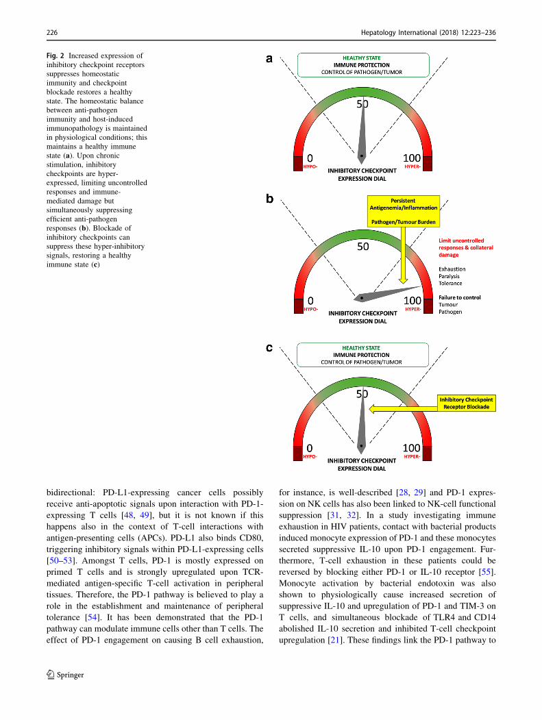
Fig. 2 Increased expression of
inhibitory checkpoint receptors
suppresses homeostatic
immunity and checkpoint
blockade restores a healthy
state. The homeostatic balance
between anti-pathogen
immunity and host-induced
immunopathology is maintained
in physiological conditions; this
maintains a healthy immune
state (a). Upon chronic
stimulation, inhibitory
checkpoints are hyper-
expressed, limiting uncontrolled
responses and immune-
mediated damage but
simultaneously suppressing
efficient anti-pathogen
responses (b). Blockade of
inhibitory checkpoints can
suppress these hyper-inhibitory
signals, restoring a healthy
immune state (c)
226 Hepatology International (2018) 12:223–236
123
the regulation of antibacterial immunity, which is key in
ALD patients.
TIM-3/CD366 was first described as a marker of acti-
vated IFNc-producing T cells [56]. TIM-3 binds to
Galectin-9 causing suppression of cytokine production, cell
cycle arrest and even cell death [57, 58]. TIM-3 is widely
expressed on several tissues and is promptly upregulated on
T cells in response to both TCR-dependent and TCR-in-
dependent stimulation by common-gamma-chain cytokines
(IL-2/7/15/21) [58–60]. Galectin-9 is widely expressed and
is further induced by proinflammatory cytokines [61].
TIM-3 is important for T-cell exhaustion both in chronic
viral infections [62–64] and in cancer [65–67]. TIM-3 is
often co-expressed with PD-1 on severely exhausted T cells
[25], where both receptors act synergistically to suppress
immune functions [68, 69] and it has been demonstrated
that blockade of TIM-3 partially restores these impaired
T-cell functions [58, 66, 70]. However, there are still
unanswered questions regarding paradoxical effects of the
TIM-3 pathway observed in bacterial infection, for instance
in tuberculosis where activation of this pathway seems to
favour immune activation and disease control [71–73] but
also suppression of T-cell functions and disease persistence
[74]. Patients with SAH compared to healthy individuals
display increased plasma Galectin-9 and higher TIM-3
expression on several immune subsets, together with pro-
duction of suppressive IL-10 and reduced antibacterial
functions, suggesting activation of the TIM-3 pathway in
these patients [21].
CTLA-4 and LAG-3
CTLA-4/CD152 is another member of the CD28/B7 family
of T-cell co-receptors and is the first negative immune
checkpoint studied in depth. CTLA-4 binds to CD80 and
CD86 with approximately 20 times greater affinity than
CD28 [40, 75], competing for CD80/CD86 binding and
lowering the probability of T-cell costimulation by pre-
venting activating interactions with APCs. Second, upon
engagement the cytoplasmic tail of CTLA-4 sequesters
factors involved in the TCR signalling, shutting down
TCR-mediated T-cell activation [54, 76, 77]. Furthermore,
CTLA-4 binding with CD80/CD86 can induce transendo-
cytosis, effectively removing B7 molecules from the sur-
face of APCs [78]. CTLA-4 can act bidirectionally,
inducing the production of immunosuppressive IDO by
APCs, which metabolically inhibits bystander T-cell
functions [51]. CTLA-4 can also bind to another B7 family
member called B7-H2, which is the only known ligand for
the activatory receptor ICOS, possibly preventing ICOS-
mediated T-cell costimulation [79]. Amongst T cells,
CTLA-4 expression is stronger in naıve T cells and Tregs,
and therefore it is believed that CTLA-4 in comparison to
PD-1 may be more relevant in the initial phases of immune
activation, preventing immune priming and the establish-
ment/maintenance of central tolerance [54].
LAG-3/CD223 is a molecular homolog of CD4 [80],
first described as a regulator of Treg activity [81]. LAG-3
binds uniquely to MHC-II molecules, which are upregu-
lated during inflammation. The exact mechanisms of action
of LAG-3 are still unclear. LAG-3 is strongly expressed on
anergic and on exhausted T cells, often in strong associa-
tion with PD-1 [82, 83]. Neutralizing antibodies against
LAG-3 can only partially reverse anergy and rescue
immune dysfunction [54, 84], but combined anti-LAG-3/
PD-1 approaches have demonstrated stronger immune
restoration [85], suggesting that LAG-3 inhibitory activity
alone may be gentler than other inhibitory checkpoints.
Immune checkpoints and checkpointblockade in diseases
Viral infections
Many pathogens have developed strategies to exploit
immune checkpoint regulation as a way to facilitate
immune escape/masking. For instance, viruses such as
HIV, HCV and HBV, which establish chronic infections in
humans, have evolved the ability to manipulate the PD-1
pathway to favour viral persistence [86–92]. In patients
with chronic HBeAg ? HBV infection, for example, we
previously found dramatic T-cell dysfunctions associated
with upregulation of PD-1 on virus-specific T cells [93].
PD-1 expression correlated directly with viremia and
decreased progressively during antiviral treatment. In these
patients, T cells displayed a skewed cytokine production
with lack of antiviral IFNc and predominant suppressive
IL-10. During antiviral treatment, HBeAg ? patients who
achieved HBeAg seroconversion, which requires the pres-
ence of immunologically active T cells, appeared to be
those with a more prominent loss of T-cell PD-1, sug-
gesting immune reactivation, and the decreased PD-1
expression was accompanied by normalisation of cytokine
production and IFNc/IL-10 ratio. Higher expression of PD-
1 on HBV-specific T cells has also been linked to failure to
spontaneously eradicate the virus during acute infections,
determining an immune milieu favourable to viral persis-
tence [90, 94], while increased expression of PD-1 on B
cells has been linked to B cell functional suppression in
HIV infection [28, 29]. Blockade of the PD-1 pathway has
been suggested as a possible host-targeted strategy to
reactivate antiviral immunity and immune-mediated viral
control in HBV, HIV or HCV infections (Figs. 1b, 2c)
[86–92].
Hepatology International (2018) 12:223–236 227
123
Cancer
Blockade of negative checkpoints first received FDA
approval in the context of anticancer treatments [54]. The
tumor microenvironment expresses high levels of negative
checkpoints and their ligands, which translates into a strong
local suppression of anticancer responses. For instance,
cancer cells of different origins express high levels of PD-
L1 and IDO [95–100], either as a direct effect of cancer-
related intracellular pathways [101, 102] or as a result of
IFNc stimulation by infiltrating immune cells [103–107].
Therefore, they can suppress tumor-infiltrating cancer-
specific T cells by PD-1 engagement and by depleting the
local milieu of essential tryptophan metabolites. Addi-
tionally, tumor-infiltrating lymphocytes have high expres-
sion of multiple checkpoint markers, with increasing
numbers of receptors correlated to the severity of immune
impairment [26]. Lastly, PD-1 is not only expressed on
tumor-infiltrating T cells, but also on NK and B cells [54],
suggesting a farther-reaching effect for a broader immune
modulation.
In vitro checkpoint receptor blockade has demonstrated
efficacy at rescuing exhausted tumor-specific T cells,
favouring increased breadth and magnitude of effector
functions and T-cell survival (Figs. 1b, 2c) [108] and an
increasing number of clinical trials for new anticancer
treatments based on immune checkpoint blockade are
showing objective therapeutic responses in several patients,
limiting disease progression and in some cases arresting or
even reverting tumor growth. The most studied pathway in
this context has been the B7 pathway [54, 109–112].
In pre-clinical studies, anti-CTLA-4 treatment showed
successful reactivation of pre-existent anti-tumor immu-
nity, alone or in combination with other immunomodula-
tory agents (such as GM-CSF) [54]. In clinical trials,
treatment of melanoma with the FDA-approved anti-
CTLA-4 antibody ipilimumab improved clinical outcome
and survival both short term and long term, with relatively
contained immune-related adverse events [113–116].
In a number of pre-clinical studies, PD-1/PD-L1
expression in the tumor microenvironment has been linked
to immune dysfunction and PD-1/PD-L1 blockade has
achieved immune rescue [54]. Clinically, anti-PD-1 and
anti-PD-L1 have been tested in several cancers displaying
good outcomes and a relatively good safety profile
[111, 112]. The anti-PD-1 antibodies nivolumab and
pembrolizumab have demonstrated better safety profiles
and far greater success rates in comparison to current
standard treatments (such as sorafenib to treat advanced
hepatocellular carcinoma, HCC) [110] and also in com-
parison to anti-CTLA-4 strategies [54] in several clinical
trials for different cancers [111, 117, 118]. In the
‘CheckMate 040’ trial for the use of nivolumab (humanised
anti-PD-1 antibody) in advanced HCC 20% of patients had
an objective response (OR), and up to 64% of them
achieved disease control (DC), compared to\ 1%OR and
43%DC, respectively, with sorafenib as standard treatment
[110, 119]. PBMC and plasma/serum samples from the
‘CheckMate 040’ patients are being collected and stored in
our laboratories and currently used to investigate a robust
biomarker of treatment response in this cohort.
Inhibitory checkpoint antibodies are currently being
tested alone or in combination with other neutralizing
antibodies in cancer to achieve a stronger immune recon-
stitution by blocking several checkpoint receptors at once
[54]. Inhibitory checkpoint antibodies currently in clinical
development or tested in clinical trials as new anticancer
agents are illustrated in Fig. 3 [120–124].
Sepsis and septic shock
According to the Third International Consensus Definitions
for Sepsis and Septic Shock (Sepsis-3), sepsis is a ‘‘life-
threatening organ dysfunction caused by a dysregulated
host response to infection (…) that arises when the body’s
response to an infection injures its own tissues and organs’’
and septic shock is a ‘‘subset of sepsis in which particularly
profound circulatory, cellular, and metabolic abnormalities
are associated with a greater risk of mortality than with
sepsis alone’’ [35]. The original biphasic description of
sepsis and septic shock included a first phase during which
an overwhelming systemic inflammatory response would
develop upon pathogen detection (systemic inflammatory
response syndrome, SIRS), characterised by uncontrolled
immune activation, cytokine storm and the occurrence of
immune-mediated tissue and organ damage, with poten-
tially lethal consequences. A second phase would then
ensue during which the hyper-active SIRS response would
subside and compensatory anti-inflammatory mechanisms
would complete pathogen clearance and tissue repair
(compensatory anti-inflammatory response syndrome,
CARS) [125–127], characterised by the induction of
physiological mechanisms of immune shut-down and the
activation of the healing response, with the acquisition of a
strong endogenous immunosuppressed state. The current
description of sepsis, however, has changed. It is now clear
that SIRS and CARS are not two distinct sequential entities
and the immune derangement that occurs during sepsis
represents the contrasting combination of concurrent
immune activation and immune exhaustion, where a state
of persistent immunosuppression arises in parallel with the
initial immune activation and drives the underlying
immune dysfunction long term [128–132]. Indeed, septic
patients who survive the initial inflammatory phase of
sepsis present with an increased susceptibility to secondary
228 Hepatology International (2018) 12:223–236
123

opportunistic infections long term, indicating that
immunosuppression and immune impairments are main-
tained over time [133, 134]. Immunosuppression rather
than hyper-immunity drives the response to sepsis, as also
supported by the evidence that clinical trials focussed on
reducing hyper-immunity/SIRS have provided conflicting
and disappointing results [135–138].
Negative immune checkpoints (including PD-1, PD-L1,
TIM-3, CTLA-4, LAG-3 and others) play a causal role in
this persistent immunosuppression [139–143]. Their
expression on both innate and adaptive immune cells is
greatly increased in septic patients, correlating with loss of
immune functions (including innate antibacterial activities
from monocytes, macrophages or neutrophils and T-cell
production of cytokines and cytotoxic factors), immune
cell apoptosis, reduced pathogen clearance and increased
patient mortality [34, 141, 143–146]. Most of these
immune dysfunctions can be at least partially restored by
blocking checkpoint pathways (Figs. 1b, 2c) [34]. This
strategy is currently being investigated in several pre-
clinical and clinical ex vivo studies with promising results
in septic patients, suggesting that host-targeted
immunotherapy may rescue suppressed antimicrobial
immunity, reduce susceptibility to infection and improve
patient survival [34]. The first clinical trial to determine the
safety profile and efficacy of treatment with the anti-PD-1
antibody nivolumab in patients with severe sepsis or septic
shock has been completed (NCT02960854), and results of
this trial are eagerly awaited.
Immune checkpoints and checkpointblockade in ALD
Many features of sepsis and septic shock resemble those
observed in ALD patients who acquire bacterial infection.
This is particularly pertinent in the context of severe ALD,
including decompensated cirrhosis, alcohol-related liver
failure, alcohol-related acute-on-chronic liver fail-
ure (ACLF) and SAH. Furthermore, in abstinent patients
immune defects persist over a long term, a common feature
with sepsis survivors. Hence, there may be a strong par-
allelism between mechanisms of immune dysfunction in
sepsis and those at play in ALD. As discussed, several
studies have investigated the contribution of negative
immune checkpoints to the immunopathophysiology of
sepsis and several pre-clinical and clinical studies are
defining the parameters of immune checkpoint blockade as
a therapeutic strategy in these patients. However, no such
clinical investigations exist in the context of ALD, high-
lighting a large gap in the possibility to develop new host-
targeted strategies for ALD and its complications, for
which there are no current specific treatment options.
In a 2015 study, we performed an in-depth ex vivo
immunological characterisation of antibacterial responses
in ARC and SAH patients and we were the first group to
show that immune dysfunctions observed in SAH patients
relate directly to increased expression of PD-1 and TIM-3
on several immune subsets [21]. Immune alterations were
directly correlated with severity of disease, and gut-derived
bacterial products were driving these immune dysfunc-
tions, therefore highlighting a parallel with bacterial sepsis.
Fig. 3 Immune checkpoints as therapeutic targets. Monoclonal anti-
bodies currently in clinical development or tested in clinical trials
against CTLA-4, PD-1, PD-L1, LAG-3 and TIM-3, as new anticancer
agents, as these same immune checkpoint antibodies also represent
the most promising therapeutic agents for future clinical trials in ALD
Hepatology International (2018) 12:223–236 229
123
First, we observed that neutrophils had reduced bacterial
phagocytosis and increased non-specific ROS production,
which may cause bystander tissue damage in the inflamed
liver. Furthermore, when challenged with bacterial cells
and antigens, neutrophils were unable to mount an oxida-
tive response, indicating a defect in antibacterial innate
functions.
Upon bacterial stimulation, we observed that antibacte-
rial responses were predominantly immunosuppressive in
SAH patients, with less IFNc-producing and more IL-10-
producing cells compared to healthy individuals. This
immune imbalance was directly correlated with increased
expression of PD-1 and TIM-3 on both CD4 and CD8 T
cells and, in addition, plasma levels of TIM-3 ligand
Galectin-9 were increased in SAH patients. PD-1 and TIM-
3 were also increased on NK and NKT cells. It remains
unclear whether PD-1 and TIM-3 or checkpoint receptors
in general have a role in modulating humoral responses in
ALD.
When we investigated the causes of PD-1/TIM-3 hyper-
expression we found that both ethanol alone and stimula-
tion with bacterial endotoxin dose-dependently upregulated
both immune checkpoints on CD4, CD8 T cells and Tregs.
The effects of ethanol and endotoxin were additive, and
skewed cytokine production preferentially towards IL-10
production.
Since monocytes are amongst the most endotoxin-re-
sponsive immune cells, we performed blocking experi-
ments directed at suppressing TLR4/CD14 signalling, and
we observed that combined blockade of both endotoxin
receptors abolished IL-10 and TNFa monocyte production
upon bacterial challenge and above all completely pre-
vented PD-1/TIM-3 upregulation on CD4 and CD8 T cells.
Most importantly, blockade of PD-1 and TIM-3 in
endotoxin-stimulated PBMCs restored IFNc and reduced
IL-10 production in SAH patients, re-establishing an
appropriate IFNc/IL-10 balance. Furthermore, checkpoint
blockade increased the phagocytic and oxidative neutrophil
response to bacterial challenge, suggesting that immune
dysfunctions in ALD patients are not permanent but
reversible and that immune checkpoint blockade may be
useful to restore defective antibacterial immunity in ALD
patients, especially in SAH patients.
The involvement of the PD-1 pathway in causing
skewed IL-10 production and pathogenic immunosup-
pression is already known in patients with non-alcoholic
sepsis, where endotoxin-driven IL-10 production and IFNcsuppression can be reversed by therapeutic PD-1/PD-L1
blockade, with improved bacterial clearance and reduced
patient mortality [147, 148]. Endotoxin levels in the liver
are higher compared to the periphery [149] and liver
inflammation correlates with intrahepatic expression of
PD-1/PD-L1 [150]. Interestingly, in mouse models of
sepsis blockade of the PD-1 pathway reduced liver
inflammation and increased survival [148, 151].
In our study, immune reconstitution driven by in vitro
PD-1/TIM-3 blockade was not accompanied by exacerba-
tion of inflammatory markers (including IL-1b/6/8, TNFaand IP-10, which are linked to immunopathology in SAH)
and did not increase spontaneous ROS production in neu-
trophils, suggesting that immune checkpoint blockade may
be a safe therapeutic approach in ALD [21]. Anti-check-
point antibodies currently used for therapeutic purposes in
cancer and sepsis have good safety profiles, with low
occurrence of severe adverse events, and it could be argued
that good safety profiles may be maintained when these
treatment strategies are extended to ALD.
Risks of checkpoint receptor therapy,biomarkers and new developments
Immunotherapy with inhibitory checkpoint blockade raises
the possibility of skewing immunity towards an injurious
hyper-active response, with increased inflammation and
loss of immune tolerance, resulting in immune-related
adverse events (irAEs). Moreover, breaking tolerance in
patients who already harbor severe immune dysfunction,
such as advanced ALD patients, may further exacerbate
this risk.
In our study we did not observe exacerbation of
inflammatory markers following immune checkpoint
blockade ex vivo, as described earlier [21]. No information
is currently available on irAEs derived from immune
checkpoint blockade in sepsis and septic shock, as the
NCT02960854 clinical trial remains unpublished at the
time of writing, but in the context of liver studies, the
majority of irAEs reported in the ‘CheckMate 040’ clinical
trial for the treatment of hepatocellular carcinoma with the
anti-PD-1 antibody nivolumab were mild (mainly derma-
tological and gastrointestinal; 75% below grade 3), with
low occurrence of severe irAEs (6%) and no treatment-
related deaths [110]. Overall, the ‘CheckMate 040’ safety
results are in line with most studies reviewing the occur-
rence of irAEs following immune checkpoint blockade
[152, 153].
The development of irAEs clearly indicates that block-
ing inhibitory checkpoint pathways is effective at reacti-
vating a dormant immune system. Conversely, a
heightened state of immunity and inflammation can also
have an effect on checkpoint blockade responses. In fact, it
has been suggested that pre-existing autoimmune condi-
tions may render some patients more susceptible to
developing irAEs, although it is still unclear whether irAEs
correlate with successful treatment response [152, 153]. On
the other hand, some patients are refractory to treatments
230 Hepatology International (2018) 12:223–236
123
using checkpoint blocking antibodies, which has been
linked, for example, to defective antigen presentation to
effector T cells, greater T-cell exhaustion and T-cell
expression of a broader array of inhibitory checkpoints or
enhanced proinflammatory signalling via interferon
responsive elements [154–156].
Overall, accurate patient selection is a key. Objective
responses to immune checkpoint-based treatments are
expected to be at least partly dependent on the presence of
pre-existing checkpoint-driven immune dysfunctions and
on the expression of high levels of checkpoint receptors in
the first place. Meta-analyses of checkpoint blockade
treatment in cancer have clearly shown this to be the case
and that, for instance, higher expression levels of PD-L1 by
the tumor microenvironment are associated with better
response to anti-PD-1 treatment and lower occurrence of
adverse events in a variety of cancers [157, 158], although
agreement on this topic is not absolute [159, 160].
Regardless, monitoring levels of immune checkpoint
expression prior to initiating treatment may provide addi-
tional indications for a more accurate selection of patients
who are more likely to have a response without developing
immunopathology [157, 158].
A further complication resides in the fact that immune
checkpoint receptors and their ligands can also exist in
soluble form [161]. Proposed mechanisms involved in the
generation of these soluble forms include alternative
mRNA splicing or protease-mediated shedding and it is
still unclear which mechanism prevails for which specific
immune checkpoint [161]. Similarly, it is unclear how
soluble immune checkpoints act, whether they are partial
or full agonists for their membrane-bound ligands, or rather
simple antagonist decoys for each other or for their mem-
brane-bound counterparts [161]. The presence of soluble
checkpoints has been demonstrated in several disorders,
often in correlation with disease severity and response to
treatment, but current studies are far from providing con-
clusive results [162–169]. Soluble checkpoint receptors
and ligands may be ideal candidate diagnostic and prog-
nostic biomarkers and it will be interesting to investigate if
and how they affect response to checkpoint blockade
treatment and in particular if and how they can be a priori
predictors of positive outcome [170].
Perspectives and conclusions
The immune checkpoint system is a very complex and
exquisitely fine-tuned endogenous network of immune
regulation. The balance between protective immunity and
immune tolerance on one side and immunopathology and
autoimmunity on the other relies on a dynamic homeostatic
equilibrium between co-stimulatory signals and a
comparatively larger number of inhibitory pathways, which
can be easily skewed causing strong and persistent
immunosuppression during chronic inflammatory diseases.
Chronic infection, cancer, sepsis and ALD present with
partly overlapping immune profiles, characterised by
hyper-expression of inhibitory checkpoint pathways on
several subsets of immune cells and consequent innate and
adaptive immune defects, which depending on the disease
will favour immune escape, immune masking or inability
to contend with secondary infections. Novel host-targeted
therapies based on blocking negative immune checkpoints
are efficacious and have good safety profiles in cancer
treatment and are being tested to resolve persistent
immunosuppression in septic patients. We have been the
first to describe a role for PD-1 and TIM-3 in the
immunopathogenesis of ALD and we believe that these and
other membrane and soluble immune checkpoints will
represent novel potential prognostic markers and safe
therapeutic targets for the treatment of ALD.
Author contributions AR wrote/revised the paper. SC revised the
paper. Both authors reviewed and approved the final version of the
paper.
Funding This study was funded by the Foundation for Liver
Research.
Compliance with ethical standards
Conflict of interest We have no conflict of interest to declare.
Open Access This article is distributed under the terms of the Creative
Commons Attribution 4.0 International License (http://creative
commons.org/licenses/by/4.0/), which permits unrestricted use, dis-
tribution, and reproduction in any medium, provided you give
appropriate credit to the original author(s) and the source, provide a
link to the Creative Commons license, and indicate if changes were
made.
References
1. WHO. Global status report on alcohol and health 2014. World
Health Organization; 2014. ISBN 978 92 4 069276 3
2. WHO. European status report on alcohol and health 2010. World
Health Organization; 2010. ISBN 978 92 890 0206 6
3. Williams R, Aspinall R, Bellis M, Camps-Walsh G, Cramp M,
Dhawan A, et al. Addressing liver disease in the UK: a blueprint
for attaining excellence in health care and reducing premature
mortality from lifestyle issues of excess consumption of alcohol,
obesity, and viral hepatitis. Lancet Lond Engl
2014;384(9958):1953–1997
4. Williams R, Alexander G, Aspinall R, Bosanquet J, Camps-
Walsh G, Cramp M, et al. New metrics for the Lancet Standing
Commission on Liver Disease in the UK. Lancet
2017;389(10083):2053–2080
Hepatology International (2018) 12:223–236 231
123
5. Cancer Research UK. Liver cancer statistics. Cancer Research
UK. 2015. http://www.cancerresearchuk.org/health-professional/
cancer-statistics/statistics-by-cancer-type/liver-cancer. Accessed
24 Jan 2018
6. Cohen C, Benjamin M. Alcoholics and liver transplantation. The
Ethics and Social Impact Committee of the Transplant and
Health Policy Center. JAMA 1991;265(10):1299–1301
7. European Association for the Study of Liver. EASL clinical
practical guidelines: management of alcoholic liver disease.
J Hepatol 2012;57(2):399–420
8. Louvet A, Mathurin P. Alcoholic liver disease: mechanisms of
injury and targeted treatment. Nat Rev Gastroenterol Hepatol
2015;12(4):231–242
9. Potts JR, Howard MR, Verma S. Recurrent severe alcoholic
hepatitis: clinical characteristics and outcomes. Eur J Gas-
troenterol Hepatol 2013;25(6):659–664
10. Albillos A, Lario M, Alvarez-Mon M. Cirrhosis-associated
immune dysfunction: Distinctive features and clinical relevance.
J Hepatol 2014;61(6):1385–1396
11. Jalan R, Fernandez J, Wiest R, Schnabl B, Moreau R, Angeli P,
et al. Bacterial infections in cirrhosis: a position statement based
on the EASL Special Conference 2013. J Hepatol
2014;60(6):1310–1324
12. Bonnel AR, Bunchorntavakul C, Reddy KR. Immune dysfunc-
tion and infections in patients with cirrhosis. Clin Gastroenterol
Hepatol Off Clin Pract J Am Gastroenterol Assoc
2011;9(9):727–738
13. Wong F, Bernardi M, Balk R, Christman B, Moreau R, Garcia-
Tsao G, et al. Sepsis in cirrhosis: report on the 7th meeting of the
International Ascites Club. Gut 2005;54(5):718–725
14. Shawcross DL, O’Grady JG. The 6-month abstinence rule in
liver transplantation. Lancet 2010;376(9737):216–217
15. Eggers V, Pascher A, Althoff H, Thiele S, Mutze J, Selignow J,
et al. Immune reactivity is more suppressed in patients with
alcoholic liver disease than in patients with virus-induced cir-
rhosis after CRH stimulation. Alcohol Clin Exp Res
2006;30(1):140–149
16. Louvet A, Wartel F, Castel H, Dharancy S, Hollebecque A,
Canva-Delcambre V, et al. Infection in patients with severe
alcoholic hepatitis treated with steroids: early response to ther-
apy is the key factor. Gastroenterology 2009;137(2):541–548
17. Hartmann P, Chen W-C, Schnabl B. The intestinal microbiome
and the leaky gut as therapeutic targets in alcoholic liver disease.
Gastrointest Sci 2012;3:402
18. Fernandez J, Bert F, Nicolas-Chanoine M-H. The challenges of
multi-drug-resistance in hepatology. J Hepatol
2016;65(5):1043–1054
19. Thursz MR, Richardson P, Allison M, Austin A, Bowers M, Day
CP, et al. Prednisolone or pentoxifylline for alcoholic hepatitis.
N Engl J Med 2015;372(17):1619–1628
20. Vergis N, Atkinson SR, Knapp S, Maurice J, Allison M, Austin
A, et al. In patients with severe alcoholic hepatitis, prednisolone
increases susceptibility to infection and infection-related mor-
tality, and is associated with high circulating levels of bacterial
DNA. Gastroenterology 2017;152(5):1068–1077.e4
21. Markwick LJL, Riva A, Ryan JM, Cooksley H, Palma E, Tranah
TH, et al. Blockade of PD1 and TIM3 restores innate and
adaptive immunity in patients with acute alcoholic hepatitis.
Gastroenterology 2015;148(3):590–602.e10
22. Riva A, Patel V, Kurioka A, Jeffery HC, Wright G, Tarff S, et al.
Mucosa-associated invariant T cells link intestinal immunity
with antibacterial immune defects in alcoholic liver disease. Gut
2018;67(5):918–930
23. Bataller R, Mandrekar P. Identifying molecular targets to
improve immune function in alcoholic hepatitis. Gastroenterol-
ogy 2015;148(3):498–501
24. Gao B, Ma J, Xiang X. MAIT cells: a novel therapeutic target
for alcoholic liver disease? Gut 2018;67(5):784–786
25. Chen L, Flies DB. Molecular mechanisms of T cell co-stimu-
lation and co-inhibition. Nat Rev Immunol 2013;13(4):227–242
26. Legat A, Speiser DE, Pircher H, Zehn D, Fuertes Marraco SA.
Inhibitory receptor expression depends more dominantly on
differentiation and activation than ‘‘Exhaustion’’ of human
CD8 T cells. Front Immunol 2013;4:455
27. Kahan SM, Wherry EJ, Zajac AJ. T cell exhaustion during
persistent viral infections. Virology 2015;479–480:180–193
28. Moir S, Fauci AS. B-cell exhaustion in HIV infection: the role
of immune activation. Curr Opin HIV AIDS 2014;9(5):472–477
29. Boliar S, Murphy MK, Tran TC, Carnathan DG, Armstrong WS,
Silvestri G, et al. B-lymphocyte dysfunction in chronic HIV-1
infection does not prevent cross-clade neutralization breadth.
J Virol 2012;86(15):8031–8040
30. Goodman A, Patel SP, Kurzrock R. PD-1-PD-L1 immune-
checkpoint blockade in B-cell lymphomas. Nat Rev Clin Oncol
2017;14(4):203–220
31. Beldi-Ferchiou A, Caillat-Zucman S. Control of NK cell acti-
vation by immune checkpoint molecules. Int J Mol Sci.
2017;18(10):2129
32. Muntasell A, Ochoa MC, Cordeiro L, Berraondo P, Lopez-Dıaz
de Cerio A, Cabo M, et al. Targeting NK-cell checkpoints for
cancer immunotherapy. Curr Opin Immunol 2017;45:73–81
33. Sipeki N, Antal-Szalmas P, Lakatos PL, Papp M. Immune
dysfunction in cirrhosis. World J Gastroenterol WJG
2014;20(10):2564–2577
34. Patil NK, Guo Y, Luan L, Sherwood ER. Targeting immune cell
checkpoints during sepsis. Int J Mol Sci 2017;18(11):2413
35. Singer M, Deutschman CS, Seymour CW, Shankar-Hari M,
Annane D, Bauer M, et al. The third international consensus
definitions for sepsis and septic shock (Sepsis-3). JAMA
2016;315(8):801–810
36. Finger LR, Pu J, Wasserman R, Vibhakar R, Louie E, Hardy RR,
et al. The human PD-1 gene: complete cDNA, genomic orga-
nization, and developmentally regulated expression in B cell
progenitors. Gene 1997;197(1–2):177–187
37. Nishimura H, Nose M, Hiai H, Minato N, Honjo T. Develop-
ment of lupus-like autoimmune diseases by disruption of the
PD-1 gene encoding an ITIM motif-carrying immunoreceptor.
Immunity 1999;11(2):141–151
38. Nishimura H, Okazaki T, Tanaka Y, Nakatani K, Hara M,
Matsumori A, et al. Autoimmune dilated cardiomyopathy in PD-
1 receptor-deficient mice. Science 2001;291(5502):319–322
39. Berger KN, Pu JJ. PD-1 pathway and its clinical application: a
20 year journey after discovery of the complete human PD-1
gene. Gene 2018;638:20–25
40. Kim ES, Kim JE, Patel MA, Mangraviti A, Ruzevick J, Lim M.
Immune checkpoint modulators: an emerging antiglioma arma-
mentarium. J Immunol Res 2016;2016:4683607
41. Parry RV, Chemnitz JM, Frauwirth KA, Lanfranco AR,
Braunstein I, Kobayashi SV, et al. CTLA-4 and PD-1 receptors
inhibit T-cell activation by distinct mechanisms. Mol Cell Biol
2005;25(21):9543–9553
42. Freeman GJ, Long AJ, Iwai Y, Bourque K, Chernova T, Nish-
imura H, et al. Engagement of the PD-1 immunoinhibitory
receptor by a novel B7 family member leads to negative regu-
lation of lymphocyte activation. J Exp Med
2000;192(7):1027–1034
43. Latchman Y, Wood CR, Chernova T, Chaudhary D, Borde M,
Chernova I, et al. PD-L2 is a second ligand for PD-1 and inhibits
T cell activation. Nat Immunol 2001;2(3):261–268
44. Petroff MG, Chen L, Phillips TA, Hunt JS. B7 family molecules:
novel immunomodulators at the maternal-fetal interface. Pla-
centa 2002;23(Suppl A):S95–S101
232 Hepatology International (2018) 12:223–236
123
45. Spranger S, Spaapen RM, Zha Y, Williams J, Meng Y, Ha TT,
et al. Up-regulation of PD-L1, IDO, and T(regs) in the mela-
noma tumor microenvironment is driven by CD8(?) T cells. Sci
Transl Med 2013;5(200):200ra116
46. Liang SC, Latchman YE, Buhlmann JE, Tomczak MF, Horwitz
BH, Freeman GJ, et al. Regulation of PD-1, PD-L1, and PD-L2
expression during normal and autoimmune responses. Eur J
Immunol 2003;33(10):2706–2716
47. Collins M, Ling V, Carreno BM. The B7 family of immune-
regulatory ligands. Genome Biol 2005;6(6):223
48. Keir ME, Butte MJ, Freeman GJ, Sharpe AH. PD-1 and its
ligands in tolerance and immunity. Annu Rev Immunol
2008;26(1):677–704
49. Azuma T, Yao S, Zhu G, Flies AS, Flies SJ, Chen L. B7-H1 is a
ubiquitous antiapoptotic receptor on cancer cells. Blood
2008;111(7):3635–3643
50. Eissner G, Kolch W, Scheurich P. Ligands working as receptors:
reverse signaling by members of the TNF superfamily enhance
the plasticity of the immune system. Cytokine Growth Factor
Rev 2004;15(5):353–366
51. Munn DH, Sharma MD, Mellor AL. Ligation of B7-1/B7-2 by
human CD4 ? T cells triggers indoleamine 2,3-dioxygenase
activity in dendritic cells. J Immunol 2004;172(7):4100–4110
52. Butte MJ, Keir ME, Phamduy TB, Freeman GJ, Sharpe AH. PD-
L1 interacts specifically with B7-1 to inhibit T cell proliferation.
Immunity 2007;27(1):111–122
53. Park J-J, Omiya R, Matsumura Y, Sakoda Y, Kuramasu A,
Augustine MM, et al. B7-H1/CD80 interaction is required for
the induction and maintenance of peripheral T-cell tolerance.
Blood 2010;116(8):1291–1298
54. Pardoll DM. The blockade of immune checkpoints in cancer
immunotherapy. Nat Rev Cancer 2012;12(4):252–264
55. Said EA, Dupuy FP, Trautmann L, Zhang Y, Shi Y, El-Far M,
et al. Programmed death-1-induced interleukin-10 production by
monocytes impairs CD4 ? T cell activation during HIV infec-
tion. Nat Med 2010;16(4):452–459
56. Monney L, Sabatos CA, Gaglia JL, Ryu A, Waldner H, Cher-
nova T, et al. Th1-specific cell surface protein Tim-3 regulates
macrophage activation and severity of an autoimmune disease.
Nature 2002;415(6871):536–541
57. Zhu C, Anderson AC, Schubart A, Xiong H, Imitola J, Khoury
SJ, et al. The Tim-3 ligand galectin-9 negatively regulates T
helper type 1 immunity. Nat Immunol 2005;6(12):1245–1252
58. Hastings WD, Anderson DE, Kassam N, Koguchi K, Greenfield
EA, Kent SC, et al. TIM-3 is expressed on activated human
CD4 ? T cells and regulates Th1 and Th17 cytokines. Eur J
Immunol 2009;39(9):2492–2501
59. Dong J, Yang X-F, Wang L-X, Wei X, Wang A-H, Hao C-Q,
et al. Modulation of Tim-3 expression by antigen-dependent and
-independent factors on T cells from patients with chronic
hepatitis B virus infection. Front Cell Infect Microbiol
2017;7:98
60. Mujib S, Jones RB, Lo C, Aidarus N, Clayton K, Sakhdari A,
et al. Antigen-independent induction of Tim-3 expression on
human T cells by the common c-chain cytokines IL-2, IL-7, IL-
15, and IL-21 is associated with proliferation and is dependent
on the phosphoinositide 3-kinase pathway. J Immunol Baltim
Md 1950 2012;188(8):3745–3756
61. Asakura H, Kashio Y, Nakamura K, Seki M, Dai S, Shirato Y,
et al. Selective eosinophil adhesion to fibroblast via IFN-
gamma-induced galectin-9. J Immunol Baltim Md 1950
2002;169(10):5912–5918
62. Jones RB, Ndhlovu LC, Barbour JD, Sheth PM, Jha AR, Long
BR, et al. Tim-3 expression defines a novel population of dys-
functional T cells with highly elevated frequencies in progres-
sive HIV-1 infection. J Exp Med 2008;205(12):2763–2779
63. Golden-Mason L, Palmer BE, Kassam N, Townshend-Bulson L,
Livingston S, McMahon BJ, et al. Negative immune regulator
Tim-3 is overexpressed on T cells in hepatitis C virus infection
and its blockade rescues dysfunctional CD4 ? and CD8 ? T
cells. J Virol 2009;83(18):9122–9130
64. Liu Y, Gao L-F, Liang X-H, Ma C-H. Role of Tim-3 in hepatitis
B virus infection: an overview. World J Gastroenterol
2016;22(7):2294–2303
65. Fourcade J, Sun Z, Benallaoua M, Guillaume P, Luescher IF,
Sander C, et al. Upregulation of Tim-3 and PD-1 expression is
associated with tumor antigen-specific CD8 ? T cell dysfunc-
tion in melanoma patients. J Exp Med 2010;207(10):2175–2186
66. Sakuishi K, Apetoh L, Sullivan JM, Blazar BR, Kuchroo VK,
Anderson AC. Targeting Tim-3 and PD-1 pathways to reverse T
cell exhaustion and restore anti-tumor immunity. J Exp Med
2010;207(10):2187–2194
67. Sakuishi K, Jayaraman P, Behar SM, Anderson AC, Kuchroo
VK. Emerging Tim-3 functions in anti-microbial and tumor
immunity. Trends Immunol 2011;32(8):345–349
68. Jin H-T, Anderson AC, Tan WG, West EE, Ha S-J, Araki K,
et al. Cooperation of Tim-3 and PD-1 in CD8 T-cell exhaustion
during chronic viral infection. Proc Natl Acad Sci USA
2010;107(33):14733–14738
69. Baitsch L, Legat A, Barba L, Fuertes Marraco SA, Rivals J-P,
Baumgaertner P, et al. Extended co-expression of inhibitory
receptors by human CD8 T-cells depending on differentiation,
antigen-specificity and anatomical localization. PLoS One
2012;7(2):e30852
70. Ngiow SF, von Scheidt B, Akiba H, Yagita H, Teng MWL,
Smyth MJ. Anti-TIM3 antibody promotes T cell IFN-c-medi-
ated antitumor immunity and suppresses established tumors.
Cancer Res 2011;71(10):3540–3551
71. Sada-Ovalle I, Chavez-Galan L, Torre-Bouscoulet L, Nava-
Gamino L, Barrera L, Jayaraman P, et al. The Tim3-galectin 9
pathway induces antibacterial activity in human macrophages
infected with Mycobacterium tuberculosis. J Immunol Baltim
Md 1950 2012;189(12):5896–5902
72. Qiu Y, Chen J, Liao H, Zhang Y, Wang H, Li S, et al. Tim-3-
expressing CD4 ? and CD8 ? T cells in human tuberculosis
(TB) exhibit polarized effector memory phenotypes and stronger
anti-TB effector functions. PLoS Pathog 2012;8(11):e1002984
73. Jayaraman P, Sada-Ovalle I, Beladi S, Anderson AC, Dardalhon
V, Hotta C, et al. Tim3 binding to galectin-9 stimulates
antimicrobial immunity. J Exp Med 2010;207(11):2343–2354
74. Wang X, Cao Z, Jiang J, Li Y, Dong M, Ostrowski M, et al.
Elevated expression of Tim-3 on CD8 T cells correlates with
disease severity of pulmonary tuberculosis. J Infect
2011;62(4):292–300
75. Collins AV, Brodie DW, Gilbert RJC, Iaboni A, Manso-Sancho
R, Walse B, et al. The interaction properties of costimulatory
molecules revisited. Immunity 2002;17(2):201–210
76. Marengere LE, Waterhouse P, Duncan GS, Mittrucker HW,
Feng GS, Mak TW. Regulation of T cell receptor signaling by
tyrosine phosphatase SYP association with CTLA-4. Science
1996;272(5265):1170–1173
77. Chuang E, Fisher TS, Morgan RW, Robbins MD, Duerr JM,
Vander Heiden MG, et al. The CD28 and CTLA-4 receptors
associate with the serine/threonine phosphatase PP2A. Immunity
2000;13(3):313–322
78. Qureshi OS, Zheng Y, Nakamura K, Attridge K, Manzotti C,
Schmidt EM, et al. Trans-endocytosis of CD80 and CD86: a
molecular basis for the cell extrinsic function of CTLA-4. Sci-
ence 2011;332(6029):600–603
79. Yao S, Zhu Y, Zhu G, Augustine M, Zheng L, Goode DJ, et al.
B7-h2 is a costimulatory ligand for CD28 in human. Immunity
2011;34(5):729–740
Hepatology International (2018) 12:223–236 233
123

80. Triebel F, Jitsukawa S, Baixeras E, Roman-Roman S, Genevee
C, Viegas-Pequignot E, et al. LAG-3, a novel lymphocyte
activation gene closely related to CD4. J Exp Med
1990;171(5):1393–1405
81. Huang C-T, Workman CJ, Flies D, Pan X, Marson AL, Zhou G,
et al. Role of LAG-3 in regulatory T cells. Immunity
2004;21(4):503–513
82. Blackburn SD, Shin H, Haining WN, Zou T, Workman CJ,
Polley A, et al. Coregulation of CD8 ? T cell exhaustion by
multiple inhibitory receptors during chronic viral infection. Nat
Immunol 2009;10(1):29–37
83. Grosso JF, Goldberg MV, Getnet D, Bruno TC, Yen H-R, Pyle
KJ, et al. Functionally distinct LAG-3 and PD-1 subsets on
activated and chronically stimulated CD8 T cells. J Immunol
Baltim Md 1950 2009;182(11):6659–6669
84. Grosso JF, Kelleher CC, Harris TJ, Maris CH, Hipkiss EL, De
Marzo A, et al. LAG-3 regulates CD8 ? T cell accumulation
and effector function in murine self- and tumor-tolerance sys-
tems. J Clin Investig 2007;117(11):3383–3392
85. Woo S-R, Turnis ME, Goldberg MV, Bankoti J, Selby M,
Nirschl CJ, et al. Immune inhibitory molecules LAG-3 and PD-1
synergistically regulate T-cell function to promote tumoral
immune escape. Cancer Res 2012;72(4):917–927
86. Barber DL, Wherry EJ, Masopust D, Zhu B, Allison JP, Sharpe
AH, et al. Restoring function in exhausted CD8 T cells during
chronic viral infection. Nature 2006;439(7077):682–687
87. Trautmann L, Janbazian L, Chomont N, Said EA, Gimmig S,
Bessette B, et al. Upregulation of PD-1 expression on HIV-
specific CD8 ? T cells leads to reversible immune dysfunction.
Nat Med 2006;12(10):1198–1202
88. Day CL, Kaufmann DE, Kiepiela P, Brown JA, Moodley ES,
Reddy S, et al. PD-1 expression on HIV-specific T cells is
associated with T-cell exhaustion and disease progression.
Nature 2006;443(7109):350–354
89. Urbani S, Amadei B, Tola D, Massari M, Schivazappa S, Mis-
sale G, et al. PD-1 expression in acute hepatitis C virus (HCV)
infection is associated with HCV-specific CD8 exhaustion.
J Virol 2006;80(22):11398–11403
90. Boni C, Fisicaro P, Valdatta C, Amadei B, Di Vincenzo P,
Giuberti T, et al. Characterization of hepatitis B virus (HBV)-
specific T-cell dysfunction in chronic HBV infection. J Virol
2007;81(8):4215–4225
91. Penna A, Pilli M, Zerbini A, Orlandini A, Mezzadri S, Sacchelli
L, et al. Dysfunction and functional restoration of HCV-specific
CD8 responses in chronic hepatitis C virus infection. Hepatol-
ogy 2007;45(3):588–601
92. Golden-Mason L, Palmer B, Klarquist J, Mengshol JA, Castel-
blanco N, Rosen HR. Upregulation of PD-1 expression on cir-
culating and intrahepatic hepatitis C virus-specific CD8 ? T
cells associated with reversible immune dysfunction. J Virol
2007;81(17):9249–9258
93. Evans A, Riva A, Cooksley H, Phillips S, Puranik S, Nathwani
A, et al. Programmed death 1 expression during antiviral treat-
ment of chronic hepatitis B: Impact of hepatitis B e-antigen
seroconversion. Hepatol Baltim Md 2008;48(3):759–769
94. Boettler T, Panther E, Bengsch B, Nazarova N, Spangenberg
HC, Blum HE, et al. Expression of the interleukin-7 receptor
alpha chain (CD127) on virus-specific CD8 ? T cells identifies
functionally and phenotypically defined memory T cells during
acute resolving hepatitis B virus infection. J Virol
2006;80(7):3532–3540
95. Zou W, Chen L. Inhibitory B7-family molecules in the tumour
microenvironment. Nat Rev Immunol 2008;8(6):467–477
96. Dong H, Strome SE, Salomao DR, Tamura H, Hirano F, Flies
DB, et al. Tumor-associated B7-H1 promotes T-cell apoptosis: a
potential mechanism of immune evasion. Nat Med
2002;8(8):793–800
97. Iwai Y, Ishida M, Tanaka Y, Okazaki T, Honjo T, Minato N.
Involvement of PD-L1 on tumor cells in the escape from host
immune system and tumor immunotherapy by PD-L1 blockade.
Proc Natl Acad Sci USA 2002;99(19):12293–12297
98. Konishi J, Yamazaki K, Azuma M, Kinoshita I, Dosaka-Akita
H, Nishimura M. B7-H1 expression on non-small cell lung
cancer cells and its relationship with tumor-infiltrating lym-
phocytes and their PD-1 expression. Clin Cancer Res Off J Am
Assoc Cancer Res 2004;10(15):5094–5100
99. Hamanishi J, Mandai M, Iwasaki M, Okazaki T, Tanaka Y,
Yamaguchi K, et al. Programmed cell death 1 ligand 1 and
tumor-infiltrating CD8 ? T lymphocytes are prognostic factors
of human ovarian cancer. Proc Natl Acad Sci USA
2007;104(9):3360–3365
100. Hino R, Kabashima K, Kato Y, Yagi H, Nakamura M, Honjo T,
et al. Tumor cell expression of programmed cell death-1 ligand
1 is a prognostic factor for malignant melanoma. Cancer
2010;116(7):1757–1766
101. Parsa AT, Waldron JS, Panner A, Crane CA, Parney IF, Barry
JJ, et al. Loss of tumor suppressor PTEN function increases B7-
H1 expression and immunoresistance in glioma. Nat Med
2007;13(1):84–88
102. Marzec M, Zhang Q, Goradia A, Raghunath PN, Liu X, Paessler
M, et al. Oncogenic kinase NPM/ALK induces through STAT3
expression of immunosuppressive protein CD274 (PD-L1, B7-
H1). Proc Natl Acad Sci USA 2008;105(52):20852–20857
103. Kim J, Myers AC, Chen L, Pardoll DM, Truong-Tran Q-A, Lane
AP, et al. Constitutive and inducible expression of b7 family of
ligands by human airway epithelial cells. Am J Respir Cell Mol
Biol 2005;33(3):280–289
104. Wilke CM, Wei S, Wang L, Kryczek I, Kao J, Zou W. Dual
biological effects of the cytokines interleukin-10 and interferon-
c. Cancer Immunol Immunother CII 2011;60(11):1529–1541
105. Kaunitz GJ, Cottrell TR, Lilo M, Muthappan V, Esandrio J,
Berry S, et al. Melanoma subtypes demonstrate distinct PD-L1
expression profiles. Lab Investig J Tech Methods Pathol
2017;97(9):1063–1071
106. Taube JM, Young GD, McMiller TL, Chen S, Salas JT,
Pritchard TS, et al. Differential Expression of immune-regula-
tory genes associated with PD-L1 display in melanoma: impli-
cations for PD-1 pathway blockade. Clin Cancer Res Off J Am
Assoc Cancer Res 2015;21(17):3969–3976
107. Taube JM, Anders RA, Young GD, Xu H, Sharma R, McMiller
TL, et al. Colocalization of inflammatory response with B7-h1
expression in human melanocytic lesions supports an adaptive
resistance mechanism of immune escape. Sci Transl Med
2012;4(127):127ra37
108. Memarnejadian A, Meilleur CE, Shaler CR, Khazaie K, Bennink
JR, Schell TD, et al. PD-1 blockade promotes epitope spreading
in anticancer CD8 ? T cell responses by preventing fratricidal
death of subdominant clones to relieve immunodomination.
J Immunol Baltim Md 1950 2017;199(9):3348–3359
109. Sangro B, Gomez-Martin C, de la Mata M, Inarrairaegui M,
Garralda E, Barrera P, et al. A clinical trial of CTLA-4 blockade
with tremelimumab in patients with hepatocellular carcinoma
and chronic hepatitis C. J Hepatol 2013;59(1):81–88
110. El-Khoueiry AB, Sangro B, Yau T, Crocenzi TS, Kudo M, Hsu
C, et al. Nivolumab in patients with advanced hepatocellular
carcinoma (CheckMate 040): an open-label, non-comparative,
phase 1/2 dose escalation and expansion trial. Lancet
2017;389(10088):2492–2502
111. Topalian SL, Hodi FS, Brahmer JR, Gettinger SN, Smith DC,
McDermott DF, et al. Safety, activity, and immune correlates of
234 Hepatology International (2018) 12:223–236
123
anti-PD-1 antibody in cancer. N Engl J Med
2012;366(26):2443–2454
112. Brahmer JR, Tykodi SS, Chow LQM, Hwu W-J, Topalian SL,
Hwu P, et al. Safety and activity of anti-PD-L1 antibody in
patients with advanced cancer. N Engl J Med
2012;366(26):2455–2465
113. Phan GQ, Yang JC, Sherry RM, Hwu P, Topalian SL,
Schwartzentruber DJ, et al. Cancer regression and autoimmunity
induced by cytotoxic T lymphocyte-associated antigen 4
blockade in patients with metastatic melanoma. Proc Natl Acad
Sci USA 2003;100(14):8372–8377
114. Downey SG, Klapper JA, Smith FO, Yang JC, Sherry RM,
Royal RE, et al. Prognostic factors related to clinical response in
patients with metastatic melanoma treated by CTL-associated
antigen-4 blockade. Clin Cancer Res Off J Am Assoc Cancer
Res 2007;13(22 Pt 1):6681–6688
115. Hodi FS, Mihm MC, Soiffer RJ, Haluska FG, Butler M, Seiden
MV, et al. Biologic activity of cytotoxic T lymphocyte-associ-
ated antigen 4 antibody blockade in previously vaccinated
metastatic melanoma and ovarian carcinoma patients. Proc Natl
Acad Sci USA 2003;100(8):4712–4717
116. Hodi FS, O’Day SJ, McDermott DF, Weber RW, Sosman JA,
Haanen JB, et al. Improved survival with ipilimumab in patients
with metastatic melanoma. N Engl J Med 2010;363(8):711–723
117. Hamid O, Robert C, Daud A, Hodi FS, Hwu W-J, Kefford R,
et al. Safety and tumor responses with lambrolizumab (anti-PD-
1) in melanoma. N Engl J Med 2013;369(2):134–144
118. Robert C, Ribas A, Wolchok JD, Hodi FS, Hamid O, Kefford R,
et al. Anti-programmed-death-receptor-1 treatment with pem-
brolizumab in ipilimumab-refractory advanced melanoma: a
randomised dose-comparison cohort of a phase 1 trial. Lancet
Lond Engl 2014;384(9948):1109–1117
119. Llovet JM, Ricci S, Mazzaferro V, Hilgard P, Gane E, Blanc
J-F, et al. Sorafenib in advanced hepatocellular carcinoma.
N Engl J Med 2008;359(4):378–390
120. Kaplon H, Reichert JM. Antibodies to watch in 2018. mAbs
2018;10(2):183–203
121. Vanpouille-Box C, Lhuillier C, Bezu L, Aranda F, Yamazaki T,
Kepp O, et al. Trial watch: Immune checkpoint blockers for
cancer therapy. OncoImmunology 2017;6(11):e1373237
122. Home—ClinicalTrials.gov. https://clinicaltrials.gov/ct2/home.
Accessed 6 Apr 2018
123. NCI Drug Dictionary. National Cancer Institute. https://www.
cancer.gov/publications/dictionaries/cancer-drug. Accessed 6
Apr 2018
124. NCI Thesaurus. https://ncit.nci.nih.gov/ncitbrowser/pages/
home.jsf. Accessed 6 Apr 2018
125. Shubin NJ, Monaghan SF, Ayala A. Anti-inflammatory mech-
anisms of sepsis. Contrib Microbiol 2011;17:108–124
126. Adib-Conquy M, Cavaillon J-M. Compensatory anti-inflamma-
tory response syndrome. Thromb Haemost 2009;101(1):36–47
127. Ward NS, Casserly B, Ayala A. The compensatory anti-in-
flammatory response syndrome (CARS) in critically ill patients.
Clin Chest Med 2008;29(4):617–625 (viii)128. Gentile LF, Cuenca AG, Efron PA, Ang D, Bihorac A,
McKinley BA, et al. Persistent inflammation and immunosup-
pression: a common syndrome and new horizon for surgical
intensive care. J Trauma Acute Care Surg
2012;72(6):1491–1501
129. Binkowska AM, Michalak G, Słotwinski R. Current views on
the mechanisms of immune responses to trauma and infection.
Cent-Eur J Immunol 2015;40(2):206–216
130. Tang BM, Huang SJ, McLean AS. Genome-wide transcription
profiling of human sepsis: a systematic review. Crit Care Lond
Engl 2010;14(6):R237
131. Boomer JS, Green JM, Hotchkiss RS. The changing immune
system in sepsis: is individualized immuno-modulatory therapy
the answer? Virulence 2014;5(1):45–56
132. Osuchowski MF, Welch K, Siddiqui J, Remick DG. Circulating
cytokine/inhibitor profiles reshape the understanding of the
SIRS/CARS continuum in sepsis and predict mortality. J Im-
munol 2006;177(3):1967–1974
133. Oberholzer A, Oberholzer C, Moldawer LL. Sepsis syndromes:
understanding the role of innate and acquired immunity. Shock
Augusta Ga 2001;16(2):83–96
134. Lu J, Goh SJ, Tng PYL, Deng YY, Ling E-A, Moochhala S.
Systemic inflammatory response following acute traumatic brain
injury. Front Biosci Landmark Ed 2009;14:3795–3813
135. Russell JA. Management of sepsis. N Engl J Med
2006;355(16):1699–1713
136. Hotchkiss RS, Karl IE. The pathophysiology and treatment of
sepsis. N Engl J Med 2003;348(2):138–150
137. Angus DC. The search for effective therapy for sepsis: back to
the drawing board? JAMA 2011;306(23):2614–2615
138. Cohen J, Opal S, Calandra T. Sepsis studies need new direction.
Lancet Infect Dis 2012;12(7):503–505
139. Brahmamdam P, Inoue S, Unsinger J, Chang KC, McDunn JE,
Hotchkiss RS. Delayed administration of anti-PD-1 antibody
reverses immune dysfunction and improves survival during
sepsis. J Leukoc Biol 2010;88(2):233–240
140. Goyert SM, Silver J. Editorial: PD-1, a new target for sepsis
treatment: better late than never. J Leukoc Biol
2010;88(2):225–226
141. Shao R, Fang Y, Yu H, Zhao L, Jiang Z, Li C-S. Monocyte
programmed death ligand-1 expression after 3–4 days of sepsis
is associated with risk stratification and mortality in septic
patients: a prospective cohort study. Crit Care 2016;20(1):124
142. Monneret G, Gossez M, Venet F. Sepsis in PD-1 light. Crit Care
2016;20:186
143. Gao D-N, Yang Z-X, Qi Q-H. Roles of PD-1, Tim-3 and CTLA-
4 in immunoregulation in regulatory T cells among patients with
sepsis. Int J Clin Exp Med 2015;8(10):18998–19005
144. Boomer JS, To K, Chang KC, Takasu O, Osborne DF, Walton
AH, et al. Immunosuppression in patients who die of sepsis and
multiple organ failure. JAMA 2011;306(23):2594–2605
145. Boomer JS, Shuherk-Shaffer J, Hotchkiss RS, Green JM. A
prospective analysis of lymphocyte phenotype and function over
the course of acute sepsis. Crit Care Lond Engl 2012;16(3):R112
146. Patera AC, Drewry AM, Chang K, Beiter ER, Osborne D,
Hotchkiss RS. Frontline Science: Defects in immune function in
patients with sepsis are associated with PD-1 or PD-L1
expression and can be restored by antibodies targeting PD-1 or
PD-L1. J Leukoc Biol 2016;100(6):1239–1254
147. Hotchkiss RS, Monneret G, Payen D. Sepsis-induced immuno-
suppression: from cellular dysfunctions to immunotherapy. Nat
Rev Immunol 2013;13(12):862–874
148. Zhu W, Bao R, Fan X, Tao T, Zhu J, Wang J, et al. PD-L1
blockade attenuated sepsis-induced liver injury in a mouse cecal
ligation and puncture model. Mediators Inflamm
2013;2013:361501
149. Lumsden AB, Henderson JM, Kutner MH. Endotoxin levels
measured by a chromogenic assay in portal, hepatic and
peripheral venous blood in patients with cirrhosis. Hepatol
Baltim Md 1988;8(2):232–236
150. Chen J, Wang X-M, Wu X-J, Wang Y, Zhao H, Shen B, et al.
Intrahepatic levels of PD-1/PD-L correlate with liver inflam-
mation in chronic hepatitis B. Inflamm Res Off J Eur Histamine
Res Soc Al 2011;60(1):47–53
151. Zhang Y, Zhou Y, Lou J, Li J, Bo L, Zhu K, et al. PD-L1
blockade improves survival in experimental sepsis by inhibiting
Hepatology International (2018) 12:223–236 235
123

lymphocyte apoptosis and reversing monocyte dysfunction. Crit
Care Lond Engl 2010;14(6):R220
152. Yoest JM. Clinical features, predictive correlates, and patho-
physiology of immune-related adverse events in immune
checkpoint inhibitor treatments in cancer: a short review.
ImmunoTargets Ther 2017;6:73–82
153. Postow MA, Sidlow R, Hellmann MD. Immune-related adverse
events associated with immune checkpoint blockade. N Engl J
Med 2018;378(2):158–168
154. O’Donnell JS, Long GV, Scolyer RA, Teng MWL, Smyth MJ.
Resistance to PD1/PDL1 checkpoint inhibition. Cancer Treat
Rev 2017;52:71–81
155. Benci JL, Xu B, Qiu Y, Wu TJ, Dada H, Twyman-Saint Victor
C, et al. Tumor interferon signaling regulates a multigenic
resistance program to immune checkpoint blockade. Cell
2016;167(6):1540–1554.e12
156. Sade-Feldman M, Jiao YJ, Chen JH, Rooney MS, Barzily-Rokni
M, Eliane J-P, et al. Resistance to checkpoint blockade therapy
through inactivation of antigen presentation. Nat Commun
2017;8(1):1136
157. Gandini S, Massi D, Mandala M. PD-L1 expression in cancer
patients receiving anti PD-1/PD-L1 antibodies: a systematic
review and meta-analysis. Crit Rev Oncol Hematol
2016;100:88–98
158. Zhang T, Fan R, Shi N, Ma W. The efficacy and safety of anti-
PD-1/PD-L1 antibodies for treatment of advanced or refractory
cancers: a meta-analysis. J Clin Oncol 2017;35(15_-
suppl):e23075–e23075
159. Maleki Vareki S, Garrigos C, Duran I. Biomarkers of response
to PD-1/PD-L1 inhibition. Crit Rev Oncol Hematol
2017;116:116–124
160. Garg AD, Vandenberk L, Van Woensel M, Belmans J, Schaaf
M, Boon L, et al. Preclinical efficacy of immune-checkpoint
monotherapy does not recapitulate corresponding biomarkers-
based clinical predictions in glioblastoma. Oncoimmunology
2017;6(4):e1295903
161. Zhu X, Lang J. Soluble PD-1 and PD-L1: predictive and prog-
nostic significance in cancer. Oncotarget
2017;8(57):97671–97682
162. Li YM, Shi YY, Li Y, Yan L, Tang JT, Bai YJ, et al. Soluble
Tim-3 and Gal-9 are associated with renal allograft dysfunction
in kidney transplant recipients: a cross-sectional study. Int
Immunopharmacol 2018;55:330–335
163. Chen D, Peng W, Jiang H, Yang H, Wu J, Wang H, et al.
Noninvasive detection of acute renal allograft rejection by
measurement of soluble Tim-3 in urine. Mol Med Rep
2017;16(1):915–921
164. Dahal LN, Basu N, Youssef H, Khanolkar RC, Barker RN,
Erwig LP, et al. Immunoregulatory soluble CTLA-4 modifies
effector T-cell responses in systemic lupus erythematosus.
Arthritis Res Ther 2016;18:180
165. Ren F, Li J, Jiang X, Xiao K, Zhang D, Zhao Z, et al. Plasma
soluble Tim-3 emerges as an inhibitor in sepsis: sepsis contrary
to membrane Tim-3 on monocytes. Tissue Antigens
2015;86(5):325–332
166. Simone R, Pesce G, Antola P, Rumbullaku M, Bagnasco M,
Bizzaro N, et al. The soluble form of CTLA-4 from serum of
patients with autoimmune diseases regulates T-cell responses.
BioMed Res Int 2014;2014:215763
167. Casati C, Camisaschi C, Rini F, Arienti F, Rivoltini L, Triebel F,
et al. Soluble human LAG-3 molecule amplifies the in vitro
generation of type 1 tumor-specific immunity. Cancer Res
2006;66(8):4450–4460
168. Horn LA, Long TM, Atkinson R, Clements V, Ostrand-Rosen-
berg S. Soluble CD80 protein delays tumor growth and pro-
motes tumor-infiltrating lymphocytes. Cancer Immunol Res
2018;6(1):59–68
169. Haile ST, Horn LA, Ostrand-Rosenberg S. A soluble form of
CD80 enhances antitumor immunity by neutralizing pro-
grammed death ligand-1 and simultaneously providing costim-
ulation. Cancer Immunol Res 2014;2(7):610–615
170. Riva A, Adams H, Patel V, Azarian S, Wright G, Tarff S, et al.
The TIM3-Gal9 immune checkpoint axis is inter-linked with
severity of Alcoholic Liver Disease. J Hepatol 2018;68:S810–
S811
236 Hepatology International (2018) 12:223–236
123