immunocytochemistry as an adjunct to diagnostic cytology · pdf fileimmunocytochemistry as an...
TRANSCRIPT

IMMUNOCYTOCHEMISTRY AS AN ADJUNCT TO DIAGNOSTIC CYTOLOGY
M Courtade-Saïdi
Department of Pathology and Cytology
Toulouse Cancer Institute, France

Specificity of immunocytochemistry
• Various kinds of specimens
• Air-dried smears
• Lymph node, thyroid, salivary glands, other organs)
• Fixed smears (spray, alcohol-based fixative…)
• Cervical smears
• Liquid samples
• Unfixed: serous fluids, cerebrospinal fluid, broncho-alveolar lavage fluid…
• Fixed: urine
• Liquid-based cytology
• Cell blocks
Specimens
Smears
- Air dried
- Often haemorrhagic
- Usually on non-adhesive slides
- May be realised at the laboratory (ex : cell pellet from serous fluid)
• Ex: cytology from lymph nodes, lung, thyroid, other organs…

Liquid samples
Cytocentrifugation :
- Serous fluid (cell blocks may also be performed)
- Cerebro-spinal fluid (CSF)
- Broncho-alveolar lavage fluid (BALF)
- Cysts…
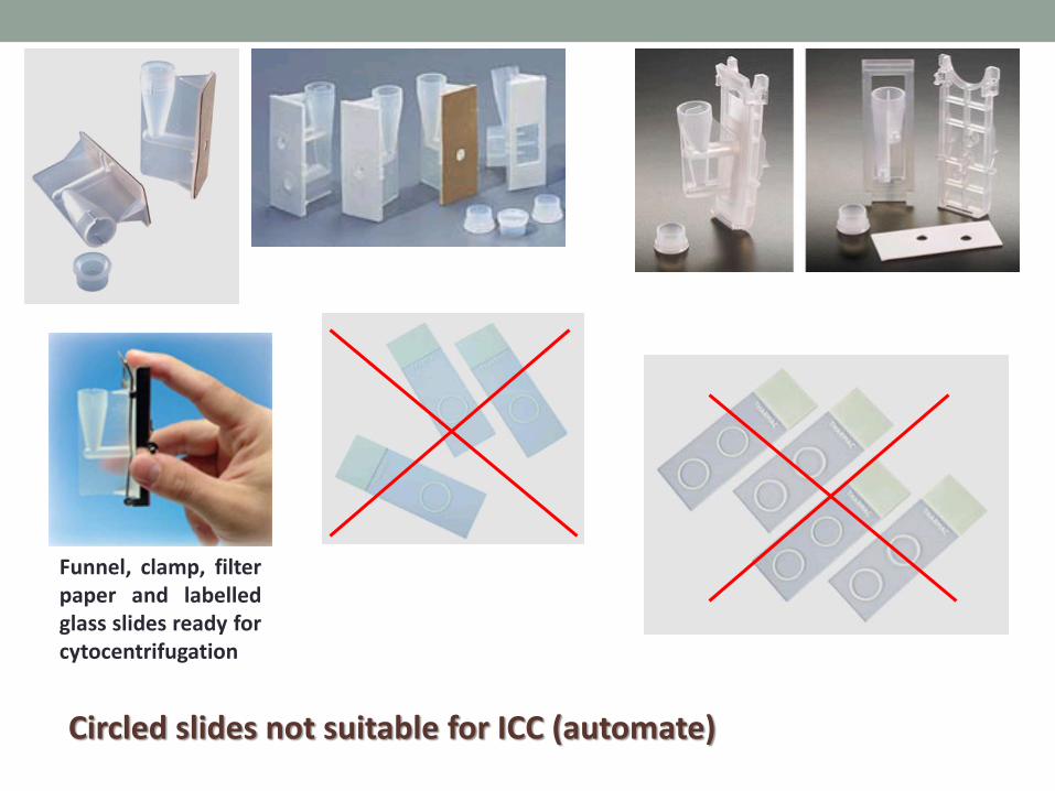
Circled slides not suitable for ICC (automate)
Funnel, clamp, filter paper and labelled glass slides ready for cytocentrifugation

Difficulty : adjust cell concentration on the slides Too much cells risk of cell detachment Too few cells difficult to analyse
Solution : cell counts
Nageotte slide : liquids with few cells (CSF)
Thomas or Malassez slide : liquids with lots of cells (BALF, serous
fluids, other….)
Kovaslide : urine

Kovaslide

Liquid based cytology Pretreated slides to increase cell adhesion Cell blocks If too few liquid remaining : cytocentrifugation (500µl/slide) many slides can be performed

Cell blocks
• Many ways to perform cell blocks • Thrombin clot +++
• Agar
• Histogel*
• Other
• Fixation: the same as tissue blocks
• Difficulties : too few cells
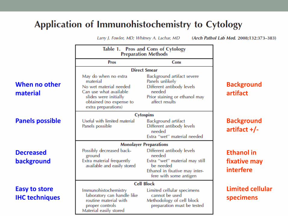
Background artifact
Background artifact +/-
Ethanol in fixative may interfere
Limited cellular specimens
When no other material
Panels possible
Decreased background
Easy to store IHC techniques


Prerequisite to immunocytochemistry
• Slides
• Fixation
•Antigen retrieval
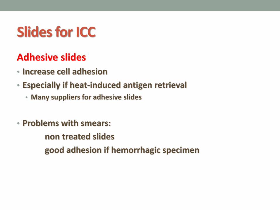
Slides for ICC
Adhesive slides
• Increase cell adhesion
• Especially if heat-induced antigen retrieval • Many suppliers for adhesive slides
• Problems with smears:
non treated slides
good adhesion if hemorrhagic specimen

Slide fixation
Smears or cytospins : air dried Fixation: Cold acetone (4°C) 10 min Ethanol (not suitable for some antigens, ex: ER, pS100) Methanol Formalin… Slides may be kept at room temperature for 7 days or -20°C several months Before use, bring them at room temperature under cover (avoids mist on the slides)

Fixatives • Acetone, Ethanol, Methanol = coagulating fixatives
(precipitate proteins) • Remove lipids (permeabilize cell membranes)
• Dehydrate the cells
• Formaldehyde (HCHO) : non-coagulant, additive fixative, cross-linking reagent • Forms intermolecular bridges, normally through free amino groups,
thus creating a network of linked antigens
• No permeablization
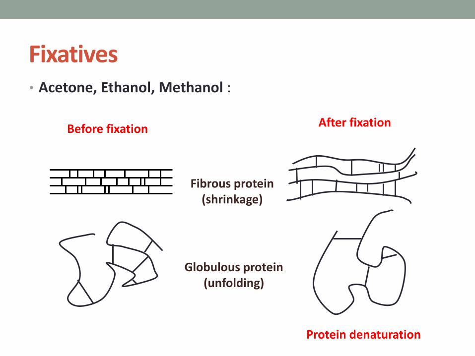
Fixatives • Acetone, Ethanol, Methanol :
Fibrous protein (shrinkage)
Globulous protein (unfolding)
After fixation Before fixation
Protein denaturation

Fixatives • Formaldehyde (HCHO) : non-coagulant, additive fixative
After fixation
Methylene bridge
Methylene bridge
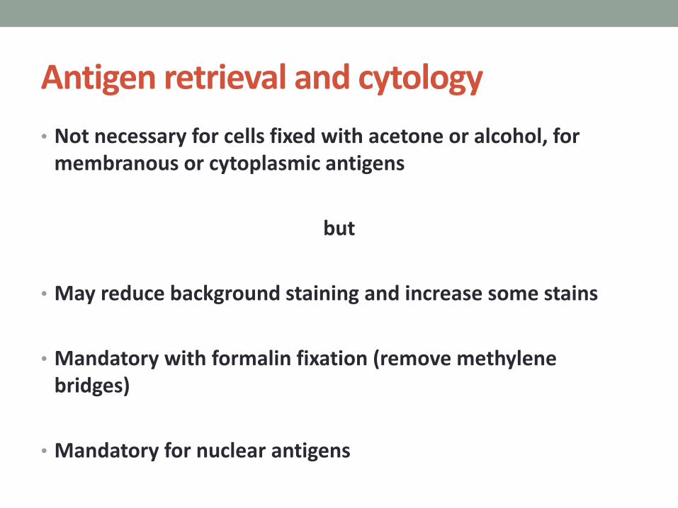
Antigen retrieval and cytology
• Not necessary for cells fixed with acetone or alcohol, for membranous or cytoplasmic antigens
but
• May reduce background staining and increase some stains
• Mandatory with formalin fixation (remove methylene bridges)
• Mandatory for nuclear antigens
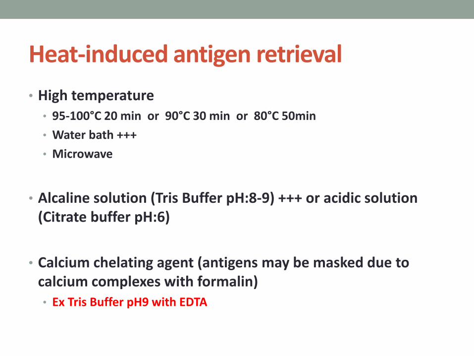
Heat-induced antigen retrieval
• High temperature • 95-100°C 20 min or 90°C 30 min or 80°C 50min
• Water bath +++
• Microwave
• Alcaline solution (Tris Buffer pH:8-9) +++ or acidic solution (Citrate buffer pH:6)
• Calcium chelating agent (antigens may be masked due to calcium complexes with formalin) • Ex Tris Buffer pH9 with EDTA
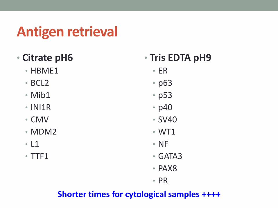
Antigen retrieval
• Citrate pH6 • HBME1
• BCL2
• Mib1
• INI1R
• CMV
• MDM2
• L1
• TTF1
• Tris EDTA pH9 • ER
• p63
• p53
• p40
• SV40
• WT1
• NF
• GATA3
• PAX8
• PR
Shorter times for cytological samples ++++

p53 (clone DO7) - Dilution : 1/30
Urine : High grade tumor (alcohol fixation)
Citrate buffer then EDTA Flex buffer Tris-EDTA-pH9
Citrate buffer alone : no staining

Revelation systems
• With Biotin : false positives with endogenous biotin • Liver, kidney, colon, thyroid, breast
• Removed by albumin
• Alkaline phosphatase: false positives with endogenous AP • Placental, intestinal, germinal
• Inhibited by Levamisole
• Peroxydase : false positives with endogenous peroxydase • Eosinophils, neutrophils, monocytes, erythrocytes, muscle cells
• Inhibited by hydrogen peroxyde

Some examples
• Litterature
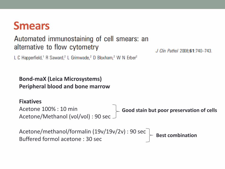
Smears
Bond-maX (Leica Microsystems) Peripheral blood and bone marrow Fixatives Acetone 100% : 10 min Acetone/Methanol (vol/vol) : 90 sec Acetone/methanol/formalin (19v/19v/2v) : 90 sec Buffered formol acetone : 30 sec
Good stain but poor preservation of cells
Best combination

Background staining of erythrocytes and polymorphonuclears with peroxydase
No background staining with alkaline phosphatase
CD45
CD61
CD3 CD10
CD68 CD138
CD20
CD235
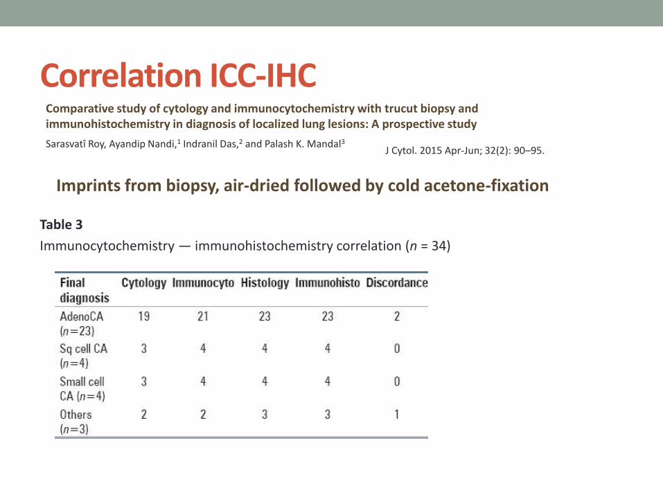
Correlation ICC-IHC
Table 3
Immunocytochemistry — immunohistochemistry correlation (n = 34)
Sarasvatî Roy, Ayandip Nandi,1 Indranil Das,2 and Palash K. Mandal3
J Cytol. 2015 Apr-Jun; 32(2): 90–95.
Comparative study of cytology and immunocytochemistry with trucut biopsy and immunohistochemistry in diagnosis of localized lung lesions: A prospective study
Imprints from biopsy, air-dried followed by cold acetone-fixation
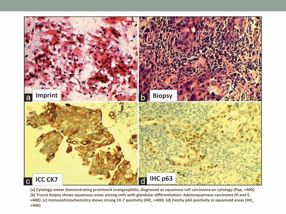
(a) Cytology smear demonstrating prominent orangeophilia, diagnosed as squamous cell carcinoma on cytology (Pap, ×400). (b) Trucut biopsy shows squamous areas among cells with glandular differentiation: Adenosquamous carcinoma (H and E, ×400). (c) Immunohistochemistry shows strong CK-7 positivity (IHC, ×400). (d) Patchy p63 positivity in squamoid areas (IHC, ×400)
Imprint Biopsy
ICC CK7 IHC p63

(a) Cytology smear showing clusters of pleomorphic cells with molding and smudging, small cell carcinoma (MGG, ×400). (b) Immunocytochemistry smear showing CD56-positive cells in a necrotic background (IHC, ×400). (c) Trucut biopsy showing solid sheets of cells with smudging (H and E, ×400). (d) Immunohistochemistry showing positivity with chromogranin-A (IHC, ×400)
Imprint
Biopsy
ICC CD56
IHC Chromogranin-A

Some examples from our experience
• Dako Autostainer Link48
• Ventana Roche BenchMark ULTRA (leucocyte antigens)
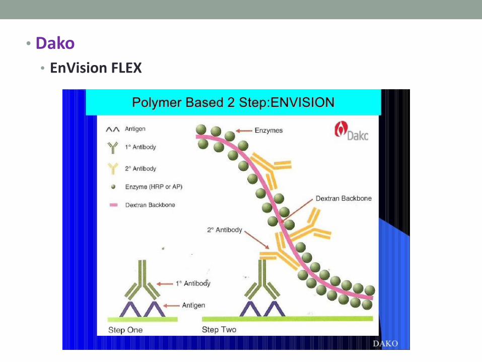
• Dako
• EnVision FLEX

• Ventana
• OptiView DAB IHC Detection Kit

Sme
ar
Ce
llblo
ck
CD56 Chromogranin

Sme
ar
Ce
llblo
ck
CK5/6 p63

Sme
ar
Ce
llblo
ck
TTF1 Synaptophysin
Comparative analysis
• ICC on smears / ICC on cell blocks
• Metastasis of lung adenocarcinoma in mediastinal lymph node
• Smears • Slides MGG stain
• Slides ICC
• Cytolyt (Hologic) Preservcyt • Slide Pap stain
• Cellblock
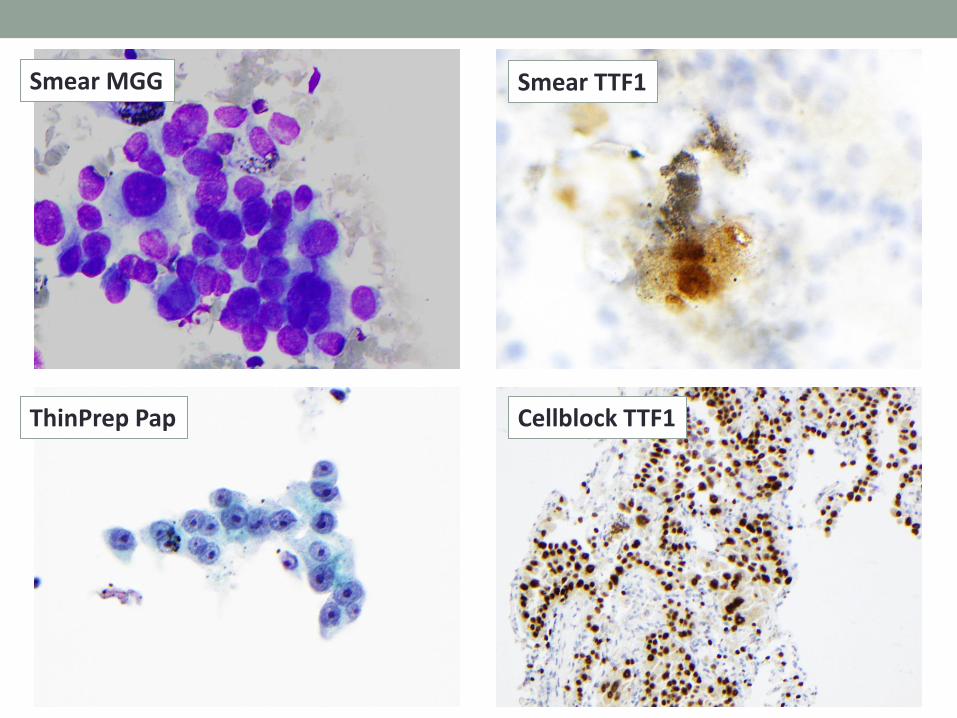
Smear MGG
Cellblock TTF1 ThinPrep Pap
Smear TTF1

Comparative analysis
• ICC on smears / ICC on cell blocks
• Metastasis of an atypical carcinoid tumor in a mediastinal lymph node
• Smears • Slide MGG stain
• Slides ICC
• Cytolyt (Hologic) Preservcyt • Slide Pap stain
• Cellblock
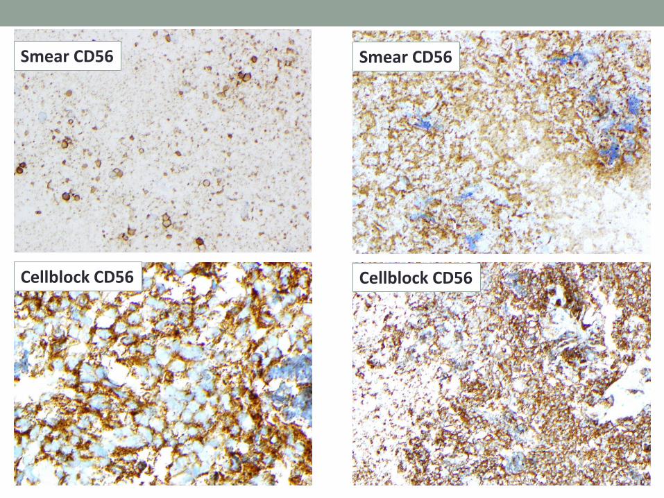
Smear CD56
Cellblock CD56
Smear CD56
Cellblock CD56

Smear Ki-67
Cellblock Synaptophysin
Cellblock Chromogranin-A
Cellblock Ki-67
Comparative analysis
• ICC on cytospins from Preservcyt/ IHC on biopsy
• Metastasis of small cell carcinoma in a mediastinal lymph node
• Cytolyt (Hologic) Preservcyt • Slide Pap stain
• Cellblock
• Cytospins for ICC
• Bronchial biopsy • IHC
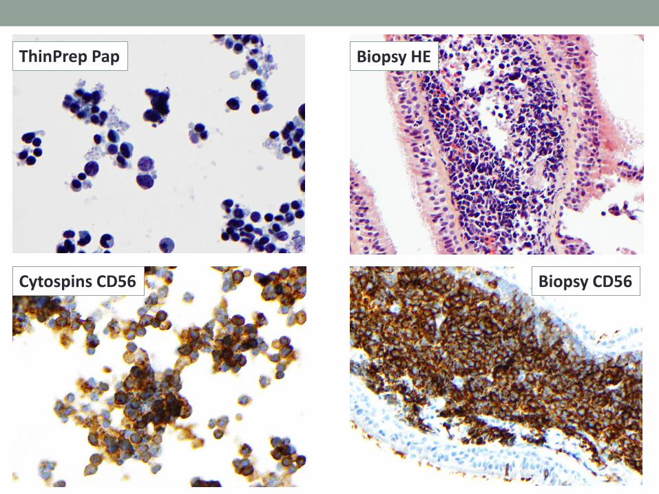
Cytospins CD56
Biopsy HE ThinPrep Pap
Biopsy CD56

Cytospin Synaptophysin
Biopsy TTF1
Cytospin TTF1
Biopsy Synaptophysin
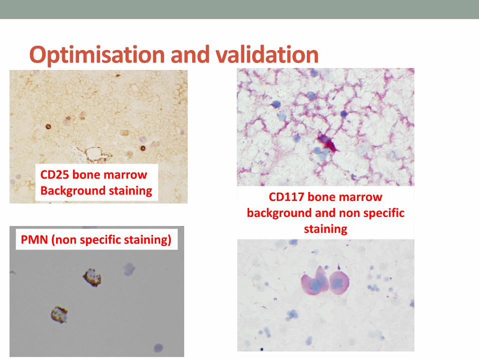
Optimisation and validation
CD25 bone marrow Background staining
PMN (non specific staining)
CD117 bone marrow background and non specific
staining
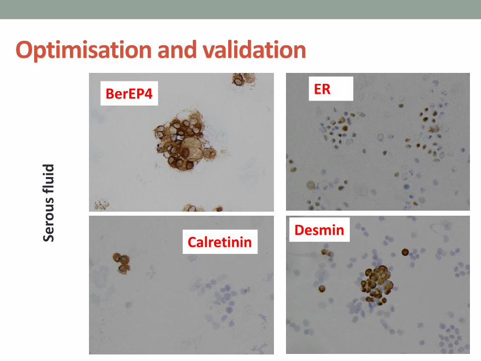
Optimisation and validation
BerEP4
Calretinin
ER
Desmin Sero
us
flu
id

Use of controls
• Internal controls +++ • Mesothelial cells in serous fluid
• Mesothelial or bronchial cells for CK7
• Small lymphocytes for CD3
• Additional slides • Positive slides fixed and stored for 12 months at -20°C
• Cell lines
• Limited for non usual antibodies

We do not perform ICC on stained slides
Positive in : - 87.3% of HG - 38% of negative or LG - 94% of CIS

Pitfalls
• No or insufficient staining: • Antibody sensitivity
• Verify storage conditions especially temperature (specification sheet). Avoid freezing and thawing (perform aliquots)
• Antibody concentration too low
• Demasking protocol inappropriate
• False positive result • Crushed or degenerated cells or marked necrosis
• Acute inflammation in background (PMN)
• Antibody dilution insufficient
• Antibody specificity
Thank you very much for your attention