immunophenotypic characterization of normal bone marrow …cdn.intechopen.com/pdfs/37443.pdf ·...
TRANSCRIPT

23
Immunophenotypic Characterization of Normal Bone Marrow Stem Cells
Paula Laranjeira, Andreia Ribeiro, Sandrine Mendes, Ana Henriques, M. Luísa Pais and Artur Paiva
Histocompatibility Center of Coimbra Portugal
1. Introduction
Despite of being described more than one decade ago (Pittenger et al., 1999), the
immunophenotypic profile of bone marrow mesenchymal stem cells (MSC) still not well
documented. The difficulty in achieving a detailed phenotypic characterization is common
in less-represented cell populations and/or populations lacking a specific known cell
marker, like bone marrow MSC.
The recent advances in flow cytometry technology and the emergence of new high-speed
flow cytometers have given a valuable contribute to diminish this problem in two different
(but complementary) aspects: 1) by reducing dramatically the acquisition time period,
making it more reasonable to study minor cell populations; and 2) by increasing the number
of parameters that can be analyzed per cell at the same time, which is critical to improve the
immunophenotypic characterization of those not-well characterized cell populations that
lack a specific known marker.
A good example of the practical usefulness of such technical developments is the
description of different cell compartments in the bone marrow CD34+ hematopoietic stem
cell (HSC) population. Detailed studies on this minor bone marrow cell population
demonstrated that each compartment is committed to a different hematopoietic cell lineage.
An extensive immunophenotypic characterization of those CD34+ compartments allowed
the development of protocols to easily and quickly identify, quantify and evaluate
phenotypic aberrations and maturational blocks in those cells, which is decisive to the
diagnosis, prognosis, or follow-up of a variety of hematological clonal diseases (del Cañizo
et al., 2003; Lochem et al., 2004; Matarraz et al., 2008; Orfao et al. 2004).
2. Bone marrow mesenchymal stem cells
After the identification of a plastic-adherent bone marrow stromal cell population in 1976 by
Friedenstein and colleagues and the first evidence of their multilineage potential (Pittenger
et al., 1999) with subsequent confirmation of their stem cell nature, an increasing interest on
these bone marrow MSC has emerged, mainly because of their promising therapeutic
applications.
www.intechopen.com

Flow Cytometry – Recent Perspectives
458
By definition, a stem cell is an undifferentiated cell with the potential ability of self-renewal and the capability of differentiation along different cell lineages (multipotency). MSC can be found on a great variety of adult tissues, where they play an important role in tissue regeneration, such as: bone marrow, adipose tissue, umbilical cord blood, umbilical cord matrix, menstrual blood, endometrium, placenta, dental pulp, skin and thymus, among others (Chamberlain et al., 2007; Ding et al. 2011; Kolf et al., 2007; Martins et al., 2009; Musina et al., 2005; Pittenger et al., 1999).
In addition to their presence in numerous adult tissues, MSC are relatively easy to isolate and have the capability to expand manyfold in culture without lose their stem cell properties. Moreover, when MSC are systemically transplanted, they are able to migrate to sites of injury and promote tissue repair, by producing growth factors or other soluble factors important to tissue regeneration, as well as by undergoing cellular differentiation (Chamberlain et al., 2007, Kolf et al., 2007; Mafi et al., 2011); such features explain the success of MSC transfusion therapy in genetic disorders affecting mesenchymal tissues (Horwitz et al., 2002; Undale et al., 2009). Furthermore, those cells have the ability of suppressing the immune response of a wide variety of immune cells, including T, B and NK lymphocytes, and antigen-presenting cells (Chamberlain et al., 2007; Stagg, 2007), and their importance in patients’ clinical outcome has already been proven in severe acute graft-versus-host disease (Remberger et al., 2011; von Bahr et al., 2011). Moreover, the results achieved in animal models of autoimmune diseases are promising and encouraged the beginning of phase I clinical trials in multiple sclerosis (Constantin et al., 2009; Darlington et al., 2011; Siatskas et al., 2009).
2.1 Identification and quantification of bone marrow MSC
As referred previously, the study of minor cell populations with no known specific cell marker toke great advantage on the development of high-speed multi-parameter flow cytometers. The use of an 8-color FACSCanto II (Becton Dickinson Biosciences, BDB) flow cytometer allowed us to identify MSC in bone marrow, quantify them and further characterize their immunophenotypic profile. We employed a monoclonal antibody panel with a backbone of 3 common markers (CD13, CD45 and CD11b) for the identification of MSC (known to be CD13+CD45-CD11b-) in each tube that, at the same time, permitted the study of the expression of five more proteins on MSC per tube.
MSC are rare in bone marrow, being reported that they represent approximately 0,01% of all nucleated bone marrow cells (Chamberlain et al., 2007; Mafi et al., 2011), although is known that their number declines with aging (Caplan, 2007). Our data point to a percentage ranging between 0,01% and 0,03% of all nucleated bone marrow cells (Martins et al., 2009).
2.2 Immunophenotypic characterization of bone marrow MSC
2.2.1 Flow cytometer quality control, compensation setup strategies and other technical issues
According to the manufacturer’s recommendations, it is done a daily quality control using the Rainbow Beads (BDB). In what concerns to cytometer’s compensation setup, it is made once per month by setting up the Rainbow Beads (BDB) values according to the EuroFlow consortium’s guidelines and then by doing a general compensation for stable fluorochromes and a specific compensation for each monoclonal antibody conjugated with tandem
www.intechopen.com

Immunophenotypic Characterization of Normal Bone Marrow Stem Cells
459
fluorochromes. Although the compensation is automatic, it is always revised by experienced staff at the end of the process.
In order to detect cellular autofluorescence, a negative control was made for each sample, where the bone marrow sample was only stained for CD45 PO and CD34 PerCPcy5.5.
SSC and FSC light dispersion properties allow a good discrimination between viable and dead cells and the doublets were excluded based on FSC-Area versus FSC-Height characteristics.
2.2.2 Material and methods
The immunophenotypic characterization of bone marrow MSC were performed in fresh EDTA-collected bone marrow samples from healthy individuals. After collection the samples were stored at 4 ºC and processed within 24 hours.
Whole bone marrow samples were stained for surface cell markers using a stain-lyse-and-
then-wash direct immunofluorescence technique. 200 μl of whole bone marrow were aliquoted in different tubes and stained with the following combinations of monoclonal antibodies in an 8-color staining protocol, detailed in table 1.
FITC PE PerCPcy5.5 PEcy7 APC APCH7 PB PO
Tube 1
CD49e (SAM1)
Beckman Coulter
CD73 (AD2)
BD Pharmingen
CD34 (8G12) BDB
CD13 (Immu103.44)
Beckman Coulter
CD90 (5E10)
BD Pharmingen
HLA-DR (L243) BDB
CD11b (ICRF44)
BD Pharmingen
CD45 (HI30)
Invitrogen
Tube 2
CD31 (WM59)
BD Pharmingen
NGFR (C40-1457)
BD Pharmingen
CD14 (M5E2)
BD Pharmingen
CD13
CD133 (293C3) Miltenyi
Biotec
- CD11b CD45
Tube 3
CD15 (HI98) BDB
CD146 (P1H12)
BD Pharmingen
CD24 (ALB9)
Beckman Coulter
CD13 CD90 CD29
(TS2/16) BioLegend
CD11b CD45
Tube 4
CD106 (51-10C9)
BD Pharmingen
CD105 (1G2)
Beckman Coulter
- CD13
HLA-A, B, C(G46-2.6)
BD Pharmingen
- CD11b CD45
Tube 5
- CD73 CD24 CD13 CD90 - CD11b CD45
Table 1. Panel of monoclonal antibodies used for the bone marrow MSC characterization. FITC - fluorescein isothiocyanate; PE – phycoerythrin; PerCPcy5.5 - peridinin chlorophyll protein cyanine 5.5; PEcy7 - R-phycoerythrin cyanine 7; APC – allophycocyanin; APCH7 - allophycocyanin H 7; PB - pacific blue; PO - pacific orange
Data acquisition was performed in a FACSCanto II flow cytometer (BDB), using FACSDiva acquisition software (BDB). The total bone marrow cellularity of the whole sample was acquired (5 x 10⁶ events, minimum) for each tube. Bone marrow MSC were identified as CD13+/CD45-/CD11b-, as shown in Figure 1.
Data analysis was performed using Infinicyt software (Cytognos, Salamanca, Spain).
www.intechopen.com

Flow Cytometry – Recent Perspectives
460
-10 0 1E1 1E2 1E3 1E4 1E5
-10
0
1E1
1E
2
1
E3
1E
4
1
E5
CD11b PB
CD
13 P
E C
y7
-10
0
1E
1
1E2
1E
3
1E4
1E5
-10 0 1E1 1E2 1E3 1E4 1E5
CD13 PE Cy7
CD
45 P
O
Fig. 1. Identification of bone marrow MSC (blue) present in a whole bone marrow sample,
phenotypically characterized as CD13+CD45-CD11b-
2.2.3 Results and discussion
Bone marrow MSC showed to be uniformly positive to CD13, CD29, CD49e, CD90, CD106,
CD146, CD73, NGFR, CD105 and HLA-A, B, C (Figure 1 and Figure 2); and negative to
CD24, CD31, CD11b, CD14, CD15, CD34, CD45, CD133 and HLA-DR, which is in agreement
with previous studies described in the literature (Chamberlain et al., 2007; Delorme et al.,
2008; Ehninger & Trumpp, 2011; Fox et al., 2007; Jones & McGonagle, 2008; Kolf et al., 2007;
Martins et al., 2009; Pittenger et al., 1999; Tormin et al., 2011). Based on the expression
profile of these markers, bone marrow MSC behave as one sole cell population, as all the
studied markers were homogeneously expressed inside the MSC population.
Several studies on adhesion molecules and chemokine receptors expression have been made
in order to shed light on MSC migratory and homing ability. CD29 (integrin β1-subunit) and
CD106 (vascular cell adhesion molecule 1, VCAM-1) seem to be important in the adhesion of
MSC to endothelial cells (Chamberlain et al., 2007; Kolf et al., 2007; Stagg, 2007) and CD29,
which when dimerized with CD49e (integrin α5-subunit) forms a receptor that binds to
fibronectin and invasin, is likely to promote MSC-extracellular matrix interaction (Gu et al.,
2009). CD146 (Muc18) plays an important role in cell-cell and cell-extracellular matrix
adhesion and an increased expression of these marker on tumor cells is associated with an
increased cell motility and invasiveness/ metastasis capability (Bardin et al., 2001; Zeng et
al., 2011). The glycoprotein CD90 (Thy-1) regulates as well cell-cell and cell-extracellular
matrix interactions, being involved in adhesion to endothelial cells, migration, metastasis
and tissue regeneration (Jurisic et al., 2010; Rege & Hagood, 2006).
The enzyme CD73 is an ecto-5’-nucleotidase that produces extracellular adenosine. In
animal tumor models, CD73-generated adenosine inhibits both homing and expansion of T
cells via adenosine-receptor signaling. In fact, recent research shows that adenosine
suppresses T cell immune response both in activation and effector phases, as well as NK cell
immune activity (Wang et al., 2011; Zhang et al., 2010).
www.intechopen.com

Immunophenotypic Characterization of Normal Bone Marrow Stem Cells
461 0
500
00
1
5000
0
0 1E2 1E3 1E4 1E5
CD13 PE Cy7
SS
C-A
0 5
0000 150000
0 1E2 1E3 1E4 1E5S
SC
-A
HLA-A, B, C APC
0 5
0000 150000
0 1E2 1E3 1E4 1E5
SS
C-A
CD146 PE
0 1E2 1E3 1E4 1E5
CD29 APCH7
0
5
0000
150
000
SSC
-A
0 1E2 1E3 1E4 1E5
CD90 APC
CD
73 P
E
0
1E
2
1E
3
1E
4
1E5
CD49 FITC
CD
73
PE
0 1E2 1E3 1E4 1E5
CD106 FITC
CD
105
PE
0
1E
2
1E
3
1E
4
1E5
0 1E2 1E3 1E4 1E5
NGFR PE
SS
C-A
0
5
0000
1
5000
0
Fig. 2. Immunophenotypic characteristics of bone marrow MSC (blue). The remaining bone
marrow nucleated cells are represented as grey events
In what concerns to growth factor receptors, NGFR (nerve growth factor receptor, CD271) is
expressed in a wide variety of tissues and, depending on the cell type, signaling through
this receptor regulates NF-kB activation, apoptosis, tissue regeneration, immune cell
activation, proliferation and cell differentiation (Micera et al., 2007; Rogers et al., 2010).
Finally, CD105 (endoglin) is one of the receptors for TGF-β, a growth factor involved in the
regulation of development, maintenance and proliferation of MSC (Stagg, 2007), and also
known to play an important role in tissue repair.
www.intechopen.com

Flow Cytometry – Recent Perspectives
462
Some discrepancies described in the expression of adhesion molecules, chemokine receptors
and other proteins, may be the reflex of the microenvironmental differences present in
different studies. Although there are a great similitude in the phenotypic profile of MSC
isolated from different tissues, differences do exist (Chamberlain et al., 2007; Kolf et al., 2007;
Martins et al., 2009). As well as different cultures conditions can also change the MSC
phenotype (Chamberlain et al., 2007; Halfon et al., 2011; Stagg, 2007; Tormin et al., 2011).
This could be a clue of MSC highly sensitiveness to microenvironment alterations, and their
potential to change their protein expression profile could be of great importance in giving an
appropriate response to physiological or pathological challenges: by changing their
migratory pattern, by initiating an immunomodulatory or immunosuppressive response, by
modifying the production and release of soluble factors, or by undergoing cell
differentiation.
As a minor bone marrow cell population easy to expand in vitro, it is attractive to
characterize the MSC immunophenotype after culture cell expansion. Nevertheless,
characterizing these cells directly (without previous culture) enables an analysis closest to
their physiological conditions, excluding the phenotypic alterations induced by factors
present in the culture medium. Moreover, this direct approach allows an accurate
quantification of MSC in bone marrow. Also, this same strategy can be applied to MSC from
other tissues.
3. Bone marrow hematopoietic stem cells
The multipotent hematopoietic stem cell is mainly located in the bone marrow of adult
animals and has the ability to differentiate along all hematopoietic cell lineages. A number
of studies based on in vitro cell culture, xeno-transplantation of hematopoietic human cells
in immunodeficient mice and in pre-immune animal fetuses, were carried out to identify the
human hematopoietic stem cell and unveil the hematopoietic precursors hierarchy (Nimer,
2008; Yin et al., 2007), becoming clear that CD34-positive cells were able to differentiate and
give rise to all blood cells. There are evidences that, within this heterogeneous population,
the more immature CD34+ HSC expresses CD133 and are CD38-negative/dim. It is also
known that the CD34+CD133+ subpopulation can arise from the CD133+CD34-CD38-
subset (Goussetis et al., 2006; Nimer, 2008; Yin et al., 1997).
3.1 Identification and quantification of the different bone marrow CD34+ HSC cell compartments
As already referred, CD34-positive cells are an heterogeneous bone marrow cell population,
consisting in various cell compartments differing in immunophenotype, size and lineage
commitment. The immunophenotypic pattern of each compartment is well described and,
with a relatively low number of markers, the majority of those subsets can be accurately and
easily identified.
Attending only to the immunophenotypic features, is possible to identify the following bone
marrow CD34+ cell subsets by flow cytometry: uncommitted (more immature) precursors,
neutrophil precursors, B cell precursors, monocytic precursors, plasmacytoid dendritic cells
precursors, erythroid precursors, basophil precursors and mast cell precursors.
www.intechopen.com

Immunophenotypic Characterization of Normal Bone Marrow Stem Cells
463
A detailed immunophenotypic description of human bone marrow CD34+ cells was
published, few years ago, by Matarraz and colleagues (Matarraz et al., 2008) and Lochem
and colleagues (Lochem et al., 2004), along with the frequency of each CD34+ cell
subpopulation in normal hematopoiesis (Matarraz et al. 2008), presented on table 2.
Bone marrow CD34+ HSC compartments Mean ±
Standard deviation
Range
% Bone marrow CD34+ HSC (of total bone marrow)
0,9 ± 0,3 (0,2-1,6)
Immature CD34+ precursor (%) (within CD34+ cells)
52 ± 12 (19-66)
CD34+ neutrophil precursors (%) (within CD34+ cells)
34 ± 7 (15-47)
CD34+ B cell precursors (%) (within CD34+ cells)
14 ± 10 (1-36)
CD34+ monocytic precursors (%) (within CD34+ cells)
10 ± 7 (0-26)
CD34+ plasmacytoid dendritic cell precursors (%) (within CD34+ cells)
5 ± 2 (0-9)
CD34+ erythroid precursors (%) (within CD34+ cells)
18 ± 8 (1-36)
CD34+ basophil precursors (%) (within CD34+ cells)
0,7 ± 0,4 (0-1,5)
CD34+ mast cell precursors (%) (within CD34+ cells)
0 ± 0,005 (0-0,02)
Table 2. Distribution of the different cell compartments of bone marrow CD34+ HSC. The
results are expressed as mean ± standard deviation (range). Adapted from Matarraz et al.
Leukemia 2008
The most immature CD34+ subset can be identified based on CD133 expression (Goussetis
et al., 2006; Pastore et al., 2008; Yin et al., 1997). When other markers are concerned, these
cell are CD34hi/CD45int/HLA-DRhi/cyMPO-/nTdT-/CD117hi and have intermediate side
scatter (SSC) and forward scatter (FSC) light dispersion properties (Matarraz et al., 2008). As
previously described by Matarraz and colleagues, the phenotypic profile of CD34+ B cell
precursors is CD34int/CD45int/dim/HLA-DRhi/cyMPO-/nTdTint/CD117- and these cells
present the lowest SSC and FSC of all CD34+ subpopulations (Lochem et al., 2004; Matarraz
et al., 2008); the CD34+ neutrophil precursors present CD34hi/CD45int/dim/HLA-
DRhi/cyMPOint/hi/nTdT-/CD117hi, along with the highest values for SSC and FSC of all
CD34+ subsets; the CD34+ plasmacytoid dendritic cell precursors are identified based on
the expression of CD34+/CD123hi/int/HLA-DRhi; CD34+ monocytic precursors display
CD34+/HLA-DRhi/CD64hi/CD45hi/CD117- immunophenotype; basophil precursors are
described as being CD34+/CD123int/hi/HLA-DR-/+; and CD34+ mast cell precursors are
CD34+/CD117hi/HLA-DR-/int (Matarraz et al., 2008). Finally, CD34+ erythroid precursors
are characterized by CD34+/CD36+/CD64-/CD45lo immunophenotype (Matarraz et al.,
2008) and by CD105 expression (Buhring et al., 1991; Rokhlin et al., 1995). As a matter of fact,
CD105 and TGF-β1 have a pivotal role in the regulation of the differentiation in the
erythroid lineage (Fortunel et al., 2000; Moody et al., 2007).
www.intechopen.com
Flow Cytometry – Recent Perspectives
464
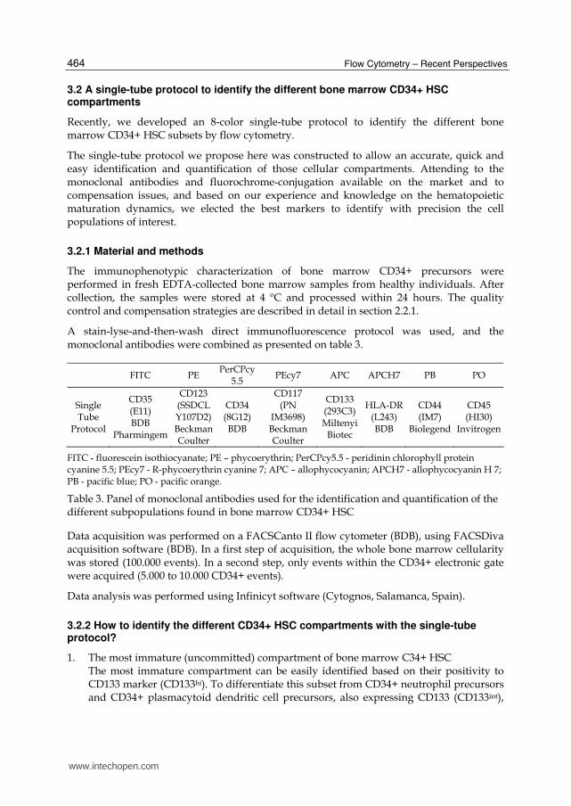
3.2 A single-tube protocol to identify the different bone marrow CD34+ HSC compartments
Recently, we developed an 8-color single-tube protocol to identify the different bone marrow CD34+ HSC subsets by flow cytometry.
The single-tube protocol we propose here was constructed to allow an accurate, quick and easy identification and quantification of those cellular compartments. Attending to the monoclonal antibodies and fluorochrome-conjugation available on the market and to compensation issues, and based on our experience and knowledge on the hematopoietic maturation dynamics, we elected the best markers to identify with precision the cell populations of interest.
3.2.1 Material and methods
The immunophenotypic characterization of bone marrow CD34+ precursors were performed in fresh EDTA-collected bone marrow samples from healthy individuals. After collection, the samples were stored at 4 ºC and processed within 24 hours. The quality control and compensation strategies are described in detail in section 2.2.1.
A stain-lyse-and-then-wash direct immunofluorescence protocol was used, and the monoclonal antibodies were combined as presented on table 3.
FITC PE PerCPcy
5.5 PEcy7 APC APCH7 PB PO
Single Tube
Protocol
CD35 (E11) BDB
Pharmingem
CD123 (SSDCLY107D2)Beckman Coulter
CD34 (8G12) BDB
CD117 (PN
IM3698) Beckman Coulter
CD133 (293C3) Miltenyi
Biotec
HLA-DR (L243) BDB
CD44 (IM7)
Biolegend
CD45 (HI30)
Invitrogen
FITC - fluorescein isothiocyanate; PE – phycoerythrin; PerCPcy5.5 - peridinin chlorophyll protein cyanine 5.5; PEcy7 - R-phycoerythrin cyanine 7; APC – allophycocyanin; APCH7 - allophycocyanin H 7; PB - pacific blue; PO - pacific orange.
Table 3. Panel of monoclonal antibodies used for the identification and quantification of the different subpopulations found in bone marrow CD34+ HSC
Data acquisition was performed on a FACSCanto II flow cytometer (BDB), using FACSDiva acquisition software (BDB). In a first step of acquisition, the whole bone marrow cellularity was stored (100.000 events). In a second step, only events within the CD34+ electronic gate were acquired (5.000 to 10.000 CD34+ events).
Data analysis was performed using Infinicyt software (Cytognos, Salamanca, Spain).
3.2.2 How to identify the different CD34+ HSC compartments with the single-tube protocol?
1. The most immature (uncommitted) compartment of bone marrow C34+ HSC The most immature compartment can be easily identified based on their positivity to CD133 marker (CD133hi). To differentiate this subset from CD34+ neutrophil precursors and CD34+ plasmacytoid dendritic cell precursors, also expressing CD133 (CD133int),
www.intechopen.com

Immunophenotypic Characterization of Normal Bone Marrow Stem Cells
465
other important phenotypic characteristics have to be taken into account:
CD35-/CD34hi/HLA-DRhi/CD117hi/FSCint/SSCint/CD123-. Figure 3 presents a detailed
immunophenotype of this compartment considering all the markers used in this
protocol.
SS
C-A
SS
C-A
SS
C-A
SS
C-A
CD45 POCD44 PBFSC-A
CD
117
PE
Cy
7
CD34 PerCP Cy5.5 HLA-DR APCH7 HLA-DR APCH7
CD
123
PE
CD
123
PE
CD
133
AP
C
CD35 FITC CD123 PE
Fig. 3. Uncommitted bone marrow CD34+ HSC (red) immunophenotype. The remaining
bone marrow CD34+ cell compartments are presented in grey
2. Bone marrow CD34+ erythroid precursors
Both CD34+ erythroid precursors and monocytic precursors express CD35. The two
CD34+ subpopulations can be distinguished in this protocol by the expression of CD117
and HLA-DR. The erythroid precursors are CD117+/HLA-DRint and the monocytic
precursors are CD117dim/-/HLA-DRhi. Moreover, the erythroid precursors are
characterized by a dim expression of CD34, CD45 and CD44 (Figure 4).
www.intechopen.com

Flow Cytometry – Recent Perspectives
466
FSC-A CD44 PB
CD45 PO
SS
C-A
SS
C-A
SS
C-A
CD34 PerCP Cy5.5 HLA-DR APCH7
HLA-DR APCH7
CD
117
PE
Cy
7C
D1
23 P
E
SS
C-A
C
D1
23 P
E
CD
133
AP
C
CD35 FITC CD123 PE0 1E2 1E3 1E4 1E5
0
1E
2
1E3
1E4
1
E5
0
1E
2
1E3
1E4
1
E5
0 1E2 1E3 1E4 1E5
0 1E2 1E3 1E4 1E5 0 1E2 1E3 1E4 1E5
0 1E2 1E3 1E4 1E50 1E2 1E3 1E4 1E5
0 1E2 1E3 1E4 1E5
0 5
00
00
1
00
00
0 2
00
00
00
5
00
00
1
00
00
0
20
00
00
0
1E
2
1E3
1E4
1
E5
0
1E
2
1E3
1E4
1
E5
0 5
00
00
1
00
00
0 2
00
00
0
0
5
00
00
10
00
00
20
00
00
0 50000 100000 150000 200000 250000
Fig. 4. Erythroid-committed bone marrow CD34+ precursors (red) immunophenotype. The remaining bone marrow CD34+ cell compartments correspond to the grey events
www.intechopen.com
Immunophenotypic Characterization of Normal Bone Marrow Stem Cells
467
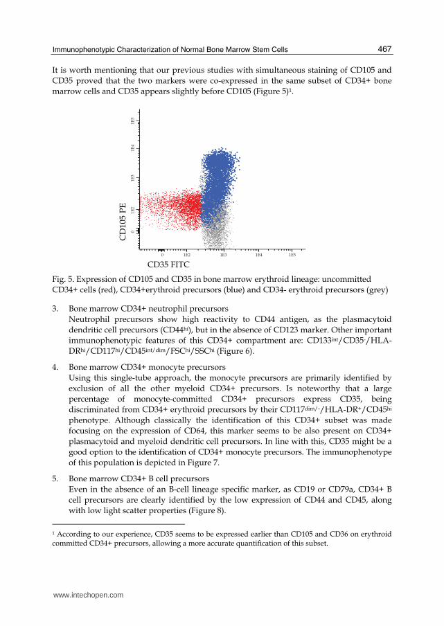
It is worth mentioning that our previous studies with simultaneous staining of CD105 and
CD35 proved that the two markers were co-expressed in the same subset of CD34+ bone
marrow cells and CD35 appears slightly before CD105 (Figure 5)1.
CD35 FITC
CD
105
PE
0 1E2 1E3 1E4 1E5
0
1E2
1
E3
1E
4
1E
5
Fig. 5. Expression of CD105 and CD35 in bone marrow erythroid lineage: uncommitted
CD34+ cells (red), CD34+erythroid precursors (blue) and CD34- erythroid precursors (grey)
3. Bone marrow CD34+ neutrophil precursors
Neutrophil precursors show high reactivity to CD44 antigen, as the plasmacytoid
dendritic cell precursors (CD44hi), but in the absence of CD123 marker. Other important
immunophenotypic features of this CD34+ compartment are: CD133int/CD35-/HLA-
DRhi/CD117hi/CD45int/dim/FSChi/SSChi (Figure 6).
4. Bone marrow CD34+ monocyte precursors
Using this single-tube approach, the monocyte precursors are primarily identified by
exclusion of all the other myeloid CD34+ precursors. Is noteworthy that a large
percentage of monocyte-committed CD34+ precursors express CD35, being
discriminated from CD34+ erythroid precursors by their CD117dim/-/HLA-DR+/CD45hi
phenotype. Although classically the identification of this CD34+ subset was made
focusing on the expression of CD64, this marker seems to be also present on CD34+
plasmacytoid and myeloid dendritic cell precursors. In line with this, CD35 might be a
good option to the identification of CD34+ monocyte precursors. The immunophenotype
of this population is depicted in Figure 7.
5. Bone marrow CD34+ B cell precursors
Even in the absence of an B-cell lineage specific marker, as CD19 or CD79a, CD34+ B
cell precursors are clearly identified by the low expression of CD44 and CD45, along
with low light scatter properties (Figure 8).
1 According to our experience, CD35 seems to be expressed earlier than CD105 and CD36 on erythroid committed CD34+ precursors, allowing a more accurate quantification of this subset.
www.intechopen.com

Flow Cytometry – Recent Perspectives
468
FSC-A
CD44 PB
CD45 PO
SS
C-A
SS
C-A
SS
C-A
CD34 PerCP Cy5.5 HLA-DR APCH7
HLA-DR APCH7
CD
117
PE
Cy
7C
D1
23 P
E
SS
C-A
C
D1
23 P
E
CD
133
AP
C
CD35 FITC CD123 PE0 1E2 1E3 1E4 1E5
0
1E
2
1
E3
1
E4
1
E5
0
1E
2
1
E3
1E
4
1E
5
0 1E2 1E3 1E4 1E5
0 1E2 1E3 1E4 1E5 0 1E2 1E3 1E4 1E5
0 1E2 1E3 1E4 1E50 1E2 1E3 1E4 1E5
0 1E2 1E3 1E4 1E5
0 5
00
00
1
00
00
0 2
00
00
00
50
00
0
10
00
00
2
00
00
0
0
1E
2
1E3
1E4
1
E5
0
1E
2
1E3
1E4
1
E5
0 5
00
00
1
00
00
0 2
00
00
0
0
50
00
0
10
00
00
2
00
00
0
0 50000 100000 150000 200000 250000
Fig. 6. Neutrophil-committed bone marrow CD34+ precursors (red) immunophenotype. The remaining bone marrow CD34+ cell compartments are presented in grey
www.intechopen.com

Immunophenotypic Characterization of Normal Bone Marrow Stem Cells
469
FSC-A CD44 PB
CD45 PO
SS
C-A
SS
C-A
SS
C-A
CD34 PerCP Cy5.5 HLA-DR APCH7
HLA-DR APCH7
CD
117
PE
Cy
7C
D1
23 P
E
SS
C-A
C
D1
23 P
E
CD
133
AP
C
CD35 FITC CD123 PE0 1E2 1E3 1E4 1E5
0
1E
2
1E
3
1E4
1E
5
0
1E2
1
E3
1E
4
1E5
0 1E2 1E3 1E4 1E5
0 1E2 1E3 1E4 1E5 0 1E2 1E3 1E4 1E5
0 1E2 1E3 1E4 1E50 1E2 1E3 1E4 1E5
0 1E2 1E3 1E4 1E5
0 5
00
00
1
00
00
0 2
00
00
00
5
00
00
1
00
00
0 2
00
00
0
0
1E
2
1E
3
1E4
1E
50
1E
2
1E
3
1E4
1E
50
5
00
00
1
00
00
0 2
00
00
0
0 5
00
00
1
00
00
0 2
00
00
0
0 50000 100000 150000 200000 250000
Fig. 7. Monocytic-committed bone marrow CD34+ precursors (red) immunophenotype. The remaining bone marrow CD34+ cell compartments are presented in grey
www.intechopen.com

Flow Cytometry – Recent Perspectives
470
FSC-A CD44 PB
CD45 PO
SS
C-A
SS
C-A
SS
C-A
CD34 PerCP Cy5.5 HLA-DR APCH7
HLA-DR APCH7
CD
117
PE
Cy
7C
D1
23 P
E
SS
C-A
C
D1
23 P
E
CD
133
AP
C
CD35 FITC CD123 PE0 1E2 1E3 1E4 1E5
0
1E
2
1E
3
1E4
1E
5
0
1E2
1E
3
1E4
1E
5
0 1E2 1E3 1E4 1E5
0 1E2 1E3 1E4 1E5 0 1E2 1E3 1E4 1E5
0 1E2 1E3 1E4 1E50 1E2 1E3 1E4 1E5
0 1E2 1E3 1E4 1E5
0 5
00
00
1
00
00
0 2
00
00
00
5
00
00
1
00
00
0 2
00
00
0
0
1E
2
1E3
1
E4
1
E5
0
1E
2
1
E3
1
E4
1
E5
0 5
00
00
1
00
00
0 2
00
00
0
0 5
00
00
1
00
00
0 2
00
00
0
0 50000 100000 150000 200000 250000
Fig. 8. B-cell-committed bone marrow CD34+ precursors (red) immunophenotype. The remaining bone marrow CD34+ cell compartments are presented in grey
www.intechopen.com

Immunophenotypic Characterization of Normal Bone Marrow Stem Cells
471
6. Bone marrow CD34+ basophil precursors
Our protocol allows the identification of basophil precursors using the classical markers
and attending to the immunophenotype HLA-DR-/dim/CD123int/hi. Of note, this CD34+
subset presents the lowest expression of CD44 among all bone marrow myeloid CD34+
cells, being easy to differentiate this precursors from all the other myeloid precursors by
using CD44 marker (Figure 9).
7. Bone marrow CD34+ plasmacytoid dendritic cell precursors
The plasmacytoid dendritic cell precursors are identified using the classical markers, as being HLA-DRhi/CD123hi/int. The most immature forms of this precursor express CD133 (CD133int). The immunophenotypic characteristics of this population are represented on Figure 10.
FSC-A CD44 PB CD45 PO
SS
C-A
SS
C-A
SS
C-A
CD34 PerCP Cy5.5 HLA-DR APCH7 HLA-DR APCH7
CD
117
PE
Cy
7
CD
123
PE
SS
C-A
C
D1
23 P
E
CD
133
AP
C
CD35 FITC CD123 PE0 1E2 1E3 1E4 1E5
0
1E
2
1E
3
1E4
1E
5
0
1E2
1E
3
1E4
1E
5
0 1E2 1E3 1E4 1E5
0 1E2 1E3 1E4 1E5 0 1E2 1E3 1E4 1E50 1E2 1E3 1E4 1E5
0 1E2 1E3 1E4 1E50 1E2 1E3 1E4 1E5
0 5
00
00
1
00
00
0 2
00
00
00
5
00
00
1
00
00
0 2
00
00
0
0
1E
2
1
E3
1
E4
1
E5
0
1E
2
1
E3
1E4
1
E5
0 5
00
00
1
00
00
0 2
00
00
0
0 5
00
00
1
00
00
0 2
00
00
0
0 50000 100000 150000 200000 250000
Fig. 9. Basophil-committed bone marrow CD34+ precursors (red) immunophenotype. The remaining bone marrow CD34+ cell compartments are presented in grey
www.intechopen.com

Flow Cytometry – Recent Perspectives
472
FSC-A CD44 PB
CD45 PO
SS
C-A
SS
C-A
SS
C-A
CD34 PerCP Cy5.5 HLA-DR APCH7
HLA-DR APCH7
CD
117
PE
Cy
7C
D1
23 P
E
SS
C-A
C
D1
23 P
E
CD
133
AP
C
CD35 FITC CD123 PE0 1E2 1E3 1E4 1E5
0
1E
2
1
E3
1
E4
1
E5
0
1E
2
1
E3
1
E4
1
E5
0 1E2 1E3 1E4 1E5
0 1E2 1E3 1E4 1E5 0 1E2 1E3 1E4 1E5
0 1E2 1E3 1E4 1E50 1E2 1E3 1E4 1E5
0 1E2 1E3 1E4 1E5
0 5
00
00
1
00
00
0 2
00
00
00
50
00
0
10
00
00
2
00
00
0
0
1E
2
1
E3
1
E4
1
E5
0
1E
2
1
E3
1
E4
1
E5
0 5
00
00
1
00
00
0 2
00
00
0
0
50
00
0
1
00
00
0
20
00
00
0 50000 100000 150000 200000 250000
Fig. 10. Plasmacytoid dendritic cell-committed bone marrow CD34+ precursors (red) immunophenotype. The remaining bone marrow CD34+ cell compartments are presented in grey
www.intechopen.com

Immunophenotypic Characterization of Normal Bone Marrow Stem Cells
473
8. Bone marrow CD34+ mast cell precursors
The classical markers for the identification of CD34+ mast cell precursors are included
in our protocol, and these cells are CD117hi/HLA-DR-/int. This subset expresses high
levels of CD44. Other immunophenotypic characteristics of this subset are illustrated in
Figure 11.
FSC-A CD44 PB
CD45 PO
SS
C-A
SS
C-A
SS
C-A
HLA-DR APCH7 HLA-DR APCH7
CD
117
PE
Cy
7
CD
123
PE
C
D1
23 P
E
CD35 FITC
0
1E
2
1E
3
1E
4
1E5
0 1E2 1E3 1E4 1E5
0 1E2 1E3 1E4 1E5 0 1E2 1E3 1E4 1E5
0 1E2 1E3 1E4 1E5
0 1E2 1E3 1E4 1E5
0 5
00
00
1
00
00
0 2
00
00
00
1
E2
1E3
1
E4
1
E5
0
1E
2
1E
3
1E
4
1E
50
5
00
00
1
00
00
0 2
00
00
0
0 5
00
00
1
00
00
0 2
00
00
0
0 50000 100000 150000 200000 250000
Fig. 11. Mast cell-committed bone marrow CD34+ precursors (red) immunophenotype. The
events in grey correspond to remaining whole bone marrow nucleated cells
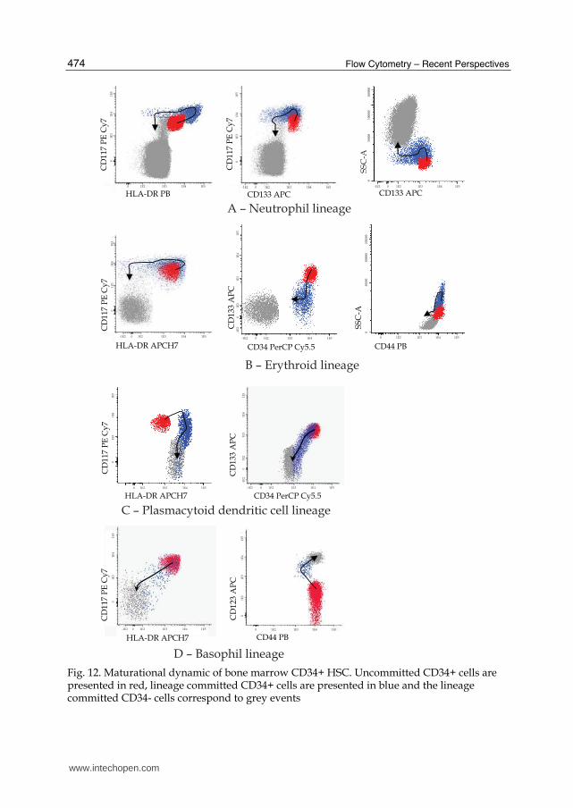
3.3 The maturation dynamic of bone marrow CD34+ hematopoietic stem cell
The possibility of a multiparameter analysis in a single cell basis conduct to a broader knowledge on the immunophenotypic characteristics of bone marrow CD34+ compartments and how it varies along the differentiation through different hematological cell lineages. Figure 12 depicts the dynamic of the maturation of different bone marrow CD34+ cell compartments.
www.intechopen.com
Flow Cytometry – Recent Perspectives
474
0
1
E3
1E
4
1
E5
0 1E2 1E3 1E4 1E5
CD
117
PE
Cy
7
HLA-DR PB-1E2 0 1E2 1E3 1E4 1E5
CD133 APC
0
1E
3
1E4
1E
5
CD
117
PE
Cy
7
SS
C-A
0
5
00
00
1
00
00
0 2
00
00
0
CD133 APC-1E2 0 1E2 1E3 1E4 1E5
HLA-DR APCH7 CD34 PerCP Cy5.5 CD44 PB
-1E2 0 1E2 1E3 1E4 1E5-1E2 0 1E2 1E3 1E4 1E5 0 1E2 1E3 1E4 1E5
CD
117
PE
Cy
7
CD
133
AP
C
SS
C-A
-1E
2
0
1E
2
1E
3
1E
4
1E
5
0 5
00
00
1
00
00
0 2
00
00
0
0
1E
3
1E4
1E
5
0 1E2 1E3 1E4 1E5
HLA-DR APCH7
CD
117
PE
Cy
70
1E
3
1E
4
1E
5
-1E2 0 1E2 1E3 1E4 1E5
CD34 PerCP Cy5.5
CD
133
AP
C-1
E2
0
1E
2
1E
3
1
E4
1E5
-1E2 0 1E2 1E3 1E4 1E5
CD
117
PE
Cy
7
CD
123
AP
C
0 1E2 1E3 1E4 1E5
CD44 PBHLA-DR APCH7
0
1E
2
1
E3
1E4
1E5
0
1E3
1
E4
1E5
D – Basophil lineage
B – Erythroid lineage
A – Neutrophil lineage
C – Plasmacytoid dendritic cell lineage
Fig. 12. Maturational dynamic of bone marrow CD34+ HSC. Uncommitted CD34+ cells are presented in red, lineage committed CD34+ cells are presented in blue and the lineage committed CD34- cells correspond to grey events
www.intechopen.com

Immunophenotypic Characterization of Normal Bone Marrow Stem Cells
475
4. Conclusion
The emergence of high-speed multi-parameter flow cytometers have given an important contribute to unveil the phenotypic characteristics of minor cell populations and/or populations without a known specific cell marker.
Using flow cytometry to characterize bone marrow MSC directly (without in vitro cell culture) represents a great advantage by enabling an analysis closest to the physiologic conditions of the cells, excluding all the phenotypic alterations induced by factors present in the culture medium. Moreover, this direct analysis allows an accurate quantification of these cells in bone marrow. In addition, the strategy used for bone marrow can also be applied in MSC from other tissues, allowing their direct quantification and characterization.
A broader knowledge about the immunophenotypic characteristics of the different compartments of bone marrow HSC could improve their identification, allow a more accurate quantification of those compartments, as well as shed light on the protein expression patterns in the earliest stages of maturation of each hematological cell lineage. Furthermore, a better knowledge of those protein expression patterns might contribute to the development of new strategies to identify aberrant phenotypes in hematological diseases affecting the more immature bone marrow cells compartments, which can be helpful in the classification of acute leukemias, diagnosis of myelodysplastic syndromes and detection of minimal residual disease. A more extensive understanding of the phenotype of CD34+ hematopoietic stem cells in the different maturational stages could also be useful to monitoring and investigate if different mobilization regimens have the capability of mobilizing distinct CD34+ hematopoietic stem cells subpopulations.
Here, we presented a simple, quick and economic approach to identify and quantify the different bone marrow CD34+ HSC compartments.
5. References
Bardin N, Anfosso F, Massé J, Cramer E, Sabatier F, Le Bivic A, Sampol J & Dignat-George F. (2001). Identification of CD146 as a component of the endothelial junction involved in the control of cell-cell cohesion. Blood, Vol.98, No.13, (December 2001), pp. 3677-3684, ISSN 0006-4971.
Bühring HJ, Müller CA, Letarte M, Gougos A, Saalmüller A, van Agthoven AJ, Busch FW. (1991). Endoglin is expressed on a subpopulation of immature erythroid cells of normal human bone marrow. Leukemia, Vol.5, No.10, (October 1991), pp. 841-847, ISSN 0887-6924.
Caplan A. (2007). Adult mesenchymal stem cells for tissue engineering versus regenerative medicine. J Cell Phisiol, Vol.213, No.2, (November 2007), pp. 341-347, ISSN 1097-4652.
Chamberlain G, Fox J, Ashton B & Middleton J. (2007). Concise review: mesenchymal stem cells: their phenotype, differentiation capacity, immunological features, and potential for homing. Stem Cells, Vol.25, No.11 (November 2007), pp. 2739-2749, ISSN 1549-4918.
Constantin G, Marconi S, Rossi B, Angiari S, Calderan L, Anghileri E, Gini B, Bach D, Martinello M, Bifari F, Galie M, Turano E, Budui S, Sbarabti A, Krampera M &
www.intechopen.com
Flow Cytometry – Recent Perspectives
476
Bonetti B. (2009). Stem Cells, Vol.27, No.10, (October 2009), pp. 2624-2635, ISSN 1549-4918.
Darlington PJ, Boivin MN, Bar-Or A. (2011). Harnessing the therapeutic potential of mesenchymal stem cells in multiple sclerosis. Expert Rev Neurother, Vol.11, no.9, (September 2011), pp. 1295-303, ISSN 1473-7175.
Del Cañizo MC, Fernández ME, López A, Vidriales B, Villarón E, Arroyo JL, Ortuño F, Orfao A & San Miguel JF. (2003). Immunophenotypic analysis of myelodysplastic syndromes. Haematologica, Vol.88, No.4, (April 2003), pp. 402-407, ISSN 0390-6078.
Delorme B, Ringe J, Gallay N, Le Vern Y, Kerboeuf D, Jorgensen C, Rosset P, Sensebé L, Layrolle P, Häupl T & Charbord P. (2008).Specific plasma membrane protein phenotype of culture-amplified and native human bone marrow mesenchymal stem cells. Blood, Vol.111, No.5, (March 2008), pp. 2631-2635, ISSN 0006-4971.
Ding DC, Shyu WC & Lin S. (2011). Mesenchymal stem cells. Cell Transplant, Vol.20, No.1, (2011), pp. 5–14, ISSN 1555-3892.
Ehninger A & Trumpp A. (2011). The bone marrow stem cell niche grows up: mesenchymal stem cells and macrophages move in. J Exp Med, Vol.208, No.3, (March 2011), pp. 421-428, ISSN 0022-1007.
Fortunel N, Hatzfeld A, & Hatzfeld J. (2000). Transforming growth factor-b: pleiotropic role in the regulation of hematopoiesis. Blood, Vol.96, No.6, (September 2000), pp. 2022-2036, ISSN 0006-4971.
Fox JM, Chamberlain G, Ashton BA & Middleton J. (2007). Recent advances into the understanding of mesenchymal stem cell trafficking. Br J Haematol, Vol.137, No.6, (June 2007), pp. 491-502, ISSN 1365-2141.
Goussetis E, Theodosaki M, Paterakis G, Tsecoura C & Graphakos S. (2006). In vitro identification of a cord blood CD133+CD34-Lin+ cell subset that gives rise to myeloid dendritic precursors. Stem Cells, Vol.24, No.4, (April 2006), pp. 1137-1140, ISSN 1549-4918.
Gu J, Isaji T, Sato Y, Kariya Y& Fukuda T. (2009).Importance of N-glycosylation on alpha5beta1 integrin for its biological functions. Biol Pharm Bull, Vol.32, No.5, (May 2009), pp. 780-785, ISSN 0918-6158.
Halfon S, Abramov N, Grinblat B & Ginis I. (2011). Markers distinguishing mesenchymal stem cells from fibroblasts are downregulated with passaging. Stem Cell Dev, Vol.20, No.1, (January 2011), ISSN 1547-3287.
Horwitz EM, Gordon PL, Koo WK, Marx JC, Neel MD, McNall RY, Muul L & Hofmann T. (2002). Isolated allogeneic bone marrow-derived mesenchymal cells engraft and stimulate growth in children with osteogenesis imperfecta: Implications for cell therapy of bone. Proc Natl Acad Sci USA, Vol.99, No.13, (June 2002), pp. 8932-8937, ISSN 0027-8424.
Jones E & McGonagle D. (2008).Human bone marrow mesenchymal stem cells in vivo. Rheumatology (Oxford). Vol.47, No.2, (February 2008), pp. 126-131, ISSN 1462-0324.
Jurisic G, Iolyeva M, Proulx ST, Halin C & Detmar M. (2010). Thymus cell antigen 1 (Thy1, CD90) is expressed by lymphatic vessels and mediates cell adhesion to lymphatic endothelium. Exp Cell Res, Vol.316, No.17, (October 2010) pp. 2982-2992, ISSN 0014-4827.
Kolf CM, Cho E & Tuan RS. (2007). Mesenchymal stromal cells. Biology of adult mesenchymal stem cells: regulation of niche, self-renewal and differentiation. Arthritis Res Ther, Vol.9, No.1, (February 2007), pp. 204-214, ISSN 1478-6354.
www.intechopen.com
Immunophenotypic Characterization of Normal Bone Marrow Stem Cells
477
Martins AA, Paiva A, Morgado JM, Gomes A & Pais ML. (2009). Quantification and immunophenotypic characterization of bone marrow and umbilical cord blood mesenchymal stem cells by multicolor flow cytometry. Transplant Proc, Vol.41, No.3, (April 2009), pp. 943-946, ISSN 0041-1345.
Matarraz S, López A, Barrena S, Fernandez C, Jensen E, Flores J, Bárcena P, Rasillo A, Sayagues JM, Sánchez ML, Hernandez-Campo P, Hernandez Rivas JM, Salvador C, Fernandez-Mosteirín N, Giralt M, Perdiguer L & Orfao A. (2008). The immunophenotype of different immature, myeloid and B-cell lineage-committed CD34+ hematopoietic cells allows discrimination between normal/reactive and myelodysplastic syndrome precursors. Leukemia, Vol.22, No.6, (June 2008), pp. 1175-1183, ISSN 0887-6924.
Micera A, Lambiase A, Stampachiacchiere B, Bonini S, Bonini S & Levi-Schaffer F. (2007). Nerve growth factor and tissue repair remodeling: trkA(NGFR) and p75(NTR), two receptors one fate. Cytokine Growth Factor Rev, Vol.18, No.3-4, (June-August 2007), pp. 245-256, ISSN 1359-6101.
Moody JL, Singbrant S, Karlsson G, Blank U, Aspling M, Flygare J, Bryder D & Karlsson S. (2007). Endoglin is not critical for hematopoietic stem cell engraftment and reconstitution but regulates adult erythroid development. Stem Cells, Vol.25, No.11, (November 2007), pp. 2809-2819, ISSN 1549-4918.
Musina RA, Bekchanova ES & Sukhikh GT. (2005). Comparison of mesenchymal stem cells obtained from different human tissues. Bull Exp Biol Med, Vol.139, No.4, (April 2005), pp. 504-9, ISSN 0007-4888.
Nimer S. (2008). MDS: a stem cell disorder--but what exactly is wrong with the primitive hematopoietic cells in this disease? Hematology Am Soc Hematol Educ Program, (2008), pp. 43-51. ISSN 1520-4391.
Orfao A, Ortuño F, Santiago M, Lopez A & San Miguel J. (2004). Immunophenotyping of Acute Leukemias and Myelodysplastic Syndromes. Cytometry A, Vol.58A, No.1, (March 2004), pp. 62-71. ISSN 1552-4930.
Pastore D, Mestice A, Perrone T, Gaudio F, Delia M, Albano F, Russo Rossi A, Carluiccio P, Leo M, Liso V& Specchia G. (2008). Subsets of CD34+ and early engraftment kinetics in allogeneic peripheral SCT for AML. Bone Marrow Transplant, Vol.41, No.11, (June 2008), pp. 977-981, ISSN 0268-3369.
Pittenger MF, Mackay AM, Beck SC, Jaiswal RK, Douglas R, Mosca JD, Moorman MA, Simonetti DW, Craig S & Marshak DR. (1999). Multilineage potential of adult human mesenchymal stem cells. Science, Vol.284, No.5411, (April 1999), pp. 143-7, ISSN 0036-8075.
Rege TA & Hagood JS. (2006). Thy-1 as a regulator of cell-cell and cell-matrix interactions in axon regeneration, apoptosis, adhesion, migration, cancer, and fibrosis. FASEB J, Vol.20, No.8, (June 2006), pp. 1045-54, ISSN: 0892-6638.
Remberger M, Uhlin M, Karlsson H, Omazic B, Svahn BM & Mattsson J. (2011). Treatment with mesenchymal stromal cells does not improve long-term survival in patients with severe acute GVHD. Transpl Immunol, 2011 Sep 10 [Epub ahead of print], ISSN 0966-3274.
Rogers ML, Bailey S, Matusica D, Nicholson I, Muyderman H, Pagadala PC, Neet KE, Zola H, Macardle P & Rush RA. ProNGF mediates death of Natural Killer cells through activation of the p75NTR-sortilin complex. J Neuroimmunol, Vol.226, No.1-2, (September 2010), pp. 93-103, ISSN: 0165-5728.
www.intechopen.com
Flow Cytometry – Recent Perspectives
478
Rokhlin OW, Cohen MB, Kubagawa H, Letarte M & Cooper MD. (2005). Differential expression of endoglin on fetal and adult hematopoietic cells in human bone marrow. J Immunol, Vol.154, No.9, (May 1995), pp. 4456-4465, ISSN: 0022-1767.
Siatskas C, Payne NL, Short MA & Bernard CC. (2010). A consensus statement addressing mesenchymal stem cell transplantation for multiple sclerosis: it's time! Stem Cell Rev, Vol.6, No,4, (December 2010), pp. 500-506, ISSN 1550-8943.
Stagg J. (2007). Immune regulation by mesenchymal stem cells: two sides to the coin. Tissue Antigens, Vol.69, No.1, (January 2007), pp. 1-9, ISSN 0001-2815.
Tormin A, Li O, Brune JC, Walsh S, Schütz B, Ehinger M, Ditzel N, Kassem M & Scheding S. (2011). CD146 expression on primary nonhematopoietic bone marrow stem cells is correlated with in situ localization. Blood, Vol.117, No.19, (May 2011), pp. 5067-77, ISSN 0006-4971.
Undale AH, Westendorf JJ, Yaszemski MJ & Khosla S. (2009). Mesenchymal stem cells for bone repair and metabolic bone diseases. Mayo Clin Proc, Vol.84, No.10, (October 2009), pp. 893-902, ISSN: 0025-6196.
van Lochem EG, van der Velden VH, Wind HK, te Marvelde JG, Westerdaal NA & van Dongen JJ. (2004). Immunophenotypic differentiation patterns of normal hematopoiesis in human bone marrow: reference patterns for age-related changes and disease-induced shifts. Cytometry B Clin Cytom, Vol.60, No.1, (July 2004), pp. 1-13, ISSN 1552-4957.
von Bahr L, Sundberg B, Lönnies L, Sander B, Karbach H, Hägglund H, Ljungman P, Gustafsson B, Karlsson H, Le Blanc K & Ringdén O. (2011). Long-Term Complications, Immunologic Effects, and Role of Passage for Outcome in Mesenchymal Stromal Cell Therapy. Biol Blood Marrow Transplant, 2011 Aug 4 [Epub ahead of print]. ISSN 1083-8791.
Wang L, Fan J, Thompson LF, Zhang Y, Shin T, Curiel TJ & Zhang B. (2011). CD73 has distinct roles in nonhematopoietic and hematopoietic cells to promote tumor growth in mice. J Clin Invest, Vol.121, No.6, (June 2011), pp. 2371-2382, ISSN 0021-9738.
Yin AH, Miraglia S, Zanjani ED, Almeida-Porada G, Ogawa M, Leary AG, Olweus J, Kearney J & Buck DW. (1997). AC133, a novel marker for human hematopoietic stem and progenitor cells. Blood, Vol.90, No.12, (December 1997), pp. 5002-5012, ISSN 0006-4971.
Zeng GF, Cai SX, Wu GJ. (2011). Up-regulation of METCAM/MUC18 promotes motility, invasion, and tumorigenesis of human breast cancer cells. BMC Cancer, Vol. 11, (March 2011), pp. 113-126, ISSN 1471-2407 .
Zhang B. (2010). CD73: A novel target for cancer immunotherapy. Cancer Res, Vol.70, No.16, (August 2010), pp. 6407–6411, ISSN 0008-5472.
www.intechopen.com

Flow Cytometry - Recent PerspectivesEdited by M.Sc. Ingrid Schmid
ISBN 978-953-51-0626-5Hard cover, 500 pagesPublisher InTechPublished online 13, June, 2012Published in print edition June, 2012
InTech EuropeUniversity Campus STeP Ri Slavka Krautzeka 83/A 51000 Rijeka, Croatia Phone: +385 (51) 770 447 Fax: +385 (51) 686 166www.intechopen.com
InTech ChinaUnit 405, Office Block, Hotel Equatorial Shanghai No.65, Yan An Road (West), Shanghai, 200040, China
Phone: +86-21-62489820 Fax: +86-21-62489821
"Flow Cytometry - Recent Perspectives" is a compendium of comprehensive reviews and original scientificpapers. The contents illustrate the constantly evolving application of flow cytometry to a multitude of scientificfields and technologies as well as its broad use as demonstrated by the international composition of thecontributing author group. The book focuses on the utilization of the technology in basic sciences and coverssuch diverse areas as marine and plant biology, microbiology, immunology, and biotechnology. It is hoped thatit will give novices a valuable introduction to the field, but will also provide experienced flow cytometrists withnovel insights and a better understanding of the subject.
How to referenceIn order to correctly reference this scholarly work, feel free to copy and paste the following:
Paula Laranjeira, Andreia Ribeiro, Sandrine Mendes, Ana Henriques, M. Luisa Pais and Artur Paiva (2012).Immunophenotypic Characterization of Normal Bone Marrow Stem Cells, Flow Cytometry - RecentPerspectives, M.Sc. Ingrid Schmid (Ed.), ISBN: 978-953-51-0626-5, InTech, Available from:http://www.intechopen.com/books/flow-cytometry-recent-perspectives/immunophenotypic-characterization-of-normal-bone-marrow-stem-cells