immunosuppressionwithantitumournecrosisfactortherapy...
TRANSCRIPT
Case ReportImmunosuppression with Antitumour Necrosis Factor TherapyLeading to Strongyloides Hyperinfection Syndrome
Muhammad Farhan Khaliq ,1 Rayan E. Ihle ,2 and James Perry3
1Department of Internal Medicine, Charleston Area Medical Center, Charleston, WV, USA2Department of Pulmonary Critical Care, West Virginia University Charleston Division, Charleston, WV, USA3Department of Pulmonary Critical Care, Charleston Area Medical Center, Charleston, WV, USA
Correspondence should be addressed to Muhammad Farhan Khaliq; [email protected]
Received 25 January 2018; Revised 1 May 2018; Accepted 8 May 2018; Published 30 May 2018
Academic Editor: Peter Olumese
Copyright © 2018Muhammad Farhan Khaliq et al.+is is an open access article distributed under theCreative CommonsAttributionLicense, which permits unrestricted use, distribution, and reproduction in any medium, provided the original work is properly cited.
Strongyloides stercoralis is an endemic parasitic infection that can remain asymptomatic for years, but it can cause death inimmunosuppressed individuals. Here, we present a case of Strongyloides hyperinfection in a 75-year-old male secondary to sepsisand chronic immunosuppression due to TNF-α inhibitors. Despite aggressive treatment including broad-spectrum antibioticsand antihelminths, his respiratory failure worsened and he died after palliative extubation. S. stercoralis infection remainsa diagnostic challenge. Presentation with Strongyloides is often nonspecific, and eosinophilia is absent in hyperinfection. Diagnosiscan be delayed, especially in low-prevalence areas where suspicion is low. Strongyloides should be considered in the differentialdiagnosis in the presence of risk factors including immunosuppressive therapy, and a travel history should be carefully obtained.Patients with recurrent enterobacterial sepsis or respiratory failure with diffuse infiltrates in the setting of eosinophilia shouldundergo testing for Strongyloides. A multidisciplinary approach can result in earlier diagnosis and favorable outcomes.
1. Introduction
Strongyloides stercoralis is an intestinal nematode that isendemic in tropical and subtropical areas and affects 370million people globally [1]. Its presentation can vary fromasymptomatic eosinophilia in immunocompetent patients tohyperinfection syndrome causing multiple organ failure inimmunocompromised patients. A weakened host immuneresponse results in an increased parasitic load, including inthe lungs, referred to as hyperinfection. Larvae migratingbeyond the lungs, for example, to the brain or skin, aretermed “disseminated Strongyloides.” Chronic glucocorti-coid therapy, malnutrition, alcoholism, and underlyinghuman T-lymphotrophic virus type 1 (HTLV-1) infectionare known risk factors. Despite therapy, prognosis remainspoor, with mortality reaching 70% [2, 3].
2. Case Report
A 75-year-old male was transferred to our facility due to acuterespiratory failure and sepsis secondary to extended-spectrum
beta-lactamase Escherichia coli urinary tract infection (ESBL-UTI). His past medical history included a recent diagnosis ofdeep vein thrombosis, after which he developed gastroin-testinal bleeding while on anticoagulation therapy. He hadrheumatoid arthritis, gout, diabetes mellitus, hypertension,atrial fibrillation, stage III chronic kidney disease, IgMmonoclonal gammopathy of undetermined significance, andchronic pain syndrome. His surgical history includedplacement of an inferior vena caval filter and recurrent bi-lateral pleural effusions requiring decortication. He recentlyhad a gout flare for which he was treated with a prednisolonetaper. +e patient’s rheumatoid arthritis was controlled withinfliximab and hydroxychloroquine sulfate. He was a formersmoker but had no significant history of alcohol or illicit druguse. He was from rural West Virginia which is located in thenorth central subregion of Appalachia. He denied traveloutside of the United States.
He originally presented with decreased appetite, nausea,vomiting, and abdominal pain at an outlying facility. At thistime, vital signs were normal with a temperature of 36.9°C,blood pressure of 131/68mmHg, a regular pulse rate of
HindawiCase Reports in Infectious DiseasesVolume 2018, Article ID 6341680, 4 pageshttps://doi.org/10.1155/2018/6341680
96 beats/minute, and a respiratory rate of 17. He was alert,awake, and oriented to time and person. +e remainder ofhis physical examination was unremarkable. Initial bloodinvestigations revealed a haemoglobin level of 8.5 g/dL, totalwhite cell count of 8×10⁹/L without eosinophilia, anda normal platelet count of 203×109/L. Biochemically, therewas evidence of impaired renal function with a creatininelevel of 2.8mg/dL, hyponatraemia (Na 122mEq/L), andhypochloraemia (Cl 92mEql/L). Liver function tests wereunremarkable including his coagulation profile. +e patienthad Clostridium difficile testing, which was negative. Anabdominal computed tomography (CT) scan demonstratedonly fecal retention. His electrolyte abnormalities were at-tributed to dehydration from vomiting, so a nasogastric tubewas placed while fluid resuscitation was administered. Adiagnosis of diabetic gastropathy was made and metoclo-pramide started. His hospital course was complicated bya UTI secondary to E. coli which was resistant to multipleantibiotics. He was treated with ciprofloxacin. However, thepatient continued to worsen, and he developed sepsis andrespiratory failure requiring intubation and transfer to ourfacility for higher care.
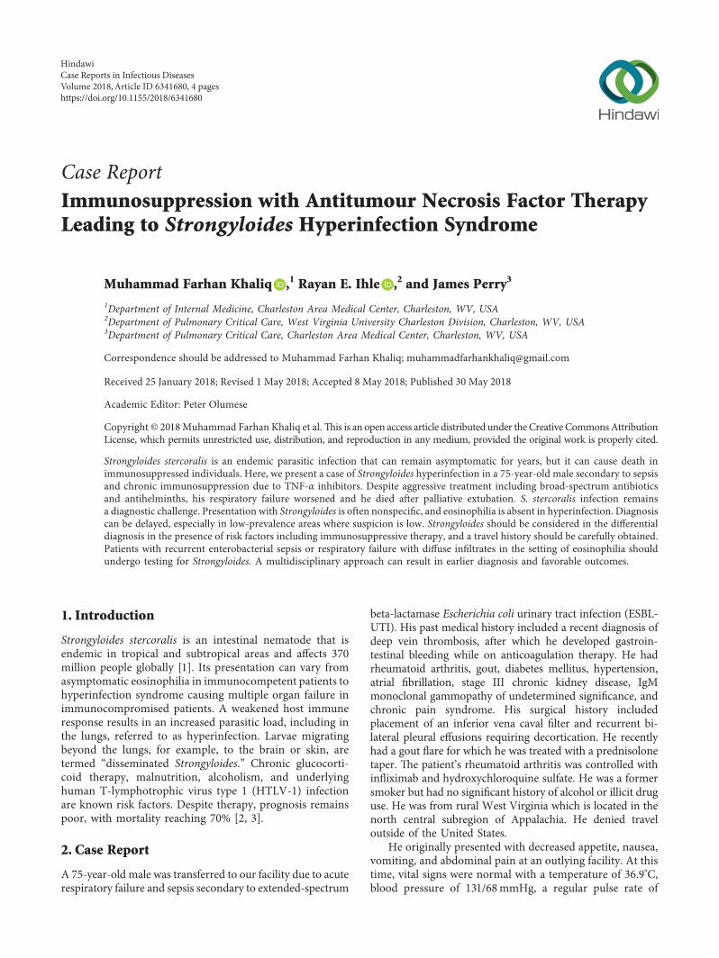
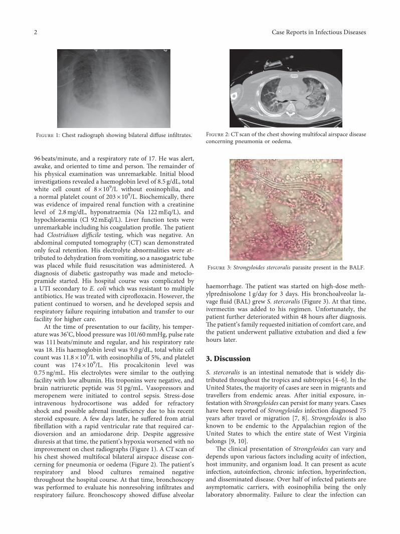
At the time of presentation to our facility, his temper-ature was 36°C, blood pressure was 101/60mmHg, pulse ratewas 111 beats/minute and regular, and his respiratory ratewas 18. His haemoglobin level was 9.0 g/dL, total white cellcount was 11.8×10⁹/L with eosinophilia of 5%, and plateletcount was 174×109/L. His procalcitonin level was0.75 ng/mL. His electrolytes were similar to the outlyingfacility with low albumin. His troponins were negative, andbrain natriuretic peptide was 51 pg/mL. Vasopressors andmeropenem were initiated to control sepsis. Stress-doseintravenous hydrocortisone was added for refractoryshock and possible adrenal insufficiency due to his recentsteroid exposure. A few days later, he suffered from atrialfibrillation with a rapid ventricular rate that required car-dioversion and an amiodarone drip. Despite aggressivediuresis at that time, the patient’s hypoxia worsened with noimprovement on chest radiographs (Figure 1). A CT scan ofhis chest showed multifocal bilateral airspace disease con-cerning for pneumonia or oedema (Figure 2). +e patient’srespiratory and blood cultures remained negativethroughout the hospital course. At that time, bronchoscopywas performed to evaluate his nonresolving infiltrates andrespiratory failure. Bronchoscopy showed diffuse alveolar
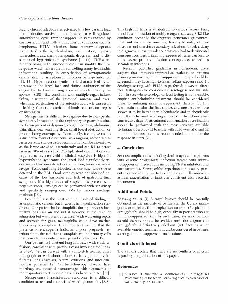
haemorrhage. +e patient was started on high-dose meth-ylprednisolone 1 g/day for 3 days. His bronchoalveolar la-vage fluid (BAL) grew S. stercoralis (Figure 3). At that time,ivermectin was added to his regimen. Unfortunately, thepatient further deteriorated within 48 hours after diagnosis.+e patient’s family requested initiation of comfort care, andthe patient underwent palliative extubation and died a fewhours later.
3. Discussion
S. stercoralis is an intestinal nematode that is widely dis-tributed throughout the tropics and subtropics [4–6]. In theUnited States, the majority of cases are seen in migrants andtravellers from endemic areas. After initial exposure, in-festation with Strongyloides can persist for many years. Caseshave been reported of Strongyloides infection diagnosed 75years after travel or migration [7, 8]. Strongyloides is alsoknown to be endemic to the Appalachian region of theUnited States to which the entire state of West Virginiabelongs [9, 10].
+e clinical presentation of Strongyloides can vary anddepends upon various factors including acuity of infection,host immunity, and organism load. It can present as acuteinfection, autoinfection, chronic infection, hyperinfection,and disseminated disease. Over half of infected patients areasymptomatic carriers, with eosinophilia being the onlylaboratory abnormality. Failure to clear the infection can
Figure 1: Chest radiograph showing bilateral diffuse infiltrates. Figure 2: CT scan of the chest showing multifocal airspace diseaseconcerning pneumonia or oedema.
Figure 3: Strongyloides stercoralis parasite present in the BALF.
2 Case Reports in Infectious Diseases
lead to chronic infection characterized by a low parasite loadthat maintains survival in the host via a well-regulatedautoinfection cycle. Immunosuppressive states induced bycorticosteroids and TNF-α inhibitors or conditions such aslymphoma, HTLV infection, bone marrow allografts,rheumatoid arthritis, alcoholism, malnutrition, leprosy,tuberculosis, and chemotherapeutic drugs can lead to dis-seminated hyperinfection syndrome [11–14]. TNF-α in-hibitors along with glucocorticoids can modify the +2response which has a role in controlling many helminthicinfestations resulting in exacerbation of asymptomaticcarrier state to symptomatic infection or hyperinfection[12, 13]. Hyperinfection syndrome is characterized by anincrease in the larval load and diffuse infiltration of theorgans by the larva causing a systemic inflammatory re-sponse- (SIRS-) like condition with multiple organ failure.Often, disruption of the intestinal mucosa and over-whelming acceleration of the autoinfection cycle can resultin leaking of enteric bacteria into bloodstream to cause sepsisor meningitis.
Strongyloides is difficult to diagnose due to nonspecificsymptoms. Infestation of the respiratory or gastrointestinaltracts can present as dyspnoea, cough, wheezing, abdominalpain, diarrhoea, vomiting, ileus, small bowel obstruction, orprotein-losing enteropathy. Occasionally, it can give rise toa distinctive form of cutaneous larva migrans, recognized aslarva currens. Standard stool examination can be insensitive,as the larvae are shed intermittently and can fail to detectlarva in 70% of cases [15]. Multiple stool examinations arerequired to increase yield if clinical suspicion is high. Inhyperinfection syndrome, the larval load significantly in-creases and becomes detectable in sputum, bronchoalveolarlavage (BAL), and lung biopsies. In our case, larvae weredetected in the BAL. Stool samples were not obtained be-cause of the low suspicion and lack of gastrointestinalsymptoms. If a high index of suspicion is present withnegative stools, serology can be performed with sensitivityand specificity ranging over 95% by various serologicmethods [16].
Eosinophilia is the most common isolated finding inasymptomatic carriers but is absent in hyperinfection syn-drome. Our patient had eosinophilia during previous hos-pitalizations and on the initial labwork at the time ofadmission but was absent otherwise. With worsening sepsisand steroids for gout, neutrophilia could have maskedunderlying eosinophilia. It is important to note that thepresence of eosinopenia indicates a poor prognosis, at-tributable to the fact that eosinophils are the primary cellsthat provide immunity against parasitic infections [17].
Our patient had bilateral lung infiltrates with small ef-fusions, consistent with previous cases involving the lungs.Strongyloides can present with a completely normal chestradiograph or with abnormalities such as pulmonary in-filtrates, lung abscesses, pleural effusions, and interstitialnodular patterns [18]. On bronchoscopy, alveolar hae-morrhage and petechial haemorrhages with hyperaemia ofthe respiratory tract mucosa have also been reported [19].
Strongyloides hyperinfection syndrome is a difficultcondition to treat and is associated with highmortality [2, 3].
+is high mortality is attributable to various factors. First,the diffuse infiltration of multiple organs causes a SIRS-likecondition. Secondly, the organism penetrates gastrointes-tinal and respiratory mucosae, leading to entry of newmicrobes and therefore secondary infections. +ird, a delayin diagnosis in low-prevalence areas can lead to detrimentalconsequences. Lastly, immunosuppressed states can lead tomore severe primary infection consequences as well assecondary infections.
Recently published guidelines in nonendemic areassuggest that immunocompromised patients or patientsplanning on starting immunosuppressant therapy should bescreened if they have high-to-intermediate exposure risk [2].Serologic testing with ELISA is preferred; however, directfecal testing can be considered if serology is not available[20]. In case where serology or fecal testing is not available,empiric antihelminthic treatment should be consideredprior to initiating immunosuppressant therapy [2, 19].Ivermectin remains the first choice, and most studies haveshown it to be better than albendazole and thiabendazole[21]. It can be used as a single dose or in two doses givenconsecutive days. Posttreatment confirmation of eradicationshould be performed with the most sensitive availabletechniques. Serology at baseline with follow-up at 6 and 12months after treatment is recommended to monitor theresponse in titers [20].
4. Conclusion
Serious complications including death may occur in patientswith chronic Strongyloides infection treated with immu-nosuppressant medications including TNF-α inhibitors andcorticosteroids. Strongyloides hyperinfection usually pres-ents as acute respiratory failure and may initially mimic anasthma exacerbation or infiltrates consistent with bacterialpneumonia.
Additional Points
Learning points. (i) A travel history should be carefullyobtained, as the majority of patients in the US are immi-grants or travellers from tropical countries. (ii) Suspicion ofStrongyloides should be high, especially in patients who areimmunosuppressed. (iii) In such cases, systemic cortico-steroid therapy should be avoided until the diagnosis ofStrongyloides is definitively ruled out. (iv) If testing is notavailable, empiric treatment should be considered in patientsstarting immunosuppressant medications.
Conflicts of Interest
+e authors declare that there are no conflicts of interestregarding the publication of this paper.
References
[1] Z. Bisoffi, D. Buonfrate, A. Montresor et al., “Strongyloidesstercoralis: a plea for action,” PLoS Neglected Tropical Diseases,vol. 7, no. 5, p. e2214, 2013.
Case Reports in Infectious Diseases 3
[2] D. Buonfrate, A. Requena-Mendez, A. Angheben et al.,“Severe strongyloidiasis: a systematic review of case reports,”BMC Infectious Diseases, vol. 13, no. 1, p. 78, 2013.
[3] S. Lim, K. Katz, S. Krajden, M. Fuksa, J. S. Keystone, andK. C. Kain, “Complicated and fatal Strongyloides infection inCanadians: risk factors, diagnosis and management,” Cana-dian Medical Association Journal, vol. 171, no. 5, pp. 479–484,2004.
[4] M. Montes, C. Sawhney, and N. Barros, “Strongyloides ster-coralis: there but not seen,” Current Opinion in InfectiousDiseases, vol. 23, no. 5, pp. 500–504, 2010.
[5] I. S. Hong, S. Y. Zaidi, P. McEvoy, and R. C. Neafie, “Diagnosisof Strongyloides stercoralis in a peritoneal effusion from anHIV-seropositive man,” Acta Cytologica, vol. 48, no. 2,pp. 211–214, 2004.
[6] B. Kakati, S. Dang, M. Heif, K. Caradine, W. McKnight, andF. Aduli, “Strongyloides duodenitis: case report and review ofliterature,” Journal of the National Medical Association,vol. 103, no. 1, pp. 60–63, 2011.
[7] V. Prendki, P. Fenaux, R. Durand, M. +ellier, andO. Bouchaud, “Strongyloidiasis in man 75 years after initialexposure,” Emerging Infectious Diseases, vol. 17, no. 5,pp. 931-932, 2011.
[8] L. Nabha, S. Krishnan, R. Ramanathan et al., “Prevalence ofStrongyloides stercoralis in an urban US AIDS cohort,” Path-ogens and Global Health, vol. 106, no. 4, pp. 238–244, 2012.
[9] R. M. Genta, “Global prevalence of strongyloidiasis: criticalreview with epidemiologic insights into the prevention ofdisseminated disease,” Reviews Infectious Diseases, vol. 11,no. 5, pp. 755–767, 1989.
[10] S. L. Berk, A. Verghese, S. Alvarez, K. Hall, and B. Smith,“Clinical and epidemiologic features of strongyloidiasis:a prospective study in rural Tennessee,” Archives of InternalMedicine, vol. 147, no. 7, pp. 1257–1261, 1987.
[11] A. Basile, S. Simzar, J. Bentow et al., “Disseminated Strong-yloides stercoralis: hyperinfection during medical immuno-suppression,” Journal of the American Academy ofDermatology, vol. 63, no. 5, pp. 896–902, 2010.
[12] P. B. Keiser and T. B. Nutman, “Strongyloides stercoralis in theimmunocompromised population,” Clinical MicrobiologyReviews, vol. 17, no. 1, pp. 208–217, 2004.
[13] F. Schar, U. Trostdorf, F. Giardina et al., “Strongyloidesstercoralis: global distribution and risk factors,” PLoSNeglected Tropical Diseases, vol. 7, no. 7, p. e2288, 2013.
[14] M. D. Boatright and B. W. Wang, “Clinical infection withStrongyloides sterocoralis following etanercept use for rheu-matoid arthritis,” Arthritis & Rheumatism, vol. 52, no. 4,pp. 1336-1337, 2005.
[15] C. D. Ericsson, R. Steffen, A. A. Siddiqui, and S. L. Berk,“Diagnosis of Strongyloides stercoralis infection,” ClinicalInfectious Diseases, vol. 33, no. 7, pp. 1040–1047, 2001.
[16] Z. Bisoffi, D. Buonfrate, M. Sequi et al., “Diagnostic accuracyof five serologic tests for Strongyloides stercoralis infection,”PLoS Neglected Tropical Diseases, vol. 8, no. 1, p. e2640, 2014.
[17] T. W. Gyorkos, R. M. Genta, P. Viens, and J. D. Maclean,“Seroepidemiology of Strongyloides infection in the SoutheastAsian refugee population in Canada,” American Journal ofEpidemiology, vol. 132, no. 2, pp. 257–264, 1990.
[18] V. K. Vijayan, “Parasitic lung infections,” Current Opinion inPulmonary Medicine, vol. 15, no. 3, pp. 274–282, 2009.
[19] A. Yee, C. T. Boylen, T. Noguchi, E. C. Klatt, andO. P. Sharma, “Fatal Strongyloides stercoralis infection ina patient receiving corticosteroids,” Western Journal ofMedicine, vol. 146, no. 3, p. 363, 1987.
[20] A. Requena-Mendez, D. Buonfrate, J. Gomez-Junyent,L. Zammarchi, Z. Bisoffi, and J. Muñoz, “Evidence-basedguidelines for screening and management of strongyloidia-sis in non-endemic countries,” American Journal of TropicalMedicine and Hygiene, vol. 97, no. 3, pp. 645–652, 2017.
[21] C. A. J. Henriquez-Camacho, E. Gotuzzo, J. Echevarria, andA. Terashima, “Ivermectin versus albendazole or thiabenda-zole for Strongyloides stercoralis infection,” Cochrane Data-base of Systematic Reviews, no. 1, 2016.
4 Case Reports in Infectious Diseases

Stem Cells International
Hindawiwww.hindawi.com Volume 2018
Hindawiwww.hindawi.com Volume 2018
MEDIATORSINFLAMMATION
of
EndocrinologyInternational Journal of
Hindawiwww.hindawi.com Volume 2018
Hindawiwww.hindawi.com Volume 2018
Disease Markers
Hindawiwww.hindawi.com Volume 2018
BioMed Research International
OncologyJournal of
Hindawiwww.hindawi.com Volume 2013
Hindawiwww.hindawi.com Volume 2018
Oxidative Medicine and Cellular Longevity
Hindawiwww.hindawi.com Volume 2018
PPAR Research
Hindawi Publishing Corporation http://www.hindawi.com Volume 2013Hindawiwww.hindawi.com
The Scientific World Journal
Volume 2018
Immunology ResearchHindawiwww.hindawi.com Volume 2018
Journal of
ObesityJournal of
Hindawiwww.hindawi.com Volume 2018
Hindawiwww.hindawi.com Volume 2018
Computational and Mathematical Methods in Medicine
Hindawiwww.hindawi.com Volume 2018
Behavioural Neurology
OphthalmologyJournal of
Hindawiwww.hindawi.com Volume 2018
Diabetes ResearchJournal of
Hindawiwww.hindawi.com Volume 2018
Hindawiwww.hindawi.com Volume 2018
Research and TreatmentAIDS
Hindawiwww.hindawi.com Volume 2018
Gastroenterology Research and Practice
Hindawiwww.hindawi.com Volume 2018
Parkinson’s Disease
Evidence-Based Complementary andAlternative Medicine
Volume 2018Hindawiwww.hindawi.com
Submit your manuscripts atwww.hindawi.com