impact of host factors on the adaptive immune response to
TRANSCRIPT

University of Pennsylvania University of Pennsylvania
ScholarlyCommons ScholarlyCommons
Publicly Accessible Penn Dissertations
2017
Impact Of Host Factors On The Adaptive Immune Response To Impact Of Host Factors On The Adaptive Immune Response To
Aav Gene Therapy Aav Gene Therapy
Scott N. Ashley University of Pennsylvania, [email protected]
Follow this and additional works at: https://repository.upenn.edu/edissertations
Part of the Allergy and Immunology Commons, Immunology and Infectious Disease Commons,
Medical Immunology Commons, and the Molecular Biology Commons
Recommended Citation Recommended Citation Ashley, Scott N., "Impact Of Host Factors On The Adaptive Immune Response To Aav Gene Therapy" (2017). Publicly Accessible Penn Dissertations. 2171. https://repository.upenn.edu/edissertations/2171
This paper is posted at ScholarlyCommons. https://repository.upenn.edu/edissertations/2171 For more information, please contact [email protected].
Impact Of Host Factors On The Adaptive Immune Response To Aav Gene Impact Of Host Factors On The Adaptive Immune Response To Aav Gene Therapy Therapy
Abstract Abstract Impact of host factor on the adaptive immune response to AAV gene therapy
Scott Ashley
James Wilson
Adaptive immune responses to the transgene product remain an active area of concern for the gene therapy field. How host factors can influence the activation of the immune system is an important consideration in the development of gene therapy for different genetic disorders. One factor considered by many to shape the adaptive immune response is an individual’s genotype. Nonsense mutations were thought to result in an absence of tolerance to a replacement protein provided by gene therapy, due to a lack of antigen presentation during T cell development and negative selection. In this work we demonstrated that a class of nonsense mutations, premature termination codons (PTC), found in ornithine transcarbamylase deficiency (OTC) patients do not inhibit antigen presentation of C-terminal epitopes. We further found that these PTC containing OTC genes were able to induce anergy in a model of peripheral tolerance. These results change how we think about the relationship between the genotype and immune response, which indicate that individuals with PTC mutations may be less at risk of an adverse immune response attenuating the effects of gene therapy. We also identified vector factors that influence the adaptive immune response, activation of TLR9 and the tissues targeted for transduction and expression of the transgene. To investigate how inflammatory signaling might impact the outcome of adaptive immune responses; we use the transgenic OT-1 mouse model to interrogate how TLR9, the primary sensing molecule for vector DNA, can activate cytotoxic T cells against the transgene product OTC. These results confirm an important role for TLR9 induced inflammation being necessary for transgene specific T cell activation. To investigate the influence of TLR9 signaling and tissue targets on the humoral response, we used a mouse model of Mucopolysaccharidosis type I (MPS1) disease, and measured antibody generation to the secreted transgene product alpha-L-iduronidase (IDUA). We report that TLR9 signaling is also instrumental for the formation of anti-IDUA antibodies, as is expression of the transgene from the muscle. This work describes a novel process by which tolerance to a peptide located downstream of a PTC can be induced. This insight can help us better define the risks associated with an adaptive immune response based on an individual’s personal mutation. We also defined important vector factors which are important for activation of an adaptive immune response, and this knowledge could be exploited to generate safer gene therapy delivery methods.
Degree Type Degree Type Dissertation
Degree Name Degree Name Doctor of Philosophy (PhD)
Graduate Group Graduate Group Cell & Molecular Biology
First Advisor First Advisor James M. Wilson

Keywords Keywords AAV, Adaptive Immunity, Alternative Translation, Antigen Presentation, Gene Therapy, Tolerance
Subject Categories Subject Categories Allergy and Immunology | Immunology and Infectious Disease | Medical Immunology | Molecular Biology
This dissertation is available at ScholarlyCommons: https://repository.upenn.edu/edissertations/2171
IMPACT OF HOST FACTORS ON THE ADAPTIVE IMMUNE
RESPONSE TO AAV GENE THERAPY
Scott N Ashley
A DISSERTATION
in
Cell and Molecular Biology
Presented to the Faculties of the University of Pennsylvania
in
Partial Fulfillment of the Requirements for the
Degree of Doctor of Philosophy
2017
Dissertation Supervisor
James M Wilson, M.D., Ph.D.
Rose H. Weiss Orphan Disease Center Director's Professor
______________________
Graduate Group Chairperson
Daniel S Kessler, Ph.D.
Associate Professor of Cell and Developmental Biology
______________________
Dissertation Committee
Youhai H. Chen, M.D., Ph.D. Professor of
Pathology and Laboratory Medicine
Narayan Avadhani, Ph.D. Harriet Ellison
Woodward Professor of Biochemistry
Michael R Betts, Ph.D. Associate Professor
of Microbiology
Hiroki Morizono, Ph.D. Associate Research
Professor of Integrated Systems Biology
and Pediatrics

IMPACT OF HOST FACTORS ON THE ADAPTIVE IMMUNE RESPONSE TO
AAV GENE THERAPY
COPYRIGHT
2017
Scott Nicholas Ashley
This work is licensed under the
Creative Commons Attribution-
NonCommercial-ShareAlike 3.0
License
To view a copy of this license, visit
https://creativecommons.org/licenses/by-nc-sa/3.0/u
iii
ACKNOWLEDGMENT
First, I would like to thank Dr. James Wilson for giving me the opportunity to join
his lab to conduct my thesis research and learn how to be a scientist. Dr. Wilson taught
me how to think critically when designing experiments so that I can both identify the
proper question and determine the answer. He also taught me the importance of
communication and how to present data to an audience with varying backgrounds. I feel
fortunate to have had the privilege of working on several interesting projects that have
shaped how I approach research. Most of all I have had an amazing experience in his lab
and have learned so much about myself and career goals through his advice that will
carry on with me into future endeavors.
I would also like to thank Sury Somanathan for his guidance when I first joined
the lab was invaluable. Sury taught me about how to approach a problem so that each
experiment I designed would lead me closer to an answer. He was also the first person to
push me toward independently moving my projects forward and taking risks pursuing an
interesting observation not entirely related to the question at hand. I would like to thank
him and Maria Limberis for helping me with my preliminary examination. Maria has
always been there for me when I have had bigger questions about career and has kept a
fire lit under me to keep me moving onward. Susan Faust was my first exposure to the
lab after Dr. Wilson as my mentor during my rotation. She took a risk by giving me an
entire mouse experiment to perform, and from her I learned about animal model studies
and how to perform RNA isolation one of the most valuable techniques I have learned.

iv
There are many current and former lab mates I would like to recognize. Christian
Hinderer has provided valuable help in discussions on results and their implications;
usually with the ELISPOT plate still freshly developed late one weekend night. I would
like to thank April Giles, William Rothwell, Virginie Adam, Brittney Gurda and Christie
Bell for helpful both with respect to scientific discussion and help learning new
techniques. Jenny Greig has helped me transition into the role of post-doc taking on new
projects and responsibilities. Lili Wang assisted me with the animal model for OTC
deficiency. Deirdre McMenamin and Christine Draper deserve special recognition for
assisting me with animal studies, and to Peter Bell, Hongwei Yu, and Yanqing Zhu for
their help with histology. Additionally, I want to thank both the Wilson lab and
PennVector cores, including Arbans Sandhu, Martin Lock, Julie Johnston, and Shu-Jen
Chen, for production of all the vectors and help with cloning strategy development.
And of course, I would like to thank my thesis committee, including Youhai
Chen, Narayan Avadhani, Michael R Betts, and Hiroki Morizono, for all of the guidance
and help they have provided over the years. I want to specially thank Hiroki Morizono
for assistance he has provided to my work, including inviting me into his home when I
was visiting his lab to learn new techniques. I also want to acknowledge the grants that
funded this research from REGENXBIO (J.M.W) and by NIH NICHD P01HD057247.
Finally, I would like to thank my friends and family for their continued support
throughout the years. My mother Ann and my father Tom who helped shaped me into
the person I am today, and gave me the opportunity to find my own path with love and
support in my endeavors. My brothers, Clayton, William, Byron and sisters, Haven and

v
Isabel, who have supported me and offered friendship and respite during periods of
difficulty. Also, all of my friends, both new and old have kept me happy and enjoying
life over the years I would like to thank them for being there.

vi
ABSTRACT
IMPACT OF HOST FACTORS ON THE ADAPTIVE IMMUNE RESPONSE TO
AAV GENE THERAPY
Scott Ashley
James Wilson
Adaptive immune responses to the transgene product remain an active area of concern for
the gene therapy field. How host factors can influence the activation of the immune
system is an important consideration in the development of gene therapy for different
genetic disorders. One factor considered by many to shape the adaptive immune
response is an individual’s genotype. Nonsense mutations were thought to result in an
absence of tolerance to a replacement protein provided by gene therapy, due to a lack of
antigen presentation during T cell development and negative selection. In this work we
demonstrated that a class of nonsense mutations, premature termination codons (PTC),
found in ornithine transcarbamylase deficiency (OTC) patients do not inhibit antigen
presentation of C-terminal epitopes. We further found that these PTC containing OTC
genes were able to induce anergy in a model of peripheral tolerance. These results
change how we think about the relationship between the genotype and immune response,
which indicate that individuals with PTC mutations may be less at risk of an adverse
immune response attenuating the effects of gene therapy. We also identified vector
factors that influence the adaptive immune response, activation of TLR9 and the tissues
targeted for transduction and expression of the transgene. To investigate how
inflammatory signaling might impact the outcome of adaptive immune responses; we use
vii
the transgenic OT-1 mouse model to interrogate how TLR9, the primary sensing
molecule for vector DNA, can activate cytotoxic T cells against the transgene product
OTC. These results confirm an important role for TLR9 induced inflammation being
necessary for transgene specific T cell activation. To investigate the influence of TLR9
signaling and tissue targets on the humoral response, we used a mouse model of
Mucopolysaccharidosis type I (MPS1) disease, and measured antibody generation to the
secreted transgene product alpha-L-iduronidase (IDUA). We report that TLR9 signaling
is also instrumental for the formation of anti-IDUA antibodies, as is expression of the
transgene from the muscle. This work describes a novel process by which tolerance to a
peptide located downstream of a PTC can be induced. This insight can help us better
define the risks associated with an adaptive immune response based on an individual’s
personal mutation. We also defined important vector factors which are important for
activation of an adaptive immune response, and this knowledge could be exploited to
generate safer gene therapy delivery methods.

viii
TABLE OF CONTENTS
ACKNOWLEDGMENT ........................................................................................................ III
ABSTRACT ................................................................................................................................ VI
LIST OF TABLES .................................................................................................................... XI
LIST OF FIGURES ................................................................................................................ XII
CHAPTER 1: INTRODUCTION ........................................................................................... 1
Gene Therapy ................................................................................................................................... 1
Viral Vectors and Immunology ......................................................................................................... 2
Development of Viral Vectors ........................................................................................................... 4
AAV Vector Structure and Biology ................................................................................................... 6
AAV Immunogenicity ....................................................................................................................... 7
Unfolded Protein Response ............................................................................................................... 8
Antigen Presentation ....................................................................................................................... 11
Goals of this Dissertation ................................................................................................................ 12
CHAPTER 2 .............................................................................................................................. 15
ix
TLR9 SIGNALING MEDIATES ADAPTIVE IMMUNITY AGAINST
TRANSGENE PRODUCTS FOLLOWING SYSTEMIC AAV GENE THERAPY 15
Abstract .......................................................................................................................................... 16
Introduction .................................................................................................................................... 16
Materials and Methods ................................................................................................................... 18
Animals ....................................................................................................................................... 18
Vectors ........................................................................................................................................ 19
Epitope Mapping .......................................................................................................................... 20
Liver Homogenization .................................................................................................................. 20
OTC Activity Assay ...................................................................................................................... 21
Biodistribution ............................................................................................................................. 21
RNA Analysis .............................................................................................................................. 22
CD8 Stain on Frozen Sections........................................................................................................ 22
Ki67 Stain on Paraffin Sections...................................................................................................... 23
Results ............................................................................................................................................ 23
Discussion ....................................................................................................................................... 29
CHAPTER 3: TOLERANCE TO ANTIGEN LOCATED DOWNSTREAM OF
NONSENSE MUTATIONS ................................................................................................... 42
Abstract .......................................................................................................................................... 43
Introduction .................................................................................................................................... 44
Materials and Methods ................................................................................................................... 46
x
Animals ....................................................................................................................................... 46
Vectors ........................................................................................................................................ 47
Ex-vivo stimulation assay .............................................................................................................. 47
Tolerance induction ...................................................................................................................... 48
Adenoviral vector immunization .................................................................................................... 49
Peptide Isolation ........................................................................................................................... 50
Protein Denaturation, Digestion, and Desalting ............................................................................... 51
Liquid Chromatography-Tandem Mass Spectrometry ...................................................................... 51
Statistical analysis......................................................................................................................... 52
Results ............................................................................................................................................ 54
Antigen presentation of epitopes downstream of a nonsense mutation ............................................... 54
Translation initiation via alternative start sites generates epitopes downstream of a nonsense mutation 55
Epitopes downstream of a stop codon can activate immune responses in vivo .................................... 58
Expression of an epitope downstream of the PTC induces peripheral tolerance in vivo ....................... 58
Discussion ....................................................................................................................................... 61
CHAPTER 4: INFLUENCE OF VECTOR ON ANTI-TRANSGENE ANTIBODY
FORMATION FOLLOWING AAV-MEDIATED GENE THERAPY ...................... 79
Abstract .......................................................................................................................................... 80
Introduction .................................................................................................................................... 80
METHODS ................................................................................................................................ 82
Vectors ........................................................................................................................................ 82
Mouse Anti hIDUA Detection Assay .............................................................................................. 83
IDUA Activity Assay .................................................................................................................... 83

xi
ELISPOT ..................................................................................................................................... 84
Results ............................................................................................................................................ 84
Discussion ....................................................................................................................................... 88
CHAPTER 5: DISCUSSION ................................................................................................. 98
General Summary ........................................................................................................................... 98
Gene Therapy, Immunology and TRL9 ........................................................................................... 98
Tolerance to Epitopes C-Terminal of a PTC .................................................................................. 104
Anti-Transgene Antibodies ........................................................................................................... 110
BIBLIOGRAPHY ................................................................................................................. 117
LIST OF TABLES
Table 1 Private mutations selected to give a range of mutation types, including nonsense,
missense, and splice variants resulting in exon-skipping. ................................................ 74
Table 2 Peptide identification using LC/MS with affinity-purified samples expressing
GFP or hOTC-C109X-StrepII........................................................................................... 78

xii
LIST OF FIGURES
Figure 1 Antigen presentation from transgene product ......................................................................... 14
Figure 2 hOTC Immunodominant Epitope Mapping in C57BL/6 mice. ................................................. 32
Figure 3 Evaluation of OTC-SIINFEKL Activity in vivo. ..................................................................... 33
Figure 4 hOTC-SIINFEKL Mitochondria Localization ........................................................................ 34
Figure 5 Systemic Inflammation Breaks Tolerance to a Transgene Product. .......................................... 35
Figure 6 Effect of T cell population on Immune Response. ................................................................... 36
Figure 7 Inflammation after T cell to Antigen Exposure ....................................................................... 37
Figure 8 T cell Infiltration and Hepatocyte Proliferation. ..................................................................... 39
Figure 9 Transgene-Specific T cell Infiltration. ................................................................................... 40
Figure 10 Extrinsic TLR9 Signaling is Required for a Destructive CTL Response .................................. 41
Figure 11. In vitro antigen presentation of epitopes downstream of reported OTC gene mutations. ......... 66
Figure 12 Alternative start sites enable expression of epitopes downstream of nonsense mutations. ........ 68
Figure 13 Amino acid coverage of C109X and unique amino acid sequences detected in samples
expressing hOTCco-C109X-StrepII. ................................................................................................... 69
Figure 14 In vivo activation of endogenous T cells to an epitope downstream of the PTC. ..................... 70
Figure 15 In vivo tolerance induction to epitopes downstream of the PTC. ........................................... 72
Figure 16 PTC near C-terminus inhibits in vivo tolerance induction to epitopes downstream of mutation. 73
Figure 17 hOTC codon sequence and transfection control .................................................................... 75
Figure 18 Impact of intronic sequence on mRNA expression and antigen presentation. .......................... 77

xiii
Figure 19 TLR9 Signaling is required for an anti-transgene antibody response. ...................................... 92
Figure 20 TLR9 Signaling is Required for an Anti-Transgene CTL Response. ....................................... 93
Figure 21 TLR9 KO B cells are intrinsically capable of mounting an anti-transgene response. ................ 94
Figure 22 CpG+ and CpG- sequences have equivalent activity in vitro. ................................................. 95
Figure 23 CpG Depletion Does Not Inhibit Anti-IDUA Formation. ....................................................... 96
Figure 24 Suppression of Muscle Expression Increase Transgene Activity. ............................................ 97
Figure 25 Model of antigen presentation in PTC containing genes ..................................................... 116
1
Chapter 1: Introduction
Gene Therapy
Gene therapy is a therapeutic approach designed to treat a wide variety of diseases by
delivery of genetic material. There are many different methods that fall under this
umbrella including, but not limited to, delivery of a gene to replace a defective gene
(Nathwani, Reiss et al. 2014), direct editing of the genome to correct an error (Yang,
Wang et al. 2016), delivery of a gene to produce a therapeutic protein agent such as an
antibody (Limberis, Adam et al. 2013), delivery of RNA or DNA to down regulated
expression of an endogenous protein (Fitzgerald, Frank-Kamenetsky et al. 2014). The
most straight forward and explored application is the delivery of a gene by a viral vector
to correct a monogenetic disorder. This type of gene therapy has its origins in protein
replacement, where individuals are given enzyme to replace a defective or missing
protein. However, protein replacement can have limits such as cost, constant re-
administration, and trafficking of the enzyme to proper target cells. One application of
gene therapy is the promise of a single dose cure that would allow the body to constantly
replace the enzyme instead of requiring continuous re-infusions. Vector design can also
lead to targeted delivery of the gene allowing for the enzyme to be delivered to its proper
location. As an early example of this potential we can look to organ transplant,
particularly of the liver, where the transplanted organ, containing the corrected gene,
produces the missing protein for the rest of the individual’s life (Starzl, Bilheimer et al.
1984). It is for these disorders that gene therapy is making strides in bringing a better

2
standard of care (Naldini 2015). For many genetic disorders, however only palliative
care is available and the promise of gene therapy could enable individuals facing difficult
challenges the opportunity to live a longer with an improved quality life.
Viral Vectors and Immunology
One theme in the development of gene therapy for monogenic diseases has been the
obstacle that a patient’s own immune system can present. Two types of immune
responses can occur, one is innate immunity driven by myeloid lineage leukocytes that
primarily respond to generic non-self-patterns often found in pathogens including non-
nuclear un-methylated DNA, double stranded RNA, or lipopolysaccharide. The other
immune response is adaptive and has the ability to target specific diseases by recognizing
either small peptide sequences or the three dimensional structure of large
macromolecules. Gene therapy attempts to deliver a corrected gene that will express a
protein. The body can potentially recognize that protein, the transgene product, as a
foreign or “non-self” entity. When this occurs the adaptive immune response develops
specifically targeting both the transgene product and the cells that produce it (Mendell,
Campbell et al.). Adaptive immune responses that are able to recognize self-antigens
from non-self-proteins are divided into two branches, the humoral, comprised of B cells,
and the cytotoxic, mediated by T cells. Both these lymphocytes lineages undergo a
maturation that will remove T and B cells which recognize self-antigen from the
population by mechanisms of clonal deletion in a process known as central tolerance
(Kappler, Roehm et al. 1987, Pelanda and Torres 2012). These safeguards however can

3
be incomplete as self-reactive T and B cell do escape the thymus and bone marrow
respectively and can cause a disease state of auto-immunity where the cells activate
against self-tissue (Yurasov, Wardemann et al. 2005, Meffre 2011, Pobezinsky, Angelov
et al. 2012). Another level of tolerance exists that helps mitigate the potential damage
self-reactive T and B cells can inflict. Peripheral tolerance involves many mechanisms to
keep naïve self-reactive lymphocytes from becoming fully active, including a) ignorance,
where the lymphocyte will have highly diminished contact with its specific antigen
mostly occurring in so called immune-privileged regions such as the eye, brain, and testes
(Streilein, Takeuchi et al. 1997) b) anergy, a state of non-responsiveness that occurs when
antigen is presented to the lymphocyte without the secondary signal provided by co-
stimulation necessary to initiate T cell activation (Schwartz 2003) or c) induced by direct
suppression of the auto-reactive lymphocytes by regulatory cells (Lim, Hillsamer et al.
2005, von Boehmer 2005). While peripheral tolerance is difficult to overcome it can
occur when danger or inflammatory signaling occurs, such as during infection, tissue
damage, or cellular stress (Lang, Georgiev et al. 2006, Zhang, Shen et al. 2006, Ferrero-
Miliani, Nielsen et al. 2007). These processes are important in understanding the
difficulties imposed by the immune system on gene therapy. Many patients that would
receive gene therapy do not express the protein at all due to a “null” mutation caused by
nonsense mutations, frameshifts, large deletions or chromosomal rearrangements. This
would increase the risk of an adverse reaction as no antigen would be presented during
central tolerance allowing for a pool of potentially high affinity lymphocytes to exist
(Naeher, Daniels et al. 2007). For patients that do express non- or low- functioning

4
protein it is believed they are at less risk to develop a transgene immune response
because lymphocytes should still undergo central tolerance, however escaped cells could
still react if enough inflammatory signaling occurs around the time of vector expression.
These signals can be caused by the vector itself and potentially by the transgene product
or the protein of replacement (Rogers, Martino et al. 2011).
Development of Viral Vectors
Delivery of therapeutic genetic material into diseased cells is one of the critical aspects of
consideration in gene therapy. In the early days of the field, adenoviral vectors were used
to package DNA and deliver it to the nucleus of infected cells. These vectors, however,
were phased out of use for treatment of genetic disorders, despite positive aspects such as
a large gene capacity and high transduction efficiency, since they were found to induce
robust immune responses (Bessis, GarciaCozar et al. 0000, Chen, Murphy et al. 0000).
Early observations of the enhanced immune responses were found in the transduction of
mouse liver with E1 and E3 deleted adenoviral vector expressing LacZ in immune
competent CBA mice which cleared both expression of the transgene and genome copies,
whereas immune compromised nude mice that lack T cells due to loss of proper thymus
development maintained expression throughout the duration of the study (Yang, Nunes et
al. 1994). Expression of adenoviral proteins by the vector was found to be a major
immunogenic stimulus that resulted in the observed destructive CTL response (Yang,
Jooss et al. 1996). These findings resulted in the rational design of the adenoviral vector
by removing both the E1 and E4 regions of the genome to produce a less immunogenic
5
vector capable of long term stable expression in mice (Gao, Yang et al. 1996, Dedieu,
Vigne et al. 1997, Wang, Greenburg et al. 1997, Christ, Louis et al. 2000). This vector
was eventually taken to the clinic with tragic results when the 18th
patient administered
the vector, a man with partial ornithine transcarbamylase (OTC) deficiency, had an acute
inflammatory system response that resulted in multi-organ failure and death (Raper,
Chirmule et al. 2003).
The safety concerns raised in this clinical trial prompted a period of introspection for the
field of gene therapy. The field redirected its focus to a different vector system, one based
on adenovirus-associated virus (AAV). This virus was originally discovered as a
contaminant of adenovirus preps (Atchison, Casto et al. 1966). A primary reason for the
use of AAV vectors was the reduced innate immune activation by AAV compared to
adenovirus (Zaiss, Liu et al. 2002). With a small viral genome comprised of only two
genes Rep, required for replication and Cap encoding for the protein capsid components
it was able to be completed gutted with the gene’s removed for the therapeutic transgene
(Zhou and Muzyczka 1998). Also during this time a number of new AAV serotypes were
discovered broadening the tissue types that could be targeted for transduction including
AAV serotype 8 for liver and AAV serotype 9 for lung and the central nervous system
(CNS) (Gao, Vandenberghe et al. 2005, Gao, Lu et al. 2006, Limberis and Wilson 2006,
Hinderer, Bell et al. 2014, Hinderer, Bell et al. 2014). Initial results reported that
transduction of mice liver and muscle mediated tolerance of expression to non-self-
antigens (Carter and Samulski 2000). However, despite the promise shown by initial

6
work, immune responses to AAV mediated gene therapy did arise albeit in a more
subdued form than those that plagued adenovirus (Zaiss, Liu et al. 2002).
AAV Vector Structure and Biology
AAV, a parvovirus, has become the viral vector of choice for many in the field primarily
due to its advantage over other vectors with its lower immunogenicity (Flotte and Carter
1995) compared to that of adenovirus vectors and its lack of integration which has caused
problems for retrovirus-based therapies (Hacein-Bey-Abina, Von Kalle et al. 2003).
AAV is a simple virus generated from only two genes Rep and Cap which code for a
helicase needed for genome replication (Im and Muzyczka 1990) and for the three
subunits of the viral capsid, respectively. The subunits that comprise the AAV capsid are
VP1, VP2 and VP3 and give the particle its three dimensional conformation and contain
regions of variability which can give different serotypes varying co-receptor affinities
and tropism (Zincarelli, Soltys et al. 2008). AAV vectors have had their genomes
entirely replaced retaining only the inverted terminal repeats (ITRs) necessary for
replication of the construct which form a double stranded hairpin at the ends of the linear
vector genome (Bohenzky, Lefebvre et al. 1988); the genome therefore has both regions
of double and single stranded DNA. The AAV vector enters the cell through endocytosis
and traffics through endosomes to the nucleus where it enters and un-coats (Ding, Zhang
et al. 2005, Xiao and Samulski 2012). The vector genome is then transcribed leading to
expression of the transgene product where leader peptides will guide the therapeutic
protein to its intended destination.
7
AAV Immunogenicity
There are three main components of the AAV vector that have the potential to induce an
immune response: a) capsid b) vector genome and c) the transgene product. The capsid
is, at best, a foreign protein the body has not yet encountered, however, since AAV is a
naturally occurring virus many individuals have had previous exposure and may have
antibodies to one or several serotypes (Calcedo, Vandenberghe et al. 2009). Capsid
specific T cells have also been found following vector administration in human clinical
trials that lead to transduction attenuation (Manno, Pierce et al. 2006). Additionally in
our lab we have demonstrated that empty AAV capsid particles devoid of any genome are
able to inherently induce an antibody response indicating that the capsid is not only a
target of the adaptive immune response, but also inherently immune activating. The
vector genome has also been implicated as a major source of immunogenic material (Zhu,
Huang et al. 2009, Martino, Suzuki et al. 2011, Rogers, Martino et al. 2011, Rogers,
Martino et al. 2014). Innate activation by vector genome is driven primarily through toll-
like receptor nine and myeloid differentiation primary response gene 88 (TLR9-MyD88)
signaling. Specifically plasmacytoid dendritic cells (pDC) express a high amount of
TLR9 in endosomes which may be responsible for the initial inflammatory signaling by
secretion of type one interferons. Studies in TLR9 KO mice have implicated its
importance in both innate and T cell activation by intramuscular administration of AAV
vector (Zhu, Huang et al. 2009, Faust, Bell et al. 2013). While MyD88 signaling seems
important to anti-transgene antibody formation, the role of TLR9 is more complicated
and it has been shown to have a limited effect on antibody generation despite downstream

8
signaling protein being vital (Rogers, Suzuki et al. 2015). Finally, the gene of
replacement and the gene product may elicit an immune response to the gene therapy.
For the transgene product, as mentioned previously, the body may not recognize the
protein as non-self and be capable of mounting an adaptive response to it (Figure 1).
This is thought to be influenced by the underlying gene mutation, where a null mutation
with no expression or non-null with some residual protein may alter the immune response
outcome following AAV gene therapy (Cao, Hoffman et al. 2009, Rogers, Martino et al.
2014). It may also be possible for a non-null mutation to be immunogenic in its own
right; the generation of a non-functional protein has the potential to cause toxicity or
cellular stress which can lead to a pro-inflammatory state setting the stage for adaptive
immune activation. (Carrell and Lomas 2002, Zhang, Wang et al. 2011).
Unfolded Protein Response
The unfolded protein response (UPR) is an important cell maintenance pathway which is
responsible for cell survival in the event of cellular stress caused by an accumulation of
improperly folded proteins usually found in the endoplasmic reticulum. This pathway is
well defined and mediated by three different branches controlled by a inositol-requiring
protein-1 (IRE1), activating transcription factor-6 (ATTF6) and protein kinase RNA
(PKR)-like ER kinase (PERK) (Ron and Walter 2007). Activation of the UPR can either
lead to resolution of the protein folding defect or death of the cell through activation of
apoptotic pathways through the pro-apoptotic protein CHOP (Lai, Teodoro et al. 2007).
Recent studies have linked the UPR to inflammation through several different

9
mechanisms. Activation of NF-κB, a key transcriptional regulator in the onset of
inflammation, has been proposed as one such mechanism. Oxidative stress or calcium
release induced by the UPR has been tentatively linked to NF-κB activation (Nakagawa
and Yuan 2000). Another direct link is through PERK-eIF2α attenuation of transcription.
PERK senses accumulation of mis-folded protein accumulation when ER chaperone BiP
disassociates with the PERK ER domains and binds to the unfolded proteins (Bertolotti,
Zhang et al. 2000). This results in PERK auto-phosphorylation which then
phosphorylates eIF2α preventing the formation of the 80s ribosome and a non-specific
protein synthesis inhibition (Kim, Son et al. 1998). In another study, UV irradiation was
used to phosphorylate eIF2α and resulted in activation of NF-κB. The authors concluded
that the higher turnover of IκB an inhibitor NF-κB resulted in a depletion of IκB when
translation was inhibited allowing NF-κB to migrate to the nucleus(Wu, Tan et al. 2004).
Cross-talk between the UPR and inflammatory pathways also occurs, an example is the
liver specific transcription regulator CREBH which can induce the acute phase response
(ARP) and is itself induced by pro-inflammatory cytokines (Zhang, Shen et al. 2006).
Activation occurs when ER stress induces CREBH to translocate to the Golgi apparatus
and becomes cleaved by SP1 and SP2. The functional transcription factor will mediate
activation of ARP proteins (Zhang, Shen et al. 2006). The mitochondrion is another
cellular compartment that can induce cellular stress under its own mitochondria unfolded
protein response (mtUPR). This pathway is less well defined than the traditional UPR
found in the endoplasmic reticulum but major signaling pathways have been discovered
using a mutated form of mitochondria matrix protein OTC (Zhao, Wang et al. 2002,
10
Horibe and Hoogenraad 2007). We also know that mitochondrial stress/oxidative stress
can result in increases of inflammation in the liver a site of unique mitochondria activity
(Satapati, Kucejova et al. 2015).
Inflammation
Successful activation of the adaptive immune response requires engagement of the innate
immune system to create the necessary conditions for recruitment and priming of T and B
cells known as inflammation. This process begins when pattern recognition receptors,
such as TLR9, start signaling upon exposure to their binding target. These target
molecules are either a molecule unique to a pathogen or, as is the case for TLR9, ones
found in a compartment where endogenous molecules of that type are not normally
located (Jounai, Kobiyama et al. 2012). These patter recognition proteins can broadly be
broken down into two classes those that recognize pathogen associated molecular
patterns (PAMP) and ones that binding to markers of damage associated molecular
patterns (DAMP). The signaling cascade for TLRs, a class of PAMPs, leads to the
freeing of transcription factor NF-kB from its inhibitor molecule, and subsequent
migration to the nucleus where it can up-regulate the transcription of pro-inflammatory
cytokines, such as type 1 interferons and IL-12 (Tsujimura, Tamura et al. 2004). These
cytokines recruit innate immune cells such as neutrophils and macrophages to which can
both begin clearance of the disease vector through non-specific cell lysis, and contribute
more cytokines to the inflammatory milieu. Professional antigen presenting cells, such as
dendritic cells, will also become licensed by inflammatory cytokines and be able to

11
present both the primary signal of the MHC-antigen complex to T cells, and stimulate co-
receptor molecules such as CD28 or inducible co-stimulator (ICOS) (Sallusto and
Lanzavecchia 2002). This secondary signaling event is important as without it T cells
exposed to their specific MHC-antigen complex alone will undergo the process of
anergy, becoming hypo-responsive and quiescent (Lin, Hensley et al. 2007).
Antigen Presentation
A major component of the adaptive immune system is its ability to recognize protein
sequences conferring individual T cells antigen specificity. The process of antigen
presentation has multiple pathways that end with the loading of a linear poly-peptide
antigen sequence onto major histocompatibility protein (MHC) which traffics to the cell
surface where it can come into contact with a T cell receptor capable of binding to it
(Hewitt 2003). It is the amino acid sequence in combination with the MHC that confers
specificity of bind to individual T cell receptors, each of which can be unique due to the
process of V(D)J recombination. MHC genes are polymorphic in both human and mouse
populations, and this diversity can lead to difference in binding strength of it to antigen.
The source of these poly-peptides has been a somewhat contentious debate in the field,
divided amongst those who think that whole protein during the normal recycling of
proteins is degraded by the proteasome and some of the products are further processed by
catalytic enzymes before transport through the TAP channel into the ER for MHC
loading (Colbert, Farfan-Arribas et al. 2013). Other scientists have described a process
where aborted translational event products comprise the majority of peptides trafficked

12
into the antigen presentation pathway, these products were first observed from early
termination and have since been found in a variety of incomplete translation events
(Yewdell, Anton et al. 1996, Dolan, Li et al. 2010, Dolan, Li et al. 2011). These
competing theories could have an important impact for gene therapy, as without antigen
presentation an individual cannot be tolerant to a self-protein and if presentation is fueled
primarily be retired full-length protein then people with a mutation that prevents all or
partial translation would be likely to be intolerant to the mutated protein.
Goals of this Dissertation
In this dissertation I decided to investigate the factors involved in mediating the
activation of an adaptive immune response to the transgene product following liver–
directed AAV gene therapy. First, I created a model to explore the necessary stimuli
needed to initiate a T cell response to a mitochondrial targeted protein OTC, and the
resultant adverse events following adaptive immune activation. I hypothesized that
TLR9 activation would be necessary and sufficient to engage a T cell response and that
this response would attenuate transgene expression by elimination of transduce cells. In
Chapter 2 I present my work using a transgenic mouse model to explore the activation
and consequence of an anti-transgene CD8+ T cell response. Also we studied the
influence that genotype has on the ability to present antigen and induce anergy in CD8+ T
cells. As discussed in Chapter 3, to investigate the antigen presentation from premature
termination codon (PTC) containing genes, I conducted a series of ex vivo antigen
presentation assays to determine the method by which epitopes downstream of a PTC are

13
presented on MHC class I. Subsequently, in in vivo experiments in mice I found that
genes containing PTC were still able to induce anergy in CD8+ T cells to epitopes
downstream of the mutation. Finally, I sought to understand the factors that drive the
activation of a humoral response against a secreted transgene product. In Chapter 4, it is
discussed how we determined that TLR9 is necessary for the generation of an anti-
transgene antibody response. Furthermore, I found that removal of transgene CpG motifs
was insufficient to reduce anti-transgene responses and that CpG islands or other factors
such as the tissue location of transgene expression play a vital role in humoral responses.
14
Figures
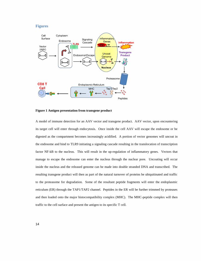
Figure 1 Antigen presentation from transgene product
A model of immune detection for an AAV vector and transgene product. AAV vector, upon encountering
its target cell will enter through endocytosis. Once inside the cell AAV will escape the endosome or be
digested as the compartment becomes increasingly acidified. A portion of vector genomes will uncoat in
the endosome and bind to TLR9 initiating a signaling cascade resulting in the translocation of transcription
factor NF-kB to the nucleus. This will result in the up-regulation of inflammatory genes. Vectors that
manage to escape the endosome can enter the nucleus through the nuclear pore. Uncoating will occur
inside the nucleus and the released genome can be made into double stranded DNA and transcribed. The
resulting transgene product will then as part of the natural turnover of proteins be ubiquitinated and traffic
to the proteasome for degradation. Some of the resultant peptide fragments will enter the endoplasmic
reticulum (ER) through the TAP1/TAP2 channel. Peptides in the ER will be further trimmed by proteases
and then loaded onto the major histocompatibility complex (MHC). The MHC-peptide complex will then
traffic to the cell surface and present the antigen to its specific T cell.

15
Chapter 2 is adapted from the manuscript Scott N. Ashley, Suryanarayan Somanathan, April Giles,
Christine Draper, Deirdre McMenamin, and James M. Wilson. In preparation.
Chapter 2
TLR9 signaling mediates adaptive immunity against transgene products
following systemic AAV gene therapy

16
Abstract
Adaptive immune responses to the transgene product remain a concern for the field of
gene therapy. Driven by the immune stimulus of vector administration, transgene
expression can elicit an adaptive immune response against the therapeutic protein,
particularly for recessive diseases in which these antigens are not presented to
lymphocytes during central tolerance induction. Here, we show that TLR9 signaling is
involved in driving the activation of CD8+ T cells against mitochondrial-targeted
ornithine transcarbamylase (OTC) following systemic AAV vector administration. Using
a CD8+ T cell receptor transgenic mouse model system, we demonstrate that TLR9
signaling extrinsic to T cells is sufficient to induce a robust cytotoxic T cell response
against OTC, resulting in transgene expression loss. Overall, our results suggest that
inflammation mediated by TLR9 signaling is important for the development of adaptive
immune responses to transgene products following AAV gene therapy.
Introduction
AAV vectors have shown potential in clinical trials to treat several recessive diseases by
replacing the missing or defective gene (Simonelli, Maguire et al. 2010, Nathwani, Reiss
et al. 2014, Jacobson, Cideciyan et al. 2015, Naldini 2015). In many preclinical animal
models, adaptive immune responses to the transgene product have been observed; this is
particularly true when a “non-self” protein is expressed from the delivered gene, as the
recipient may be more likely to develop an immune response to antigens that are not
present during lymphocyte development (Fields, Kowalczyk et al. 2000, Ding, Hodges et

17
al. 2001, Limberis, Figueredo et al. 2007, Bradbury, Cochran et al. 2013, Ciesielska,
Hadaczek et al. 2013). However, the adaptive immune response is tightly controlled and
exposure to a novel protein often results in tolerance rather than immunity, unless an
inflammatory signal is present to initiate an adaptive immune response to the novel
antigen (Lang, Georgiev et al. 2006). One potential immunogenic stimulus associated
with AAV-mediated gene therapy is the vector genome. The immune system’s major
sensing agent for this foreign material is TLR9, located in the endosomal compartment,
which recognizes unmethylated CpG DNA and induces pro-inflammatory cytokine
signaling that can result in an adaptive response (Hemmi, Takeuchi et al. 2000, Rogers,
Martino et al. 2011).
The ability of TLR9 to upregulate pro-inflammatory cytokines in response to AAV
vectors has been observed both by ex vivo stimulation of plasmacytoid dendritic cells
(pDCs) and by intramuscular (IM) AAV2 vector administration in mice (Zaiss, Liu et al.
2002, Zhu, Huang et al. 2009). Self-complementary AAV genomes containing double-
stranded DNA were implicated in more robust TLR9 activation resulting in a diminished
induction of tolerance (Martino, Suzuki et al. 2011). Additional work has been done to
avoid TLR9-induced inflammatory signaling by CpG depletion or by increasing the CpG
methylation state in DNA-based therapies (Reyes-Sandoval and Ertl 2004, Faust, Bell et
al. 2013, Shapir, Miari et al. 2015). However, there is evidence that TLR9 binds the
phosphodiester backbone of DNA regardless of the nucleotide sequence, and that
activation is merely enhanced by CpG motifs (Haas, Metzger et al. 2008).

18
I sought to investigate the influence of TLR9 on adaptive immune responses following
AAV gene therapy using a systemic of administration for delivery to the liver. I found
that systemic inflammation mediated by a TLR9 agonist was able to break tolerance to
the mitochondrially targeted ornithine transcarbamylase (OTC) protein and cause the
destruction of transduced cells. These findings suggest that, while TLR9 activation can
be consequential in eliciting a transgene specific adaptive immune response, our ability to
curb its influence may be limited.
Materials and Methods
Animals
6-10 weeks of age OT-1 mice on a C57BL/6 background, C57BL/6, and B-cell -/- muMt-
(stock number 002288) mice were acquired from The Jackson Laboratory (Bar Harbor,
ME). 6-8 weeks of age TLR9 KO mice on a C57BL/6 background mice were a kind gift
from Dr. Koretsky (University of Pennsylvania, Philadelphia, PA); these mice were
created by Dr. Akira (Osaka University, Osaka, Japan) (Hemmi, Takeuchi et al. 2000).
All mice were housed and bred under specific pathogen-free conditions in the
Translational Research Laboratory Animal Facility at the University of Pennsylvania.
Procedures were performed under IACUC approved protocols.

19
Vectors
AAV2/8sc.TBG.hOTCco (self-complementary codon-optimized human ornithine
transcarbamylase transgene under the control of a TBG promoter containing the
G4SVPA poly A signal sequence) was produced by the University of Pennsylvania
Vector Core. To generate the sc.hOTCco-SIINFEKL transgene cassette, the
immunodominant epitope of chicken ovalbumin (Ova257-264 SIINFEKL) was cloned into
the sc.hOTCco transgene immediately following the C-terminal residue.
Adoptive Transfer
Mice were administered intravenously via the tail vein 1x10¹¹
genome copies (GCs) of
AAV8 expressing either sc.hOTCco-SIINFEKL or mutant variants in a total volume of
100 µl PBS. On day 14 post vector administration, CD8+ T cells were collected from
OT-1 mouse spleens using CD8a+ T Cell Isolation Kit (Miltenyi Biotec, San Diego, CA).
CD8+ T cell purity was >90%, as measured by flow cytometry. Vector treated mice were
then given 1x10⁶ CD8
+ T cells in 100 µl PBS by tail vein injection. Concurrent with
adoptive cell transfer and for three days following, mice were given intraperitoneal (IP) a
mixture of 20 µg TLR9 ODN 2395 and 20 ug ODN M363 (InvivoGen, San Diego, CA).
For labeling T cells, CFSE (eBioscience, San Diego, CA) was diluted in DMSO to a 5
mM stock solution. T cells were resuspended to 5x10⁷
cells/ml. 1 µl of 0.5 mM CSFE
was added to each ml of cell suspension and incubated at 37 ºC for 10 minutes. Cells
were then washed two times with DMEM and two times with PBS, before resuspension
in PBS at a concentration of 1x10⁷ cells per ml. Retro-orbital bleeds were performed

20
weekly throughout the study and samples were submitted to Antech Diagnostics (Irvine,
CA) for analysis of transaminase and bilirubin levels.
Epitope Mapping
C57Bl/6 mice received IM 5x10¹¹
GC of AAVrh32.33.CB7.hOTCco followed 12 weeks
later by an intramuscular injection of 5x10¹⁰
particles (delivered as two 50 µl injections)
of Ad5 vector expressing hOTCco. On day 10 post Ad5 vector immunization, mice were
sacrificed and splenocytes were harvested as described above (Section: Adoptive
Transfer). An interferon (IFN-) ELISPOT assay (BD Biosciences, San Jose, CA) was
performed according to the manufacturer’s instructions. Cells were seeded at a
concentration of 2x10⁵ or 2x10
4 cells per well along with 2 g/ml of hOTC peptide pools
or pool B containing an overlapping 15-mer peptide library (Mimotopes, Victoria,
Australia) spanning the full length of the Ad5 hexon. Splenocytes were incubated at
37°C in 5% CO2 for 18 h. Spot forming units (SFUs) were counted using the AID
ELISPOT Reader System (Cell Technology, Columbia, MD).
Liver Homogenization
Livers previously harvested and frozen at -80c were ground in liquid nitrogen. 20 mg of
ground liver and added 20 µL of 50 mM Tris buffer ph 7.5 (Sigma-Aldrich, St. Louis,
MO) per mg liver powder, then homogenize on ice for 5 seconds, 10 times. We repeat
homogenization routine once, then centrifuged at 13,200 rpm for 2 min and transfer 20

21
µL supernatant into another new labeled tube with 180 µL 50 mM Tris buffer ph 7.5,
homogenate can be kept on ice until use in activity assay.
OTC Activity Assay
We added 50 µl of diluted supernatant to 500 µl reaction buffer consisting of 5mM
ornithine (Sigma-Aldrich, St. Louis, MO) in 50 mM Tris buffer pH 7.5. To start the
reaction we added 50 ul 4.8mM carbamyl phosphate (Sigma-Aldrich, St. Louis, MO) to
reaction mixture and allow reaction to continue for 5 min at 25c. The reaction was
stopped by quenching with 500 µl of 5 mM C14 citrulline, 30% trichloroacetic acid
(Sigma-Aldrich, St. Louis, MO). Activity was determined by formation of citrulline as a
ratio of C14 citrulline as measured by liquid chromatography–mass spectrometry.
Biodistribution
Liver samples were frozen on dry ice at the time of necropsy, and DNA was extracted
using the QIAamp DNA Mini Kit (Qiagen, Valencia, CA). Detection and quantification
of vector GCs in extracted DNA were performed by real-time PCR, as described
previously (Bell, Moscioni et al. 2006). Briefly, genomic DNA was isolated, and vector
GCs were quantified using primers/probes designed against the sequence of the TBG
promoter. Quantification of GCs from liver was performed on one liver sample from
each mouse.

22
RNA Analysis
Liver samples were frozen in liquid nitrogen at the time of necropsy. RNA was extracted
using TRIZOL (Life Technologies, Carlsbad, CA), according to the manufacturer's
protocol. 12 µg of RNA was then treated with DNase I (Roche, Basel, Switzerland),
according to the manufacturer's protocol. The RNeasy Mini Kit (Qiagen, Valencia, CA)
was used to remove DNase prior to cDNA synthesis by reverse transcription using the
Applied Biosystems High Capacity cDNA Reverse Transcriptase Kit (Life Technologies,
Carlsbad, CA). Real-time PCR was then performed on cDNA with primers binding to
the hOTCco transgene with Power SYBR Master Mix for detection (Life Technologies,
Carlsbad, CA).
CD8 Stain on Frozen Sections
Frozen liver sections were fixed in acetone at -20 ºC for 7 minutes, air dried, blocked in
1% donkey serum (Jackson Immuno West Grove, PA) in PBS for 20 minutes, and
incubated with 1:20 primary rat antibody against CD8 (clone 53-6.7, BD Biosciences)
diluted in blocking buffer for 45 minutes. After washing in PBS, sections were stained
with secondary donkey antibodies labeled with FITC (Jackson Immunoresearch
Laboratories, West Grove, PA) for 30 minutes, washed in PBS, and mounted in
Vectashield immunofluorescence stain containing DAPI (Vector Laboratories,
Burlingame, CA).
23
Ki67 Stain on Paraffin Sections
Paraffin sections were deparaffinized through an ethanol and xylene series, boiled in a
microwave for 6 minutes in 10 mM citrate buffer, pH 6.0 for antigen retrieval, and treated
sequentially with 2% H2O2 (Sigma-Aldrich, St. Louis, MO) for 15 minutes, avidin/biotin
blocking reagents (Vector Laboratories, Burlingame, CA) for 15 minutes, and blocking
buffer (1% donkey serum (Jackson Immunoresearch, West Grove, PA) in PBS with 0.2%
Triton) for 10 minutes. Sections were then incubated for 1 hour with 1:500 primary
rabbit serum against Ki67 (ab15580, Abcam, Cambridge, United Kingdom) and 45
minutes with FITC-labeled secondary donkey antibody (Jackson Immunoresearch, West
Grove, PA) diluted in blocking buffer. Sections were mounted using Vectashield
mounting medium (Vector Laboratories, Burlingame, CA) containing DAPI as a nuclear
counterstain.
Results
In previous work, our group investigated the importance of inflammation on gene therapy
in the liver using reporter gene, nuclear-targeted beta-galactosidase (nlacZ) (Somanathan,
Breous et al. 2010, Breous, Somanathan et al. 2011). Our results suggested that in the
presence of a strong inflammatory signal, T cell targeting of a transgene product could
result in the loss of transgene expression and destruction of transduced cells. Thus, I
probed the ability of TLR9 signaling to initiate a destructive cytotoxic T cell (CTL)
response following gene therapy for human OTC (hOTC), a promising candidate for
clinical applications. First we attempted to determine if hOTC could be targeted by a
24
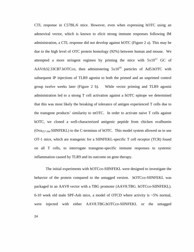
CTL response in C57BL/6 mice. However, even when expressing hOTC using an
adenoviral vector, which is known to elicit strong immune responses following IM
administration, a CTL response did not develop against hOTC (Figure 2 a). This may be
due to the high level of OTC protein homology (92%) between human and mouse. We
attempted a more stringent regimen by priming the mice with 5x10¹¹
GC of
AAVrh32.33CB7.hOTCco, then administering 5x10¹⁰
particles of Ad5.hOTC with
subsequent IP injections of TLR9 agonist to both the primed and an unprimed control
group twelve weeks later (Figure 2 b). While vector priming and TLR9 agonist
administration led to a strong T cell activation against a hOTC epitope we determined
that this was most likely the breaking of tolerance of antigen experienced T cells due to
the transgene products’ similarity to mOTC. In order to activate naive T cells against
hOTC, we cloned a well-characterized antigenic peptide from chicken ovalbumin
(Ova257-264 SIINFEKL) to the C-terminus of hOTC. This model system allowed us to use
OT-1 mice, which are transgenic for a SIINFEKL-specific T cell receptor (TCR) found
on all T cells, to interrogate transgene-specific immune responses to systemic
inflammation caused by TLR9 and its outcome on gene therapy.
The initial experiments with hOTCco-SIINFEKL were designed to investigate the
behavior of the protein compared to the untagged version. hOTCco-SIINFEKL was
packaged in an AAV8 vector with a TBG promoter (AAV8.TBG. hOTCco-SIINFEKL).
6-10 week old male SPF-Ash mice, a model of OTCD where activity is ~5% normal,
were injected with either AAV8.TBG.hOTCco-SIINFEKL or the untagged

25
AAV8.TBG.hOTCco. Mice were evaluated for OTC enzyme activity at day 14 post
administration (Figure 3 a) which showed minimal activity compare to control. We also
evaluated urine orotate a metabolic marker of OTCD and showed that correction was
slower to occur in hOTCco-SIINFEKL treated mice (Figure 3 b). We also investigated
the ability of hOTCco-SIINFEKL to properly migrate to the mitochondria by transfecting
the human liver cell line HuH7 on slide wells with OTC plasmids including one lacking
the leader sequence required to guide the protein. Transfected cells were probed with
Mito Tracker CMXRos to visualize the mitochondria and following up take, they were
fixed for OTC immunohistochemistry (Figure 4). Staining revealed that hOTC-
SIINFEKL translocated to the mitochondria similar to wild type hOTC, whereas without
the leader sequence hOTC is dispersed throughout the cytoplasm indicating a disruption
of normal biogenesis which could impact antigen presentation. Despite impaired OTC
activity due to the addition of the tag, mitochondrial localization remained intact, and we
will continue our investigation using hOTCco-SIINFEKL.
Male C57BL/6 mice 6-8 weeks old were treated with an intravenous (IV)
injection of AAV8.TBG. hOTC-SIINFEKL. Fourteen days after vector injection, 1x10⁶
SIINFEKL specific T cells were adoptively transferred from OT-1 mice. Concurrent
with the adoptive transfer, we gave 40 µg of TLR9 agonist by IP injection (Figure 5 a).
As anticipated, we only observed a CTL response (measured by transaminase levels) to
the transgene product when both a population of transgene-specific OT-1 T cells and
TLR9 inflammatory signals were present; transaminases aspartate transaminase (AST)
26
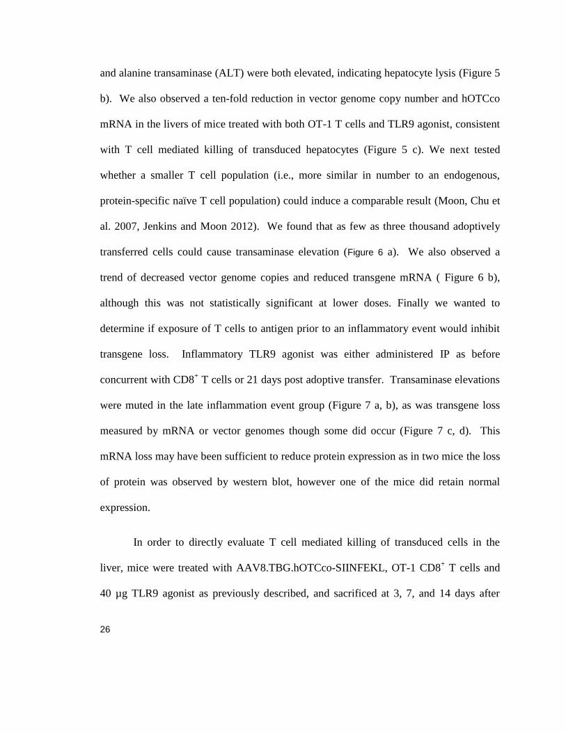
and alanine transaminase (ALT) were both elevated, indicating hepatocyte lysis (Figure 5
b). We also observed a ten-fold reduction in vector genome copy number and hOTCco
mRNA in the livers of mice treated with both OT-1 T cells and TLR9 agonist, consistent
with T cell mediated killing of transduced hepatocytes (Figure 5 c). We next tested
whether a smaller T cell population (i.e., more similar in number to an endogenous,
protein-specific naïve T cell population) could induce a comparable result (Moon, Chu et
al. 2007, Jenkins and Moon 2012). We found that as few as three thousand adoptively
transferred cells could cause transaminase elevation (Figure 6 a). We also observed a
trend of decreased vector genome copies and reduced transgene mRNA ( Figure 6 b),
although this was not statistically significant at lower doses. Finally we wanted to
determine if exposure of T cells to antigen prior to an inflammatory event would inhibit
transgene loss. Inflammatory TLR9 agonist was either administered IP as before
concurrent with CD8+ T cells or 21 days post adoptive transfer. Transaminase elevations
were muted in the late inflammation event group (Figure 7 a, b), as was transgene loss
measured by mRNA or vector genomes though some did occur (Figure 7 c, d). This
mRNA loss may have been sufficient to reduce protein expression as in two mice the loss
of protein was observed by western blot, however one of the mice did retain normal
expression.
In order to directly evaluate T cell mediated killing of transduced cells in the
liver, mice were treated with AAV8.TBG.hOTCco-SIINFEKL, OT-1 CD8+ T cells and
40 µg TLR9 agonist as previously described, and sacrificed at 3, 7, and 14 days after

27
adoptive transfer for histological evaluation of the liver. Liver sections were stained for
CD8a to mark CD8+ T cells, and infiltration was quantified by image analysis (Figure 8
a, b). Marked CD8+ T cell infiltration occurred at periportal and pericentral locations
beginning on day 3; by day 7 the infiltrates with TLR9 agonist had penetrated deeper into
the liver, whereas further penetration was not seen for T cells without the enhanced TLR9
stimulation from administered agonist. In both groups, infiltration was resolved by day
14. Our previous experiment suggested that hepatocyte death occurred following
treatment with TLR9 agonist; however terminal deoxynucleotidyl transferase dUTP nick-
end labeling (TUNEL) staining and caspase-9 immunofluorescence did not show
differences between control and experimental groups (data not shown). As an indicator
of cell death, we also examined the regrowth of hepatocytes that were predicted to have
undergone lysis resulting in loss of AAV genomes (Figure 8 c). Using Ki-67 staining to
mark proliferation, we observed hepatocyte division to be spread out evenly across liver
regions in the experimental groups, mirroring the degree of CD8+ T cell infiltration. We
determined the percent of hepatocytes that were Ki-67 positive by image analysis (Figure
8 d), and found hepatocyte division to be much higher than the normal expected rate of
turnover, indicating increased cell death above background (Miyaoka, Ebato et al. 2012,
Miyaoka and Miyajima 2013). By determining genome copies at each time point (Figure
8 e), we observed that most loss occurred between three and seven days post adoptive
transfer, correlating with the histology results (Figure 8 a, c); together, these data indicate
that the majority of T cell infiltration and hepatocyte death occurs during this time span 7
to 14 days.

28
Next, we examined whether the infiltrating cells were indeed transgene-specific
CTLs. We labeled CD8+ T cells with CFSE fluorescent cell staining dye before delivery
into C57BL/6 mice previously administered with AAV8.TBG.hOTCco-SIINFEKL.
Seven days after adoptive transfer and TLR9 stimulation, we isolated lymphocytes from
the liver or spleen, and stained them with anti-CD8a and SIINFEKL tetramer (Figure 9 b,
c). The majority of liver-isolated lymphocytes were tetramer positive, regardless of
whether mice received TLR9 agonist (Figure 9 c). In contrast, the majority of spleen
isolated T cells were tetramer positive only if the mice received TLR9 agonist. In
addition, CFSE staining of tetramer positive splenocytes remained high in mice that did
not receive TLR9 agonist (Figure 9 d) suggesting that transgene-specific activation of
CD8+ T cells in the presence of TLR9 stimulation increased proliferation. We then
sought to determine if intrinsic TLR9 signaling in transferred T cells alone would trigger
an immune response, or if extrinsic signaling was required for the effect. We
administered 6-8 week old male C57BL/6 mice or TRL9 -/- mice with
AAV8.TBG.hOTCco-SIINFEKL. Fourteen days later, we adoptively transferred 1x10⁶
OT-1 CD8+ T cells into recipient mice that were also given 40 µg TLR9 agonist (Figure
10 a). AST and ALT elevation only occurred in the C57BL/6 group; despite TLR9
signaling being intact in transferred OT-1 T cells, this was insufficient to activate a robust
CTL response (Figure 10 b,c). These results indicate that the TLR9 signaling required
for initiating T cell responses must be extrinsic to the T cells themselves, perhaps
occurring in transduced hepatocytes or antigen-presenting cells.

29
Discussion
Adaptive immune responses to a transgene product are complex from activation
of antigen presenting cells to the balance of a productive to destructive immune response.
Thus, when determining the best method for delivering a successful treatment, there are
many factors to consider, such as the route of administration and vector genome
composition. These components may have a dramatic effect on the likelihood that a
given therapy will induce an inflammatory environment, elicit an adaptive anti-transgene
response, and lead to an ineffective treatment (Cao, Hoffman et al. 2009, Martino, Suzuki
et al. 2011, Rogers, Martino et al. 2014). Here, we show that loss of TLR9 signaling
dramatically attenuates the CTL responses against the transgene following IV
administration of an AAV vector.
Relevant to our interest in liver-directed gene therapy for metabolic disorders, we
examined transgene-specific T cell response to a mitochondrially targeted protein, OTC.
Given the previous evidence of minimal immune responses following liver-directed gene
transfer with AAV vectors, we anticipated that hepatic OTC expression would not elicit a
robust immune response (Lang, Georgiev et al. 2006). The high homology between
mouse and human protein complicated matters. In an ideal model, there would be no
prior exposure to the transgene product; however, the OTC -/- model is neonatal lethal,
and the SPF-Ash OTCD mouse line still expresses residual (around 5% of normal)
activity(Wang, Wang et al.). Therefore, we added the immune-dominant epitope Ova257-
264 to the C-terminus of hOTC to increase its immunogenicity. While addition of the
30
SIINFEKL epitope reduced OTC activity of the construct, mitochondrial localization was
retained and so therefore we would expect turnover and antigen processing to be similar.
Using the OVA epitope allowed us to measure and manipulate the transgene-specific T
cell population for more sensitive readouts of immune activation. Using OT-1 transgenic
T cells, we demonstrated that a robust response against the transgene product could be
mediated by the activation of TLR9. This immune response was characterized by large
elevations in transaminases and loss of transgene expression, as measured by hOTC-
SIINFEKL mRNA and genome copy levels. We also observed that antigen-specific T
cell populations of as low as 3,000 cells could induce transaminase elevation and
modestly decrease genome copy number. In addition we showed that inflammation after
transgene expression and presumed exposure to CD8+ T cells resulted in an attenuated
clearance of the transgene and product, indicating that the most at risk timeframe for a
robust immune response is during first contact of antigen to the T cells which will occur
when expression begins a few days following vector administration. Further, we
observed increased T cell infiltration of predominantly transgene-specific T cells, which
corresponded to an increase in hepatocytes entering the cell cycle and subsequent loss of
genome copies. Together, these data suggest that as hepatocytes are destroyed, dividing
cells replicate to replace them. An alternative hypothesis could be that the cell division is
responsible for the loss of the genome copies that are not retained when the nuclear
envelope is dissolved and reformed; this possibility is supported by the lack of an
increase in TUNEL or caspase 9 staining. As systemic administration of TLR9 agonist
induced T cell activation against the transgene product, we were curious as to whether
31
TLR9 signaling intrinsic to T cells alone would be sufficient to activate them. We used
TLR9 KO mice as recipients of the OT-1 T cell transfer. Our results showed that TLR9
signaling extrinsic to T cells was required for activation, such as both B cells and pDCs
that respond strongly to TLR9 stimulation (Montoya, Jie et al. 2006, Jiang, Lederman et
al. 2007). Therefore, future studies may help to further our knowledge and ability to
design less immunogenic AAV vectors.

32
Figure 2 hOTC Immunodominant Epitope Mapping in C57BL/6 mice.
(A) C57BL/6 mice were given IM 5x10¹⁰
p Ad5 hOTC. Mice (n=3) were sacrificed 28 days later, and IFN-
gamma ELISPOT was performed on isolated T cells. (B) One cohort of C57Bl/6 mice was injected IM
with 5x10¹¹
AAVrh32.33 CB7.hOTCco twelve weeks later mice were injected IM with 5x10¹⁰
of
Ad5.hOTC vector and TLR9 agonist 40 µg IP concurrent with ad administration and for the three following
days N=4. 10 days after vector administration mice were sacrificed for IFN ɣ ELISPOT.

33
Figure 3 Evaluation of OTC-SIINFEKL Activity in vivo.
Male SPF-Ash mice (n=3) ages 6-10 weeks were injected IV with AAV8.TBG.hOTCco or
AAV8.TBG.hOTCco-SIINFEKL 1x10¹¹
GC. (A) Mice urine was collected and urine orotate was
measured. (B) OTC enzyme activity was assayed from liver collected at day 14 necropsy.

34
Figure 4 hOTC-SIINFEKL Mitochondria Localization
Huh7 cells were transfected in slide well plates with plasids that contained hOTC transgenes. 48 hours
after transfection MitoTracker is added to media to visualize mitochondria (red) cells are fixed thirty
minutes later for OTC immunohistochemistry (green) and nuclear DAPI (blue).

35
Figure 5 Systemic Inflammation Breaks Tolerance to a Transgene Product.
(A) Schematic of experimental design detailing IV administration of AAV8.TBGhOTCco-SIINFEKL and
subsequently OT-1 CD8+T cell transfer at 14 days after vector delivery, concurrent with administration of
40 µg TLR9 agonist for three days following adoptive transfer, n=3. (B) Serum AST and (C) Serum ALT
levels were monitored every seven days following vector delivery. (D) Genome copies and (E) transgene
mRNA from liver were determined by qPCR following termination of the study. These experiments were
independently performed three times. Statistical analysis was performed by one-way ANOVA with
Bonferroni test comparing to vector only control *P<0.05, **P<0.001, ***P<0.0001.

36
Figure 6 Effect of T cell population on Immune Response.
6-8 week old male C57Bl/6 mice (n=3) were given IV XX GC of AAV8.TBGhOTCco-SIINFEKL.
Twenty-eight days after vector delivery, OT-1 CD8+T cells were transferred to recipient mice concurrent
with 40 µg TLR9 agonist administration for three days following adoptive transfer. (A) Serum AST (B)
ALT and (C) bilirubin levels were monitored every seven days following vector delivery. (D) Genome
copies and (E) transgene mRNA from liver were determined by qPCR following termination of the study.
Analysis was performed by one-way ANOVA with Bonferroni test comparing to no transfer control
*P<0.05, **P<0.001, ***P<0.0001.
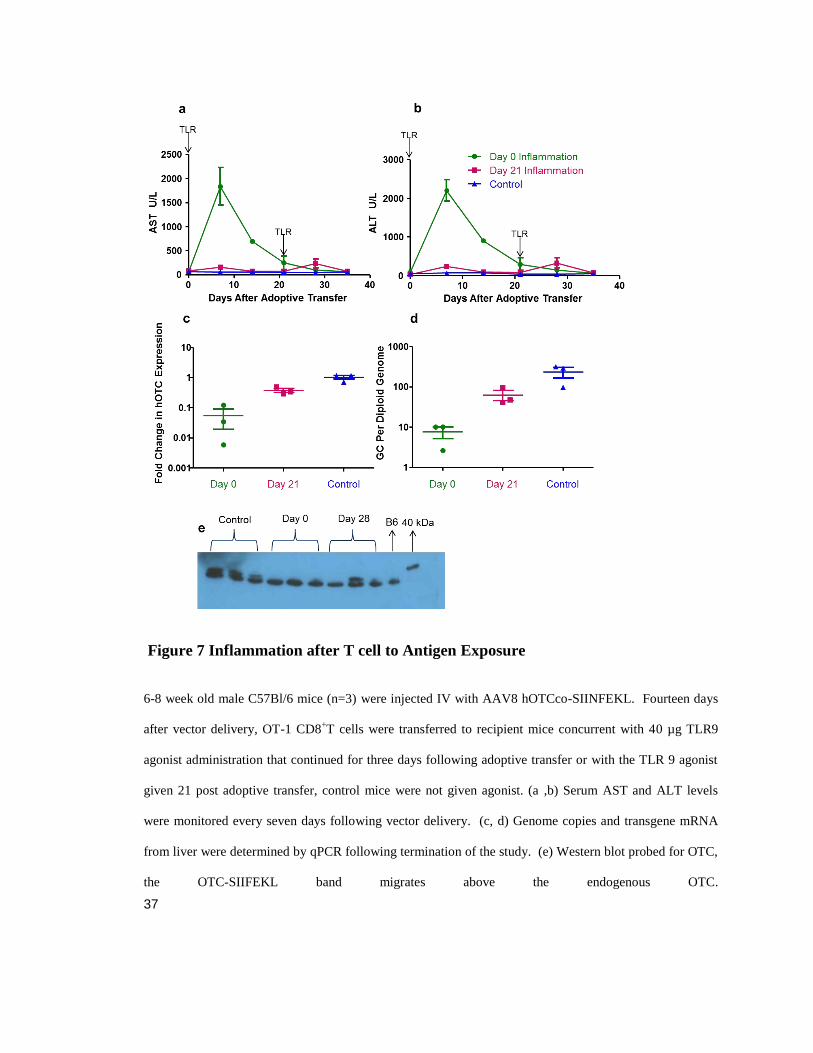
37
Figure 7 Inflammation after T cell to Antigen Exposure
6-8 week old male C57Bl/6 mice (n=3) were injected IV with AAV8 hOTCco-SIINFEKL. Fourteen days
after vector delivery, OT-1 CD8+T cells were transferred to recipient mice concurrent with 40 µg TLR9
agonist administration that continued for three days following adoptive transfer or with the TLR 9 agonist
given 21 post adoptive transfer, control mice were not given agonist. (a ,b) Serum AST and ALT levels
were monitored every seven days following vector delivery. (c, d) Genome copies and transgene mRNA
from liver were determined by qPCR following termination of the study. (e) Western blot probed for OTC,
the OTC-SIIFEKL band migrates above the endogenous OTC.

38

39
Figure 8 T cell Infiltration and Hepatocyte Proliferation.
Vector was administered to 6-8 week old male mice (n=4) IV and 14 days later OT-1 CD8+T cell were
transferred, concurrent with administration of 40 ug TLR9 agonist for three days following adoptive
transfer. Mice were sacrificed on days three, seven, and 14 following CD+ T cell transfer. (a, b) Liver
sections were stained for CD8, and percent area covered by infiltrating T cells was determined using Image
J. (c, d) Liver sections were stained for Ki-67 proliferation marker, and percent positive hepatocytes were
calculated. (e) AAV vector genome copies were determined by qPCR following termination of each
cohort. For image analysis experiments, five images per mouse were analyzed. Analysis was performed
by one-way ANOVA with Bonferroni test comparing columns to their day appropriate control *P<0.05,
**P<0.001, ***P<0.0001. NS, not significant.

40
Figure 9 Transgene-Specific T cell Infiltration.
6-8 week old male C57Bl/6 mice (n=4) were given AAV8 hOTC-SIINFEKL; 14 days later TLR9 agonist
and CFSE-labeled OT-1 CD8+ T cells were administered. Mice were sacrificed seven days following CD8
+
T cell transfer, and lymphocytes were isolated from liver and spleen. (a ,c) Lymphocytes isolated from
spleen or liver by forward and side scatter were stained for CD8. (b) Spleen- and (d) liver-isolated CD8+ T
cells were stained with SIINFEKL tetramer. (f, e) Tetramer positive splenocytes were analyzed for CFSE
retention.

41
Figure 10 Extrinsic TLR9 Signaling is Required for a Destructive CTL Response
(a) C57BL/6 or TLR9 KO male mice (n=5) ages 6-8 weeks were administered IV AAV8 expressing
hOTCco-SIINFEKL. Fourteen days after vector delivery, OT-1 CD8+T cells were transferred to recipient
mice concurrent with 40 µg TLR9 agonist administration that continued for three days following adoptive
transfer. (b-c) Serum (b) AST and (c) ALT levels were monitored every seven days following vector
delivery. Genome copies and transgene mRNA expression was determined following termination of the
study by the study.

42
Chapter 3 is adapted from the manuscript Scott N. Ashley, Suryanarayan Somanathan, Christian Hinderer,
Maxwell Arias, Deirdre McMenamin, Christine Draper, and James M. Wilson. In preparation.
Chapter 3:
Tolerance to Antigens C-Terminal to Premature Termination Codon

43
Abstract
Cytotoxic T cell (CTL) responses to the transgene product remain an active area of
concern for the gene therapy field. The patient’s underlying genetic mutation may
influence the qualitative nature of these potentially destructive T cell responses.
Individuals with a mutation that introduces a premature termination codon (PTC)
preventing synthesis of full-length peptide have been considered more likely to mount a
transgene-specific T cell response due to a lack of endogenous antigen presentation and,
therefore, no immune tolerance to C-terminal epitopes. Here, we demonstrate that a
human ornithine transcarbamylase (OTC) gene containing various PTC-inducing
nonsense mutations is able to generate and present epitopes downstream of the
termination codon. Generation of these epitopes occurs primarily from alternative
translation start sites downstream of the stop codon. Furthermore, we show that
expression of these genes from AAV vectors in C57Bl/6 mice is able to induce peripheral
tolerance to epitopes downstream of the PTC. These results suggest that, despite the lack
of full-length endogenous protein, patients with PTC-inducing nonsense mutations may
still present T cell epitopes downstream to the premature termination that may render the
subject tolerant to wild-type transgene products, and therefore less likely to generate a
transgene-specific immune response resulting in an overall improved therapeutic
outcome.

44
Introduction
OTC deficiency is a rare X-linked metabolic disorder that is considered a therapeutic
target for gene therapy (Hudak, Jones et al. 1985, Nguyen and Ferry 2004). Most OTC
gene mutations are “private”, meaning that they are unique to a particular individual or
family, and thereby result in a patient population with a diverse array of mutations
(Yamaguchi, Brailey et al. 2006). While some groups have begun to investigate the
influence of genotype on immune responses to the transgene product in other genetic
disorders (Cao, Hoffman et al. 2009, Nayak, Doerfler et al. 2014, Rogers, Martino et al.
2014), the impact of these diverse mutations on the outcome of gene therapy for OTC
remains unknown. Because OTC localizes to the mitochondria, the development of
transgene-specific T cells may be more likely and problematic than an antibody response
(Limberis, Figueredo et al. 2007, Mendell, Campbell et al. 2010, Flotte, Trapnell et al.
2011). Whereas a healthy individual could deplete transgene-specific T cells via
mechanisms of central and peripheral tolerance (Mendell, Campbell et al. 2010, Flotte,
Trapnell et al. 2011), the OTC-deficient patient with mutations that eliminate expression
of the protein (or parts of the protein) may be unable to induce tolerance to the wild-type
gene, rendering them susceptible to CTL responses upon transgene delivery. Indeed,
preclinical evidence in other disease models suggests that certain mutations may limit
tolerance induction and predispose patients receiving gene therapy to a T cell mediated
transgene response (4-6).
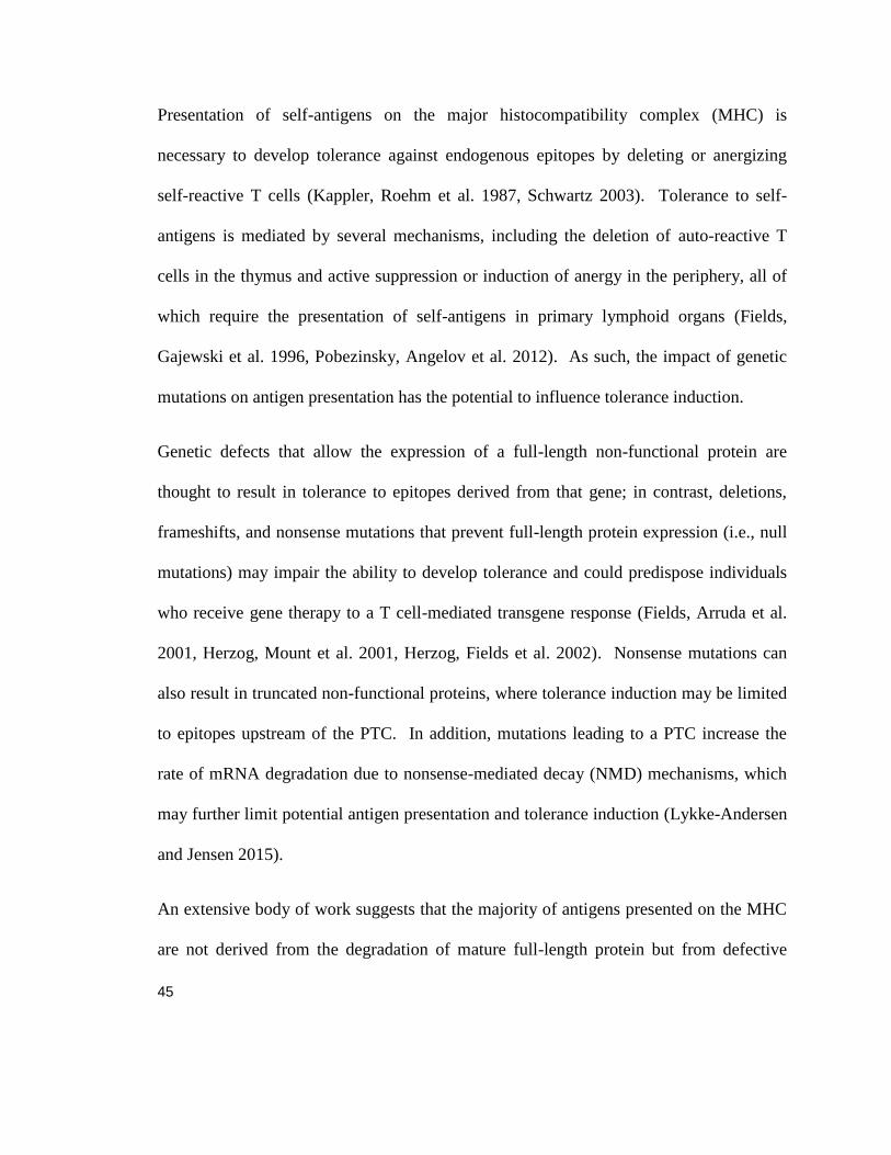
45
Presentation of self-antigens on the major histocompatibility complex (MHC) is
necessary to develop tolerance against endogenous epitopes by deleting or anergizing
self-reactive T cells (Kappler, Roehm et al. 1987, Schwartz 2003). Tolerance to self-
antigens is mediated by several mechanisms, including the deletion of auto-reactive T
cells in the thymus and active suppression or induction of anergy in the periphery, all of
which require the presentation of self-antigens in primary lymphoid organs (Fields,
Gajewski et al. 1996, Pobezinsky, Angelov et al. 2012). As such, the impact of genetic
mutations on antigen presentation has the potential to influence tolerance induction.
Genetic defects that allow the expression of a full-length non-functional protein are
thought to result in tolerance to epitopes derived from that gene; in contrast, deletions,
frameshifts, and nonsense mutations that prevent full-length protein expression (i.e., null
mutations) may impair the ability to develop tolerance and could predispose individuals
who receive gene therapy to a T cell-mediated transgene response (Fields, Arruda et al.
2001, Herzog, Mount et al. 2001, Herzog, Fields et al. 2002). Nonsense mutations can
also result in truncated non-functional proteins, where tolerance induction may be limited
to epitopes upstream of the PTC. In addition, mutations leading to a PTC increase the
rate of mRNA degradation due to nonsense-mediated decay (NMD) mechanisms, which
may further limit potential antigen presentation and tolerance induction (Lykke-Andersen
and Jensen 2015).
An extensive body of work suggests that the majority of antigens presented on the MHC
are not derived from the degradation of mature full-length protein but from defective
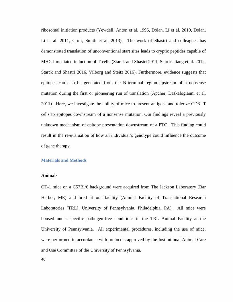
46
ribosomal initiation products (Yewdell, Anton et al. 1996, Dolan, Li et al. 2010, Dolan,
Li et al. 2011, Croft, Smith et al. 2013). The work of Shastri and colleagues has
demonstrated translation of unconventional start sites leads to cryptic peptides capable of
MHC I mediated induction of T cells (Starck and Shastri 2011, Starck, Jiang et al. 2012,
Starck and Shastri 2016, Vilborg and Steitz 2016). Furthermore, evidence suggests that
epitopes can also be generated from the N-terminal region upstream of a nonsense
mutation during the first or pioneering run of translation (Apcher, Daskalogianni et al.
2011). Here, we investigate the ability of mice to present antigens and tolerize CD8+ T
cells to epitopes downstream of a nonsense mutation. Our findings reveal a previously
unknown mechanism of epitope presentation downstream of a PTC. This finding could
result in the re-evaluation of how an individual’s genotype could influence the outcome
of gene therapy.
Materials and Methods
Animals
OT-1 mice on a C57Bl/6 background were acquired from The Jackson Laboratory (Bar
Harbor, ME) and bred at our facility (Animal Facility of Translational Research
Laboratories [TRL], University of Pennsylvania, Philadelphia, PA). All mice were
housed under specific pathogen-free conditions in the TRL Animal Facility at the
University of Pennsylvania. All experimental procedures, including the use of mice,
were performed in accordance with protocols approved by the Institutional Animal Care
and Use Committee of the University of Pennsylvania.
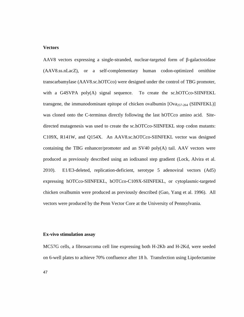
47
Vectors
AAV8 vectors expressing a single-stranded, nuclear-targeted form of β-galactosidase
(AAV8.ss.nLacZ), or a self-complementary human codon-optimized ornithine
transcarbamylase (AAV8.sc.hOTCco) were designed under the control of TBG promoter,
with a G4SVPA poly(A) signal sequence. To create the sc.hOTCco-SIINFEKL
transgene, the immunodominant epitope of chicken ovalbumin [Ova257-264 (SIINFEKL)]
was cloned onto the C-terminus directly following the last hOTCco amino acid. Site-
directed mutagenesis was used to create the sc.hOTCco-SIINFEKL stop codon mutants:
C109X, R141W, and Q154X. An AAV8.sc.hOTCco-SIINFEKL vector was designed
containing the TBG enhancer/promoter and an SV40 poly(A) tail. AAV vectors were
produced as previously described using an iodixanol step gradient (Lock, Alvira et al.
2010). E1/E3-deleted, replication-deficient, serotype 5 adenoviral vectors (Ad5)
expressing hOTCco-SIINFEKL, hOTCco-C109X-SIINFEKL, or cytoplasmic-targeted
chicken ovalbumin were produced as previously described (Gao, Yang et al. 1996). All
vectors were produced by the Penn Vector Core at the University of Pennsylvania.
Ex-vivo stimulation assay
MC57G cells, a fibrosarcoma cell line expressing both H-2Kb and H-2Kd, were seeded
on 6-well plates to achieve 70% confluence after 18 h. Transfection using Lipofectamine
48
(Life Technologies, Grand Island, NY) was performed according to the manufacturer’s
instructions. Cells were transfected with a plasmid containing either hOTCco-SIINFEKL
or the hOTCco-SIINFEKL stop codon mutants that were developed using site-directed
mutagenesis kit (Agilent Technologies, Santa Clara, CA) according to the manufacturer’s
instructions. After 24 h post-transfection, 6-well plates were washed once with PBS,
cells were mechanically scraped off the plate and into suspension, and washed once more
before being resuspended at 2x10⁶ cells/ml, seeding 100 l/well on an enzyme-linked
immunospot (IFN-ɣ ELISPOT) plate (BD Biosciences, San Jose, CA). OT-1 splenocytes
were harvested from OT-1 mice as previously described (Limberis, Figueredo et al. 2007)
and then seeded 1:1 or 1:10 with the MC57G cells. This combination was then incubated
at 37°C in 5% CO2 for 18 h. The ELISPOT plate was developed according to the
manufacturer’s protocol, and spots were counted using the AID ELISPOT Reader System
(Cell Technology, Columbia, MD).
Tolerance induction
Mice received 1x10¹¹
genome copies of AAV8 expressing either sc.hOTCco-SIINFEKL,
sc.hOTCco-SIINFEKL stop codon mutants, or an ss.nLacZ control, delivered IV in 100
l of PBS. On day 14 post vector administration, 1x10¹⁰
GC of Ad5.TBG.hOTCco-
SIINFEKL or 3x10⁹ particles of Ad5.TBG.OVA were injected IV. Mice were sacrificed
and splenocytes were harvested on day 27 after vector administration as previously
described (Limberis, Figueredo et al. 2007). An interferon (IFN-) ELISPOT assay
(BD Biosciences, San Jose, CA) was performed according to the manufacturer’s
49
instructions. Cells were seeded at a concentration of 2x10⁵ or 2x10
4 cells per well along
with 2 g/ml SIINFEKL peptide or pool B containing an overlapping 15-mer peptide
library (Mimotopes, Victoria, Australia) spanning the full length of the Ad5 hexon.
Splenocytes were incubated at 37°C in 5% CO2 for 18 h. Spot forming units (SFUs)
were counted using the AID ELISPOT Reader System (Cell Technology, Columbia,
MD). Tetramer staining was performed according to the manufacturer’s instructions
(MBL International, Woburn, MA). Splenocytes were resuspended to a concentration of
5x10⁶
cells/ml. 200 l of the cell suspension was stained with either 2 l iTAg
Tetramer/PE-H-2 Kb OVA (MBL International, Woburn, MA) and 3 l anti-mouse
CD8-APC (Clone 53-6.7; eBiosciences, San Diego, CA), or a cocktail containing 2 l
iTAg Tetramer/PE-H-2 Kb OVA, 2 l CD62L-FITC (Clone MEL-14), 3 l PD-1-APC
(Clone RMPI-30), and 1 l CD44-PE-Cyanine7 (Clone IM7; eBiosciences, San Diego,
CA), then incubated for 30 min in the dark at room temperature. Cells were washed with
PBS, resuspended in 500 l PBS with 1% FBS, and run on an FC 500 series flow
cytometer (Beckman Coulter, Brea, CA). Analysis was performed using FlowJo software
(Becton Dickinson, Franklin Lakes, NJ)
Adenoviral vector immunization
Mice received 5x10¹⁰
p of Ad5 expressing either hOTCco-SIINFEKL, nonsense mutant
variant C109X (hOTCco-C109X-SIINFEKL), or an nLacZ control delivered IV in 100 l
PBS. On Day 10 post vector immunization, mice were sacrificed and splenocytes were

50
harvested as described above. An IFN- ELISPOT assay was performed on harvested
splenocytes as described in the previous section.
Peptide Isolation
Cloning was used to swap out SIINFEKL with a StrepII tag with a SerAla linker (SA-
WSHPQFEK) on hOTCco-C109X. Nine, 15 cm plates of HEK 293T cells were
transfected per condition using 3 g polyethylenimine per 1 g DNA with either
hOTCco-C109X-StrepII or eGFP; cells were grown for 24 h. Proteasome inhibitor
MG132 (APExBIO, Houston, TX) was added to the cells to reach a concentration of 1
M. Two h after proteasome addition, cells were harvested using a gravity-flow Strep-
Tactin sepharose column (IBA, Goettingen, Germany). Media was removed and cells
were washed twice with PBS containing 1 M proteasome inhibitor MG132 at 1500 rpm;
all spins are performed at 4C. Cells were resuspended in 2 ml of Strep-Tactin wash
buffer with 25 M MG132 and 1X complete protease inhibitor (Sigma Aldrich, St Louis,
MO). Cells were lysed by five freeze-thaw cycles in liquid nitrogen, followed by a 37C
water bath. DNA was sheared by passing lysate through 18G needles five times over ice.
Lysate was spun down at 4000 rpm for 15 min. Supernatant was collected and spun
down at 14,000 rpm for 5 min. StrepII-tagged peptides were collected using 1 ml gravity
flow Strep-Tactin sepharose columns (IBA, Goettingen, Germany), as per manufacturer’s
instructions. Fractions were analyzed using Novex NuPAGE SDS-PAGE (Invitrogen,
Carlsbad, CA), per manufacturer’s instructions and stained with Sypro Ruby Protein stain

51
(Thermo Fisher Scientific, Waltham, MA). Sample fractions were submitted for mass
spectrometry analysis (Bioproximity, Chantilly, VA).
Protein Denaturation, Digestion, and Desalting
Samples were sent to Bioproximity (Chantilly, VA) for digestion using the filter-assisted
sample preparation method (Wisniewski, Zougman et al. 2009). Briefly, the samples
were suspended in 5% sodium deoxycholate SDC, 50 mM Tris-HCl, pH 7.6, 3 mM DTT;
they were sonicated briefly and incubated in a ThermoMixer (Eppendorf, Hauppauge,
NY) at 60C with 1000 rpm for 20 min. Samples were centrifuged, and the supernatant
was transferred to a 30 kD molecular weight cut-off device (Millipore, Billerica, MA)
and centrifuged at 13,000 g for 30 min. The remaining sample was buffer exchanged
with 1% SDC in 100 mM Tris-HCl, pH 7.6 and alkylated with 15 mM iodoacetamide.
Samples were digested using trypsin at an enzyme to substrate ratio of 1:100, overnight at
37C in a ThermoMixer at 1000 rpm. Digested peptides were collected by
centrifugation. A portion of the digested peptides (about 20 µg), were desalted using
reversed-phase stop-and-go extraction tips (Rappsilber, Mann et al. 2007). Peptides were
eluted with 80% acetonitrile, 5% ammonium hydroxide and lyophilized for
approximately 1 h in a Savant SpeedVac (Thermo Fisher Scientific, Waltham, MA) to
remove volatile components.
Liquid Chromatography-Tandem Mass Spectrometry
Each digestion mixture was analyzed by ultra-high pressure liquid chromatography (LC)
mass spectrometry (MS)/mass spectrometry by Bioproximity (Chantilly, VA) . LC was

52
performed on an EASY-nLC 1000 system (Thermo Fisher Scientific, Waltham, MA).
Mobile phase A was 97.5% Milli-Q water (Millipore, Billerica, MA), 2% acetonitrile,
and 0.5% acetic acid; mobile phase B was 99.5% acetonitrile and 0.5% acetic acid. The
35 min LC gradient ran from 0% B to 25% B over 20 min, and then to 80% B for 3 min.
Samples were loaded directly onto the 15 cm x 50 µm I.D. EASY-Spray column (Thermo
Fisher Scientific, Waltham, MA), packed with 2 µm PepMap C18 media (Thermo Fisher
Scientific, Waltham, MA). The LC system was interfaced to a Q Exactive Hybrid
Quadrupole Orbitrap Mass Spectrometer (Thermo Fisher Scientific, Waltham, MA) via
nano-electrospray ionization using an EASY-Spray source with an integrated column
heater (Thermo Fisher Scientific, Waltham, WA). The column was heated to 50C, and
an electrospray voltage of 2.2 kV was applied. The mass spectrometer was programmed
to acquire, by data-dependent acquisition, tandem mass spectra from the top 20 ions in
the full scan from 400-1200 m/z. Dynamic exclusion was set to 15 s, singly-charged ions
were excluded, isolation width was set to 1.6 Da, full MS resolution was set to 70,000
and MS/MS resolution was set to 17,500. Normalized collision energy was set to 25,
automatic gain control to 2e5, max fill MS to 20 ms, max fill MS/MS to 60 ms, and the
underfill ratio to 0.1%.
Statistical analysis
All statistics were performed using Prism 5 (GraphPad, La Jolla, CA). One-way
ANOVA analysis with Dunnett's Test was performed on each experimental replicate in
the study. Data are shown from a representative experiment for each study. For all

53
figures, significance is depicted as *p<0.05, **p<0.001, ***p<0.0001. The number of
animals per group, biological replicates and experimental replicates are defined in the
figure legends.

54
Results
Antigen presentation of epitopes downstream of a nonsense mutation
We set out to investigate the impact that mutations in hOTC cDNA could have on
MHC class I antigen presentation. We designed a panel of hOTC constructs (Figure 11
a) into which we inserted an array of mutations previously reported in patients (Table 1)
(Grompe, Muzny et al. 1989, Carstens, Fenton et al. 1991, Oppliger Leibundgut,
Wermuth et al. 1996, Oppliger Leibundgut, Wermuth et al. 1996, Matsuda and Tanase
1997, Calvas, Segues et al. 1998). The codons selected for translation of the hOTC
protein in these cDNA constructs were optimized for efficient translation using standard
algorithms (Wang, Morizono et al.). Redundancy in the genetic code allows for several
codons to translate into same amino acid. We used the more preferred human codon
when multiple codons existed for a single amino acid, by selectively replacing the rare
codons, this should result in an increase in the expression without changing the amino
acid sequence (Mauro and Chappell 2014). An immunodominant H2-Kb epitope from
chicken ovalbumin (amino acid residues 257-264; SIINFEKL) was inserted at the C-
terminus of each construct (Figure 11 a). To facilitate the presentation of the SIINFEKL
epitope on MHC class I molecules, we transfected mouse-derived MC57G fibroblast cells
with each of the plasmid constructs. Then we performed IFN- ELISPOT assays to
determine if the transfected cells were able to stimulate splenocytes derived from OT-1
mice, which express a transgenic T cell receptor that recognizes SIINFEKL presented in
MHC class I molecules (Figure 11). Our results indicate that all missense mutations

55
(R26Q, L88N, P225L, T264A, and H302Y) showed comparable antigen presentation and
T cell activation to wild-type hOTCco-SIINFEKL which was expected since this
category of mutation should not interfere with translation and presentation of the C
terminal SIINFEKL epitope. Interestingly, the three nonsense mutants (C109X, R141X,
and E154X) also generated robust T cell activation indicative of antigen presentation
(Figure 11 b). This finding indicates that, despite the presence of a PTC, the downstream
SIINFEKL epitope was still translated and presented.
To determine whether codon optimization impacted the ability to present C-
terminal epitopes in this system, we compared codon-optimized with non-codon-
optimized human sequences (Figure 17 a). T cell activation was still observed in wild-
type codon-optimized sequences as well as those containing nonsense and frameshift
mutations upstream of the SIINFEKL epitope (Figure 17 a). In non-Lipofectamine
transfection controls, T cell activation was ablated, demonstrating that transfection of our
plasmid constructs, and not residual protein contamination within the plasmid preps, was
responsible for generating the observed anti-SIINFEKL immune response (Figure 17 b).
Translation initiation via alternative start sites generates epitopes downstream of a
nonsense mutation
We considered two general mechanisms by which epitopes downstream of a
nonsense mutation could be presented on MHC class I molecules: a) read through of the
stop mutation and b) initiation of translation downstream of the stop mutation. To
investigate these two mechanisms, we designed an expanded panel of hOTCco-

56
SIINFEKL stop codon mutants including the original C109X, R141X, and E154X
nonsense mutants as well as three new constructs containing: three consecutive nonsense
mutations (CPL-109-111X), a +1 frameshift mutation (P110+1), and a nonsense mutation
near the C-terminus (K353X) (Figure 12 a). Interestingly, the triple-stop CPL-109-111X
and frameshift P110+1 mutants did not inhibit antigen presentation, which would have
been expected if the driving mechanism were stop codon read through (Figure 12 b). On
the other hand, the K353X mutation, which is very close to the C-terminus, did result in a
substantial reduction in antigen presentation, suggesting that an alternative start site
upstream of residue 353 may be responsible for epitope expression. Unlike endogenous
genome sequences, our OTC construct lacks an intron. Since an intronic sequence is
required for eukaryotic NMD (Chang, Imam et al. 2007, Isken, Kim et al. 2008, Lykke-
Andersen and Jensen 2015), we created constructs that inserted a mouse intron at amino
acid 180 (Figure 18 a) and observed mRNA levels using RT-PCR of total RNA harvested
from transfected 293 cells (Figure 18 b). We also performed an antigen presentation
assay using the intron-containing constructs (Figure 18 c).
Despite a non-significant trend of reduced RNA levels in those intron-containing
constructs with a PTC (Figure 18 b), significant levels of SIINFEKL-specific T cell
activation were still observed for both wild-type and mutant intron-containing constructs
(Figure 18 c). Thus, NMD did not appear to inhibit the C-terminal translation and MHC
loading of the SIINFEKL epitope. We cannot rule-out NMD reducing hOTC mRNA

57
levels from endogenous transcipts to a point that would eliminate the impact of read-
through or downstream translation.
To assess the location of the alternative start site, we generated a new series of
constructs by mutating each of the in-frame methionine codons located downstream of
C109X into a stop codon, while retaining the C109X mutation. Except for the construct
containing the most downstream mutation (M338X), antigen presentation was observed
in all other methionine-to-stop mutation constructs (
Figure 12 c). This finding indicates that the alternative start site is between
residues M335 and M338. As no strong alternative start sites are predicted here, it is
possible that the final methionine at residue 338 or an unconventional in-frame start site
in this region initiates protein synthesis residues. However, we found that T cell
activation still occurs if we mutate individual methionine residues (data not shown) or all
methionine residues (
Figure 12 d) to arginine, indicating that at least one of the driving alternative start
site is non-canonical. While these studies do not precisely map the most distal start site
capable of initiating translation it appears distinct from, but in close proximity to, the
most distant methionine.
To directly confirm expression of protein sequences downstream of C109X, we
replaced the SIINFEKL tag with a strep-tag II and affinity purified hOTC fusion protein
from plasmid transfected 293 cells. Tandem LCMS of tryptic digests of these samples
58
identified multiple hOTC derived peptides from hOTCco-C109X-StrepII transfected cells
and no hOTCco-C109X-StrepII derived peptides from GFP-transfected cells (Table 2).
All hOTC derived peptides detected by LC/MS were distal to the C109X mutation
(Figure 13); total coverage of the gene downstream of the stop codon was 63.
Epitopes downstream of a stop codon can activate immune responses in vivo
We hypothesize that, with the help of appropriate inflammatory signals, antigen
presentation from our PTC mutant constructs could also induce a CD8+ T cell response to
the C-terminal SIINFEKL epitope in vivo. Other groups have shown that viral vectors
can be used to investigate expression of epitopes via read through, and we adopted a
similar approach (Goodenough, Robinson et al. 2014). We packaged into an adenoviral
vector our most immunogenic PTC mutant (hOTCco-C109X-SIINFEKL), and
immunized IM C57Bl/6 mice. Our results demonstrate a T cell response to SIINFEKL in
the context of the C109X stop codon that was as robust as that observed for the wild-type
Ad5.hOTCco-SIINFEKL positive control (Figure 14). No T cell activation to SIINFEKL
was observed in mice immunized with Ad5.nLacZ, the negative control.
Expression of an epitope downstream of the PTC induces peripheral tolerance in
vivo
A potentially interesting extension of our observations is that expression of
epitopes in genes containing a PTC could induce peripheral or central tolerance if
presented to T cells in the appropriate stage of their maturation cycle. Previous research
has shown that peripheral tolerance to a foreign protein or epitope can be achieved by
59
expressing it in the liver using an AAV vector (Lin, Hensley et al. 2007). The in vivo
model system we used to study the induction of AAV mediated tolerance is as follows:
AAV vector expressing wild type or one of three hOTCco-SIINFEKL mutants was
injected IV into mice; 14 days later the animals were infused IV with an Ad vector
expressing hOTCco-SIINFEKL) (Figure 15 a). As expected, administration of the
AAV8.TBG. hOTCco-SIINFEKL vectors alone (wild type or mutants) did not elicit T
cell responses to the SIINFEKL or Ad5 epitopes. Administration of the Ad5 vector
without antecedent AAV vector administration did elicit robust T cell responses to both
SIINFKL and Ad5 hexon confirming the potential immunogenicity of the Ad vector.
Exposure of mice to AAV vectors expressing wild type and mutant hOTCco alleles
(C109X, R141X and E154X) eliminated or statically significantly reduced the Ad
induced response to SIINFEKL without affecting the response to Ad hexon in much the
same way that the AAV vector expressing wild type hOTCco did. This indicates that
sufficient expression of the SIINFEKL epitope occurs downstream of the PTC mutation
to allow for induction of tolerance to it.
Tolerance from AAV administration and transgene expression in the liver has
been attributed to one of several mechanisms such as anergy or suppression driven by
CD25+CD4
+ regulatory T cells (Dobrzynski, Fitzgerald et al. 2006). For regulatory T
cells to play a role, there needs to be shared CD4+ epitopes between the tolerizing AAV
vector and the immunizing vector; the only shared sequence capable of eliciting a CD4+
T cell response in the system described above is hOTCco. We eliminated the shared

60
hOTCco sequence while retaining the shared CTL epitope SIINFEKL by immunizing
mice with an Ad5 vector containing cytoplasmic-targeted chicken ovalbumin, instead of
the Ad5.hOTC-SIINFEKL vector, after exposure to the tolerance inducing
AAV8.hOTCco-SIINFEKL vector. SIINFEKL would be the only overlapping peptide
sequence expressed by both the AAV and Ad5 vectors, and therefore would be
insufficient to generate a CD25+CD4
+ T cell response specific for hOTC. Interestingly,
we continued to observe the reduction of T cell activation in groups receiving
AAV8.hOTCco-SIINFEKL wild-type or mutant vectors compared to AAV8.hOTCco,
AAV8.nLacZ, or no AAV8 vector controls when the immunizing vector was Ad
expressing ovalbumin (Figure 15 b). This suggests that anergy plays a role in the
induction of tolerance to epitopes downstream of a PTC although a component of Treg
induced suppression cannot be ruled-out. The observation of AAV induced tolerance to
an antigen downstream of a PTC, as demonstrated by a reduction of Ad5 induced IFN ɣ
ELISPOT, was confirmed using tetramer staining for SIINFEKL specific T cells (Figure
15 c). We repeated the previous experiment this time including two new AAV vectors
based on previously used mutant constructs M338X and K353X both which lacked the
ability to stimulate T cell in antigen presentation assays. (Figure 16). While neither
allowed for as robust response as the control no AAV administration group, the responses
were significantly higher than hOTC-SIINFEKL and equivalent to the non-specific
control OTC. Based on these results, it appears that alternative start site expression can
result in tolerance to epitopes downstream of a PTC via a mechanism that likely involves
61
anergy although other redundant pathways such as regulatory T cell activation could
contribute.
Discussion
Antigen presentation has a profound impact on shaping immune tolerance, where
location, cell type, and the cytokine milieu can all vary the outcome (Thompson and
Thomas 2002, Lee, Epardaud et al. 2007, Hadeiba, Lahl et al. 2012, Riedhammer and
Weissert 2015). A point of controversy in the field is the question of which antigen
source has the most impact on antigen presentation and tolerance (Yewdell, Anton et al.
1996, Dolan, Li et al. 2010, Dolan, Li et al. 2011, Croft, Smith et al. 2013): full-length
proteins that are degraded by the proteasome (Colbert, Farfan-Arribas et al. 2013), or
defective ribosomal initiation products that were initially discovered in viral presentation
pathways, and later observed in mammalian translation (de Verteuil, Granados et al.
2012). In fact, Shastri and colleagues suggested that non-conventional translation leading
to expression cryptic peptides contributes substantially to the repertoire of peptide loaded
MHC molecules (Starck and Shastri 2011, Starck, Jiang et al. 2012, Starck and Shastri
2016, Vilborg and Steitz 2016). Transcription of substantial portions of the genome not
thought to be involved in traditional translation could expand the scope and complexity
of the MHC I/peptide repertoire. Previously, the field of gene therapy assumed that the
complete absence of full-length protein expression due to null mutations results in a loss
of tolerance via lack of antigen presentation (Mendell, Campbell et al. 2010). However,
studies have documented differences in immune responses following gene therapy

62
between true nulls that lack the gene entirely, compared with expected nulls caused by
nonsense mutations (Cao, Hoffman et al. 2009). As nonsense mutations may still lead to
truncated, non-functional proteins, this finding was hypothesized to result from epitopes
upstream of the nonsense mutation, which has been supported by recent work (Apcher,
Daskalogianni et al. 2011).
Interestingly, in the present study, we show that nonsense mutations in the OTC
gene do not prevent expression and presentation of downstream peptides that could result
in a more immune-tolerant phenotype in the OTC-targeted gene therapy setting. First, we
demonstrated that a SIINFEKL epitope downstream of nonsense mutations was still
presented at a high enough efficiency in vivo to stimulate a SIINFEKL-specific T cell
response. We hypothesized that this finding could be due to two potential mechanisms:
a) read-through of the stop codon by the incorporation of cognate tRNA, which could
allow for the generation of the downstream epitopes (Goodenough, Robinson et al. 2014)
or b) the presence of alternative start sites that could allow for translation initiation
downstream of the stop codon and synthesis of the SIINFEKL antigen (Kochetov 2008,
Bazykin and Kochetov 2011). Using a second panel of hOTCco-SIINFEKL mutants, we
determined that a high degree of antigen presentation and T cell activation still occurred
in the presence of both a triple-stop mutant (i.e. CPL109-111X), as well as a construct
with a frameshift mutation (i.e. P110 +1). As stop codon read-through would have
resulted in reduced or ablated expression of epitopes downstream of the triple-stop and
frameshift mutations, respectively, our results suggested that alternative start sites were

63
responsible for driving C-terminal antigen presentation in this model. Furthermore, the
Met-to-stop mutants in this system suggested the presence of multiple start sites that were
able to continue driving translation downstream of the PTC. By mutating all methionine
residues in the sequence, we also demonstrated that at least one non-canonical start
appears to be involved. The fact that T cell activation was lost when a PTC mutation was
introduced very close to the C-terminus of the protein indicates that there we no viable
alternative translation initiation sites downstream of this mutation. With the use of two
viral vector systems in a mammalian model of OTC deficiency (i.e., AAV to induce
tolerance and Ad to activate immunity), we were able to confirm the induction of
tolerance to epitopes downstream of nonsense mutations in the OTC gene as the result of
anergy although other mechanisms of tolerance such as suppression by Tregs could not
be ruled-out. In addition we reconfirmed that position of the mutation that seems to be
important in allowing for antigen presentation as PTC closer to the c-terminus inhibited
both antigen presentation and tolerance induction.
Together, these findings suggest that the lack of immune tolerance to a variety of
mutations that prevent protein expression could be overcome by the presence of an
alternative translational start site allowing for expression in primary lymphoid organs and
resultant central tolerance. An important practical implication of this work relates to the
relevance of the patient’s genotype in assessing the risk of pathologic adaptive immune
responses in the setting of in vivo gene therapy. Patients with PTC mutations may not be
as susceptible to CTL responses as are those patients with large deletions. We do not

64
believe that there exists enough evidence to exclude patients with PTC mutations from
participating in gene therapy trial due to the theoretical risk of T cell mediated immunity.

65
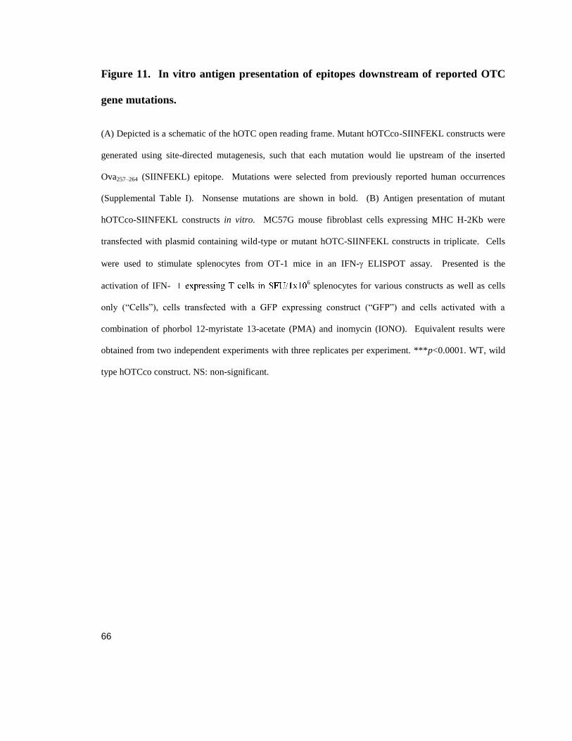
66
Figure 11. In vitro antigen presentation of epitopes downstream of reported OTC
gene mutations.
(A) Depicted is a schematic of the hOTC open reading frame. Mutant hOTCco-SIINFEKL constructs were
generated using site-directed mutagenesis, such that each mutation would lie upstream of the inserted
Ova257–264 (SIINFEKL) epitope. Mutations were selected from previously reported human occurrences
(Supplemental Table I). Nonsense mutations are shown in bold. (B) Antigen presentation of mutant
hOTCco-SIINFEKL constructs in vitro. MC57G mouse fibroblast cells expressing MHC H-2Kb were
transfected with plasmid containing wild-type or mutant hOTC-SIINFEKL constructs in triplicate. Cells
were used to stimulate splenocytes from OT-1 mice in an IFN- ELISPOT assay. Presented is the
activation of IFN-6 splenocytes for various constructs as well as cells
only (“Cells”), cells transfected with a GFP expressing construct (“GFP”) and cells activated with a
combination of phorbol 12-myristate 13-acetate (PMA) and inomycin (IONO). Equivalent results were
obtained from two independent experiments with three replicates per experiment. ***p<0.0001. WT, wild
type hOTCco construct. NS: non-significant.

67

68
Figure 12 Alternative start sites enable expression of epitopes downstream of
nonsense mutations.
(A) Diagram showing within the hOTC open reading frame the location of new mutations designed to
elucidate the mechanism of C-terminal expression. (B – D) MC57G mouse fibroblasts were transfected in
triplicate with plasmids containing a control transgene (“GFP”), hOTCco-SIINFEKL wild-type, or the
hOTCco-SIINFEKL mutant plasmids. Cells were then cultured with stimulated OT-1 splenocytes in an
IFN- ELISPOT assay. An additional control was only cells (“Cells”). Presented is the activation of IFN-ɣ
expressing T cells in SFU/1x106 splenocytes for various constructs as well as the Cells and GFP controls.
This experiment was independently performed five times (B) hOTCco-SIINFEKL constructs included
CPL109-111X, P110+1, and K353X mutants. (C) Antigen presentation assays for a series of Met-to-stop
substitutions were performed to locate the alternative start site. Each construct includes a single Met-to-stop
mutation in a hOTCco gene that has a C109X mutation. (D) Antigen presentation using a construct with all
methionines downstream of C109X substituted to arginine to determine the alternative start site. These
experiments were independently performed twice. **p<0.001, ***p<0.0001 in comparison with cells only
and GFP negative controls.

69
Figure 13 Amino acid coverage of C109X and unique amino acid sequences
detected in samples expressing hOTCco-C109X-StrepII.
HEK 293T cells were transfected with either hOTCco-C109X-StrepII or control eGFP plasmid constructs.
Cells were grown for 24 h post transfection, and peptides were isolated by column chromatography. Eluted
peptides were submitted to mass spectrometry analysis. Detected peptides that localized to OTC are
highlighted in alternating black and underlined dark grey. Light grey amino acids were not isolated within
a peptide.

70
Figure 14 In vivo activation of endogenous T cells to an epitope downstream of the
PTC.
C57Bl/6 mice (n = 3) were immunized IV with 5x10¹⁰
particles of Ad5 vector containing either wild-type
hOTCco-SIINFEKL, hOTCco-C109X-SIINFEKL, or nLacZ. Day 10 after immunization, mice were
sacrificed and splenocytes were harvested for an IFN- ELISPOT assay. Cells were stimulated with either
media, SIINFEKL peptide, or Ad5 hexon. Presented is the activation of IFN-ɣexpressing T cells in
SFU/1x106 splenocytes. NS, not significant in comparison with Ad5.nLacZ control.

71

72
Figure 15 In vivo tolerance induction to epitopes downstream of the PTC.
(A – C) C57Bl/6 mice (n = 3/group) were administered IV with AAV8 vectors expressing different
hOTCco genes or a no-AAV8? vector control. At day 14, mice were immunized with either (A)
Ad5.hOTCco-SIINFEKL or (B, C) Ad5.OVA, which naturally contains the SIINFEKL epitope. At day 27,
mice were sacrificed and splenocytes were harvested for either (A, B) IFN- ELISPOT assays or (C) CD8
tetramer staining. ( A, B) cells were stimulated with media (black), SIINFEKL (white), or Ad5 Hexon
(grey). (C) the lymphocyte population was gated by forward and side scatter, and then gated by CD8
staining. The percent of SIINFEKL-specific T cells within the CD8+ population was determined by
tetramer staining. Presented for panels A and B is the activation of IFN-ɣexpressing T cells in SFU/1x106
splenocytes. (C) is the percent of CD8+ T cells that are tetramer positive. These experiments were
independently performed twice. *p<0.05, **p<0.001, ***p<0.0001 in comparison with no-AAV vector
negative control.

73
Figure 16 PTC near C-terminus inhibits in vivo tolerance induction to epitopes
downstream of mutation.
C57Bl/6 mice (n = 4) were administered IV with AAV8 vectors expressing different
hOTCco genes or a no-AAV vector control. At day 14, mice were immunized with
Ad5.OVA, which naturally contains the SIINFEKL epitope. At day 27, mice were
sacrificed and splenocytes were harvested for IFN- ELISPOT assay. Cells were
stimulated with media (black), SIINFEKL (white), or Ad5 Hexon (grey). Presented for
is the activation of IFN-g expressing T cells in SFU/1x106 splenocytes. These
experiments were *p<0.05, **p<0.001, ***p<0.0001 in comparison with OTC-
SIINFEKL vector positive control. NS: non-significant. SFU: spot forming units

74
Table 1 Private mutations selected to give a range of mutation types, including
nonsense, missense, and splice variants resulting in exon-skipping.
Mutation Codon
Nucleotide
change Exon/Intron Gender Age
OTC
Activity Reference
Missense R26Q CGG>CAG Exon1 Male 4 days 0%
Grompe et al.,
1989, PNAS
Reduced
activity/late
onset L88N A>T Exon 3 Male 4 months 23%
Oppliger et al.,
1996, Hum
Mutat
Nonsense C109X T>A Exon 4 Male 3 days ND
Calvas et al.,
1998, Hum
Mutat
Nonsense R141X CGA>TGA Exon 5 Male Neonatal 0%
Oppliger et al.,
1996, Hum
Mutat
Nonsense Q154X GAA>TAA Exon 6 ND ND ND
Grompe et al.,
1989, PNAS
Missense P225L CCG>CTG Exon 7 Male 4 days 2%
Matsuda and
Tanake, 1997,
Am J Med
Genet
Reduced
activity/late
onset T264A ACT>GCT Exon 8 Male 5 months 22%
Matsuda and
Tanake, 1997,
Am J Med
Genet
Missense H302Y CAC>TAC Exon 9 Male Neonatal 0%
Oppliger et al.,
1996, Hum
Mutat
Exon-
skipping - GT > GC Intron 7 Male ND ND
Carstens et al.,
1991, Am J
Med Genet
ND, not determined

75
Figure 17 hOTC codon sequence and transfection control
(A) MC57G mouse fibroblasts were not transfected (“Cells”) or transfected with plasmids containing a
control transgene (“GFP”), codon optimized wild-type amino acid sequence hOTCco-SIINFEKL, codon
optimized mutant amino acid sequence hOTCco-SIINFEKL, native codon wild-type amino acid sequence
hOTC-SIINFEKL, or native codon mutant amino acid sequence hOTC-SIINFEKL plasmids. Cells were
then cultured with stimulated OT-1 splenocytes in an IFN-ɣELISPOT assay (B) MC57G mouse fibroblasts
were either not transfected or transfected with plasmids containing a control transgene (GFP), wild-type
hOTCco-SIINFEKL, or mutant hOTCco-SIINFEKL plasmids with or without Lipofectamine. Cells were
then cultured with stimulated OT-1 splenocytes in an IFN-ɣELISPOT assay. Presented is the activation of
IFN-ɣ expressing T cells in SFU/1x106 splenocytes for various constructs as well as the Cells and GFP
controls. Experiments were independently performed twice and in each experiment the individual groups
were evaluated in triplicate. ***p<0.0001 in comparison with cells only and GFP negative controls. SFU:
spot forming units.
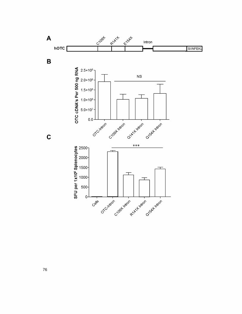
76

77
Figure 18 Impact of intronic sequence on mRNA expression and antigen
presentation.
(A) Diagram showing the location of the intron that is 78 bp downstream of E154X. (B) RT-qPCR analysis
was conducted in 293 cells transfected with wild-type or mutant plasmid constructs and is presented as
OTC cDNA’s per 500 ng RNA. (C) MC57G mouse fibroblasts were transfected in triplicate with plasmids
containing wild-type hOTCco-SIINFEK-Intron, or mutant hOTCco-SIINFEKL intron constructs. Cells
were then cultured with stimulated OT-1 splenocytes in an IFN ɣ ELISPOT assay. Presented is the
activation of IFN-g expressing T cells in SFU/1x106 splenocytes for various constructs as well as the Cells
and GFP controls. Both experiments were independently performed twice, n=3 mice / group. ***p<0.0001
in comparison with cells only negative control. NS, not significant

78
Table 2 Peptide identification using LC/MS with affinity-purified samples
expressing GFP or hOTC-C109X-StrepII.
Algorithm Sample OTC Peptides Identified Readsa E-Value
Xtandem! C109X 9 54 -394
Xtandem! GFP - - -
aNumber of total reads in the sample that align to the OTC sequence.

79
Chapter 4
Influence of Vector on Anti-Transgene Antibody Formation Following AAV-
Mediated Gene Therapy
80
Abstract
Antibody formation to secreted transgene products remains as a potential barrier to many
gene therapies. Driven by exposure of naïve B cells to a transgene product and the
immune stimulus of vector administration, expression can result in an adaptive immune
response and antibody formation against the therapeutic protein resulting in attenuation
of efficacy. Here, we show that TLR9 signaling and the expression location profile is
involved in driving the activation of B cells against secreted protein alpha-L-iduronidase
(IDUA), following AAV administration. This TLR9-dependent response occurs
independently of transgene CpG content, however it is strongly influenced by the
location of transgene expression or lack therefore of. Overall, our results suggest that
TLR9 signaling is important for the development of adaptive immune responses to
transgene products following AAV gene therapy, and that elimination of CpG motifs in
the vector genome may not be sufficient to prevent TLR9 activation.
Introduction
Adeno-associated virus (AAV) vectors have been investigated in many diseases
where cross correction of transgene product expressed into the blood is vital for
therapeutic efficacy. In many of these preclinical models, antibody formation to the
transgene product has been observed. However, the adaptive immune response is tightly
controlled; exposure to a novel protein often results in tolerance rather than immunity,
unless an inflammatory signal is present to initiate an adaptive immune response to the
novel antigen (Lang, Georgiev et al. 2006). One potential immunogenic stimulus

81
associated with AAV-mediated gene therapy is the vector genome. The immune
system’s major sensing agent for this foreign material is TLR9, located in the endosomal
compartment, which recognizes unmethylated CpG DNA and induces pro-inflammatory
reactions that can result in an adaptive response (Hemmi, Takeuchi et al. 2000, Rogers,
Martino et al. 2011). Additional work has been done to avoid TLR9-induced
inflammatory signaling by CpG depletion or by increasing the CpG methylation state in
DNA-based therapies (Reyes-Sandoval and Ertl 2004, Faust, Bell et al. 2013, Shapir,
Miari et al. 2015). However, there is evidence that TLR9 binds the phosphodiester
backbone of DNA regardless of the nucleotide sequence, and that activation is merely
enhanced by CpG motifs (Haas, Metzger et al. 2008).
The involvement of TLR9 signaling following intramuscular (IM) AAV administration
has been found to be important in the generation of both anti-transgene antibodies and
AAV neutralizing antibodies (Zhu, Huang et al. 2009, Rogers, Suzuki et al. 2015). TLR9
signaling was shown to have a more limited role in the production of anti-transgene
antibodies following intravenous (IV) vector administration, with myeloid differentiation
primary response gene 88 (MyD88) being required to generate this immune response
(Rogers, Suzuki et al. 2015). A tolerizing phenomenon has been observed following
hepatic gene transfer using AAV vectors expressing the serine protease factor IX, leading
to reduced or ablated anti-transgene antibodies (Cao, Dobrzynski et al. 2007), and even
elimination of existing transgene antibodies in large animal studies (Finn, Ozelo et al.
2010, Crudele, Finn et al. 2015). However, our experience with different monogenetic

82
disorders indicates that this phenomenon is transgene-dependent (Hinderer, Bell et al.
2015). AAV-mediated hepatic gene therapy for mucopolysaccharidosis I (MPS I) in a
dog model provoked an antibody response to the transgene product, alpha-L iduronidase
(IDUA), demonstrating that hepatic expression alone is insufficient to induce tolerance
(Hinderer, Bell et al. 2015).
In this study, we investigated the influence of TLR9 and transgene expression on
antibody formation following AAV gene therapy using different routes of administration,
primarily focusing on systemic delivery. We found a role for TLR9 in anti-transgene
antibody formation against human IDUA (hIDUA) following IV vector administration;
this response was also modified by the promoter. Unexpectedly, CpG depletion of the
transgene did not influence the immune response to IDUA. Further, we discovered a link
between generation of anti-IDUA antibodies and tissue restricted expression. These
findings suggest that, while TLR9 activation can be consequential in eliciting a transgene
specific adaptive immune response, our ability to curb its influence may be limited.
Methods
Vectors
AAV9 hIDUAco (human codon-optimized IDUA under the control of a CMV, CB7, or
EF1alpha enhancer/promoter containing SV40 poly A) was produced along with other
AAV vectors using an iodixanol step gradient (Lock, Alvira et al. 2010).

83
Mouse Anti hIDUA Detection Assay
Clear, flat-bottom polystyrene high-binding plates were coated with 50 μl/well of 0.58
mg/kg Aldurazyme (Biomarin, San Rafael, CA) diluted 1:100 in pH 5.8 PBS. Plates
were washed five times using 0.05% PBS-Tween solution and then blocked for two hours
with 2% BSA (GE Healthcare, Little Chalfont, UK) diluted in pH 5.8 Dulbecco’s PBS
(Thermo Fisher Scientific, Waltham, MA). Serum was diluted 1:1,000 in Dulbecco’s
PBS (Thermo Fisher Scientific, Waltham, MA); 50μL of diluted sample was plated and
incubated at room temperature for two hours. Positive serum for the standard was diluted
1:10 and then diluted four fold for a series of seven dilutions and a negative control well.
Plates were washed as previously described. Streptavidin-conjugated goat anti-mouse
IgG antibody (BD Biosciences, San Jose, CA) was diluted 1:10,000 in 2% BSA (GE
Healthcare, Little Chalfont, UK), and 100μl of diluted goat anti-mouse IgG antibody was
added per well. Plates were incubated for 1 hour and washed as previously described.
150μl of TMB solution was added per well. Plates were read up to 20 minutes after
adding stop read value to 450 nm.
IDUA Activity Assay
4-MU substrate (Glycosynth, Winwick Quay, Warrington, UK) was diluted 1:25 in
IDUA assay buffer (0.1 M sodium acetate, 0.15 M NaCl, 0.05% TritonX, pH 3.8). In 96-
well flat bottom tissue culture plates, 95 ul di-H20, 100 ul diluted 4-MU substrate, and 5
µl serum sample was added to wells in duplicate. Plates were sealed, gently shaken, and
incubated at 37 ºC for 2 hours. 180 µl IDUA stop buffer (0.29 M glycine, 0.18 M sodium

84
citrate, pH 10.9) was added per well. 20 µl sample was removed to black well plates
(Corning, Corning New York) for fluorescence readings (365 nm excitation and 450 nm
emission).
ELISPOT
Mice were administered IM 2x10¹¹
GC of AAV9 expressing either hIDUA or a CpG-
depleted variant in 50 µl PBS. On day 14 after vector administration, mice were
sacrificed and splenocytes were harvested, as previously described (Limberis, Figueredo
et al. 2007). Interferon ɣ ELISPOT assays were performed, according to the
manufacturer’s instructions (BD Biosciences, San Jose, CA). Cells were seeded at a
concentration of 2x10⁵ or 2x10
4 cells/well along with 2 ug/ml peptide pool A, B, or C of
an overlapping 15-mer peptide library spanning the full-length hIDUA (Mimotopes,
Victoria, Australia). Splenocytes were incubated at 37 °C in 5% CO2 for 18 hours. Spots
were counted using the AID ELISPOT Reader System (Cell Technology, Columbia,
MD).
Results
Previously we demonstrated that TLR9 signaling is capable of triggering a strong
CTL response to transgene product following AAV-mediated gene therapy. We next
investigated its ability to modulate a humoral response. C57BL/6 or TLR9 KO mice
were given IV AAV9.hIDUAco under the control of one of three promoters: the universal
promoters CMV or CB7, or the liver-specific promoter TBG (Bell, Wang et al. 2011).
We determined transgene product expression by measuring hIDUA activity in serum,

85
which inversely correlated with the presence of antibodies for all three groups. AAV9
vectors with either universal promoter (CMV or CB7) triggered the production of a
robust anti-hIDUA antibody that reduced serum activity (Figure 19 a, b); in contrast,
TBG-driven expression resulted in low anti-hIDUA antibody levels and a high, consistent
serum activity level (Figure 19 c). However, in the TLR9 KO mice, vectors with the
universal promoters CMV or CB7 had dramatically reduced antibody generation to
hIDUA, indicating that TLR9 signaling is required for the anti-transgene response
(Figure 19 a, b).
To further investigate the role of TLR9 signaling in IDUA-specific responses, we
administered 1x10¹¹
GC AAV9.hIDUAco IV or IM under the control of either the CMV
or the CB7 promoters to either C57BL/6 or TLR9 KO mice. After ten days, we
sacrificed the mice and performed an IFN-ɣ ELISPOT on splenocytes. As we
anticipated, T cells from TLR9 KO mice stimulated with hIDUA pools did not respond as
robustly as those from C57BL/6 mice across all treatment regimens (Figure 20 a, b).
Interestingly, the largest response occurred in mice given AAV9.CB7.hIDUAco IV
compared to IM; CMV-containing vectors had an overall lower T cell response (Figure
20 a, b) corresponding with an overall lower peak activity. TLR9 signaling is thought to
be important for proper B-cell development (Hua and Hou 2013). To confirm that the
observed effect was due to loss of TLR9 signaling upon administration of vector and not
an intrinsic defect in the B cells, we adoptively transferred 1.5 x 10⁷ B cells from
C57BL/6 mice or TLR9 KO mice into muMt- mice that lack mature B cells. One week
86
later, we administered 1x10¹¹
GC AAV9 CMV.hIDUAco IV and monitored serum for
transgene activity and transgene-specific antibody expression (Figure 21 a). We found
that B cells from both groups were capable of producing anti-transgene antibodies and
ablating serum transgene activity levels, indicating that the absence of TLR9 signaling in
B cells did not inhibit these immune responses (Figure 21 b).
We next assessed whether modulating the CpG content would influence the
antibody response to hIDUA, as we found previously removal of CpG content can reduce
T cell responses in the muscle to a reporter transgene (Faust, Bell et al. 2013). We
utilized an AAV vector with the hIDUAco transgene under the control of an EF1-α
promoter depleted of CpGs. We designed a second transgene construct in which the
hIDUA transgene was depleted of CpG content while retaining the amino acid sequence;
we confirmed that the activity of the construct was not changed due to codon effects
using an in vitro activity assay (Figure 22 a). We administered mice with vectors AAV9
EF1-α.hIDUAco or EF1-α.hIDUA,CpG- either IV or IM, and assayed IDUA activity and
antibodies from serum for each cohort (Figure 23 a, b). IDUA activity peaked at day 7
and remained steady throughout the study in both groups (Figure 23 a, b). Anti-IDUA
antibodies rose steadily throughout the experiment in both groups, but did not inhibit
IDUA activity or reach levels as high as those measured when AAV9.CMV. hIDUAco or
AAV9.CB7.hIDUAco was administered (Figure 23 a, b; Figure 19 a, b). To confirm that
CpG depletion did not impact the cytotoxic immune response to IDUA, which would
otherwise reduce transgene activity, we examined CTL activation. We administered

87
mice with either 1x10¹¹
AAV9.EF1-α.hIDUAco or AAV9. EF1-α.hIDUA.CpG- IM
vector, and ten days later performed IFN-ɣ ELISPOT on splenocytes. T cell responses to
IDUA were detectable following IM or IV vector administration; however, we did not
observe a difference between groups in the magnitude of the response, indicating
effective T cell activation regardless of transgene CpG content (Figure 23 c).
Finally we examined the differences in antibody formation and transgene
expression mediated by promoter restriction. As shown earlier, CMV expression results
in both low serum activity and high antibody titer as opposed to TBG driven expression.
When AAV9.CMV.hDUA and AAV9.CB7.hDUA are mixed 1:1 and administered serum
activity peaks and then reduces by 2 fold corresponding with a rise in antibody titer that
is similar to mice injected only with CMV containing vectors (Figure 24 a). There is
evidence in mice that muscle cells express MHC II under inflammatory conditions and
allow for priming of CD4+ T cells, which is a crucial step for the ultimate activation of B
cells (Keller, Fokken et al. 2011, Faust, Bell et al. 2013). To reduce muscle expression
we cloned four miR206 sequences, that target mRNA in skeletal muscle for degradation,
into the poly A section of our CB7.hIDUA which has both initial high expression
followed by a robust antibody increase and loss of serum expression (Greig, Peng et al.
2014). Mice injected with the AAV9.CB7.hIDUA.4xmiR206 maintained higher
expression and lower antibody titer until the last time point of the study where it
converged with control group; despite this the activity did not decrease (Figure 24 b).
88
Discussion
Here we considered the influence of TLR9 and other host factors on humoral
responses. For these experiments, we could not use OTC as the transgene, as it would
likely be sequestered in the mitochondria and not come into contact with B cells. Instead,
we used a vector expressing hIDUA, a secreted transgene that caused antibodies to form
against it in our previous studies in both MPS1 -/- and C57BL/6 mice. Indeed, we saw
differences in the relative titers of anti-hIDUA antibodies between constructs with
expression driven by the universal promoters CMV and CB7 compared to the liver-
specific TBG promoter. Other groups previously reported similar results when using a
muscle-specific promoter (Doerfler, Todd et al. 2016). Importantly, we demonstrated
that a lack of TLR9 signaling dramatically reduced the anti-IDUA antibody response in
mice that received either CMV or CB7 hIDUA vector, indicating that its signaling is
critical for developing a robust humoral response following global expression.
We next assessed the impact of TLR9 signaling, promoter selection, and route of
administration on CTL responses. We compared C57BL/6 or TLR9 KO mice that
received AAV9.CMV.hIDUA or AAV9.CB7.hIDUA IV or IM. We found that
splenocytes isolated from TLR9 KO mice did not respond to stimulation with hIDUA
peptide pools compared to C57BL/6 mice across all experimental conditions.
Interestingly, the largest response occurred in mice that received AAV9.CB7.hIDUA IV
compared to IM; CMV-containing vectors had an overall lower T cell response. This
could indicate that expression level is tied to the strength of immune activation, where the
89
high activity of CB7 and higher expression of the transgene product could enable better
priming of APCs. To further probe this observation, we reduced the CpG content of
transgene, as we have done previously (Faust, Bell et al. 2013). We used a universal
promoter, EF1-α, as this was previously CpG depleted (Faust, Bell et al. 2013). When
given the vectors IV, groups that had retained our codon-optimized hIDUA sequence did
not generate a higher antibody titer than those receiving CpG-depleted hIDUA.
Surprising, both maintained high levels of serum IDUA activity. The antibody titers
were lower in mice that received vectors driven by EF1-α compared to CMV or CB7,
suggesting that there is a threshold for transgene-specific antibody levels to negatively
affect activity and that some inherent differences between CB7, CMV and EF1-α account
for the gap in transgene-specific antibody and serum levels. We next confirmed that CpG
depletion did not impact transgene-specific immune responses, as measured by IFN-ɣ
ELISPOT. Although this result was consistent with our antibody data, it did contradict
previous results in studies where we modified the CpG content of nuclear-targeted LacZ
(Faust, Bell et al. 2013). This difference could be explained by a number of factors,
including the vector (in our previous work we used the more immunogenic AAVrh32.33
(Mays, Vandenberghe et al. 2009)) or enhancement of the CpG content of the LacZ
control transgene sequence (the depletion could have caused a decrease in pro-
inflammatory cytokines that was not capable of inhibiting larger responses to the
transgene product).

90
Lastly, we investigated one aspect of how the site of expression can affect enzyme
activity and potentially therapeutic efficacy. To accomplish this we mixed hIDUA
expressing AAV9 vectors driven by either CMV or TBG promoters. We demonstrated
that the dominant effect of CMV expression is high antibody titer capable of overcoming
a presumed tolerogenic effect that liver driven AAV expression can induce (Cao,
Dobrzynski et al. 2007, Hinderer, Bell et al. 2015, Doerfler, Todd et al. 2016). It has
suggested that one organ group where expression could positively influence antibody
generation is skeletal which is capable of presenting on MHC II potentially priming
CD4+ T cells for B cell activation (Keller, Fokken et al. 2011, Faust, Bell et al. 2013,
Doerfler, Todd et al. 2016). By eliminating muscle expression using miR206 sites in a
CB7.hIDUA construct we were able to observe stable expression of the transgene product
and a slower raise in anti-transgene antibodies. An interesting observation was both
CB7.hIDAU.miR206 and EF1α driven constructs reached an antibody titer elevated to
levels that in CMV or CB7 containing vectors serum activity was dramatically reduced,
yet in CB7.hIDAU.miR206 and EF1α they were able to maintain higher activity
indicating that serum activity loss could be caused by factors beyond antibody titer.
Together, these results demonstrate the importance of TLR9 in developing a
robust humoral response to a secreted transgene product and highlight an important but
complicated role for promoters. While CpG depletion did not improve the outcome of
AAV9 vector administration, this informed us that TLR9 signaling might be independent
of CpG influence on our vector genomes. This could be due to the natural DNA

91
backbone retaining a basal level of immunogenicity (Haas, Metzger et al. 2008). Some
groups have also reported the importance of methylation and its inhibition of TLR9
binding; this potentially could be explored to rationally design vectors to be less
immunogenic. In larger animal models in which transgene antibodies seem to be more
common, the effect of CpG depletion may become more pronounced. Therefore, future
studies may help to further our knowledge and ability to design less immunogenic
vectors.

92
Figure 19 TLR9 Signaling is required for an anti-transgene antibody response.
Male C57BL/6 or TLR9 KO mice (n=5/group) age 6-8 weeks were given IV 1x10¹¹
GC of (a)
AAV9.CMV.hIDUA vector (b) AAV9.CB7.hIDUA vector promoter, or (c) AAV9.TBG.hIDUA vector.
Mice were bled weekly, and IDUA activity and anti-transgene antibodies were determined from serum.

93
Figure 20 TLR9 Signaling is Required for an Anti-Transgene CTL Response.
Male C57BL/6 or TLR9 KO mice (n=5/group) age 6-8 weeks were given 1x10¹¹
IV GC (a) or IM (b)
AAV9 hIDUA vector with CMV or CB7 promoter. Mice were sacrificed ten days later and INF-gamma
ELISPOT was performed on isolated T cells. Analysis was performed by one-way ANOVA with the
Bonferroni test comparing columns in C57BL/6 mice to TLR9 KO mice for each stimulation., *P<0.05,
**P<0.001, ***P<0.0001.
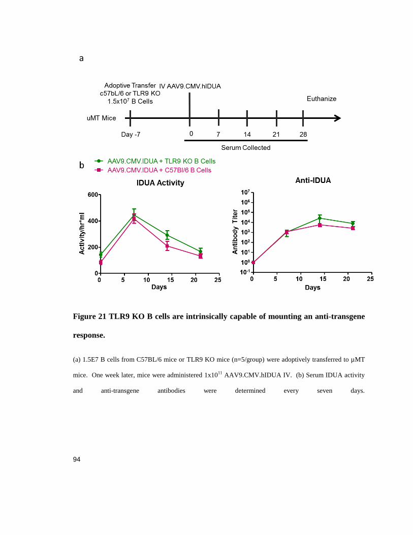
94
Figure 21 TLR9 KO B cells are intrinsically capable of mounting an anti-transgene
response.
(a) 1.5E7 B cells from C57BL/6 mice or TLR9 KO mice (n=5/group) were adoptively transferred to µMT
mice. One week later, mice were administered 1x10¹¹
AAV9.CMV.hIDUA IV. (b) Serum IDUA activity
and anti-transgene antibodies were determined every seven days.
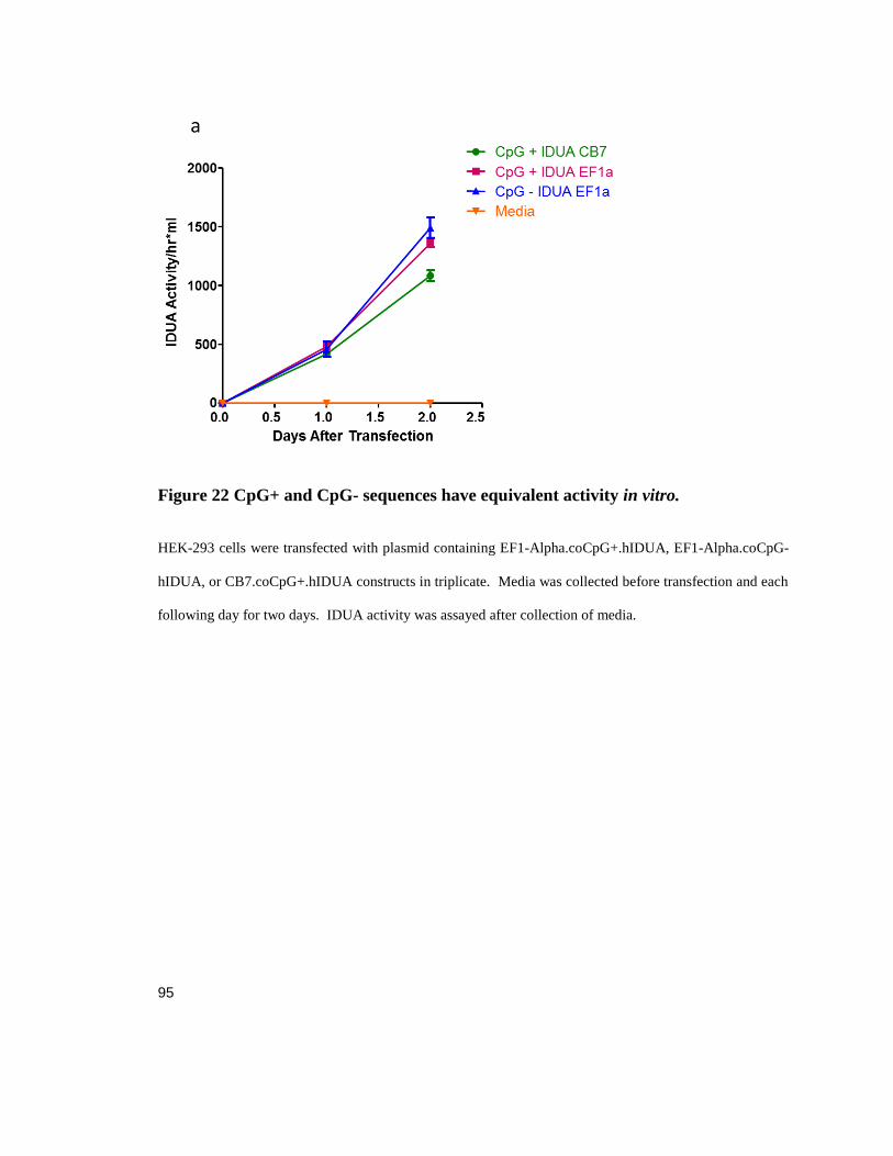
95
Figure 22 CpG+ and CpG- sequences have equivalent activity in vitro.
HEK-293 cells were transfected with plasmid containing EF1-Alpha.coCpG+.hIDUA, EF1-Alpha.coCpG-
hIDUA, or CB7.coCpG+.hIDUA constructs in triplicate. Media was collected before transfection and each
following day for two days. IDUA activity was assayed after collection of media.

96
Figure 23 CpG Depletion Does Not Inhibit Anti-IDUA Formation.
Male C57BL/6 mice (n=5) ages 6-8 weks were given IV or IM 1x10¹¹
AAV9 EF1α hIDUA vector with
normal or CpG-depleted transgene sequence. (a, b) IDUA activity and antibodies were determined from
serum. (c) T cell IFN-gamma ELISPOT was performed on splenocytes isolated from mice ten days after
vector administration.

97
Figure 24 Suppression of Muscle Expression Increase Transgene Activity.
Male C57BL/6 mice (n=5) ages 6-8 weeks were given IV 1x10¹¹
of AAV9.hIDUA vector with CMV
promoter, TBG promoter, or a dose of both. Serum IDUA activity and anti-transgene antibodies were
determined every seven days. (a) Results show that be expressing IDUA with a universal promoter anti-
body titer rises and serum IDUA activity lowers even when there is concurrent high levels of expression
from the liver. (b) Male C57BL/6 mice (n=5) ages 6-8 weeks were given IV 1x10¹¹
of AAV9.CB7.IDUA
with or without a 4X miRNA 206 sequence. Serum IDUA activity and anti-transgene antibodies were
determined every seven days.
98
Chapter 5: Discussion
General Summary
AAV gene therapy holds promise for safe efficacious treatment of genetic disorders with
limited available therapeutics. Preclinical development in many disease models,
including MPS1, hemophilia, muscular dystrophy, pompe disease, have both
demonstrated efficacy and the potential for an immune response to AAV mediated gene
therapy (Manno, Pierce et al. 2006, Mendell, Campbell et al. 2010, Hinderer, Bell et al.
2015, Doerfler, Todd et al. 2016). Following these observations and previous challenges
with adenoviral vector immunogenicity studies we have investigated the immune
response and its effect on gene therapy efficacy. The goal of my dissertation work was to
investigate the underlying host factors that modulate the activation of the adaptive
immune response to the transgene product. With deeper understanding of these agents
we could better assess the risks of each new AAV clinical product and tailor AAV
treatment based on an individual’s likelihood for immune activation. Through these
efforts, we may discover critical points in adaptive immune activation and lead to a safer
more thought-out methodology for reducing the risk of adverse immunity to gene
therapy.
Gene Therapy, Immunology and TRL9
Previous work performed in the Wilson laboratory had shown that CD8+ T cells
required inflammatory signaling in addition to the presence of their specific antigen to
become active and attenuate transgene expression of highly immunogenic bacterial

99
protein (Somanathan, Breous et al. 2010, Breous, Somanathan et al. 2011). As described
in chapter 2, I opted to develop a model to explore transgene-specific immunity against
the mitochondrial protein OTC and to further investigate the influence of specific host
factors that are critical to immune activation. Initially I sought to develop a scheme
similar to ones used previously to activate CD8+ T cells against a specific transgene by
immunizing C57Bl/6 mice with Ad5 vector expressing hOTCco, then harvest the T cells
for adoptive transfer (Somanathan, Breous et al. 2010). Our first experiments did not
yield the anticipated result, as T cell activation, measured by IFN-ɣ ELISPOT, did not
induce a robust OTC-specific response immune response. Inducing and immune response
required a more robust methodology, first we administered the immunogenic
AAVrh32.33, then 12 weeks later administered both Ad5 and TLR9 agonist concurrent
with vector and for three days following (Figure 2 a ,b) (Mays, Vandenberghe et al.
2009). These results indicated that we were not activating naive hOTC-specific T cells,
but rather breaking tolerance of anergized T cells which could have occurred due to the
highly conserved nature of OTC between species (P11725, P00480) (Lang, Georgiev et
al. 2006). Under such stringent conditions necessary to generate a population of OTC-
specific CD8+ T cells I did not consider it feasible to continue with that methodology.
Further, I opted against using cells that had previously been exposed to the transgene as
this is not what I anticipate will occur in AAV gene therapy for the patients most likely to
have an adverse T cell activation, namely, those that express no protein from the mutated
gene. To develop our model I took advantage of the transgenic OT-1 mouse which
contains genes for a T cell receptor that specifically binds the immune-dominant epitope

100
of chicken ovalbumin (SIINFKEL) by generating a construct with a SIIFEKL tag
attached to the c-terminus of hOTCco (Figure 11 a) (Hogquist, Jameson et al. 1994).
Though the epitope tag did diminish OTC activity (Figure 3), biogenesis of the protein
and its translocation to the mitochondria remained intact, which was important for our
design study. Maintenance of the biogenesis pathway was important since I expected
protein degradation and processing into antigen to be similar to the untagged hOTCco
(Yewdell, Reits et al. 2003). To investigate the influence of TLR9 on the activation of a
cell mediated immune response to the transgene product we packaged hOTCco-
SIINFEKL into an AAV8 vector (Figure 5). C57Bl/6 mice were given IV either TLR9,
OT-1 CD8+ T cells or both and analyzed for toxicity by evaluation of transaminase levels
and the presence of genome copies four weeks after adoptive transfer. Elevations of AST
and ALT were only observed in the group of mice that received both transgene specific T
cells and TLR9 agonist indicating a requirement for a naïve CD8+ T cell population, and
TLR9 can provide the necessary inflammatory signals to activate a cellular response.
Further, we observed both a transgene mRNA and genome copy loss indicating clearance
of the episomes potentially driven by cell death.
To evaluate the number of naïve T cells required to initiate such an immune
response we titrated the number of CD8+ T cells that were adoptively transferred to
recipient mice. Surprisingly even at the lowest dose of 3,000 transferred cells we
observed a rise in transaminase levels (AST and ALT) (Figure 6 a). This low dose also
resulted in a decrease of GC, although not as dramatic as that observed in the higher dose

101
groups. These findings were corroborated by our experiment designed to examine the
activation of the adoptively transferred cells infiltrating the liver, in which the majority of
cells were activated (Figure 9 d). We investigated the importance of naïve T cells in
developing a cell mediated response by withholding TLR9 agonist until three weeks after
the adoptive transfer to allow for CD8+ OT-1 T cells to come in contact with the
SIINFEKL antigen presented by liver cells. While toxicity was reduced with
transaminase elevation being substantially decreased in the late inflammation group;
protein expression was not consistent between mice in the group despite genome copies
and RNA expression being similar. These findings suggest that antigen naïve T cells are
able to generate a more robust immune response compared to T cells that have previously
been exposed to a transgene product prior to an inflammatory event.
Clearance of transduced cells by an adaptive immune response has been a concern
for the development of AAV-based gene therapy; in our experiments we address this
likelihood. C57Bl/6 mice were given IV AAV8.TBG.hOTCco-SIINFEKL and fourteen
days later administered CD8+ OT-1 T cells and TLR9 agonist. Mice from this group and
control groups were then sacrificed on days 3, 7 and 17 following adoptive transfer.
Liver sections were analyzed for CD8+ T cell infiltration (Figure 8 a) noting extreme
infiltration in the TLR9 treated group into regions beyond the periportal or pericentral
regions. We also observed some infiltration in the periportal liver region in mice
administered only AAV8.TBG.hOTCco-SIINFEKL and T cells indicating some homing
to the liver but potentially no activation. Death markers capsase9 and TUNEL staining

102
were performed on liver sections, however, no differences were observed between groups
including a control AAV8.TBG.hOTCco-SIINFEKL only treated cohort. The possibility
that we missed the time-point of maximal cell death led us to using a marker for cell
growth KI-67 to evaluate the regeneration of the liver (Figure 8 b). Upon examination we
observed a large percentage of hepatocytes marked as having left G0 of the cell cycle in
mice that received TLR9 agonist indicating that active division was occurring to replace
cells lost as a consequence of the immune response. Further, we observed significant GC
loss in TLR9 treated mice corroborating our KI67 data suggesting that transduced cells
were lysed by the infiltrating CD8+ T cells (Figure 8c). Whereas previous loss of
transgene expression was not highly correlated with loss of GC and was potentially
caused by shut-off of transcription, here we have evidence that massive cell death can
occur prior to regeneration of hepatocytes (Somanathan, Breous et al. 2010, Breous,
Somanathan et al. 2011).
To confirm that the adoptively transferred T cells were migrating to the liver,
mice were given IV AAV8.hOTC.SIINFEKL. Fourteen days later, mice were
administered TLR9 and CFSE labeled CD8+ OT-1 T cells. Seven days following
adoptive transfer, at peak liver infiltration, mice were sacrificed and liver and spleen were
harvested for lymphocyte isolation. In mice receiving TRL9 agonist, the CD8+ T cell
population, as a percentage of total lymphocytes, was increased as was the amount of
SIINFEKL-tetramer positive cells (Figure 9a, b). In the liver, the majority of infiltrating
T cells were transgene-specific when TLR9 was given, however since the number CFSE

103
positive cells was reduced in this cohort, we hypothesize that by this point of the immune
activation the transferred T cells may have undergone many rounds of division resulting
in dilution of the CFSE to below our threshold for positive detection (Figure 9 b,c). This
indicates that most of the transferred cell population remaining had expanded as opposed
to only a subset of that population suggesting that most of the T cells that survive migrate
to the liver.
Finally it was important to determine if TLR9 signaling in CD8+ T cells was
responsible for immune activation. C57Bl/6 or TLR9 KO mice were administered AAV8
hOTCco-SIINFEKL. Fourteen days later, mice were given OT-1 T cells and TLR9
agonist. In this experiment TLR9 KO mice that received the CD8+ T cells will only have
signaling in the adoptively transferred cells. We found that transaminase levels did not
elevate in TLR9 -/- mice given both agonist and T cells (Figure 10 a). GC and mRNA
levels were similar to vector only control in treated TLR9 KO (Figure 10 b). Taken
together, these results demonstrate the need for TLR9 signaling extrinsic to the T cells
that will become activated. This is important as during gene delivery TLR9 signaling
should occur only in cells transduced by the vector or potentially by the antigen
presenting cells that take up vector particles.
These results establish a method for investigating a cytotoxic T cell response to a
mitochondrial targeted gene therapy of the liver. We observed that it was necessary to
have both a strong inflammatory event and transgene specific T cells. Our evidence
suggests that TLR9 could provide that inflammatory signaling, and as both other groups

104
and our own investigation into the humoral response has shown AAV provides a
sufficient amount of DNA to initiate such a response (Zhu, Huang et al. 2009). If we are
able to either avoid or suppress this signaling we would anticipate a much reduced
immune response to the transgene product. An important question for the field would be
how long does the vector DNA remain in endosomes after transduction and the likely
hood that it could lead to inflammation in the months following gene therapy. The
requirement for having a pool of naïve antigen specific T cells also provides optimism as
in the third chapter we describe a mechanism that could potentially reduce the number of
these T cells from individuals with a PTC mutation.
Tolerance to Epitopes C-Terminal of a PTC
One guiding principal for the dangers of transgene-specific T cell activation is
how the genotype of the afflicted individual can alter the outcome. Specifically, that
people with missense mutations that are expected to allow reduced or non-functional
protein expression will retain tolerance to the transgene product as both negative
selection and anergy should still be occurring; compared to those that lack expression of
protein altogether whom we would assume lack those mechanisms for preventing an
auto-immune response to gene therapy. In our work on OTC we thought it was important
to explore the potential effects of a patient's genotype on the outcome of gene therapy as
most individuals in OTC tend to have private mutations as opposed to some disorders that
have a single mutation predominate, such as the delta 508 CFTR mutation in cystic
fibrosis. A potential area of concern was related to missense mutations resulting in

105
proteins that could aggregate or in some other way cause cellular stress in ways similar to
the Z allele of ATAT (Carrell and Lomas 2002, Flotte, Trapnell et al. 2011). In less
extreme forms, misfolding can lead to endoplasmic reticulum stress releasing oxygen
species, which could lead to a pre-inflammatory environment which could be prone to
initiating an immune response upon AAV mediated gene therapy (Nakagawa and Yuan
2000, Malhotra, Miao et al. 2008). This would deviate from our understating that any
specific missense mutation would not carry an increased risk of immune activation. To
investigate these potential effects we chose to create a panel of hOTCco-SIINFEKL
constructs that contained missense mutations or nonsense mutations originally discovered
in individuals with OTC deficiency (Table 1). In our initial screen of these mutants we
found that only one of our selected missense mutants had increased antigen presentation
as measured by IFN-ɣ ELISPOT, which was anticipated since this category of mutation
should not interfere with translation and the mutation, R26Q, occurs in the leader
sequence of OTC potentially interfering with biogenesis. (Grompe, Muzny et al. 1989)
(Figure 11 a, b). Of noted interest were the results obtained from our nonsense mutant
constructs, where each was capable of expressing antigen that is C-terminal to the PTC.
This result contrasted our assumed model where antigen presentation should not
occur, and indicated that expression of epitopes in spite of a PTC could occur and that
potential exists for tolerance either peripheral or central to transpire in individuals with
nonsense mutations (Figure 25 a). The source of antigens presented on MHC has been
contested in the past with two competing theories; one holds that antigens presented are

106
primarily from fully formed proteins degraded by the proteasome as part of the natural
turnover cycle (Colbert, Farfan-Arribas et al. 2013) the second states that the primary
reservoir of epitopes are generated from DRiPS, defective ribosomal products that
include early terminated peptides chains (Yewdell, Anton et al. 1996, Dolan, Li et al.
2010, Dolan, Li et al. 2011). In our model, it is possible that one or both are occurring,
read-through of the PTC by incorporation of a cognate tRNA would allow for continuous
synthesis of our constructs with the finished protein being degraded by turnover
processes (Goodenough, Robinson et al. 2014), another option is that translation is being
initiated downstream of the PTC creating peptide chains that are immediately sent to the
proteasome for degradations (Bazykin and Kochetov 2011, Kochetov, Prayaga et al.
2013) (Figure 25 b, c). Our investigation required the production of several more
constructs to interrogate these hypotheses and we took advantage of inducing PTC at
different location or in volume. Our in vitro work demonstrated that read-through was
unlikely to be occurring, our construct with three mutations, CLP109-111X, did not have
any decrease in T cell activation compared to a single stop codon at position 109 and we
believe it unlikely that cognate tRNA incorporation would occur at the same rate in both
instances (Figure 12 a). Conversely, mutation K353X is one removed from the start of the
SIINFEKL epitope and effectively shut down expression of the peptides measured by our
T cell assay. We believe this demonstrates that by having more gene sequence between
the PTC and epitope one increases the chance of an alternative start site initiating
translation, whereas if read-through we the dominant cause of expression position would
have less of an effect. Finally using a +1 frame shift mutation at P110 we show that C-
107
terminal expression occurs, this is important as the frame shift generates 17 PTC, in
addition even if all stop codons were read-through the downstream product would be out
of frame and SIINFEKL would never be translated, therefore we conclude that enough
translation events to generate a pool of antigens must begin C-terminal to the PTC
(Figure 12 a).
We attempted to identify the site of alternative translation initiation and to
accomplish this we created two panels of constructs, one where each methionine down-
stream of the C109X mutation was substituted with a stop codon, and a second where
each methionine is changed to an arginine. Our results showed that alternative initiation
did not require an AUG site as all our Met-Arg substitution constructs did not prevent C-
terminal expression (Figure 12 c). From our Met-Stop mutants we saw that only the final
mutation M338X inhibited translation, indicating that either it or the M335X mutation
were involved in some capacity to start translation as the intervening codons are not
common alternative start codons such as CUG or GUG (Figure 12 b) (Starck, Jiang et al.
2012). Taken together we hypothesize that multiple codons contribute to translation
initiation including both canonical and non-canonical start codons.
While these observations are interesting with respect to antigen presentation and
offer more evidence for the importance of the DRiP theory to explain the source of
antigen presentation, we think that in the context of gene therapy it could be very
important to considering how we think individual mutations can influence the outcome of
gene therapy. To determine if these phenomena occur in an animal model, we packaged
108
the C109X construct into an Ad5 vector and injected into mice. We observed an immune
activation against SIINFEKL as measured by the INF-ɣ ELISPOT assay against both the
WT and mutant gene containing vectors indicating that epitopes C-terminal to a PTC
were expressed and presented on MHC class I to CD8+ T cells (Figure 14).
Induction of tolerance to the transgene product is an important potential outcome
of our work. If antigen presentation is occurring even in individuals with a PTC or other
null mutation, tolerance could still be maintained if alternative translation occurs at a
significant enough rate to either create central tolerance and thereby deplete the body of
transgene specific T cells or by peripheral tolerance inducing anergy in transgene specific
T cells. To explore these possibilities we used a model of peripheral tolerance and
anergy where expression of an antigen from the liver will induce tolerance to antigens in
the transgene product by exposure of naïve T cells to their epitopes without major
inflammation signals (Lin, Hensley et al. 2007). To determine if tolerance to epitopes C-
terminal to a PTC could induce tolerance similar to normal protein we created AAV8
vectors containing hOTCco-SIINFEKL constructs with stop codon mutations. Mice were
given the AAV vectors and then challenged with Ad5 hOTCco-SIINFEKL, followed by
analysis by INF-ɣ ELISPOT assay. The results showed that a successful reduction of
SIINFEKL specific cells occurred, providing evidence that an individual with a PTC
could still induce tolerance to their defective protein (Figure 15 a). Next, we challenged
a second cohort of mice with Ad5 OVA to prevent activation of hOTC specific CD4+
CD25+ T-regulatory cells that can be induced by liver directed AAV gene transfer. This
109
was important as OTC-specific T regulatory cells could have become active against N-
terminal OTC epitopes; by using Ad5 OVA to provide the SIINFEKL we avoided that
possibility. Again we showed that the PTC containing constructs reduce T cell activation
and lower the amount of SIINFEKL specific T cells (Figure 15 a, b).
Taken as a whole these studies provide insight into the source of antigen
presented on MHC class 1 and the potential for tolerance to intracellular proteins that are
expressed in the liver. Our work provides strong evidence that a large contributor to the
antigen pool comes from defective ribosomal products, specifically from peptides derived
from start sites within the gene. Peptide synthesis starting from internal start sites was
sufficient to create the requisite amount of peptides to load onto MHC class 1 and present
antigen, even with the possibility of mRNA degradation of mutant transcripts by
nonsense mediated decay. While other groups have observed presentation from peptides
formed N-terminal to a PTC this is the first evidence that epitopes found downstream of a
PTC could lead to T cell activation (Apcher, Daskalogianni et al. 2011). A clinically
relevant application is that antigen presentation of epitopes C-terminal to a PTC could
allow for T cell tolerance to be generated to the defective gene’s product. Based on these
results we should reevaluate how individuals are chosen for clinical trials. Patients that
contain a PTC mutation in the defective gene should not be denied the opportunity to
contribute to the advancement of gene therapy development and the potential of receiving
a positive health outcome.

110
Anti-Transgene Antibodies
The humoral response is the other major branch in the adaptive immune system
and while it shares many systems with the cytotoxic response including assistance in
activation via CD4+ T cell help it seems to have a different threshold for activation based
results from pre-clinical models and enzyme replacement therapy (Nayak, Doerfler et al.
2014, Hinderer, Bell et al. 2015, Doerfler, Todd et al. 2016). We believed that immune
activation was strongly tied to signaling through TLR9 an endosomal protein that
becomes active upon binding unmethylated CpG motifs. Signaling through TLR9 results
in translocation of NF-Kb to the nuclease and up-regulation of pro-inflammatory genes,
which could cause activation of antigen presenting cells and lead to an adaptive immune
response. AAV vector genome can provide unmethlyated CpG to TLR9 if the vector
genome is released during its transition from the cell surface to the nucleus. Previously
we had investigated the importance of TLR9 signaling on the transduction of β-
glycosidase in mouse muscle and demonstrated that removing CpG motifs prevented loss
of transduction due to T cell activation (Faust, Bell et al. 2013). In a furtherance of that
study we chose to investigate the impact of TLR9 signaling on gene therapy of MPS1 by
transduction of the IDUA gene. While not part of its normal biogenesis pathway, IDUA,
can be secreted through Golgi transport and then taken back up by other cells due to its
binding of mannose-6-phosphate, this allows for cross correction of the disease but also
leaves it vulnerability to a humoral response (Huang, Wong et al. 1997).

111
To investigate the effect of TLR9 on the humoral response to IDUA we
administered either C57Bl/6 mice or TLR9 KO mice with AAV9 vector containing the
IDUA transgene. Using three different cohorts we varied the promoters between groups
to have two universal expressers (CB7, CMV) and one liver restricted expresser (TBG)
(Figure 19). These studies contribute two important findings, first we demonstrate that
location of transgene expression has a great deal of impact on antibody formation with
expression from liver peaking high and maintaining stable with no anti-transgene
antibody generation compared to either universal expresser that peak and fall off
presumable due to the corresponding rise in antibody titer. The second finding was that
without TLR9, mice could not mount a humoral response against the transgene product.
To control for any B cell developmental differences in TLR9 KO mice we adoptively
transferred both KO mice B cells and C57Bl/6 B cells to µMT mice which lack B cells
due to loss of the IgM gene. In both groups mice were capable of generating an antibody
response that attenuated transgene expression indicating that the TLR9 KO B cells are
comparable functional to WT mice (Figure 21). We also confirmed that TLR9 signaling
is vital to T cell activation by measuring IFN- ɣ ELISPOT of splenocytes following
AAV9.IDUA transduction (Figure 20). Together these results demonstrate a very
important finding that the unmethylated AAV vector genome is not only the primary
immunogenic material, but that TLR9 signal is necessary to generate a humoral response.
It would be invaluable to the field if the of AAV if vector immunogenicity could be
decreased, and our lab has had experience in CpG depletion of AAV vectors a method
that is possible due to multiple codons available for the same tRNA.

112
We systematically removed all CpG motifs from the IDUA gene while still
retaining the same protein coding sequence. For the poly A and promoter we used ones
previously generated to be CpG less as both CMV and CB7 contain large CpG islands
and their removal would most likely impair functionality of the promoter (Faust, Bell et
al. 2013). C57Bl/6 mice were administered AAV9.Ef1α.IDUA with either WT or CpG
depleted IDUA gene sequence and our results showed that there was no difference
between the two groups in either antibody titer or transgene activity (Figure 22 a, b). In
addition T cell activation against the transgene as measured by the INF-ɣ ELISPOT assay
did not shown any meaningful differences. Interestingly, unlike other universal
promoters the EF1α vector resulted in a lower antibody titer and maintained stable levels
of transgene activity. One possible explanation for this and the results as a whole is that
the CpG islands in CMV and CB7 were more important for TLR9 activation than the
CpG motifs found in IDUA. This may mean that using promoters with lower CpG
content and avoidance of CpG islands could be as effective as CpG depletion.
The location of transgene expression has also been noted to have an influence on
the immune response to transgene expression. Most of the research to date has been
focused on the ability of the liver to “tolerize” the body against a secreted product, less
has been explored into which cell type of expression result in such antibody responses
(Dobrzynski, Fitzgerald et al. 2006, Cao, Dobrzynski et al. 2007, Doerfler, Todd et al.
2016). As we observed in Chapter 4 anti-IDUA antibody responses could be modulated
by the promoter used to drive expression, with more universal promoters allowing for

113
robust formation of anti-transgene antibodies. To determine if expression from the liver
was able to tolerize or that expression from other locations would overwhelm any T
regulatory cell activation and push the immune system toward a humoral response we
treated mice with equal amounts of both TBG and CMV driven AAV9 IDUA vectors.
From our results we observed a similar response to (Doerfler, Todd et al. 2016) where
despite having a large dose of liver specific expression it seems that the driving force of
anti-body formation is expression from non-liver tissues (Figure 24 a). One tissue group
that we hypothesized was the most important for antibody formation to a secreted
transgene delivered by AAV9 is the muscle as it is both highly transduced and capable of
expressing MHC class II (Vandendriessche, Thorrez et al. 2007, Zincarelli, Soltys et al.
2008) (Englund, Lindroos et al. 2001). We injected mice with AAV9.CB7.IDUA vectors
some of which contained a micro RNA site (miR206) that is targeted only in muscle
(Greig, Peng et al. 2014) and observed that mice injected with the miR206 containing
vectors had both lower anti-IDUA antibodies and higher long term expression. This
result is important as it demonstrates that expression from the muscle may be the most
important transduction target that results in the generation of anti-transgene antibodies. In
future work, if we can achieve cross correction with a transgene we may want to limit the
organs of transduction to exclude the muscle or diminish expression from that location by
inclusion of micro RNA sites.
These results describe the vital role TLR9 and location of transgene expression
play in the generation of a humoral immune response to a transgene product. As
114
demonstrated in our knockout experiments TLR9 is both necessary and sufficient to
activate B cell secretion of anti-IDUA antibodies, provided that transgene expression is
not restricted to the liver. One attempt to evade the TRL9 mediated B cell activation was
CpG depletion of our transgene construct, our results show no improvement was made in
avoiding antibody detection compared to our control. We did observe that changing from
a CpG rich CB7 promoter, to a CpG depleted EF1α promoter we had a dramatic effect on
serum IDUA activity and antibody-formation, however the interpretation of this result is
somewhat complicated by the fact that despite both being considered “universal
expressers” there may be small difference in expression from different cell types that
could impact on the result. We further interrogated our observation and found that for
AAV9 vectors, expression from the muscle was an important factor in formation of a
robust anti-IDUA immune response. This has important implications for delivery of gene
therapy where we would surmise that expression of secreted transgene would be more
tolerated if it only originated from the liver. I would recommend that for a therapy where
a cross correction strategy is viable one should either restrict expression to the liver, or
design the vector construct as to avoid transgene expression from skeletal muscle cells.
Final Remarks
Throughout this dissertation, my extensive investigation of the adaptive immune
response to transgene products following AAV transduction have given us important
insight into the factors which influence the outcome. I determined influential host factors
that are required for adaptive immune activation, giving us the ability to design better

115
AAV vector constructs in hopes of reducing the likelihood of an adverse immune
response in patients. My hope is that my research will contribute to the safety and
effectiveness of the AAV gene therapy delivery platform and will allow for a broader
group of individuals to be candidates for treatment.
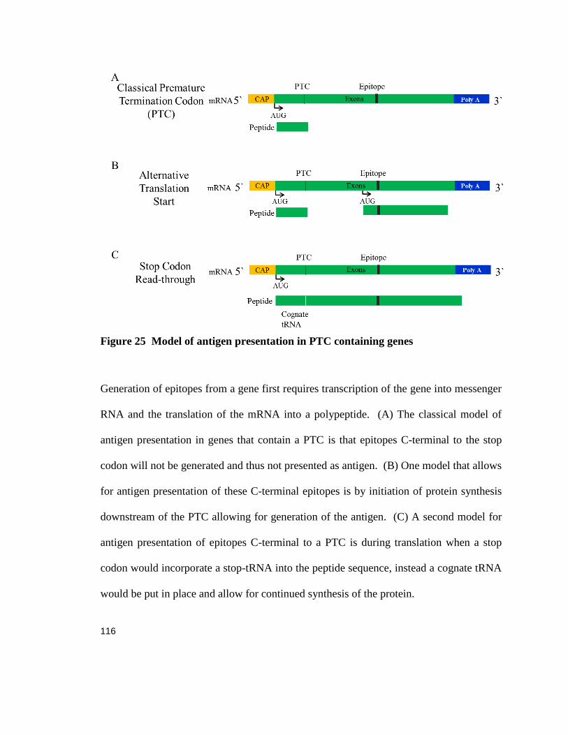
116
Figure 25 Model of antigen presentation in PTC containing genes
Generation of epitopes from a gene first requires transcription of the gene into messenger
RNA and the translation of the mRNA into a polypeptide. (A) The classical model of
antigen presentation in genes that contain a PTC is that epitopes C-terminal to the stop
codon will not be generated and thus not presented as antigen. (B) One model that allows
for antigen presentation of these C-terminal epitopes is by initiation of protein synthesis
downstream of the PTC allowing for generation of the antigen. (C) A second model for
antigen presentation of epitopes C-terminal to a PTC is during translation when a stop
codon would incorporate a stop-tRNA into the peptide sequence, instead a cognate tRNA
would be put in place and allow for continued synthesis of the protein.

117
BIBLIOGRAPHY
Apcher, S., C. Daskalogianni, F. Lejeune, B. Manoury, G. Imhoos, L. Heslop and R. Fahraeus (2011). "Major source of antigenic peptides for the MHC class I pathway is produced during the pioneer round of mRNA translation." Proc Natl Acad Sci U S A 108(28): 11572-11577. Atchison, R. W., B. C. Casto and W. M. Hammon (1966). "Electron microscopy of adenovirus-associated virus (AAV) in cell cultures." Virology 29(2): 353-357. Bazykin, G. A. and A. V. Kochetov (2011). "Alternative translation start sites are conserved in eukaryotic genomes." Nucleic Acids Res 39(2): 567-577. Bell, P., A. D. Moscioni, R. J. McCarter, D. Wu, G. Gao, A. Hoang, J. C. Sanmiguel, X. Sun, N. A. Wivel, S. E. Raper, E. E. Furth, M. L. Batshaw and J. M. Wilson (2006). "Analysis of Tumors Arising in Male B6C3F1 Mice with and without AAV Vector Delivery to Liver." Mol Ther 14(1): 34-44. Bell, P., L. Wang, G. Gao, M. E. Haskins, A. F. Tarantal, R. J. McCarter, Y. Zhu, H. Yu and J. M. Wilson (2011). "Inverse zonation of hepatocyte transduction with AAV vectors between mice and non-human primates." Mol Genet Metab 104(3): 395-403. Bertolotti, A., Y. Zhang, L. M. Hendershot, H. P. Harding and D. Ron (2000). "Dynamic interaction of BiP and ER stress transducers in the unfolded-protein response." Nat Cell Biol 2(6): 326-332. Bessis, N., F. J. GarciaCozar and M. C. Boissier (0000). "Immune responses to gene therapy vectors: influence on vector function and effector mechanisms." Gene Ther 11(S1): S10-S17. Bohenzky, R. A., R. B. Lefebvre and K. I. Berns (1988). "Sequence and symmetry requirements within the internal palindromic sequences of the adeno-associated virus terminal repeat." Virology 166(2): 316-327. Bradbury, A. M., J. N. Cochran, V. J. McCurdy, A. K. Johnson, B. L. Brunson, H. Gray-Edwards, S. G. Leroy, M. Hwang, A. N. Randle, L. S. Jackson, N. E. Morrison, R. C. Baek, T. N. Seyfried, S. H. Cheng, N. R. Cox, H. J. Baker, M. B. Cachon-Gonzalez, T. M. Cox, M. Sena-Esteves and D. R. Martin (2013). "Therapeutic response in feline sandhoff disease despite immunity to intracranial gene therapy." Mol Ther 21(7): 1306-1315. Breous, E., S. Somanathan, P. Bell and J. M. Wilson (2011). "Inflammation promotes the loss of adeno-associated virus-mediated transgene expression in mouse liver." Gastroenterology 141(1): 348-357, 357.e341-343. Calcedo, R., L. H. Vandenberghe, G. Gao, J. Lin and J. M. Wilson (2009). "Worldwide epidemiology of neutralizing antibodies to adeno-associated viruses." J Infect Dis 199(3): 381-390. Calvas, P., B. Segues, J. M. Rozet, D. Rabier, J. P. Bonnefond and A. Munnich (1998). "Novel intragenic deletions and point mutations of the ornithine transcarbamylase gene in congenital hyperammonemia." Hum Mutat Suppl 1: S81-84. Cao, O., E. Dobrzynski, L. Wang, S. Nayak, B. Mingle, C. Terhorst and R. W. Herzog (2007). "Induction and role of regulatory CD4+CD25+ T cells in tolerance to the transgene product following hepatic in vivo gene transfer." Blood 110(4): 1132-1140. Cao, O., B. E. Hoffman, B. Moghimi, S. Nayak, M. Cooper, S. Zhou, H. C. Ertl, K. A. High and R. W. Herzog (2009). "Impact of the underlying mutation and the route of vector administration on immune responses to factor IX in gene therapy for hemophilia B." Mol Ther 17(10): 1733-1742. Carrell, R. W. and D. A. Lomas (2002). "Alpha1-antitrypsin deficiency--a model for conformational diseases." N Engl J Med 346(1): 45-53.
118
Carstens, R. P., W. A. Fenton and L. R. Rosenberg (1991). "Identification of RNA splicing errors resulting in human ornithine transcarbamylase deficiency." Am J Hum Genet 48(6): 1105-1114. Carter, P. J. and R. J. Samulski (2000). "Adeno-associated viral vectors as gene delivery vehicles." Int J Mol Med 6(1): 17-27. Chang, Y. F., J. S. Imam and M. F. Wilkinson (2007). "The nonsense-mediated decay RNA surveillance pathway." Annu Rev Biochem 76: 51-74. Chen, D., B. Murphy, R. Sung and J. S. Bromberg (0000). "Adaptive and innate immune responses to gene transfer vectors: role of cytokines and chemokines in vector function." Gene Ther 10(11): 991-998. Christ, M., B. Louis, F. Stoeckel, A. Dieterle, L. Grave, D. Dreyer, J. Kintz, D. Ali Hadji, M. Lusky and M. Mehtali (2000). "Modulation of the inflammatory properties and hepatotoxicity of recombinant adenovirus vectors by the viral E4 gene products." Hum Gene Ther 11(3): 415-427. Ciesielska, A., P. Hadaczek, G. Mittermeyer, S. Zhou, J. F. Wright, K. S. Bankiewicz and J. Forsayeth (2013). "Cerebral infusion of AAV9 vector-encoding non-self proteins can elicit cell-mediated immune responses." Mol Ther 21(1): 158-166. Colbert, J. D., D. J. Farfan-Arribas and K. L. Rock (2013). "Substrate-induced protein stabilization reveals a predominant contribution from mature proteins to peptides presented on MHC class I." J Immunol 191(11): 5410-5419. Croft, N. P., S. A. Smith, Y. C. Wong, C. T. Tan, N. L. Dudek, I. E. Flesch, L. C. Lin, D. C. Tscharke and A. W. Purcell (2013). "Kinetics of antigen expression and epitope presentation during virus infection." PLoS Pathog 9(1): e1003129. Crudele, J. M., J. D. Finn, J. I. Siner, N. B. Martin, G. P. Niemeyer, S. Zhou, F. Mingozzi, C. D. Lothrop, Jr. and V. R. Arruda (2015). "AAV liver expression of FIX-Padua prevents and eradicates FIX inhibitor without increasing thrombogenicity in hemophilia B dogs and mice." Blood 125(10): 1553-1561. de Verteuil, D., D. P. Granados, P. Thibault and C. Perreault (2012). "Origin and plasticity of MHC I-associated self peptides." Autoimmun Rev 11(9): 627-635. Dedieu, J. F., E. Vigne, C. Torrent, C. Jullien, I. Mahfouz, J. M. Caillaud, N. Aubailly, C. Orsini, J. M. Guillaume, P. Opolon, P. Delaere, M. Perricaudet and P. Yeh (1997). "Long-term gene delivery into the livers of immunocompetent mice with E1/E4-defective adenoviruses." J Virol 71(6): 4626-4637. Ding, E. Y., B. L. Hodges, H. Hu, A. J. McVie-Wylie, D. Serra, F. K. Migone, D. Pressley, Y. T. Chen and A. Amalfitano (2001). "Long-term efficacy after [E1-, polymerase-] adenovirus-mediated transfer of human acid-alpha-glucosidase gene into glycogen storage disease type II knockout mice." Hum Gene Ther 12(8): 955-965. Ding, W., L. Zhang, Z. Yan and J. F. Engelhardt (2005). "Intracellular trafficking of adeno-associated viral vectors." Gene Ther 12(11): 873-880. Dobrzynski, E., J. C. Fitzgerald, O. Cao, F. Mingozzi, L. Wang and R. W. Herzog (2006). "Prevention of cytotoxic T lymphocyte responses to factor IX-expressing hepatocytes by gene transfer-induced regulatory T cells." Proc Natl Acad Sci U S A 103(12): 4592-4597. Doerfler, P. A., A. G. Todd, N. Clement, D. J. Falk, S. Nayak, R. W. Herzog and B. J. Byrne (2016). "Copackaged AAV9 Vectors Promote Simultaneous Immune Tolerance and Phenotypic Correction of Pompe Disease." Hum Gene Ther 27(1): 43-59. Dolan, B. P., L. Li, K. Takeda, J. R. Bennink and J. W. Yewdell (2010). "Defective ribosomal products are the major source of antigenic peptides endogenously generated from influenza A virus neuraminidase." J Immunol 184(3): 1419-1424. Dolan, B. P., L. Li, C. A. Veltri, C. M. Ireland, J. R. Bennink and J. W. Yewdell (2011). "Distinct pathways generate peptides from defective ribosomal products for CD8+ T cell immunosurveillance." J Immunol 186(4): 2065-2072. Englund, P., E. Lindroos, I. Nennesmo, L. Klareskog and I. E. Lundberg (2001). "Skeletal muscle fibers express major histocompatibility complex class II antigens independently of inflammatory infiltrates in inflammatory myopathies." Am J Pathol 159(4): 1263-1273.

119
Faust, S. M., P. Bell, B. J. Cutler, S. N. Ashley, Y. Zhu, J. E. Rabinowitz and J. M. Wilson (2013). "CpG-depleted adeno-associated virus vectors evade immune detection." J Clin Invest 123(7): 2994-3001. Ferrero-Miliani, L., O. H. Nielsen, P. S. Andersen and S. E. Girardin (2007). "Chronic inflammation: importance of NOD2 and NALP3 in interleukin-1beta generation." Clin Exp Immunol 147(2): 227-235. Fields, P. A., V. R. Arruda, E. Armstrong, K. Chu, F. Mingozzi, J. N. Hagstrom, R. W. Herzog and K. A. High (2001). "Risk and prevention of anti-factor IX formation in AAV-mediated gene transfer in the context of a large deletion of F9." Mol Ther 4(3): 201-210. Fields, P. A., D. W. Kowalczyk, V. R. Arruda, E. Armstrong, M. L. McCleland, J. N. Hagstrom, K. J. Pasi, H. C. J. Ertl, R. W. Herzog and K. A. High (2000). "Role of Vector in Activation of T Cell Subsets in Immune Responses against the Secreted Transgene Product Factor IX." Mol Ther 1(3): 225-235. Fields, P. E., T. F. Gajewski and F. W. Fitch (1996). "Blocked Ras activation in anergic CD4+ T cells." Science 271(5253): 1276-1278. Finn, J. D., M. C. Ozelo, D. E. Sabatino, H. W. Franck, E. P. Merricks, J. M. Crudele, S. Zhou, H. H. Kazazian, D. Lillicrap, T. C. Nichols and V. R. Arruda (2010). "Eradication of neutralizing antibodies to factor VIII in canine hemophilia A after liver gene therapy." Blood 116(26): 5842-5848. Fitzgerald, K., M. Frank-Kamenetsky, S. Shulga-Morskaya, A. Liebow, B. R. Bettencourt, J. E. Sutherland, R. M. Hutabarat, V. A. Clausen, V. Karsten, J. Cehelsky, S. V. Nochur, V. Kotelianski, J. Horton, T. Mant, J. Chiesa, J. Ritter, M. Munisamy, A. K. Vaishnaw, J. A. Gollob and A. Simon (2014). "Effect of an RNA interference drug on the synthesis of proprotein convertase subtilisin/kexin type 9 (PCSK9) and the concentration of serum LDL cholesterol in healthy volunteers: a randomised, single-blind, placebo-controlled, phase 1 trial." Lancet 383(9911): 60-68. Flotte, T. R. and B. J. Carter (1995). "Adeno-associated virus vectors for gene therapy." Gene Ther 2(6): 357-362. Flotte, T. R., B. C. Trapnell, M. Humphries, B. Carey, R. Calcedo, F. Rouhani, M. Campbell-Thompson, A. T. Yachnis, R. A. Sandhaus, N. G. McElvaney, C. Mueller, L. M. Messina, J. M. Wilson, M. Brantly, D. R. Knop, G. J. Ye and J. D. Chulay (2011). "Phase 2 clinical trial of a recombinant adeno-associated viral vector expressing alpha1-antitrypsin: interim results." Hum Gene Ther 22(10): 1239-1247. Gao, G., Y. Lu, R. Calcedo, R. L. Grant, P. Bell, L. Wang, J. Figueredo, M. Lock and J. M. Wilson (2006). "Biology of AAV serotype vectors in liver-directed gene transfer to nonhuman primates." Mol Ther 13(1): 77-87. Gao, G., L. H. Vandenberghe and J. M. Wilson (2005). "New recombinant serotypes of AAV vectors." Curr Gene Ther 5(3): 285-297. Gao, G. P., Y. Yang and J. M. Wilson (1996). "Biology of adenovirus vectors with E1 and E4 deletions for liver-directed gene therapy." J Virol 70(12): 8934-8943. Goodenough, E., T. M. Robinson, M. B. Zook, K. M. Flanigan, J. F. Atkins, M. T. Howard and L. C. Eisenlohr (2014). "Cryptic MHC class I-binding peptides are revealed by aminoglycoside-induced stop codon read-through into the 3' UTR." Proc Natl Acad Sci U S A 111(15): 5670-5675. Greig, J. A., H. Peng, J. Ohlstein, C. A. Medina-Jaszek, O. Ahonkhai, A. Mentzinger, R. L. Grant, S. Roy, S. J. Chen, P. Bell, A. P. Tretiakova and J. M. Wilson (2014). "Intramuscular injection of AAV8 in mice and macaques is associated with substantial hepatic targeting and transgene expression." PLoS One 9(11): e112268. Grompe, M., D. M. Muzny and C. T. Caskey (1989). "Scanning detection of mutations in human ornithine transcarbamoylase by chemical mismatch cleavage." Proc Natl Acad Sci U S A 86(15): 5888-5892.

120
Haas, T., J. Metzger, F. Schmitz, A. Heit, T. Muller, E. Latz and H. Wagner (2008). "The DNA sugar backbone 2' deoxyribose determines toll-like receptor 9 activation." Immunity 28(3): 315-323. Hacein-Bey-Abina, S., C. Von Kalle, M. Schmidt, M. P. McCormack, N. Wulffraat, P. Leboulch, A. Lim, C. S. Osborne, R. Pawliuk, E. Morillon, R. Sorensen, A. Forster, P. Fraser, J. I. Cohen, G. de Saint Basile, I. Alexander, U. Wintergerst, T. Frebourg, A. Aurias, D. Stoppa-Lyonnet, S. Romana, I. Radford-Weiss, F. Gross, F. Valensi, E. Delabesse, E. Macintyre, F. Sigaux, J. Soulier, L. E. Leiva, M. Wissler, C. Prinz, T. H. Rabbitts, F. Le Deist, A. Fischer and M. Cavazzana-Calvo (2003). "LMO2-associated clonal T cell proliferation in two patients after gene therapy for SCID-X1." Science 302(5644): 415-419. Hadeiba, H., K. Lahl, A. Edalati, C. Oderup, A. Habtezion, R. Pachynski, L. Nguyen, A. Ghodsi, S. Adler and E. C. Butcher (2012). "Plasmacytoid dendritic cells transport peripheral antigens to the thymus to promote central tolerance." Immunity 36(3): 438-450. Hemmi, H., O. Takeuchi, T. Kawai, T. Kaisho, S. Sato, H. Sanjo, M. Matsumoto, K. Hoshino, H. Wagner, K. Takeda and S. Akira (2000). "A Toll-like receptor recognizes bacterial DNA." Nature 408(6813): 740-745. Herzog, R. W., P. A. Fields, V. R. Arruda, J. O. Brubaker, E. Armstrong, D. McClintock, D. A. Bellinger, L. B. Couto, T. C. Nichols and K. A. High (2002). "Influence of vector dose on factor IX-specific T and B cell responses in muscle-directed gene therapy." Hum Gene Ther 13(11): 1281-1291. Herzog, R. W., J. D. Mount, V. R. Arruda, K. A. High and C. D. Lothrop, Jr. (2001). "Muscle-directed gene transfer and transient immune suppression result in sustained partial correction of canine hemophilia B caused by a null mutation." Mol Ther 4(3): 192-200. Hewitt, E. W. (2003). "The MHC class I antigen presentation pathway: strategies for viral immune evasion." Immunology 110(2): 163-169. Hinderer, C., P. Bell, B. L. Gurda, Q. Wang, J. P. Louboutin, Y. Zhu, J. Bagel, P. O'Donnell, T. Sikora, T. Ruane, P. Wang, M. E. Haskins and J. M. Wilson (2014). "Intrathecal gene therapy corrects CNS pathology in a feline model of mucopolysaccharidosis I." Mol Ther 22(12): 2018-2027. Hinderer, C., P. Bell, J. P. Louboutin, Y. Zhu, H. Yu, G. Lin, R. Choa, B. L. Gurda, J. Bagel, P. O'Donnell, T. Sikora, T. Ruane, P. Wang, A. F. Tarantal, M. L. Casal, M. E. Haskins and J. M. Wilson (2015). "Neonatal Systemic AAV Induces Tolerance to CNS Gene Therapy in MPS I Dogs and Nonhuman Primates." Mol Ther 23(8): 1298-1307. Hinderer, C., P. Bell, C. H. Vite, J. P. Louboutin, R. Grant, E. Bote, H. Yu, B. Pukenas, R. Hurst and J. M. Wilson (2014). "Widespread gene transfer in the central nervous system of cynomolgus macaques following delivery of AAV9 into the cisterna magna." Mol Ther Methods Clin Dev 1: 14051. Hogquist, K. A., S. C. Jameson, W. R. Heath, J. L. Howard, M. J. Bevan and F. R. Carbone (1994). "T cell receptor antagonist peptides induce positive selection." Cell 76(1): 17-27. Horibe, T. and N. J. Hoogenraad (2007). "The chop gene contains an element for the positive regulation of the mitochondrial unfolded protein response." PLoS One 2(9): e835. Hua, Z. and B. Hou (2013). "TLR signaling in B-cell development and activation." Cell Mol Immunol 10(2): 103-106. Huang, M. M., A. Wong, X. Yu, E. Kakkis and D. B. Kohn (1997). "Retrovirus-mediated transfer of the human alpha-L-iduronidase cDNA into human hematopoietic progenitor cells leads to correction in trans of Hurler fibroblasts." Gene Ther 4(11): 1150-1159. Hudak, M. L., M. D. Jones, Jr. and S. W. Brusilow (1985). "Differentiation of transient hyperammonemia of the newborn and urea cycle enzyme defects by clinical presentation." J Pediatr 107(5): 712-719. Im, D. S. and N. Muzyczka (1990). "The AAV origin binding protein Rep68 is an ATP-dependent site-specific endonuclease with DNA helicase activity." Cell 61(3): 447-457.

121
Isken, O., Y. K. Kim, N. Hosoda, G. L. Mayeur, J. W. Hershey and L. E. Maquat (2008). "Upf1 phosphorylation triggers translational repression during nonsense-mediated mRNA decay." Cell 133(2): 314-327. Jacobson, S. G., A. V. Cideciyan, A. J. Roman, A. Sumaroka, S. B. Schwartz, E. Heon and W. W. Hauswirth (2015). "Improvement and Decline in Vision with Gene Therapy in Childhood Blindness." New England Journal of Medicine 372(20): 1920-1926. Jenkins, M. K. and J. J. Moon (2012). "The role of naive T cell precursor frequency and recruitment in dictating immune response magnitude." J Immunol 188(9): 4135-4140. Jiang, W., M. M. Lederman, C. V. Harding, B. Rodriguez, R. J. Mohner and S. F. Sieg (2007). "TLR9 stimulation drives naive B cells to proliferate and to attain enhanced antigen presenting function." Eur J Immunol 37(8): 2205-2213. Jounai, N., K. Kobiyama, F. Takeshita and K. J. Ishii (2012). "Recognition of damage-associated molecular patterns related to nucleic acids during inflammation and vaccination." Front Cell Infect Microbiol 2: 168. Kappler, J. W., N. Roehm and P. Marrack (1987). "T cell tolerance by clonal elimination in the thymus." Cell 49(2): 273-280. Keller, C. W., C. Fokken, S. G. Turville, A. Lunemann, J. Schmidt, C. Munz and J. D. Lunemann (2011). "TNF-alpha induces macroautophagy and regulates MHC class II expression in human skeletal muscle cells." J Biol Chem 286(5): 3970-3980. Kim, Y. M., K. Son, S. J. Hong, A. Green, J. J. Chen, E. Tzeng, C. Hierholzer and T. R. Billiar (1998). "Inhibition of protein synthesis by nitric oxide correlates with cytostatic activity: nitric oxide induces phosphorylation of initiation factor eIF-2 alpha." Mol Med 4(3): 179-190. Kochetov, A. V. (2008). "Alternative translation start sites and hidden coding potential of eukaryotic mRNAs." Bioessays 30(7): 683-691. Kochetov, A. V., P. D. Prayaga, O. A. Volkova and R. Sankararamakrishnan (2013). "Hidden coding potential of eukaryotic genomes: nonAUG started ORFs." J Biomol Struct Dyn 31(1): 103-114. Lai, E., T. Teodoro and A. Volchuk (2007). "Endoplasmic reticulum stress: signaling the unfolded protein response." Physiology (Bethesda) 22: 193-201. Lang, K. S., P. Georgiev, M. Recher, A. A. Navarini, A. Bergthaler, M. Heikenwalder, N. L. Harris, T. Junt, B. Odermatt, P. A. Clavien, H. Pircher, S. Akira, H. Hengartner and R. M. Zinkernagel (2006). "Immunoprivileged status of the liver is controlled by Toll-like receptor 3 signaling." J Clin Invest 116(9): 2456-2463. Lee, J. W., M. Epardaud, J. Sun, J. E. Becker, A. C. Cheng, A. R. Yonekura, J. K. Heath and S. J. Turley (2007). "Peripheral antigen display by lymph node stroma promotes T cell tolerance to intestinal self." Nat Immunol 8(2): 181-190. Lim, H. W., P. Hillsamer, A. H. Banham and C. H. Kim (2005). "Cutting edge: direct suppression of B cells by CD4+ CD25+ regulatory T cells." J Immunol 175(7): 4180-4183. Limberis, M. P., V. S. Adam, G. Wong, J. Gren, D. Kobasa, T. M. Ross, G. P. Kobinger, A. Tretiakova and J. M. Wilson (2013). "Intranasal antibody gene transfer in mice and ferrets elicits broad protection against pandemic influenza." Sci Transl Med 5(187): 187ra172. Limberis, M. P., J. Figueredo, R. Calcedo and J. M. Wilson (2007). "Activation of CFTR-specific T Cells in cystic fibrosis mice following gene transfer." Mol Ther 15(9): 1694-1700. Limberis, M. P. and J. M. Wilson (2006). "Adeno-associated virus serotype 9 vectors transduce murine alveolar and nasal epithelia and can be readministered." Proc Natl Acad Sci U S A 103(35): 12993-12998. Lin, S. W., S. E. Hensley, N. Tatsis, M. O. Lasaro and H. C. Ertl (2007). "Recombinant adeno-associated virus vectors induce functionally impaired transgene product-specific CD8+ T cells in mice." J Clin Invest 117(12): 3958-3970. Lock, M., M. Alvira, L. H. Vandenberghe, A. Samanta, J. Toelen, Z. Debyser and J. M. Wilson (2010). "Rapid, simple, and versatile manufacturing of recombinant adeno-associated viral vectors at scale." Hum Gene Ther 21(10): 1259-1271.

122
Lykke-Andersen, S. and T. H. Jensen (2015). "Nonsense-mediated mRNA decay: an intricate machinery that shapes transcriptomes." Nat Rev Mol Cell Biol 16(11): 665-677. Malhotra, J. D., H. Miao, K. Zhang, A. Wolfson, S. Pennathur, S. W. Pipe and R. J. Kaufman (2008). "Antioxidants reduce endoplasmic reticulum stress and improve protein secretion." Proc Natl Acad Sci U S A 105(47): 18525-18530. Manno, C. S., G. F. Pierce, V. R. Arruda, B. Glader, M. Ragni, J. J. Rasko, M. C. Ozelo, K. Hoots, P. Blatt, B. Konkle, M. Dake, R. Kaye, M. Razavi, A. Zajko, J. Zehnder, P. K. Rustagi, H. Nakai, A. Chew, D. Leonard, J. F. Wright, R. R. Lessard, J. M. Sommer, M. Tigges, D. Sabatino, A. Luk, H. Jiang, F. Mingozzi, L. Couto, H. C. Ertl, K. A. High and M. A. Kay (2006). "Successful transduction of liver in hemophilia by AAV-Factor IX and limitations imposed by the host immune response." Nat Med 12(3): 342-347. Martino, A. T., M. Suzuki, D. M. Markusic, I. Zolotukhin, R. C. Ryals, B. Moghimi, H. C. Ertl, D. A. Muruve, B. Lee and R. W. Herzog (2011). "The genome of self-complementary adeno-associated viral vectors increases Toll-like receptor 9-dependent innate immune responses in the liver." Blood 117(24): 6459-6468. Matsuda, I. and S. Tanase (1997). "The ornithine transcarbamylase (OTC) gene: mutations in 50 Japanese families with OTC deficiency." Am J Med Genet 71(4): 378-383. Mauro, V. P. and S. A. Chappell (2014). "A critical analysis of codon optimization in human therapeutics." Trends Mol Med 20(11): 604-613. Mays, L. E., L. H. Vandenberghe, R. Xiao, P. Bell, H. J. Nam, M. Agbandje-McKenna and J. M. Wilson (2009). "Adeno-associated virus capsid structure drives CD4-dependent CD8+ T cell response to vector encoded proteins." J Immunol 182(10): 6051-6060. Meffre, E. (2011). "The establishment of early B cell tolerance in humans: lessons from primary immunodeficiency diseases." Ann N Y Acad Sci 1246: 1-10. Mendell, J. R., K. Campbell, L. Rodino-Klapac, Z. Sahenk, C. Shilling, S. Lewis, D. Bowles, S. Gray, C. Li, G. Galloway, V. Malik, B. Coley, K. R. Clark, J. Li, X. Xiao, J. Samulski, S. W. McPhee, R. J. Samulski and C. M. Walker "Dystrophin immunity in Duchenne's muscular dystrophy." N Engl J Med 363(15): 1429-1437. Mendell, J. R., K. Campbell, L. Rodino-Klapac, Z. Sahenk, C. Shilling, S. Lewis, D. Bowles, S. Gray, C. Li, G. Galloway, V. Malik, B. Coley, K. R. Clark, J. Li, X. Xiao, J. Samulski, S. W. McPhee, R. J. Samulski and C. M. Walker (2010). "Dystrophin immunity in Duchenne's muscular dystrophy." N Engl J Med 363(15): 1429-1437. Miyaoka, Y., K. Ebato, H. Kato, S. Arakawa, S. Shimizu and A. Miyajima (2012). "Hypertrophy and unconventional cell division of hepatocytes underlie liver regeneration." Curr Biol 22. Miyaoka, Y. and A. Miyajima (2013). "To divide or not to divide: revisiting liver regeneration." Cell Division 8(1): 1-12. Montoya, C. J., H. B. Jie, L. Al-Harthi, C. Mulder, P. J. Patino, M. T. Rugeles, A. M. Krieg, A. L. Landay and S. B. Wilson (2006). "Activation of plasmacytoid dendritic cells with TLR9 agonists initiates invariant NKT cell-mediated cross-talk with myeloid dendritic cells." J Immunol 177(2): 1028-1039. Moon, J. J., H. H. Chu, M. Pepper, S. J. McSorley, S. C. Jameson, R. M. Kedl and M. K. Jenkins (2007). "Naive CD4(+) T cell frequency varies for different epitopes and predicts repertoire diversity and response magnitude." Immunity 27(2): 203-213. Naeher, D., M. A. Daniels, B. Hausmann, P. Guillaume, I. Luescher and E. Palmer (2007). "A constant affinity threshold for T cell tolerance." J Exp Med 204(11): 2553-2559. Nakagawa, T. and J. Yuan (2000). "Cross-talk between two cysteine protease families. Activation of caspase-12 by calpain in apoptosis." J Cell Biol 150(4): 887-894. Naldini, L. (2015). "Gene therapy returns to centre stage." Nature 526(7573): 351-360. Nathwani, A. C., U. M. Reiss, E. G. Tuddenham, C. Rosales, P. Chowdary, J. McIntosh, M. Della Peruta, E. Lheriteau, N. Patel, D. Raj, A. Riddell, J. Pie, S. Rangarajan, D. Bevan, M. Recht, Y. M. Shen, K. G. Halka, E. Basner-Tschakarjan, F. Mingozzi, K. A. High, J. Allay, M. A. Kay, C. Y. Ng, J. Zhou, M. Cancio, C. L. Morton, J. T. Gray, D. Srivastava, A. W. Nienhuis and A. M.
123
Davidoff (2014). "Long-term safety and efficacy of factor IX gene therapy in hemophilia B." N Engl J Med 371(21): 1994-2004. Nayak, S., P. A. Doerfler, S. L. Porvasnik, D. D. Cloutier, R. Khanna, K. J. Valenzano, R. W. Herzog and B. J. Byrne (2014). "Immune responses and hypercoagulation in ERT for Pompe disease are mutation and rhGAA dose dependent." PLoS One 9(6): e98336. Nguyen, T. H. and N. Ferry (2004). "Liver gene therapy: advances and hurdles." Gene Ther 11 Suppl 1: S76-84. Oppliger Leibundgut, E., B. Wermuth, J. P. Colombo and S. Liechti-Gallati (1996). "Identification of four novel splice site mutations in the ornithine transcarbamylase gene." Hum Genet 97(2): 209-213. Oppliger Leibundgut, E. O., B. Wermuth, J. P. Colombo and S. Liechti-Gallati (1996). "Ornithine transcarbamylase deficiency: characterization of gene mutations and polymorphisms." Hum Mutat 8(4): 333-339. Pelanda, R. and R. M. Torres (2012). "Central B-cell tolerance: where selection begins." Cold Spring Harb Perspect Biol 4(4): a007146. Pobezinsky, L. A., G. S. Angelov, X. Tai, S. Jeurling, F. Van Laethem, L. Feigenbaum, J. H. Park and A. Singer (2012). "Clonal deletion and the fate of autoreactive thymocytes that survive negative selection." Nat Immunol 13(6): 569-578. Raper, S. E., N. Chirmule, F. S. Lee, N. A. Wivel, A. Bagg, G. P. Gao, J. M. Wilson and M. L. Batshaw (2003). "Fatal systemic inflammatory response syndrome in a ornithine transcarbamylase deficient patient following adenoviral gene transfer." Mol Genet Metab 80(1-2): 148-158. Rappsilber, J., M. Mann and Y. Ishihama (2007). "Protocol for micro-purification, enrichment, pre-fractionation and storage of peptides for proteomics using StageTips." Nat. Protocols 2(8): 1896-1906. Reyes-Sandoval, A. and H. C. Ertl (2004). "CpG methylation of a plasmid vector results in extended transgene product expression by circumventing induction of immune responses." Mol Ther 9(2): 249-261. Riedhammer, C. and R. Weissert (2015). "Antigen Presentation, Autoantigens, and Immune Regulation in Multiple Sclerosis and Other Autoimmune Diseases." Front Immunol 6: 322. Rogers, G. L., A. T. Martino, G. V. Aslanidi, G. R. Jayandharan, A. Srivastava and R. W. Herzog (2011). "Innate Immune Responses to AAV Vectors." Front Microbiol 2: 194. Rogers, G. L., A. T. Martino, I. Zolotukhin, H. C. Ertl and R. W. Herzog (2014). "Role of the vector genome and underlying factor IX mutation in immune responses to AAV gene therapy for hemophilia B." J Transl Med 12: 25. Rogers, G. L., M. Suzuki, I. Zolotukhin, D. M. Markusic, L. M. Morel, B. Lee, H. C. Ertl and R. W. Herzog (2015). "Unique Roles of TLR9- and MyD88-Dependent and -Independent Pathways in Adaptive Immune Responses to AAV-Mediated Gene Transfer." J Innate Immun 7(3): 302-314. Ron, D. and P. Walter (2007). "Signal integration in the endoplasmic reticulum unfolded protein response." Nat Rev Mol Cell Biol 8(7): 519-529. Sallusto, F. and A. Lanzavecchia (2002). "The instructive role of dendritic cells on T-cell responses." Arthritis Research & Therapy 4(3): S127. Satapati, S., B. Kucejova, J. A. Duarte, J. A. Fletcher, L. Reynolds, N. E. Sunny, T. He, L. A. Nair, K. Livingston, X. Fu, M. E. Merritt, A. D. Sherry, C. R. Malloy, J. M. Shelton, J. Lambert, E. J. Parks, I. Corbin, M. A. Magnuson, J. D. Browning and S. C. Burgess (2015). "Mitochondrial metabolism mediates oxidative stress and inflammation in fatty liver." J Clin Invest 125(12): 4447-4462. Schwartz, R. H. (2003). "T cell anergy." Annu Rev Immunol 21: 305-334. Shapir, N., R. Miari, S. Blum, D. Schwartz, G. Chernin, G. Neil, D. Afik and A. Panet (2015). "Preclinical and preliminary clinical evaluation of genetically transduced dermal tissue implants for the sustained secretion of erythropoietin and interferon alpha." Hum Gene Ther Clin Dev.

124
Simonelli, F., A. M. Maguire, F. Testa, E. A. Pierce, F. Mingozzi, J. L. Bennicelli, S. Rossi, K. Marshall, S. Banfi, E. M. Surace, J. Sun, T. M. Redmond, X. Zhu, K. S. Shindler, G. S. Ying, C. Ziviello, C. Acerra, J. F. Wright, J. W. McDonnell, K. A. High, J. Bennett and A. Auricchio (2010). "Gene therapy for Leber's congenital amaurosis is safe and effective through 1.5 years after vector administration." Mol Ther 18(3): 643-650. Somanathan, S., E. Breous, P. Bell and J. M. Wilson (2010). "AAV vectors avoid inflammatory signals necessary to render transduced hepatocyte targets for destructive T cells." Mol Ther 18(5): 977-982. Starck, S. R., V. Jiang, M. Pavon-Eternod, S. Prasad, B. McCarthy, T. Pan and N. Shastri (2012). "Leucine-tRNA initiates at CUG start codons for protein synthesis and presentation by MHC class I." Science 336(6089): 1719-1723. Starck, S. R. and N. Shastri (2011). "Non-conventional sources of peptides presented by MHC class I." Cell Mol Life Sci 68(9): 1471-1479. Starck, S. R. and N. Shastri (2016). "Nowhere to hide: unconventional translation yields cryptic peptides for immune surveillance." Immunol Rev 272(1): 8-16. Starzl, T. E., D. W. Bilheimer, H. T. Bahnson, B. W. Shaw, Jr., R. L. Hardesty, B. P. Griffith, S. Iwatsuki, B. J. Zitelli, J. C. Gartner, Jr., J. J. Malatack and et al. (1984). "Heart-liver transplantation in a patient with familial hypercholesterolaemia." Lancet 1(8391): 1382-1383. Streilein, J. W., M. Takeuchi and A. W. Taylor (1997). "Immune privilege, T-cell tolerance, and tissue-restricted autoimmunity." Hum Immunol 52(2): 138-143. Thompson, A. G. and R. Thomas (2002). "Induction of immune tolerance by dendritic cells: implications for preventative and therapeutic immunotherapy of autoimmune disease." Immunol Cell Biol 80(6): 509-519. Tsujimura, H., T. Tamura, H. J. Kong, A. Nishiyama, K. J. Ishii, D. M. Klinman and K. Ozato (2004). "Toll-like receptor 9 signaling activates NF-kappaB through IFN regulatory factor-8/IFN consensus sequence binding protein in dendritic cells." J Immunol 172(11): 6820-6827. Vandendriessche, T., L. Thorrez, A. Acosta-Sanchez, I. Petrus, L. Wang, L. Ma, D. E. W. L, Y. Iwasaki, V. Gillijns, J. M. Wilson, D. Collen and M. K. Chuah (2007). "Efficacy and safety of adeno-associated viral vectors based on serotype 8 and 9 vs. lentiviral vectors for hemophilia B gene therapy." J Thromb Haemost 5(1): 16-24. Vilborg, A. and J. A. Steitz (2016). "Readthrough transcription: How are DoGs made and what do they do?" RNA Biol: 1-5. von Boehmer, H. (2005). "Mechanisms of suppression by suppressor T cells." Nat Immunol 6(4): 338-344. Wang, L., H. Morizono, J. Lin, P. Bell, D. Jones, D. McMenamin, H. Yu, M. L. Batshaw and J. M. Wilson "Preclinical evaluation of a clinical candidate AAV8 vector for ornithine transcarbamylase (OTC) deficiency reveals functional enzyme from each persisting vector genome." Mol Genet Metab 105(2): 203-211. Wang, L., H. Wang, H. Morizono, P. Bell, D. Jones, J. Lin, D. McMenamin, H. Yu, M. L. Batshaw and J. M. Wilson "Sustained correction of OTC deficiency in spf( ash) mice using optimized self-complementary AAV2/8 vectors." Gene Ther 19(4): 404-410. Wang, Q., G. Greenburg, D. Bunch, D. Farson and M. H. Finer (1997). "Persistent transgene expression in mouse liver following in vivo gene transfer with a delta E1/delta E4 adenovirus vector." Gene Ther 4(5): 393-400. Wisniewski, J. R., A. Zougman, N. Nagaraj and M. Mann (2009). "Universal sample preparation method for proteome analysis." Nat Meth 6(5): 359-362. Wu, S., M. Tan, Y. Hu, J. L. Wang, D. Scheuner and R. J. Kaufman (2004). "Ultraviolet light activates NFkappaB through translational inhibition of IkappaBalpha synthesis." J Biol Chem 279(33): 34898-34902. Xiao, P. J. and R. J. Samulski (2012). "Cytoplasmic trafficking, endosomal escape, and perinuclear accumulation of adeno-associated virus type 2 particles are facilitated by microtubule network." J Virol 86(19): 10462-10473.

125
Yamaguchi, S., L. L. Brailey, H. Morizono, A. E. Bale and M. Tuchman (2006). "Mutations and polymorphisms in the human ornithine transcarbamylase (OTC) gene." Hum Mutat 27(7): 626-632. Yang, Y., K. U. Jooss, Q. Su, H. C. Ertl and J. M. Wilson (1996). "Immune responses to viral antigens versus transgene product in the elimination of recombinant adenovirus-infected hepatocytes in vivo." Gene Ther 3(2): 137-144. Yang, Y., F. A. Nunes, K. Berencsi, E. E. Furth, E. Gonczol and J. M. Wilson (1994). "Cellular immunity to viral antigens limits E1-deleted adenoviruses for gene therapy." Proc Natl Acad Sci U S A 91(10): 4407-4411. Yang, Y., L. Wang, P. Bell, D. McMenamin, Z. He, J. White, H. Yu, C. Xu, H. Morizono, K. Musunuru, M. L. Batshaw and J. M. Wilson (2016). "A dual AAV system enables the Cas9-mediated correction of a metabolic liver disease in newborn mice." Nat Biotechnol 34(3): 334-338. Yewdell, J. W., L. C. Anton and J. R. Bennink (1996). "Defective ribosomal products (DRiPs): a major source of antigenic peptides for MHC class I molecules?" J Immunol 157(5): 1823-1826. Yewdell, J. W., E. Reits and J. Neefjes (2003). "Making sense of mass destruction: quantitating MHC class I antigen presentation." Nat Rev Immunol 3(12): 952-961. Yurasov, S., H. Wardemann, J. Hammersen, M. Tsuiji, E. Meffre, V. Pascual and M. C. Nussenzweig (2005). "Defective B cell tolerance checkpoints in systemic lupus erythematosus." J Exp Med 201(5): 703-711. Zaiss, A. K., Q. Liu, G. P. Bowen, N. C. Wong, J. S. Bartlett and D. A. Muruve (2002). "Differential activation of innate immune responses by adenovirus and adeno-associated virus vectors." J Virol 76(9): 4580-4590. Zhang, K., X. Shen, J. Wu, K. Sakaki, T. Saunders, D. T. Rutkowski, S. H. Back and R. J. Kaufman (2006). "Endoplasmic reticulum stress activates cleavage of CREBH to induce a systemic inflammatory response." Cell 124(3): 587-599. Zhang, K., S. Wang, J. Malhotra, J. R. Hassler, S. H. Back, G. Wang, L. Chang, W. Xu, H. Miao, R. Leonardi, Y. E. Chen, S. Jackowski and R. J. Kaufman (2011). "The unfolded protein response transducer IRE1alpha prevents ER stress-induced hepatic steatosis." Embo j 30(7): 1357-1375. Zhao, Q., J. Wang, I. V. Levichkin, S. Stasinopoulos, M. T. Ryan and N. J. Hoogenraad (2002). "A mitochondrial specific stress response in mammalian cells." Embo j 21(17): 4411-4419. Zhou, X. and N. Muzyczka (1998). "In vitro packaging of adeno-associated virus DNA." J Virol 72(4): 3241-3247. Zhu, J., X. Huang and Y. Yang (2009). "The TLR9-MyD88 pathway is critical for adaptive immune responses to adeno-associated virus gene therapy vectors in mice." J Clin Invest 119(8): 2388-2398. Zincarelli, C., S. Soltys, G. Rengo and J. E. Rabinowitz (2008). "Analysis of AAV serotypes 1-9 mediated gene expression and tropism in mice after systemic injection." Mol Ther 16(6): 1073-1080.