impact of retinal disease-associated rpe65 mutations … · impact of retinal disease-associated...
TRANSCRIPT
Impact of Retinal Disease-Associated RPE65 Mutations on Retinoid Isomerization†
Grzegorz Bereta,‡ Philip D. Kiser,‡ Marcin Golczak,‡ Wenyu Sun,‡ Elise Heon,§,| David A. Saperstein,⊥ andKrzysztof Palczewski*,‡
Department of Pharmacology, School of Medicine, Case Western ReserVe UniVersity, CleVeland, Ohio 44106-4965, Departmentof Ophthalmology, UniVersity of Washington, Seattle, Washington 98195, Department of Ophthalmology and Vision Sciences,
The Hospital for Sick Children, UniVersity of Toronto, Toronto, Ontario, Canada, and Program of Genetics and GenomicBiology, The Hospital for Sick Children, Toronto, Ontario, Canada M5G 1X8
ReceiVed May 14, 2008; ReVised Manuscript ReceiVed June 10, 2008
ABSTRACT: Pathogenic mutations in the RPE65 gene are associated with a spectrum of congenital blindingdiseases in humans. We evaluated changes in the promoter region, coding regions, and exon/intron junctionsof the RPE65 gene by direct sequencing of DNA from 36 patients affected with Leber’s congenitalamaurosis (LCA), 62 with autosomal recessive retinitis pigmentosa (arRP), and 21 with autosomal dominant/recessive cone-rod dystrophies (CORD). Fifteen different variants were found, of which 6 were novel.Interesting was Gly244Val, a novel mutation close to the catalytic center. To assess the role of this mutationin RPE65 inactivation, we performed detailed biochemical studies of the mutant along with a structuralanalysis of the 244 amino acid position with respect to amino acids known to be important for RPE65-dependent retinoid isomerization. Bicistronic plasmid expression of the RPE65 Gly244Val mutant andenhanced green fluorescent protein (EGFP) allowed us to document both its instability in cultured cellsby cell sorting and immunoblotting methodology and its loss of RPE65-dependent isomerase activity byenzymatic assays. Further insights into the structural requirements for retinoid isomerization by RPE65were obtained by using the carotenoid oxygenase (ACO) from Synechocystis (PDB accession code 2BIW)as a structural template to construct a RPE65 homology model and locating all known inactivating mutationsincluding Gly244Val within this model.
The visual (retinoid) cycle is the fundamental processresponsible for production of 11-cis-retinal, the chromophoreof rhodopsin and cone pigments (1). The importance ofretinoid metabolic transformations that occur in photorecep-tors and retinal pigmented epithelium (RPE)1 is underscoredby identification of retinal dystrophy-associated mutationsin all genetically encoded enzymes and retinoid-bindingproteins of that cycle (2). Among retinal dystrophies the mostsevere is Leber’s congenital amaurosis (LCA), a leadingcause of inherited childhood blindness. LCA is an autosomalrecessive, early onset severe retinal dystrophy that accountsfor 5% of all inherited retinal dystrophies (3). Of the visualcycle enzymes, mutations in RDH12 (retinol dehydrogenase12) (4, 5), RPE65 (retinal pigmented epithelium-specificprotein with molecular mass 65 kDa) (6, 7), and LRAT
(lecithin:retinol acyltransferase) (8, 9) are known to causethis type of blindness.
RPE65 is the long sought after retinoid isomerase thatproduces 11-cis-retinol from all-trans retinyl esters (10–12).This enzyme is a highly abundant protein in the RPE andbelongs to the carotenoid oxygenase family (13, 14).Although the mechanism of isomerization is not fullyunderstood, RPE65, like other carotenoid oxygenase familymembers, contains an iron cofactor essential for its activity(11). A crystal structure of one of the carotenoid oxygenasefamily members reveals the general architecture of this familyof enzymes featuring a Fe2+-(His)4 arrangement in the centerof a seven-bladed �-propeller fold where the Fe2+ ion isaccessible through a long nonpolar tunnel through whichcarotenoids or retinoids travel to reach the active site (15).
Mutations in RPE65 cause LCA (6) or retinitis pigmentosa(RP) (7). Since the original studies were done, a large numberof individuals with retinal disease have been investigatedfor mutations in this gene (2). Detailed analyses of visualfunction in patients of different ages with mutations in RPE65were evaluated (16, 17). In primates, the central RPE layershowed greater retinoid isomerase activity and protein levelsof RPE65 compared to the more peripheral RPE (18).
Significant progress has been made in understanding thepathological basis of diseases caused by inactivating muta-tions in the RPE65. First, the RPE of RPE65 knockout micewas found to contain large amounts of retinyl esters thataccumulated because they could not be processed further togenerate 11-cis-retinol (19). Naturally inactivating mutations
† This research was supported in part by grants from the NationalEye Institute (EY009339), National Institutes of Health (K.P.), Founda-tion Fighting Blindness (K.P.), Visual Science Training Program Grant2T32EY007157 from the National Eye Institute (P.D.K.), and MiraGodard Research Fund (E.H.).
* Address correspondence to this author. Phone: 216-368-4631. Fax:216-368-1300. E-mail: [email protected].
‡ Case Western Reserve University.§ University of Toronto.| The Hospital for Sick Children.⊥ University of Washington.1 Abbreviations: ACO, carotenoid oxygenase; ad, autosomal domi-
nant; ar, autosomal recessive; CORD, cone-rod dystrophy; EGFP,enhanced green fluorescent protein; LCA, Leber’s congenital amaurosis;LRAT, lecithin:retinol acyltransferase; RPE, retinal pigmented epithe-lium; RPE65, retinal pigmented epithelium protein with molecular mass65 kDa; RP, retinitis pigmentosa.
Biochemistry 2008, 47, 9856–98659856
10.1021/bi800905v CCC: $40.75 2008 American Chemical SocietyPublished on Web 08/23/2008
of RPE65 in mice and dogs also were identified that causedgenerally poor visual function (20, 21). Moreover, partialinactivation of RPE65 leads to a milder visual deteriorationas demonstrated in mice carrying an Arg91Trp mutation thatalso has been identified in humans (22). Availability of LCAand RP animals led to rapid development of gene transferand pharmacological interventions to restore vision as aprelude for treating these diseases in humans (23–26).Outcomes of the first two human gene transfer trials recentlyhave been reported (27, 28).
Biochemical analysis of the spectrum of mutations foundin a human population is important for understanding normalmechanisms of RPE65 action and disease pathology as wellas identifying patients who would most benefit from emerg-ing therapies. To assess the role of key residues in RPE65function, we used direct DNA sequencing to analyze genomicDNA from 119 patients afflicted with LCA, RP, and CORDfor mutations of the coding sequences, intron/exon junctionregions, and promoter region of the RPE65 gene. Fifteendifferent exonic variants were found, of which seven werenovel. Detailed biochemical analyses of a novel Gly244Valmutation in the RPE65 gene and structural modeling withthe carotenoid oxygenase (ACO) template provided sup-portive information about the role of certain critical residues,
such as the Fe2+-coordinating His residues, in the mechanismof retinoid isomerization.
MATERIALS AND METHODS
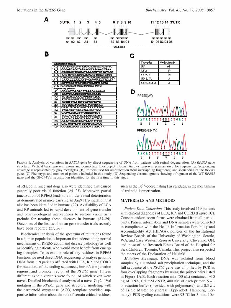
Patient Data Collection. This study involved 119 patientswith clinical diagnoses of LCA, RP, and CORD (Figure 1C).Consent and/or assent forms were obtained from all partici-pants. Patient information and DNA samples were collectedin compliance with the Health Information Portability andAccountability Act (HIPAA), policies of the InstitutionalReview Boards of the University of Washington, Seattle,WA, and Case Western Reserve University, Cleveland, OH,and those of the Research Ethics Board of the Hospital forSick Children, Toronto, Canada. This project also respectedthe tenets of the Declaration of Helsinki.
Mutation Screening. DNA was isolated from bloodsamples by a standard salt precipitation technique, and thefull sequence of the RPE65 gene was amplified by PCR asfour overlapping fragments by using the primer pairs listedin Figure 1A,B. Each reaction mix (50 µL) contained ∼10ng of DNA, 0.5 mM dNTP, 400 nM of each primer, 5 µLof reaction buffer (provided with polymerase), and 0.3 µLof Triple Master polymerase (Eppendorf, Hamburg, Ger-many). PCR cycling conditions were 93 °C for 3 min, 10×
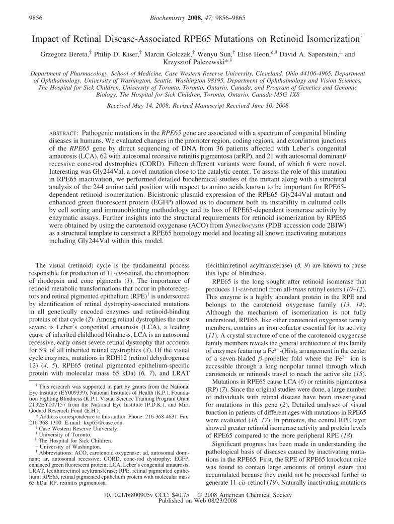
FIGURE 1: Analysis of variations in RPE65 gene by direct sequencing of DNA from patients with retinal degeneration. (A) RPE65 genestructure. Vertical bars represent exons and connecting lines depict introns. Arrows represent primers used for sequencing. Sequencingcoverage is represented by gray rectangles. (B) Primers used for amplification (four overlapping fragments) and sequencing of the RPE65gene. (C) Phenotype and number of patients included in this study. (D) Sequencing chromatograms showing a fragment of the WT RPE65gene and the Gly244Val substitution identified for the first time in this study.
Mutations in the RPE65 Gene Biochemistry, Vol. 47, No. 37, 2008 9857
(93 °C for 15 s, 62 °C for 30 s, and 68 °C for 8 min), and30× (93 °C for 15 s, 62 °C for 30 s, and 68 °C for 8 min +20 s/cycle). Following amplification, a 0.5 µL aliquot of eachsample was visualized on 1% agarose gel to confirm thepresence of a properly sized product.The remaining sample(49.5 µL) was incubated at 37 °C for 1 h with shrimp alka-line phosphatase (USB Corp., Cleveland, OH) and exonu-clease I (USB), 5 units each, to dephosphorylate nucleotidesand remove unused primers that interfere with sequencingreactions. Enzymes then were deactivated by a 15 minincubation at 80 °C. Sequencing samples were prepared byusing BigDye Terminator v3.0 chemistry (Applied Biosys-tems, Foster City, CA) and the primers listed in Figure 1B.Sequence data were acquired with an ABI 3730 DNAanalyzer and analyzed by Phred/Phrap/Consed/PolyPhredsoftware (University of Washington, Seattle, WA). Identifiednovel changes were validated by assessment of nucleotide/amino acid conservation, biochemical assays, and screeningof a control population of a minimum of 300 chromosomes.Segregation analysis was not possible in most cases.
Mutagenesis and Expression of RPE65. The RPE65 clonewas purchased from the American Type Culture Collection(Manassas, VA) and cloned into the multicloning site (MCS)of the pMXs-IG3 retroviral vector by using EcoRI and NotIrestriction sites appended to the coding sequence by PCR.The pMXs-IG3 vector was derived from a pMXs-IG vector
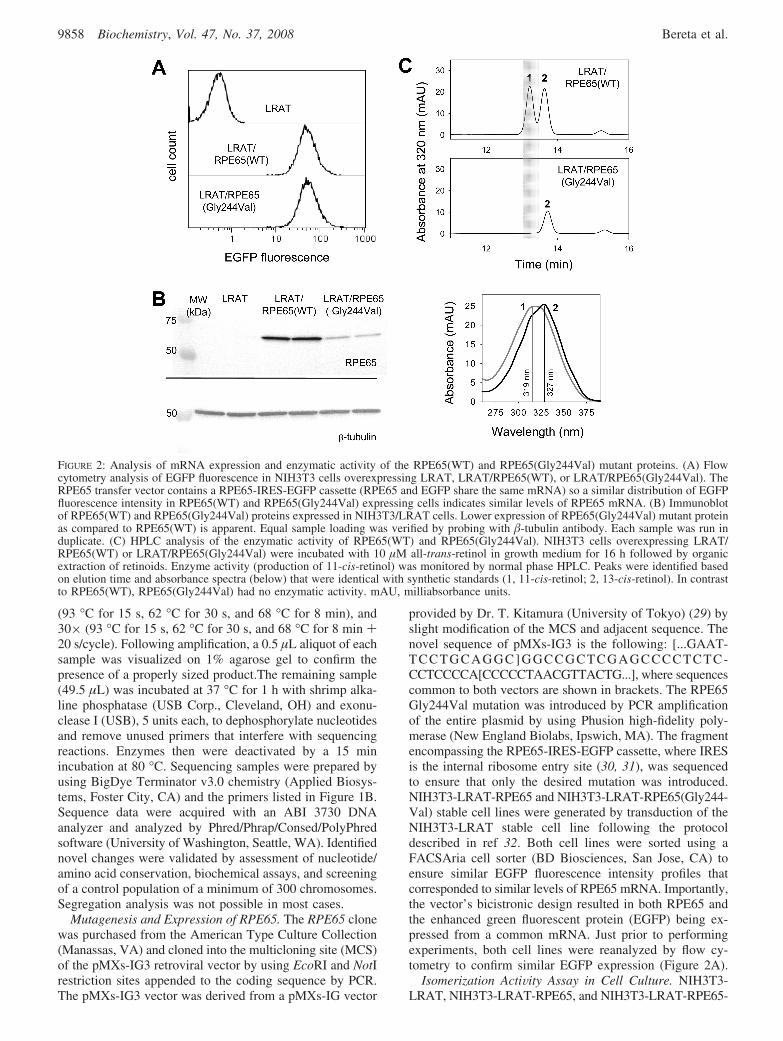
provided by Dr. T. Kitamura (University of Tokyo) (29) byslight modification of the MCS and adjacent sequence. Thenovel sequence of pMXs-IG3 is the following: [...GAAT-TCCTGCAGGC]GGCCGCTCGAGCCCCTCTC-CCTCCCCA[CCCCCTAACGTTACTG...], where sequencescommon to both vectors are shown in brackets. The RPE65Gly244Val mutation was introduced by PCR amplificationof the entire plasmid by using Phusion high-fidelity poly-merase (New England Biolabs, Ipswich, MA). The fragmentencompassing the RPE65-IRES-EGFP cassette, where IRESis the internal ribosome entry site (30, 31), was sequencedto ensure that only the desired mutation was introduced.NIH3T3-LRAT-RPE65 and NIH3T3-LRAT-RPE65(Gly244-Val) stable cell lines were generated by transduction of theNIH3T3-LRAT stable cell line following the protocoldescribed in ref 32. Both cell lines were sorted using aFACSAria cell sorter (BD Biosciences, San Jose, CA) toensure similar EGFP fluorescence intensity profiles thatcorresponded to similar levels of RPE65 mRNA. Importantly,the vector’s bicistronic design resulted in both RPE65 andthe enhanced green fluorescent protein (EGFP) being ex-pressed from a common mRNA. Just prior to performingexperiments, both cell lines were reanalyzed by flow cy-tometry to confirm similar EGFP expression (Figure 2A).
Isomerization ActiVity Assay in Cell Culture. NIH3T3-LRAT, NIH3T3-LRAT-RPE65, and NIH3T3-LRAT-RPE65-
FIGURE 2: Analysis of mRNA expression and enzymatic activity of the RPE65(WT) and RPE65(Gly244Val) mutant proteins. (A) Flowcytometry analysis of EGFP fluorescence in NIH3T3 cells overexpressing LRAT, LRAT/RPE65(WT), or LRAT/RPE65(Gly244Val). TheRPE65 transfer vector contains a RPE65-IRES-EGFP cassette (RPE65 and EGFP share the same mRNA) so a similar distribution of EGFPfluorescence intensity in RPE65(WT) and RPE65(Gly244Val) expressing cells indicates similar levels of RPE65 mRNA. (B) Immunoblotof RPE65(WT) and RPE65(Gly244Val) proteins expressed in NIH3T3/LRAT cells. Lower expression of RPE65(Gly244Val) mutant proteinas compared to RPE65(WT) is apparent. Equal sample loading was verified by probing with �-tubulin antibody. Each sample was run induplicate. (C) HPLC analysis of the enzymatic activity of RPE65(WT) and RPE65(Gly244Val). NIH3T3 cells overexpressing LRAT/RPE65(WT) or LRAT/RPE65(Gly244Val) were incubated with 10 µM all-trans-retinol in growth medium for 16 h followed by organicextraction of retinoids. Enzyme activity (production of 11-cis-retinol) was monitored by normal phase HPLC. Peaks were identified basedon elution time and absorbance spectra (below) that were identical with synthetic standards (1, 11-cis-retinol; 2, 13-cis-retinol). In contrastto RPE65(WT), RPE65(Gly244Val) had no enzymatic activity. mAU, milliabsorbance units.
9858 Biochemistry, Vol. 47, No. 37, 2008 Bereta et al.

(Gly244Val) cells were seeded in six-well culture plates at0.8 × 106 cells per well in growth medium (GM) consistingof Dulbecco’s modified Eagle’s medium, pH 7.2, with 4 mML-glutamine, 4500 mg/L glucose, and 110 mg/L sodiumpyruvate supplemented with 10% fetal bovine serum, 100units/mL penicillin, and 100 units/mL streptomycin. Theisomerization reaction was initiated 24 h later by exchangeof GM for one containing all-trans-retinol delivered in N,N-dimethylformamide to a 10 µM final concentration andcarried out for 16 h in a cell culture incubator (37 °C, 5%CO2). Subsequently, the cells and medium were collected,mixed with an equal volume of 4 M KOH in methanol, andincubated at 52 °C for 2.5 h to hydrolyze retinoid esters.Next, an equal volume of hexane was added, and retinoidswere extracted by vigorous shaking. Following 15 mincentrifugation at 4000 rpm to facilitate phase separation, theorganic phase was collected, dried down, and redissolved in250 µL of hexane. Extracted retinoids were separated on anormal phase HPLC column (Sil; 5 µm, 4.6 × 250 mm;Agilent Technologies, Santa Clara, CA) equilibrated with10% ethyl acetate in hexane at an isocratic flow rate of 1.4mL/min.
Immunoblotting of RPE65. NIH3T3/LRAT, NIH3T3/LRAT/RPE65, and NIH3T3/LRAT/RPE65(Gly244Val) cellswere plated on six-well plates at 8 × 10⁵ cells/well, grownfor 24 h, and washed with PBS (154 mM NaCl, 5.6 mMNa2HPO4, 1 mM KH2PO4, pH 7.1). Next, cells were collectedby scraping in PBS with 1 µM leupeptin, pelleted bycentrifugation, resuspended in 200 µL of PBS with 1 µMleupeptin, sonicated for 10 s to shear the DNA, mixed with100 µL of SDS loading buffer, incubated for 5 min at 95°C, and separated on 10% SDS-PAGE gels (15 µL of eachsample). Following transfer to Immobilon-P (Millipore,Bedford, MA) polyvinylidene fluoride (PVDF) membranes,RPE65, and �-tubulin (the control for equal sample loading)were detected by using anti-RPE65 monoclonal antibody(Novus Biologicals, Littleton, CO) diluted 1:2000 and E7anti-�-tubulin monoclonal antibody (Developmental StudiesHybridoma Bank, contributor Dr. M. Klymkowsky) diluted1:3000 and the ProtoBlot II AP System (Promega, Madison,WI) following the manufacturers’ protocols.
Generation of the RPE65 Homology Model. Initial RPE65homology models were generated with the Fugue (33), Phyre(34), and 3D-Jigsaw servers. All three algorithms identifiedthe carotenoid oxygenase (ACO) from Synechocystis (PDBaccession code 2BIW) as the most appropriate template forconstruction of the RPE65 homology model. In general, eachof the models was similar in those regions containing Hisresidues 180, 241, 313, and 527 which are highly conservedamong all RPE65 family members, including the apocaro-tenoid and �-carotenoid oxygenases (11). The model pro-duced by Fugue was selected as our starting model since itappeared to portray the seven-bladed �-propeller fold mostcompletely. Amino acid side chains were inserted usingCOOT (35) and manually adjusted to optimize both thestereochemistry and interresidue contact distances. The modelwas then energy-minimized by using CNS (36) with har-monic restraints placed on the residues surrounding the activesite Fe2+. Because residues 380-415 were predicted toconstitute a long unstructured loop in all three homologymodels, this region was omitted from our final model becauseit was likely to be portrayed incorrectly. All figures of the
RPE65 homology model were generated by using PyMOLversion 1.0 (DeLano Scientific LLC, San Francisco, CA).
RESULTS
Mutational Analysis. Exons, exon-intron junctions, andregulatory elements of the RPE65 gene were analyzed bydirect sequencing to identify the retinal dystrophy-associatedsequence variations within a group of 119 unrelated patientsof different ages and ethnic origins. A total of 49 sequencevariants were found, of which 3 were in the 5′-regulatoryregion, 15 within exons, 24 within introns, and 7 in the3′UTRs. The great majority of these alterations were singlenucleotide variations (SNVs), but we also detected twoduplications and one insertion (Supporting Information Table1). Among 15 exonic variants found, 9 were predicted tocause missense substitutions, 4 silent substitutions, 1 trunca-tion, and 1 splicing defect (Table 1). The novel exonicvariants found here include Gly244Val, His182Arg, Val189Ile,Trp288Cys, Ile291Val (all missense substitutions), Ile34Ile(a silent substitution), and 1059_1060insG (an insertion). TheHis182Arg and Gly244Val variants were detected in ho-mozygous form while the other novel variants were het-erozygous (Table 1, Figure 1D). We expected the His182Argvariant to be disease-associated since other substitutions atthis position have been previously reported to cause LCA(discussed below). The most intriguing substitution to us wasthe Gly244Val variant since it is located in a critical regionof the RPE65 structure (discussed below). This finding ledto a detailed biochemical analysis of this variant.
Impact of the Gly244Val Mutation on RPE65 ActiVity.Gly244 is a highly conserved residue within RPE65 se-quences of a number of vertebrate species (SupportingInformation Table 2). It is localized close to His241, a keyresidue involved in coordination of Fe2+ at the enzymaticactive site (11). Thus, mutation of Gly244 could have asignificant effect on the enzymatic activity or stability of thisprotein. To test this hypothesis, we generated two lines ofNIH3T3 cells stably expressing LRAT and wild-type RPE65(LRAT/RPE65WT) or LRAT and the RPE65 mutantGly244Val (LRAT/RPE65-Gly244Val). WT and mutatedRPE65 cDNAs were positioned in the retrovirus pMXs-IG3vector upstream of the internal ribosomal entry site (IRES)and EGFP sequence (29). Thus, the protein of interest andEGFP were expressed from a common mRNA. This ar-rangement allowed easy visualization of the transduced cellsand maximization of expression levels by sorting cells basedon GFP fluorescence intensity. Moreover, to ensure equiva-lent mRNA levels for WT and mutated RPE65, we collectedpopulations of cells characterized by identical fluorescencesignals for both cell lines (Figure 2A).
Immunoblotting with an anti-RPE65 monoclonal antibodyconfirmed robust expression of WT RPE65 upon infectionof NIH3T3/LRAT cells with the retrovirus. However, theGly244Val mutant protein level was found to be greatlyreduced as compared with WT protein (Figure 2B). Con-sidering that both LRAT/RPE65(WT) and LRAT/RPE65-(Gly244Val) cell lines had similar levels of transcript forRPE65, the observed differences in protein levels clearlysuggest low stability and accelerated degradation of theGly244Val variant.
Mutations in the RPE65 Gene Biochemistry, Vol. 47, No. 37, 2008 9859
To test whether mutation of Gly244 leads to alteration ofRPE65 isomerase activity, we incubated LRAT/RPE65-(Gly244Val) cells overnight with 10 µM all-trans-retinol.Analysis of retinoids extracted from the cells revealed theabsence of 11-cis-retinol, indicating a complete lack ofenzymatic activity (Figure 2C). The parallel experiment withWT RPE65 resulted in a robust 11-cis-retinol production of500 pmol per 1 × 10⁶ of LRAT/RPE65(WT) cells (Figure2C). Because the protein level of the Gly244Val mutant wassignificantly reduced as compared with WT, we scaled upthe mutant isomerase reaction ten times but still could notdetect 11-cis-retinol production (data not shown). Thus,substitution at position 244 completely abolished the enzy-matic activity of RPE65 under conditions where the mutantmRNA was fully expressed.
Homology Modeling of RPE65. Availability of a crystalstructure of a caroteinoid oxygenase family member, Syn-echocystis ACO (15), allowed us to build a three-dimensionalmodel of RPE65 to explore for possible mechanisms bywhich the pathogenic mutations identified in this (Table 1)and other studies (Supporting Information Table 2) mightdisrupt protein function. The accuracy of our model dependson the homology between RPE65 and ACO. Regions of theprotein where homology is higher (e.g., the catalytic centerand its vicinity) could be modeled more accurately thanregions of low homology such as loops and helices at theprotein exterior. His182 and Gly244 were found in closeproximity to the conserved Fe2+-binding His residues ofthe catalytic core (Figure 3A,B). Therefore, substitutions atthese positions should cause displacement of Fe2+-bindingHis residues, resulting in a decreased affinity of RPE65 forFe2+ and a decrease in this protein’s enzymatic activity and/or stability. Our biochemical studies of the Gly244Val variant
as well as reports regarding substitutions of His182 (11) agreewith this hypothesis.
DISCUSSION
Characterization of Sequence Variants Found in theRPE65 Gene. In this study, sequence analysis of the RPE65gene (encoding the retinoid isomerohydrolase of the visualcycle) revealed multiple sequence variants throughout thegene (Supporting Information Table 1). We restricted ourfocus to variants detected in exons (Table 1) since the diseaserelevance of variants within other sequences (introns, regula-tory regions) would be more difficult to establish.
The novel variants, Gly244Val and His182Arg, werehomozygous and appeared especially intriguing because thesesubstitutions are located near the Fe2+-chelating core andadjoin each other in the model RPE65 structure (Figure3A,B) based on homology to carotenoid oxygenase (Materi-als and Methods). Furthermore, both substitutions are alsopositioned close to the Fe2+-chelating His residues; His182is separated by a single amino acid and Gly244 by two aminoacids from His180 and His241, respectively. These observa-tions suggest that substitutions of Gly244 and His182 byother residues could displace the Fe2+-chelating His residuecausing loss of Fe2+ from the active site with ensuing lossof enzymatic activity and possibly structural stability as well.This concept is supported by the nature of the Gly residue,because it lacks a side chain and therefore can adopt a widerange of conformations due to minimal steric hindrance.Replacement of Gly with a Val residue could thus preventthe Fe2+-chelating center from adopting the correct confor-mation. Here we did show with the Gly244Val RPE65mutant expressed in a heterologous system that the Gly244Val
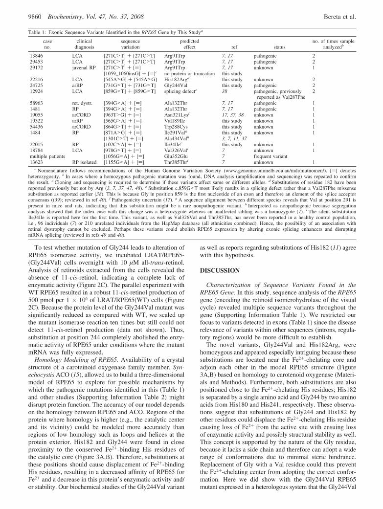
Table 1: Exonic Sequence Variants Identified in the RPE65 Gene by This Studya
caseno.
clinicaldiagnosis
sequencevariation
predictedeffect ref status
no. of times sampleanalyzedb
13846 LCA [271C>T] + [271C>T] Arg91Trp 7, 17 pathogenic 229453 LCA [271C>T] + [271C>T] Arg91Trp 7, 17 pathogenic 229172 juvenal RP [271C>T] + [)] Arg91Trp 7, 17 unknown 1
[1059_1060insG] + [)]c no protein or truncation this study22216 LCA [545A>G] + [545A>G] His182Argd this study unknown 224725 arRP [731G>T] + [731G>T] Gly244Val this study pathogenic 212924 LCA [859G>T] + [859G>T] splicing defecte 38 pathogenic, previously
reported as Val287Phe2
58963 ret. dystr. [394G>A] + [)] Ala132Thr 7, 17 pathogenic 11481 RP [394G>A] + [)] Ala132Thr 7, 17 pathogenic 119055 arCORD [963T>G] + [)] Asn321Lysf 17, 37, 38 unknown 119322 arRP [565G>A] + [)] Val189Ile this study unknown 154436 arCORD [864G>T] + [)] Trp288Cys this study unknown 11484 RP [871A>G] + [)] Ile291Valg this study unknown 1
[1301C>T] + [)] Ala434Valh 3, 7, 11, 3722015 RP [102C>A] + [)] Ile34Ilei this study unknown 118784 LCA [978G>T] + [)] Val326Vali 7 unknown 1multiple patients [1056G>A] + [)] Glu352Glu 7 frequent variant13623 RP isolated [1155G>A] + [)] Thr385Thri 7 unknown 1
a Nomenclature follows recommendations of the Human Genome Variation Society (www.genomic.unimelb.edu.au/mdi/mutnomen/). [)] denotesheterozygosity. b In cases where a homozygous pathogenic mutation was found, DNA analysis (amplification and sequencing) was repeated to confirmthe result. c Cloning and sequencing is required to determine if these variants affect same or different alleles. d Substitutions of residue 182 have beenreported previously but not by Arg (3, 7, 37, 47, 48). e Substitution c.859G>T most likely results in a splicing defect rather than a Val287Phe missensesubstitution as reported earlier (38). This is because Gly in position 859 is the first nucleotide of an exon and therefore an element of the splice acceptorconsensus ((39); reviewed in ref 40). f Pathogenicity uncertain (17). g A sequence alignment between different species reveals that Val at position 291 ispresent in mice and rats, indicating that this substitution might be a rare nonpathogenic variant. h Interpreted as nonpathogenic because segregationanalysis showed that the index case with this change was a heterozygote whereas an unaffected sibling was a homozygote (7). i The silent substitutionIle34Ile is reported here for the first time. This variant, as well as Val326Val and Thr385Thr, has never been reported in a healthy control population,i.e., 96 individuals (7) or 210 unrelated individuals from the HapMap database (all ethnicities combined). Hence, the possibility of an association withretinal dystrophy cannot be excluded. Perhaps these variants could abolish RPE65 expression by altering exonic splicing enhancers and disruptingmRNA splicing (reviewed in refs 49 and 40).
9860 Biochemistry, Vol. 47, No. 37, 2008 Bereta et al.
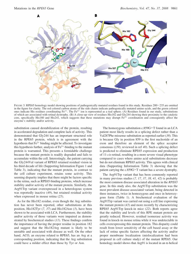
substitution caused destabilization of the protein, resultingin accelerated degradation and complete lack of activity. Thisdemonstrated that Gly244 has an important structural rolein the RPE65 protein, which is in agreement with thehypothesis that Fe2+ binding might be affected. To investigatethis hypothesis further, analysis of Fe2+-binding to the mutantprotein is warranted. This presents a formidable challengebecause the mutant protein is readily degraded and fails toaccumulate within the cell. Interestingly, the patient carryingthe Gly244Val variant of RPE65 retained residual vision inhis third decade of life (Supporting Information Figure 1 andTable 3), indicating that the mutant protein, in contrast tothe cell culture experiment, retains some activity. Thisseeming disparity implies that there might be factors specificto the retina, such as RPE65-binding proteins, which increasestability and/or activity of the mutant protein. Similarly, theArg91Trp variant overexpressed in a heterologous systemwas reportedly inactive (49), but retained residual activitywhen expressed in mouse retina (25).
As for the His182 residue, even though the Arg substitu-tion has never been reported, other substitutions at thisposition, His182Tyr (7, 37) and His182Asn (3), have beenshown to be associated with LCA. Furthermore, the stabilityand/or activity of those variants were impaired as demon-strated by biochemical studies (11). These findings confirmthe importance of having the proper residue in position 182and suggest that the His182Arg mutant is likely to beunstable and associated with disease as well. On the otherhand, ACO, an enzyme related to RPE65, has an Arg at acorresponding position, indicating that the Arg substitutioncould have a milder effect than those by Tyr or Asn.
The homozygous substitution c.859G>T found in an LCApatient most likely results in a splicing defect rather than aVal287Phe missense substitution as reported earlier (38). Thisis because Gly in position 859 is the first nucleotide of anexon and therefore an element of the splice acceptorconsensus ((39); reviewed in ref 40). Such a splicing defectis predicted to eliminate RPE65 expression and productionof 11-cis-retinal, resulting in a more severe visual phenotypecompared to cases where amino acid substitutions decreasebut do not eliminate RPE65 activity. This agrees with clinicaldata (Supporting Information Table 3) showing that thepatient carrying the c.859G>T variant has a severe dystrophy.
The Arg91Trp variant that has been commonly reportedin many previous studies (7, 17, 37, 38, 41, 42) is probablythe most common disease-associated alteration in the RPE65gene. In this study also, the Arg91Trp substitution was themost prevalent disease-associated variant, being detected inthree instances, twice in homozygous and once in heterozy-gous form (Table 1). A biochemical evaluation of theArg91Trp variant was carried out using a cell line expressingthe mutant protein (43) and more recently by characterizingRPE65 Arg91Trp knock-in mice (22). Both reports agreethat the stability and levels of this RPE mutant protein aregreatly reduced. However, residual isomerase activity wasfound in knock-in mouse retina while it was undetectable incells overexpressing this mutant protein. This disparity couldresult from lower sensitivity of the cell based assay or thelack of retina specific factors affecting the activity and/orproper membrane localization (mislocalization had beenproposed in cell culture study) of the mutant RPE65. Ourhomology model shows that Arg91 is located in an R-helical
FIGURE 3: RPE65 homology model showing positions of pathogenically mutated residues found in this study. Residues 200-235 are omittedin the figure for clarity. The red colored carbon atoms of the side chains indicate pathogenically mutated amino acids, and the green coloredones indicate His residues coordinating Fe2+. The Fe2+ ion is represented as a teal sphere. (A) Residues found in our study, substitutionsof which are associated with retinal dystrophy. (B) A close-up view of residues His182 and Gly244 showing their proximity to the catalyticcore, specifically His180 and His241, which suggests that these mutations may disrupt Fe2+ coordination and consequently affect theenzyme’s stability and/or activity.
Mutations in the RPE65 Gene Biochemistry, Vol. 47, No. 37, 2008 9861

region at the exterior of the protein (Figure 3A), and thus,exchange of a positively charged Arg residue for an aromatic,uncharged Trp residue could destabilize local structure andaccelerate degradation of this enzyme.
The Ala132Thr variant has been reported in RP patientsin homozygous (7) and heterozygous forms (17), and thedisease relevance of this variant was supported by segregationanalysis (7). Surprisingly, biochemical studies indicated thatthis variant retained 50% of WT activity (44) and mightprovide enough 11-cis-retinal to maintain a healthy retina.To resolve this discrepancy, more comprehensive patient dataand a better understanding of RPE65 function are required.We found the Ala132Thr variant in heterozygous form intwo patients. Our inability to find a sequence alteration inthe other allele could have resulted from incomplete se-quencing coverage of the RPE65 gene or another gene mighthave caused the clinical disorder. In our model of the RPE65structure, the Ala132 residue is hidden inside the moleculebut not close to the catalytic center (Figure 3A). Since theamino acid sequence in the vicinity of Ala132 is notconserved between the RPE65 and ACO proteins, ourhomology model of the protein may not be accurate in thisregion so we can only speculate as to the molecular effect(s)of the Ala132Thr substitution.
We colored all affected residues in the RPE65 homologymodel red (Figure 4) to determine whether the disease-associated RPE65 variants reported to date are localized todiscrete regions of the protein or are distributed randomly.This revealed that the mutations are located in many regionsof the protein. Thus, they could exert deleterious effects inmany different ways, e.g., by impairing Fe2+ binding orsubstrate channeling, disrupting the local or global structure,or affecting binding of putative protein partners. Interestingly,some residues altered in retinal dystrophy seem to beclustered rather than distributed randomly. An especiallyobvious example is the location of Arg44, His68, Tyr79, andGly528 residues within a single �-sheet (Figure 5). Substitu-tion of any of these residues could destabilize this �-sheetand displace the Fe2+-chelating His527 located on itsinnermost strand. This in turn could affect Fe2+ binding andcause a reduction in RPE65 enzymatic activity. Similarly,residues Thr457, Leu450, Tyr435, and Glu436 are also foundwithin a single �-sheet (Figure 6). Substitutions of theseresidues may affect the positioning of Glu417 predicted to
form an ion-dipole interaction with His313 (Figure 6). Lossof this interaction could alter the Lewis base properties ofHis313 and reduce its metal binding capability. Regardingthe aforementioned Leu450 residue, its naturally occurringMet substitutions are frequently found in laboratory micewhere they affect the rate of chromophore regeneration (seebelow).
Effect of the Leu450Met Mutation on RPE65 Structure andEnzymatic ActiVity. In addition to human mutations inRPE65, spontaneously occurring variants in RPE65 havebeen identified in other species that contribute to ourunderstanding of this protein’s role in ocular physiology. Onesuch example is that a greatly improved resistance to light-induced retinal damage in C57Bl/6 mice was directlyassociated with a hypomorphic variant of mouse RPE65 (45).
FIGURE 4: Location of all pathogenically mutated residues reported to date within the RPE65 structure. Residues affected in retinal dystrophy(colored red) are located throughout many regions of this protein and therefore are predicted to exert their deleterious effects by severaldifferent mechanisms. Some mutations seem to cluster, stressing the importance of the affected regions for the protein’s activity and/orstability (discussed below).
FIGURE 5: An example of clustering of pathogenically mutatedresidues in human RPE65. Residues 416-523 are omitted fromthe model for clarity. Substitutions of residues Arg44, His68, Tyr79,and Gly528 (colored red) were identified as a cause of retinaldystrophy. These residues are located in a single �-sheet, suggestinga common disease mechanism that destabilizes the �-sheet. Thisin turn might result in displacement of the Fe2+-chelating His527,impaired Fe2+ binding, and a consequent reduction in RPE65enzymatic activity.
9862 Biochemistry, Vol. 47, No. 37, 2008 Bereta et al.

Light damage susceptibility was proportional to the rate ofrhodopsin regeneration, which determines rhodopsin avail-ability during light exposure. Light damage found in twostrains with Leu450 in RPE65 was correlated with theoccurrence of photoreceptor apoptosis after short bright lightexposure. In contrast, mice with the Leu450Met variationregenerated rhodopsin more slowly and evidenced increasedresistance to light-induced retinal degeneration (46). Themechanism of retinal protection can be explained by the factthat a change of a single amino acid residue (Leu450Met)in mouse RPE65 causes both lower protein expression andlower overall activity of this enzyme. Consequently, the rateof rhodopsin regeneration and its subsequent activation isreduced (44). The importance of amino acid 450 is reinforcedby the greatly diminished activity and severe ocular pheno-type of humans carrying the Leu450Arg mutation (50). Basedon homology models to ACO, the effect of the Met450variant on RPE65 activity was proposed to relate to theability of the Met side chain to form hydrogen bonds withother residues in blade 6 of the protein. So this mutationmight affect the secondary structure of the �-strand, flexibilityof the blade, and overall stability and enzymatic activity ofthe protein (44). Analysis of our RPE65 model does notchallenge this hypothesis. But we do note that the Glu417residue that seems to stabilize the Fe2+-chelating His residuethrough an ion-dipole interaction lies just at the edge ofthe affected �-sheet (Figure 6). This residue might be
displaced as a result of changes within that �-sheet, therebyimpairing Fe2+ coordination. On the other hand, Leu450 islocated at the periphery of RPE65 some distance from theactive site of the protein. Thus, an alternative explanationfor the reduced enzymatic activity of the Leu450Met variantmight be a reduced ability to interact with proteins such asLRAT, RDH5, or others involved in the isomerizationprocess. Interestingly, although Leu450 is highly conservedwithin RPE65 sequences of different species, effects of theLeu450 mutation on enzymatic activity may vary. In Vitrostudies show that the impact of the Leu450Met substitutionis more severe in murine RPE65 as compared to the canineprotein. Such differences might indicate additional, species-specific variations in surrounding residues that play importantroles in modulating RPE65 activity, as was proposed forresidue 446 (44).
CONCLUSIONS
Mutations in the RPE65 gene can cause severe retinaldystrophies. Here we report 15 exonic sequence variants ofRPE65 gene, 7 of which are novel and might be associatedwith retinal dystrophies. The novel RPE65 Gly244Val mutationwas characterized in biochemical detail. We introduced bicis-tronic plasmid expression of this mutant together with EGFPenabling tight control and monitoring of the mRNA level, whichpermitted precise evaluation of this mutant’s stability andactivity in cell culture experiments. Molecular modeling ofRPE65 provided important information as to how severalmutations might impact retinoid isomerization by RPE65 andlead to photoreceptor degeneration.
ACKNOWLEDGMENT
We thank Yesmino Elia for coordinating patient schedulingand Alex Levin for contributing to patient recruitment. Wealso thank Dr. T. Kitamura (University of Tokyo) for thegenerous gift of pMXs-IG and pMXs-IP vectors and Dr.Leslie Webster, Jr., for help during manuscript preparation.
SUPPORTING INFORMATION AVAILABLE
Table 1, genetic variation identified in the RPE65 gene inthis study in patients with retinal degeneration (frequencydata included); Table 2, sequence variation in the RPE65gene and sequence alignments among the RPE65 genes fromdifferent species; Table 3, clinical characteristics of patientswith RPE65 mutations; Figure 1, retinal imaging of patientcarrying the Gly244Val mutation. This material is availablefree of charge via the Internet at http://pubs.acs.org.
REFERENCES
1. Palczewski, K. (2006) G protein-coupled receptor rhodopsin. Annu.ReV. Biochem. 75, 743–767.
2. Travis, G. H., Golczak, M., Moise, A. R., and Palczewski, K. (2007)Diseases caused by defects in the visual cycle: retinoids as potentialtherapeutic agents. Annu. ReV. Pharmacol. Toxicol. 47, 469–512.
3. Hanein, S., Perrault, I., Gerber, S., Tanguy, G., Barbet, F., Ducroq,D., Calvas, P., Dollfus, H., Hamel, C., Lopponen, T., Munier, F.,Santos, L., Shalev, S., Zafeiriou, D., Dufier, J. L., Munnich, A.,Rozet, J. M., and Kaplan, J. (2004) Leber congenital amaurosis:comprehensive survey of the genetic heterogeneity, refinement ofthe clinical definition, and genotype-phenotype correlations as astrategy for molecular diagnosis. Hum. Mutat. 23, 306–317.
4. Janecke, A. R., Thompson, D. A., Utermann, G., Becker, C.,Hubner, C. A., Schmid, E., McHenry, C. L., Nair, A. R.,
FIGURE 6: A hypothetical mechanism by which Leu450 substitutionsmight affect the RPE65 enzymatic activity. Residues 365-379 and323-331 are omitted from the model for clarity. Leu450 is locatedin a �-sheet close to other residues known to be pathogenicallymutated (colored red). This encourages us to propose a possiblemechanism by which its substitutions might affect RPE65 activity.Mutations in this region could disrupt the �-sheet fold and affectpositioning of Glu417 which is predicted to form an ion-dipoleinteraction with His313 (shown as a dashed black line). Loss ofthis interaction could alter the Lewis acid properties of His313 andreduce its metal binding capacity. The well-known Leu450Metsubstitution found in mice, which reduces but does not eliminateisomerase activity, may only slightly perturb this region due to therelative similarities between the Leu residue and Met side chains.In contrast, the Leu450Arg substitution found in retinal dystrophypatients introduces a positive charge into this region so it is likelyto disrupt the fold to a greater extent.
Mutations in the RPE65 Gene Biochemistry, Vol. 47, No. 37, 2008 9863

Ruschendorf, F., Heckenlively, J., Wissinger, B., Nurnberg, P., andGal, A. (2004) Mutations in RDH12 encoding a photoreceptor cellretinol dehydrogenase cause childhood-onset severe retinal dys-trophy. Nat. Genet. 36, 850–854.
5. Perrault, I., Hanein, S., Gerber, S., Barbet, F., Ducroq, D., Dollfus,H., Hamel, C., Dufier, J. L., Munnich, A., Kaplan, J., and Rozet,J. M. (2004) Retinal dehydrogenase 12 (RDH12) mutations in lebercongenital amaurosis. Am. J. Hum. Genet. 75, 639–646.
6. Gu, S. M., Thompson, D. A., Srikumari, C. R., Lorenz, B., Finckh,U., Nicoletti, A., Murthy, K. R., Rathmann, M., Kumaramanickavel,G., Denton, M. J., and Gal, A. (1997) Mutations in RPE65 causeautosomal recessive childhood-onset severe retinal dystrophy. Nat.Genet. 17, 194–197.
7. Morimura, H., Fishman, G. A., Grover, S. A., Fulton, A. B., Berson,E. L., and Dryja, T. P. (1998) Mutations in the RPE65 gene inpatients with autosomal recessive retinitis pigmentosa or lebercongenital amaurosis. Proc. Natl. Acad. Sci. U.S.A. 95, 3088–3093.
8. Thompson, D. A., Li, Y., McHenry, C. L., Carlson, T. J., Ding,X., Sieving, P. A., Apfelstedt-Sylla, E., and Gal, A. (2001)Mutations in the gene encoding lecithin retinol acyltransferase areassociated with early-onset severe retinal dystrophy. Nat. Genet.28, 123–124.
9. Senechal, A., Humbert, G., Surget, M. O., Bazalgette, C., Bazal-gette, C., Arnaud, B., Arndt, C., Laurent, E., Brabet, P., and Hamel,C. P. (2006) Screening genes of the retinoid metabolism: novelLRAT mutation in leber congenital amaurosis. Am. J. Ophthalmol.142, 702–704.
10. Moiseyev, G., Chen, Y., Takahashi, Y., Wu, B. X., and Ma, J. X.(2005) RPE65 is the isomerohydrolase in the retinoid visual cycle.Proc. Natl. Acad. Sci. U.S.A. 102, 12413–12418.
11. Redmond, T. M., Poliakov, E., Yu, S., Tsai, J. Y., Lu, Z., andGentleman, S. (2005) Mutation of key residues of RPE65 abolishesits enzymatic role as isomerohydrolase in the visual cycle. Proc.Natl. Acad. Sci. U.S.A. 102, 13658–13663.
12. Jin, M., Li, S., Moghrabi, W. N., Sun, H., and Travis, G. H. (2005)Rpe65 is the retinoid isomerase in bovine retinal pigment epithe-lium. Cell 122, 449–459.
13. Schwartz, S. H., Tan, B. C., Gage, D. A., Zeevaart, J. A., andMcCarty, D. R. (1997) Specific oxidative cleavage of carotenoidsby VP14 of maize. Science 276, 1872–1874.
14. von Lintig, J., and Wyss, A. (2001) Molecular analysis of vitaminA formation: cloning and characterization of beta-carotene 15,15′-dioxygenases. Arch. Biochem. Biophys. 385, 47–52.
15. Kloer, D. P., Ruch, S., Al-Babili, S., Beyer, P., and Schulz, G. E.(2005) The structure of a retinal-forming carotenoid oxygenase.Science 308, 267–269.
16. Jacobson, S. G., Cideciyan, A. V., Aleman, T. S., Sumaroka, A.,Schwartz, S. B., Windsor, E. A., Roman, A. J., Heon, E., Stone,E. M., and Thompson, D. A. (2007) RDH12 and RPE65, visualcycle genes causing leber congenital amaurosis, differ in diseaseexpression. InVest. Ophthalmol. Visual Sci. 48, 332–338.
17. Thompson, D. A., Gyurus, P., Fleischer, L. L., Bingham, E. L.,McHenry, C. L., Apfelstedt-Sylla, E., Zrenner, E., Lorenz, B.,Richards, J. E., Jacobson, S. G., Sieving, P. A., and Gal, A. (2000)Genetics and phenotypes of RPE65 mutations in inherited retinaldegeneration. InVest. Ophthalmol. Visual Sci. 41, 4293–4299.
18. Jacobson, S. G., Aleman, T. S., Cideciyan, A. V., Heon, E.,Golczak, M., Beltran, W. A., Sumaroka, A., Schwartz, S. B.,Roman, A. J., Windsor, E. A., Wilson, J. M., Aguirre, G. D., Stone,E. M., and Palczewski, K. (2007) Human cone photoreceptordependence on RPE65 isomerase. Proc. Natl. Acad. Sci. U.S.A.104, 15123–15128.
19. Redmond, T. M., Yu, S., Lee, E., Bok, D., Hamasaki, D., Chen,N., Goletz, P., Ma, J. X., Crouch, R. K., and Pfeifer, K. (1998)Rpe65 is necessary for production of 11-cis-vitamin A in the retinalvisual cycle. Nat. Genet. 20, 344–351.
20. Pang, J. J., Chang, B., Hawes, N. L., Hurd, R. E., Davisson, M. T.,Li, J., Noorwez, S. M., Malhotra, R., McDowell, J. H., Kaushal,S., Hauswirth, W. W., Nusinowitz, S., Thompson, D. A., andHeckenlively, J. R. (2005) Retinal degeneration 12 (rd12): a new,spontaneously arising mouse model for human Leber congenitalamaurosis (LCA). Mol. Vision 11, 152–162.
21. Aguirre, G. D., Baldwin, V., Pearce-Kelling, S., Narfstrom, K.,Ray, K., and Acland, G. M. (1998) Congenital stationary nightblindness in the dog: common mutation in the RPE65 gene indicatesfounder effect. Mol. Vision 4, 23.
22. Samardzija, M., von Lintig, J., Tanimoto, N., Oberhauser, V.,Thiersch, M., Reme, C. E., Seeliger, M., Grimm, C., and Wenzel,A. (2008) R91W mutation in Rpe65 leads to milder early-onset
retinal dystrophy due to the generation of low levels of 11-cis-retinal. Hum. Mol. Genet. 17, 281–292.
23. Acland, G. M., Aguirre, G. D., Ray, J., Zhang, Q., Aleman, T. S.,Cideciyan, A. V., Pearce-Kelling, S. E., Anand, V., Zeng, Y.,Maguire, A. M., Jacobson, S. G., Hauswirth, W. W., and Bennett,J. (2001) Gene therapy restores vision in a canine model ofchildhood blindness. Nat. Genet. 28, 92–95.
24. Acland, G. M., Aguirre, G. D., Bennett, J., Aleman, T. S.,Cideciyan, A. V., Bennicelli, J., Dejneka, N. S., Pearce-Kelling,S. E., Maguire, A. M., Palczewski, K., Hauswirth, W. W., andJacobson, S. G. (2005) Long-term restoration of rod and cone visionby single dose rAAV-mediated gene transfer to the retina in acanine model of childhood blindness. Mol. Ther. 12, 1072–1082.
25. Van Hooser, J. P., Aleman, T. S., He, Y. G., Cideciyan, A. V.,Kuksa, V., Pittler, S. J., Stone, E. M., Jacobson, S. G., andPalczewski, K. (2000) Rapid restoration of visual pigment andfunction with oral retinoid in a mouse model of childhoodblindness. Proc. Natl. Acad. Sci. U.S.A. 97, 8623–8628.
26. Van Hooser, J. P., Liang, Y., Maeda, T., Kuksa, V., Jang, G. F.,He, Y. G., Rieke, F., Fong, H. K., Detwiler, P. B., and Palczewski,K. (2002) Recovery of visual functions in a mouse model of Lebercongenital amaurosis. J. Biol. Chem. 277, 19173–19182.
27. Maguire, A. M., Simonelli, F., Pierce, E. A., Pugh, E. N., Jr.,Mingozzi, F., Bennicelli, J., Banfi, S., Marshall, K. A., Testa, F.,Surace, E. M., Rossi, S., Lyubarsky, A., Arruda, V. R., Konkle,B., Stone, E., Sun, J., Jacobs, J., Dell’Osso, L., Hertle, R., Ma,J. X., Redmond, T. M., Zhu, X., Hauck, B., Zelenaia, O., Shindler,K. S., Maguire, M. G., Wright, J. F., Volpe, N. J., McDonnell,J. W., Auricchio, A., High, K. A., and Bennett, J. (2008) Safetyand efficacy of gene transfer for Leber’s congenital amaurosis.N. Engl. J. Med. 358, 2240–2248.
28. Bainbridge, J. W., Smith, A. J., Barker, S. S., Robbie, S.,Henderson, R., Balaggan, K., Viswanathan, A., Holder, G. E.,Stockman, A., Tyler, N., Petersen-Jones, S., Bhattacharya, S. S.,Thrasher, A. J., Fitzke, F. W., Carter, B. J., Rubin, G. S., Moore,A. T., and Ali, R. R. (2008) Effect of gene therapy on visualfunction in Leber’s congenital amaurosis. N. Engl. J. Med. 358,2231–2239.
29. Kitamura, T., Koshino, Y., Shibata, F., Oki, T., Nakajima, H.,Nosaka, T., and Kumagai, H. (2003) Retrovirus-mediated genetransfer and expression cloning: powerful tools in functionalgenomics. Exp. Hematol. 31, 1007–1014.
30. Liu, X., Constantinescu, S. N., Sun, Y., Bogan, J. S., Hirsch, D.,Weinberg, R. A., and Lodish, H. F. (2000) Generation ofmammalian cells stably expressing multiple genes at predeterminedlevels. Anal. Biochem. 280, 20–28.
31. Mancia, F., Patel, S. D., Rajala, M. W., Scherer, P. E., Nemes, A.,Schieren, I., Hendrickson, W. A., and Shapiro, L. (2004) Optimiza-tion of protein production in mammalian cells with a coexpressedfluorescent marker. Structure 12, 1355–1360.
32. Golczak, M., Maeda, A., Bereta, G., Maeda, T., Kiser, P. D.,Hunzelmann, S., von Lintig, J., Blaner, W. S., and Palczewski, K.(2008) Metabolic basis of visual cycle inhibition by retinoid andnonretinoid compounds in the vertebrate retina. J. Biol. Chem. 283,9543–9554.
33. Shi, J., Blundell, T. L., and Mizuguchi, K. (2001) FUGUE:sequence-structure homology recognition using environment-specific substitution tables and structure-dependent gap penalties.J. Mol. Biol. 310, 243–257.
34. Bennett-Lovsey, R. M., Herbert, A. D., Sternberg, M. J., and Kelley,L. A. (2008) Exploring the extremes of sequence/structure spacewith ensemble fold recognition in the program Phyre. Proteins 70,611–625.
35. Emsley, P., and Cowtan, K. (2004) Coot: model-building tools formolecular graphics. Acta Crystallogr. 60, 2126–2132.
36. Brunger, A. T., Adams, P. D., Clore, G. M., DeLano, W. L., Gros,P., Grosse-Kunstleve, R. W., Jiang, J. S., Kuszewski, J., Nilges,M., Pannu, N. S., Read, R. J., Rice, L. M., Simonson, T., andWarren, G. L. (1998) Crystallography & NMR system: A newsoftware suite for macromolecular structure determination. ActaCrystallogr. 54, 905–921.
37. Simovich, M. J., Miller, B., Ezzeldin, H., Kirkland, B. T., McLeod,G., Fulmer, C., Nathans, J., Jacobson, S. G., and Pittler, S. J. (2001)Four novel mutations in the RPE65 gene in patients with Lebercongenital amaurosis. Hum. Mutat. 18, 164.
38. Lotery, A. J., Namperumalsamy, P., Jacobson, S. G., Weleber,R. G., Fishman, G. A., Musarella, M. A., Hoyt, C. S., Heon, E.,Levin, A., Jan, J., Lam, B., Carr, R. E., Franklin, A., Radha, S.,Andorf, J. L., Sheffield, V. C., and Stone, E. M. (2000) Mutation
9864 Biochemistry, Vol. 47, No. 37, 2008 Bereta et al.
analysis of 3 genes in patients with Leber congenital amaurosis.Arch. Ophthalmol. 118, 538–543.
39. Mount, S. M. (1982) A catalogue of splice junction sequences.Nucleic Acids Res. 10, 459–472.
40. Wang, Z., and Burge, C. B. (2008) Splicing regulation: from aparts list of regulatory elements to an integrated splicing code. RNA(New York) 14, 802–813.
41. Lorenz, B., Gyurus, P., Preising, M., Bremser, D., Gu, S., Andrassi,M., Gerth, C., and Gal, A. (2000) Early-onset severe rod-conedystrophy in young children with RPE65 mutations. InVest.Ophthalmol. Visual Sci. 41, 2735–2742.
42. El Matri, L., Ambresin, A., Schorderet, D. F., Kawasaki, A.,Seeliger, M. W., Wenzel, A., Arsenijevic, Y., Borruat, F. X., andMunier, F. L. (2006) Phenotype of three consanguineous Tunisianfamilies with early-onset retinal degeneration caused by an R91Whomozygous mutation in the RPE65 gene. Graefe’s Arch. Clin.Exp. Ophthalmol. 244, 1104–1112.
43. Takahashi, Y., Chen, Y., Moiseyev, G., and Ma, J. X. (2006) Twopoint mutations of RPE65 from patients with retinal dystrophiesdecrease the stability of RPE65 protein and abolish its isomero-hydrolase activity. J. Biol. Chem. 281, 21820–21826.
44. Redmond, T. M., Weber, C. H., Poliakov, E., Yu, S., and Gentleman,S. (2007) Effect of Leu/Met variation at residue 450 on isomeraseactivity and protein expression of RPE65 and its modulation byvariation at other residues. Mol. Vision 13, 1813–1821.
45. Danciger, M., Matthes, M. T., Yasamura, D., Akhmedov, N. B.,Rickabaugh, T., Gentleman, S., Redmond, T. M., La Vail, M. M.,and Farber, D. B. (2000) A QTL on distal chromosome 3 thatinfluences the severity of light-induced damage to mouse photo-receptors. Mamm. Genome 11, 422–427.
46. Wenzel, A., Reme, C. E., Williams, T. P., Hafezi, F., and Grimm,C. (2001) The Rpe65 Leu450Met variation increases retinalresistance against light-induced degeneration by slowing rhodopsinregeneration. J. Neurosci. 21, 53–58.
47. Galvin, J. A., Fishman, G. A., Stone, E. M., and Koenekoop, R. K.(2005) Evaluation of genotype-phenotype associations in lebercongenital amaurosis. Retina (Philadelphia) 25, 919–929.
48. Galvin, J. A., Fishman, G. A., Stone, E. M., and Koenekoop, R. K.(2005) Clinical phenotypes in carriers of Leber congenital amau-rosis mutations. Ophthalmology 112, 349–356.
49. Wang, G. S., and Cooper, T. A. (2007) Splicing in disease:disruption of the splicing code and the decoding machinery. Nat.ReV. Genet. 8, 749–761.
50. Wada, Y., Nakazawa, M., Abe, T., Fuse, N., and Tamai, M. (2000)Clinical variability also is found in patients with mutations in genesencoding other visual cycle proteins, e.g., arrestin, RPE65 andRDH5. InVest. Ophthalmol. Visual Sci. 41, S617.
BI800905V
Mutations in the RPE65 Gene Biochemistry, Vol. 47, No. 37, 2008 9865