impaired osteogenesis in menkes disease-derived induced ... · impaired osteogenesis in menkes...
TRANSCRIPT

Kim et al. Stem Cell Research & Therapy (2015) 6:160 DOI 10.1186/s13287-015-0147-5
RESEARCH Open Access
Impaired osteogenesis in Menkes disease-derived induced pluripotent stem cells
Dongkyu Kim1, Jieun Choi1, Kyu-Min Han1, Beom Hee Lee2, Jin-Ho Choi2, Han-Wook Yoo2 and Yong-Mahn Han1*Abstract
Introduction: Bone abnormalities, one of the primary manifestations of Menkes disease (MD), include a weakenedbone matrix and low mineral density. However, the molecular and cellular mechanisms underlying these bonedefects are poorly understood.
Methods: We present in vitro modeling for impaired osteogenesis in MD using human induced pluripotent stemcells (iPSCs) with a mutated ATP7A gene. MD-iPSC lines were generated from two patients harboring differentmutations.
Results: The MD-iPSCs showed a remarkable retardation in CD105 expression with morphological anomaliesduring development to mesenchymal stem cells (MSCs) compared with wild-type (WT)-iPSCs. Interestingly,although prolonged culture enhanced CD105 expression, mature MD-MSCs presented with low alkalinephosphatase activity, reduced calcium deposition in the extracellular matrix, and downregulated osteoblast-specificgenes during osteoblast differentiation in vitro. Knockdown of ATP7A also impaired osteogenesis in WT-MSCs. Lysyloxidase activity was also decreased in MD-MSCs during osteoblast differentiation.
Conclusions: Our findings indicate that ATP7A dysfunction contributes to retardation in MSC development and impairsosteogenesis in MD.
IntroductionMenkes disease (MD) is a copper metabolism disorderthat is caused by a loss-of-function of a major coppertransporter, ATP7A [1, 2]. The ATP7A gene is locatedon the long arm of X chromosome and encodes a P-typeATPase, which plays crucial roles in cellular copper me-tabolism by controlling copper export and intracellularcopper trafficking [3, 4]. Although its primary physio-logical function is copper absorption in the small intes-tine, ATP7A is also implicated in intracellular copperdelivery to copper-dependent enzymes [5, 6]. A varietyof copper-dependent enzymes become nonfunctionaldue to a lack of ATP7A activity in MD patients, whichcan lead to multisystemic clinical symptoms [6, 7].Clinical manifestations of MD patients include pro-
gressive neurodegeneration, connective tissue defects,sparse and kinky hairs, vascular defects, and manyothers. Connective tissue defects comprise tortuous
* Correspondence: [email protected] of Biological Science, Korea Advanced Institute of ScienceTechnology (KAIST), Daejeon 305-701, Republic of KoreaFull list of author information is available at the end of the article
© 2015 Kim et al. Open Access This articleInternational License (http://creativecommonsreproduction in any medium, provided you gthe Creative Commons license, and indicate if(http://creativecommons.org/publicdomain/ze
vessels, skeletal change, loose skin, laxity of joints, andso forth [7–9]. Similarly, MD mouse models presentwith fragmentation of the internal elastic lamina, defect-ive synthesis of bone collagen, reduced skin tensilestrength, and weak blood vessels [10–12]. Among thevarious symptoms, bone abnormalities are a typicalphenotype in MD patients [13–15]. Bone abnormalitiesin Menkes patients include osteoporosis, metaphysealspurs, diaphyseal fractures, and wormian occipital bones[15–19]. Bone defects have also been shown in occipitalhorn syndrome, a mild phenotype of ATP7A-deficientdisease [20]. Defective phenotypes in bone formation arefrequently used as a diagnostic test in the early stagealong with a blood test to measure low serum copperlevels [16]. However, in vitro model systems that investi-gate how ATP7A mutations result in abnormal bone for-mation in MD have not been reported. Here, we attemptto model MD pathogenesis at the cellular level using in-duced pluripotent stem cells (iPSCs) in vitro. HumaniPSCs, which have the capability to differentiate intovarious cell types and to proliferate indefinitely, are
is distributed under the terms of the Creative Commons Attribution 4.0.org/licenses/by/4.0/), which permits unrestricted use, distribution, andive appropriate credit to the original author(s) and the source, provide a link tochanges were made. The Creative Commons Public Domain Dedication waiverro/1.0/) applies to the data made available in this article, unless otherwise stated.

Kim et al. Stem Cell Research & Therapy (2015) 6:160 Page 2 of 12
useful cell sources for studying the pathogenesis of hu-man diseases [21, 22].In this study, MD-iPSCs were differentiated into oste-
oblasts (OBs) to investigate the effect of ATP7A dys-function on bone formation. Intriguingly, MD-iPSCsshowed delayed mesenchymal stem cell (MSC) matur-ation compared with wild-type (WT)-iPSCs. Subse-quently, MD-MSCs showed impaired osteogenesis interms of alkaline phosphatase (ALP) activity, calciummineralization, and transcription of osteogenic genes.Copper chelation in WT-MSCs resembled defectivephenotypes shown in MD-MSCs. Our results demon-strate that dysfunction of copper utilization in MDgives rise to delayed MSC development and impairedOB differentiation.
Materials and methodsRetrovirus productionFor retrovirus packaging, retroviral vectors encodingOCT4, SOX2, KLF4, cMYC (Addgene, Cambridge, MA,USA) were co-transfected with VSV-G vector (Takara Bio,Mountain View, CA, USA) in GP2 293 cells. Transfectantswere further incubated in Dulbecco’s modified Eagle’smedium (DMEM; Welgene, Seoul, Korea) supplementedwith 10 % fetal bovine serum (FBS; Invitrogen, Carlsbad,CA, USA) and 1 % penicillin-streptomycin (Invitrogen) at37 °C, 5 % CO2 in air. The medium was changed 8 h aftertransfection. Then, supernatants were harvested 48 and72 h after incubation. Supernatants harvested from fourdishes (10 cm in diameter) per factor were ultracentri-fuged at 90,000 × g for 90 min at 4 °C. The viral pellet wasdissolved in 2 ml of the medium and kept at −70 °C beforeuse.
Generation and maintenance of MD-iPSCsTo generate MD-iPSCs, patient fibroblasts were infectedwith four retroviruses and then plated onto mitomycinC-treated (MMC; A.G. Scientific, San Diego, CA, USA)MEF feeder layers at a density of 103 cells/cm2. Thisstudy using patient fibroblasts was approved by theInstitutional Review Board of Asan Medical Center, andwritten informed consent was obtained from their par-ents. Infected cells were cultured in human embryonicstem cell (ESC) medium at 37 °C, 5 % CO2 in air for 2 to3 weeks. The human ESC medium consists of DMEM/F12 (Invitrogen) supplemented with 20 % Knockout SR(Invitrogen), 1 % nonessential amino acids (Invitrogen),1 % penicillin-streptomycin, 0.1 mM β-mercaptoethanol(Sigma, St. Louis, MO, USA), and 10 ng/ml fibroblastgrowth factor (FGF)2 (R&D systems, Minneapolis, MN,USA). Respective human ESC-like colonies were mechan-ically transferred onto new MMC-treated MEF feedersand subcultured for stabilization for 10 to 20 passages.iPSC characteristics were analyzed by the expression of
human ESC markers, karyotypes, methylation states onpromoters of human ESC marker genes, and teratoma for-mation. WT-iPSCs derived from foreskin fibroblasts [23]were used as a control group (Additional file 1: Figure S1).
Real-time quantitative PCRTotal mRNA was extracted from iPSCs and differentiatedcells using easy-Blue™ (Intron Biotechnology, Seongnam,Korea). Briefly, approximately 1 × 10⁵ cells were washedin phosphate-buffered saline (PBS) and treated with 1 mleasy-Blue™ solution. After mixing with 200 μl chloroform,cell lysates were centrifuged and the upper layer of thesupernatants was harvested to isolate RNA. Then, RNAwas precipitated and rehydrated for cDNA synthesis. Atotal of 1 μg RNA was annealed with oligo(dT), and cDNAwas synthesized using M-MLV Reverse Transcriptase(Enzynomics, Daejeon, Korea). The real-time polymerasechain reaction (RT-PCR) was performed using the follow-ing cycle conditions: 95 °C denaturation, 60 °C annealing,and 72 °C elongation. The cycle numbers for each reactionvaried between 30 and 40. Red safe (Intron Biotechnology)was used for visualization of PCR products in gel electro-phoresis. For quantitative comparison, the relative expres-sion level was measured by CFX-Connect real-timesystem (Bio-Rad, Hercules, CA, USA). The relativeexpression level of each gene was analyzed using a com-parative threshold cycle method, and the transcriptionlevel of GAPDH was used for normalization. The primersused in this study are listed in Additional file 2 (Tables S1and S2).
Bisulfite sequencingGenomic DNA was isolated from cell samples using aG-DEX Genomic DNA Extraction Kit (Intron Biotech-nology). Briefly, 2 × 10⁶ cells were lysed in cell lysis buf-fer (300 μl) at room temperature for 5 min and thenincubated at 37 °C for 30 min in the presence of RNaseA. After the addition of PPT Buffer (100 μl), cell lysateswere centrifuged at 16,000 × g for 5 min, and the super-natant was harvested. Isopropanol (300 μl) was addedto the supernatant. After centrifugation at 16,000 × gfor 1 min, the DNA pellet was dissolved in distilledwater (DW). Bisulfite treatment was performed using aZymo EZ DNA methylation kit (Zymo Research, Irvine,CA, USA) according to manufacturer’s instructions.Briefly, 1 μg of genomic DNA was denatured at 95 °Cfor 10 min, and CT-conversion was performed byaddition of the CT Conversion Reagent. CT-convertedDNA was desulfonated in M-Desulfonation Buffer,washed with M-Wash Buffer, and dissolved in 20 μl ofDW. Bisulfite-treated genomic DNA was amplified byPCR, individually cloned into a pGEM®-T vector (Pro-mega, Madison, WI, USA), and sequenced using an ABI3730XL DNA Analyzer (Applied Biosystems, Foster City,

Kim et al. Stem Cell Research & Therapy (2015) 6:160 Page 3 of 12
CA, USA). Methylation quantification was performed usingthe QUMA program (Riken, Kobe, Japan). The primersused in bisulfate sequencing are listed in Additional file 2(Table S3).
ImmunostainingThe cells were fixed with 4 % formaldehyde for 30 min,washed twice in PBST (PBS containing 0.1 % Tween 20),and permeabilized in PBS containing 0.1 % Triton X-100(Sigma) for 20 min. After blocking with 2 % bovineserum albumin (BSA; Sigma) for 1 h, the cells weretreated with each primary antibody and incubated at 4 °Covernight. The primary antibodies used in this study wereas follows: OCT4 (Santa Cruz Biotechnology, Santa Cruz,CA, USA); SOX2 (Cell Signaling, Danvers, MA, USA);NANOG (R&D Systems); SSEA4 (Abcam, Cambridge,England); TRA-1-60 (Millipore, Billerica, MA, USA);TRA-1-81 (Millipore); NESTIN (Millipore); α-SMA (R&DSystems); and GATA4 (Santa Cruz Biotechnology). Thecells were washed several times in PBST and incubatedwith secondary antibodies (Alexa Fluor 488 or 594; Invi-trogen) for 1 h. Then, the cells were washed several timesin PBST and counter-stained with 4′-6-diamidino-2-phe-nylindole (DAPI; Sigma) during the washing step. Afterwashing with PBST, fluorescence images were observedon a Zeiss LSM 510 confocal microscope equipped withargon and helium–neon lasers (Carl Zeiss, Germany).
Teratoma formation of MD-iPSCsAnimal care and experimental procedures were per-formed under the approval of the Animal Care Commit-tees of KAIST. MD-iPSCs (1 × 10⁷ cells) were collectedby scraping, mixed with Matrigel (BD Biosciences,Franklin Lakes, NJ, USA), and subcutaneously injectedinto the dorso-lateral area of CAnN.Cg-Foxn1 nu/CrljOri mice (Orient, Seongnam, Korea). Approximately2 months after injection, the tumor tissues were dis-sected and embedded in paraffin wax. Tissue sectionswere placed on slide glasses. Hematoxylin and eosin(H&E; Sigma) staining was performed to observe variouscell types and tissues.
Differentiation of human iPSCs into MSCsDifferentiation of human iPSCs into MSCs was per-formed as previously described [24, 25]. Briefly, humaniPSC colonies were mechanically dissected and trans-ferred to low-adhesion petri dishes (SPL Lifesciences,Pocheon, Korea). Dissected human iPSCs spontaneouslyaggregated to form embryoid bodies (EBs) in EB mediumat 37 °C with 5 % CO2 for 1 day. The EB medium consistsof DMEM/F12 supplemented with 10 % Knockout SR, 1% nonessential amino acids, 1 % penicillin-streptomycin,and 0.1 mM β-mercaptoethanol. EBs were further cul-tured in the EB medium containing 10 μM SB431542
(Abcam) at 37 °C with 5 % CO2 for 10 days, and thenattached to fibronectin-coated dishes (BD Biosciences).The attached cells were further cultured in DMEM/F12supplemented with 1 μM SB431542, 1 % ITS Liquidmedia supplement (Sigma), 1 % B27 supplement (Invi-trogen), and 1 % CD lipid concentrate (Invitrogen) for4 days. Then, the cells were cultured in α-minimum es-sential medium (α-MEM; Invitrogen) containing 10 %FBS for 20 days for MSC induction.
FACS analysisCells were dissociated by treatment with trypsin-EDTA(0.25 %; Invitrogen) for 5 min followed by the addition offresh culture medium containing FBS for enzyme inactiva-tion. After centrifugation at 300 × g for 5 min, the pelletswere resuspended in FACS buffer (PBS containing 2 %FBS) and filtered through a cell strainer with a 40-μm poresize (SPL Lifesciences). Dissociated cells were incubatedwith specific FACS antibodies against CD44, CD73, CD90,CD105, and respective isotype controls (Biolegend, SanDiego, CA, USA) at 4 °C for 30 min. After washing withFACS buffer, samples were analyzed using a FACSCaliburflow cytometer (BD Biosciences). The positive populationfor each antibody was evaluated with FlowJo software(Tree Star, Ashland, OR, USA). Gating strategy for thisanalysis is summarized in Additional file 3 (Figure S2).
Western blottingCells were lysed with Pro-Prep protein extraction solution(Intron Biotechnology) on ice for 1 h. After centrifugationat 16,000 × g for 30 min, the supernatant was harvested.The concentration of protein lysates was determined byBradford protein assay (Bio-Rad). Proteins were loadedonto an SDS-PAGE gel (Elpis Biotech, Daejeon, Korea) andthen transferred to a nitrocellulose membrane (Whatman,Maidstone, England). The membrane was blocked withTBST (0.1 % Tween in TBS) containing 4 % skim milk,washed in TBST, and treated with the appropriate primaryantibodies. The primary antibodies used in this study wereas follows: SMAD2 (Cell Signaling); p-SMAD2 (Cell Signal-ing); ACTIN (Santa Cruz Biotechnology); ATP7A (HycultBiotech, Uden, Netherlands); SMAD1 (Cell Signaling); andp-SMAD1 (Cell Signaling). After washing in TBST, themembrane was incubated with an horseradish peroxidase(HRP)-conjugated secondary antibody (Thermo Fisher Sci-entific, Waltham, MA, USA) at room temperature for 1 h.The membrane was developed using the ECL system(Thermo Fischer Scientific), and images were captured byLAS-3000 (Fuji Film, Tokyo, Japan).
MSC proliferation and apoptosisCells were seeded on a gelatin-coated dish at a densityof 2 × 10³ cells/cm2 and cultured in α-MEM containing10 % FBS for 9 days. The number of cells was

Kim et al. Stem Cell Research & Therapy (2015) 6:160 Page 4 of 12
calculated daily using a hemocytometer (Marienfeld,Lauda-Königshofen, Germany). To examine cell viability,cells were first plated on a gelatin-coated dish at a densityof 2 × 10⁴ cells/cm2 and then cultured for 1 day. After theaddition of the CCK-8 reagents (Dojindo, Kumamoto,Japan), the cells were incubated at 37 °C for 30 min, andabsorbance was measured at 450 nm. To detect apoptosis,cells were resuspended in 1X annexin V binding buffer(eBioscience, San Diego, CA, USA) and incubated withannexin V-FITC (eBioscience) and propidium iodide (PI;Sigma) for 15 min in the dark. After FACS analysis,annexin-positive cells were counted and graphed. For cellcycle analysis, cells were fixed in cold 70 % ethanol for 1 hand then treated with RNase A (Sigma) for 30 min. Aftertreatment with PI for 15 min, the distribution of cells inthe cell cycle was analyzed on a FACSCalibur.
MSC differentiation into OBs and chondrocytesFor osteogenesis, MSCs were seeded on to gelatin-coated dishes at a density of 2 × 10⁴ cells/cm2 and cul-tured in α-MEM containing 10 % FBS for 1 day. Then,MSCs were cultured in StemPro® Osteogenesis Differ-entiation Medium (Invitrogen) at 37 °C with 5 % CO2
for 21 days. Medium was changed every 3–4 days. Differ-entiated cells were analyzed by ALP activity, alizarin red Sstaining, and Von Kossa staining. For chondrogenesis,MSCs were concentrated in α-MEM containing 10 % FBSat a density of 1 × 10⁷ cells/ml. A 10 μl droplet wasplaced into noncoated wells of a 96-well plate (SPLLifesciences) for 1 h, and 100 μl StemPro® ChondrogenesisDifferentiation Medium (Invitrogen) was added to eachwell. The next day, the MSC spheroids that formed werefurther cultured in the same medium for 2–3 weeks. Themedium was changed every 3–4 days. The Alcian bluestaining method was utilized to confirm chondrogenicdifferentiation.
ALP assayFixative solution and ALP staining solution are requiredfor the ALP assay. Fixative solution is a mixture of 25 mlcitrate solution (Sigma), 65 ml acetone (Junsei Chemical,Tokyo, Japan) and 8 ml 37 % formaldehyde. To createthe ALP staining solution, 1 ml sodium nitrite solution(Sigma) was mixed with 1 ml FBV solution (Sigma).After incubation at room temperature for 2 min, 45 mlDW and 1 ml naphthol As-BI alkaline solution (Sigma)were added to the mixture. For the ALP assay, the cellswere fixed in fixative for 30 s and then incubated in ALPstaining solution for 20 min in the dark.
Alizarin red S, Von Kossa, and Alcian blue stainingFor alizarin red S staining, the cells were fixed with 10 %formalin for 20 min and then incubated with alizarin redS staining solution (Millipore) for 20 min. For Von Kossa
staining, cells were fixed with 10 % formalin for 20 minand then exposed to ultraviolet light in 5 % silver nitrate(American Master Tech, Lodi, CA, USA) for 1 h. Afterwashing with DW, the cells were incubated in 5 % sodiumthiosulfate (American Master Tech) at room temperaturefor 3 min and then observed under an inverted micro-scope (Olympus, Tokyo, Japan). For Alcian blue staining,chondrogenic spheroids were fixed with 10 % formalin for30 min and embedded in 2 % agarose (LPS solution, Seoul,Korea) in PBS. Sections of chondrogenic spheroids weretreated with 3 % acetic acid (Millipore) for 3 min and thenincubated in Alcian blue staining solution (AmericanMaster Tech) for 30 min. The stains were detected usingan inverted microscope.
Transfection of ATP7A-siRNA into WT-MSCsPredesigned siRNAs (Bioneer, Daejeon, Korea) targetingATP7A were transfected into WT- MSCs using Lipofec-tamine® RNAiMAX (Invitrogen). Briefly, a total of 5pmol siRNA was diluted in 50 μl Opti-MEM® medum(Invitrogen) and mixed with 3 μl Lipofectamine® RNAi-MAX diluted in 50 μl Opti-MEM® medum. After 5 minincubation at room temperature, siRNA-lipid complexeswere added to the culture medium.
Lysyl oxidase activityTo measure lysyl oxidase (LOX) activity, supernatantswere obtained 7 days after the induction of MSCs toOBs. The LOX assay reaction solution is a mixture of20 μl Amplite™ HRP substrate stock solution (Abcam),20 μl HRP (50 U/ml, Abcam), and 5 ml assay buffer(Abcam). A total of 50 μl supernatant and 50 μl LOXassay reaction solution were added to 96 wells. Afterincubation at 37 °C for 20 min in the dark, fluorescencewas measured at Ex/Em = 540/590 using a microreader(Tecan, Maennedorf, Switzerland).
Matrix collagen assayTo measure collagen deposition in the extracellularmatrix (ECM), human iPSC-derived OBs were fixed in200 μl Kahle fixative solution (Chondrex, Redmond,WA, USA) in four-well dishes for 10 min. After washingwith DW, the cells were incubated in 200 μl dye solution(Chondrex) at room temperature for 30 min. Afterremoval of the dye solution, 500 μl dye extraction buffer(Chondrex) was added to elute the bound dye solution.The OD values of the eluted dye solution were measuredat 540 nm and 605 nm using a microreader.
Measurement of intracellular copper concentrationWT-MSCs and MD-MSCs were detached with trypsin-EDTA and digested in a mixture of 2 ml 65 % HNO3
(J.T. Baker®, PA, USA), and 7 ml deionized water. Pre-pared samples were further digested in the Microwave

Kim et al. Stem Cell Research & Therapy (2015) 6:160 Page 5 of 12
Digestion System (Milestone Inc., CT, USA) for 4 h at150 °C. Then, copper concentration was analyzed byinductively coupled plasma (ICP) mass spectrometry(Agilent Technologies, CA, USA).
Statistical analysisThe statistical significance of the real-time RT-PCRdata and other assays was evaluated by Student’s t-test,and p < 0.05 was considered significant.
ResultsGeneration of iPSCs from MD patient fibroblastsDermal fibroblasts were obtained from two differentpatients who each had a mutation in the ATP7A gene[26]. The 2-year-old Patient 1 (Menkes disease case 1,MD1) had an intronic mutation (c.4005 + 5G > A) thatcauses a splicing error on exon 20. The newborn Patient2 (Menkes disease case 2, MD2) had a large genomicdeletion (c.121-930_2626 + 488del) encompassing theexon 3–12 region (Table 1). The mutated region ofpatient MD1 is in the ATP-binding domain, which modu-lates the catalytic activity, and the deleted regions of MD2include five copper-binding domains, four transmembraneregions, and fragments of a phosphatase domain thatdisturb a large portion of ATP7A (Additional file 4:Figure S3A). The two patients showed typical symp-toms of MD, including severe neurodegeneration andintensive connective tissue abnormality (Table 1).MD-iPSCs were generated from dermal fibroblasts of
patients MD1 and MD2 by ectopic expression ofOCT4, SOX2, cMYC, and KLF4. A MD1-iPSC clone, aMD2-iPSC clone, and WT-iPSCs were used in thisstudy. MD1- and MD2-iPSCs had a typical morphologywith tightly packed clusters and sharp boundaries andexpressed pluripotency-associated marker genes (Fig. 1a
Table 1 Genetic information of Menkes patients
Identification number MD Case1 (MD1)
Gender Male
Age at diagnosis 4 months
Genotype c.4005 + 5G > A of ATP7A (Splic
Protein Exon 20 deletion
Manifestations Lethargy
Seizure
Hypotonia
Hypsarrhythmic pattern on EEG
Elongated tortuos intracranial ve
Brittle hair & loose skin
Copper (68–168 μg/dl) 17
Initial ceruloplasmin (13.1–42.8 mg/dl) 4.6
Current outcomes Death (4.7 years)
and Additional file 4: Figure S3B). Exogenous geneswere silenced after iPSC generation (Fig. 1b). MD1- andMD2-iPSCs differentiated into various cell types of thethree germ layers in vitro (Additional file 4: Figure S3C)and formed teratomas after subcutaneous injection intonude mice (Fig. 1c). Furthermore, methylation of CpGdinucleotides in the promoter of OCT4, NANOG, REX1genes were highly demethylated in MD1- and MD2-iPSCs compared with each patient’s fibroblasts (Fig. 1d),indicating successful epigenetic reprogramming. MD1-iPSCs had a normal karyotype, and MD2-iPSCs showeda polymorphic variant (pericentric inversion of chromo-some 9) that was the same as the karyotype of the MD2patient (Additional file 4: Figure S3D). Mutations of theATP7A gene were confirmed again at the genomic andtranscriptional levels in MD1- and MD2-iPSCs (Additionalfile 4: Figure S3E and S3F).
Differentiation of MD-iPSCs into MSCsMSCs were differentiated from MD-iPSCs using an EB-based method (Fig. 2a). In this method, EBs were treatedwith an inhibitor of transforming growth factor-beta sig-naling, SB431542 (SB), to enhance differentiation into car-diac mesoderm and neuro-ectoderm lineages. Treatmentwith SB efficiently blocked SMAD2 phosphorylation in allWT, MD1 and MD2 EBs (Additional file 5: Figure S4A).SB-treated EBs were morphologically normal in thethree groups (Fig. 2b), and showed upregulated expressionof a cardiac mesodermal gene, cTNT, and a neuro-ectodermal gene, NEUROD1, compared to undifferenti-ated cells (Additional file 5: Figure S4B). After attachmentof SB-treated EBs to fibronectin-coated dishes, develop-ment of mesenchymal cells appeared to be retarded inMD-iPSCs (MD1- and MD2-iPSCs) compared with thatof WT-iPSCs. Mesenchymal morphology could be
MD case2 (MD2)
Male
36 days
e site mutation) c.121-930_2626 + 488del of ATP7A (Large deletion)
Exon 3–12 deletion
Diffuse cerebral dysfunction
Developmental delay
Hypotonia
Elongated tortuos intracranial vessels
ssels Brittle hair & loose skin
8
<3
Bed ridden (2.6 years)

Fig. 1 Generation of MD-iPSCs. a Expression of pluripotent markers in MD-iPSCs. MD1- and MD2-iPSCs had normal morphologies and expressedpluripotent markers. Scale bars = 500 μm. b Transcriptional expression of transgenes such as OCT4, SOX2, cMYC, and KLF4 in MD-Fib, MD-inf,and MD-iPSCs. Transcription of the transgenes was detected only in infected MD1 and MD2 fibroblasts. c Teratoma formation of MD-iPSCs inimmunodeficient mice. H&E staining was performed to detect diverse cell types and tissues (neural rosette, ectoderm; adipose tissue,mesoderm; and secretory gland, endoderm). Scale bars = 100 μm. d Epigenetic reprogramming in MD-iPSCs. Promoters of pluripotent geneswere highly demethylated in MD1- and MD2-iPSCs compared with fibroblasts. Each circle represents the methylation status of single CpGdinucleotides: empty circle, unmethylated; filled circle, methylated. ALP alkaline phosphatase, iPSC induced pluripotent stem cell, Inf infectedfibroblasts, Fib normal fibroblasts, MD1/2 Menkes disease patient 1/2, Tg transgene
Kim et al. Stem Cell Research & Therapy (2015) 6:160 Page 6 of 12
observed at 1 week after α-MEM induction in the WT-iPSC group, whereas mesenchymal morphology was ob-served at 2 weeks in the MD1- and MD2-iPSC groups(Fig. 2c). Differences in mesenchymal development be-tween the WT- and MD-iPSC groups were also apparentafter FACS analysis (Fig. 2d). CD105 expression in theMD1- and MD2-iPSC groups was relatively low by 3weeks during mesenchymal development compared withthe WT-iPSC group. The MD1- and MD2-iPSC groupsalso showed a slight reduction in CD90 expression after1 week of α-MEM induction, but no difference wasdetected in the expression of other MSC markers, suchas CD44 and CD73, between the WT- and MD-iPSCgroups. These results demonstrate that the induction ofMD-EBs towards the mesenchyme may be delayed inthe early stage. Intriguingly, however, MD1- and MD2-MSCs achieved the normal MSC morphology and celldensity of WT-MSCs after a long-term culture of 5weeks (Fig. 3a). Furthermore, the expression level of
CD105 in MD-MSCs was similar to that of WT-MSCs(Fig. 3b), indicating the complete maturation of MSCs.In addition, mature MD-MSCs had normal cellularfunctions, including cell growth (Fig. 3c), viability(Fig. 3d), apoptosis (Fig. 3e) and cell cycle (Fig. 3f )compared with WT-MSCs. Thus, ATP7A mutations didnot influence fundamental cellular functions in MSCs.Genetic mutations of the ATP7A gene were confirmedagain in the MD1- and MD2-MSCs (Additional file 6:Figure S5A and S5B, respectively). The ATP7A proteinwas not detected in either the MD1- or MD2-MSCs(Additional file 6: Figure S5C), and MD-MSCs exhib-ited higher levels of intracellular copper than WT-MSCs (Additional file 2: Table S4).
Impaired osteogenesis of MD-MSCsTo test the effect of ATP7A mutations on osteogenesisduring bone formation, MD-MSCs were differentiatedinto OBs. To monitor OB differentiation, an ALP assay,
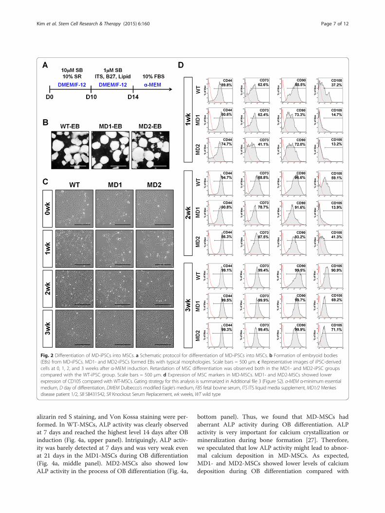
Fig. 2 Differentiation of MD-iPSCs into MSCs. a Schematic protocol for differentiation of MD-iPSCs into MSCs. b Formation of embryoid bodies(EBs) from MD-iPSCs. MD1- and MD2-iPSCs formed EBs with typical morphologies. Scale bars = 500 μm. c Representative images of iPSC-derivedcells at 0, 1, 2, and 3 weeks after α-MEM induction. Retardation of MSC differentiation was observed both in the MD1- and MD2-iPSC groupscompared with the WT-iPSC group. Scale bars = 500 μm. d Expression of MSC markers in MD-MSCs. MD1- and MD2-MSCs showed lowerexpression of CD105 compared with WT-MSCs. Gating strategy for this analysis is summarized in Additional file 3 (Figure S2). α-MEM α-minimum essentialmedium, D day of differentiation, DMEM Dulbecco’s modified Eagle’s medium, FBS fetal bovine serum, ITS ITS liquid media supplement, MD1/2 Menkesdisease patient 1/2, SB SB431542, SR Knockout Serum Replacement, wk weeks, WT wild type
Kim et al. Stem Cell Research & Therapy (2015) 6:160 Page 7 of 12
alizarin red S staining, and Von Kossa staining were per-formed. In WT-MSCs, ALP activity was clearly observedat 7 days and reached the highest level 14 days after OBinduction (Fig. 4a, upper panel). Intriguingly, ALP activ-ity was barely detected at 7 days and was very weak evenat 21 days in the MD1-MSCs during OB differentiation(Fig. 4a, middle panel). MD2-MSCs also showed lowALP activity in the process of OB differentiation (Fig. 4a,
bottom panel). Thus, we found that MD-MSCs hadaberrant ALP activity during OB differentiation. ALPactivity is very important for calcium crystallization ormineralization during bone formation [27]. Therefore,we speculated that low ALP activity might lead to abnor-mal calcium deposition in MD-MSCs. As expected,MD1- and MD2-MSCs showed lower levels of calciumdeposition during OB differentiation compared with
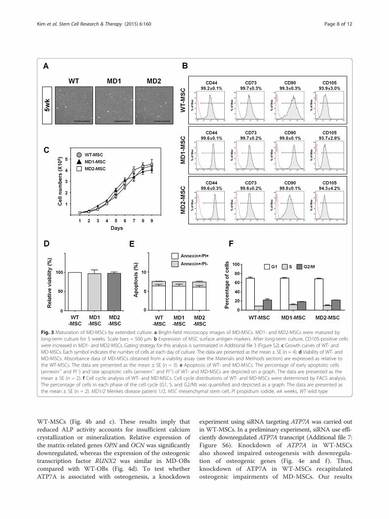
Fig. 3 Maturation of MD-MSCs by extended culture. a Bright-field microscopy images of MD-MSCs. MD1- and MD2-MSCs were matured bylong-term culture for 5 weeks. Scale bars = 500 μm. b Expression of MSC surface antigen markers. After long-term culture, CD105-positive cellswere increased in MD1- and MD2-MSCs. Gating strategy for this analysis is summarized in Additional file 3 (Figure S2). c Growth curves of WT- andMD-MSCs. Each symbol indicates the number of cells at each day of culture. The data are presented as the mean ± SE (n = 4). d Viability of WT- andMD-MSCs. Absorbance data of MD-MSCs obtained from a viability assay (see the Materials and Methods section) are expressed as relative tothe WT-MSCs. The data are presented as the mean ± SE (n = 3). e Apoptosis of WT- and MD-MSCs. The percentage of early apoptotic cells(annexin+ and PI−) and late apoptotic cells (annexin+ and PI+) of WT- and MD-MSCs are depicted on a graph. The data are presented as themean ± SE (n = 2). f Cell cycle analysis of WT- and MD-MSCs. Cell cycle distributions of WT- and MD-MSCs were determined by FACS analysis.The percentage of cells in each phase of the cell cycle (G1, S, and G2/M) was quantified and depicted as a graph. The data are presented asthe mean ± SE (n = 2). MD1/2 Menkes disease patient 1/2, MSC mesenchymal stem cell, PI propidium iodide, wk weeks, WT wild type
Kim et al. Stem Cell Research & Therapy (2015) 6:160 Page 8 of 12
WT-MSCs (Fig. 4b and c). These results imply thatreduced ALP activity accounts for insufficient calciumcrystallization or mineralization. Relative expression ofthe matrix-related genes OPN and OCN was significantlydownregulated, whereas the expression of the osteogenictranscription factor RUNX2 was similar in MD-OBscompared with WT-OBs (Fig. 4d). To test whetherATP7A is associated with osteogenesis, a knockdown
experiment using siRNA targeting ATP7A was carried outin WT-MSCs. In a preliminary experiment, siRNA use effi-ciently downregulated ATP7A transcript (Additional file 7:Figure S6). Knockdown of ATP7A in WT-MSCsalso showed impaired osteogenesis with downregula-tion of osteogenic genes (Fig. 4e and f ). Thus,knockdown of ATP7A in WT-MSCs recapitulatedosteogenic impairments of MD-MSCs. Our results

Fig. 4 Impaired osteogenesis in MD-MSCs. a Representative images of ALP activity in WT- and MD-MSCs during OB differentiation. ALP activitywas observed as red granules. D, days after osteogenesis induction. b Representative images of alizarin red S staining in WT- and MD-MSCs duringOB differentiation. Alizarin red S staining presented as red granules. c Representative images of Von Kossa staining in WT- and MD-MSCs duringOB differentiation. Von Kossa staining was observed as black dots. Scale bars = 500 μm. d Relative expression of osteogenic genes RUNX2, OPN,and OCN in MD-MSCs during osteogenesis. The data are presented as the mean ± SE (n = 3). e, f Effects of ATP7A knock-down on osteogenesisin WT-MSCs. e Representative images of ALP activity, alizarin red S staining, and Von Kossa staining after transfection of siRNAs targeting ATP7Agene. Scramble siRNA (si-SCR) was also transfected in WT-MSCs as a control. f Relative expression of RUNX2, OPN, and OCN after ATP7A knockdown.The data are represented as the mean ± SE (n = 3). *p < 0.05, **p < 0.01. ALP alkaline phosphatase, D day of differentiation, MD1/2 Menkes diseasepatient 1/2, WT wild type
Kim et al. Stem Cell Research & Therapy (2015) 6:160 Page 9 of 12
indicate that ATP7A plays an important role in boneformation.Previous findings have shown that bone morpho-
genetic protein 2 (BMP2) induces OB differentiation ofMSCs [28, 29]. We therefore examined whether impairedosteogenesis in MD-MSCs is caused by insufficient activa-tion of the BMP2 signaling pathway. No differences
were detected in the activity of p-SMAD1 between theWT- and MD-MSCs (Additional file 8: Figure S7A). Inaddition, chondrogenesis appeared normal in MD-MSCs (Additional file 8: Figure S7B). These results sug-gest that decrements of ALP activity and mineralizationadversely affect osteogenesis in MD-MSCs. Next, activ-ity of LOX, a copper-dependent enzyme, was measured

Kim et al. Stem Cell Research & Therapy (2015) 6:160 Page 10 of 12
to test whether it is associated with osteogenesis. TheMD1- and MD2-OBs showed lower LOX activity thanWT-OBs (Additional file 9: Figure S8A), but the amountof matrix collagen was not different between WT- andMD-OBs (Additional file 9: Figure S8B). Therefore, it isconceivable that impaired mineralization is not due toreduced deposition of collagen in MD-MSCs. Takentogether, we suggest that the ATP7A mutation causesdecreased ALP activity and mineralization, eventuallyresulting in impaired osteogenesis in MD.
DiscussionHere, we provide novel insight into the impairedosteogenesis in MD using iPSCs. ATP7A, which is amajor copper transporter, plays important roles incopper absorption and delivery of copper to the hu-man body [6, 8]. Copper is one of the essential traceelements in normal development, and its homeostasisshould be tightly regulated [30]. Disability of copperutilization in MD patients who have a defective ATP7Agene causes severe multisystemic phenotypes such as con-nective tissue abnormalities. In this study, MD-iPSCs withATP7A mutations showed retardation of MSC develop-ment (Fig. 2d), although MD-MSCs matured after ex-tended culturing in vitro (Fig. 3). Subsequently, severalosteogenic defects, including decreased ALP activity andweak calcium mineralization, were observed in MD-MSCsand ATP7A-knockdown WT-MSCs during OB differenti-ation (Fig. 4).ALP activity appears to be associated with calcium de-
position during osteogenesis. During osteogenesis, ALPproduces inorganic phosphate (Pi) from pyrophosphate(PPi), and controls the balance between Pi and PPi levelsin the ECM [31, 32]. Pi is further crystallized with cal-cium and accelerates calcium mineralization in theECM. Dysfunction of the ALP gene causes a genetic dis-order called hypophosphatasia, which is characterized byabnormal bone formation [27]. Here, low activity of ALPresulted in decreased calcium mineralization in MD-MSCs during OB differentiation as shown by alizarin redS and Von Kossa staining (Fig. 4).LOX mediates cross-linking of collagen and elastin in
the ECM, which enhance tensile strength and structuralintegrity of connective tissues [3]. It has been postulatedthat decreased LOX activity accounts for the impairedconnective tissue in MD patients [33, 34]. Reducedactivity of LOX is also implicated in abnormal vascu-lopathy such as the vascular tortuosity and peripheralaneurysms in MD and its allelic variant, occipital hornsyndrome (OHS) [35]. Aberrant internal elastic laminastructure is observed in the MD patient and theMenkes mouse model [36]. Cultured fibroblasts of MDand OHS show abnormalities in the expression of con-nective tissue genes [37]. Furthermore, bladder diverticula,
inguinal hernia, skin laxity, hyperelasticity, and occipitalexostosis are caused by reduced LOX activity [38].In this study, LOX activity was decreased in MD-MSCs
during OB differentiation. Nonetheless, there were nochanges in collagen deposition in this study. These resultsraise the possibility that another role of LOX might beinvolvement in the aberrant OB differentiation in MD-MSCs. In fact, it has been reported that LOX activity isinvolved in many biological functions other than collagencross-linking, such as metastasis, tumor cell growth, cellmigration and motility, angiogenesis, cell signaling, andtranscription [39–42]. Thus, the identification of a newrole for LOX in MD-MSCs during OB differentiationwould be very interesting.Taken together, these data show that utilization of
intracellular copper is unavailable in MD cells due todysfunctional ATP7A. This study provides additionalinsight into the pathophysiology of bone defects causedby the ATP7A mutation in MD.
ConclusionsHere we described the important role of ATP7A and cop-per during osteogenesis using MD-derived iPSCs. DuringOB differentiation, several osteogenic impairments suchas low ALP activity, reduced calcium mineralization anddecreased expression of osteogenic marker genes were ob-served. Knockdown of ATP7A in WT-MSCs recapitulatedthe impaired osteogenesis observed in MD-MSCs. Ourresults provide new insight into the important role ofATP7A in bone formation.
Additional files
Additional file 1: Figure S1. Characterization of WT-iPSCs. (A) Expressionof pluripotent markers in WT-iPSCs. (B) Teratoma formation of MD-iPSCs inimmunodeficient mice. (C) Epigenetic reprogramming in WT-iPSCs.(TIFF 2486 kb)
Additional file 2: Table S1. Primers used in RT-PCR analysis. Table S2Primers used in ATP7A genotyping of MD-derived cells. Table S3 Primersused in methylation analysis. Table S4 Copper concentration in WT- andMD-MSCs. (DOCX 19 kb)
Additional file 3: Figure S2. Detailed gating strategy for each MSCsurface antigens. Gating strategy for CD44 surface antigen shown as arepresentative. Live cells (gate A) were gated based on forward scatterand side scatter. Staining with isotype control (PE-conjugated) was usedto exclude CD44-negative events. This gate was then applied to samplesstained with anti-CD44 antibody (PE-conjugated) to identify CD44 positiveevents. In merged image, red line indicates isotype control and black tintedarea indicates CD44 positive events. (TIFF 1455 kb)
Additional file 4: Figure S3. Characterization of MD-iPSCs. (A) Schematicdefective regions of ATP7A in MD1 and MD2 patients. The functionaldomains of ATP7A are depicted. ATP7A has six copper-binding domains,one phosphatase domain, one phosphorylation domain, one ATP-bindingdomain, and eight transmembrane domains. The defective regions of eachpatient are marked as boxes. (B) Transcriptional expression of pluripotentgenes in MD-fibroblasts and MD-iPSCs. GAPDH was used as a control. (C) Invitro differentiation of MD-iPSCs. Immunostaining of NESTIN (ectoderm, red),α-SMA (mesoderm, green), and GATA4 (endoderm, green) was performed 7days after spontaneous EB differentiation. DAPI showed nuclear counterstaining

Kim et al. Stem Cell Research & Therapy (2015) 6:160 Page 11 of 12
(blue). Scale bar = 500 μm. (D) Karyotypes of MD1- and MD2-iPSCs. (E) Geneticmutations in MD1-fibroblasts and MD1-iPSCs. A single-base substitution wasconfirmed in MD1-fibroblasts and MD1-iPSCs (Figure E-a). The resultant PCRproducts were different in MD1-fibroblasts and MD1-iPSCs (Figure E-b). The sizedifference (450 bp in WT and 246 bp in MD1) generated by exon skippingwas analyzed by gel electrophoresis. (F) Mutations in MD2-fibroblasts andMD2-iPSCs. Strategy for duplex PCR was explained as an illustration(Figure F-a). Size differences (532 bp in WT and 224 bp in MD2)generated by a large genomic deletion between WT- and MD2-fibroblastswere detected (Figure F-b). The primers used in this study are listed inAdditional file 1 (Table S2). (TIFF 4321 kb)
Additional file 5: Figure S4. Characterization of EBs during MSCdifferentiation. (A) SMAD2 phosphorylation in WT- and MD-EBs duringMSC differentiation. SB treatment suppressed p-SMAD2 in WT- and MD-EBs.(B) Relative expression of neuro-ectoderm and cardiac mesoderm markergenes (NEUROD1 and cTNT, respectively) in WT- and MD-EBs. The data arepresented as the mean ± SE (n = 3); *p < 0.05, **p < 0.01. (TIFF 2052 kb)
Additional file 6: Figure S5. Confirmation of MD-MSCs. (A) Geneticmutation of the ATP7A gene in MD1-MSCs. A single-base substitution wasconfirmed again in MD1-MSCs. (B) Genetic mutation of the ATP7A genein MD2-MSCs. Size differences in the PCR product were observed inMD2-MSCs. (C) Expression of ATP7A in WT- and MD-MSCs. ATP7A proteinwas not detected in MD1- and MD2-MSCs. (TIFF 936 kb)
Additional file 7: Figure S6. Relative expression of ATP7A after siRNAtransfection. The data are represented as the mean ± SE (n = 3); **p < 0.01.(TIFF 326 kb)
Additional file 8: Figure S7. Osteogenesis and chondrogenesis fromMSCs. (A) SMAD1 phosphorylation in WT- and MD-MSCs during osteogenesis.(B) Differentiation of WT- and MD-MSCs into chondrocytes. WT- and MD-MSCsdifferentiated into chondrocytes following the procedures described in theMaterials and Methods section. Morphologies of chondrocyte spheroids weresimilar between WT- and MD-MSCs (upper panel). Scale bar = 500 μm.Additionally, MD-MSCs stained normally with Alcian blue solution(lower panel). Scale bar = 50 μm. (TIFF 1998 kb)
Additional file 9: Figure S8. Activity of LOX and matrix collagen inWT- and MD-OBs. (A) LOX activity in WT- and MD-OBs. Fluorescencedata showing the LOX activity of MD-OBs (see the Materials andMethods section) are expressed as values relative to those of WT-OBs.The data are presented as the mean ± SE (n = 3); *p < 0.05, **p < 0.01.(B) Relative amount of matrix collagen in WT- and MD-OBs. The absorbancedata obtained from a matrix collagen assay (see the Materials and Methodssection) are expressed as values relative to that of WT-OBs. The data arepresented as the mean ± SE (n = 3). (TIFF 813 kb)
AbbreviationsALP: Alkaline phosphatase; α-MEM: α-Minimum essential medium;BMP2: Bone morphogenetic protein 2; BSA: Bovine serum albumin;DAPI: 4′-6-diamidino-2-phenylindole; DMEM: Dulbecco’s modified Eagle’smedium; DW: Distilled water; EBs: Embryoid bodies; ECM: Extracellular matrix;ESC: Embryonic stem cell; FBS: Fetal bovine serum; FGF: Fibroblast growthfactor; H&E: Hematoxylin and eosin; HRP: Horseradish peroxidase;ICP: Inductively coupled plasma; iPSC: Induced pluripotent stem cell;LOX: Lysyl oxidase; MD: Menkes disease; MMC: Mitomycin C;MSC: Mesenchymal stem cell; OB: Osteoblast; OHS: Occipital horn syndrome;PBS: Phosphate-buffered saline; PBST: Phosphate-buffered saline + 0.1 %Tween 20; Pi: Inorganic phosphate; PI: Propidium iodide; PPI: Pyrophosphate;RT-PCR: Real-time polymerase chain reaction; SB: SB431542; WT: Wild type.
Competing interestsThe authors declare that they have no competing interests.
Authors’ contributionsDK designed the study, carried out disease modeling, interpreted data anddrafted the manuscript. JC carried out differentiation study, interpreted data,and helped to revise the manuscript. KMH performed the characterizationstudy, interpreted data, and drafted the manuscript. BHL studied patientsamples, interpreted data, and helped to draft the manuscript. JHCparticipated in genetic analysis and helped to revise the manuscript. HWYhelped to design the study and drafted the manuscript. YMH managed the
project, designed the study, interpreted data, and drafted the manuscript. Allauthors read and approved the final manuscript.
AcknowledgementsWe thank Ms. Sora Oh for the maintenance of human iPSCs. This work wassupported by the NRF Stem Cell Program (2011–0019509) funded by MSIPand a grant (A120275) from Korea Healthcare technology R&D project, MHW,Republic of Korea.
Author details1Department of Biological Science, Korea Advanced Institute of ScienceTechnology (KAIST), Daejeon 305-701, Republic of Korea. 2Department ofPediatrics, Asan Medical Center Children’s Hospital, University of UlsanCollege of Medicine, Seoul, South Korea.
Received: 22 January 2015 Revised: 30 April 2015Accepted: 5 August 2015
References1. Danks DM, Campbell PE, Stevens BJ, Mayne V, Cartwright E. Menkes’s kinky
hair syndrome. An inherited defect in copper absorption with widespreadeffects. Pediatrics. 1972;50:188–201.
2. Kaler SG. Diagnosis and therapy of Menkes syndrome, a genetic form ofcopper deficiency. Am J Clin Nutr. 1998;67:1029S–34.
3. La Fontaine S, Mercer JF. Trafficking of the copper-ATPases, ATP7A andATP7B: role in copper homeostasis. Arch Biochem Biophys. 2007;463:149–67.doi:10.1016/j.abb.2007.04.021.
4. Barry AN, Shinde U, Lutsenko S. Structural organization of humanCu-transporting ATPases: learning from building blocks. J Biol Inorg Chem.2010;15:47–59. doi:10.1007/s00775-009-0595-4.
5. Monty JF, Llanos RM, Mercer JF, Kramer DR. Copper exposure inducestrafficking of the menkes protein in intestinal epithelium of ATP7Atransgenic mice. J Nutr. 2005;135:2762–6.
6. Lutsenko S, Barnes NL, Bartee MY, Dmitriev OY. Function and regulation ofhuman copper-transporting ATPases. Physiol Rev. 2007;87:1011–46.doi:10.1152/physrev.00004.2006.
7. Kaler SG. ATP7A-related copper transport diseases—emerging concepts andfuture trends. Nat Rev Neurol. 2011;7:15–29. doi:10.1038/nrneurol.2010.180.
8. de Bie P, Muller P, Wijmenga C, Klomp LW. Molecular pathogenesis ofWilson and Menkes disease: correlation of mutations with moleculardefects and disease phenotypes. J Med Genet. 2007;44:673–88.doi:10.1136/jmg.2007.052746.
9. Tumer Z, Horn N. Menkes disease: recent advances and new aspects.J Med Genet. 1997;34:265–74.
10. Hunt DM. Primary defect in copper transport underlies mottled mutants inthe mouse. Nature. 1974;249:852–4.
11. Mercer JF. Menkes syndrome and animal models. Am J Clin Nutr.1998;67:1022S–8.
12. Rowe DW, McGoodwin EB, Martin GR, Grahn D. Decreased lysyl oxidaseactivity in the aneurysm-prone, mottled mouse. J Biol Chem.1977;252:939–42.
13. Kreuder J, Otten A, Fuder H, Tumer Z, Tonnesen T, Horn N, et al. Clinicaland biochemical consequences of copper-histidine therapy in Menkesdisease. Eur J Pediatr. 1993;152:828–32.
14. Danks DM, Campbell PE, Walker-Smith J, Stevens BJ, Gillespie JM, BlomfieldJ, et al. Menkes’ kinky-hair syndrome. Lancet. 1972;1:1100–2.
15. Nassogne MC, Sharrard M, Hertz-Pannier L, Armengaud D, Touati G,Delonlay-Debeney P, et al. Massive subdural haematomas in Menkes diseasemimicking shaken baby syndrome. Childs Nerv Syst. 2002;18:729–31.doi:10.1007/s00381-002-0630-z.
16. Amador E, Domene R, Fuentes C, Carreno JC, Enriquez G. Long-term skeletalfindings in Menkes disease. Pediatr Radiol. 2010;40:1426–9.doi:10.1007/s00247-010-1551-8.
17. Kanumakala S, Boneh A, Zacharin M. Pamidronate treatment improves bonemineral density in children with Menkes disease. J Inherit Metab Dis.2002;25:391–8. doi:10.1023/A:1020103901969.
18. Moller LB, Lenartowicz M, Zabot MT, Josiane A, Burglen L, Bennett C, et al.Clinical expression of Menkes disease in females with normal karyotype.Orphanet J Rare Dis. 2012;7:6. doi:10.1186/1750-1172-7-6.

Kim et al. Stem Cell Research & Therapy (2015) 6:160 Page 12 of 12
19. Baerlocher KE, Steinmann B, Rao VH, Gitzelmann R, Horn N. Menkesdisease— clinical, therapeutic and biochemical studies. J Inherit Metab Dis.1983;6:87–8. doi:10.1007/Bf01810339.
20. Kaler SG. Metabolic and molecular bases of Menkes disease and occipitalhorn syndrome. Pediatr Dev Pathol. 1998;1:85–98.
21. Grskovic M, Javaherian A, Strulovici B, Daley GQ. Induced pluripotent stemcells—opportunities for disease modelling and drug discovery. Nat RevDrug Discov. 2011;10:915–29. doi:10.1038/nrd3577.
22. Maury Y, Gauthier M, Peschanski M, Martinat C. Human pluripotent stemcells for disease modelling and drug screening. Bioessays. 2012;34:61–71.doi:10.1002/bies.201100071.
23. Kim H, Kim D, Jang MJ, Han YM. Variations in the epigenetic regulation oflineage-specific genes among human pluripotent stem cell lines. BiochemBiophys Res Commun. 2012;424:331–7. doi:10.1016/j.bbrc.2012.06.122.
24. Mahmood A, Harkness L, Schroder HD, Abdallah BM, Kassem M. Enhanceddifferentiation of human embryonic stem cells to mesenchymal progenitorsby inhibition of TGF-beta/activin/nodal signaling using SB-431542. J BoneMiner Res. 2010;25:1216–33. doi:10.1002/jbmr.34.
25. Tran NT, Trinh QM, Lee GM, Han YM. Efficient differentiation of humanpluripotent stem cells into mesenchymal stem cells by modulatingintracellular signaling pathways in a feeder/serum-free system. Stem CellsDev. 2012;21:1165–75. doi:10.1089/scd.2011.0346.
26. Kim JH, Lee BH, Kim YM, Choi JH, Kim GH, Cheon CK, et al. Novel mutationsand clinical outcomes of copper-histidine therapy in Menkes diseasepatients. Metab Brain Dis. 2014;30:75–81. doi:10.1007/s11011-014-9569-5.
27. Whyte MP. Hypophosphatasia and the role of alkaline phosphatase inskeletal mineralization. Endocr Rev. 1994;15:439–61.doi:10.1210/edrv-15-4-439.
28. Gersbach CA, Guldberg RE, Garcia AJ. In vitro and in vivo osteoblasticdifferentiation of BMP-2- and Runx2-engineered skeletal myoblasts. J CellBiochem. 2007;100:1324–36. doi:10.1002/jcb.21118.
29. Chen D, Harris MA, Rossini G, Dunstan CR, Dallas SL, Feng JQ, et al. Bonemorphogenetic protein 2 (BMP-2) enhances BMP-3, BMP-4, and bone celldifferentiation marker gene expression during the induction ofmineralized bone matrix formation in cultures of fetal rat calvarialosteoblasts. Calcif Tissue Int. 1997;60:283–90.
30. Festa RA, Thiele DJ. Copper: an essential metal in biology. Curr Biol.2011;21:R877–83. doi:10.1016/j.cub.2011.09.040.
31. Murshed M, Harmey D, Millan JL, McKee MD, Karsenty G. Uniquecoexpression in osteoblasts of broadly expressed genes accounts for thespatial restriction of ECM mineralization to bone. Genes Dev. 2005;19:1093–104.doi:10.1101/gad.1276205.
32. Russell RG, Bisaz S, Donath A, Morgan DB, Fleisch H. Inorganic pyrophosphatein plasma in normal persons and in patients with hypophosphatasia,osteogenesis imperfecta, and other disorders of bone. J Clin Invest.1971;50:961–9. doi:10.1172/JCI106589.
33. Kagan HM, Li WD. Lysyl oxidase: Properties, specificity, and biological rolesinside and outside of the cell. J Cell Biochem. 2003;88:660–72.doi:10.1002/Jcb.10413.
34. Lucero HA, Kagan HM. Lysyl oxidase: an oxidative enzyme and effector of cellfunction. Cell Mol Life Sci. 2006;63:2304–16. doi:10.1007/s00018-006-6149-9.
35. Godwin SC, Shawker T, Chang B, Kaler SG. Brachial artery aneurysms inMenkes disease. J Pediatr. 2006;149:412–5. doi:10.1016/j.jpeds.2006.05.041.
36. Murakami H, Kodama H, Nemoto N. Abnormality of vascular elastic fibers inthe macular mouse and a patient with Menkes’ disease: ultrastructural andimmunohistochemical study. Med Electron Microsc. 2002;35:24–30.doi:10.1007/s007950200003.
37. Kemppainen R, Hamalainen ER, Kuivaniemi H, Tromp G, Pihlajaniemi T,Kivirikko KI. Expression of mRNAs for lysyl oxidase and type III procollagenin cultured fibroblasts from patients with the menkes and occipital hornsyndromes as determined by quantitative polymerase chain reaction. ArchBiochem Biophys. 1996;328:101–6. doi:10.1006/abbi.1996.0148.
38. Kuivaniemi H, Peltonen L, Palotie A, Kaitila I, Kivirikko KI. Abnormalcopper-metabolism and deficient lysyl oxidase activity in a heritableconnective-tissue disorder. J Clin Investig. 1982;69:730–3.doi:10.1172/Jci110503.
39. Rodriguez C, Martinez-Gonzalez J, Raposo B, Alcudia JF, Guadall A,Badimon L. Regulation of lysyl oxidase in vascular cells: lysyl oxidase as anew player in cardiovascular diseases. Cardiovasc Res. 2008;79:7–13.doi:10.1093/cvr/cvn102.
40. Baker AM, Cox TR, Bird D, Lang G, Murray GI, Sun XF, et al. The role of lysyloxidase in SRC-dependent proliferation and metastasis of colorectal cancer.J Natl Cancer Inst. 2011;103:407–24. doi:10.1093/jnci/djq569.
41. Baker AM, Bird D, Welti JC, Gourlaouen M, Lang G, Murray GI, et al. Lysyloxidase plays a critical role in endothelial cell stimulation to drive tumorangiogenesis. Cancer Res. 2013;73:583–94.doi:10.1158/0008-5472.CAN-12-2447.
42. Erler JT, Giaccia AJ. Lysyl oxidase mediates hypoxic control of metastasis.Cancer Res. 2006;66:10238–41. doi:10.1158/0008-5472.CAN-06-3197.
Submit your next manuscript to BioMed Centraland take full advantage of:
• Convenient online submission
• Thorough peer review
• No space constraints or color figure charges
• Immediate publication on acceptance
• Inclusion in PubMed, CAS, Scopus and Google Scholar
• Research which is freely available for redistribution
Submit your manuscript at www.biomedcentral.com/submit