implants in periodontal patients
DESCRIPTION
PeriodonticsTRANSCRIPT

Seediscussions,stats,andauthorprofilesforthispublicationat:http://www.researchgate.net/publication/6823391
Outcomeofimplanttherapyinpatientswithprevioustoothlossduetoperiodontitis
ARTICLEinCLINICALORALIMPLANTSRESEARCH·NOVEMBER2006
ImpactFactor:3.12·DOI:10.1111/j.1600-0501.2006.01347.x·Source:PubMed
CITATIONS
94
4AUTHORS,INCLUDING:
PalleHolmstrup
UniversityofCopenhagen
197PUBLICATIONS4,885CITATIONS
SEEPROFILE
HelenVWorthington
TheUniversityofManchester
419PUBLICATIONS9,174CITATIONS
SEEPROFILE
MarcoEsposito
UniversityofGothenburg
227PUBLICATIONS5,717CITATIONS
SEEPROFILE
Availablefrom:PalleHolmstrup
Retrievedon:25August2015

Outcome of implant therapy in patientswith previous tooth loss due toperiodontitis
S�ren SchouPalle HolmstrupHelen V. WorthingtonMarco Esposito
Authors’ affiliations:S�ren Schou, Department of Oral and MaxillofacialSurgery, Aalborg Hospital, Aarhus UniversityHospital, Aalborg, DenmarkPalle Holmstrup, Department of Periodontology,School of Dentistry, University of Copenhagen,Copenhagen, DenmarkHelen V. Worthington, Marco Esposito, School ofDentistry, University of Manchester, Manchester,UK
Correspondence to:S�ren SchouDepartment of Oral and Maxillofacial SurgeryAalborg HospitalAarhus University Hospital18-22 HobrovejDK-9000 AalborgDenmarkTel.: þ45 99 32 35 51Fax: þ45 99 32 28 04e-mail: [email protected]
Key words: complications, dental implants, oral implants, osseointegration, pathology,
peri-implantitis, periodontal diseases, systematic review
Abstract
Background: It is frequently debated whether implant treatment in individuals with
previous tooth loss due to periodontitis is characterized by an increased incidence of
implant loss and peri-implantitis.
Objective: The objective of the present systematic review was to assess whether individuals
with previous tooth loss due to periodontitis have an increased risk of loss of
suprastructures, loss of implants, peri-implantitis, and peri-implant marginal bone loss as
compared with individuals with previous tooth loss due to reasons other than periodontitis.
Search strategy: Studies considered for inclusion were searched in MEDLINE (PubMed) and
relevant journals were hand searched. Moreover, reference lists of articles selected for full-
text screening as well as previously published reviews relevant for the present systematic
review were searched. The search was performed by one reviewer and was restricted to
human studies published from January 1, 1980 to January 1, 2006. No language restrictions
were applied.
Selection criteria: Prospective and retrospective cohort studies with at least a 5-year follow-
up comparing the outcome of implant treatment in individuals with periodontitis-
associated and non-periodontitis-associated tooth loss, respectively, were included. The
outcome measures were survival of suprastructures, survival of implants, occurrence of peri-
implantitis, and peri-implant marginal bone loss. The 5- and 10-year time points were
evaluated.
Data collection and analysis: Screening of eligible studies, methodological quality
assessment, and data extraction were conducted in duplicate and independently by two of
the authors. The authors were contacted for missing information. Results were expressed as
random effect models using weighted mean differences for continuous outcomes and
relative risk for dichotomous outcomes with 95% confidence intervals (CIs).
Main results: Two studies with a 5- and 10-year follow-up, respectively, were identified
including a total of 33 patients with tooth loss due to periodontitis and 70 patients with
non-periodontitis-associated tooth loss. There was no significant difference in the survival
of the suprastructures after 5 years. Furthermore, there were no significant differences in
the survival of the implants after 5 and 10 years. However, there were significantly more
patients affected by peri-implantitis in the group with periodontitis-associated tooth loss
during the 10-year follow-up period, risk ratio (RR) 9 (95% CI 3.94–20.57). Moreover,
significantly increased peri-implant marginal bone loss was observed in patients with
periodontitis-associated tooth loss after 5 years, mean difference 0.5 mm (95% CI 0.06–
0.94).
Conclusions: The survival of the suprastructures and the implants was not significantly
different in individuals with periodontitis-associated and non-periodontitis-associated
tooth loss. However, significantly increased incidence of peri-implantitis and significantly
increased peri-implant marginal bone loss were revealed in individuals with periodontitis-
associated tooth loss. The small sample size and the methodological quality assessment of
the two studies suggest that the results should be interpreted with caution. Consequently,
further long-term studies focusing particularly on the outcome of implant treatment in
young adults with aggressive periodontitis are needed before final conclusions can be
drawn about the outcome of implant treatment in patients with a history of periodontitis.r 2006 The Authors
Journal compilation r Blackwell Munksgaard 2006
To cite this article:Schou S, Holmstrup P, Worthington HV, Esposito M.Outcome of implant therapy in patients with previoustooth loss due to periodontitis.Clin. Oral Imp. Res. 17 (Suppl. 2), 2006; 104–123
104

The first long-term study on implant treat-
ment involving fixed complete dentures
was published in 1981 (Adell et al. 1981).
During the following decades, implant
treatment has been assessed in numerous
reports involving both totally and partially
edentulous patients (Esposito et al. 1998;
Berglundh et al. 2002). A systematic review
on clinical studies with at least a 5-year
follow-up showed that 5% of the implants
with fixed dentures were lost before loading
or during function (Berglundh et al. 2002).
It was concluded that biological and me-
chanical complications occurred, but im-
plant treatment in general was to be
considered a treatment modality with pre-
dictable outcome and high survival rates.
In most long-term studies, the causality
behind tooth loss before implant placement
has not been specified, and presumably
some of the included patients have lost
some or all teeth due to periodontitis.
The susceptibility to periodontitis-asso-
ciated attachment and tooth loss shows
major individual variation, and accordingly
periodontitis has been classified into ag-
gressive and chronic subtypes (Armitage
1999). It is frequently debated whether
implant therapy in individuals with pre-
viously periodontitis-associated tooth loss
is associated with an increased incidence of
implant loss and peri-implantitis.
Objective
The objective of the present systematic
review was to assess whether individuals
with previous tooth loss due to perio-
dontitis have an increased risk of loss of
suprastructures, loss of implants, peri-im-
plantitis, and peri-implant marginal bone
loss as compared with individuals with
previous tooth loss due to reasons other
than periodontitis.
Criteria for considering studiesfor this review
Types of studies, participants, andintervention
Prospective and retrospective cohort stu-
dies with at least a 5-year follow-up com-
paring the outcome of implant treatment in
partially edentulous individuals with,
respectively, periodontitis-associated and
non-periodontitis-associated tooth loss
were assessed. More than 10 patients
should be included in the study, and the
treatment should involve osseointegrated
oral implants.
Types of outcome measures
The outcome measures included the
following:
� Loss of suprastructures.
� Loss of implants defined as implant
mobility of previously clinically os-
seointegrated implants and removal of
non-mobile implants due to progressive
peri-implant marginal bone loss and
infection.
� Occurrence of peri-implantitis defined
as progressive peri-implant marginal
bone loss associated with infection
signs.
� Radiographic peri-implant marginal
bone loss on intraoral radiographs taken
with a paralleling technique.
Search strategy foridentification of studies
The search strategy used for identification
of studies is summarized in Fig. 1. Studies
Hand-searched journals (1980-2005):
Br J Oral Maxillofac Surg (1984-2005)Br J Oral Surg (1980-1983)Clin Implant Dent Relat Res (1999-2005)Clin Oral Implants Res (1990-2005)Implant Dent (1992-2005)Int J Oral Maxillofac Implants (1986-2005)Int J Oral Maxillofac Surg (1986-2005)Int J Oral Surg (1980-1985)Int J Periodontics Restorative Dent (1985-2005)Int J Prosthodont (1988-2005)J Clin PeriodontolJ Craniomaxillofac Surg (1987-2005)J Maxillofac Surg (1980-1986)J Periodontal ResJ PeriodontolJ Oral ImplantolJ Oral Surg (1980-1981)J Oral Maxillofac Surg (1982-2005)J Prosthet DentJ Prosthodont (1992-2005)
Medline (PubMed) search (1980-2005, human trials):2116
Abstracts reviewed:
Articles reviewed:
Articles included:
547
49
2
(0)
Number
(exp Periodontal diseases) AND(exp Dental implants OR exp Dental implantation OR “Oral implants”)
Fig. 1. Search strategy for identification of studies.
Schou et al . Periodontitis and implants
105 | Clin. Oral Impl. Res. 17 (Suppl. 2), 2006 / 104–123

considered for inclusion were searched in
MEDLINE (PubMed) with a broad search
strategy involving controlled vocabulary
(MeSH) and free text terms:
#1 exp Periodontal diseases
#2 exp Dental implantation
#3 exp Dental implants
#4 ‘Oral implants’
#5 2 OR 3 OR 4
#6 1 AND 5.
In addition, relevant journals were hand
searched page by page for relevant studies
(Fig. 1). Manual search also included the
bibliographies of all articles selected for
full-text screening as well as previously
published reviews relevant for the present
systematic review. Finally, the ‘related
article’ feature of PubMed was used for
all articles selected for full-text screening.
The search was performed by one reviewer
(S. S.) and was restricted to human studies
published from January 1, 1980 to January
1, 2006. No language restrictions were
applied.
Methods of the review
The titles of the identified reports were
initially screened (Fig. 1). The abstract was
assessed when the title indicated that the
study fulfilled the inclusion criteria. Full-
text analysis was carried out when an ab-
stract was unavailable or when the abstract
indicated that the above-described inclusion
criteria were fulfilled. The study selection
was performed by one reviewer (S. S.).
Quality assessment
The quality assessment of the included
studies was undertaken independently
and in duplicate by two authors (S. S., M.
E.) as part of the data-extraction process.
When disagreement between the two re-
viewers was revealed, consensus was
achieved by discussion. Additional infor-
mation provided by the authors of the
studies was taken into account, as de-
scribed in the following paragraph on data
extraction. The quality assessment was
performed according to the following para-
meters:
� Description of tooth loss, attachment
loss, and health status of periodontal
tissues at the time of implant place-
ment for each treatment group (yes, no).
� Blinding of outcome assessment (yes,
no).
� Completeness of follow-up. A clear
explanation for withdrawals and drop-
outs in each treatment group (yes, no).
The studies were grouped according to:
� Low risk of bias (plausible bias unlikely
to alter the results seriously) if all
quality criteria were met.
� High risk of bias (plausible bias that
seriously weakens confidence in the
results) if one or more quality criteria
were not met.
Data extraction
Data were extracted independently by two
reviewers (S. S., M. E.) according to a
specially designed data-collection form,
which ensured systematic recording of
data. When disagreement between the
two reviewers was revealed, consensus
was achieved by discussion. Characteris-
tics of patients, their treatment, follow-up
period, and treatment outcome, i.e., survi-
val of suprastructures, survival of implants,
occurrence of peri-implantitis, and peri-
implant marginal bone loss were obtained.
Finally, recording of study quality assess-
ment was included. The authors were con-
tacted for clarification or missing
information.
Data synthesis
For binary outcomes (loss of suprastruc-
tures, loss of implants, occurrence of peri-
implantitis), the estimate of effect of inter-
vention was expressed as risk ratios (RR),
together with 95% confidence intervals
(CIs). For continuous outcomes (peri-im-
plant marginal bone loss), weighted mean
differences (MD) and standard deviations
were used to summarize the data for each
group using MD and 95% CIs. The statis-
tical unit was the patient and not the
implant. Meta-analysis was only at-
tempted if there were studies of similar
comparisons reporting the same outcome
measures. RRs combined for binary data
were for implant loss only.
Description of studies andmethodological quality
The search result is outlined in Fig. 1. A
total of 2116 titles were identified, and 547
abstracts were reviewed. Full-text analysis
included 49 articles, and two studies were
finally included in the review (Hardt et al.
2002; Karoussis et al. 2003). The main
reason for exclusion was: no control group
included with non-periodontitis-associated
tooth loss, less than 10 patients included,
focus on aspects other than implant treat-
ment in patients with periodontitis-asso-
ciated tooth loss, relevant results reported
in other publications, and inclusion of
patients both with and without perio-
dontitis-associated tooth loss. No articles
were added as the result of hand-searching.
The two studies are described below and
summarized in Table 1.
The patients included in the retrospec-
tive study by Hardt et al. (2002) were
selected among patients treated at the Bra-
nemark Clinic, Goteborg, Sweden. A total
of 97 partially edentulous patients with
346 Branemark implants inserted in the
posterior part of the maxilla without bone
regeneration were included. The marginal
bone loss of the remaining teeth at the time
of the implant treatment planning was
assessed on panoramic radiographs and an
age-related periodontal marginal bone loss
score was estimated to describe the perio-
dontal destruction. Two-end quartiles were
used to define a periodontitis group of
individuals with susceptibility to perio-
dontitis (n¼25) and a non-periodontitis
group of individuals with minimal perio-
dontal breakdown (n¼ 25). All patients
were recalled according to the standard
protocol at the Branemark Clinic after 1,
3, and 5 years. In addition, all patients
visited their regular dentist once a year. It
could not be assessed whether adequate
treatment of plaque-induced inflammatory
reactions was performed during the 5-year
period. The peri-implant marginal bone
level was assessed blindly on intraoral
radiographs mesially and distally. The pri-
mary outcome measures were implant loss
and peri-implant marginal bone loss during
a 5-year follow-up period.
A total of 100 and 92 implants, respec-
tively, were inserted in the periodontitis
and non-periodontitis group. Penicillin was
prescribed for 10 days after implant place-
ment, and fixed partial dentures were in-
serted after a 6-month healing period. At
the time of implant installation, the mean
age of the periodontitis and non-perio-
dontitis group was, respectively, 54 and
Schou et al . Periodontitis and implants
106 | Clin. Oral Impl. Res. 17 (Suppl. 2), 2006 / 104–123

Tab
le1.
Su
mm
ary
of
incl
ud
ed
stu
die
s
Pati
en
tsIm
pla
nts
an
dan
tib
ioti
csSu
pra
-st
ruct
ure
Foll
ow
-up
peri
od
(years
)
Resu
lts
Refe
ren
ces
Peri
-im
pla
nt
tiss
ues
Peri
o-
do
nta
lti
ssu
es
Sup
rast
ruct
ure
an
dim
pla
nt
surv
ival
rate
Oth
er
resu
lts
an
dco
mm
en
ts
25
PE
susc
ep
tib
leto
peri
od
on
titi
sTe
eth
wit
ho
50%
bo
ne
sup
po
rt:
26%
No
.o
fte
eth
:16
Ag
e:
54
years
Wo
men
:52%
,m
en
:48%
Smo
kin
g:
NR
100
Bra
nem
ark
Pen
icil
lin
for
10
days
FDin
po
steri
or
part
of
maxi
lla
5B
on
elo
ss:
2.2
mm
NR
Sup
rast
ruct
ure
:92%
Imp
lan
ts:
92%
Sig
nifi
can
tco
rrela
tio
nb
etw
een
peri
-im
pla
nt
bo
ne
loss
an
dp
eri
od
on
tal
bo
ne
loss
befo
reim
pla
nt
thera
py
Hard
tet
al.
(2002)
25
PE
wit
hm
inim
al
peri
od
on
tal
bre
akd
ow
nTe
eth
wit
ho
50%
bo
ne
sup
po
rt:
1%
No
.o
fte
eth
:17
Ag
e:
57
years
Wo
men
:64%
,m
en
:36%
Smo
kin
g:
NR
92
Bra
nem
ark
Pen
icil
lin
for
10
days
Bo
ne
loss
:1.7
mm
Sup
rast
ruct
ure
:100%
Imp
lan
ts:
97%
8PE
wit
hto
oth
loss
du
eto
chro
nic
peri
od
on
titi
sA
ge:
NR
Gen
der:
NR
Pro
po
rtio
no
fim
pla
nts
insm
okers
:48%
21
ITI
(ho
llo
wsc
rew
)A
nti
bio
tics
:N
RFD
,ST
R10
BO
P:
29%
PD
:3
mm
Bo
ne
loss
,m
esi
al:
1m
mB
on
elo
ss,
dis
tal:
0.9
mm
Imp
lan
tsw
ith
peri
-im
pla
nti
tis
(BO
P,PD
�5
mm
,an
db
on
elo
ss):
38%
NR
Sup
rast
ruct
ure
:N
RIm
pla
nts
:91%
Imp
lan
tsw
ith
PD4
5m
m,
BO
P,an
nu
al
bo
ne
loss
�0.2
mm
:48%
Sig
nifi
can
tly
hig
her
inci
den
ceo
fb
iolo
gic
al
com
pli
cati
on
sin
pati
en
tsw
ith
too
thlo
ssd
ue
toch
ron
icp
eri
od
on
titi
s
Karo
uss
iset
al.
(2003)
45
PE
wit
hto
oth
loss
du
eto
oth
er
reaso
ns
than
peri
od
on
titi
sA
ge:
NR
Gen
der:
NR
Pro
po
rtio
no
fim
pla
nts
insm
okers
:20%
91
ITI
(ho
llo
wsc
rew
)A
nti
bio
tics
:N
RB
OP:
40%
PD
:2.5
mm
Bo
ne
loss
,mesi
al:
0.5
mm
Bo
ne
loss
,d
ista
l:0.5
mm
Imp
lan
tsw
ith
peri
-im
pla
nti
tis
(BO
P,PD
�5
mm
,an
db
on
elo
ss):
5%
Sup
rast
ruct
ure
:N
RIm
pla
nts
:97%
Imp
lan
tsw
ith
PD4
5m
m,
BO
P,an
nu
al
bo
ne
loss
�0.2
mm
:21%
All
gro
up
valu
es
refe
rred
toare
exp
ress
ed
as
mean
valu
es.
Cli
nic
al
para
mete
rsre
cord
ed
at
fou
rsi
tes
per
imp
lan
t.
BO
P,b
leed
ing
on
pro
bin
g;
FD,
fixe
dd
en
ture
;N
R,
no
tre
po
rted
;PD
,p
rob
ing
dep
th;
PE,
part
ial
ed
en
tulo
us;
STR
,si
ng
leto
oth
rep
lace
men
t.
Schou et al . Periodontitis and implants
107 | Clin. Oral Impl. Res. 17 (Suppl. 2), 2006 / 104–123

57 years. The gender distribution was
comparable in the periodontitis group (wo-
men: 52%, men: 48%), while women
prevailed in the non-periodontitis group
(women: 64%, men: 36%). The proportion
of smokers was not reported. The number
of remaining teeth was comparable in the
two groups (periodontitis group: 16, non-
periodontitis group: 17). The proportion of
teeth with less than 50% bone support
was, respectively, 26% in the periodontitis
group and 1% in the non-periodontitis
group. Data on the periodontal health sta-
tus could not be obtained either at implant
placement or at 5-year follow-up. No pa-
tients were excluded from the study due to
loss of all implants.
The patients included in the retrospec-
tive study by Karoussis et al. (2003) were
treated at the Department of Perio-
dontology and Fixed Prosthodontics,
School of Dental Medicine, University of
Berne, Switzerland. A total of 53 partially
edentulous patients with 112 ITI hollow
screw implants were included. Two patient
groups were identified, namely individuals
with tooth loss due to chronic periodontitis
(n¼ 8) and individuals with tooth loss due
to other seasons (caries, fracture, tooth
agenesia, and trauma) (n¼ 45). The pa-
tients were incorporated in an individually
designed maintenance care program at the
Department of Periodontology and Fixed
Prosthodontics, School of Dental Medi-
cine, University of Berne, or at the referring
dentist. Patients with periodontitis-asso-
ciated tooth loss were recalled usually at
3–5-month intervals, while patients with
non-periodontitis-associated tooth loss
were not recalled more frequently than at
4–8-month intervals. At every recall exam-
ination during the 10-year period, peri-im-
plantitis was recorded and treated according
to the so-called cumulative interceptive
supportive therapy (CIST) (Lang et al.
2000). Moreover, clinical and radiographic
examination was performed after 1 and 10
years of function. The peri-implant mar-
ginal bone level was assessed on intraoral
radiographs mesially and distally. The as-
sessment was performed blindly. The pri-
mary outcome measures were implant
loss, implant success, and incidence of
peri-implantitis during the 10-year follow-
up period. Peri-implantitis was defined by
probing depths of 5 mm or more, bleeding
on probing, and radiographic signs of mar-
ginal bone loss.
A total of 21 and 91 implants, respec-
tively, were inserted in the periodontitis
and non-periodontitis group without the
use of antibiotics. Single-tooth restorations
or fixed partial dentures were inserted after
a 4–6-month healing period. The age at the
time of implant installation was unre-
ported. When the two groups were consid-
ered together, 67 of the implants (60%)
were inserted in women and 45 (40%) in
men. In the periodontitis group, 48% of the
implants were inserted in smokers, while
the same figure for the non-periodontitis
group was 20%. The number of remaining
teeth at the time of implant placement was
unreported. Finally, the periodontal health
status was not reported either at implant
placement or at 10-year follow-up. No
patients were excluded from the study
due to loss of all implants.
Based on the above-described criteria for
quality assessment, the risk of bias was
judged to be high for both studies (Table 2).
Results
Survival of suprastructures
After the 5-year follow-up period, two of
the 25 suprastructures were removed in the
periodontitis group (8%), while none were
lost in the non-periodontitis group in the
study by Hardt et al. (2002). These data can
be seen in Fig. 2, along with the RR of 5
and its 95% CI from 0.25 to 99.16.
Although the risk of loss of the suprastruc-
tures in the periodontitis group was five
times that of the non-periodontitis group,
the P-value of 0.29 indicates that no sig-
nificant difference in survival of the supra-
structures was observed between the two
groups after 5-year follow-up.
The survival rate of the suprastructures
was unreported by Karoussis et al. (2003).
Survival of implants
After a 5-year follow-up, six patients lost
one implant each and one patient lost two
implants in the periodontitis group in the
study by Hardt et al. (2002). Consequently,
Table 2. Quality assessment of included studies
Quality assessment parameters Hardtet al.(2002)
Karoussiset al.(2003)
Description of tooth loss, attachment loss and health status ofperiodontal tissues at the time of implant placement for eachtreatment group (yes, no)
No No
Blinding of outcome assessment (yes, no) Yes Yes
Completeness of follow-up. A clear explanation for withdrawals anddrop-outs in each treatment group (yes, no)
Yes Yes
Review: testing data01 periodontitis versus non-periodontitis patients01 prosthesis failure
Comparison:Outcome:
01 5 years
25Subtotal (95% CI)
Studyor sub-category
Hardt 2002 2/2525 100.00 5.00
2/25 100.00
0.01 0.1 1 10 100
Favours treatment Favours control
5.00 0[0.25, 99.16][0.25, 99.16]
Total events: 2 (periodontitis), 0 (non-periodontitis)Test for heterogeneity: not applicableTest for overall effect: Z = 1.06 (P = 0.29)
non-periodontitisn/N
periodontitisn/N
RR (random)95% CI
RR (random)95% CI
Weight% Order
Fig. 2. Comparison between patients with and without periodontitis-associated tooth loss: loss of suprastructures.
Schou et al . Periodontitis and implants
108 | Clin. Oral Impl. Res. 17 (Suppl. 2), 2006 / 104–123

eight of the 100 implants (8%) were lost in
seven patients in the periodontitis group,
while three of the 92 implants (3%) were
lost in three patients in the non-perio-
dontitis group. After a 5-year follow-up,
none of the 21 implants were lost in the
periodontitis group, while one of the 92
implants (2%) was lost in the non-perio-
dontitis group within the study by Karous-
sis et al. (2003). A meta-analysis was
conducted for the two studies at 5 years,
and no significant difference in survival of
the implants was observed. The black dia-
mond in Fig. 3 shows the combined RR of
2.24 (95% CI 0.71–7.04), which was not
significant (P¼ 0.17).
After the 10-year follow-up period, two
implants (9.5%) were lost in two indivi-
duals in the periodontitis group and three
implants (3.5%) in three individuals in the
non-periodontitis group within the study
by Karoussis et al. (2003). These data are
shown in Fig. 3, which also shows an RR of
3.75 (95% CI 0.74–19.02), which was not
significant (P¼ 0.11).
Incidence of peri-implantitis
The incidence of peri-implantitis was un-
reported by Hardt et al. (2002).
Peri-implantitis was revealed around
eight of the 21 implants (38%) in eight
patients in the periodontitis group and
around five of the 91 implants (5%) in
five patients in the non-periodontitis group
in the study by Karoussis et al. (2003).
Based on these data, significantly more
patients were affected by peri-implantitis
in the group with periodontitis-associated
than in the group with non-periodontitis-
associated tooth loss during the 10-year
follow-up period, RR of 9 (95% CI 3.94–
20.57) (Karoussis et al. 2003). These data
are shown in Fig. 4, along with the P-value
for this comparison (Po0.00001).
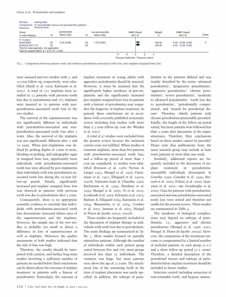
Peri-implant marginal bone loss
At the patient level, the peri-implant mar-
ginal bone loss from the time of abutment
placement to the 5-year follow-up was
2.2 mm (SD¼0.8 mm) in the periodontitis
group and 1.7 mm (SD¼ 0.8 mm) in the
non-periodontitis group in the study by
Hardt et al. (2002). Based on these data,
significantly increased peri-implant mar-
ginal bone loss was observed around im-
plants placed in patients with periodontitis-
associated tooth loss at 5-year follow-up,
MD 0.5 mm (95% CI 0.06–0.94) (P¼0.03)
(Fig. 5).
The peri-implant marginal bone loss was
unreported at the patient level by Karoussis
et al. (2003).
Discussion
The current scientific knowledge about
implant treatment in individuals with pre-
vious tooth loss due to periodontitis was
assessed in the present systematic review.
Prospective and retrospective cohort stu-
dies with at least a 5-year follow-up com-
paring the outcome of implant treatment
in partially edentulous individuals with,
respectively, periodontitis-associated and
non-periodontitis-associated tooth loss
Review: testing data01 periodontitis versus non-periodontitis patients02 implant failure
Comparison:Outcome:
Studyor sub-category
non-periodontitisn/N
periodontitisn/N
RR (random)95% CI
RR (random)95% CI
Weight% Order
01 5 years
33 70
02 10 years
Subtotal (95% CI)
Subtotal (95% CI)
Hardt 2002 7/25
2/88 45
3/25 86.48 2.331.70
3.75 [0.74, 19.02][0.74, 19.02]3.75
[0.08, 38.58][0.68, 8.01] 0
0
0
13.52100.00
100.00100.00
2.24 [0.71, 7.04]1/45
3/45
0/8Karoussis 2003
Karoussis 2003
Total events: 7 (periodontitis), 4 (non-periodontitis)
Total events: 2 (periodontitis), 3 (non-periodontitis)Test for heterogeneity: not applicable
Test for overall effect: Z = 1.37 (P = 0.17)
Test for overall effect: Z = 1.60 (P = 0.11)
Test for heterogeneity: Chi = 0.03, df = 1 (P = 0.85), l = 0%
0.01 0.1 1 10 100
Favours treatment Favours control
Fig. 3. Comparison between patients with and without periodontitis-associated tooth loss: loss of implants.
Review: testing data01 periodontitis versus non-periodontitis patients03 periimplantitis
Comparison:Outcome:
02 10 years
Subtotal (95% CI)
Studyor sub-category
Karoussis 2003 8/8 5/45
Total events: 8 (periodontitis), 5 (non-periodontitis)Test for heterogeneity: not applicableTest for overall effect: Z = 5.21 (P < 0.00001)
non-periodontitisn/N
periodontitisn/N
RR (random)95% CI
RR (random)95% CI
Weight%
100.00 9.00 0
0.01 0.1 1 10 100
Favours treatment Favours control
[3.94, 20.57]9.00 [3.94, 20.57]100.00
Order
8 45
Fig. 4. Comparison between patients with and without periodontitis-associated tooth loss: incidence of peri-implantitis.
Schou et al . Periodontitis and implants
109 | Clin. Oral Impl. Res. 17 (Suppl. 2), 2006 / 104–123
were assessed and two studies with 5- and
10-year follow-up, respectively, were iden-
tified (Hardt et al. 2002; Karoussis et al.
2003). A total of 121 implants were in-
stalled in 33 patients with previous tooth
loss due to periodontitis and 183 implants
were inserted in 70 patients with non-
periodontitis-associated tooth loss in the
two studies.
The survival of the suprastructures was
not significantly different in individuals
with periodontitis-associated and non-
periodontitis-associated tooth loss after 5
years. Also, the survival of the implants
was not significantly different after 5 and
10 years. When peri-implantitis was de-
fined by probing depths of 5 mm or more,
bleeding on probing, and radiographic signs
of marginal bone loss, significantly more
individuals with periodontitis-associated
tooth loss were affected by peri-implantitis
than individuals with non-periodontitis-as-
sociated tooth loss during the 10-year fol-
low-up period. Finally, significantly
increased peri-implant marginal bone loss
was observed in patients with previous
tooth loss due to periodontitis after 5 years.
Consequently, there is no appropriate
scientific evidence to conclude that indivi-
duals with periodontitis-associated tooth
loss demonstrate increased failure rates of
the suprastructures and the implants.
However, the sample size of the two stu-
dies is probably too small to detect a
difference in loss of suprastructures as
well as implants. Moreover, the quality
assessment of both studies indicated that
the risk of bias was high.
Therefore, the results should be inter-
preted with caution, and further long-term
studies involving a sufficient number of
patients are needed before final conclusions
can be drawn about the outcome of implant
treatment in patients with a history of
periodontitis. Particularly, the outcome of
implant treatment in young adults with
aggressive periodontitis should be assessed.
However, it must be assumed that the
significantly higher incidence of peri-im-
plantitis and the significantly increased
peri-implant marginal bone loss in patients
with a history of periodontitis may jeopar-
dize the longevity of implant treatment. In
general, these conclusions are in accor-
dance with a recently published systematic
review including four studies with more
than a 5-year follow-up (van der Weijden
et al. 2005).
A total of 47 studies were excluded from
the present review because the inclusion
criteria were not fulfilled. When studies of
titanium implants, more than five patients
with periodontitis-associated tooth loss,
and a follow-up period of more than 1
year are considered, 19 studies were iden-
tified (Ericsson et al. 1986; Nevins &
Langer 1995; Mengel et al. 1996; Daele-
mans et al. 1997; Ellegaard et al. 1997a,
1997b; Schwartz-Arad & Chaushu 1998;
Buchmann et al. 1999; Sbordone et al.
1999; Mengel et al. 2001; Yi et al. 2001;
Leonhardt et al. 2002; Feloutzis et al. 2003;
Baelum & Ellegaard 2004; Karoussis et al.
2004; Wennstrom et al. 2004; Cordaro
et al. 2005; Jansson et al. 2005; Mengel
& Flores-de-Jacoby 2005a, 2005b).
These studies are frequently included in
the discussion of implant therapy in indi-
viduals with tooth loss due to periodontitis.
The main findings are summarized in Ta-
ble 3. Most studies focused on partially
edentulous patients. Although the number
of individuals within each patient group
varied between five and 766, most groups
involved less than 25 individuals. The
variation was huge, but most patients
were above the age of 55 years. The attach-
ment loss of the remaining teeth at the
time of implant placement was rarely spe-
cified. In addition, the subtype of perio-
dontitis in the patients differed and was
mainly described by the terms ‘advanced
periodontitis’, ‘progressive periodontitis’,
‘aggressive periodontitis’, ‘chronic perio-
dontitis’, ‘severe periodontitis’, ‘moderate
to advanced periodontitis’, ‘tooth loss due
to periodontitis’, ‘periodontally compro-
mised’, and ‘treated for periodontal dis-
ease’. Therefore, elderly patients with
chronic periodontitis presumably prevailed.
Finally, the length of the follow-up period
varied, but most patients were followed less
than 3 years after placement of the supra-
structures. Therefore, firm conclusions
based on these studies cannot be provided.
Please note that publications from the
same research group may include at least
some patients in more than one study.
Similarly, additional reports are fre-
quently included in the discussion of im-
plant treatment in periodontitis-
susceptible individuals (Rosenquist &
Grenthe 1996; Grunder et al. 1999; Bro-
card et al. 2000; Polizzi et al. 2000; Quir-
ynen et al. 2001; van Steenberghe et al.
2002). Data for patients with periodontitis-
associated and non-periodontitis-associated
tooth loss were mixed and therefore not
usable for the present review. These studies
are summarized in Table 4.
The incidence of biological complica-
tions may depend on subtype of perio-
dontitis, i.e., aggressive and chronic
periodontitis (Mengel et al. 1996, 2001;
Mengel & Flores-de-Jacoby 2005a). How-
ever, the comparison of the treatment out-
come is compromised by a limited number
of included patients in each group (5–15)
and a short follow-up period (3–5 years).
Therefore, a detailed description of the
periodontal tissues and subtype of perio-
dontitis before implant treatment should be
included in future studies.
Infection control including extraction of
non-retainable teeth, oral hygiene instruc-
Review: testing data01 periodontitis versus non-periodontitis patients04 bone level
Comparison:Outcome:
Studyor sub-category
non-periodontitisMean (SD)
periodontitisMean (SD)N N
WMD (fixed)95% CI
WMD (fixed)95% CI
Weight% Order
01 5 years
Subtotal (95% CI)25 2.20 (0.80) 1.70 (0.80) 100.00 [0.06, 0.94]0.50 0
−4 −2 0 2 4Favours treatment Favours control
[0.06, 0.94]0.50100.00252525
Test for heterogeneity: not applicableTest for overall effect: Z = 2.21 (P = 0.03)
Hardt 2002
Fig. 5. Comparison between patients with and without periodontitis-associated tooth loss: peri-implant marginal bone loss.
Schou et al . Periodontitis and implants
110 | Clin. Oral Impl. Res. 17 (Suppl. 2), 2006 / 104–123

Tab
le3.
Su
mm
ary
of
stu
die
so
nim
pla
nt
treatm
en
tin
ind
ivid
uals
wit
hp
eri
od
on
titi
s-ass
oci
ate
dto
oth
loss
invo
lvin
gm
ore
than
a1-y
ear
follo
w-u
pp
eri
od
an
dm
ore
than
five
ind
ivid
uals
Pati
en
tsIm
pla
nts
an
dan
tib
ioti
csSu
pra
stru
ctu
reFo
llo
w-u
pp
eri
od
Resu
lts
Refe
ren
ces
Peri
-im
pla
nt
tiss
ues
Peri
od
on
tal
tiss
ues
Sup
rast
ruct
ure
an
dim
pla
nt
surv
ival
rate
Oth
er
resu
lts
an
dco
mm
en
ts
10
PE
wit
had
van
ced
peri
od
on
titi
s
Ag
e:
31–6
0ye
ars
Wo
men
:70%
,m
en
:30%
Smo
kin
g:
NR
41
Bra
nem
ark
An
tib
ioti
cs:
NR
FDo
nim
pla
nts
an
dte
eth
18
(6–3
0)
mo
nth
sPla
qu
e:
15%
BO
P:
8%
PD
:3.3
mm
PD�
3m
m:
60%
PD
4–5
mm
:38%
PD�
6m
m:
2%
Bo
ne
loss
:M
ost
case
s
o1
mm
,3
imp
lan
ts:
1–3
mm
Pla
qu
e:
13%
BO
P:
4%
PD
:2.3
mm
PD�
3m
m:
90%
PD
4–5
mm
:10%
PD�
6m
m:
0%
No
bo
ne
loss
Sup
rast
ruct
ure
:100%
Imp
lan
ts:
100%
Sig
nifi
can
t
dif
fere
nce
inPD
aro
un
dim
pla
nts
an
dte
eth
Eri
csso
net
al.
(1986)
59
wit
hPE
an
dE
jaw
s
Too
thlo
ssd
ue
to
reca
lcit
ran
tp
eri
od
on
titi
s
(fail
ed
tore
spo
nd
to
ap
pro
pri
ate
peri
od
on
tal
treatm
en
t)
Ag
e:
42–8
6ye
ars
Gen
der:
NR
Smo
kin
g:
NR
309
Bra
nem
ark
Maxi
lla:
177
Man
dib
le:
132
An
tib
ioti
cs:
NR
FD,
RD
1ye
ar:
23
imp
lan
ts
2ye
ars
:42
imp
lan
ts
3–5
years
:185
imp
lan
ts
6–7
years
:38
imp
lan
ts
8ye
ars
:21
imp
lan
ts
Seve
ral
pati
en
tsw
ith
1
or
mo
reim
pla
nts
wit
h
bo
ne
loss
toth
e1st
or
2n
dth
read
Seve
nim
pla
nts
wit
hb
on
e
loss
toth
e4th
thre
ad
NR
Sup
rast
ruct
ure
:100%
Imp
lan
ts,
maxi
lla:
98%
Imp
lan
ts,
man
dib
le:
97%
NR
Nevi
ns
&
Lan
ger
(1995)
19
PE
wit
hto
oth
loss
du
eto
pro
gre
ssiv
e
peri
od
on
titi
s
No
.o
fm
axi
llary
teeth
:7
No
.o
fm
an
dib
ula
rte
eth
:
11
Ag
e:
60
years
Wo
men
:79%
,m
en
:21%
Smo
kers
:63%
31
Ast
ra(T
iO2
bla
sted
)
An
tib
ioti
cs:
NR
Pre
do
min
an
tly
STR
an
d
FD Two
pati
en
tstr
eate
d
wit
hp
art
ial
RD
30
(12–4
0)
mo
nth
s1,
3ye
ar
Imp
lan
tsw
ith
pla
qu
e:
0%
,17%
Imp
lan
tsw
ith
BO
P:
0%
,
32%
Imp
lan
tsw
ith
PDo
4m
m:
88%
,44%
Imp
lan
tsw
ith
PD4
6m
m:
0%
,0%
Imp
lan
tsw
ith
bo
ne
losso
1.5
mm
:100%
,76%
Imp
lan
tsw
ith
bo
ne
loss4
3.5
mm
:0%
,0%
NR
1,
3ye
ar
Sup
rast
ruct
ure
:N
R
Imp
lan
ts:
100%
,
100%
No
sig
nifi
can
t
dif
fere
nce
in
imp
lan
tsu
rviv
al
rate
Ell
eg
aard
et
al.
(1997a)
56
PE
wit
hto
oth
loss
du
eto
pro
gre
ssiv
e
peri
od
on
titi
s
No
.o
fm
axi
llary
teeth
:8
No
.o
fm
an
dib
ula
rte
eth
:
10
Ag
e:
60
years
Wo
men
:75%
,m
en
:25%
Smo
kers
:64%
93
ITI
(ho
llo
w
scre
w)
An
tib
ioti
cs:
NR
33
(3–8
4)
mo
nth
s1,
3,
5ye
ar
Imp
lan
tsw
ith
pla
qu
e:
17%
,31%
,45%
Imp
lan
tsw
ith
BO
P:
11%
,
30%
,45%
Imp
lan
tsw
ith
PDo
4m
m:
90%
,63%
,18%
Imp
lan
tsw
ith
PD4
6m
m:
4%
,8%
,31%
Imp
lan
tsw
ith
bo
ne
losso
1.5
mm
:96%
,86%
,
55%
Imp
lan
tsw
ith
bo
ne
loss4
3.5
mm
:3%
,7%
,
21%
1,
3,
5ye
ar
Sup
rast
ruct
ure
:N
R
Imp
lan
ts:
97%
,95%
,
95%
Schou et al . Periodontitis and implants
111 | Clin. Oral Impl. Res. 17 (Suppl. 2), 2006 / 104–123

24
PE
wit
hto
oth
loss
du
eto
pro
gre
ssiv
e
peri
od
on
titi
s
Ag
e:
57
(42–7
3)
years
Wo
men
:88%
,m
en
:12%
Smo
kers
:63%
Main
lyim
pla
nts
in
maxi
lla
wit
ho
rw
ith
ou
t
sin
us
lift
pro
ced
ure
No
bo
ne
gra
ft
25
Ast
ra
(TiO
2b
last
ed
)
No
sin
us
lift
An
tib
ioti
cs:
NR
Pre
do
min
an
tly
STR
an
d
FD Fou
rp
ati
en
tstr
eate
d
wit
hp
art
ial
RD
31
mo
nth
s1,
3ye
ar
Imp
lan
tsw
ith
pla
qu
e:
0%
,20%
Imp
lan
tsw
ith
BO
P:
0%
,
35%
Imp
lan
tsw
ith
PDo
4m
m:
86%
,44%
Imp
lan
tsw
ith
PD4
6m
m:
0%
,0%
Imp
lan
tsw
ith
bo
ne
losso
1.5
mm
:100%
,76%
Imp
lan
tsw
ith
bo
ne
loss4
3.5
mm
:0%
,0%
NR
1,
3ye
ar
Sup
rast
ruct
ure
:N
R
Imp
lan
ts:
100%
,
100%
No
sig
nifi
can
t
dif
fere
nce
in
imp
lan
tsu
rviv
al
rate
an
dfr
eq
uen
cyo
f
imp
lan
tsw
ith
ou
t
pla
qu
e,
BO
P,
incr
ease
dPD
,an
d
bo
ne
loss
aro
un
d
imp
lan
tsw
ith
or
wit
ho
ut
sin
us
lift
Ell
eg
aard
et
al.
(1997b
)
26
Ast
ra
(TiO
2b
last
ed
)
Sin
us
lift
An
tib
ioti
cs:
NR
30
mo
nth
s1,
3ye
ar
Imp
lan
tsw
ith
pla
qu
e:
0%
,11%
Imp
lan
tsw
ith
BO
P:
0%
,
27%
Imp
lan
tsw
ith
PDo
4m
m:
100%
,59%
Imp
lan
tsw
ith
PD4
6m
m:
0%
,0%
Imp
lan
tsw
ith
bo
ne
losso
1.5
mm
:95%
,82%
Imp
lan
tsw
ith
bo
ne
loss4
3.5
mm
:5%
,5%
1,
3ye
ar
Sup
rast
ruct
ure
:N
R
Imp
lan
ts:
95%
,95%
17
ITI
(ho
llo
wo
rso
lid
scre
w)
No
sin
us
lift
An
tib
ioti
cs:
NR
29
mo
nth
s1,
3ye
ar
Imp
lan
tsw
ith
pla
qu
e:
8%
,18%
Imp
lan
tsw
ith
BO
P:
8%
,
28%
Imp
lan
tsw
ith
PDo
4m
m:
100%
,80%
Imp
lan
tsw
ith
PD4
6m
m:
0%
,0%
Imp
lan
tsw
ith
bo
ne
losso
1.5
mm
:91%
,71%
Imp
lan
tsw
ith
bo
ne
loss4
3.5
mm
:9%
,9%
1,
3ye
ar
Sup
rast
ruct
ure
:N
R
Imp
lan
ts:
91%
,91%
12
ITI
(so
lid
scre
w)
Sin
us
lift
An
tib
ioti
cs:
NR
25
mo
nth
s1,
3ye
ar
Imp
lan
tsw
ith
pla
qu
e:
0%
,14%
Imp
lan
tsw
ith
BO
P:
0%
,
14%
Imp
lan
tsw
ith
PDo
4m
m:
100%
,64%
Imp
lan
tsw
ith
PD4
6m
m:
0%
,0%
Imp
lan
tsw
ith
bo
ne
losso
1.5
mm
:73%
,29%
Imp
lan
tsw
ith
bo
ne
loss4
3.5
mm
:14%
,14%
1,
3ye
ar
Sup
rast
ruct
ure
:N
R
Imp
lan
ts:
86%
,86%
Tab
le3.
Co
nti
nu
ed
Pati
en
tsIm
pla
nts
an
dan
tib
ioti
csSu
pra
stru
ctu
reFo
llo
w-u
pp
eri
od
Resu
lts
Refe
ren
ces
Peri
-im
pla
nt
tiss
ues
Peri
od
on
tal
tiss
ues
Sup
rast
ruct
ure
an
dim
pla
nt
surv
ival
rate
Oth
er
resu
lts
an
dco
mm
en
ts
Schou et al . Periodontitis and implants
112 | Clin. Oral Impl. Res. 17 (Suppl. 2), 2006 / 104–123

33
Ean
dPE
wit
hto
oth
loss
du
eto
peri
od
on
tal
dis
ease
Ag
e:
52
(27–7
5)
years
Wo
men
:48%
,m
en
:52%
Smo
kin
g:
NR
Imp
lan
tsin
sert
ed
inth
e
po
steri
or
part
of
maxi
lla
con
com
itan
tw
ith
the
sin
us
lift
pro
ced
ure
an
d
AB
fro
mil
iac
crest
121
Bra
nem
ark
Am
oxi
cill
in
500
mg�
4o
r
clin
dam
ycin
e
300
mg�
3
FD40
(3–8
0)
mo
nth
sN
RN
RSu
pra
stru
ctu
re:
98%
Imp
lan
ts:
93%
NR
Daele
man
s
et
al.
(1997)
25
PE
wit
hm
od
era
teto
ad
van
ced
ad
ult
peri
od
on
titi
s
Ag
e:
37–6
8ye
ars
Wo
men
:52%
,m
en
:48%
Smo
kin
g:
NR
42
Bra
nem
ark
(MK
III)
An
tib
ioti
cs:
NR
NR
3ye
ars
1,
2,
3ye
ar
PI:
0.9
,1,
1
GI:
1.6
,1.7
PD
:3.2
,3.3
,3.4
mm
1,
2,
3ye
ar
PI:
1,
0.9
,0.7w
GI:
1.6
,1.7w
PD
:3.2
,3,
3m
mw
1,
2,
3ye
ar
Sup
rast
ruct
ure
:N
R
Imp
lan
ts:
100%
,
100%
,100%
Co
mp
ara
ble
mic
rofl
ora
aro
un
d
imp
lan
tsan
dte
eth
thro
ug
ho
ut
stu
dy
Peri
od
on
tal
path
og
en
sse
ldo
m
dete
cted
an
dw
hen
dete
cted
at
low
leve
ls
Sbo
rdo
ne
et
al.
(1999)
22
wit
hse
vere
peri
od
on
titi
s
Ag
e:
54
(36–6
6)
years
Wo
men
:73%
,m
en
:27%
Smo
kin
g:
NR
AB
chip
sin
top
eri
-
imp
lan
td
efe
ctw
hen
nece
ssary
214
scre
w-t
ype
tita
niu
mim
pla
nts
inse
rted
imm
ed
iate
lyaft
er
ext
ract
ion
of
all
teeth
Maxi
lla:
128
Man
dib
le:
86
Am
oxi
cill
ino
r
ery
thro
myc
info
r
5–7
days
Full
-arc
hFD
1ye
ar:
211
imp
lan
ts
5ye
ars
:28
imp
lan
ts
NR
NR
1,
5ye
ar
Sup
rast
ruct
ure
:N
R
Imp
lan
ts:
99%
,99%
NR
Sch
wart
z-A
rad
&C
hau
shu
(1998)
50
pati
en
tsw
ith
chro
nic
ad
ult
peri
od
on
titi
s
treate
dw
ith
the
sin
us
lift
pro
ced
ure
,A
B,
an
d
sim
ult
an
eo
us
imp
lan
t
pla
cem
en
t
Ag
e:
52
years
Wo
men
:58%
,m
en
:42%
Smo
kin
g:
NR
167
Bra
nem
ark
,
IMZ,
Fria
lit-
2
An
tib
ioti
cs:
NR
FD,
RD
5ye
ars
PI:
0.4
GI:
0.4
PD
:2.9
mm
NR
Sup
rast
ruct
ure
:100%
Imp
lan
ts:
100%
NR
Bu
chm
an
n
et
al.
(1999)
Schou et al . Periodontitis and implants
113 | Clin. Oral Impl. Res. 17 (Suppl. 2), 2006 / 104–123

37
peri
od
on
tall
yh
ealt
hy
pati
en
tstr
eate
dw
ith
maxi
llary
imp
lan
ts
wit
ho
ut
the
sin
us
lift
pro
ced
ure
an
dA
B
Ag
e:
44
years
Gen
der:
NR
Smo
kin
g:
NR
60
IMZ,
ITI,
Led
erm
an
n
An
tib
ioti
cs:
NR
PI:
0.5
GI:
0.6
PD
:3
mm
5PE
wit
hg
en
era
lize
d
ag
gre
ssiv
ep
eri
od
on
titi
s
Healt
hy
peri
od
on
tal
tiss
ues
(PDo
3m
m,
no
BO
P)
No
.o
fte
eth
:10
Ag
e:
31–4
4ye
ars
Wo
men
:100%
Smo
kers
:20%
36
Bra
nem
ark
Maxi
lla:
21
Man
dib
le:
15
An
tib
ioti
cs:
NR
Pre
do
min
an
tly
FD
On
ep
ati
en
ttr
eate
d
wit
hR
D
5ye
ars
1,
2,
3,
4,
5ye
ar
PI:
0.8
,0.5
,0.3
,0.6
,0.7
GI:
0.2
,0,
0.2
,0.5
,0.5
PD
:2,
2,
2.2
,3.8
,3.3
mm
AL:
2,
2.3
,2.4
,4.7
,
5.6
mm
1,
3,
5ye
ar
Bo
ne
loss
:0.6
,0.8
,
0.9
mm
Base
lin
ean
d1,
2,
3,
4,
5ye
ar
PI:
0.3
,0.9
,0.6
,0.7
,
0.6
,0.8
GI:
0,
0.2
,0,
0.1
,0.3
,
0.5
PD
:3,
3,
2.8
,3,
4.1
,
3.5
mm
AL:
4.1
,4.5
,4.7
,4.9
,
6.1
,6.3
mm
1,
3,
5ye
ar
Bo
ne
loss
:1.6
%,
3.4
%,
5.1
%
Sup
rast
ruct
ure
:100%
Imp
lan
ts,
maxi
lla:
86%
Imp
lan
ts,
man
dib
le:
93%
Sig
nifi
can
tly
mo
re
att
ach
men
tlo
ssat
imp
lan
tsth
an
teeth
Two
teeth
ext
ract
ed
ino
ne
pati
en
t
Men
gel
et
al.
(1996,
2001)
5PE
wit
hg
en
era
lize
d
chro
nic
peri
od
on
titi
s
Healt
hy
peri
od
on
tal
tiss
ues
(PDo
3m
m,
no
BO
P)
No
.o
fte
eth
:23
Ag
e:
35–4
2ye
ars
Wo
men
:100%
Smo
kin
g:
NR
12
Bra
nem
ark
An
tib
ioti
cs:
NR
FD,
STR
3ye
ars
1,
2,
3ye
ar
PI:
0.4
,0.5
,0.5
GI:
0.2
,0,
0.2
PD
:3.1
,3,
3m
m
AL:
4,
4.4
,5.5
mm
1,
3ye
ar
Bo
ne
loss
:0.1
,0.2
mm
Base
lin
ean
d1,
2,
3
years
PI:
0.5
,0.3
,0.3
,0.3
GI:
0.2
,0.1
,0.2
,0.2
PD
:2.7
,3,
2.8
,
2.6
mm
AL:
3.2
,3.7
,3.9
,
3.6
mm
1,
3ye
ar
Bo
ne
loss
:1.5
%,
2.7
%
Sup
rast
ruct
ure
:100%
Imp
lan
ts,
maxi
lla:
100%
Imp
lan
ts,
man
dib
le:
100%
No
sig
nifi
can
t
dif
fere
nce
in
att
ach
men
tlo
ssat
imp
lan
tsan
dte
eth
43
PE
an
dE
treate
dfo
r
ad
van
ced
peri
od
on
titi
s
Ag
e:
50
(26–6
5)
years
Wo
men
:53%
,m
en
:47%
Smo
kin
g:
NR
AB
an
dePTFE
mem
bra
ne
con
com
itan
tw
ith
imp
lan
tp
lace
men
t(n¼
5)1
25
Ast
ra(T
iO2
bla
sted
)
Am
oxi
cill
in
500
mg�
2fo
r
10
days
FD3
years
3ye
ar
Surf
ace
sw
ith
pla
qu
e:o
10%
Bo
ne
loss
:0.2
mm
Imp
lan
tsw
ith
bo
ne
loss
�0.5
mm
:81%
Imp
lan
tsw
ith
0.5
–2m
m
bo
ne
loss
:19%
NR
Sup
rast
ruct
ure
:100%
Imp
lan
ts:
100%
NR
Yi
et
al.
(2001)
15
PE
wit
had
van
ced
peri
od
on
titi
s
Tota
lN
o.
of
maxi
llary
teeth
:125
Tota
lN
o.
of
man
dib
ula
r
teeth
:136
Ag
e:
21–7
1ye
ars
Wo
men
:47%
,m
en
:53%
Smo
kin
g:
NR
57
Bra
nem
ark
Maxi
lla:
31
Man
dib
le:
26
An
tib
ioti
cs:
NR
FD10
years
Imp
lan
tsw
ith
BO
P:
61%
Bo
ne
loss
:1.7
mm
Imp
lan
tsw
ith
bo
ne
loss
�0.5
mm
:15%
Imp
lan
tsw
ith
bo
ne
loss
0.6
–2m
m:
52%
Imp
lan
tsw
ith
bo
ne
loss
�2.1
mm
:33%
Teeth
wit
hB
OP:
35%
Teeth
wit
hPD
�4
mm
:16%
Teeth
wit
hPD
�6
mm
:3%
Bo
ne
loss
:0.8
mm
Sup
rast
ruct
ure
:N
R
Imp
lan
ts,
maxi
lla:
94%
Imp
lan
ts,
man
dib
le:
96%
No
sig
nifi
can
t
corr
ela
tio
nb
etw
een
bo
ne
loss
aro
un
d
imp
lan
tsan
dte
eth
Surv
ivalo
fte
eth
:87%
5p
ati
en
tsw
ith
ou
t
too
thlo
ss
Leo
nh
ard
t
et
al.
(2002)
Tab
le3.
Co
nti
nu
ed
Pati
en
tsIm
pla
nts
an
dan
tib
ioti
csSu
pra
stru
ctu
reFo
llo
w-u
pp
eri
od
Resu
lts
Refe
ren
ces
Peri
-im
pla
nt
tiss
ues
Peri
od
on
tal
tiss
ues
Sup
rast
ruct
ure
an
dim
pla
nt
surv
ival
rate
Oth
er
resu
lts
an
dco
mm
en
ts
Schou et al . Periodontitis and implants
114 | Clin. Oral Impl. Res. 17 (Suppl. 2), 2006 / 104–123

90
PE
treate
dfo
r
chro
nic
peri
od
on
titi
s
Ag
e:
60
(33–8
8)
years
Gen
der:
NR
No
n-s
mo
kers
:43%
,
form
er
smo
kers
(sm
okin
g
cess
ati
on4
5ye
ars
):26%
,
mo
dera
tesm
okers
(5–1
9
cig
are
ttes/
day)
:16%
,
heavy
smo
kers
(20
cig
are
ttes/
day)
:16%
182
ITI
(ho
llo
w
cyli
nd
er,
scre
w
cyli
nd
er,
soli
d
scre
ws)
An
tib
ioti
cs:
NR
FD,
STR
5.6
(2–1
2)
years
BO
P:
15%
n
Bo
ne
loss
,IL
-1p
osi
tive
gen
oty
pe:
0.2
mm
n
Bo
ne
loss
,IL
-1n
eg
ati
ve
gen
oty
pe:
0.5
mm
n
BO
P:
13%
nSu
pra
stru
ctu
re:
NR
Imp
lan
ts:
96%
Sig
nifi
can
tly
incr
ease
db
on
elo
ss
inh
eavy
smo
kers
as
com
pare
dto
no
n-
smo
kers
No
sig
nifi
can
tly
dif
fere
nt
bo
ne
loss
betw
een
IL-1
po
siti
ve
an
dn
eg
ati
ve
gen
oty
pe
ind
ivid
uals
IL-1
po
siti
veg
en
oty
pe
ind
ivid
uals
:
Sig
nifi
can
tly
incr
ease
db
on
elo
ssin
heavy
smo
kers
as
com
pare
dto
no
n-
smo
kers
IL-1
neg
ati
ve
gen
oty
pe
ind
ivid
uals
:
No
sig
nifi
can
tly
dif
fere
nt
bo
ne
loss
in
heavy
smo
kers
as
com
pare
dto
no
n-
smo
kers
Felo
utz
iset
al.
(2003)
32
peri
od
on
tall
y
com
pro
mis
ed
PE
No
.o
fm
axi
llary
teeth
:8
No
.o
fm
an
dib
ula
rte
eth
:
11
Ag
e:
60
(44–7
8)
years
Wo
men
:75%
,m
en
:25%
Smo
kers
:66%
57
Ast
ra
(TiO
2b
last
ed
)
An
tib
ioti
cs:
NR
Pre
do
min
an
tly
STR
an
dFD
Fou
rp
ati
en
tstr
eate
d
wit
hp
art
ial
RD
68
(0–1
28)
mo
nth
s1,
5,
10
year
Imp
lan
tsw
ith
BO
P:
0%
,
51%
,91%
Imp
lan
tsw
ith
PDo
4m
m:
98%
,55%
,25%
Imp
lan
tsw
ith
PD4
6m
m:
0%
,6%
,23%
Imp
lan
tsw
ith
bo
ne
losso
1.5
mm
:100%
,
85%
,70%
Imp
lan
tsw
ith
bo
ne
loss4
3.5
mm
:0%
,5%
,
5%
NR
1,
5,
10
year
Sup
rast
ruct
ure
:N
R
Imp
lan
ts:
100%
,
97%
,97%
39
imp
lan
tsin
20
pati
en
tstr
eate
d
surg
icall
yd
ue
to
peri
-im
pla
nti
tis
Ho
weve
r,18
rem
ove
d
du
eto
un
succ
ess
ful
treatm
en
t
Smo
kin
gsi
gn
ifica
ntl
y
ass
oci
ate
dw
ith
incr
ease
dim
pla
nt
fail
ure
rate
Baelu
m&
Ell
eg
aard
(2004)
108
peri
od
on
tall
y
com
pro
mis
ed
PE
No
.o
fm
axi
llary
teeth
:8
No
.o
fm
an
dib
ula
rte
eth
:
10
Ag
e:
58
(34–8
7)
years
Wo
men
:66%
,m
en
:34%
Smo
kers
:64%
201
ITI
(ho
llo
w
an
dso
lid
scre
w)
An
tib
ioti
cs:
NR
74
(0–1
68)
mo
nth
s1,
5,
10
year
Imp
lan
tsw
ith
BO
P:
2%
,
46%
,70%
Imp
lan
tsw
ith
PDo
4m
m:
95%
,40%
,24%
Imp
lan
tsw
ith
PD4
6m
m:
1%
,15%
,25%
Imp
lan
tsw
ith
bo
ne
losso
1.5
mm
:98%
,72%
,
60%
Imp
lan
tsw
ith
bo
ne
loss4
3.5
mm
:0%
,6%
,
14%
1,
5,
10
year
Sup
rast
ruct
ure
:N
R
Imp
lan
ts:
100%
,94%
,
78%
Schou et al . Periodontitis and implants
115 | Clin. Oral Impl. Res. 17 (Suppl. 2), 2006 / 104–123

51
PE
wit
hm
od
era
teto
ad
van
ced
chro
nic
peri
od
on
titi
s
Peri
od
on
tal
bo
ne
leve
l:
44%
No
.o
fte
eth
:19
Ag
e:
60
(36–8
0)
years
Wo
men
:61%
,m
en
:39%
Smo
kers
:33%
149
Ast
ra
Maxi
lla:
83
Man
dib
le:
66
Min
imu
mo
ne
TiO
2b
last
ed
an
d
on
ew
ith
turn
ed
surf
ace
ineach
pati
en
t
Pen
icil
lin
1g�
2
for
7d
ays
FD5
years
Pla
qu
e:
5%
BO
P:
5%
PD
:3.1
mm
PD�
3m
m:
80%
PD
4–5
mm
:15%
PD�
6m
m:
5%
Bo
ne
loss
,im
pla
nts
wit
h
TiO
2b
last
ed
surf
ace
:
0.5
mm
Bo
ne
loss
,im
pla
nts
wit
h
turn
ed
surf
ace
:0.3
mm
Bo
ne
loss
,m
axi
lla:0.6
mm
Bo
ne
loss
,m
an
dib
le:
0.2
mm
Imp
lan
tsw
ith4
2m
m
bo
ne
loss
:11%
Bo
ne
loss
,sm
okers
:
0.8
mm
Bo
ne
loss
,n
on
-sm
okers
:
0.2
mm
NR
Sup
rast
ruct
ure
:95%
Imp
lan
ts:
97%
No
sig
nifi
can
tly
dif
fere
nt
bo
ne
loss
aro
un
dim
pla
nts
wit
h
TiO
2b
last
ed
an
d
mach
ined
surf
ace
Wen
nst
rom
et
al.
(2004)
89
PE
treate
dfo
r
peri
od
on
tal
dis
ease
No
.o
fte
eth
:20
Ag
e:
49
(19–7
8)
years
Wo
men
:62%
,m
en
:38%
Smo
kin
g:
NR
179
ITI
(ho
llo
w
scre
w,
ho
llo
w
cyli
nd
er,
an
gu
late
dh
oll
ow
cyli
nd
er)
Maxi
lla:
104
Man
dib
le:
75
An
tib
ioti
cs:
NR
FD,
STR
10
(8–1
2)
years
PI:
0.4
BO
P:
42%
PD
:2.8
mm
Bo
ne
loss
:0.7
mm
Co
ntr
ala
tera
lte
eth
:
PI:
0.4
BO
P:
30%
PD
:2
mm
Bo
ne
loss
:0.6
mm
Sup
rast
ruct
ure
:N
R
Imp
lan
ts:
92%
PD
aro
un
dim
pla
nts
sig
nifi
can
tly
infl
uen
ced
by
full-
mo
uth
PDan
d
full-
mo
uth
att
ach
men
t
loss
Bo
ne
loss
aro
un
d
imp
lan
tssi
gn
ifica
ntl
y
infl
uen
ced
by
smo
kin
g,
full-
mo
uth
att
ach
men
t
leve
l,an
dch
an
ge
in
full-
mo
uth
PD
Surv
ival
of
teeth
:95%
Karo
uss
iset
al.
(2004)
766
PE
treate
dfo
r
peri
od
on
tal
dis
ease
Ag
e:
NR
Wo
men
:56%
,m
en
:44%
Smo
kin
g:
NR
1796
Bra
nem
ark
Maxi
lla:
1091
Man
dib
le:
705
An
tib
ioti
cs:
NR
NR
Up
to10
years
NR
NR
Sup
rast
ruct
ure
:N
R
Imp
lan
ts,
maxi
lla:
97%
Imp
lan
ts,
man
dib
le:
92%
An
aly
sis
invo
lvin
g22
pati
en
tsw
ith
loss
of
32
imp
lan
ts(a
tle
ast
on
ein
each
pati
en
t):
No
sig
nifi
can
t
dif
fere
nce
inim
pla
nt
loss
betw
een
IL-1
po
siti
vean
dn
eg
ati
ve
gen
oty
pe
ind
ivid
uals
Sig
nifi
can
tly
incr
ease
dim
pla
nt
loss
insm
okers
than
no
n-
smo
kers
wit
hIL
-1
po
siti
veg
en
oty
pe
No
sig
nifi
can
t
dif
fere
nce
inim
pla
nt
loss
betw
een
smo
kers
an
dn
on
-sm
okers
wit
hIL
-1n
eg
ati
ve
gen
oty
pe
Jan
sso
net
al.
(2005)
Tab
le3.
Co
nti
nu
ed
Pati
en
tsIm
pla
nts
an
dan
tib
ioti
csSu
pra
stru
ctu
reFo
llo
w-u
pp
eri
od
Resu
lts
Refe
ren
ces
Peri
-im
pla
nt
tiss
ues
Peri
od
on
tal
tiss
ues
Sup
rast
ruct
ure
an
dim
pla
nt
surv
ival
rate
Oth
er
resu
lts
an
dco
mm
en
ts
Schou et al . Periodontitis and implants
116 | Clin. Oral Impl. Res. 17 (Suppl. 2), 2006 / 104–123

15
PE
wit
hg
en
era
lize
d
ag
gre
ssiv
ep
eri
od
on
titi
s
(gen
era
lize
datt
ach
men
t
loss�
3m
min
1ye
ar
at
43
teeth
exc
lud
ed
1st
mo
lars
an
din
ciso
rs)
Healt
hy
peri
od
on
tal
tiss
ues
(PD�
3m
m,
no
BO
P)
No
.o
fte
eth
:15
Ag
e:
32
years
Wo
men
:53%
,m
en
:47%
No
n-s
mo
kers
52
Bra
nem
ark
(MK
II)
an
d25
Oss
eo
tite
3i
Maxi
lla:
47
Man
dib
le:
30
An
tib
ioti
cs:
NR
FD3
years
1,
2,
3ye
ar
PI:
0.5
,0.4
,0.3
GI:
0.1
,0.1
,0.2
PD
:2.5
,2.9
,3.2
mm
AL:
2.9
,3.7
,4.3
mm
1,
3ye
ar
Bo
ne
loss
:0.8
,1.1
mm
Base
lin
ean
d1,
2,
3
year
PI:
0.4
,0.5
,0.5
,0.5
GI:
0.1
,0.1
,0.1
,0.1
PD
:3.2
,2.7
,2.9
,
3.2
mm
AL:
4.4
,4.1
,4.4
,
4.9
mm
1,
3ye
ar
Bo
ne
loss
:1.7
%,
3.5
%
1,
2,
3ye
ar
Sup
rast
ruct
ure
:
100%
,100%
,100%
Imp
lan
ts,
maxi
lla:
96%
,96%
,96%
Imp
lan
ts,
man
dib
le:
100%
,100%
,100%
Sig
nifi
can
tly
gre
ate
r
att
ach
men
tlo
ssat
imp
lan
tsth
an
teeth
inall
gro
up
s
No
sig
nifi
can
t
dif
fere
nce
incl
inic
al
an
dra
dio
gra
ph
ic
para
mete
rsb
etw
een
gro
up
s
Men
gel
&
Flo
res-
de-
Jaco
by
(2005a)
12
PE
wit
hg
en
era
lize
d
chro
nic
peri
od
on
titi
s
(gen
era
lize
datt
ach
men
t
losso
3m
min
1ye
ar
at
43
teeth
)
Healt
hy
peri
od
on
tal
tiss
ues
(PD�
3m
m,
no
BO
P)
No
.o
fte
eth
:19
Ag
e:
34
years
Wo
men
:50%
,m
en
:50%
No
n-s
mo
kers
17
Bra
nem
ark
(MK
II)
an
d26
Oss
eo
tite
3i
Maxi
lla:
20
Man
dib
le:
23
An
tib
ioti
cs:
NR
1,
2,
3ye
ar
PI:
0.4
,0.4
,0.6
GI:
0.1
,0.1
,0
PD
:2.7
,2.7
,2.7
mm
AL:
3.9
,4.8
,4.6
mm
1,
3ye
ar
Bo
ne
loss
:0.7
,0.9
mm
Base
lin
ean
d1,
2,
3
year
PI:
0.3
,0.3
,0.3
,0.4
GI:
0.3
,0.1
,0.2
,0.1
PD
:2.8
,2.8
,2.6
,
2.6
mm
AL:
3.9
,4.2
,4,
3.9
mm
1,
3ye
ar
Bo
ne
loss
:1.6
%,
3%
1,
2,
3ye
ar
Sup
rast
ruct
ure
:
100%
,100%
,100%
Imp
lan
ts,
maxi
lla:
100%
,100%
,100%
Imp
lan
ts,
man
dib
le:
100%
,100%
,100%
12
PE
peri
od
on
tall
y
healt
hy
(PD�
3m
m,
no
BO
P)
No
.o
fte
eth
:25
Ag
e:
31
years
Wo
men
:58%
,m
en
:
42%
No
n-s
mo
kers
14
Bra
nem
ark
(MK
II)
an
d16
Oss
eo
tite
3i
Maxi
lla:
15
Man
dib
le:
15
An
tib
ioti
cs:
NR
1,
2,
3ye
ar
PI:
0.3
,0.4
,0.5
GI:
0.1
,0.1
,0.1
PD
:2.4
,3.2
,3.3
mm
AL:
2.8
,4.3
,4
mm
1,
3ye
ar
Bo
ne
loss
:0.6
,0.7
mm
Base
lin
ean
d1,
2,
3
year
PI:
0.6
,0.2
,0.4
,0.5
GI:
0.2
,0,
0.1
,0.1
PD
:2.5
,2.5
,2.6
,
2.5
mm
AL:
2.8
,2.8
,3.1
,
3m
m
1,
3ye
ar
Bo
ne
loss
:1.3
%,
2%
1,
2,
3ye
ar
Sup
rast
ruct
ure
:
100%
,100%
,100%
Imp
lan
ts,
maxi
lla:
100%
,100%
,100%
Imp
lan
ts,
man
dib
le:
100%
,100%
,100%
9PE
wit
hre
du
ced
peri
od
on
tal
sup
po
rt
(o2/3
bo
ne
sup
po
rtfo
r
all
teeth
)
Ag
e:
NR
Gen
der:
NR
Smo
kin
g:
NR
71
ITI
(so
lid
scre
w)
an
d19
3i
(tu
rned
surf
ace
)
An
tib
ioti
cs:
NR
Full
-arc
hFD
on
imp
lan
ts
an
dte
eth
wit
hri
gid
or
no
n-r
igid
con
nect
ion
37
(24–9
4)
mo
nth
sN
RN
RSu
pra
stru
ctu
re:
100%
Imp
lan
tsu
rviv
al:
100%
Pro
sth
eti
c
com
pli
cati
on
s:0%
Reg
ard
less
of
con
nect
ion
typ
e,
no
too
thin
tru
sio
nw
hen
peri
od
on
tal
sup
po
rt
red
uce
d
Co
rdaro
et
al.
(2005)
10
PE
wit
hn
orm
al
peri
od
on
tal
sup
po
rt
(42/3
bo
ne
sup
po
rt
for
all
teeth
)
Ag
e:
NR
Gen
der:
NR
Smo
kin
g:
NR
Sup
rast
ruct
ure
:100%
(tw
on
ew
pro
sth
esi
s)
Imp
lan
tsu
rviv
al:
98%
Pro
sth
eti
c
com
pli
cati
on
s:40%
Schou et al . Periodontitis and implants
117 | Clin. Oral Impl. Res. 17 (Suppl. 2), 2006 / 104–123

10
PE
wit
hg
en
era
lized
ag
gre
ssiv
ep
eri
od
on
titi
s
(att
ach
men
tlo
ss�
3m
m
in1
year
at4
3te
eth
)
Healt
hy
peri
od
on
tal
tiss
ues
(PD�
3m
m,n
oB
OP)
Ag
e:
NR
Wo
men
:10
0%
No
n-s
mo
kers
Ver
tica
lan
dh
ori
zon
tal
gu
ided
bo
ne
reg
en
era
tio
n
by
tita
niu
m-r
ein
forc
ed
ePT
FEm
emb
ran
e
sup
po
rted
by
tita
niu
m
scre
ws
6–8
mo
nth
sb
efo
re
imp
lan
tp
lace
men
t
15
Bra
nem
ark
(MK
II)
inm
axi
lla
(in
ciso
ran
d
pre
mo
lar
reg
ion
s)
Am
oxi
cill
in
500
mg�
3fo
r3
days
STR
3ye
ars
1,
2,
3ye
ar
PI:
0.3
,0.3
,0.3
GI:
0,
0,
0.3
PD
:3.1
,3.4
,3.4
mm
AL:
5.1
,5.4
,5.7
mm
1,
3ye
ar
Bo
ne
loss
:1.2
,1.8
mm
Sig
nifi
can
tly
gre
ate
r
att
ach
men
tlo
ssat
imp
lan
tsth
an
teeth
Base
lin
ean
d1,
2,
3
year
PI:
0.2
,0.4
,0.5
,0.5
GI:
0,
0,
0.2
,0.1
PD
:2.8
,2.7
,2.9
,
2.9
mm
AL:
3.7
,3.7
,4.1
,
4.1
mm
1,
3ye
ar
Bo
ne
loss
:2.2
%,
5.4
%
Sup
rast
ruct
ure
:100%
Imp
lan
ts:
100%
Sig
nifi
can
tly
gre
ate
r
att
ach
men
tlo
ss
aro
un
dim
pla
nts
in
reg
en
era
ted
bo
ne
than
inn
on
-
reg
en
era
ted
bo
ne
Men
gel
&
Flo
res-
de-
Jaco
by
(2005b
)
10
peri
od
on
tall
yh
ealt
hy
PE
(PDo
3m
m,
no
BO
P)
Ag
e:
NR
Wo
men
:80%
,m
en
:20%
No
n-s
mo
kers
11
Bra
nem
ark
(MK
II)
inm
axi
lla
(in
ciso
ran
d
pre
mo
lar
reg
ion
s)
An
tib
ioti
cs:
NR
STR
1,
2,
3ye
ar
PI:
0.3
,0.4
,0.5
GI:
0.1
,0.1
,0.1
PD
:3.4
,3.1
,3.3
mm
AL:
3.9
,4.1
,4
mm
1,
3ye
ar
Bo
ne
loss
:1.1
,1.4
mm
Sig
nifi
can
tly
gre
ate
r
att
ach
men
tlo
ssat
teeth
than
imp
lan
ts
Base
lin
ean
d1,
2,
3
year
PI:
0.6
,0.2
,0.4
,0.5
GI:
0.2
,0,
0.1
,0.1
PD
:2.5
,2.5
,2.6
,
2.5
mm
AL:
2.3
,2,
2.6
,2.5
mm
1,
3ye
ar
Bo
ne
loss
:1.5
%,
2.3
%
Sup
rast
ruct
ure
:100%
Imp
lan
ts:
100%
All
gro
up
valu
es
refe
rred
toare
exp
ress
ed
as
mean
valu
es,
ifn
ot
oth
erw
ise
speci
fied
.C
lin
ical
para
mete
rsre
cord
ed
at
fou
rsi
tes
per
imp
lan
t/to
oth
,if
no
to
therw
ise
speci
fied
.nM
ed
ian
.wR
eco
rded
at
6si
tes
per
too
that
on
era
nd
om
lyse
lect
ed
too
th.
AB
,au
tog
en
ou
sb
on
e;A
L,att
ach
men
tle
vel;
BO
P,b
leed
ing
on
pro
bin
g;E,ed
en
tulo
us;
ePTFE
,exp
an
ded
po
lyte
trafl
uo
roeth
yle
ne;FD
,fi
xed
den
ture
;G
I,g
ing
ivalin
dex;
NR
,n
ot
rep
ort
ed
;PD
,p
rob
ing
dep
th;PE,
part
ial
ed
en
tulo
us;
PI,
pla
qu
ein
dex;
RD
,re
mo
vab
led
en
ture
;ST
R,
sin
gle
-to
oth
rep
lace
men
t.
Tab
le3.
Co
nti
nu
ed
Pati
en
tsIm
pla
nts
an
dan
tib
ioti
csSu
pra
stru
ctu
reFo
llo
w-u
pp
eri
od
Resu
lts
Refe
ren
ces
Peri
-im
pla
nt
tiss
ues
Peri
od
on
tal
tiss
ues
Sup
rast
ruct
ure
an
dim
pla
nt
Oth
er
resu
lts
an
dco
mm
en
ts
Schou et al . Periodontitis and implants
118 | Clin. Oral Impl. Res. 17 (Suppl. 2), 2006 / 104–123

Tab
le4.
Su
mm
ary
of
stu
die
so
nvari
ou
sp
ote
nti
al
risk
fact
ors
for
imp
lan
tlo
ssan
dp
eri
-im
pla
nt
marg
inal
bo
ne
loss
Pati
en
tsIm
pla
nts
an
dan
tib
ioti
csSu
pra
-st
ruct
ure
Foll
ow
-up
peri
od
Resu
lts
Refe
ren
ces
Peri
-im
pla
nt
tiss
ues
Peri
od
on
tal
tiss
ues
Sup
rast
ruct
ure
an
dim
pla
nt
surv
ival
rate
Oth
er
resu
lts
an
dco
mm
en
ts
51
PE
Ag
e:
33
(16–7
2)
years
Wo
men
:41%
,m
en
:59%
Smo
kin
g:
NR
109
Bra
nem
ark
(tu
rned
surf
ace
)in
sert
ed
imm
ed
iate
lyaft
er
too
th
ext
ract
ion
ePTFE
mem
bra
ne
infi
ve
pati
en
ts
Pen
icil
lin
for
10
days
FD,
STR
31
(1–6
7)
mo
nth
sN
RN
RSu
pra
stru
ctu
re:
100%
Imp
lan
tsw
hen
too
th
ext
ract
ion
du
eto
peri
od
on
titi
s:92%
Imp
lan
tsw
hen
too
th
ext
ract
ion
du
eto
reaso
ns
oth
er
than
peri
od
on
titi
s:
96%
Infe
ctio
naft
er
3–5
weeks
infi
vep
ati
en
ts(f
ou
r
pati
en
tsw
ith
too
thlo
ss
du
eto
peri
od
on
titi
s)
Exp
osu
reo
fco
ver
scre
win
12
pati
en
ts(1
0p
ati
en
ts
wit
hto
oth
loss
du
eto
peri
od
on
titi
s)
Ro
sen
qu
ist
&
Gre
nth
e
(1996)
440
PE
an
dE
Ag
e:
53
(16–9
0)
years
Wo
men
:58%
,m
en
:42%
Smo
kers
:30%
(main
lyo
5
cig
are
ttes/
day)
147
(33%
)tr
eate
dfo
r
peri
od
on
tal
dis
ease
(ora
l
hyg
ien
ein
stru
ctio
n,
scali
ng
,
roo
tp
lan
ing
foll
ow
ed
in
som
eca
ses
by
peri
od
on
tal
surg
ery
)b
efo
reim
pla
nt
pla
cem
en
t
Bo
ne
reg
en
era
tio
n
(bio
ab
sorb
ab
leco
llag
en
mem
bra
ne
wit
ho
rw
ith
ou
t
hyd
roxy
ap
ati
tesp
ace
r)
befo
reo
rco
nco
mit
an
tw
ith
pla
cem
en
to
f177
imp
lan
ts
1022
ITI
(ho
llo
wsc
rew
,so
lid
scre
w,
ho
llo
wcy
lin
der)
Maxi
lla:
415
Man
dib
le:
607
An
tib
ioti
cs:
NR
NR
5ye
ars
:371
imp
lan
ts
7ye
ars
:132
imp
lan
ts
NR
NR
Sup
rast
ruct
ure
:N
R
All
pati
en
tsaft
er
4–5
,6–7
year
Imp
lan
tsu
ccess
rate
:94%
,
83%
Pati
en
tstr
eate
dfo
r
peri
od
on
tal
dis
ease
befo
re
imp
lan
ttr
eatm
en
taft
er
4–5
,6–7
year
Imp
lan
tsu
ccess
rate
:89%
,
75%
Sig
nifi
can
tly
low
er
imp
lan
t
succ
ess
rate
inp
ati
en
ts
treate
dfo
rp
eri
od
on
tal
dis
ease
aft
er
6–7
years
Imp
lan
tsu
ccess
rate
no
t
sig
nifi
can
tly
infl
uen
ced
by
smo
kin
g
Bro
card
et
al.
(2000)
143
PE
an
dE
Ag
e:
40–4
7ye
ars
Wo
men
:52%
,m
en
:48%
Smo
kin
g:
NR
264
Bra
nem
ark
Maxi
lla:
165
Man
dib
le:
99
Pla
ced
imm
ed
iate
lyaft
er
too
thext
ract
ion
(82%
)o
r3–
5w
eeks
aft
er
too
th
ext
ract
ion
(18%
)w
ith
or
wit
ho
ut
mem
bra
ne,
freeze
-
dri
ed
bo
ne,
AB
,o
rco
llag
en
An
tib
ioti
cs:
NR
FD,
STR
5ye
ars
Base
lin
ean
d1,
3,
5ye
ar
BO
P,m
axi
lla:
24%
,19%
,
21%
,25%
BO
P,m
an
dib
le:
28%
,
13%
,16%
,30%
PDo
4m
m,
maxi
lla:
82,
79%
,80%
,73%
PDo
4m
m,
man
dib
le:
91%
,92%
,95%
,88%
PD�
4m
m,
maxi
lla:
18%
,
21%
,20%
,27%
PD�
4m
m,
man
dib
le:
9%
,
8%
,5%
,12%
1,
5ye
ar
Bo
ne
loss
,m
axi
lla,m
esi
al:
1.4
,1.2
mm
Bo
ne
loss
,m
axi
lla,
dis
tal:
1.6
,1.2
mm
Bo
ne
loss
,m
an
dib
le,
mesi
al:
0.8
,0.7
mm
Bo
ne
loss
,m
an
dib
le,
dis
tal:
0.9
,0.6
mm
NR
3,
5ye
ar
Sup
rast
ruct
ure
:N
R
Maxi
lla:
92%
,92%
Man
dib
le:
95%
,95%
Slig
ht
ten
den
cyo
f
corr
ela
tio
nb
etw
een
reaso
n
for
too
thlo
ssan
dim
pla
nt
fail
ure
In14
of
17
pati
en
tsw
ith
imp
lan
tfa
ilu
re,
reaso
nfo
r
too
thext
ract
ion
was
peri
od
on
titi
salo
ne
or
on
eo
f
reaso
ns
Ho
weve
r,78%
of
imp
lan
ts
inse
rted
aft
er
too
th
ext
ract
ion
du
eto
peri
od
on
titi
s
Gru
nd
er
et
al.
(1999);
Po
lizz
i
et
al.
(2000)
Schou et al . Periodontitis and implants
119 | Clin. Oral Impl. Res. 17 (Suppl. 2), 2006 / 104–123

tion, scaling, root planing, and periodontal
surgery, if indicated, was performed before
implant treatment. The importance of pre-
implant infection control is supported by
experimental studies in humans and ani-
mals demonstrating that bacterial coloniza-
tion of the implant surface may cause peri-
implant mucositis (Berglundh et al. 1992;
Ericsson et al. 1992; Pontoriero et al. 1994;
Abrahamsson et al. 1998; Zitzmann et al.
2001). This has been further substantiated
by experimental studies in animals with
peri-implantitis due to ligature-enhanced
plaque accumulation (Lindhe et al. 1992;
Lang et al. 1993; Schou et al. 1993a,
1993b). Moreover, the microflora around
implants and teeth appear to be similar in
partially edentulous patients with a history
of periodontitis, why periodontitis-asso-
ciated microorganisms may be transmitted
to implants from residual pockets of teeth
in partially edentulous patients (Leonhardt
et al. 1993; Mombelli et al. 1995; Pa-
paioannou et al. 1996; Quirynen et al.
1996; Lee et al. 1999; Sbordone et al.
1999).
It is likely that the presence of perio-
dontitis-associated microorganisms for an
extended period of time increases the risk
of peri-implantitis, especially in indivi-
duals with tooth loss due to periodontitis.
Thus, it has been reported that the health
status of the peri-implant tissues is influ-
enced by the health status of the perio-
dontal tissues (Bragger et al. 1997;
Karoussis et al. 2004). In other words, the
periodontal conditions may influence the
conditions of the peri-implant tissues.
Although such a relation was not found
in one study (Quirynen et al. 2001), it is
generally accepted that neglected or impro-
perly treated periodontitis may compro-
mise the prognosis of implant treatment
by increasing the risk for biological com-
plications. This is why the importance of
adequate infection control before implant
placement and an individualized supportive
periodontal maintenance regimen was em-
phasized early and repeatedly underlined
for many years (Newman & Flemmig
1988; Bragger et al. 1990).
Various prophylactic antibiotic regimens
have been used to minimize the infection
risk after implant installation, but this use
of antibiotics is controversial and no rando-
mized clinical trial has been published so
far (Esposito et al. 2003). It was seldom
Bo
ne
loss4
2m
m,m
axi
lla,
mesi
al:
20%
,18%
Bo
ne
loss4
2m
m,m
axi
lla,
dis
tal:
24%
,18%
Bo
ne
loss4
2m
m,
man
dib
le,m
esi
al:
8%
,8%
Bo
ne
loss4
2m
m,
man
dib
le,
dis
tal:
9%
,
12%
84
PE
Ag
eat
last
visi
t:63
(30–8
1)
years
Wo
men
:67%
,m
en
:33%
Smo
kin
g:
NR
289
Bra
nem
ark
(tu
rned
surf
ace
)
An
tib
ioti
cs:
NR
FD5
(3–1
1)
years
Bo
ne
loss
:0.1
mm
Att
ach
men
tlo
ss:
NR
Bo
ne
loss
:
0.5
mm
Att
ach
men
t
loss
:0.7
mm
Sup
rast
ruct
ure
:N
R
Imp
lan
ts:
96%
Sig
nifi
can
tly
incr
ease
d
bo
ne
loss
aro
un
dte
eth
as
com
pare
dto
imp
lan
ts
Bo
ne
loss
aro
un
dim
pla
nts
corr
ela
ted
wit
hn
eit
her
att
ach
men
tn
or
bo
ne
loss
aro
un
dte
eth
Bo
ne
loss
aro
un
dim
pla
nts
no
tin
flu
en
ced
by
smo
kin
g
Qu
iryn
en
et
al.
(2001)
399
PE
an
dE
Ag
e:
50
(15–8
0)
years
Wo
men
:59%
,m
en
:41%
Smo
kin
g:
NR
1263
Bra
nem
ark
(tu
rned
surf
ace
)
An
tib
ioti
cs:
NR
FD,
RD
,ST
RFr
om
imp
lan
t
pla
cem
en
tto
1
mo
nth
aft
er
ab
utm
en
t
pla
cem
en
t
NR
NR
Imp
lan
ts:
98%
Sig
nifi
can
tly
mo
reearl
y
fail
ure
sin
heavy
smo
kers
(410
cig
are
ttes/
day)
Sig
nifi
can
tly
mo
reearl
y
fail
ure
sw
hen
too
thlo
ssd
ue
totr
au
ma
van
Steen
berg
he
et
al.
(2002)
All
gro
up
valu
es
refe
rred
toare
exp
ress
ed
as
mean
valu
es.
Cli
nic
al
para
mete
rsre
cord
ed
at
4si
tes
per
imp
lan
t/to
oth
.
AB
,au
tog
en
ou
sb
on
e;
BO
P,b
leed
ing
on
pro
bin
g;
E,
ed
en
tulo
us;
ePTFE
,exp
an
ded
po
lyte
trafl
uo
roeth
ylen
e;
FD,
fixe
dd
en
ture
;N
R,
no
tre
po
rted
;PD
,p
rob
ing
dep
th;
PE,
part
ial
ed
en
tulo
us;
RD
,re
mo
vab
le
den
ture
;ST
R,
sin
gle
-to
oth
rep
lace
men
t.
Tab
le4.
Co
nti
nu
ed
Pati
en
tsIm
pla
nts
an
dan
tib
ioti
csSu
pra
-st
ruct
ure
Foll
ow
-up
peri
od
Resu
lts
Refe
ren
ces
Peri
-im
pla
nt
tiss
ues
Peri
od
on
tal
tiss
ues
Sup
rast
ruct
ure
an
dim
pla
nt
surv
ival
rate
Oth
er
resu
lts
an
dco
mm
en
ts
Schou et al . Periodontitis and implants
120 | Clin. Oral Impl. Res. 17 (Suppl. 2), 2006 / 104–123
reported whether a prophylactic antibiotic
regimen was used at implant placement
(Tables 1 and 3). When reported, amoxicil-
lin was usually the treatment of choice.
Therefore, the scientific evidence to re-
commend use or non-use of antibiotics to
prevent infection after implant placement
is lacking and further studies are needed. It
is important to mention that infection
shortly after implant installation has been
reported more often in individuals with
tooth loss due to periodontitis than in
individuals with tooth loss due to reasons
other than periodontitis (Rosenquist &
Grenthe 1996). However, infection shortly
after implant installation and early implant
failure do not, even in patients with perio-
dontitis-associated tooth loss, seem to be a
major problem.
Implants inserted in regenerated bone
have demonstrated high survival rates, as
shown in three systematic reviews (Ham-
merle et al. 2002; del Fabbro et al. 2004;
Esposito et al. 2006). Three studies with a
3–5-year follow-up period were identified
in which bone regeneration in individuals
with periodontitis-associated tooth loss
was assessed (Ellegaard et al. 1997b; Buch-
mann et al. 1999; Mengel & Flores-
de-Jacoby 2005b). These studies showed a
similar survival of implants inserted in
either pristine bone or bone regenerated
with a barrier membrane or a sinus lift
procedure with or without autogenous
bone grafts.
It was beyond the scope of this review to
discuss the periodontal condition appropri-
ate for a tooth to be extracted and replaced
by an implant. However, it has been sug-
gested that periodontitis-affected teeth
should be extracted before the alveolar
bone is extensively resorbed (Nevins &
Langer 1995). On the other hand, tooth
extraction with the only purpose of preser-
ving bone is in general not justified today,
because methods have been developed to
regenerate bone (Hammerle et al. 2002; del
Fabbro et al. 2004; Esposito et al. 2006). It
is also important to emphasize that prop-
erly treated periodontitis-affected teeth can
usually be preserved for a long period of
time (Lindhe & Nyman 1984; Leonhardt
et al. 2002; Fardal et al. 2004; Karoussis
et al. 2004), and no study has ever docu-
mented that the survival of an implant
exceeds that of a tooth properly treated for
periodontitis.
Conclusions
In the present systematic review, it was
assessed whether individuals with previous
tooth loss due to periodontitis have an
increased risk of loss of suprastructures,
loss of implants, peri-implantitis, and
peri-implant marginal bone loss as com-
pared with individuals with tooth loss due
to reasons other than periodontitis. Two
retrospective cohort studies with, respec-
tively, 5- and 10-year follow-ups were
identified.
The review warrants the following main
conclusions:
� The survival of the suprastructures was
not significantly different in indivi-
duals with periodontitis-associated and
non-periodontitis-associated tooth loss
after a 5-year follow-up.
� The survival of the implants was not
significantly different in individuals
with periodontitis-associated and non-
periodontitis-associated tooth loss after
5- and 10-year follow-up.
� Significantly more individuals were af-
fected by peri-implantitis among indi-
viduals with periodontitis-associated
tooth loss than with non-perio-
dontitis-associated tooth loss during
the 10-year follow-up period.
� Significantly increased peri-implant
marginal bone loss was revealed in
individuals with periodontitis-asso-
ciated tooth loss than with non-perio-
dontitis-associated tooth loss after a
5-year follow-up.
The sample size of the two studies is
probably too small to detect a difference in
loss of the suprastructures as well as the
implants, and the methodological quality
assessment of the two studies suggests that
the results should be interpreted with cau-
tion. It may be assumed that the signifi-
cantly higher incidence of peri-implantitis
and the significantly increased peri-im-
plant marginal bone loss in patients with
a history of periodontitis may jeopardize
the longevity of the implant treatment.
Therefore, further long-term studies
involving a sufficient number of patients
are needed before final conclusions can
be drawn about the outcome of implant
treatment in patients with a history of
periodontitis. Particularly, the outcome
of implant treatment in young adults
with aggressive periodontitis should be
assessed.
Acknowledgements: The authors wish
to thank Prof. N. P. Lang and Prof. J.
Wennstrom for providing additional
information on their studies.
References
Abrahamsson, I., Berglundh, T. & Lindhe, J. (1998)
Soft tissue response to plaque formation at differ-
ent implant systems. A comparative study in the
dog. Clinical Oral Implants Research 9: 73–79.
Adell, R., Lekholm, U., Rockler, B. & Branemark,
P.-I. (1981) A 15-year study of osseointegrated
implants in the treatment of the edentulous
jaw. International Journal of Oral Surgery 10:
387–416.
Armitage, G.C. (1999) Development of a classifica-
tion system for periodontal diseases and condi-
tions. Annals of Periodontology 4: 1–6.
Baelum, V. & Ellegaard, B. (2004) Implant survival
in periodontally compromised patients. Journal of
Periodontology 75: 1404–1412.
Berglundh, T., Lindhe, J., Marinello, C., Ericsson, I.
& Liljenberg, B. (1992) Soft tissue reaction to de
novo plaque formation on implants and teeth. An
experimental study in the dog. Clinical Oral
Implants Research 3: 1–8.
Berglundh, T., Persson, L. & Klinge, B. (2002) A
systematic review of the incidence of biological
and technical complications in implant dentistry
reported in prospective longitudinal studies of at
least 5 years. Journal of Clinical Periodontology
29 (Suppl.): 197–212.
Brocard, D., Barthet, P., Baysse, E., Duffort, J.F.,
Eller, P., Justumus, P., Marin, P., Oscaby, F.,
Simonet, T., Benque, E. & Brunel, G. (2000) A
multicenter report on 1,022 consecutively placed
ITI implants: a 7-year longitudinal study. Inter-
national Journal of Oral & Maxillofacial Im-
plants 15: 691–700.
Bragger, U., Burgin, W.B., Hammerle, C.H.F. &
Lang, N.P. (1997) Associations between
clinical parameters assessed around implants and
Schou et al . Periodontitis and implants
121 | Clin. Oral Impl. Res. 17 (Suppl. 2), 2006 / 104–123

teeth. Clinical Oral Implants Research 8:
412–421.
Bragger, U., Hammerle, C. & Weber, H.-P. (1990)
Fixed reconstructions in partially edentulous
patients using two-part ITI implants (Bonefits
)
as abutments. Treatment planning, indications
and prosthetic aspects. Clinical Oral Implants
Research 10: 41–49.
Buchmann, R., Khoury, F., Faust, C. & Lange, D.E.
(1999) Peri-implant conditions in periodontally
compromised patients following maxillary sinus
augmentation. A long-term post-therapy trial.
Clinical Oral Implants Research 10: 103–110.
Cordaro, L., Ercoli, C., Rossini, C., Torsello, F. &
Feng, C. (2005) Retrospective evaluation of com-
plete-arch fixed partial dentures connecting teeth
and implant abutments in patients with normal
and reduced periodontal support. Journal of Pros-
thetic Dentistry 94: 313–320.
Daelemans, P., Hermans, M., Godet, F. & Malevez,
C. (1997) Autologous bone graft to augment the
maxillary sinus in conjunction with immediate
endosseous implants: a retrospective study up to 5
years. International Journal of Periodontics and
Restorative Dentistry 17: 27–39.
Ellegaard, B., Baelum, V. & Karring, T. (1997a)
Implant therapy in periodontally compromised
patients. Clinical Oral Implants Research 8:
180–188.
Ellegaard, B., K�lsen-Petersen, J. & Baelum, V.
(1997b) Implant therapy involving maxillary si-
nus lift in periodontally compromised patients.
Clinical Oral Implants Research 8: 305–315.
Ericsson, I., Berglundh, T., Marinello, C.,
Liljenberg, B. & Lindhe, J. (1992) Long-standing
plaque and gingivitis at implants and teeth in
the dog. Clinical Oral Implants Research 3:
99–103.
Ericsson, I., Lekholm, U., Branemark, P.-I., Lindhe,
J., Glantz, P.-O. & Nyman, S. (1986) A clinical
evaluation of fixed-bridge restorations supported
by the combination of teeth and osseointegrated
titanium implants. Journal of Clinical Perio-
dontology 13: 307–312.
Esposito, M., Coulthard, P., Oliver, R., Thomsen,
P. & Worthington, H.V. (2003) Antibiotics to
prevent complications following dental implant
treatment (Review). The Cochrane Database of
Systematic Reviews 3: CD004152.
Esposito, M., Grusovin, M.G., Worthington, H.V.
& Coulthard, P. (2006) Interventions for replac-
ing missing teeth: bone augmentation techni-
ques for dental implant treatment. The
Cochrane Database of Systematic Reviews 1:
CD003607.
Esposito, M., Hirsch, J.-M., Lekholm, U. &
Thomsen, P. (1998) Biological factors con-
tributing to failures of osseointegrated oral
implants (I). Success criteria and epidemiology.
European Journal of Oral Sciences 106:
527–551.
del Fabbro, M., Testori, T., Francetti, L. & Wein-
stein, R. (2004) Systematic review of survival
rates for implants placed in the grafted maxillary
sinus. International Journal of Periodontics and
Restorative Dentistry 24: 565–577.
Fardal, Ø., Johannessen, A.C. & Linden, G.J. (2004)
Tooth loss during maintenance following
periodontal treatment in a periodontal practice in
Norway. Journal of Clinical Periodontology 31:
550–555.
Feloutzis, A., Lang, N.P., Tonetti, M.S., Burgin, W.,
Bragger, U., Buser, D., Duff, G.W. & Kornman,
K.S. (2003) IL-1 gene polymorphism and smoking
as risk factors for peri-implant bone loss in a well-
maintained population. Clinical Oral Implants
Research 14: 10–17.
Grunder, U., Polizzi, G., Goene, R., Hatano, N.,
Henry, P., Jackson, W.J., Kawamura, K.,
Kohler, S., Renouard, F., Rosenberg, R.,
Triplett, G., Werbitt, M. & Lithner, B. (1999)
A 3-year prospective multicenter follow-up
report on the immediate and delayed-
immediate placement of implants. International
Journal of Oral & Maxillofacial Implants 14:
210–216.
Hardt, C.R.E., Grondahl, K., Lekholm, U. &
Wennstrom, J.L. (2002) Outcome of implant
therapy in relation to experienced loss of
periodontal bone support. A retrospective 5-year
study. Clinical Oral Implants Research 13:
488–494.
Hammerle, C.H.F., Jung, R.E. & Feloutzis, A.
(2002) A systematic review of the survival of
implants in bone sites augmented with barrier
membranes (guided bone regeneration) in partially
edentulous patients. Journal of Clinical Perio-
dontology 29 (Suppl.): 226–231.
Jansson, H., Hamberg, K., de Bruyn, H. & Bratthall,
G. (2005) Clinical consequences of IL-1 genotype
on early implant failures in patients under perio-
dontal maintenance. Clinical Implant Dentistry
& Related Research 7: 51–59.
Karoussis, I.K., Muller, S., Salvi, G.E., Heitz-May-
field, L.J.A., Bragger, U. & Lang, N.P. (2004)
Association between periodontal and peri-implant
conditions: a 10-year prospective study. Clinical
Oral Implants Research 15: 1–7.
Karoussis, I.K., Salvi, G.E., Heitz-Mayfield, L.J.A.,
Bragger, U., Hammerle, C.H.F. & Lang, N.P.
(2003) Long-term implant prognosis in patients
with and without a history of chronic perio-
dontitis: a 10-year prospective cohort study of
the ITI Dental Implant System. Clinical Oral
Implants Research 14: 329–339.
Lang, N.P., Bragger, U., Walther, D., Beamer, B. &
Kornman, K.S. (1993) Ligature-induced peri-im-
plant infection in cynomolgus monkeys. I. Clin-
ical and radiographic findings. Clinical Oral
Implants Research 4: 2–11.
Lang, N.P., Wilson, T.G. & Corbet, E.F. (2000)
Biological complications with dental implants:
their prevention, diagnosis and treatment.
Clinical Oral Implants Research 11 (Suppl.):
146–155.
Lee, K.H., Maiden, M.F.J., Tanner, A.C.R. &
Weber, H.P. (1999) Microbiota of successful
osseointegrated dental implants. Journal of Perio-
dontology 70: 131–138.
Leonhardt, A., Adolfsson, B., Lekholm, U., Wik-
strom, M. & Dahlen, G. (1993) A longitudinal
microbiological study on osseointegrated titanium
implants in partially edentulous patients. Clinical
Oral Implants Research 4: 113–120.
Leonhardt, A., Grondahl, K., Bergstrom, C. &
Lekholm, U. (2002) Long-term follow-up of
osseointegrated titanium implants using clinical,
radiographic and microbiological parameters.
Clinical Oral Implants Research 13: 127–132.
Lindhe, J., Berglundh, T., Ericsson, I., Liljenberg, B.
& Marinello, C. (1992) Experimental breakdown
of peri-implant and periodontal tissues. A study in
the beagle dog. Clinical Oral Implants Research
3: 9–16.
Lindhe, J. & Nyman, S. (1984) Long-term main-
tenance of patients treated for advanced
periodontal disease. Journal of Clinical Perio-
dontology 11: 504–514.
Mengel, R. & Flores-de-Jacoby, L. (2005a) Implants
in patients treated for generalized aggressive and
chronic periodontitis: a 3-year prospective long-
itudinal study. Journal of Periodontology 76: 534–
543.
Mengel, R. & Flores-de-Jacoby, L. (2005b) Implants
in regenerated bone in patients treated for general-
ized aggressive periodontitis: a prospective longi-
tudinal study. International Journal of
Periodontics and Restorative Dentistry 25: 331–
341.
Mengel, R., Schroder, T. & Flores-de-Jacoby, L.
(2001) Osseointegrated implants in patients trea-
ted for generalized chronic periodontitis and gen-
eralized aggressive periodontitis: 3- and 5-year
results of a prospective long-term study. Journal
of Periodontology 72: 977–989.
Mengel, R., Stelzel, M., Hasse, C. & Flores-de-
Jacoby, L. (1996) Osseointegrated implants in
patients treated for generalized severe adult perio-
dontitis. An interim report. Journal of Perio-
dontology 67: 782–787.
Mombelli, A., Marxer, M., Gaberthuel, T., Grun-
der, U. & Lang, N.P. (1995) The microbiota of
osseointegrated implants in patients with a history
of periodontal disease. Journal of Clinical Perio-
dontology 22: 124–130.
Nevins, M. & Langer, B. (1995) The successful use
of osseointegrated implants for the treatment of
the recalcitrant periodontal patient. Journal of
Periodontology 66: 150–157.
Newman, M.G. & Flemmig, T.F. (1988) Perio-
dontal considerations of implants and implant
associated microbiota. Journal of Dental Educa-
tion 52: 737–744.
Papaioannou, W., Quirynen, M. & van Steenberghe,
D. (1996) The influence of periodontitis on the
subgingival flora around implants in partially
edentulous patients. Clinical Oral Implants
Research 7: 405–409.
Polizzi, G., Grunder, U., Goene, R., Hatano, N.,
Henry, P., Jackson, W.J., Kawamura, K., Re-
nouard, F., Rosenberg, R., Triplett, G., Werbitt,
M. & Lithner, B. (2000) Immediate and delayed
implant placement into extraction sockets: a
5-year report. Clinical Implant Dentistry &
Related Research 2: 93–99.
Pontoriero, R., Tonelli, M.P., Carnevale, G., Mom-
belli, A., Nyman, S.R. & Lang, N.P. (1994)
Experimentally induced peri-implant mucositis.
A clinical study in humans. Clinical Oral
Implants Research 5: 254–259.
Quirynen, M., Papaioannou, W. & van Steenberghe,
D. (1996) Intraoral transmission and the coloniza-
tion of oral hard surfaces. Journal of Perio-
dontology 67: 986–993.
Schou et al . Periodontitis and implants
122 | Clin. Oral Impl. Res. 17 (Suppl. 2), 2006 / 104–123

Quirynen, M., Peeters, W., Naert, I., Coucke, W. &
van Steenberghe, D. (2001) Peri-implant health
around screw-shaped c.p. titanium machined im-
plants in partially edentulous patients with or
without ongoing periodontitis. Clinical Oral Im-
plants Research 12: 589–594.
Rosenquist, B. & Grenthe, B. (1996) Immediate
placement of implants into extraction sockets:
implant survival. International Journal of Oral
& Maxillofacial Implants 11: 205–209.
Sbordone, L., Barone, A., Ciaglia, R.N., Ramaglia,
L. & Iacono, V.J. (1999) Longitudinal study of
dental implants in a periodontally compromised
population. Journal of Periodontology 70:
1322–1329.
Schou, S., Holmstrup, P., Reibel, J., Juhl, M.,
Hj�rting-Hansen, E. & Kornman, K.S. (1993a)
Ligature-induced marginal inflammation around
osseointegrated implants and ankylosed teeth:
stereologic and histologic observations in cyno-
molgus monkeys (Macaca fascicularis). Journal of
Periodontology 64: 529–537.
Schou, S., Holmstrup, P., Stoltze, K., Hj�rting-
Hansen, E. & Kornman, K.S. (1993b)
Ligature-induced marginal inflammation around
osseointegrated implants and ankylosed teeth.
Clinical and radiographic observations in cyno-
molgus monkeys (Macaca fascicularis). Clinical
Oral Implants Research 4: 12–22.
Schwartz-Arad, D. & Chaushu, G. (1998) Full-arch
restoration of the jaw with fixed ceramometal
prosthesis. International Journal of Oral & Max-
illofacial Implants 13: 819–825.
van Steenberghe, D., Jacobs, R., Desnyder, M.,
Maffei, G. & Quirynen, M. (2002) The relative
impact of local and endogenous patient-related
factors on implant failure up to the abutment stage.
Clinical Oral Implants Research 13: 617–622.
van der Weijden, G.A., van Bemmel, K.M. &
Renvert, S. (2005) Implant therapy in partially
edentulous, periodontally compromised patients:
a review. Journal of Clinical Periodontology 32:
506–511.
Wennstrom, J.L., Ekestubbe, A., Grondahl, K.,
Karlsson, S. & Lindhe, J. (2004) Oral rehabilita-
tion with implant-supported fixed partial dentures
in periodontitis-susceptible subjects. A 5-year
prospective study. Journal of Clinical Perio-
dontology 31: 713–724.
Yi, S.-W., Ericsson, I., Kim, C.-K., Carlsson, G.E. &
Nilner, K. (2001) Implant-supported fixed pros-
theses for the rehabilitation of periodontally com-
promised dentitions: a 3-year prospective clinical
study. Clinical Implant Dentistry & Related
Research 3: 125–134.
Zitzmann, N.U., Berglundh, T., Marinello, C.P. &
Lindhe, J. (2001) Experimental peri-implant mu-
cositis in man. Journal of Clinical Periodontology
28: 517–523.
Schou et al . Periodontitis and implants
123 | Clin. Oral Impl. Res. 17 (Suppl. 2), 2006 / 104–123