important issues in immunotoxicity testing of chemicals · important issues in immunotoxicity...
TRANSCRIPT

Important issues in immunotoxicity testing of
chemicalsPr Marc Pallardy, Toxicologie and INSERM UMR-S 749, Faculté de
Pharmacie, Châtenay-Malabry, France

• Innate immune response: non antigen specific– Physical barriers– IgA, Complement– Neutrophils phagocytosis– Macrophages– NK cells cytotoxicity
• Adaptative immune response: antigen specific– Lymphocytes (T cells: TH1, TH2, TH17,
Treg; B cells)
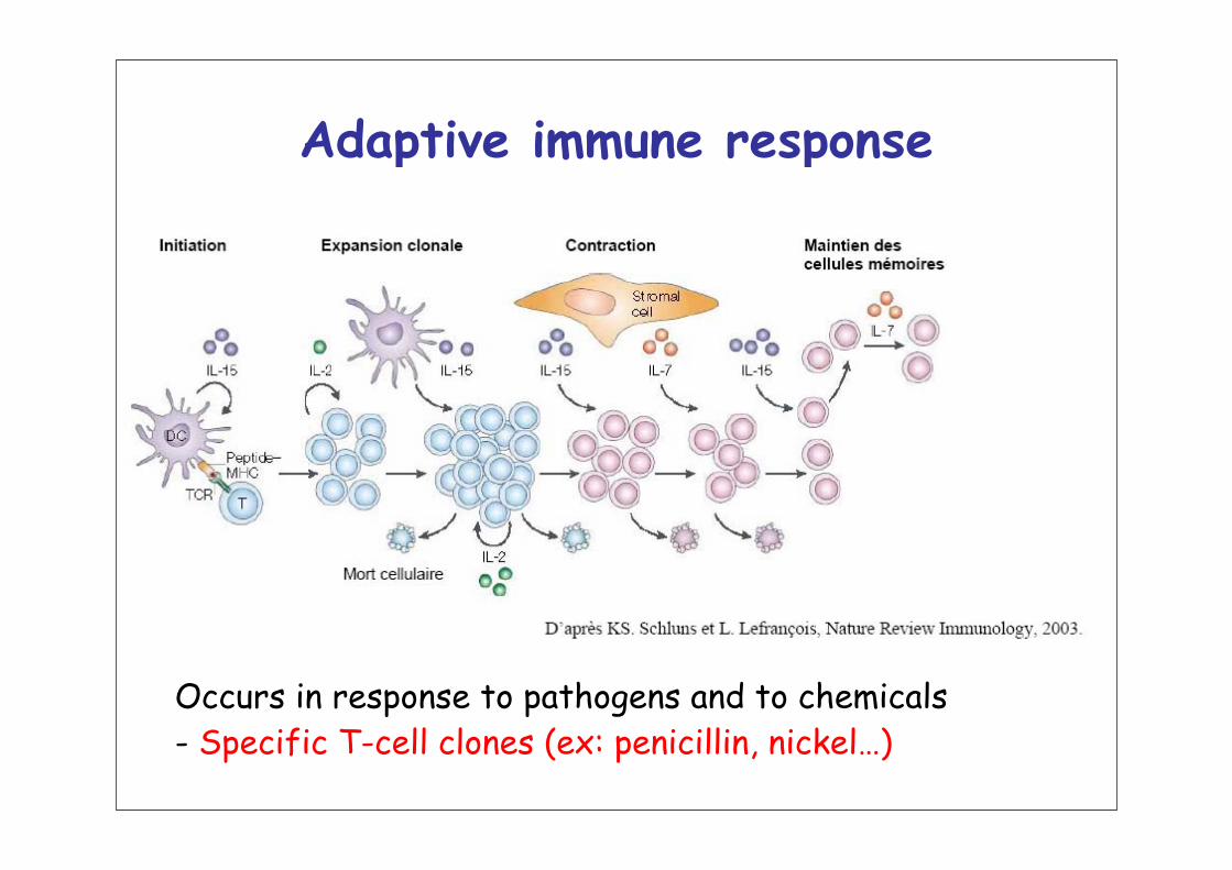
Adaptive immune response
Occurs in response to pathogens and to chemicals- Specific T-cell clones (ex: penicillin, nickel…)

IMMUNOTOXICITY
• Immunosuppression– Down-regulation of immune responses by chemicals
• Hypersensitivity– Innapropriate specific immune response to
chemicals• Stimulation of the immune system leading to
pathology– Cytokine release
• Auto-immunity– Immune response to auto-antigens induced by
chemicals

IMMUNOSUPPRESSION
Strategies for in vitro testing
Strategies for in vivo testing
based on the functional evaluation ofthe different components of the immune system

In vitro testing for Immunosuppression
• Initial evaluation of myelotoxicity– Hematopoietic progenitors (CFU-GM assay)
• Determination of lymphotoxicity– Lymphocytes (trypan blue, MTT, LDH release)
• Determination of potential effects on NK cells– Cytotoxicity (51Cr, flow cytometry) on selected target cells
(YAC for rodent, K562 for human)• Lymphocyte proliferation
– Mitogens (ConA, PHA), Anti-CD3 + anti-CD28– 3H-thymidine, flow cytometry (PKH26, CFSE)
• Cytokine production– Whole blood assay (IL-2, IFN-gamma)
• Determination of potential effects on antibody induction/production
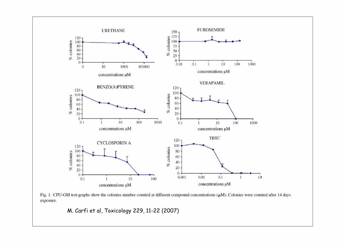
M. Carfi et al, Toxicology 229, 11-22 (2007)
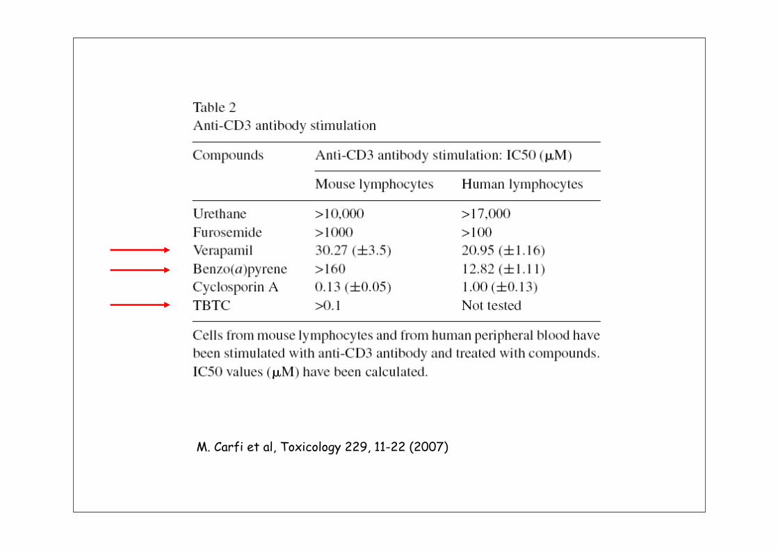
M. Carfi et al, Toxicology 229, 11-22 (2007)

M. Carfi et al, Toxicology 229, 11-22 (2007)
IC50 obtained when combining all tests
Verapamil, a calcium channel blocker, was found positiveTrue in the real life ?Remember: no drugs have been withdrawn from the marketdue to immunosuppression; why ? impossible to detect !
Chemical Hypersensitivity

Steps of chemical sensitization
• Pre-immunological phase– Chemical reactivity– Metabolism– Genetic polymorphism
• Sensitization phase– Dendritic cell activation– Hapten presentation to T-cells– T-lymphocyte activation and proliferation
• Effector phase– Th1 vs Th2 response

Strategies for detecting chemical
• Strategy for in vivo testing– Chemicals induce immunopathology (GMPT tests)– Chemicals induce an immune response (LLNA)
• In vitro– Try to mimick an immune response (ie lymphocyte
proliferation) to chemical sensitizers using and in vitro approach ? Failed so far
– Try to find unique properties of chemical sensitizers that distinghish them form other chemicals ?

Development ofIn silico/in vitro methods• QSAR
– Patlewicz G, Aptula AO, Uriarte E, Roberts DW, Kern PS, Gerberick GF, Kimber I, Dearman RJ, Ryan CA, Basketter DA. An evaluation of selected global (Q)SARs/expert systems for the prediction of skin sensitisation potential. SAR QSAR Environ Res. 2007 Jul-Sep;18(5-6):515-41.
• Peptide reactivity assay– 2006, 82 molecules tested
• Cell-based models– Human dendritic cells (murine,
human)– Cell lines (THP-1, U937, MUTZ-3)
Chemicalreactivity

From Gerberick GF et al, Tox Sci 2004
Chemical reactivity: peptide reactivity assay
GSH Lysine
Cystéine Histidine

Statistics for non-sensitizers vs sensitizers(cys 1/10 and lys 1/50 prediction model)
Gerberick et al, 2006
6 molecules failed
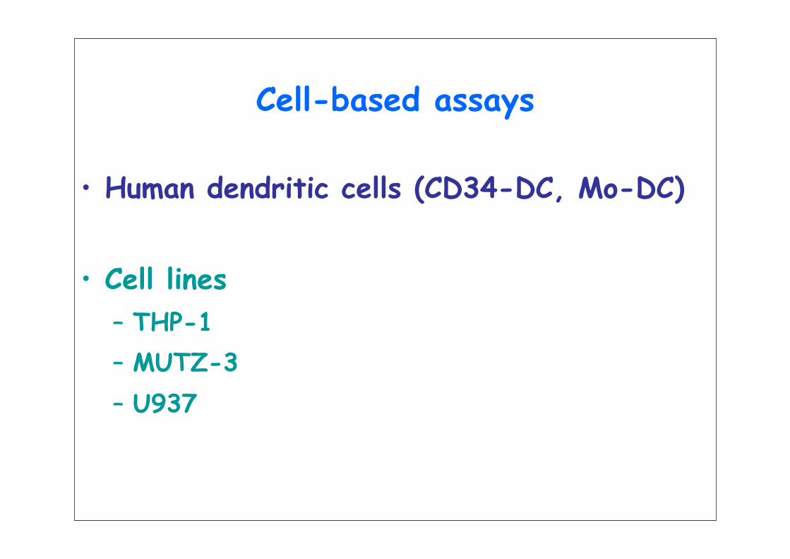
Cell-based assays
• Human dendritic cells (CD34-DC, Mo-DC)
• Cell lines– THP-1– MUTZ-3– U937

Dendritic cell migration after antigen uptake
CCR6 +
CCR7 -
Blood
CCR6 +
CCR7 –
E-cadherin +
Antigens/chemicals
Skin
CCR6 –E-cadherin –CCR7 +
Lymphaticvessel
Lymph node
CCL20 productionby keratinocytes
Recruitment of LC precursors
Migration of activated LCthrough lymphatics
Homing in T cell areas of maturing LCs in reponse to CCL19 and CCL21
T cell area
CCL21
CCL21CCL19

Kissenpfenig et al, Immunity 2005
Murine epidermis
Murine skinEGP-langerin
Green: LCRed = CMH IIYellow: LC/CMH II

Peripheral organ
Antigen
Immature dendritic cell
Lymph
1- Antigen uptakeand Activation
2- Migration
T cell
Lymph node
Mature dendritic cell
3- Antigen Presentation
Danger signals (microbial products, RNA, CpG DNA…
Toll-Like Receptor

CD 1a -Mannose receptor +++ FcR +++
Mannose receptor +/-FcR +/-
MHC II +++CD 80, CD 86 +++CD83 ++CD 40 +++IL-12 production
MHC II +/-CD 80, CD 86 –CD 83 -CD 40 -
E-cadherin +++
CCR7 – CCR6 ++
CCR7 +++CCR6 -
E-cadherin -
Antigen capture
Antigen presentation
Migration
CD 1a +
SKIN LYMPH NODE

Can chemical sensitizers play the role of danger signals and
Hypothesis:
chemicals mimick « danger signals » (TLR agonists…) signalling in Dendritic Cells
orDendritic cells perceived chemical sensitizers as
« danger »
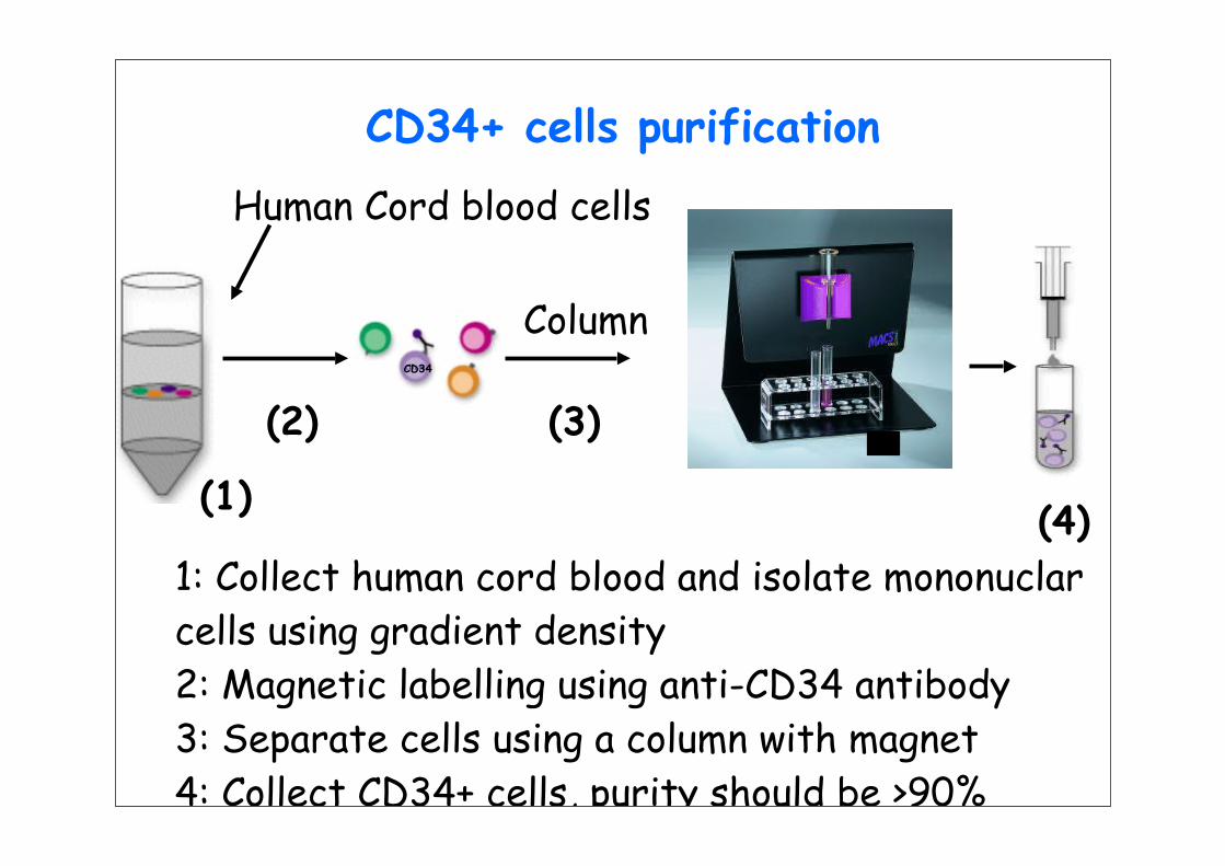
CD34+ cells purification
CD34
Column
Human Cord blood cells
1: Collect human cord blood and isolate mononuclarcells using gradient density2: Magnetic labelling using anti-CD34 antibody3: Separate cells using a column with magnet4: Collect CD34+ cells, purity should be >90%
(1)
(2) (3)
(4)
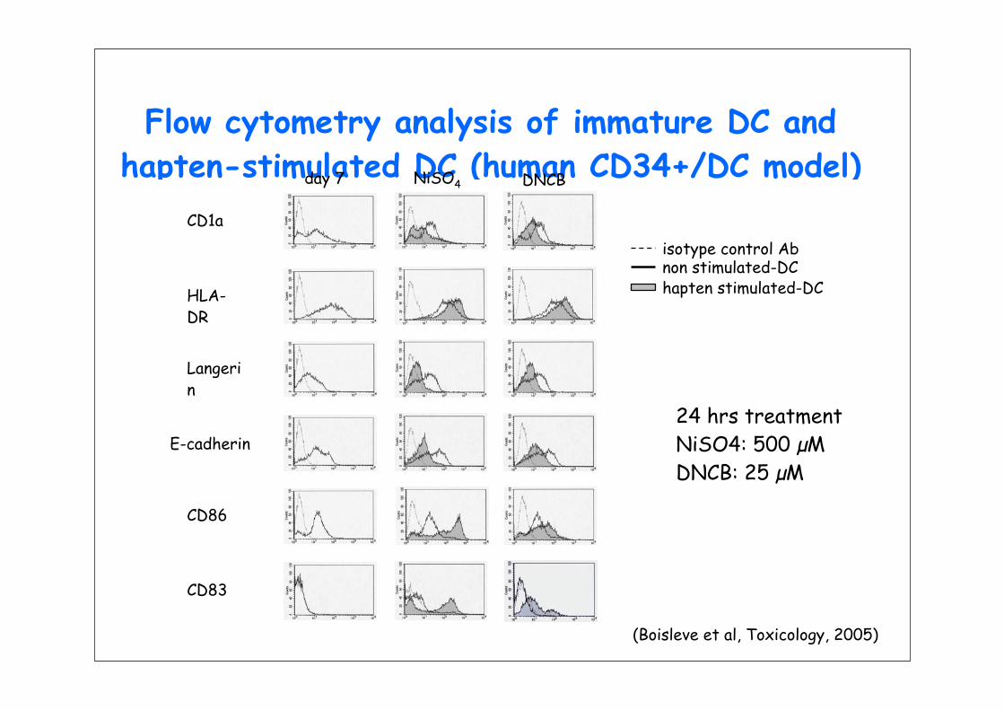
Flow cytometry analysis of immature DC and hapten-stimulated DC (human CD34+/DC model)NiSO4 DNCBday 7
CD1a
HLA-DR
Langerin
E-cadherin
CD86
CD83
isotype control Ab
hapten stimulated-DC non stimulated-DC
24 hrs treatmentNiSO4: 500 µMDNCB: 25 µM
(Boisleve et al, Toxicology, 2005)

Haptens alter DC phenotype
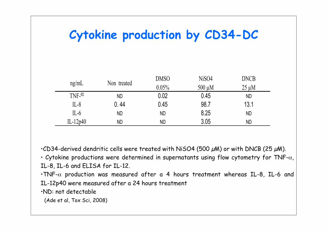
Cytokine production by CD34-DC
•CD34-derived dendritic cells were treated with NiSO4 (500 µM) or with DNCB (25 µM).• Cytokine productions were determined in supernatants using flow cytometry for TNF-α, IL-8, IL-6 and ELISA for IL-12.•TNF-α production was measured after a 4 hours treatment whereas IL-8, IL-6 and IL-12p40 were measured after a 24 hours treatment •ND: not detectable
(Ade et al, Tox Sci, 2008)
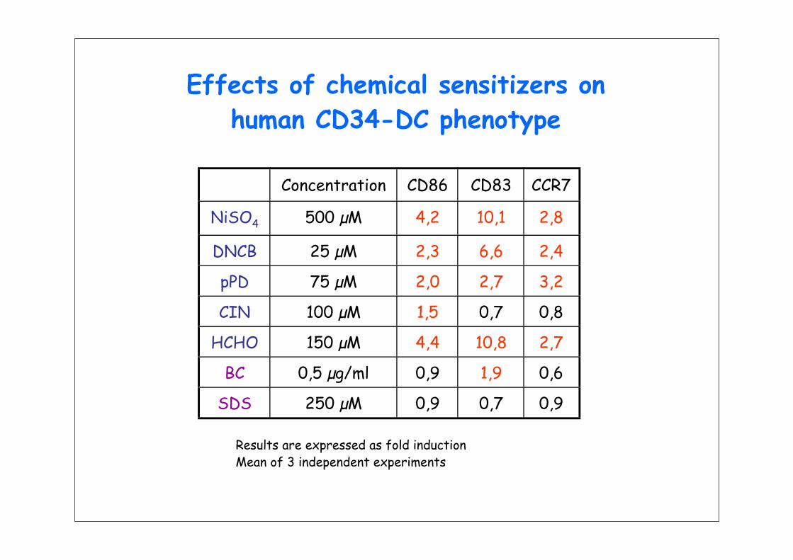
Concentration CD86 CD83 CCR7
NiSO4 500 µM 4,2 10,1 2,8
DNCB 25 µM 2,3 6,6 2,4
pPD 75 µM 2,0 2,7 3,2
CIN 100 µM 1,5 0,7 0,8
HCHO 150 µM 4,4 10,8 2,7
BC 0,5 µg/ml 0,9 1,9 0,6
SDS 250 µM 0,9 0,7 0,9
Effects of chemical sensitizers onhuman CD34-DC phenotype
Results are expressed as fold inductionMean of 3 independent experiments
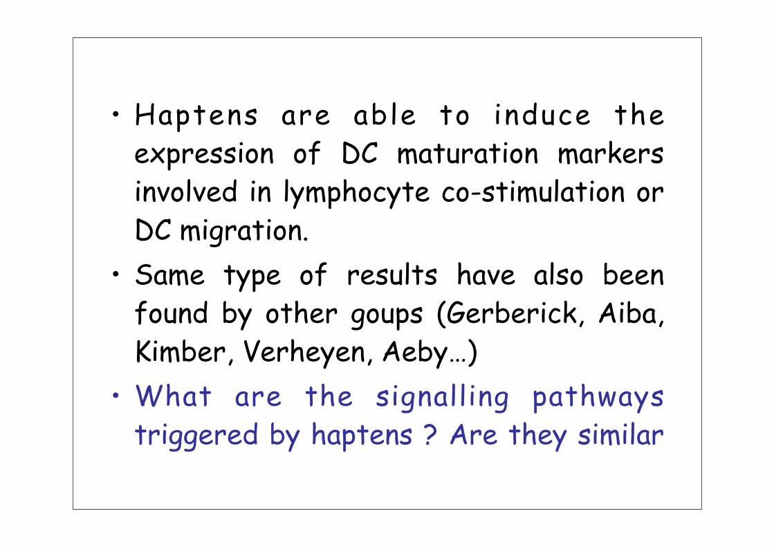
• Haptens are able to induce the expression of DC maturation markers involved in lymphocyte co-stimulation or DC migration.
• Same type of results have also been found by other goups (Gerberick, Aiba, Kimber, Verheyen, Aeby…)
• What are the signalling pathways triggered by haptens ? Are they similar

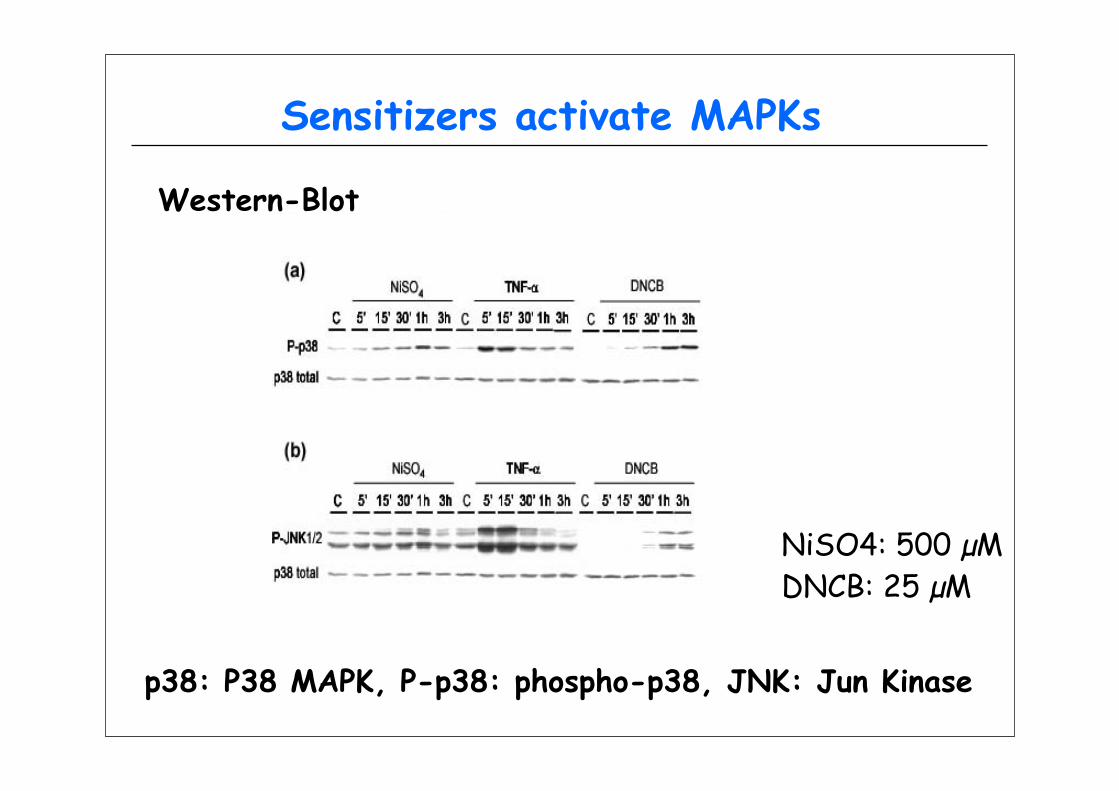
Sensitizers activate MAPKs
Western-Blot
p38: P38 MAPK, P-p38: phospho-p38, JNK: Jun Kinase
NiSO4: 500 µMDNCB: 25 µM

Chemical sensitizers activate NFKB
NiSO4: 500 µMDNCB: 25 µM
3h 5h 7h 9hTNF-a 0 1h 2h 4h 6h 8h 0 1h 2hDNCB
3h 5h 7h 9h0 1h 2h 4h 6h 8h 0 1h 2h 3h 5h 7h 9h0 1h 2h 4h 6h 8h 0 1h 2hNiSO4
IkB-αpp38P38 total
3h 5h 7h 9h0 1h 2h 4h 6h 8h 0 1h 2h
NiSO4- +
DNCB- +
Total protein
NiSO4- +
DNCB- +
NiSO4- +
DNCB- +
NF-kB NF-kB-mut
p65
Western-Blot
DNA-binding activity

DNCBNiSO4
NF-kB
IkBMAPKs(p38, JNK)
NF-kB
IkB
MAPKs(p38, JNK)
CCR7CD83
CD86IL-8
TNF-α
CD86,CD83 HLA-DR, CD40
IL-8, IL-6, IL-12p40CCR7
CD40, HLA-DR,IL12p40, IL-6
?
Signaling pathways in CD34-DC activated by NiSO4 or DNCB
How DC handle the chemical stress ?
• Chemical sensitizers modify the DC phenotype
• Current hypothesis: chemical reactivity is necessary for a chemical to be a sensitizer
• However, chemical stress is known to induce cell signaling leading to cell death
• Necessity for the DC to handle the chemical stress to survive

Nrf2
Kinases
ARE
Proteasome degradation
Nrf2
Nrf2
Keap1
SH SH
P
sMaf
Cell SurvivalDC phenotype alteration ?
Stressors/inducers
NUCLEUSCYTOPLASM
Target gene functions
PP
Nrf2 HMOXNQO1…
Kensler TW, 2007
Keap1
S S

0
1
2
3
4
5
6
NiS
O4
300
NiS
O4
400
NiS
O4
500
pPD
25
pPD
50
pPD
75
HC
HO
50
HC
HO
100
HC
HO
150
DN
CB
6,2
5
DN
CB
12,
5
DN
CB
25
BC
0,5
BC
1
BC
2
tBH
Q
CIN
25
CIN
50
CIN
100
CIN
150
SD
S 1
50
SD
S 2
00
SD
S 2
50
HMOXNQO1
mRNAs expression of NQO1 and HMOX1after chemical treatment
(CD34-DC model)
Results of a representative experiment

Nrf2 protein level in response to chemicals (CD34-DC model)
DMSO CIN NT SDS DMSO DNCB
Donor 2 Donor 3Cells were treated with chemicals for 5h3x106 cells lysed in Laemmli BufferIncubation at 100°C during 5 minutesCentrifugation 20 min at 15,000 gMigration of 20 µL of template
Nrf2
NT NiSO4 NT pPD NT BC Eth tBHQ
Donor 1
β-tubulin
500µM 75µM 2µg/ml 0.05% 30µM
100 KDa150 KDa
100 KDa150 KDaNrf2
β-tubulin
Models for in vitro evaluation
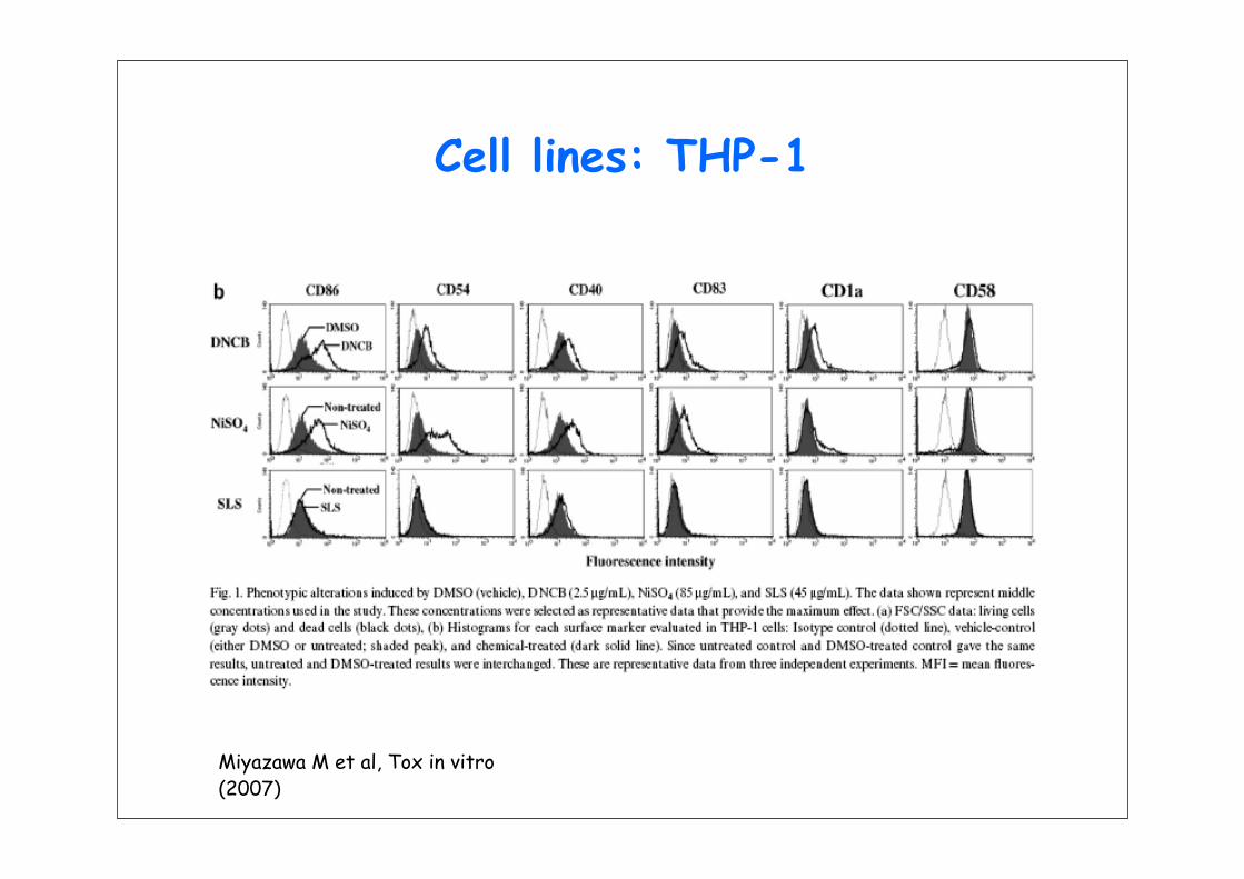
Cell lines: THP-1
Miyazawa M et al, Tox in vitro (2007)

Cell lines: THP-1
Miyazawa M et al, Tox in vitro (2007)

• THP-1 based assay = hCLAT (human cell line activation test)– Evaluated by 5 labs since 2004: P&G,
Shiseido, Kao, Henkel, L’Oréal (2nd ring study ongoing)
• U937/CD86– Originally developed by L’Oréal and
Cosmital SA (now P&G) with LVMH. Recently transferred in Shiseido and
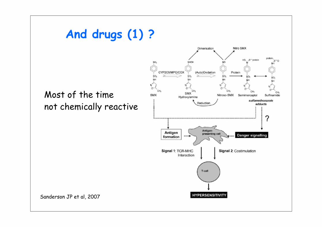
And drugs (1) ?
Sanderson JP et al, 2007
Most of the timenot chemically reactive

And drugs (2) ?
Sanderson JP et al, 2007
Detection of SMX adducts inMo-DC following incubationwith both SMX and SMX-NO
Cell surface covalent binding
Intracellular binding
Protein-SMX adducts
Are these adducts providing signalsin DC allowing their maturation ?

Prediction of chemical sensitization• In vivo preclinical test are in the process of
prevalidation for the detection of contact sensitizers
• Prediction of the potential of chemical to be a sensitizer may be possible based on the following basis:– Protein reactivity is necessary for a majority of
molecules– This property may be obtain only after
metabolisation (the case for drugs ?)– A chemical sensitizer = danger danger
signal for DC
• And of course: more work is needed,

Conclusions and perspectives
• The amount of results obtained in the field of immunotoxicology is highly dependent on the regulatory activity– Ex: EMEA guideline in immunotoxicology and TDR
test• The 7th amendment to the cosmetics
directive and the REACH program are at the initiative of a great industrial and academic efforts in the search for alternative/in vitro tests
• Always important to keep in mind that to generate new tests it is necessary to

Success ?• Hypersensitivity
– The peptide reactivity assay and cell assays are very promising; prevalidation studies are ongoing
– Combination of results from these assays and others (QSAR…) may provide a robust decision tree
• Immunosuppression– Efforts are ongoing to validate well-described
existing tests– Work is needed to develop new tests and to refine
the existing ones• Immunostimulation
– TG1412 accident due to CRS (cytokine release syndrome: there is a need for tests predicting this type of events


SUBSETS OF DCs
Myeloid lineage Langerhans cells Dermal DC
Lymphoid lineage Plasmacytoid DC Thymic DC
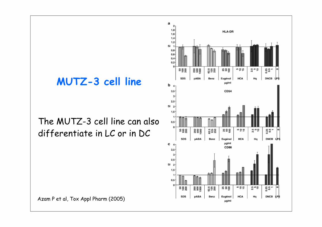
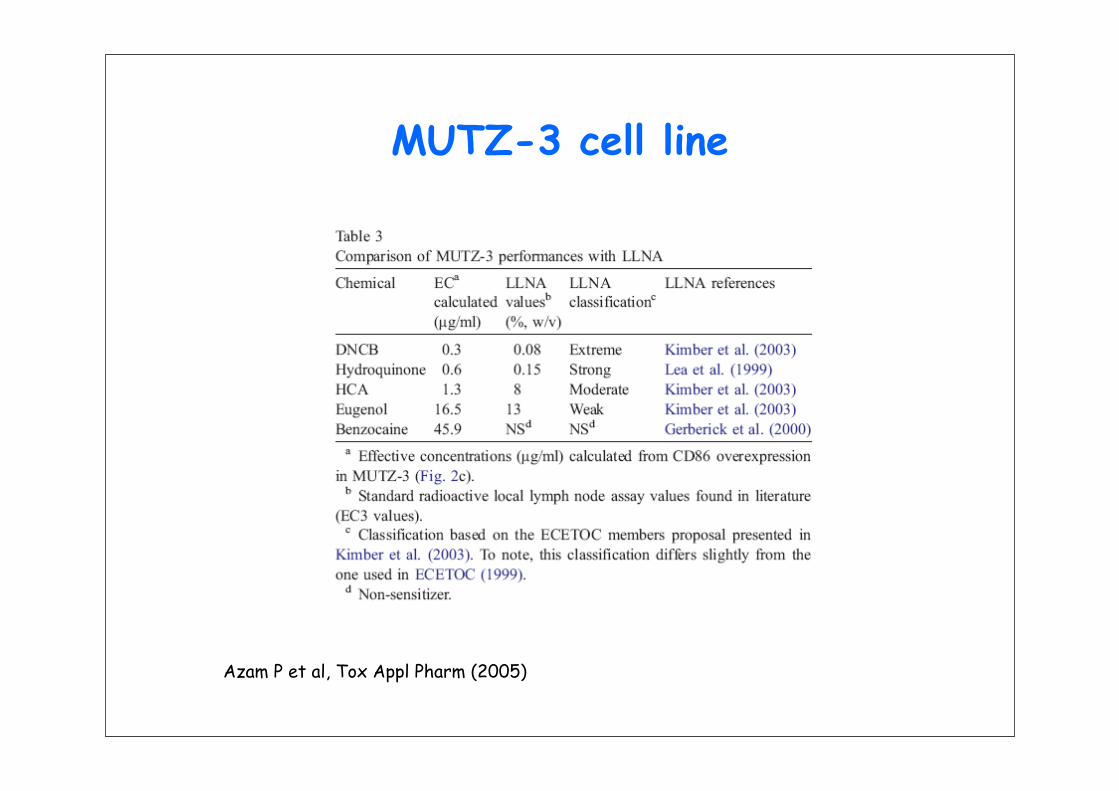
MUTZ-3 cell line
Azam P et al, Tox Appl Pharm (2005)
The MUTZ-3 cell line can alsodifferentiate in LC or in DC
MUTZ-3 cell line
Azam P et al, Tox Appl Pharm (2005)

DC generation from CD34+ cells
CD34+
D0: GM-CSF, TNF-αD4: GM-CSF , TNF-α iDCs
D0 D6
D0: GM-CSF, TNF-α, Flt-3LD4: GM-CSF, TNF-α iDCsCD34+
D0: GM-CSF, TNF-α, Flt-3L, SCFD4: GM-CSF, TNF-α, IL-4
iDCsCD34+
D0: GM-CSF, TNF-α, Flt-3L, SCFD4: GM-CSF, TNF-α
iDCsCD34+


M. Carfi et al, Toxicology 229, 11-22 (2007)

Regulatory tests for the detection of chemical sensitizers
• Guinea pig tests: mimick the elicitation phase of contact dermatitis– Buëhler test– Magnusson & Kligman test (+ adjuvant, GPMT)– OCDE 406, FDA, US-EPA, EMEA (local tolerance
guideline)
• Local Lymph Node Assay: measure the sensitization phase of contact allergy– OCDE 429, EMEA (local tolerance guideline), FDA,
US-EPA– A revolution : a test not based on the evaluation of

ECVAM exploratory study
• RIVM (H. van Loveren, R. Vandenbriel)• University of Milan (E. Corsini)• Bayer (HW. Vohr)• University of Utrecht (R. Pieters)• University of Paris 11 (M. Pallardy, A. Biola)
• ECVAM (L. Gribaldo, M. Carfi)

Sources of cells
• Mouse and rats– Splenocytes– CD34+ cells (hematopoietic progenitor)
• Human– Peripheral Blood Monocyte Cells after
density gradient purification– CD34+ cells (hematopoietic progenitor)
• DC were first identified in the epidermis: Langerhans cells (1868)
• Their presence in other tissues was identified in 1973 (Steinman and Cohn)
• Early 1990s: in vitro generation of human DCs from CD34+ progenitor cells
• Mid 1990s: human DC can also be

% o
f migra
ted
DCs
DNCB
- CCL19 + CCL190
5
10
15
20
7,8 % 5%
12%
18%
+ CCL19
NiSO4
- CCL19
23,2%
0
5
10
15
20
25
30
0,4%
8,0% 8,9%
% o
f migra
ted
DCs
DC migration after hapten stimulation
Up-regulation of CCR7 allows the migration of DC after CCL19 addition (Boisleve et al, J Invest Dermatol, 2004)
culture medium +/- CCL19
200 000 cellsTranswell® system
control cells 24hDNCB 24hNiSO4 24h