improved extracellular vesicle detection and characterization
TRANSCRIPT

IMPROVED EXTRACELLULAR VESICLE
DETECTION AND CHARACTERIZATION
Ph.D. thesis
Xabier Osteikoetxea
Doctoral School of Molecular Medicine
Semmelweis University
Supervisor: Edit I Buzás MD, D.Sc
Official reviewers: Dr. Mihály Józsi Ph.D.
Zoltán Jakus MD, Ph.D.
Head of the Final Examination Committee: Dr. Éva Szökő, D.Sc
Members of the Final Examination Committee: Zoltán Benyó MD, D.Sc
Dr. Zsuzsanna Bajtay, D.Sc
Budapest, 2016
2
TABLE OF CONTENTS
1. ABBREVIATIONS ................................................................................................................. 4
2. INTRODUCTION ................................................................................................................... 7
2.1 Theoretical Background ...................................................................................................... 8
2.1.1 Extracellular vesicles and their classification .............................................................. 8
2.1.1.1 Exosomes ............................................................................................................ 10
2.1.1.2 Microvesicles ...................................................................................................... 12
2.1.1.3 Apoptotic vesicles ............................................................................................... 14
2.1.1.4 Other vesicle types .............................................................................................. 15
2.1.2 Extracellular vesicles and their functions ................................................................... 15
2.1.2.1 Extracellular vesicles in health ............................................................................ 16
2.1.2.2 Extracellular vesicles in cancer ........................................................................... 18
2.1.2.3 Extracellular vesicles in cardiovascular diseases ................................................ 20
2.1.2.4 Extracellular vesicles in other diseases ............................................................... 21
2.1.3 Clinical applications of extracellular vesicles ............................................................ 22
2.1.3.1 Extracellular vesicles as diagnostic tools ............................................................ 23
2.1.3.2 Extracellular vesicles as therapeutic tools ........................................................... 24
2.1.4 Extracellular vesicle isolation and characterization techniques ................................. 25
2.1.4.1 Extracellular vesicle isolation techniques ........................................................... 25
2.1.4.2 Extracellular vesicle characterization techniques ................................................ 29
3. OBJECTIVES ....................................................................................................................... 33
4. MATERIALS AND METHODS .......................................................................................... 35
4.1 Cell line cultures................................................................................................................ 35
4.2 Human blood, platelet, and red blood cell concentrate collection and processing ............ 36
4.3 Extracellular vesicle isolation ........................................................................................... 36
4.4 Extracellular vesicle concentration and size ..................................................................... 37
4.5 Transmission electron microscopy of extracellular vesicle preparations .......................... 38
3
4.6 Flow cytometry of cells and extracellular vesicles............................................................ 38
4.7 Protein and lipid determination of extracellular vesicle preparations ............................... 40
4.8 Membrane lipid bilayer order of extracellular vesicle preparations .................................. 43
4.9 Detergent lysis of extracellular vesicles ............................................................................ 43
4.10 Statistical analyses ........................................................................................................... 44
5. RESULTS ............................................................................................................................... 45
5.1 Improved characterization of extracellular vesicle preparations based on protein to lipid
ratios and lipid properties ........................................................................................................ 45
5.1.1 Optimization of the sulfophosphovanilin total lipid assay for EV studies ................. 45
5.1.2 Interference of different compounds with lipid and protein assays ........................... 46
5.1.3 Accuracy and variability of lipid and protein assays.................................................. 48
5.1.4 Characterization of different EV subpopulations ....................................................... 50
5.1.5 Characterization of protein to lipid ratios of the EV subtypes ................................... 54
5.1.6 Protein to lipid ratios of damaged or contaminated EV samples ............................... 56
5.1.7 Lipid membrane order of the EV subtypes ................................................................ 58
5.2 Differential detergent sensitivity of extracellular vesicle subpopulations......................... 61
5.2.1 Morphology and concentrations of EVs found in the serum-free conditioned media of
various human cell lines ...................................................................................................... 61
5.2.2 Detergent sensitivity of apoptotic bodies and microvesicles ..................................... 63
5.2.3 Higher detergent resistance of exosomes ................................................................... 65
5.2.4 Comparison of detergent capacity to disrupt EVs ...................................................... 66
5.2.5 Flow cytometric validation of detergent sensitivity of extracellular vesicles ............ 70
6. DISCUSSION ........................................................................................................................ 72
7. CONCLUSIONS ................................................................................................................... 78
8. SUMMARY ........................................................................................................................... 79
9. ÖSSZEFOGLALÁS .............................................................................................................. 80
10. REFERENCES .................................................................................................................... 82
11. BIBLIOGRAPHY OF THE CANDIDATE .................................................................... 111
12. ACKNOWLEDGEMENTS .............................................................................................. 113
4
1. ABBREVIATIONS
% CV Percentage coefficient
variation
% Error Percentage error
16K PRL 16 kDa N-terminal
prolactin fragment
A Ampere
Ab/AM Antibiotic Antimycotic
Solution
AC8 Anti-cholesterol
antibody
ACD Acid-citrate dextrose
ADP Adenosine diphosphate
Alix Alg-2
interacting protein X
ANOVA Analysis of Variance
APC Allophycocyanin
APO Apoptotic body
ARF6 ADP-ribosylation factor
6
BBB Blood–brain barrier
BCA Bicinchoninic acid assay
BSA Bovine serum albumin
CTX Cholera toxin
DAPI 4',6-diamidino-2-
phenylindole
DC Dendritic cell
DEX Dendritic cell exosome
DMEM Dulbecco's Modified
Eagle's Medium
DRF3/Dia2 Diaphanous Related
Formin 3
EBV Epstein-Barr virus
ECM Extracellular matrix
EM Electron microscopy
EMT Epithelial-mesenchymal
transition
ESCRT Endosomal sorting
complex required for
transport
EV Extracellular vesicle
EXO Exosome
FBS Fetal bovine serum
FITC Fluorescein
Isothiocyanate
FSC Forward Scatter
g g-Force, equivalent to
RCF

5
GM-CSF Granulocyte-macrophage
colony-stimulating factor
GP General polarization
GTPase Guanosine triphosphate
phosphohydrolase
HGFR Hepatocyte growth
factor receptor
HIV Human
immunodeficiency virus
hr Hour
HSV Herpes simplex virus
IL-6 Interleukin-6
ISEV International Society for
Extracellular Vesicles
ISTH International Society on
Thrombosis and
Haemostasis
min Minute
miRNA microRNA
MMP Matrix metalloproteinase
mRNA messenger RNA
MV Microvesicle
MVB Multivesicular body
NK Cell Natural killer cell
nSMase 2 Neutral
sphingomyelinase 2
OMV Outer membrane vesicle
OSE Oxidation-specific
epitopes
PBS Phosphate Buffered
Saline
PE Phycoerytherin
PFP Platelet-free plasma
P-gp P-glycoprotein
PS Phosphatidylserine
Rab Ras-related proteins in
brain
RCF Relative Centrifugal
Force, equivalent to g-
Force
Refs References
RPMI Roswell Park Memorial
Institute Medium
RT Room temperature
SD Standard deviation
SDS Sodium dodecyl sulphate
SEM Standard error mean
SPV Sulfophosphovanilin
SSC Side Scatter
TEX Tumor exosome
TF Tissue factor
TfR Transferrin receptor

6
TRAIL TNF-related apoptosis-
inducing ligand
Treg Regulatory T cell
TrpC5 Transient receptor
potential channel 5
TRPS Tunable resistive pulse
sensing
V Volt
VEGF Vascular endothelial
growth factor
VPS Vacuolar protein sorting
7
2. INTRODUCTION
Extracellular vesicles (EVs) are a heterogeneous group of lipid bilayer enclosed
particles found to be released by most, if not all, cells. To date EVs have been found to
be present in most biological fluids as well as in the environment around us. These
vesicles can efficiently carry and protect from degradation different biological molecules
such as functionally active proteins, lipids, and nucleic acids. By transferring such
molecules between cells, different EVs participate in a diversity of biological processes
in health and disease such as inflammation, immune suppression, antigen presentation,
tumor development, as well as in the transfer of genetic information, morphogens and
signaling molecules.
Given their ability to participate in the above biological processes and to transfer
molecules among cells, EVs have become increasingly attractive to researchers from
various disciplines for the development of novel diagnostic and therapeutic tools. As a
consequence, the field of EV research has seen tremendous growth and an exponential
increase in number of publications year after year which has profoundly impacted our
understanding of intercellular communication, tumor and stem cell biology,
inflammation, virology, circulating extracellular RNA and DNA research among other
fields
Despite of the fast growth experienced in the field of EVs there are still remain
many questions to address about the fundamental characteristics of distinct EV
subpopulations. One important limitation for the field is that there are still no universally
accepted molecular markers with which to characterize EVs. Additionally, there are no
gold standard EV isolation techniques. Because of these reasons some disparity exists in
the literature in regards to the different isolation and characterization techniques used for
various studies. Consequently, even if there is ample evidence clearly supporting the
effects of EVs in many biological processes there are still some areas where the evidence
is ambiguous due to dissimilar findings.
Considering these important limitations in the field, the present work focused on
improving existing EV characterization and detection techniques by introducing to the
field new approaches based on lipid properties with which to quantify and characterize
EV preparations.
8
2.1 Theoretical Background
2.1.1 Extracellular vesicles and their classification
Before the full realization of their biological significance, classification, or
functions, different observations provided evidence for the existence of EVs. Some early
evidence for these vesicles was provided in 1946 by the findings of procoagulant platelet-
derived particles in healthy human plasma (1). Later, these particles were termed “platelet
dust” (2) and in the ensuing decades more reports surfaced of such particles found in other
biological settings including rectal adenomas (3) and tumor tissues (4), during bone
calcification (5, 6), in human semen (7, 8), in bovine sera (9) and human cell cultures
(10). Only until 1983 did more evidence emerge about how some of these vesicles were
generated when detailed electron microscopic studies revealed that these particles were
released upon fusion of inner multi-vesicular bodies (MVBs) with the cell membrane
during the differentiation of immature reticulocytes (11-13).
Following these discoveries more research attention was brought unto EVs with
further reports paving the way for a rapidly emerging interest. One early report showed
that EVs released by B cells were capable of inducing T cell responses (14). Later, other
reports showed that the vesicles contain and can horizontally transfer functional RNA
between cells (15, 16). Recently more reports have expanded the known functions and
biological roles of EVs impacting our understanding of many fields of biology such as
immunology (17, 18), virology (19), neuroscience (20, 21), cardiovascular (22) and
reproductive (23) biology, parasitology (24), and bacteriology (25, 26). With this rapid
growth in the amount of reports about EV biology some classification and nomenclature
of different subsets of EVs has emerged. Although there is still some debate over the
nomenclature used, most in the field utilize the classification of different EVs based on
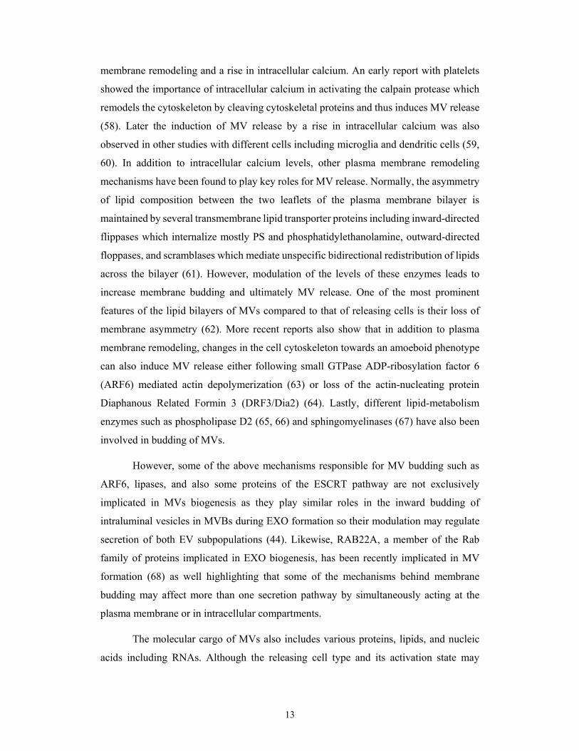
their biogenesis and size ranges as exosomes (EXOs) of approximately 100 nm in size,
microvesicles (MVs) of 100 to 1000 nm in size, and apoptotic bodies (APOs) of more
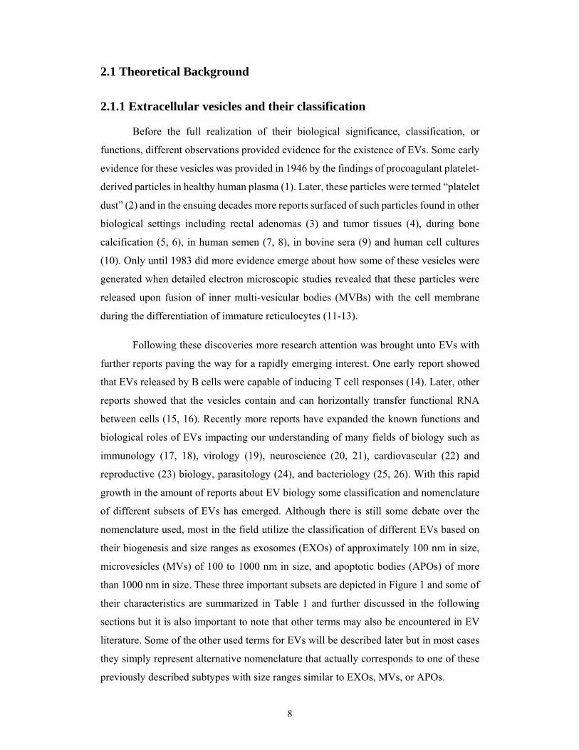
than 1000 nm in size. These three important subsets are depicted in Figure 1 and some of
their characteristics are summarized in Table 1 and further discussed in the following
sections but it is also important to note that other terms may also be encountered in EV
literature. Some of the other used terms for EVs will be described later but in most cases
they simply represent alternative nomenclature that actually corresponds to one of these
previously described subtypes with size ranges similar to EXOs, MVs, or APOs.
9
Figure 1. Size ranges of different EV subtypes (27).
10
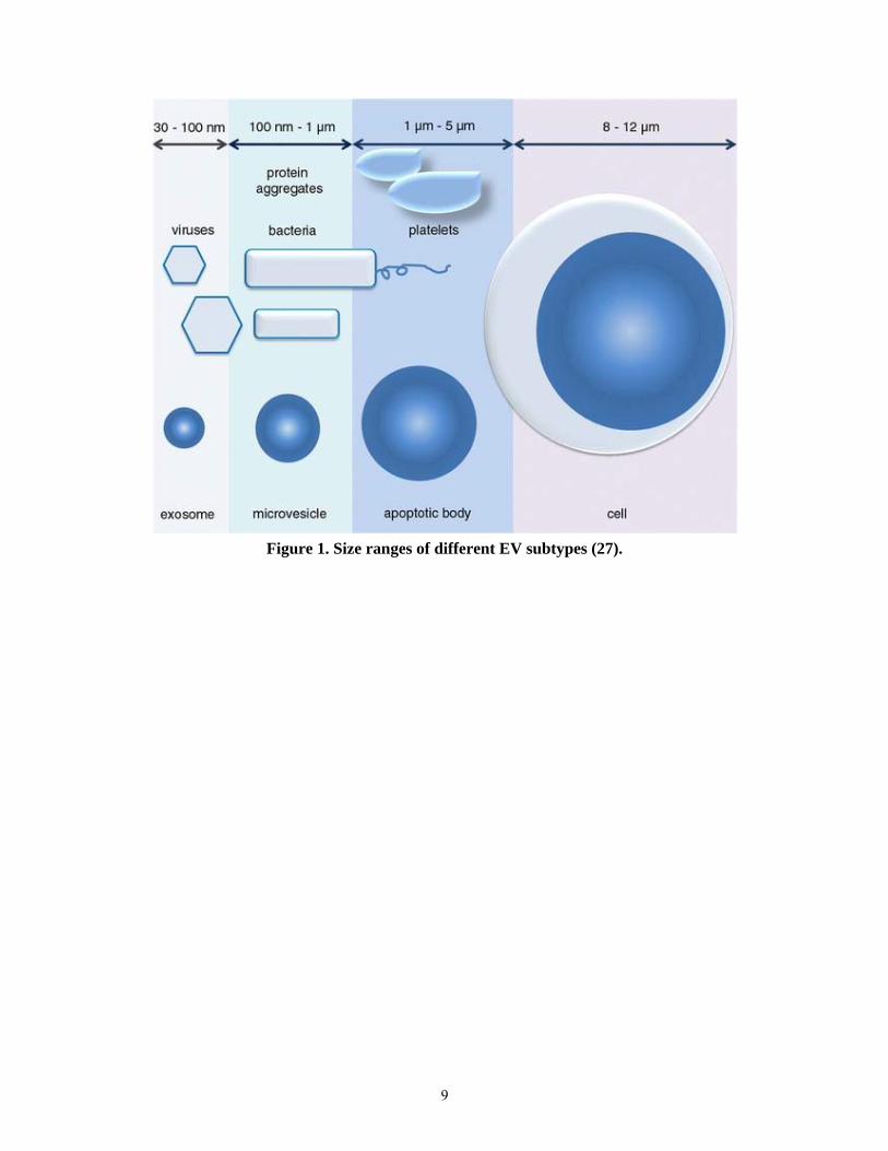
Table 1. Characteristic parameters of different EV subtypes
In the following sections the basic characteristics of the different EV subtypes are
discussed in further detail with special emphasis on their defining parameters and
mechanisms of biogenesis. Focus on their functions will be discussed in later sections.
2.1.1.1 Exosomes
Exosomes comprise the smallest particles among the EV subpopulations with an
approximate size of 100 nm and currently attract the most research interest. Importantly,
this vesicle subtype is not only distinguished from the other subtypes based solely on size
but also based on having a separate molecular mechanism of formation. In contrast to
MVs and APOs which may bud directly from the plasma membrane, the biogenesis of
EXOs occurs intracellularly within the endosomal network when intraluminal vesicles
are formed inside multi-vesicular bodies which later fuse with the plasma membrane for
subsequent release instead of fusing with lysosomes for degradation (44). This unique
Parameter Exosomes Microvesicles Apoptotic bodies
Refs
Size 50–120 nm 100–1000 nm ≥1000 nm (18, 27-30)
Homogeneity relatively uniform
heterogeneous ≥2 subpopulations
(31, 32)
Nucleic acid content
miRNA, mRNA, DNA
diverse RNAs rRNAs, DNA (15, 32-36)
Lipid content high cholesterol, high glyco-sphingolipids,
phosphatidyl-serine
phosphatidyl-serine
(37-40)
Approximate density
1.13–1.19 g/mL 1.03–1.20 g/mL 1.16–1.28 g/mL (14, 18, 41-43)
EM morphology
cup shaped
heterogeneous
heterogeneous
(14, 18, 27, 29, 32)
11
type of vesicular formation was originally described in studies about the maturation steps
of rat reticulocytes when these cells need to discard their membrane bound transferrin
receptors (TfR). After an initial endocytosis of TfRs into multivesicular endosomes it was
observed that, instead of an expected fusion with lysosomes for degradation of TfR, these
endosomes later fused with the plasma membrane for subsequent release of TfR-
containing intraluminal vesicles (12, 45).
Given their endosomal origin, EXO secretion is coordinated by several members
of the Rab GTPase family of proteins which are essential regulators of intracellular
vesicle transport between different compartments (46). Early work showed that among
this large family of more than 60 GTPases, RAB4 and RAB5 were enriched in EXOs
(47). Further studies later found that RAB11 is important for EXO release pathway since
its inhibition decreased EXO secretion (48). More recent studies have further expanded
the members of this family of proteins known to have functions in the EXO release
pathway to include RAB2B, RAB5A, RAB9A, RAB27A/B (49), as well as RAB35 (50).
In addition to Rab GTPases, other proteins have also been found to mediate EXO
secretion and include elements of the endosomal sorting complex required for transport
(ESCRT) pathway such as ALIX and VPS4 as well as syndecan and syntenin (51).
Similarly to the other vesicle subpopulations, EXOs may be released
constitutively or upon activation and contain various proteins, lipids, and nucleic acids
including RNAs. Although there are some differences in cargo depending on the releasing
cell and its activation state, some molecules may be commonly found in most EXOs.
Among these molecules EXOs contain high amounts of cholesterol and
glycosphingolipids (37-40), small RNAs including mRNA and miRNA (15, 52), as well
as different transmembrane and cytosolic proteins (53, 54). A few reports have also found
DNA to be associated with EXOs (36, 55). In recent past, different candidates have
emerged for markers of EXOs including CD63, CD81, CD9, TSG101, and externalized
phosphatidylserine (PS). However, although these molecules are indeed found in most
EXOs, one problem is that they may also be found in other EV subpopulations and thus
may not be considered markers of EXOs but instead of EVs. EXOs have also been
characterized to have a cup shaped morphology by electron microscopy (14), although
there is much debate about whether this is an artifact of the preparation needed to take
electron microscopic images. An important parameter for EV isolation and
characterization is the density of the vesicles which allows to separate them from denser

12
protein aggregates. In this regard EXOs have and approximate buoyant density of 1.13 to
1.19 g/mL (14, 41).
Figure 2. Schematic representation of selected genes involved in the molecular
machinery of exosome and microvesicle secretion (44).
2.1.1.2 Microvesicles
Microvesicles comprise the intermediate particles among the EV subpopulations
with an approximate size range of 100 to 1000 nm. Not only are these particles larger but
also more heterogeneous in terms of sizes and morphology which reflects in their wider
buoyant density range of 1.03 to 1.20 g/mL (18, 42, 52). Initially many reports
characterized these vesicles in blood and termed them microparticles and most were
found to be secreted by platelets, red blood cells, and endothelial cells. However, by now
many reports have found that MVs are secreted constitutively by most if not all cells or
upon activation.
Similarly to APOs and in contrast to EXOs, MVs are secreted by budding at the
cells’ plasma membranes. The release of MVs occurs by the budding of small cytoplasmic
protrusions which then detach from the plasma membrane (56, 57). There is evidence
from several reports that this budding process is regulated and induced by plasma
13
membrane remodeling and a rise in intracellular calcium. An early report with platelets
showed the importance of intracellular calcium in activating the calpain protease which
remodels the cytoskeleton by cleaving cytoskeletal proteins and thus induces MV release
(58). Later the induction of MV release by a rise in intracellular calcium was also
observed in other studies with different cells including microglia and dendritic cells (59,
60). In addition to intracellular calcium levels, other plasma membrane remodeling
mechanisms have been found to play key roles for MV release. Normally, the asymmetry
of lipid composition between the two leaflets of the plasma membrane bilayer is
maintained by several transmembrane lipid transporter proteins including inward-directed
flippases which internalize mostly PS and phosphatidylethanolamine, outward-directed
floppases, and scramblases which mediate unspecific bidirectional redistribution of lipids
across the bilayer (61). However, modulation of the levels of these enzymes leads to
increase membrane budding and ultimately MV release. One of the most prominent
features of the lipid bilayers of MVs compared to that of releasing cells is their loss of
membrane asymmetry (62). More recent reports also show that in addition to plasma
membrane remodeling, changes in the cell cytoskeleton towards an amoeboid phenotype
can also induce MV release either following small GTPase ADP-ribosylation factor 6
(ARF6) mediated actin depolymerization (63) or loss of the actin-nucleating protein
Diaphanous Related Formin 3 (DRF3/Dia2) (64). Lastly, different lipid-metabolism
enzymes such as phospholipase D2 (65, 66) and sphingomyelinases (67) have also been
involved in budding of MVs.
However, some of the above mechanisms responsible for MV budding such as
ARF6, lipases, and also some proteins of the ESCRT pathway are not exclusively
implicated in MVs biogenesis as they play similar roles in the inward budding of
intraluminal vesicles in MVBs during EXO formation so their modulation may regulate
secretion of both EV subpopulations (44). Likewise, RAB22A, a member of the Rab
family of proteins implicated in EXO biogenesis, has been recently implicated in MV
formation (68) as well highlighting that some of the mechanisms behind membrane
budding may affect more than one secretion pathway by simultaneously acting at the
plasma membrane or in intracellular compartments.
The molecular cargo of MVs also includes various proteins, lipids, and nucleic
acids including RNAs. Although the releasing cell type and its activation state may
14
influence their cargo most MVs contain externalized PS, heterogenous RNAs, integrins,
selectins, and CD40 ligand, CD63, CD81 and CD9 (62, 69).
2.1.1.3 Apoptotic vesicles
Apoptotic bodies comprise the largest particles among the EV subpopulations
with an approximate size range of more than 1000 nm. Unlike EXOs and MVs, there are
not so many reports about APOs and for this reason it is not entirely clear whether APOs
are only secreted as membrane surrounded fragments of cells undergoing apoptosis (70,
71) or if they may also be secreted constitutively by different cells as has been reported
more recently with amoeboid type of tumor cells (64, 72). Although there are not many
reports about the biogenesis of APOs, given their common plasma membrane origin it
probably shares many similarities to the biogenesis of MVs. Among the important
molecular players discussed for MVs, those related to cytoskeletal remodeling and
changes towards amoeboid phenotype have already been implicated in the formation of
very large EVs (64, 72).
However, regardless of whether APOs are produced constitutively or not, they can
be detected and isolated from most biofluids and cell culture supernatants due to the low
level of apoptosis found in different cells and tissues or due to their constitutive secretion
by non-apoptotic or tumor cells, but most likely due to a combination of both since they
can be detected even in proliferating cell cultures with less than 5% apoptosis. Isolated
APOs have also been found to be heterogeneous in size and morphology with some
evidence showing that there might be different subpopulations of APOs depending on
whether they are derived from the apoptotic cells’ endoplasmic reticulum (ER) or plasma
membrane (73). So far the reported buoyant density of APOs is higher than other EVs at
1.16 to 1.28 g/mL but only one study has looked into their density (41).
Similarly to other EV subtypes, the molecular cargo of APOs also includes
various proteins, lipids, and nucleic acids including RNAs. Additionally, since APOs can
be secreted following apoptosis, any cellular component may be packed within them (70)
resulting in a higher diversity of molecular cargo than MVs or EXOs. Notably, APOs
may contain DNA fragments (74) and other nuclear components in addition to
externalized PS, RNAs, CD63, CD81 and CD9.
15
2.1.1.4 Other vesicle types
In addition to the three previously described EV subtypes there are also other
vesicles terms to be found in the literature. Some simply represent alternative
nomenclature that actually corresponds to one of the previously described EV subtypes.
Such is the case for example of microparticles, ectosomes, or membrane particles which
are all released directly from the plasma membrane and can be viewed as alternative terms
for MVs. Other types of EVs are named after their sources such as tumor exosomes
(TEXs), dendritic cell exosomes (DEXs), oncosomes derived from tumors, and
prostasomes which are released by the prostate and found in seminal fluid, among others.
More interestingly, there are EVs found to be secreted by gram positive and
negative as well as archaeal species of bacteria (25, 26). The term OMV emerged because
initially most reports of bacterial EVs found these vesicles to be secreted by gram
negative bacteria which have an outer membrane from where the vesicles were secreted
(75-78) but now we know that also other bacteria without outer membranes can secrete
vesicles too (25, 26, 79-81). Since the mechanisms for OMV formation are not completely
understood and since they fall within the size ranges of previously described EV subtypes,
it is still debated whether bacterial derived EVs should be considered part of the
previously described EV subtypes or if they should instead be viewed as a completely
separate subtype of EVs.
2.1.2 Extracellular vesicles and their functions
In the last decades, EVs have been found to exert many different biological
functions in both physiology and pathology explaining the rapid growth of interest in this
research field. These functions can be explained due to their unique ability to carry and
protect from degradation various biologically active molecules between different cells,
tissues, or even across species. In the following sections will be discussed several of the
biological roles in which EVs have been involved. In discussing these roles the collective
term EV is chosen for all cases where the studied particles are not thoroughly
characterized or supported by the described methodology to belong to a particular EV
subtype. Nevertheless, the originally reported EV subtype is retained for all other cases
in which the described methodology and characterization matches with those widely
utilized and accepted for the described subtype.
16
2.1.2.1 Extracellular vesicles in health
Many of the early reports about EVs described their roles in normal physiological
processes such as coagulation (1, 2) reticulocyte maturation (12, 45), and antigen
presentation (14). To date EVs have been found in most biofluids and thus many new
functions have been unraveled for their roles in different physiological processes. In this
section are described a few of the roles of EVs in physiological processes.
Since EVs were first described to participate in coagulation many reports have
further elucidated their roles in the process. Due to the externalized PS, which is a
negatively charged phospholipid, EVs can promote coagulation by binding to several
coagulation factors in the presence of calcium (1, 2). Additional support for the
procoagulant properties of EVs emerged when the key initiator of the coagulation cascade
tissue factor (TF), formerly known as thromboplastin, was discovered to be present in
EVs shed by different cells (82, 83). More recent studies have shown that blood contains
large amounts of procoagulant EVs derived mainly from platelets and that they are
important players in normal hemostasis at their baseline levels. However different disease
states may also be presented with increased levels of EVs which promote pathological
thrombosis (84-86).
Following the early report on the capacity of B cell derived EVs to carry both
antigenic material and peptide–MHC complexes (14) lots of research interest was sparked
in the field of immunology about the potential roles of vesicles. Since then many different
reports have been published further clarifying how EVs participate in various
immunological processes in health and EVs derived from many cell types and biofluids
have been found to be capable of antigen presentation and immune regulation. By now
different studies have reported that in addition to B cells other cell types can carry
preformed antigen-MHC complexes capable of activating T Cells (87-90) and EVs
isolated from malignant effusions have also been found to induce the activation of
antigen-specific T cells in vitro (91). In addition to their antigen presentation roles, EVs
have been found to have participate in immunoregulation in antigen-independent
manners. One example is that EVs derived from thymic cells have been found to induce
the development of Foxp3+ regulatory T cells (Treg) in the lung and liver as well as
inducing the conversion of CD4+CD25- T cells into Tregs. These EV-induced Tregs were
capable of suppressing the proliferation of CD4+CD25- T cells in vitro and in vivo (92).

17
EVs secreted by other cells have also been found to have immunoregulatory effects such
as those derived from neutrophils and erythrocytes which can restrain inflammation by
inhibiting macrophages from releasing pro-inflammatory tumor necrosis factor (TNF)
and IL-8 (93). Likewise, EVs isolated from different biofluids such as milk (94), pregnant
women plasma (95), or serum and bronchoalveolar fluid (96) have also been found to
have immunosuppressive effects.
In contrast to the above reports, EV derived from other sources have been found
to have immunostimulatory effects. EVs secreted from activated platelets have been
reported to carry CD40L and activate B cells through interaction with their CD40
costimulatory proteins (97). EXOs derived from DCs were also described to be capable
of activating Natural Killer cells (NK Cells) by delivering ligands for IL-15Rα and
Natural Killer Group 2 member D (NKG2D) dependent proliferation and activation in
humans and mice (98). Another example of immunostimulation is encountered in a study
where T cell proliferation was synergistically increased when these cells were exposed to
a combination of EVs and IL-2 (99).
Interestingly, EVs have also been implicated with important roles in non-synaptic
communication among various cells of the nervous system including neurons and glia.
Importantly, EVs are able to cross the blood–brain barrier (BBB) in both directions and
thus can play a role in transport to the brain of various molecules including folate which
plays an important role in neuronal development (100, 101). Moreover, EV release is also
found in the normal synaptic activity between neurons. EVs secreted during glutamatergic
synaptic activity carry away neurotransmitter receptor subunits, modulating synaptic
strength by eliminating part of these receptors from the post-synaptic membranes (102).
EVs can also facilitate the communication between two different types of cells of the
nervous system. In one report, EVs from microglia were found to act at the presynaptic
site and stimulate the synaptic activity of receiving neurons by enhancing their
sphingolipid metabolism (103). More reports have demonstrated that microglia derived
EVs modulate neurotransmission through lipid-signalling (104, 105). Lastly, Schwann
cell and oligodendrocyte derived EVs mediate neuroprotection through pro-survival
signalling pathways and modulation of gene expression in target neurons by making them
more resistant to stress and enhancing axonal regeneration (106, 107).
18
In healthy pregnancy EVs have been reported to carry different roles in the
immunological communication between the fetus and the mother. Several reports have
shown that trophoblast derived EVs are present in maternal blood and interact with both
immune and endothelial cells. It is now known that trophoblast derived EVs are involved
in the mechanisms of fetal tolerance and can elicit pregnancy-specific
immunosuppression of activated T cells by delivery of Fas ligand and Programmed death-
ligand 1 (PD-L1) (108-110).
Finally, by transferring different molecules such as RNAs and proteins, EVs have
been described to play important roles in tissue homeostasis and repair. Thus far, EVs
have been implicated with roles in lung repair (111), cardiovascular protection (112-114),
kidney repair (115, 116), hepatic regeneration (117), as well as bone calcification and
remodeling (118-120).
2.1.2.2 Extracellular vesicles in cancer
So far many studies have reported on various functions of EVs in cancer making
it one of the most intensely studied areas within the field of EV research. EVs have been
found to be involved with tumor development, survival, progression, and metastasis.
One important mechanism by which EVs are implicated with tumor biology is by
their ability to transfer pro-oncogenic molecules to other cells and tissues. For example
EVs can transfer functional pro-oncogenic proteins such as epidermal growth factor
receptor, EGFRvIII, to other cells and impart them more aggressive oncogenic
phenotypes (121). In a similar manner different studies have found that drug resistance
can be transmitted from resistant to non-resistant cells by EVs. EVs carrying P-
glycoprotein (P-gp) can transfer this drug transport membrane protein to cells lacking
drug resistance (122). Transient receptor potential channel 5 (TrpC5) (123) and
Akt/mTOR protein complexes (124) are other proteins that have been found in tumor
derived EVs that can transfer drug resistance to sensitive cells. In addition to proteins
EVs can also transfer miRNAs such as associated with chemoresitance to drug sensitive
cells and make them drug resistant (125, 126). Other pro-oncogenic phenotypes aside
from drug resistance may also be transmitted by EV associated RNA and DNA. In one
report EVs released by metastatic tumor cell lines contained miR-200 and transferred the
metastatic capacity to non-metastatic cells (127). Another study showed that transcript
19
mRNA for the pro-oncogenic EGFRvIII can be found in serum EVs of glioblastoma
patients (33). Moreover, genomic DNA can also be transferred between cells by EV, with
one study finding that the oncogenic BCR-ABL fusion gene can be transferred to recipient
cells (128).
Given their ability to transfer various molecules, tumor derived EVs have different
pro-oncogenic effects on surrounding cells and tissues. Several studies have found that
tumor derived EVs can promote tumor migration by remodeling of the extracellular
matrix (ECM). Indeed several members of the matrix metalloproteinase (MMP) family
of proteins have been found in tumor derived EVs including MT1-MMP, MMP9, MMP2,
and A disintegrin and metalloproteinase domain-containing protein 10 (ADAM10) (63,
72, 129, 130). Consequently, these MMPs may remodel the surrounding ECM and
promote tumor progression as has been shown in a study that identified tumor cell derived
EVs with functionally active MT1-MMP (131).
Another mechanism by which EVs have been found to promote tumor
development is by activation and reprograming of fibroblasts toward the phenotype of
cancer associated fibroblast (CAF) which support tumor growth and establishment of
favorable tumoral microenvironment (130, 132). In turn, CAF derived EVs can increase
recipient tumor cell motility and invasiveness as well as promoting epithelial-
mesenchymal transition (EMT) (133-135).
Yet another important role of tumor derived EVs in cancer is related to their pro-
angiogenic functions. It is well known that formation of new vasculature within tumors
for the supply of oxygen and nutrients and the removal of waste products is a critical step
for cancer cell growth and later metastasis (136). Cancer cell derived EVs have been
found to be able to promote this process by delivering pro-angiogenic interleukin-6 (IL-
6) and vascular endothelial growth factor (VEGF) (33, 137), EGFR (138), miR-9 (139)
and miR-210 (140), as well as neutral sphingomyelinase 2 (nSMase 2) (141).
In more recent studies, tumor derived EVs have been described to play an
important role in preconditioning the future metastatic sites. In one milestone study, EVs
derived from highly metastatic melanoma cell lines were found to localize in vivo at the
site of future metastasis and to educate the site towards a pro-metastatic phenotype
inducing vascular leakiness via upregulation of hepatocyte growth factor receptor
(HGFR) also called MET (142). Similarly, an earlier study had found that EVs derived
20
from human renal cell carcinoma could induce pro-metastatic phenotype changes by
delivery of pro-angiogenic mRNAs and microRNAs at the site of future metastasis in the
lung (143).
One last mechanism by which tumor derived EVs can support cancer progression
is by inducing tolerogenic immune responses to evade recognition and elimination by the
immune system. Different studies have found tumor derived EVs to be
immunosuppresive and to induce apoptosis of T cells by delivery of FasL and TNF-
related apoptosis-inducing ligand (TRAIL) (144-146).
2.1.2.3 Extracellular vesicles in cardiovascular diseases
The field of cardiovascular research has also been impacted with new
understandings of the roles of EVs in the development of disease. Different studies now
show that EVs can contribute to cardiovascular disease progression. In one report,
following induction with the peripartum cardiomyopathy factor 16 kDa N-terminal
prolactin fragment (16K PRL), endothelial cells secrete EXOs that transfer miR-146a to
cardiomyocytes and inhibit their metabolic activity and contractile function (147).
Under different conditions, several reports have shown that cardiomyocyte-
derived EVs transfer functional proteins and nucleic acids which have an adverse effect
on other cardiac cells. Extracellular heat shock protein 60 (exHSP60) has been shown to
induce apoptosis in a TLR4-dependent manner (148) and one report found that it was
mostly found in EXOs released by either non-stressed or mildly stressed cardiomyocytes
while it was absent from EXO-depleted fractions (149). Another study has found that
EXOs derived from hypoxic cardiomyocytes contained functional TNF-α and were
capable of inducing apoptosis when administered to normal cardiomyocytes (150). EXOs
derived from cardiomyocytes of a type 2 diabetes rat model were shown to contain and
transfer functional miR-320 to cardiac endothelial cells inhibiting their proliferation and
decreasing angiogenesis (151).
In addition to cardiomyocytes, other cells can secrete EVs that contribute to
cardiovascular disease. For example, EXOs secreted by cardiac fibroblasts induce a
hypertrophic phenotype in cardiomyocytes by transferring miR-21 (152). Similarly,
different reports show that EXOs derived from platelets of septic patients contribute to
21
cardiomyopathy by inducing apoptosis in endothelial cells and cardiomyocytes leading
to vascular and cardiac dysfunctions as well as coagulation (153-155).
Until now there has been growing evidence that many cardiovascular diseases are
associated with an increased oxidative stress (156, 157). Correspondingly, MVs from
myocardial infarction patients carry oxidation-specific epitopes (OSE) and are increased
at the site of coronary occlusion. Importantly, these MVs were found to be able to induce
pro-inflammatory IL-8 secretion by monocytes in an OSE-dependent manner (158).
Finally, different studies have implicated EVs with promoting atherogenesis and
atherosclerotic plaque formation by enhancing recruitement of inflammatory cells (159,
160).
2.1.2.4 Extracellular vesicles in other diseases
In addition to the previously described conditions, EVs have been found to play
important roles in many other diseases. In this section some of the important findings are
described for different conditions but the list of other diseases in which EVs play roles
will surely expand in the future.
Different studies have shown that EVs are key mediators in the progression of
neurodegenerative diseases such as Creutzfeldt–Jakob, Parkinson’s, Alzheimer’s,
Gerstmann–Straüssler–Scheinker syndrome, and amyotrophic lateral sclerosis (ALS) by
transferring the prions (161-164) or pathogenic proteins (165-170) responsible for each
of these conditions.
In parasitic infections EVs can function in immunosuppresion of the host and in
functionally preparing for the dissemination of the parasites. To date EVs have been
described to be secreted by most parasitic protozoa and helminthes. EVs derived from
these parasites can delivery various immunomodulatory signals to the host immune
system by transferring small RNAs and proteins (171-177). Additionally, parasite derived
EVs can modulate the adhesion of parasites to host cells (178).
Similarly to parasites, other pathogens release EVs that can be uptaken by the host
and are involved in the development of pathology and different studies have found
bacterial EVs in clinical samples (179-182). To date, several reports have shown that
bacterial derived EVs are readily uptaken by host epithelial and immune cells (183-185).
22
One of the most important mechanisms by which bacterial EVs can contribute to
pathogenesis is by the transfer of various biologically active toxins to host cells (79, 184,
186-188). Interestingly, in some cases delivery of equal amounts of toxins, either in
soluble form or in association with EVs, produced different effects on host cells showing
that EVs do not serve as mere transporters but also can have modulate the effects of the
toxins (189, 190). Another important mechanism by which bacterial EVs interact with the
host is by modulation of the immune system and induction of inflammation. In many
studies upregulation of proinflammatory cytokines such as IL-6 and IL-8 has been
observed following exposure to EVs derived from many pathogenic bacteria, including
Helicobacter pylori (191), Klebsiella pneumonia (192), Salmonella typhimurium (193),
Pseudomonas aeruginosa (194, 195), enterotoxigenic Escherichia coli (189), and
Neisseria meningitidis (196, 197). Lastly, bacterial EVs play important roles in
communication not only with the host but also with other bacteria and this has been found
to be especially relevant for the formation and maintenance of biolfilms (198, 199).
Finally, viruses are another class of pathogens in which EVs have been found to
play important roles. Different reports have shown that several viruses, including herpes
simplex virus (HSV), human immunodeficiency virus (HIV), and Epstein-Barr virus
(EBV), can exploit EV pathways for their own assembly and release (200-204).
Furthermore, EVs released from infected cells can contain and transfer viral particles,
proteins, and miRNA to non-infected cells and therefore allow propagation to cells that
may not have been permissive for entry of the conventional EV free virus while also
avoiding recognition by the immune system (205-209). One more important mechanism
by which EVs can assist viruses is by immunoregulation of the host (210-214).
2.1.3 Clinical applications of extracellular vesicles
There currently is much interest in developing clinical applications for EVs
considering the vast amount of findings showing a wide diversity of EV molecular cargos
and biological roles in health and disease. Since EVs released by tissues and cells in
pathological states have been found in several biofluids, one area of promise is to detect
and isolate these EVs for diagnostic purposes as disease biomarkers earlier than it would
be feasible to detect the affected releasing cells and tissues. Furthermore, EVs also offer
great potential for therapeutics given their ability to protect from degradation and transfer
23
molecules. Some approaches are based on disrupting or removing pathological EVs so
that they do not contribute to disease. Another interesting approach is to use unmodified
or engineered EVs to deliver signaling molecules or therapeutics of choice to different
cells or tissues while protecting them from degradation or immune detection. In the
following section will be highlighted some of the important studies that attempt to
translate some of the novel findings in EV biology into clinical applications for
diagnostics and therapeutics.
2.1.3.1 Extracellular vesicles as diagnostic tools
So far many studies have reported on the release of EVs by cells implicated in
various diseases and their molecular cargos. Since EVs have been detected in many
biofluids such as saliva, urine, blood, ascites, breast milk, and cerebrospinal fluid there is
great potential in the search for disease derived EVs for diagnosis from these sources (30).
However, an important aspect to consider is that the majority of EVs found in circulation
are derived from platelets and other blood cells (30, 158). Thus, in some conditions it may
be technically unfeasible to reach the required sensitivity to detect the few EVs derived
from the tissue or cells of interest, even if their secretion is strongly upregulated, within
the large background of mostly blood derived EVs. For this reason different studies also
assess the origin of EV used for diagnosis with specific cell or tissue markers.
Different approaches may be used independently or in combination when
assessing EVs for diagnostic purposes. One approach is based on measuring the
concentration of EVs in a given biofluid and assesing how different diseases may have
changes in EV concentration compared to that found in normal healthy conditions. Such
approach can be found in studies of EV as diagnostic tools for severe hypertension (215),
preeclampsia (216), and heart diseases (158, 217, 218) as well as ovarian (219), lung
(220), and gastric (221, 222) cancers.
Another promising approach with EVs for diagnosis of various conditions is to
look at their cargo for disease specific molecules including proteins or nucleic acids. This
approach is especially interesting considering that EVs pack and protect from degradation
molecular cargo that reflects the composition of the secreting cells and thus unique
disease associated molecules can be identified in circulation or at distant sites from the
originating cells or tissues. With this approach several studies have shown promise for
24
diagnosis of many conditions with EVs based on analysis of their proteins (33, 146, 219,
221, 223, 224), lipids (225-228), RNA (33, 220, 229-232), and DNA (36, 55).
2.1.3.2 Extracellular vesicles as therapeutic tools
Considering their ability to transfer and protect various molecules from
degradation EVs have been exploited in various studies for therapeutic purposes. To date
several approaches have been employed to use EVs for anti-tumor therapy, vaccination,
drug or nucleic acid delivery, and regenerative therapy as well.
So far different clinical trials have attempted to use EVs in anti-tumor therapies.
One trial utilized ascites-derived EXOs in combination with granulocyte-macrophage
colony-stimulating factor (GM-CSF) for colorectal cancer immunotherapy finding
beneficial anti-tumor cytotoxic T cell responses (233). Other clinical trials used EV
secreted by DCs, pulsed with antigenic peptides from patient tumors, for anti-tumor
therapy against metastatic melanoma (234) and non-small cell lung cancer (235).
Other reports have also shown promise in use of EVs for vaccination. Similarly
to some of the anti-tumor therapy clinical trials, EVs secreted by DCs pulsed with
pathogenic antigens have shown promise for vaccination. One example is found with
several reports showing that EVs released by DCs pulsed with Toxoplasma gondii
antigens can act as an effective vaccine conferring protection against infection (236-238).
Alternatively, EVs released directly by the pathogens have also been used for vaccination
with a notable example of using EVs secreted by Neisseria meningitidis in a Novartis
FDA-approved vaccine against meningococcal diseases in children named Bexsero®
(239, 240).
One more important application of EVs in therapeutics is to modify them for
delivery of drugs or nucleic acids. Use of EVs for drug or nucleic acid delivery is
especially attractive since they can overcome several important barriers of other existing
delivery systems, including liposomes and viruses, because of their lower
immunogenicity and toxicity, increased stability in circulation and tissue, and intrinsic
homing abilities. Among the potential therapeutic cargos, already several reports have
successfully shown EV mediated delivery of various drugs (241-244) and nucleic acids
such as small RNAs (245-247).
25
Lastly, EVs derived from various cells, including stem cells and immune cells,
have shown promise in regenerative medicine. There is emerging evidence that in various
cell based therapies EVs play an important role in mediating the therapeutic effects of
such cells. Originally cell based regenerative therapies were envisioned to function
mostly by replacing the diseased cell and tissues with therapeutic cells. However, in many
cases the therapeutic cells are rarely found among the treated tissues leading the
increasingly accepted conclusion that instead of directly engrafting the therapeutic cells
were exerting beneficial effects through soluble mediators including EVs (248, 249).
Many reports have shown by now that EVs derived from immune or pluripotent stem
cells can be used for therapeutics in various diseases including acute kidney failure (116,
250-254), neurological disorders such as ischemic stroke and multiple sclerosis (255-
260), lung (261, 262) and liver injury (117, 263, 264), cardiovascular conditions such as
myocardial ischemia (112, 265, 266), and graft versus host disease (GVHD) (267).
2.1.4 Extracellular vesicle isolation and characterization techniques
In this section the commonly used methods for EV isolation and characterization
are summarized with emphasis on their recommended applications and some of their
advantages and disadvantages over other used methods.
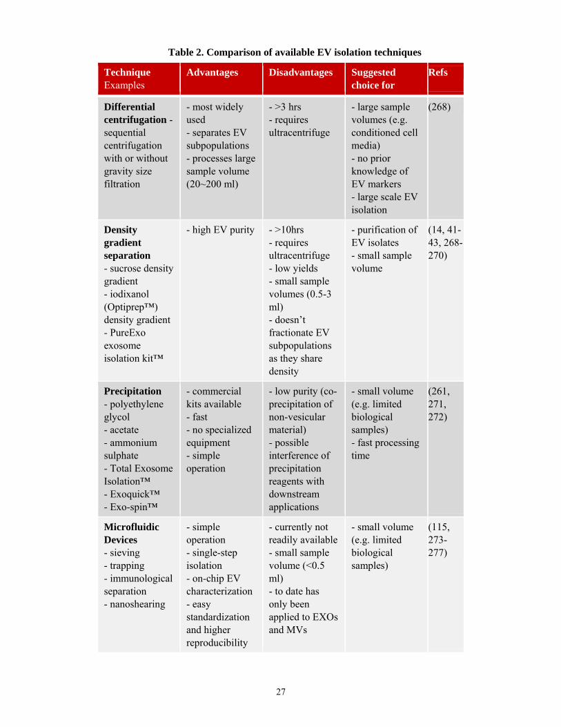
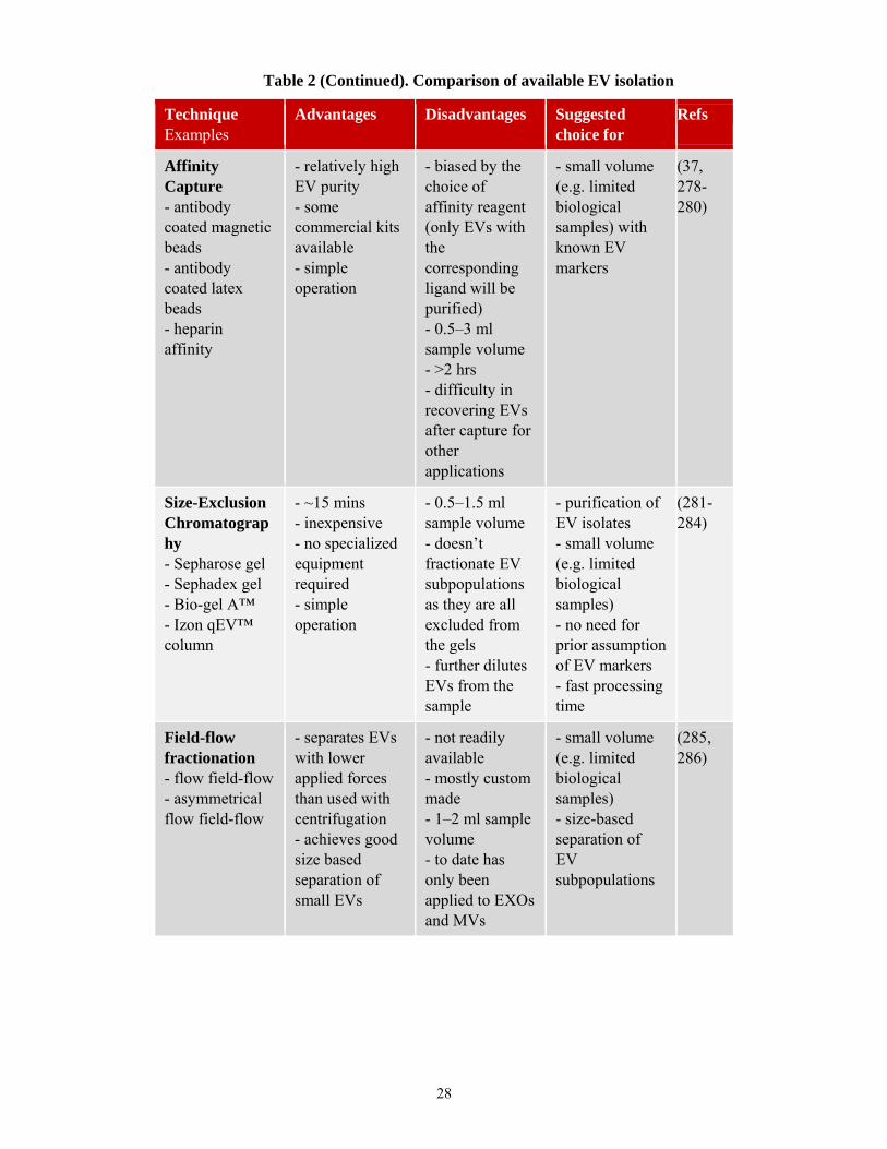
2.1.4.1 Extracellular vesicle isolation techniques
Currently used EV isolation techniques are summarized with their strengths and
best suited applications in Table 2. Traditionally, EVs have been isolated using
differential centrifugation which also includes high speed ultracentrifugation (15, 32,
268). This common technique, encountered in many studies, allows to separate size-based
EV subpopulations with different centrifugation speeds and to process high volumes of
sample. Several other techniques for EV isolation have also emerged to complement or
replace differential centrifugation. Some techniques such as density gradient isolation
(sucrose or iodixanol) are well suited to obtain EV preparations of higher purity than
differential centrifugation alone (14, 41-43, 268-270). Other isolation techniques which
may be either faster or less dependent on instrumentation include precipitation techniques
(261, 271, 272), microfluidic devices (115, 273-277), affinity capture (37, 278-280), size-
exclusion chromatography (281-284), and field-flow fractionation (285, 286). The type
26
of isolation procedure best suited for a given experiment is dependent on the origin of the
sample (e.g. biological fluid or cell conditioned medium), its volume, equipment
availability, and the subsequent type of analysis. These different techniques for isolation
of EVs are currently used due to their relative strengths. However, it is conceivable that
future technical advances in isolation methods may bring about protocols and techniques
that can be applicable to most type of experiments and samples with equal efficiency.
27
Technique Examples
Advantages Disadvantages Suggested choice for
Refs
Differential centrifugation - sequential centrifugation with or without gravity size filtration
- most widely used - separates EV subpopulations - processes large sample volume (20~200 ml)
- >3 hrs - requires ultracentrifuge
- large sample volumes (e.g. conditioned cell media) - no prior knowledge of EV markers - large scale EV isolation
(268)
Density gradient separation - sucrose density gradient - iodixanol (Optiprep™) density gradient - PureExo exosome isolation kit™
- high EV purity - >10hrs - requires ultracentrifuge - low yields - small sample volumes (0.5-3 ml) - doesn’t fractionate EV subpopulations as they share density
- purification of EV isolates - small sample volume
(14, 41-43, 268-270)
Precipitation - polyethylene glycol - acetate - ammonium sulphate - Total Exosome Isolation™ - Exoquick™ - Exo-spin™
- commercial kits available - fast - no specialized equipment - simple operation
- low purity (co-precipitation of non-vesicular material) - possible interference of precipitation reagents with downstream applications
- small volume (e.g. limited biological samples) - fast processing time
(261, 271, 272)
Microfluidic Devices - sieving - trapping - immunological separation - nanoshearing
- simple operation - single-step isolation - on-chip EV characterization - easy standardization and higher reproducibility
- currently not readily available - small sample volume (<0.5 ml) - to date has only been applied to EXOs and MVs
- small volume (e.g. limited biological samples)
(115, 273-277)
Table 2. Comparison of available EV isolation techniques
28
Technique Examples
Advantages Disadvantages Suggested choice for
Refs
Affinity Capture - antibody coated magnetic beads - antibody coated latex beads - heparin affinity
- relatively high EV purity - some commercial kits available - simple operation
- biased by the choice of affinity reagent (only EVs with the corresponding ligand will be purified) - 0.5–3 ml sample volume - >2 hrs - difficulty in recovering EVs after capture for other applications
- small volume (e.g. limited biological samples) with known EV markers
(37, 278-280)
Size-Exclusion Chromatography - Sepharose gel - Sephadex gel - Bio-gel A™ - Izon qEV™ column
- ~15 mins - inexpensive - no specialized equipment required - simple operation
- 0.5–1.5 ml sample volume - doesn’t fractionate EV subpopulations as they are all excluded from the gels - further dilutes EVs from the sample
- purification of EV isolates - small volume (e.g. limited biological samples) - no need for prior assumption of EV markers - fast processing time
(281-284)
Field-flow fractionation - flow field-flow - asymmetrical flow field-flow
- separates EVs with lower applied forces than used with centrifugation - achieves good size based separation of small EVs
- not readily available - mostly custom made - 1–2 ml sample volume - to date has only been applied to EXOs and MVs
- small volume (e.g. limited biological samples) - size-based separation of EV subpopulations
(285, 286)
Table 2 (Continued). Comparison of available EV isolation

29
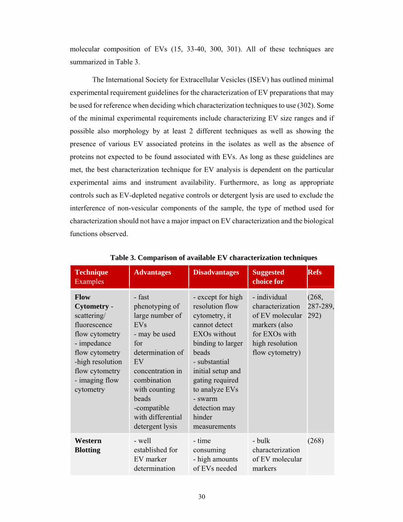
2.1.4.2 Extracellular vesicle characterization techniques
Similarly to EV isolation techniques, there is also a wide diversity of detection
and characterization techniques currently available for EV studies. Two common
techniques used for molecular characterization of EVs are Western blotting (14, 268) and
flow cytometry (268, 287-289). Western blotting is routinely used for bulk molecular
characterization of all EVs present in an isolate. After initial setup and bead or liposome
based gating for EV detection, fluorescent flow cytometry allows for the molecular
characterization of larger sized EVs such as MVs (100–1000 nm) and APOs (≥1 µm).
Furthermore, flow cytometry enables bulk molecular characterization of bead-bound
exosomes (EXOs, 50–100 nm). The coupling of EXOs to beads for flow cytometry is
necessitated since the sizes of EXOs fall below the limit of detection of most instruments.
EVs may be bound to beads with antibodies against specific markers or by unspecific
adsorption of vesicular molecules to chemically modified surfaces such as sulfate
aldehyde. Using antibodies for capture of EVs unto beads can result in highly specific
binding and limited adsorption of other non-vesicular proteins, but this approach may also
bias the measurement since EVs lacking a specific marker will be excluded. On the other
hand, unspecific adsorption to chemically modified surfaces does not bias the
measurements towards only those vesicles bearing a specific marker but other
contaminating non-vesicular proteins may also bind. High resolution flow cytometry
proves capable of circumventing the need for bead coupling allowing for direct molecular
characterization of individual EXOs by employing optimized setups with improved
fluidics, lasers, and detectors.
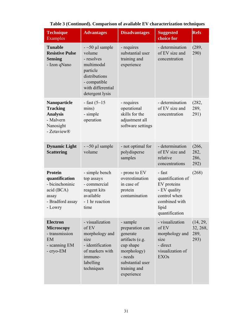
To assess EV particle size and concentration, tunable resistive pulse sensing (289,
290), nanoparticle tracking analysis (282, 289, 291), and dynamic light scattering (266,
282, 286, 292) may be used. For fast quantification of EVs, protein (268) content can be
measured. Additionally, several microscopy methods are routinely used for EV size and
morphology determination including transmission electron microscopy, scanning
electron microscopy, and cryo-electron microscopy (29, 32, 268, 293) as well as atomic
force microscopy (292, 294). Many new studies are also showing the applicability of
label-free techniques such as grating coupled interferometry, surface plasmon resonance,
as well as Raman, infrared, and electrochemical impedance spectroscopy for
characterization EVs (295-299). Lastly, different “omics” techniques such as proteomics,
lipidomics, and genomics among others have been applied to determine the precise
30
molecular composition of EVs (15, 33-40, 300, 301). All of these techniques are
summarized in Table 3.
The International Society for Extracellular Vesicles (ISEV) has outlined minimal
experimental requirement guidelines for the characterization of EV preparations that may
be used for reference when deciding which characterization techniques to use (302). Some
of the minimal experimental requirements include characterizing EV size ranges and if
possible also morphology by at least 2 different techniques as well as showing the
presence of various EV associated proteins in the isolates as well as the absence of
proteins not expected to be found associated with EVs. As long as these guidelines are
met, the best characterization technique for EV analysis is dependent on the particular
experimental aims and instrument availability. Furthermore, as long as appropriate
controls such as EV-depleted negative controls or detergent lysis are used to exclude the
interference of non-vesicular components of the sample, the type of method used for
characterization should not have a major impact on EV characterization and the biological
functions observed.
Technique Examples
Advantages Disadvantages Suggested choice for
Refs
Flow Cytometry - scattering/ fluorescence flow cytometry - impedance flow cytometry -high resolution flow cytometry - imaging flow cytometry
- fast phenotyping of large number of EVs - may be used for determination of EV concentration in combination with counting beads -compatible with differential detergent lysis
- except for high resolution flow cytometry, it cannot detect EXOs without binding to larger beads - substantial initial setup and gating required to analyze EVs - swarm detection may hinder measurements
- individual characterization of EV molecular markers (also for EXOs with high resolution flow cytometry)
(268, 287-289, 292)
Western Blotting
- well established for EV marker determination
- time consuming - high amounts of EVs needed
- bulk characterization of EV molecular markers
(268)
Table 3. Comparison of available EV characterization techniques
31
Technique Examples
Advantages Disadvantages Suggested choice for
Refs
Tunable Resistive Pulse Sensing - Izon qNano
- ~50 µl sample volume - resolves multimodal particle distributions - compatible with differential detergent lysis
- requires substantial user training and experience
- determination of EV size and concentration
(289, 290)
Nanoparticle Tracking Analysis - Malvern Nanosight - Zetaview®
- fast (5~15 mins) - simple operation
- requires operational skills for the adjustment all software settings
- determination of EV size and concentration
(282, 289, 291)
Dynamic Light Scattering
- ~50 µl sample volume
- not optimal for polydisperse samples
- determination of EV size and relative concentrations
(266, 282, 286, 292)
Protein quantification - bicinchoninic acid (BCA) assay - Bradford assay - Lowry
- simple bench top assays - commercial reagent kits available - 1 hr reaction time
- prone to EV overestimation in case of protein contamination
- fast quantification of EV proteins - EV quality control when combined with lipid quantification
(268)
Electron Microscopy - transmission EM - scanning EM - cryo-EM
- visualization of EV morphology and size - identification of markers with immune-labelling techniques
- sample preparation can generate artifacts (e.g. cup shape morphology) - needs substantial user training and experience
- visualization of EV morphology and size - direct visualization of EXOs
(14, 29, 32, 268, 289, 293)
Table 3 (Continued). Comparison of available EV characterization techniques

32
Technique Examples
Advantages Disadvantages Suggested choice for
Refs
Atomic Force Microscopy
- visualization of EV morphology and size
- needs substantial user training and experience
- visualization of EV morphology and size including EXOs
(292, 294)
Label free techniques - grating coupled interferometry - surface plasmon resonance - Raman, infrared, or electrochemical impedance spectroscopy
- determination of EV markers - determination of EV concentration - detection of EV binding - label free study of EV surface interactions
- methodology still in development for EV applications - needs substantial user training and experience
- dynamic study of molecular interaction with EVs
(295-299)
“OMICS” techniques - proteomics - lipidomics - genomics - transcriptomics - glycomics
- complete molecular characterization of EVs
- relatively high amounts of EV sample needed
- determination of EV molecular composition
(15, 33-40, 300, 301)
Table 3 (Continued). Comparison of available EV characterization techniques
33
3. OBJECTIVES
In the previous sections have been described many of the novel biological
functions that have been discovered so far for EVs as well as their potential applications
in the clinic. The field of EV research has truly emerged as one of the fastest growing
areas due to all these recent findings and more scientists from other areas continue to get
interested and start exploring the functions of EVs in their respective fields. However,
despite this rapid growth one concern is that in regards to some biological processes it
seems that different reports have found discordant roles for EVs. Therefore, it might seem
puzzling whether EVs are in fact beneficial or detrimental in various biological processes
in health and disease.
Some of the observed discordance in biological roles reported for EVs can be
explained by the inherent differences among various subtypes of EVs simultaneously
found in the body which can also be released by different cells possibly in different
activation states. Furthermore, the EVs and their cargo can also undergo various post-
synthetic modifications or combine with other EVs or soluble molecules which can alter
their biological effects. Thus, when comparing different studies showing effects of EVs
in any biological process all these inherent biological differences should be accounted for
as they may explain discordant results if each report is analyzing completely different
EVs.
Even if it may be that many of the discordances can be explained by biological
differences among the studies EVs there are still some examples where various reports
supposedly investigating the same particles do not obtain comparable results. One such
example can be found with reports of EVs released during remote ischemic
preconditioning (RIPC) which were observed to either reduce or to have no effect in
infarct sizes (114, 303, 304). Based on this problem, we believe that in such a rapidly
growing field it is also very important to have sound methodology and controls with
which to characterize the material that is being studied so that researchers can easily
compare their results to each other. As the field currently stands that is not always the
case as many groups utilize different isolation and characterization methods and there are
still no universally accepted protocols and molecular markers for EVs.
34
For these reasons, the work undertaken during this Ph.D. had the following
objectives.
I. Improve existing EV methodology.
II. Introduce novel assays for EV analysis.
III. Develop a workflow for the quantification of EV lipid content.
IV. Investigate the protein to lipid ratio of the different EV subpopulations.
V. Characterize the membrane lipid order of the different EV subpopulations.
VI. Establish the detergent lysis sensitivity of EVs.
By pursuing these objectives we attempt to develop methods which we believe
will expand the existing toolkit for EV research and hopefully will allow researchers to
better characterize the material under study.
35
4. MATERIALS AND METHODS
In the following sections the methods used for EV isolation and characterization
as well as for cell culturing and performing all experiments are described in detail.
4.1 Cell line cultures
Jurkat (TIB-152) human T cell lymphoma, THP-1 (TIB202) human acute
monocytic leukemia, and U937 (CRL-1593.2) human histiocytic lymphoma cell lines
were all obtained from ATCC (Manassas, VA). The BV-2 murine microglia cell line was
a generous gift of Prof. Rosario Donato (Università degli Studi di Perugia, Perugia, Italy)
(305). The MiaPaCa-2 (MiaPaCa) pancreatic cancer cells were kindly provided by Dr.
Klaus Felix, (Universität Heidelberg, Heidelberg, Germany) and the MH-S murine
alveolar macrophage cell line was kindly provided by Dr. Dolores Solis (Instituto de
Química Física Rocasolano, Madrid, Spain). Both MiaPaCa and MH-S cell lines were
originally obtained from ATCC.
All cell lines were cultured as indicated by the suppliers in RPMI-1640 or DMEM
medium containing 10% (v/v) fetal bovine serum (FBS), 2 mM glutamine, and 1% (v/v)
Antibiotic Antimycotic Solution (Ab/AM) (all from Sigma-Aldrich, St Louis, MO), at 37
oC in 5% CO2/air. The cell lines were regularly tested for Mycoplasma contamination by
fluorescence microscopy using DAPI staining (Molecular Probes Life Technologies,
Carlsbad, CA). Cells used for the production of EVs were first washed three times with
phosphate buffered saline (PBS) and placed in FBS-free medium to avoid contamination
of the preparations with EVs present in FBS. Cells were grown in this condition for 24
hrs at concentrations of 0.3–1 x 10⁶/mL depending on the optimal density indicated by
the supplier of each cell line. Cell viability was always monitored by flow cytometry to
be >90–95% as confirmed by staining with Annexin V FITC and propidium iodide (both
from BD Biosciences, Franklin Lakes, NJ). Prior to isolation, EV-containing conditioned
FBS-free media was first submitted to 300 g centrifugation for 10 mins followed by
gravity driven filtration through a 5 μm filter (Millipore, Billerica, MA) to deplete cells.
36
4.2 Human blood, platelet, and red blood cell concentrate collection and processing
The use of human blood samples was approved by the Scientific Ethics
Committee of the Hungarian Health Scientific Council (ETT TUKEB), and human blood
donors provided written informed consent.
For EV isolation from platelets, acid-citrate dextrose (ACD) anti-coagulated
plasma samples were centrifuged at 400 g for 15mins at room temperature (RT) to remove
most of the blood cells, and then the platelet containing supernatant was diluted 4x in
TRIS-citrate buffer. Next, the platelet-rich plasma was again centrifuged at 600 g for 20
mins at RT, and the pellet was resuspended in 3 mL PBS. Platelets were then incubated
at 37 oC for an hour in the presence of 10 μM adenosine diphosphate (ADP) (Sigma-
Aldrich). Next, 4 mL of PBS was added to reduce the viscosity of the samples, and
centrifuged at 800 g for 20 mins at RT. For EV isolation from red blood cell concentrates,
samples were first diluted 2x in PBS to reduce the viscosity.
To obtain EVs from human blood plasma, we collected 30–40 mL of blood from
healthy adult donors in ACD-A tubes (Greiner Bio-One, Kremsmünster, Austria) to
prevent ex vivo vesiculation related to blood sample handling and storage (306). The
samples were processed following the International Society on Thrombosis and
Hemostasis (ISTH) protocol for preparation of platelet-free plasma (PFP) (307, 308).
Briefly, the ACD anti-coagulated blood was centrifuged twice at 2,500 g for 15 mins at
RT to remove platelets using a HermLe Z206A bench top centrifuge (HermLe
Labortechnik GmbH, Wehingen, Germany).
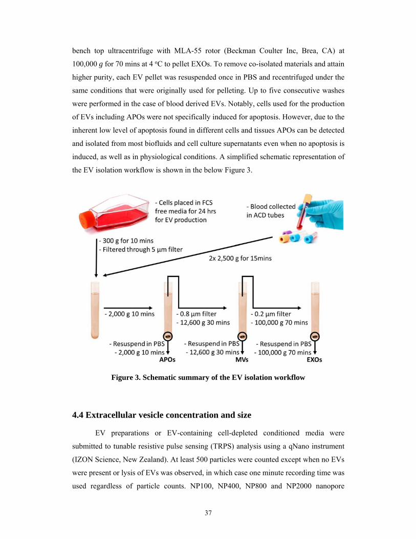
4.3 Extracellular vesicle isolation
Three different EV subpopulations were isolated including APOs, MVs, and
EXOs by the combination of gravity driven filtration and differential centrifugation.
Initially cells were removed by centrifugation at 300 g for 10 mins, and then the
supernatant was filtered by gravity through a 5 µm filter (Millipore) and submitted to a
2,000 g centrifugation for 10 mins at RT to pellet APOs. The supernatant was next filtered
by gravity through a 0.8 µm filter (Millipore) and centrifuged at 12,600 g for 30 mins at
RT to pellet MVs. Finally, the supernatant was ultracentrifuged in an Optima MAX-XP
37
bench top ultracentrifuge with MLA-55 rotor (Beckman Coulter Inc, Brea, CA) at
100,000 g for 70 mins at 4 oC to pellet EXOs. To remove co-isolated materials and attain
higher purity, each EV pellet was resuspended once in PBS and recentrifuged under the
same conditions that were originally used for pelleting. Up to five consecutive washes
were performed in the case of blood derived EVs. Notably, cells used for the production
of EVs including APOs were not specifically induced for apoptosis. However, due to the
inherent low level of apoptosis found in different cells and tissues APOs can be detected
and isolated from most biofluids and cell culture supernatants even when no apoptosis is
induced, as well as in physiological conditions. A simplified schematic representation of
the EV isolation workflow is shown in the below Figure 3.
Figure 3. Schematic summary of the EV isolation workflow
4.4 Extracellular vesicle concentration and size
EV preparations or EV-containing cell-depleted conditioned media were
submitted to tunable resistive pulse sensing (TRPS) analysis using a qNano instrument
(IZON Science, New Zealand). At least 500 particles were counted except when no EVs
were present or lysis of EVs was observed, in which case one minute recording time was
used regardless of particle counts. NP100, NP400, NP800 and NP2000 nanopore
38
membranes stretched between 43–47 mm were used. Voltage was set in between 0.04–
0.7 V to achieve a stable current between 115–145 nA. Particle-size histograms were
recorded when root mean square noise was below 12 pA, and particle rate in time was
linear using calibration beads CPC100B, CPC400E, CPC800D and CPC2000D (mode
diameters 110 nm, 340 nm, 740 nm and 1900 nm, respectively) (all from IZON). For
detergent lysis experiments shown in Section 5.2, the calibration beads were also
measured at every detergent concentration step at least twice in order to detect any
possible differences in their modal size and particle rate possibly due to the presence of
detergent micelles. For none of the detergents used in Section 5.2 this was found to be the
case.
4.5 Transmission electron microscopy of extracellular vesicle preparations
In order to characterize the morphology and size of the different EV preparations,
pellets were fixed with 4% paraformaldehyde in 0.01M PBS for 60mins at RT. Following
washing with PBS, the preparations were postfixed in 1% OsO4 (Taab, Aldermaston,
Berks, UK) for 30 mins. After rinsing the intact fixed pellets within the centrifugation
tubes with distilled water, the pellets were dehydrated in graded ethanol, including block
staining with 1% uranyl-acetate in 50% ethanol for 30 mins, and were subsequently
embedded in Taab 812 (Taab). Overnight polymerization of samples at 60 °C was
followed by sectioning, and the ultrathin sections were analyzed using a Hitachi 7100
electron microscope (Hitachi Ltd., Japan) equipped with a Megaview II (lower resolution,
Soft Imaging System, Germany) digital camera.
4.6 Flow cytometry of cells and extracellular vesicles
For flow cytometric measurements, EV preparations were incubated for 30mins
at RT in the dark with different fluorochrome conjugated antibodies and affinity reagents.
These included cholera toxin (CTX) subunit B-Alexa Fluor647 and di-4-ANEPPDHQ
which were purchased from Life Technologies, an anti-cholesterol antibody (AC8) which
was produced at Eötvös Loránd University (Budapest, Hungary) (309) and conjugated to
CF488A fluorophore according to the Mix-n-Stain protocol (Biotium, Sigma-Aldrich) as

39
well as annexin V FITC or APC conjugated (in annexin binding buffer from BD
Biosciences), anti-CD9 FITC and anti-CD63 PE-conjugated antibodies (all from BD
Biosciences). Isotype controls (all from BD Biosciences) were used for samples stained
with fluorochrome-conjugated antibodies, whereas autofluorescence was detected in the
absence of either annexin V or CTX. To verify the vesicular nature of MVs and APOs,
and to exclude the presence of antibody aggregates, we added Triton X-100 to 0.05%
final concentration to the samples. This step resulted in prompt disappearance of
fluorescent event counts from the MV and APO gates suggesting the presence of
membranous structures within these gates. EXOs were resuspended in a total volume 50
µL of PBS, and aliquots of 5µL were incubated with 5 µL of 4 µm aldehyde/sulfate latex
beads (Life Technologies), followed by an incubation of 5 mins at RT. Then 20 µL of
PBS was added, and incubation was continued for another 15 mins at RT. Thirty µL of
2% bovine serum albumin (BSA) in PBS was added and samples were blocked for 2 hrs
at RT. Then the Eppendorf tubes were filled with PBS, and were centrifuged at 2,700 g
for 3 mins at RT. The supernatant was discarded, and the pellet was resuspended in 200
µL of 100 mM glycine in PBS, and was incubated for 30 mins at RT. The samples were
washed with PBS, and centrifuged at 2,700 g for 3 mins at RT. Then the supernatant was
discarded, and the pellet was resuspended in 100 µL of PBS for staining as described
above. EV preparations were analyzed by using FACSAria III and FACSCalibur flow
cytometers (both from BD Biosciences). Events were counted for 1min from equal sample
volumes at medium flow rate and data were analyzed by FlowJo software (Treestar,
Ashland, USA).
In case of APOs and MVs, instrument settings and gates were set as recommended
by the International Society on Thrombosis and Haemostasis (ISTH) (307, 308) using
Megamix-Plus SSC beads (BioCytex, Marseille, France) and further optimized with 1 µm
Silica Beads Fluo-Green (Kisker, Steinfurt, Germany). Megamix-Plus SSC beads are a
mix of fluorescent polystyrene beads of different diameters covering the MV size range
(0.16 µm, 0.20 µm, 0.24 µm, and 0.5 µm). Since the forward scatter (FSC) of most flow
cytometers is not sensitive enough to differentiate within the size range of EVs we used
these beads to calibrate the settings of the instrument to be able to resolve each of the
distinct sized beads by their side scatter (SSC) (Figure 4). However, one important
consideration is that side scatter is not only due to the diameter of the particles but also
their refractive index and thus EV size estimation based on SSC after calibration with
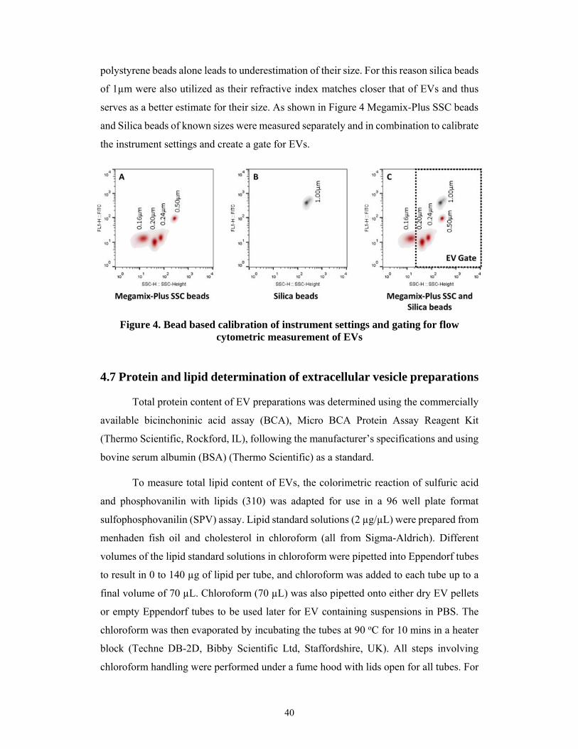
40
polystyrene beads alone leads to underestimation of their size. For this reason silica beads
of 1µm were also utilized as their refractive index matches closer that of EVs and thus
serves as a better estimate for their size. As shown in Figure 4 Megamix-Plus SSC beads
and Silica beads of known sizes were measured separately and in combination to calibrate
the instrument settings and create a gate for EVs.
Figure 4. Bead based calibration of instrument settings and gating for flow cytometric measurement of EVs
4.7 Protein and lipid determination of extracellular vesicle preparations
Total protein content of EV preparations was determined using the commercially
available bicinchoninic acid assay (BCA), Micro BCA Protein Assay Reagent Kit
(Thermo Scientific, Rockford, IL), following the manufacturer’s specifications and using
bovine serum albumin (BSA) (Thermo Scientific) as a standard.
To measure total lipid content of EVs, the colorimetric reaction of sulfuric acid
and phosphovanilin with lipids (310) was adapted for use in a 96 well plate format
sulfophosphovanilin (SPV) assay. Lipid standard solutions (2 µg/µL) were prepared from
menhaden fish oil and cholesterol in chloroform (all from Sigma-Aldrich). Different
volumes of the lipid standard solutions in chloroform were pipetted into Eppendorf tubes
to result in 0 to 140 µg of lipid per tube, and chloroform was added to each tube up to a
final volume of 70 µL. Chloroform (70 µL) was also pipetted onto either dry EV pellets
or empty Eppendorf tubes to be used later for EV containing suspensions in PBS. The
chloroform was then evaporated by incubating the tubes at 90 oC for 10 mins in a heater
block (Techne DB-2D, Bibby Scientific Ltd, Staffordshire, UK). All steps involving
chloroform handling were performed under a fume hood with lids open for all tubes. For
41
EV suspensions, instead of pellets, 50 µL (with at least 10–30 µg/mL of protein
depending on the EV type) was added to the empty chloroform-pretreated Eppendorf
tubes and 50µL of PBS was also added to each tube containing the lipid standards. The
pre-treatment with equal volumes of chloroform, and PBS in the case of EV suspensions,
ensured the same background color development in all Eppendorf tubes. As a following
step, 250 µL of 96% sulfuric acid was added to the tubes followed by incubation with
open lids at 90 oC for 20 min in a heater block. Next, 220 µL of samples and standards in
sulfuric acid were transferred into a 96-well polystyrene plate (Nunc, Thermo Scientific),
and allowed to cool down to RT. Finally, 110 µL of 0.2 mg/mL vanillin in 17%
phosphoric acid (both from Sigma-Aldrich) was added to each well, and the plate was
incubated for 10 mins at RT. Absorbance was measured at 540 nm by an MS Reader
(Multiskan MS; Labsystems, Helsinki, Finland). Figure 5 shows a simplified schematic
summary of the total lipid determination protocol.

42
Figure 5. Schematic summary of the total lipid determination protocol for EV preparations
43
4.8 Membrane lipid bilayer order of extracellular vesicle preparations
In order to investigate the membrane lipid bilayer order of EV preparations, EVs
were stained with the membrane probe Di-4-ANEPPDHQ (Life Technologies) at 5 µM
final concentration for 30 mins at 37 oC as described previously (311). EV
subpopulations, immobilized on poly-L-lysine (Sigma-Aldrich) coated cover slips, were
analyzed by confocal microscopy (IX81 inverted microscope based Fluoview500 laser
scanning confocal system and software; Olympus Europe, Hamburg, Germany) at 60x
magnification. Suspensions of APOs, MVs as well as 4-µm latex bead-bound EXOs were
also assessed by flow cytometry. The Di-4-ANEPPDHQ probe exhibits a 60 nm spectral
shift between the liquid-ordered and liquid-disordered phases. Ratiometric measurement
of the fluorescence intensity by confocal microscopy was performed in the 560–600 nm
and the >660 nm emission channels, defined by appropriate band pass and cut off filters,
respectively. Background-corrected fluorescence intensities of EV samples in each
channel were calculated by ImageJ software (http://imagej.nih.gov/ij/). General
polarization (GP) values were determined as follows: GP = (I560–600– I>660)/(I560–
600+I>660). In flow cytometry, PE (585±21 nm) and PE-Texas Red (616±12 nm) filters
were used to define the corresponding two emission channels and GP values were
calculated in similar way.
4.9 Detergent lysis of extracellular vesicles
Sodium dodecyl sulphate (SDS), Triton X-100, Tween 20 and sodium
deoxycholate were all purchased from Sigma-Aldrich. Detergents were added to EV-
containing samples to different final concentrations in the range of 0.005–15% (w/v for
SDS and deoxycholate and v/v for Triton X-100 and Tween 20), vortexed for 30 seconds,
and analyzed by TRPS or flow cytometry. All steps of detergent lysis were carried out at
RT. These conditions were established based on preliminary experiments using different
incubation times of EVs with detergents and showing essentially no difference in lysis
efficiency with increasing time.
44
4.10 Statistical analyses
For data analysis GraphPad Prism (GraphPad Software, La Jolla, USA) was used.
For comparison of different parameters of EV subpopulations, Wilcoxon matched-pairs
signed rank test was used. For membrane lipid order data obtained by confocal
microscopy, 1-way ANOVA was used followed by Dunnetts post-hoc test. For analysis
of separate cell line-derived EV counts two-way ANOVA was used. For the analysis of
combined EVs counts for all cells and all detergents, one-way ANOVA was used, and for
parameters not showing normal distribution, Kruskal-Wallis non-parametric test was
applied. P values <0.05 were considered statistically significant (* p<0.05, ** p<0.01 and
*** p<0.001).
45
5. RESULTS
5.1 Improved characterization of extracellular vesicle preparations based on protein to lipid ratios and lipid properties
Given the important need for better tools for EV research, the goal of this first
study was to find improved parameters to characterize EV preparations. Here a simple 96
well plate-based total lipid assay was introduced for the determination of lipid content as
well as protein to lipid ratios of EV preparations from various myeloid and lymphoid cell
lines as well as blood plasma. These preparations included APOs, MVs, and EXOs
isolated by differential centrifugation and gravity filtration. Furthermore, membrane lipid
bilayer order of EV subpopulations was measured with the Di-4-ANEPPDHQ lipid probe
and their lipid composition was assessed with affinity reagents to clustered cholesterol
and ganglioside GM1. Different protein to lipid ratios were found to be characteristic for
the investigated EV subpopulations and these were substantially altered in the case of
vesicular damage or protein contamination. Spectral ratiometric imaging and flow
cytometric analysis also revealed marked differences between the various vesicle
populations in regards to their lipid order and their clustered membrane cholesterol and
GM1 content. Thus, this study introduced for the first time a simple and readily available
lipid assay to complement the widely used protein assays in order to better characterize
EV preparations. Besides differentiating extracellular vesicle subpopulations, the novel
parameters introduced in this work (protein to lipid ratio, lipid bilayer order, and lipid
composition), may prove useful for quality control of EV related basic and clinical
studies.
5.1.1 Optimization of the sulfophosphovanilin total lipid assay for EV studies
Given the unmet need in the EV research field for a simple and inexpensive lipid
assay with the sensitivity to detect EVs, we first attempted to adapt the colorimetric SPV
assay for total lipid determination of EV subpopulations (310, 312). One very important
limitation when working with EVs is that typically the yields are much smaller than most
other biological samples. Therefore, prior to any further experimentation it was crucial to
first assess whether the SPV reaction had the necessary sensitivity to detect the few
micrograms of lipids commonly found in most EV preparations. Thus we evaluated
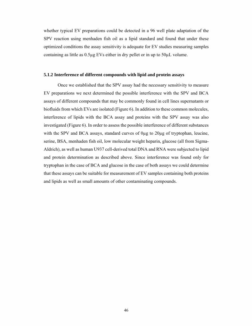
46
whether typical EV preparations could be detected in a 96 well plate adaptation of the
SPV reaction using menhaden fish oil as a lipid standard and found that under these
optimized conditions the assay sensitivity is adequate for EV studies measuring samples
containing as little as 0.5µg EVs either in dry pellet or in up to 50µL volume.
5.1.2 Interference of different compounds with lipid and protein assays
Once we established that the SPV assay had the necessary sensitivity to measure
EV preparations we next determined the possible interference with the SPV and BCA
assays of different compounds that may be commonly found in cell lines supernatants or
biofluids from which EVs are isolated (Figure 6). In addition to these common molecules,
interference of lipids with the BCA assay and proteins with the SPV assay was also
investigated (Figure 6). In order to assess the possible interference of different substances
with the SPV and BCA assays, standard curves of 0µg to 20µg of tryptophan, leucine,
serine, BSA, menhaden fish oil, low molecular weight heparin, glucose (all from Sigma-
Aldrich), as well as human U937 cell-derived total DNA and RNA were subjected to lipid
and protein determination as described above. Since interference was found only for
tryptophan in the case of BCA and glucose in the case of both assays we could determine
that these assays can be suitable for measurement of EV samples containing both proteins
and lipids as well as small amounts of other contaminating compounds.
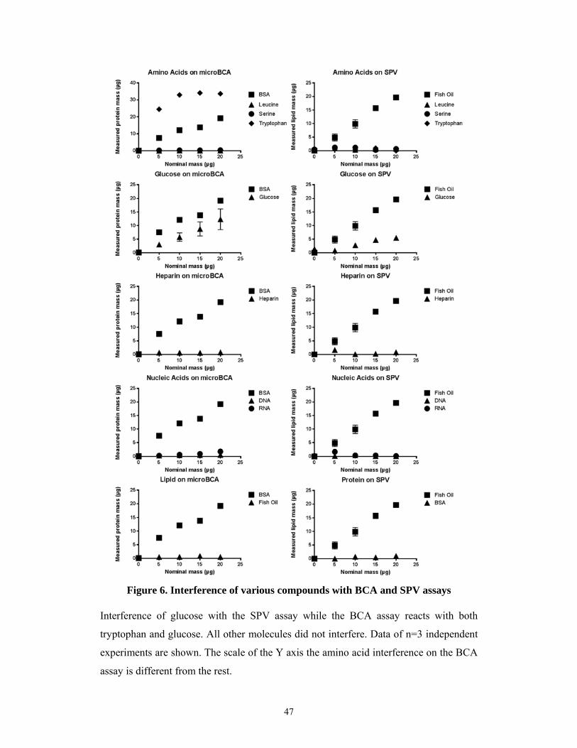
47
Figure 6. Interference of various compounds with BCA and SPV assays
Interference of glucose with the SPV assay while the BCA assay reacts with both
tryptophan and glucose. All other molecules did not interfere. Data of n=3 independent
experiments are shown. The scale of the Y axis the amino acid interference on the BCA
assay is different from the rest.

48
5.1.3 Accuracy and variability of lipid and protein assays
After observing that the SPV assay had adequate sensitivity for EV research and
lesser interference by other compounds than the BCA assay we next assessed how these
two assays compared in regards to accuracy and variability. To this end, equal
concentrations of menhaden fish oil and BSA (both from Sigma-Aldrich) were subjected
to SPV and BCA assays respectively in 6 parallel technical repeats of 0, 10, 20, 30, 40,
50, 100, and 200 µg/mL concentrations in three independent experiments. Accuracy was
assessed by calculating the percentage error (% Error) as follows: (|measured value -
nominal value|/nominal value) x 100. Variability was assessed by determining the intra-
assay variation calculated as percentage coefficient variation (% CV) using the following
formula: standard deviation (SD) of measured values/measured values x 100. The results
are shown in Figure 7.

49
Figure 7. Working concentration ranges and coefficients of variations of the BCA and SPV assays
Intra-assay % Error is shown for the two assays reflecting higher accuracy of SPV. 7B
shows a precision profile as defined earlier (313) indicating the working concentration
range by % CV plotted against concentration for both SPV and BCA assays. The
suggested working range (gray box) is defined as the range of concentrations for which
the % CV < 20%. The scales of the Y axis are not the same. Inter-assay variation was also
determined as follows: average of 10 µg/mL (lowest) and 200 µg/mL (highest) % CVs,
and were found to be 11.4 and 3.1 for the SPV and BCA assays, respectively. n = 3.
50
5.1.4 Characterization of different EV subpopulations
Separate EV subpopulations (APOs, MVs, and EXOs) were isolated from the
supernatants of the cell lines THP-1, BV-2, MH-S, and Jurkat as described in earlier work
form our research group and others (32, 268, 293). The vesicles from the BV-2 cell line
were characterized by tunable resistive pulse sensing (TRPS) and transmission electron
microscopy. EVs from the three subpopulations were found to have different size and
morphology (Figure 8). The APO preparation predominantly contained vesicles between
1250–2500 nm in diameter (1627.05 nm mode diameter). In contrast, most vesicles in the
MV preparation had approximately 200 nm diameter (208.32 nm mode diameter). The
most uniform population was the one of EXOs with a peak size around 100 nm (98.76
nm mode diameter). In electron microphotographs some of the APOs contained highly
electron dense intravesicular content presumably corresponding to fragmented DNA.
MVs were somewhat variable in shape, size and electron density, whereas EXOs had
uniform size and cup shaped morphology (Figure 8).
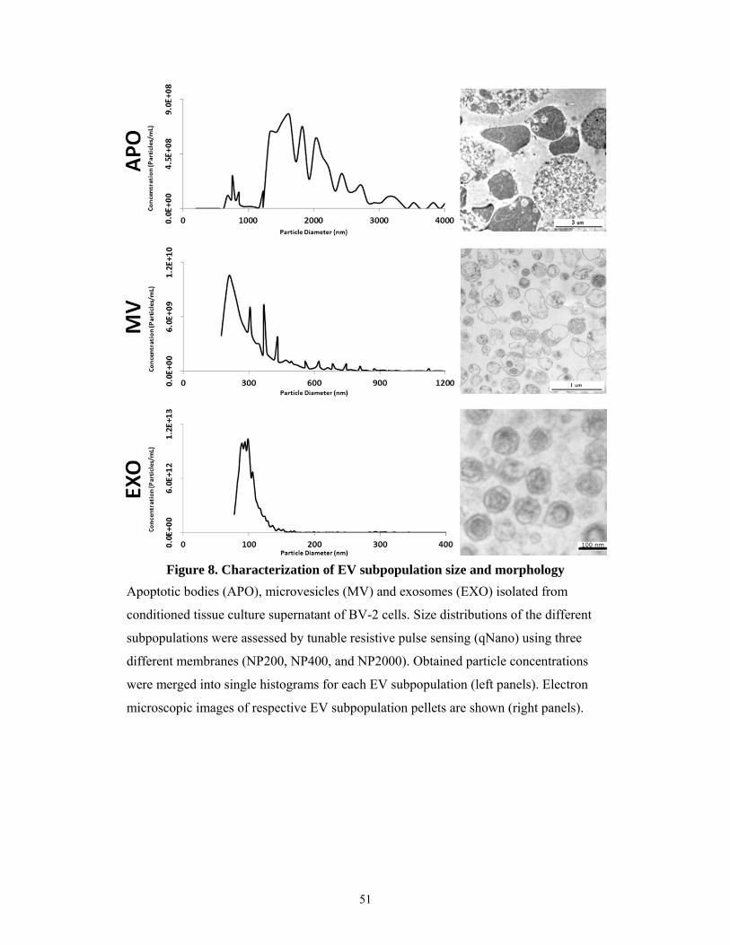
51
Apoptotic bodies (APO), microvesicles (MV) and exosomes (EXO) isolated from
conditioned tissue culture supernatant of BV-2 cells. Size distributions of the different
subpopulations were assessed by tunable resistive pulse sensing (qNano) using three
different membranes (NP200, NP400, and NP2000). Obtained particle concentrations
were merged into single histograms for each EV subpopulation (left panels). Electron
microscopic images of respective EV subpopulation pellets are shown (right panels).
Figure 8. Characterization of EV subpopulation size and morphology
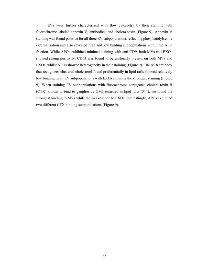
52
EVs were further characterized with flow cytometry by their staining with
fluorochrome labeled annexin V, antibodies, and cholera toxin (Figure 9). Annexin V
staining was found positive for all three EV subpopulations reflecting phosphatidylserine
externalization and also revealed high and low binding subpopulations within the APO
fraction. While APOs exhibited minimal staining with anti-CD9, both MVs and EXOs
showed strong positivity. CD63 was found to be uniformly present on both MVs and
EXOs, whilst APOs showed heterogeneity in their staining (Figure 9). The AC8 antibody
that recognizes clustered cholesterol found preferentially in lipid rafts showed relatively
low binding to all EV subpopulations with EXOs showing the strongest staining (Figure
9). When staining EV subpopulations with fluorochrome-conjugated cholera toxin B
(CTX) known to bind to ganglioside GM1 enriched in lipid rafts (314), we found the
strongest binding to MVs while the weakest one to EXOs. Interestingly, APOs exhibited
two different CTX binding subpopulations (Figure 9).
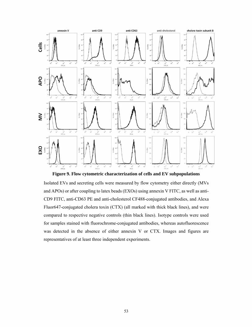
53
Isolated EVs and secreting cells were measured by flow cytometry either directly (MVs
and APOs) or after coupling to latex beads (EXOs) using annexin V FITC, as well as anti-
CD9 FITC, anti-CD63 PE and anti-cholesterol CF488-conjugated antibodies, and Alexa
Fluor647-conjugated cholera toxin (CTX) (all marked with thick black lines), and were
compared to respective negative controls (thin black lines). Isotype controls were used
for samples stained with fluorochrome-conjugated antibodies, whereas autofluorescence
was detected in the absence of either annexin V or CTX. Images and figures are
representatives of at least three independent experiments.
Figure 9. Flow cytometric characterization of cells and EV subpopulations
54
5.1.5 Characterization of protein to lipid ratios of the EV subtypes
Following the characterization of distinct EV subpopulations obtained by our
isolation methods we next determined the lipid content of the EV preparations using
menhaden fish oil as a lipid standard. In parallel with the total lipid determination,
aliquots of the same EV preparations were also submitted to protein determination by
BCA assay. Total protein to total lipid ratios of EV preparations from three different cell
lines (Figure 10A-C). Protein to lipid ratios of EV subpopulations measured
independently for all tested cell lines and human blood plasma were combined into Figure
10D to show that they are characteristic of the respective EV subpopulations regardless
of their cell origin. APOs were consistently characterized by the highest protein to lipid
ratio followed by an intermediate ratio for MVs and the lowest ratio was in all cases found
for EXOs (Figure 10). As shown in Figure 10E, we found differences in the combined
protein concentrations of EV subpopulations of different cellular origin with MVs
showing the lowest protein concentration. Interestingly a striking elevation in lipid
concentrations of the combined EXOs, was observed as compared to the combined MVs
or APOs (Figure 10F).

55
Protein to lipid ratios are shown for APOs, MVs, and EXOs isolated from MH-S (A),
THP-1 (B), and BV-2 (C) cell lines (n ≥ 3 independent experiments for each EV type
from all cell lines). Combined protein to lipid ratios obtained using EVs derived from
MH-S, THP-1, BV-2, Jurkat, U937 as well as from human blood plasma are shown (D)
(results of ≥ 12 independent experiments each EV type). Combined protein (E) and lipid
concentrations (F) for the above cell line derived and blood plasma derived EVs are also
shown. Mean values are represented by horizontal lines, and standard error means
(SEMs) are indicated by error bars. The mean values ± SEM of lipid and protein
concentrations (µg/mL) of conditioned media are reported below each respective EV
subpopulation.
Figure 10. Protein to lipid ratios of EV subpopulations
56
5.1.6 Protein to lipid ratios of damaged or contaminated EV samples
To show the applicability of protein to lipid ratio as a quality control parameter of
EV preparations, we tested EVs from different conditions. Firstly, platelets were
incubated with ADP for one hour in PBS, and the supernatant was subsequently submitted
for EV isolation. The low speed (20,000 g) MV pellet was characterized by the expected
protein to lipid ratio (Figure 11A) and the presence of MVs was confirmed by using
transmission electron microscopy (Figure 11B). However, the protein to lipid ratio of the
high speed “EXO” (100,000 g) pellet was found to be abnormally elevated (Figure 11A).
In accordance with this value, electron microscopy showed the lack of any vesicular
structures in the 100,000 g pellet (Figure 11C), suggesting that under these conditions the
pellet was mostly composed of proteins. This is in line with the previous finding that ADP
is a poor inducer of EXO production (315).
Secondly, we exposed freshly isolated MVs to overnight incubation at 37 oC, a
condition that we have found earlier to damage MV preparations. Protein to lipid ratio
was determined in the low speed MV pellet before and after overnight incubation. The
overnight incubated and re-pelleted MV preparation was characterized by an abnormal
protein to lipid ratio compared to that of freshly isolated MVs (Figure 11D). This was in
agreement with the electron microscopy showing vesicular damage following the
overnight incubation (Figure 11E and F). Finally MV preparations derived from red blood
cell concentrates were compared after 2, 3, 4 or 5 consecutive washes. As shown in Figure
11G, the 3rd washing step resulted in a shift from elevated protein to lipid ratio to the
ratio expected for MVs suggesting the removal of contaminating proteins, and this ratio
remained stable upon further washes.
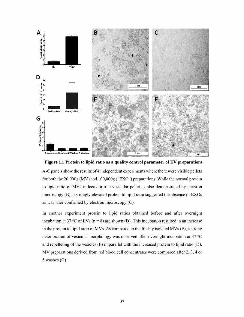
57
A-C panels show the results of 4 independent experiments where there were visible pellets
for both the 20,000g (MV) and 100,000g (“EXO”) preparations. While the normal protein
to lipid ratio of MVs reflected a true vesicular pellet as also demonstrated by electron
microscopy (B), a strongly elevated protein to lipid ratio suggested the absence of EXOs
as was later confirmed by electron microscopy (C).
In another experiment protein to lipid ratios obtained before and after overnight
incubation at 37 oC of EVs (n = 8) are shown (D). This incubation resulted in an increase
in the protein to lipid ratio of MVs. As compared to the freshly isolated MVs (E), a strong
deterioration of vesicular morphology was observed after overnight incubation at 37 oC
and repelleting of the vesicles (F) in parallel with the increased protein to lipid ratio (D).
MV preparations derived from red blood cell concentrates were compared after 2, 3, 4 or
5 washes (G).
Figure 11. Protein to lipid ratio as a quality control parameter of EV preparations
58
5.1.7 Lipid membrane order of the EV subtypes
Finally, in order to determine if membrane lipid order could be a distinguishing
feature of different EV subpopulations, we stained EVs with the fluorescent polarity-
sensitive lipid probe di-4-ANEPPDHQ. Using confocal microscopy and ImageJ software,
we determined the general polarization (GP) values for APO, MV and EXO preparations,
respectively, and found significant differences between subpopulations of EVs (one-way
ANOVA, p<0.001). The GP value was calculated from the fluorescence intensities at
560–600 nm and 660 nm resulting in values between -1 and +1 with higher GP values
reflecting higher membrane lipid order (low liquid disordered, high liquid ordered
structure). APOs and MVs were characterized by intermediate and partially overlapping
lipid orders with GP values of -0.15 ± 0.15 and 0.00 ± 0.21 (median ± SEM), respectively.
In contrast, EXOs displayed a GP value of 0.36 ± 0.29 (median ± SEM), reflecting a
remarkably higher degree of lipid order than either MVs or APOs (Figure 12). The
observed GP values were also assessed by flow cytometry and the results were close to
those of the confocal microscope measurement. The GP values measured by flow
cytometry were -0.14, -0.14, and 0.25 for APOs, MVs, and EXOs, respectively (Figure
13). The differences between the GP values measured by confocal microscopy and flow
cytometry could be ascribed to the small differences in the available filters in each
instrument (560–600 nm and >660 nm in the confocal microscope as opposed to 585 ±
21 nm and 616 ± 12 in the flow cytometer). However, the pattern of higher order in EXOs
was observed by both instruments.

59
Quantitative assessment of membrane lipid order in subpopulations of EVs secreted by
BV-2 cells is shown (≥3 independent experiments each EV type). Ratiometric
measurements of the fluorescent intensities at 560–600 nm and at >660 nm were carried
out by confocal microscopy, and results are expressed as general polarization (GP) values.
The higher GP value reflects a higher membrane lipid order. EXOs showed the highest
order, while APOs and MVs were characterized by partially overlapping, intermediate
order.
Figure 12. Confocal microscopy determination of EV membrane lipid order
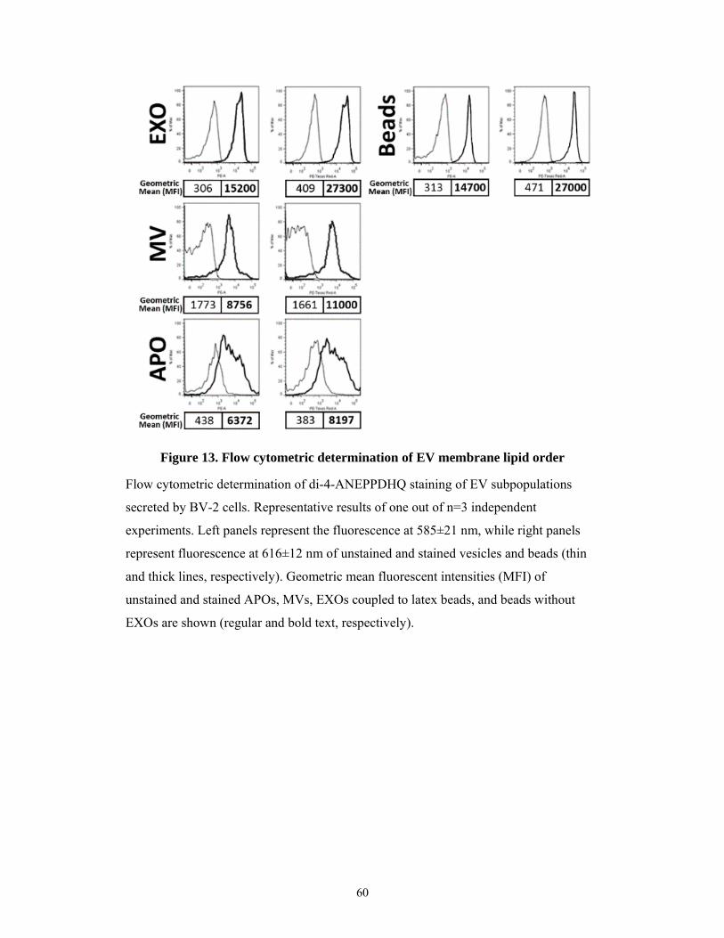
60
Flow cytometric determination of di-4-ANEPPDHQ staining of EV subpopulations
secreted by BV-2 cells. Representative results of one out of n=3 independent
experiments. Left panels represent the fluorescence at 585±21 nm, while right panels
represent fluorescence at 616±12 nm of unstained and stained vesicles and beads (thin
and thick lines, respectively). Geometric mean fluorescent intensities (MFI) of
unstained and stained APOs, MVs, EXOs coupled to latex beads, and beads without
EXOs are shown (regular and bold text, respectively).
Figure 13. Flow cytometric determination of EV membrane lipid order
61
5.2 Differential detergent sensitivity of extracellular vesicle subpopulations
Based on previous findings of our group (292), in this study we carried out a
comprehensive analysis of the effect of detergents on various EV subpopulations. Here
we investigated the sensitivity of various subpopulations of EVs to different
concentrations of detergents including sodium dodecyl sulphate (SDS), Triton X-100,
Tween 20 and deoxycholate. We determined the required detergent concentration that
lysed each of the vesicle subpopulations secreted by Jurkat, THP-1, MiaPaCa and U937
human cell lines.
5.2.1 Morphology and concentrations of EVs found in the serum-free conditioned media of various human cell lines
EV-containing conditioned media of Jurkat, THP-1, U937, and MiaPaCa cell lines
were collected for analysis of their secreted EVs as described in Section 4. We found that
all EV subtypes were simultaneously present in the cell line supernatants as shown with
a representative transmission electron microscopic image of pelleted EVs from
conditioned media collected from THP-1 cells in Figure 14. We also found that the
concentration of EVs released by each cell type differed significantly from one another
as shown in Figure 15. However there was no correlation between the original
concentrations of EVs present in the supernatants and the concentrations of the detergents
required for their lysis as seen in Figures 16-19.
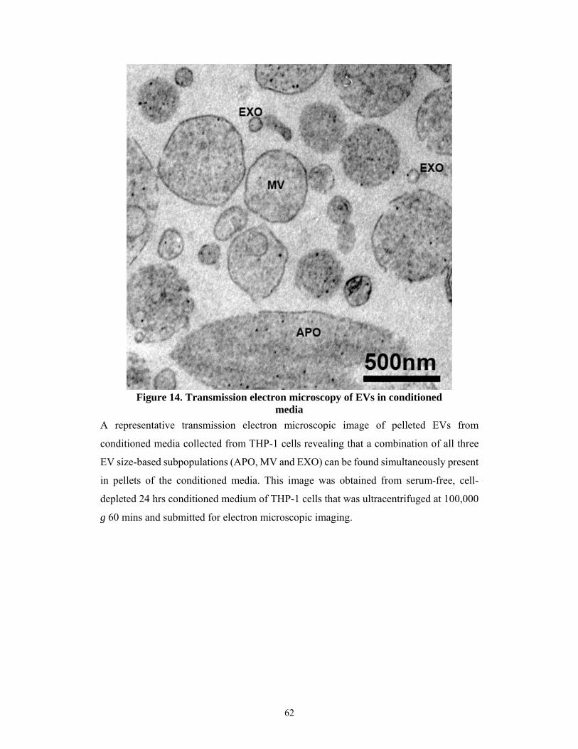
62
A representative transmission electron microscopic image of pelleted EVs from
conditioned media collected from THP-1 cells revealing that a combination of all three
EV size-based subpopulations (APO, MV and EXO) can be found simultaneously present
in pellets of the conditioned media. This image was obtained from serum-free, cell-
depleted 24 hrs conditioned medium of THP-1 cells that was ultracentrifuged at 100,000
g 60 mins and submitted for electron microscopic imaging.
Figure 14. Transmission electron microscopy of EVs in conditioned media

63
MiaPaCa cells produced significantly higher amounts of both MVs and EXOs as
compared to the other three cell lines. In contrast, significantly different amounts of APOs
were produced by the four cell lines with U937 producing the most. The mean values ±
SEM of EV concentrations (EV/mL) of conditioned media are reported below each
respective EV subpopulation for the different cell lines. Concentrations were compared
by one-way ANOVA.
5.2.2 Detergent sensitivity of apoptotic bodies and microvesicles
Various size-based subpopulations of EVs derived from the four different cell
lines were analyzed by different sized pores in the TRPS system before and after
treatment with increasing concentrations of detergents. Prior to TRPS analysis, cell-
depleted conditioned media were supplemented with detergents and were vortexed for 30
seconds. For the detection of APOs in the conditioned media a NP2000 nanopore
Figure 15. Concentration of EVs present in the supernatant of various cell lines
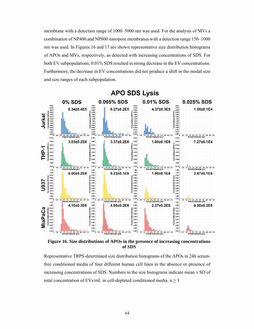
64
membrane with a detection range of 1000–5000 nm was used. For the analysis of MVs a
combination of NP400 and NP800 nanopore membranes with a detection range 150–1000
nm was used. In Figures 16 and 17 are shown representative size distribution histograms
of APOs and MVs, respectively, as detected with increasing concentrations of SDS. For
both EV subpopulations, 0.01% SDS resulted in strong decrease in the EV concentrations.
Furthermore, the decrease in EV concentrations did not produce a shift in the modal size
and size ranges of each subpopulation.
Representative TRPS-determined size distribution histograms of the APOs in 24h serum-
free conditioned media of four different human cell lines in the absence or presence of
increasing concentrations of SDS. Numbers in the size histograms indicate mean ± SD of
total concentration of EVs/mL in cell-depleted conditioned media. n ≥ 3
Figure 16. Size distributions of APOs in the presence of increasing concentrations of SDS
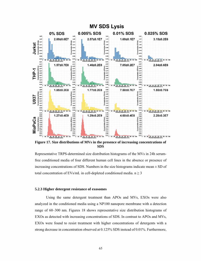
65
Representative TRPS-determined size distribution histograms of the MVs in 24h serum-
free conditioned media of four different human cell lines in the absence or presence of
increasing concentrations of SDS. Numbers in the size histograms indicate mean ± SD of
total concentration of EVs/mL in cell-depleted conditioned media. n ≥ 3
5.2.3 Higher detergent resistance of exosomes
Using the same detergent treatment than APOs and MVs, EXOs were also
analyzed in the conditioned media using a NP100 nanopore membrane with a detection
range of 60–300 nm. Figures 18 shows representative size distribution histograms of
EXOs as detected with increasing concentrations of SDS. In contrast to APOs and MVs,
EXOs were found to resist treatment with higher concentrations of detergents with a
strong decrease in concentration observed at 0.125% SDS instead of 0.01%. Furthermore,
Figure 17. Size distributions of MVs in the presence of increasing concentrations of SDS
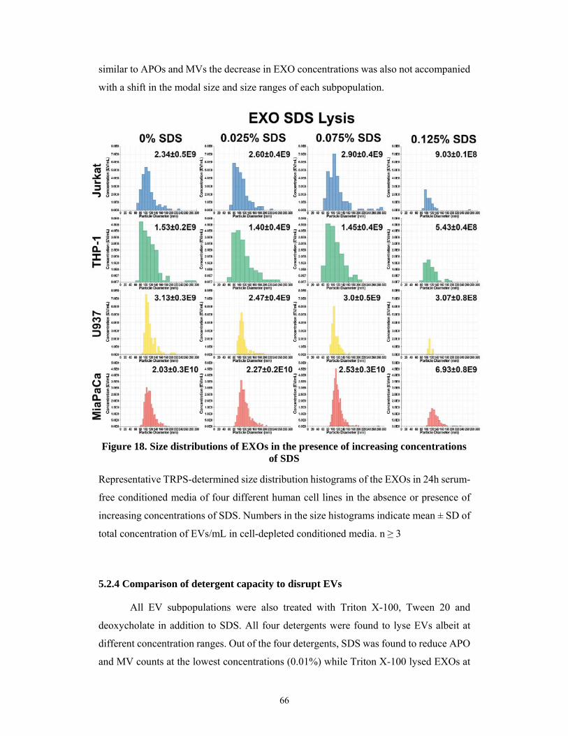
66
similar to APOs and MVs the decrease in EXO concentrations was also not accompanied
with a shift in the modal size and size ranges of each subpopulation.
Representative TRPS-determined size distribution histograms of the EXOs in 24h serum-
free conditioned media of four different human cell lines in the absence or presence of
increasing concentrations of SDS. Numbers in the size histograms indicate mean ± SD of
total concentration of EVs/mL in cell-depleted conditioned media. n ≥ 3
5.2.4 Comparison of detergent capacity to disrupt EVs
All EV subpopulations were also treated with Triton X-100, Tween 20 and
deoxycholate in addition to SDS. All four detergents were found to lyse EVs albeit at
different concentration ranges. Out of the four detergents, SDS was found to reduce APO
and MV counts at the lowest concentrations (0.01%) while Triton X-100 lysed EXOs at
Figure 18. Size distributions of EXOs in the presence of increasing concentrations of SDS
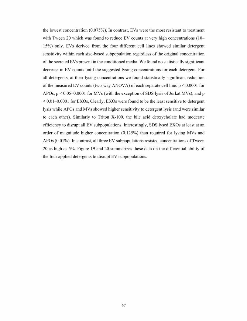
67
the lowest concentration (0.075%). In contrast, EVs were the most resistant to treatment
with Tween 20 which was found to reduce EV counts at very high concentrations (10–
15%) only. EVs derived from the four different cell lines showed similar detergent
sensitivity within each size-based subpopulation regardless of the original concentration
of the secreted EVs present in the conditioned media. We found no statistically significant
decrease in EV counts until the suggested lysing concentrations for each detergent. For
all detergents, at their lysing concentrations we found statistically significant reduction
of the measured EV counts (two-way ANOVA) of each separate cell line: p < 0.0001 for
APOs, p < 0.05–0.0001 for MVs (with the exception of SDS lysis of Jurkat MVs), and p
< 0.01–0.0001 for EXOs. Clearly, EXOs were found to be the least sensitive to detergent
lysis while APOs and MVs showed higher sensitivity to detergent lysis (and were similar
to each other). Similarly to Triton X-100, the bile acid deoxycholate had moderate
efficiency to disrupt all EV subpopulations. Interestingly, SDS lysed EXOs at least at an
order of magnitude higher concentration (0.125%) than required for lysing MVs and
APOs (0.01%). In contrast, all three EV subpopulations resisted concentrations of Tween
20 as high as 5%. Figure 19 and 20 summarizes these data on the differential ability of
the four applied detergents to disrupt EV subpopulations.

68
Comparative sensitivity of APOs, MVs and EXOs secreted by different cell lines (Jurkat,
THP-1, U937 and MiaPaCa) to increasing concentrations of detergents SDS and Triton
X-100. EV concentration is shown as a percentage of the initial concentration prior to
detergent treatment. Note that the detergent concentration ranges differ due to the large
variation among detergents in working ranges for EV lysis. n ≥ 3
Figure 19. Comparison of detergent lysis sensitivity of EV subpopulations
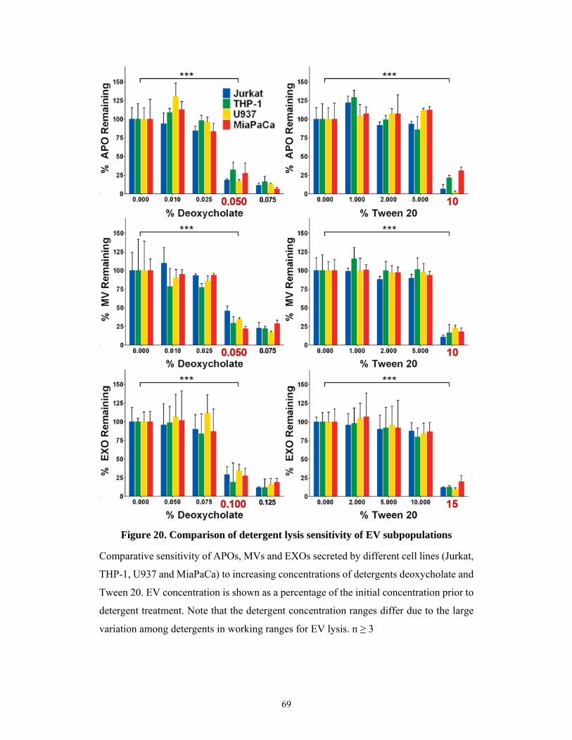
69
Comparative sensitivity of APOs, MVs and EXOs secreted by different cell lines (Jurkat,
THP-1, U937 and MiaPaCa) to increasing concentrations of detergents deoxycholate and
Tween 20. EV concentration is shown as a percentage of the initial concentration prior to
detergent treatment. Note that the detergent concentration ranges differ due to the large
variation among detergents in working ranges for EV lysis. n ≥ 3
Figure 20. Comparison of detergent lysis sensitivity of EV subpopulations

70
These results demonstrate that SDS is the detergent that disrupts MVs and APOs
at the lowest concentration which is in good accordance with the fact that ionic detergents,
such as SDS, are extremely effective in the solubilisation of membrane proteins (316).
Given the large difference in SDS concentration that lyses EXOs compared to MVs and
APOs, the use of this detergent for differential lysis of the different EV subpopulations
may appear superior to other detergents. However, for downstream applications it may
be needed to also consider that in most instances ionic detergents are denaturing to some
extent, while bile acids and non-ionic detergents are relatively mild and are usually non-
denaturing (316). Thus, the choice of detergent for EV studies might dictated by the type
of work to be carried out. Non-ionic detergents such as Triton X-100 and Tween 20 are
known to break lipid–lipid interactions and lipid–protein interactions rather than protein–
protein interactions, a feature that can enable them to differentiate EVs from protein
aggregates (292, 317).
These data also point to the similarity of the lipid membrane composition of APOs
and MVs as opposed to EXOs in line with the results described earlier related to lipid
membrane composition and membrane liquid order of different size-based EV
subpopulations and falls in accordance with the findings that liquid ordered as opposed
to liquid-disordered membranes are more resistant to detergents (318, 319).
5.2.5 Flow cytometric validation of detergent sensitivity of extracellular vesicles
Lastly, EVs were characterized with flow cytometry by their staining with
fluorochrome labelled annexin V and anti-CD9 antibody and the results are shown in
Figure 21. Annexin V and anti-CD9 stained all three EV subpopulations. Using SDS
concentrations shown in the present study by TRPS to lyse vesicles, we could demonstrate
the prompt disappearance of fluorescent events validating our prior TRPS observations.
71
Serum-free, cell-depleted 24h conditioned media of cell lines were submitted for APO,
MV and EXO isolation using differential centrifugation/ultracentrifugation and gravity
driven size filtration. EVs were stained with Annexin V APC and anti-CD9 FITC. APOs
and MVs were analyzed directly, whereas EXOs were detected upon binding to latex
beads. All fluorescent events promptly disappeared upon addition of the concentration of
SDS found to lyse each EV subpopulation. The figure shows representative data for the
lysis of THP-1 cell-derived EVs.
Figure 21. Flow cytometric validation of EV lysing concentration of SDS
72
6. DISCUSSION
In the past decades EV research emerged as a novel field of cell biology.
Understanding of the roles of EVs has profoundly impacted our understanding of many
biological processes. In the meantime, different methods have been adopted to quantify
EVs. However, currently used assays do not discriminate between vesicular or non-
vesicular particles (such as NTA and TRPS), or solely quantify EVs in a given preparation
based on the total protein content. Since protein aggregates have been shown to be co-
purified with EVs (292), and in some samples soluble proteins and protein aggregates are
more abundant than lipid bilayer enclosed EVs, such approaches are error-prone. We
hypothesized that as an alternative to protein-based EV quantification, simultaneous
determination of both the protein and lipid content may lead to a better quantification of
the EVs in a sample. In spite of the large body of published data on EVs, virtually none
of the studies have used an assay to determine total lipids in EV preparations besides
determining the protein content. Thus, there has been an unmet need in the field for a
simple lipid assay to detect EVs. Furthermore, there have been no universally accepted
molecular markers to distinguish among the subpopulations of EVs.
Thus the work of this Ph.D. dissertation was undertaken to establish for the first
time a simple lipid assay for EVs and to find good quality control parameters to
characterize EV preparations that might be utilized both in basic research and in clinical
laboratory settings. To this end, we used and optimized the SPV total lipid assay for EV
studies and combined this lipid assay with the conventional microBCA protein
determination that is used widely in the EV field. This total lipid assay is simple and fast,
requiring only 30 min of incubation, and 0.5µg protein containing EVs either in dry pellet
or in up to 50µL volume. Furthermore, we showed if used under the optimized conditions
described in this dissertation, the SPV assay sensitivity is adequate for EV studies and
compares favorably to microBCA in terms of accuracy, although shows slightly higher
intra and inter-assay variabilities. However, at concentrations >50 µg/mL lipid for SPV
or protein for Micro BCA, both assays show low variability and good accuracy as shown
earlier in Figure 7. Using these two assays, we introduced protein to lipid ratio as a novel
parameter to characterize EV preparations enriched in EXOs, MVs, and APOs.
As expected, we found increasing protein to lipid ratios for EV subpopulations of
increasing diameter. Our data are in line with results of an earlier study that determined
73
protein and lipid composition of platelet microparticle size fractions using mass
spectrometry and a detection of lipid phosphorous with the method of Bartlett (320). We
found that the protein to lipid values depended on the lipid standard used in the assay.
Although the reference lipids (such as menhaden fish oil) that we used in this study may
not exactly reflect the complex composition of EV lipid membranes, they are produced
under standardized conditions, they are inexpensive and they are commercially available
for the broad scientific community. More sophisticated lipid standards may be further
developed as the lipid composition of EVs will be better understood. Regardless of the
method used, if adhering to the same lipid standard consistently throughout the
experiments, protein to lipid ratio determination may prove very useful for quality control
of EV preparations. In addition to the SPV method introduced in this study, more
sophisticated methods such as infrared spectroscopy could be alternatively used for the
determination of protein and lipid content of EVs. However, the requirement for special
equipment may prevent the use of infrared spectroscopy in many laboratories.
Furthermore, cholesterol as well as other sterols do not have many useful infrared features
by which to distinguish them from other molecules. The vibrational bands due to C–C or
C-H bonds, which are commonly used to measure sterols with infrared spectroscopy, are
not only very weak but are also shared by most biological molecules including proteins
(321). Its reduced cholesterol sensitivity may thus, limit the wide use of infrared
spectroscopy for EV studies.
In addition to EVs, we also determined the protein to lipid ratio of cells releasing
EVs. We found that, as compared to the EVs, cells showed higher protein to lipid ratios
(2.19) but with substantially higher variation (SEM = 0.95) probably reflecting
differences in cell types and in cell cycle phases. Lipid sorting mechanisms (such as
preferential sorting of cholesterol into EXOs) during the generation of different EV
subpopulations have been reported (40, 322). Such sorting process may result in the
enrichment of given lipids in different EV subpopulations which may contribute to the
relatively low variation of protein to lipid ratios of the same EV subpopulation compared
to the producing cells.
In an attempt to find novel parameters to characterize EV subpopulations, using
flow cytometry we stained our EVs with an anti-cholesterol antibody, and found strongest
staining for EXOs. Furthermore, we found staining of all EV subpopulations with CTX
reflecting the presence of GM1 gangliosides in all EV subpopulations. GM1 gangliosides
74
have been previously detected in EXOs (323, 324). Here we found that GM1 was not only
present in EXOs but also especially enriched in MVs and differentiated two
subpopulations of APOs. Intriguingly, heterogeneity within APOs was not only suggested
by bimodal CTX binding, but also by the annexin V and anti-CD63 staining.
Transmission electron microscopy also revealed the presence of highly electron dense,
possibly fragmented DNA containing APOs and less electron dense and more granular
APOs possibly corresponding to the recently described different subpopulations of APOs
(73).
The spectral ratiometric approach of this study provides evidence that EV
subpopulations can be distinguished based on the difference in their membrane lipid
order. This parameter reflects the degree of lipid packing and is one of the most important
biophysical parameters of membranes (311). In low ordered membrane domains the
probability of protein-protein interactions is decreased. Therefore membrane lipid order
can impact signaling pathways. This is strongly supported by the findings that high
membrane lipid order is typically found at the immunological synapse, sites of cell
adhesion, viral entry and budding as well as in exosomes (the latter described for the first
time in this work). Our data suggest that EXOs are characterized by highest membrane
lipid order, while APOs and MVs showed low to intermediate, partially overlapping lipid
order reflecting important differences in membrane lipid composition. A high degree of
membrane lipid order and the relatively high content of cholesterol in EXOs may be
important factors explaining their distinguished role in intercellular signaling.
Without universally accepted molecular markers reflecting different biogenic
origin of EV subpopulations, most studies simply classify EVs based on their size. Such
a size-based classification could be challenged by observations that exosome-sized EVs
can also be shed from the plasma membrane (202). In an attempt to assess whether this
kind of observations represent exceptions to the rule or if size-based classification is
arbitrary, we looked beyond size, and compared lipid properties and protein to lipid ratios
in EVs fractionated by size. We found differential binding of an anti-cholesterol antibody
and CTX to different sized populations.
Additionally, our data show an increasing protein to lipid ratio with an increasing
diameter for each EV subpopulation. This was as expected because with an increasing
diameter, the amount of protein that may be contained inside an EV increases as a
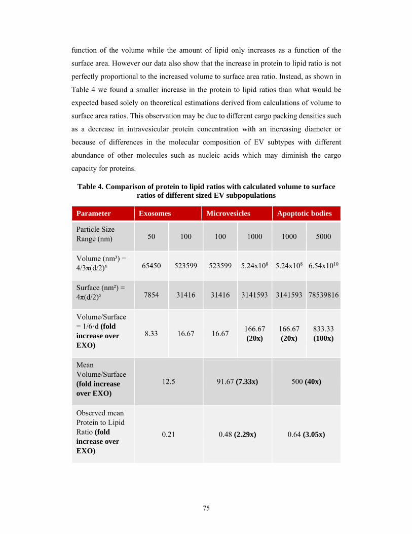
75
function of the volume while the amount of lipid only increases as a function of the
surface area. However our data also show that the increase in protein to lipid ratio is not
perfectly proportional to the increased volume to surface area ratio. Instead, as shown in
Table 4 we found a smaller increase in the protein to lipid ratios than what would be
expected based solely on theoretical estimations derived from calculations of volume to
surface area ratios. This observation may be due to different cargo packing densities such
as a decrease in intravesicular protein concentration with an increasing diameter or
because of differences in the molecular composition of EV subtypes with different
abundance of other molecules such as nucleic acids which may diminish the cargo
capacity for proteins.
Table 4. Comparison of protein to lipid ratios with calculated volume to surface ratios of different sized EV subpopulations
Parameter Exosomes Microvesicles Apoptotic bodies
Particle Size Range (nm) 50 100 100 1000 1000 5000
Volume (nm³) = 4/3π(d/2)³ 65450 523599 523599 5.24x10⁸ 5.24x10⁸ 6.54x10¹⁰
Surface (nm²) = 4π(d/2)² 7854 31416 31416 3141593 3141593 78539816
Volume/Surface = 1/6·d (fold increase over EXO)
8.33 16.67 16.67 166.67 (20x)
166.67 (20x)
833.33 (100x)
Mean Volume/Surface (fold increase over EXO)
12.5 91.67 (7.33x) 500 (40x)
Observed mean Protein to Lipid Ratio (fold increase over EXO)
0.21 0.48 (2.29x) 0.64 (3.05x)
76
Table 4 shows the calculated volumes to surface area ratios for the size ranges of
different EV subpopulations in comparison with the observed protein to lipid ratios.
Together, these observations suggest that simple size-based classification enables
distinguishing EVs with different membrane lipid properties. Until selective molecular
markers become available, established protein to lipid ratios may serve as suitable
parameters to characterize different EV subpopulations. In addition, protein to lipid ratio
may also prove useful for routine quality control of EV preparations.
Based on previous findings of our group, we also carried out a comprehensive
analysis of the effect of detergents on size-based EV subpopulations. This work provides
a practical tool to confirm the vesicular nature of particles present in biological samples
using either TRPS or flow cytometry. Given that both TRPS and flow cytometry are
commonly used methods to characterize EVs, the use of detergent control in EV analysis
may prove useful in a wide variety of experimental settings and may also be combined
with other techniques of EV analysis such as those described previously in Table 3.
Results of the experiments with detergents clearly demonstrate that SDS disrupts
MVs and APOs at the lowest concentration. This is in good accordance with the fact that
ionic detergents, such as SDS, are extremely effective in the solubilization of membrane
proteins (316). It was also found that there is an order of magnitude difference between
the concentration of SDS that lyses both APOs and MVs and the one that lyses EXOs.
Therefore the use of SDS for differential lysis of APOS, MVs and EXOs may appear
superior to other detergents. However, for downstream applications it also has to be
considered that in most instances ionic detergents are denaturing to some extent, while
bile acids and non-ionic detergents are relatively mild and are usually non-denaturing
(316). The choice of detergent for EV studies is thus, dictated by the type of work to be
carried out. Non-ionic detergents are known to break lipid–lipid interactions and lipid–
protein interactions rather than protein–protein interactions, a feature that enables them
to differentiate EVs from protein aggregates (292, 317).
Interestingly, a non-statistically significant increase in EV concentrations was
observed in the case of treatment with SDS and deoxycholate. This observation is
probably due to a small decrease in instrumental noise due to the used of these detergents
and thus slightly enhanced lower detection limit for smaller particles which would be
below the noise threshold without detergent. Furthermore, it is also possible that a small

77
percentage of previously aggregated EVs might dissociate from each other at low
detergent concentration and thus be counted as more individual particles instead of their
aggregated form.
Taken together, our results point to the similarity of the lipid membrane
composition of APOs and MVs as opposed to EXOs which were found to be more liquid
ordered while both APOs and MVs had similar and lower membrane liquid orders.
Accordingly, several studies show that liquid ordered as opposed to liquid-disordered
membranes are resistant to detergents (318, 319).
Moreover, our findings show that using detergents at the suggested
concentrations, vesicle enclosed cytokines, RNAs and metabolites may be released for
detection. Also, analysis of vesicle cargo by Western blotting or mass spectrometry
requires both the disruption of the vesicle membrane and the release of membrane
proteins. Recently, particular attention follows RNA as a vesicular cargo. RNA extraction
from EVs may involve the use of detergents (325). Results of our study also have
important implications in other research methods such as in ELISAs where Tween 20 is
used at lower concentrations (typically 0.05–0.1%) than what we found to lyse EVs. Thus
this study introduced a practical tool for confirmation of the vesicular nature of particles
present in biological samples using either TRPS or flow cytometry. Given that both TRPS
and flow cytometry are commonly used methods to characterize EVs, the use of simple
and fast detergent controls to differentiate between EXOs and other extracellular vesicle
subpopulations as well as between vesicular and non-vesicular structures in EV analysis
may prove useful in a wide variety of experimental settings.
In conclusion, for the first time our work introduced a simple lipid assay and novel
EV characterization parameter as well as the appropriate concentrations of different
detergents to lyse EVs. We demonstrated that different size-based subpopulations of EVs
secreted by highly diverse human cell lines show similar protein to lipid ratios, membrane
lipid orders, and detergent sensitivity patterns. Taken together, our data validate the use
of the SPV lipid assay, membrane lipid order assessment, and detergent lysis as integral
components of the EV detection toolbox.
78
7. CONCLUSIONS
In conclusion, with this work the existing methodology of extracellular vesicle
research has been improved with the introduction of a new assay as well as different
parameters useful for the detection and characterization, as well as quality control and
validation of extracellular vesicle preparations.
This was achieved by introducing a new assay for quantification of total lipids.
Using this assay we found a novel parameter consisting of the protein to lipid ratio that is
suitable for the characterization of the subtype as well as the purity and quality of
extracellular vesicle preparations.
Furthermore we have characterized the membrane lipid order of different
extracellular vesicle subpopulations finding a higher degree of order in exosomes lipid
membranes as compared to microvesicles and apoptotic bodies. Interestingly we also
found an increased detergent resistance with the increased membrane lipid order of
exosomes.
79
8. SUMMARY
Extracellular vesicles including exosomes, microvesicles, and apoptotic bodies
have so far attracted rapidly increasing attention from various fields of biology due to
their ability to carry complex information and act as autocrine, paracrine and even
endocrine intercellular messengers. Despite this growth, there is still an important need
to improve the methodology and controls with which to characterize extracellular
vesicles.
For this reason the aim of this Ph.D. research was to introduce to the field of
extracellular vesicles new and improved methods for detection and characterization.
Extracellular vesicles were isolated by differential centrifugation and gravity
driven filtration from the conditioned media of cell lines as well as human blood plasma.
Extracellular vesicles were characterized by using tunable resistive pulse sensing,
transmission electron microscopy, and flow cytometry. Additionally, we introduced for
the first time a simple bench top lipid assay for determination of lipid content of
extracellular vesicles based on a 96 well plate adaptation of the sulfophosphovanilin
colorimetric reaction.
Using this novel lipid assay and an available protein assay we found characteristic
protein to lipid ratios for each of the investigated extracellular vesicle subpopulations.
Moreover, these ratios were substantially altered in the case of vesicular membrane
damage or protein contamination. We also found marked differences in membrane lipid
order between the various vesicle subpopulations using ratiometric spectrophotometry.
Lastly, we determined the sensitivity of extracellular vesicles subpopulations to different
concentrations of various detergents. Interestingly, we found that microvesicles and
apoptotic bodies had similar and higher sensitivity to detergent lysis than the smallest
sized extracellular vesicles, exosomes.
Together, all these observations may contribute to a more comprehensive
detection of extracellular vesicles. Protein to lipid ratio that we introduced to the field,
may prove useful as a quality control parameter for extracellular vesicle preparations both
in basic research and in clinical studies.
80
9. ÖSSZEFOGLALÁS
Az extracelluláris vezikulák (mint pl. az exoszómák, mikrovezikulák és
apoptotikus testek) az elmúlt években igen nagy érdeklődést váltottak ki a biológia
számos területén amiatt, hogy komplex információhordozóként működnek, továbbá
annak kapcsán, hogy autokrin, parakrin és endokrin intercelluláris hírvivőként képesek a
sejtek működését szabályozni. Az extracelluláris vezikulákra irányuló egyre intenzívebb
kutatások ellenére nagy szükség van a jellemzésükre alkalmazható alapmódszerek
továbbfejlesztésére.
PhD munkám célja az extracelluláris vezikulák detektálására és karakterizálására
alkalmas új alapvető módszerek és módszerkombinációk keresése.
Az extracelluláris vezikulákat sejtvonalakból felülúszójából és humán
vérplazmából izoláltuk differenciálcentrifugálással és hidrosztatikus filtrációval. Tunable
resistive puls sensing („hangolható rezisztív impulzus érzékelő”) módszerrel,
transzmissziós elektron mikroszkópiával és áramlási citometriával jellemeztük őket.
Elsőként alkalmaztuk lipid assay-t az extracelluláris vezikulák lipidtartalmának
meghatározására. Az extracelluláris vezikulák lipidtartalmának mérésére adaptált 96
lyukú lemez alapú szulfo-foszfo-vanillin kolorimetriás assay gyors és egyszerű
lehetőséget kínál az extracelluláris vezikulák jellemzésére. Az új lipid assay és a Micro
BCA fehérje assay együttes használatával minden extracelluláris vezikula szubpopuláció
esetében jellegzetes fehérje/lipid arányt tapasztaltunk. Igazoltuk, hogy az extracelluláris
vezikulák membránjának károsodása vagy a vezikula preparátum fehérjeszennyeződése
hatására ez az arány lényegesen megváltozik. Emellett sajátos különbséget találtunk a
membrán lipid rendezettségben a különböző vezikula szubpopulációk között
ratiometrikus spektrofotometriás vizsgálatokkal. Végül, a differenciál detergens lízisnek
a tunable resistive pulse sensing módszerrel illetőleg áramlási citometriával való
kombinációjával elsőként határoztuk meg az extracelluláris vezikula alpopulációk
érzékenységét különböző detergensek általi lízisre. Igazoltuk, hogy a mikrovezikulák és
apoptotikus testek egymáshoz igan hasonló módon érzékenyebbek a detergens lízisre
mint a legkisebb méretű extracelluláris vezikulák, az exoszómák.
Eredményeink hozzájárulhatnak az extracelluláris vezikulák pontosabb
detektálásához. A bevezetett protein/lipid hasznos lehet az extracelluláris vezikulák

81
„quality control” paramétereként az extracelluláris vezikulákra irányuló alapkutatási és
klinikai kutatási vizsgálatok során.
82
10. REFERENCES
1. Chargaff E and West R. (1946) The biological significance of the thromboplastic protein
of blood. J Biol Chem, 166(1): 189-197.
2. Wolf P. (1967) The nature and significance of platelet products in human plasma. Br J
Haematol, 13(3): 269-288.
3. De Broe M, Wieme R, and Roels F. (1975) Letter: Membrane fragments with koinozymic
properties released from villous adenoma of the rectum. Lancet, 2(7946): 1214-1215.
4. Taylor DD, Homesley HD, and Doellgast GJ. (1980) Binding of specific peroxidase-
labeled antibody to placental-type phosphatase on tumor-derived membrane fragments.
Cancer Res, 40(11): 4064-4069.
5. Bonucci E. (1967) Fine structure of early cartilage calcification. J Ultrastruct Res, 20(1):
33-50.
6. Anderson HC. (1967) Electron microscopic studies of induced cartilage development and
calcification. J Cell Biol, 35(1): 81-101.
7. Stegmayr B and Ronquist G. (1982) Promotive effect on human sperm progressive
motility by prostasomes. Urol Res, 10(5): 253-257.
8. Ronquist G, Brody I, Gottfries A, and Stegmayr B. (1978) An Mg2+ and Ca2+-stimulated
adenosine triphosphatase in human prostatic fluid: part I. Andrologia, 10(4): 261-272.
9. Benz EW, Jr. and Moses HL. (1974) Small, virus-like particles detected in bovine sera
by electron microscopy. J Natl Cancer Inst, 52(6): 1931-1934.
10. Dalton AJ. (1975) Microvesicles and vesicles of multivesicular bodies versus "virus-like"
particles. J Natl Cancer Inst, 54(5): 1137-1148.
11. Harding C, Heuser J, and Stahl P. (1984) Endocytosis and intracellular processing of
transferrin and colloidal gold-transferrin in rat reticulocytes: demonstration of a pathway
for receptor shedding. Eur J Cell Biol, 35(2): 256-263.
12. Pan BT and Johnstone RM. (1983) Fate of the transferrin receptor during maturation of
sheep reticulocytes in vitro: selective externalization of the receptor. Cell, 33(3): 967-
978.
13. Johnstone RM, Adam M, Hammond JR, Orr L, and Turbide C. (1987) Vesicle formation
during reticulocyte maturation. Association of plasma membrane activities with released
vesicles (exosomes). J Biol Chem, 262(19): 9412-9420.
14. Raposo G, Nijman HW, Stoorvogel W, Liejendekker R, Harding CV, Melief CJ, and
Geuze HJ. (1996) B lymphocytes secrete antigen-presenting vesicles. J Exp Med, 183(3):
1161-1172.

83
15. Valadi H, Ekström K, Bossios A, Sjöstrand M, Lee JJ, and Lötvall JO. (2007) Exosome-
mediated transfer of mRNAs and microRNAs is a novel mechanism of genetic exchange
between cells. Nat Cell Biol, 9(6): 654-659.
16. Ratajczak J, Miekus K, Kucia M, Zhang J, Reca R, Dvorak P, and Ratajczak MZ. (2006)
Embryonic stem cell-derived microvesicles reprogram hematopoietic progenitors:
evidence for horizontal transfer of mRNA and protein delivery. Leukemia, 20(5): 847-
856.
17. Robbins PD and Morelli AE. (2014) Regulation of Immune Responses by Extracellular
Vesicles. Nat Rev Immunol, 14(3): 195-208.
18. Théry C, Ostrowski M, and Segura E. (2009) Membrane vesicles as conveyors of immune
responses. Nat Rev Immunol, 9(8): 581-593.
19. Wurdinger T, Gatson NN, Balaj L, Kaur B, Breakefield XO, and Pegtel DM. (2012)
Extracellular Vesicles and Their Convergence with Viral Pathways. Adv Virol, 2012:
767694.
20. Budnik V, Ruiz-Canada C, and Wendler F. (2016) Extracellular vesicles round off
communication in the nervous system. Nat Rev Neurosci, 17(3): 160-172.
21. Thompson AG, Gray E, Heman-Ackah SM, Mager I, Talbot K, Andaloussi SE, Wood
MJ, and Turner MR. (2016) Extracellular vesicles in neurodegenerative disease -
pathogenesis to biomarkers. Nat Rev Neurol, 12(6): 346-357.
22. Osteikoetxea X, Nemeth A, Sodar BW, Vukman KV, and Buzas EI. (2016) Extracellular
vesicles in cardiovascular diseases, are they Jedi or Sith? J Physiol, 594(11): 2881-2894.
23. Tannetta D, Dragovic R, Alyahyaei Z, and Southcombe J. (2014) Extracellular vesicles
and reproduction–promotion of successful pregnancy. Cell Mol Immunol, 11(6): 548-
563.
24. Marcilla A, Martin-Jaular L, Trelis M, de Menezes-Neto A, Osuna A, Bernal D,
Fernandez-Becerra C, Almeida IC, and Del Portillo HA. (2014) Extracellular vesicles in
parasitic diseases. J Extracell Vesicles, 3: 25040.
25. Deatherage BL and Cookson BT. (2012) Membrane vesicle release in bacteria,
eukaryotes, and archaea: a conserved yet underappreciated aspect of microbial life. Infect
Immun, 80(6): 1948-1957.
26. MacDonald IA and Kuehn MJ. (2012) Offense and defense: microbial membrane vesicles
play both ways. Res Microbiol, 163(9-10): 607-618.
27. György B, Szabó T, Pásztói M, Pál Z, Misják P, Aradi B, László V, Pállinger É, Pap E,
Kittel Á, Nagy G, Falus A, and Buzás E. (2011) Membrane vesicles, current state-of-the-
art: emerging role of extracellular vesicles. Cell Mol Life Sci, 68(16): 2667-2688.
28. Théry C. (2011) Exosomes: secreted vesicles and intercellular communications. F1000
Biol Rep, 3.
84
29. Buzás EI, Gyorgy B, Nagy G, Falus A, and Gay S. (2014) Emerging role of extracellular
vesicles in inflammatory diseases. Nat Rev Rheumatol.
30. Yáñez-Mó M, Siljander PRM, Andreu Z, Bedina Zavec A, Borràs FE, Buzas EI, Buzas
K, Casal E, Cappello F, Carvalho J, Colás E, Cordeiro-da Silva A, Fais S, Falcon-Perez
JM, Ghobrial IM, Giebel B, Gimona M, Graner M, Gursel I, Gursel M, Heegaard NHH,
Hendrix A, Kierulf P, Kokubun K, Kosanovic M, Kralj-Iglic V, Krämer-Albers E-M,
Laitinen S, Lässer C, Lener T, Ligeti E, Linē A, Lipps G, Llorente A, Lötvall J, Manček-
Keber M, Marcilla A, Mittelbrunn M, Nazarenko I, Nolte-‘t Hoen ENM, Nyman TA,
O'Driscoll L, Olivan M, Oliveira C, Pállinger É, del Portillo HA, Reventós J, Rigau M,
Rohde E, Sammar M, Sánchez-Madrid F, Santarém N, Schallmoser K, Stampe Ostenfeld
M, Stoorvogel W, Stukelj R, Van der Grein SG, Vasconcelos MH, Wauben MHM, and
De Wever O. (2015) Biological properties of extracellular vesicles and their physiological
functions. J Extracell Vesicles, 4(0).
31. Bilyy RO, Shkandina T, Tomin A, Munoz LE, Franz S, Antonyuk V, Kit YY, Zirngibl
M, Furnrohr BG, Janko C, Lauber K, Schiller M, Schett G, Stoika RS, and Herrmann M.
(2012) Macrophages discriminate glycosylation patterns of apoptotic cell-derived
microparticles. J Biol Chem, 287(1): 496-503.
32. Crescitelli R, Lasser C, Szabo TG, Kittel A, Eldh M, Dianzani I, Buzas EI, and Lotvall J.
(2013) Distinct RNA profiles in subpopulations of extracellular vesicles: apoptotic
bodies, microvesicles and exosomes. J Extracell Vesicles, 2.
33. Skog J, Würdinger T, van Rijn S, Meijer DH, Gainche L, Curry WT, Carter BS,
Krichevsky AM, and Breakefield XO. (2008) Glioblastoma microvesicles transport RNA
and proteins that promote tumour growth and provide diagnostic biomarkers. Nat Cell
Biol, 10(12): 1470-1476.
34. Hong B, Cho J-H, Kim H, Choi E-J, Rho S, Kim J, Kim J, Choi D-S, Kim Y-K, Hwang
D, and Gho Y. (2009) Colorectal cancer cell-derived microvesicles are enriched in cell
cycle-related mRNAs that promote proliferation of endothelial cells. BMC Genomics,
10(1): 556.
35. Waldenström A, Gennebäck N, Hellman U, and Ronquist G. (2012) Cardiomyocyte
Microvesicles Contain DNA/RNA and Convey Biological Messages to Target Cells.
PLoS ONE, 7(4): e34653.
36. Thakur BK, Zhang H, Becker A, Matei I, Huang Y, Costa-Silva B, Zheng Y, Hoshino A,
Brazier H, Xiang J, Williams C, Rodriguez-Barrueco R, Silva JM, Zhang W, Hearn S,
Elemento O, Paknejad N, Manova-Todorova K, Welte K, Bromberg J, Peinado H, and
Lyden D. (2014) Double-stranded DNA in exosomes: a novel biomarker in cancer
detection. Cell Res, 24(6): 766-769.
85
37. Wubbolts R, Leckie RS, Veenhuizen PTM, Schwarzmann G, Mobius W,
Hoernschemeyer J, Slot JW, Geuze HJ, and Stoorvogel W. (2003) Proteomic and
Biochemical Analyses of Human B Cell-derived Exosomes: POTENTIAL
IMPLICATIONS FOR THEIR FUNCTION AND MULTIVESICULAR BODY
FORMATION. J Biol Chem, 278(13): 10963-10972.
38. Trajkovic K, Hsu C, Chiantia S, Rajendran L, Wenzel D, Wieland F, Schwille P, Brugger
B, and Simons M. (2008) Ceramide Triggers Budding of Exosome Vesicles into
Multivesicular Endosomes. Science, 319(5867): 1244-1247.
39. Carayon K, Chaoui K, Ronzier E, Lazar I, Bertrand-Michel J, Roques V, Balor S, Terce
F, Lopez A, Salome L, and Joly E. (2011) Proteolipidic Composition of Exosomes
Changes during Reticulocyte Maturation. J Biol Chem, 286(39): 34426-34439.
40. Llorente A, Skotland T, Sylvänne T, Kauhanen D, Róg T, Orłowski A, Vattulainen I,
Ekroos K, and Sandvig K. (2013) Molecular lipidomics of exosomes released by PC-3
prostate cancer cells. Bba-Mol Cell Biol L, 1831(7): 1302-1309.
41. Théry C, Boussac M, Veron P, Ricciardi-Castagnoli P, Raposo G, Garin J, and
Amigorena S. (2001) Proteomic Analysis of Dendritic Cell-Derived Exosomes: A
Secreted Subcellular Compartment Distinct from Apoptotic Vesicles. J Immunol,
166(12): 7309-7318.
42. Marzesco AM, Janich P, Wilsch-Bräuninger M, Dubreuil V, Langenfeld K, Corbeil D,
and Huttner WB. (2005) Release of extracellular membrane particles carrying the stem
cell marker prominin-1 (CD133) from neural progenitors and other epithelial cells. J Cell
Sci, 118(13): 2849-2858.
43. Keerthikumar S, Gangoda L, Liem M, Fonseka P, Atukorala I, Ozcitti C, Mechler A,
Adda CG, Ang C-S, and Mathivanan S. (2015) Proteogenomic analysis reveals exosomes
are more oncogenic than ectosomes. Oncotarget, 6(17): 15375-15396.
44. Colombo M, Raposo G, and Thery C. (2014) Biogenesis, secretion, and intercellular
interactions of exosomes and other extracellular vesicles. Annu Rev Cell Dev Biol, 30:
255-289.
45. Harding C, Heuser J, and Stahl P. (1983) Receptor-mediated endocytosis of transferrin
and recycling of the transferrin receptor in rat reticulocytes. J Cell Biol, 97(2): 329-339.
46. Stenmark H. (2009) Rab GTPases as coordinators of vesicle traffic. Nat Rev Mol Cell
Biol, 10(8): 513-525.
47. Vidal MJ and Stahl PD. (1993) The small GTP-binding proteins Rab4 and ARF are
associated with released exosomes during reticulocyte maturation. Eur J Cell Biol, 60(2):
261-267.
48. Savina A, Vidal M, and Colombo MI. (2002) The exosome pathway in K562 cells is
regulated by Rab11. J Cell Sci, 115(Pt 12): 2505-2515.

86
49. Ostrowski M, Carmo NB, Krumeich S, Fanget I, Raposo G, Savina A, Moita CF, Schauer
K, Hume AN, Freitas RP, Goud B, Benaroch P, Hacohen N, Fukuda M, Desnos C, Seabra
MC, Darchen F, Amigorena S, Moita LF, and Thery C. (2010) Rab27a and Rab27b
control different steps of the exosome secretion pathway. Nat Cell Biol, 12(1): 19-30; sup
pp 11-13.
50. Hsu C, Morohashi Y, Yoshimura S, Manrique-Hoyos N, Jung S, Lauterbach MA, Bakhti
M, Gronborg M, Mobius W, Rhee J, Barr FA, and Simons M. (2010) Regulation of
exosome secretion by Rab35 and its GTPase-activating proteins TBC1D10A-C. J Cell
Biol, 189(2): 223-232.
51. Baietti MF, Zhang Z, Mortier E, Melchior A, Degeest G, Geeraerts A, Ivarsson Y,
Depoortere F, Coomans C, Vermeiren E, Zimmermann P, and David G. (2012) Syndecan-
syntenin-ALIX regulates the biogenesis of exosomes. Nat Cell Biol, 14(7): 677-685.
52. Crescitelli R, Lässer C, Szabó TG, Kittel A, Eldh M, Dianzani I, Buzás EI, and Lötvall J.
(2013) Distinct RNA profiles in subpopulations of extracellular vesicles: apoptotic
bodies, microvesicles and exosomes. J Extracell Vesicles, 2(0).
53. Kowal J, Arras G, Colombo M, Jouve M, Morath JP, Primdal-Bengtson B, Dingli F,
Loew D, Tkach M, and Thery C. (2016) Proteomic comparison defines novel markers to
characterize heterogeneous populations of extracellular vesicle subtypes. Proc Natl Acad
Sci U S A, 113(8): E968-977.
54. Mathivanan S, Fahner CJ, Reid GE, and Simpson RJ. (2012) ExoCarta 2012: database of
exosomal proteins, RNA and lipids. Nucleic Acids Res, 40(Database issue): D1241-1244.
55. Kahlert C, Melo SA, Protopopov A, Tang J, Seth S, Koch M, Zhang J, Weitz J, Chin L,
Futreal A, and Kalluri R. (2014) Identification of Double Stranded Genomic DNA
Spanning all Chromosomes with Mutated KRAS and p53 DNA in the Serum Exosomes
of Patients with Pancreatic Cancer. J Biol Chem.
56. Dolo V, Li R, Dillinger M, Flati S, Manela J, Taylor BJ, Pavan A, and Ladisch S. (2000)
Enrichment and localization of ganglioside G(D3) and caveolin-1 in shed tumor cell
membrane vesicles. Biochim Biophys Acta, 1486(2-3): 265-274.
57. Cocucci E, Racchetti G, Podini P, and Meldolesi J. (2007) Enlargeosome traffic:
exocytosis triggered by various signals is followed by endocytosis, membrane shedding
or both. Traffic, 8(6): 742-757.
58. Pasquet JM, Dachary-Prigent J, and Nurden AT. (1996) Calcium influx is a determining
factor of calpain activation and microparticle formation in platelets. Eur J Biochem,
239(3): 647-654.
59. Bianco F, Pravettoni E, Colombo A, Schenk U, Moller T, Matteoli M, and Verderio C.
(2005) Astrocyte-derived ATP induces vesicle shedding and IL-1 beta release from
microglia. J Immunol, 174(11): 7268-7277.
87
60. Pizzirani C, Ferrari D, Chiozzi P, Adinolfi E, Sandona D, Savaglio E, and Di Virgilio F.
(2007) Stimulation of P2 receptors causes release of IL-1beta-loaded microvesicles from
human dendritic cells. Blood, 109(9): 3856-3864.
61. Bevers EM, Comfurius P, Dekkers DW, and Zwaal RF. (1999) Lipid translocation across
the plasma membrane of mammalian cells. Biochim Biophys Acta, 1439(3): 317-330.
62. Hugel B, Martinez MC, Kunzelmann C, and Freyssinet JM. (2005) Membrane
microparticles: two sides of the coin. Physiology (Bethesda), 20: 22-27.
63. Muralidharan-Chari V, Clancy J, Plou C, Romao M, Chavrier P, Raposo G, and D'Souza-
Schorey C. (2009) ARF6-regulated shedding of tumor cell-derived plasma membrane
microvesicles. Curr Biol, 19(22): 1875-1885.
64. Di Vizio D, Kim J, Hager MH, Morello M, Yang W, Lafargue CJ, True LD, Rubin MA,
Adam RM, Beroukhim R, Demichelis F, and Freeman MR. (2009) Oncosome formation
in prostate cancer: association with a region of frequent chromosomal deletion in
metastatic disease. Cancer Res, 69(13): 5601-5609.
65. Ghossoub R, Lembo F, Rubio A, Gaillard CB, Bouchet J, Vitale N, Slavik J, Machala M,
and Zimmermann P. (2014) Syntenin-ALIX exosome biogenesis and budding into
multivesicular bodies are controlled by ARF6 and PLD2. Nat Commun, 5: 3477.
66. Laulagnier K, Grand D, Dujardin A, Hamdi S, Vincent-Schneider H, Lankar D, Salles
JP, Bonnerot C, Perret B, and Record M. (2004) PLD2 is enriched on exosomes and its
activity is correlated to the release of exosomes. FEBS Lett, 572(1-3): 11-14.
67. Bianco F, Perrotta C, Novellino L, Francolini M, Riganti L, Menna E, Saglietti L,
Schuchman EH, Furlan R, Clementi E, Matteoli M, and Verderio C. (2009) Acid
sphingomyelinase activity triggers microparticle release from glial cells. Embo j, 28(8):
1043-1054.
68. Wang T, Gilkes DM, Takano N, Xiang L, Luo W, Bishop CJ, Chaturvedi P, Green JJ,
and Semenza GL. (2014) Hypoxia-inducible factors and RAB22A mediate formation of
microvesicles that stimulate breast cancer invasion and metastasis. Proc Natl Acad Sci U
S A, 111(31): E3234-3242.
69. Heijnen HF, Schiel AE, Fijnheer R, Geuze HJ, and Sixma JJ. (1999) Activated platelets
release two types of membrane vesicles: microvesicles by surface shedding and exosomes
derived from exocytosis of multivesicular bodies and alpha-granules. Blood, 94(11):
3791-3799.
70. Elmore S. (2007) Apoptosis: A Review of Programmed Cell Death. Toxicol Pathol,
35(4): 495-516.
71. Charras GT. (2008) A short history of blebbing. J Microsc, 231(3): 466-478.
72. Di Vizio D, Morello M, Dudley AC, Schow PW, Adam RM, Morley S, Mulholland D,
Rotinen M, Hager MH, Insabato L, Moses MA, Demichelis F, Lisanti MP, Wu H,

88
Klagsbrun M, Bhowmick NA, Rubin MA, D'Souza-Schorey C, and Freeman MR. (2012)
Large oncosomes in human prostate cancer tissues and in the circulation of mice with
metastatic disease. Am J Pathol, 181(5): 1573-1584.
73. Bilyy RO, Shkandina T, Tomin A, Munoz LE, Franz S, Antonyuk V, Kit YY, Zirngibl
M, Furnrohr BG, Janko C, Lauber K, Schiller M, Schett G, Stoika RS, and Herrmann M.
(2011) Macrophages Discriminate Glycosylation Patterns of Apoptotic Cell-derived
Microparticles. J Biol Chem, 287(1): 496-503.
74. Holmgren L, Szeles A, Rajnavolgyi E, Folkman J, Klein G, Ernberg I, and Falk KI.
(1999) Horizontal transfer of DNA by the uptake of apoptotic bodies. Blood, 93(11):
3956-3963.
75. Birdsell DC and Cota-Robles EH. (1967) Production and ultrastructure of lysozyme and
ethylenediaminetetraacetate-lysozyme spheroplasts of Escherichia coli. J Bacteriol,
93(1): 427-437.
76. Knox KW, Vesk M, and Work E. (1966) Relation Between Excreted Lipopolysaccharide
Complexes and Surface Structures of a Lysine-Limited Culture of Escherichia coli. J
Bacteriol, 92(4): 1206-1217.
77. Zhou L, Srisatjaluk R, Justus DE, and Doyle RJ. (1998) On the origin of membrane
vesicles in gram-negative bacteria. FEMS Microbiol Lett, 163(2): 223-228.
78. Mug-Opstelten D and Witholt B. (1978) Preferential release of new outer membrane
fragments by exponentially growing Escherichia coli. Biochim Biophys Acta, 508(2):
287-295.
79. Rivera J, Cordero RJ, Nakouzi AS, Frases S, Nicola A, and Casadevall A. (2010) Bacillus
anthracis produces membrane-derived vesicles containing biologically active toxins. Proc
Natl Acad Sci U S A, 107(44): 19002-19007.
80. Lee EY, Choi DY, Kim DK, Kim JW, Park JO, Kim S, Kim SH, Desiderio DM, Kim
YK, Kim KP, and Gho YS. (2009) Gram-positive bacteria produce membrane vesicles:
proteomics-based characterization of Staphylococcus aureus-derived membrane vesicles.
Proteomics, 9(24): 5425-5436.
81. Soler N, Marguet E, Verbavatz JM, and Forterre P. (2008) Virus-like vesicles and
extracellular DNA produced by hyperthermophilic archaea of the order Thermococcales.
Res Microbiol, 159(5): 390-399.
82. Dvorak HF, Quay SC, Orenstein NS, Dvorak AM, Hahn P, Bitzer AM, and Carvalho AC.
(1981) Tumor shedding and coagulation. Science, 212(4497): 923-924.
83. Bastida E, Ordinas A, Escolar G, and Jamieson GA. (1984) Tissue factor in microvesicles
shed from U87MG human glioblastoma cells induces coagulation, platelet aggregation,
and thrombogenesis. Blood, 64(1): 177-184.
89
84. Owens AP and Mackman N. (2011) Microparticles in Hemostasis and Thrombosis. Circ
Res, 108(10): 1284-1297.
85. del Conde I, Shrimpton CN, Thiagarajan P, and López JA. (2005) Tissue-factor–bearing
microvesicles arise from lipid rafts and fuse with activated platelets to initiate
coagulation. Blood, 106(5): 1604-1611.
86. Gardiner C, Harrison P, Belting M, Böing A, Campello E, Carter BS, Collier ME,
Coumans F, Ettelaie C, van Es N, Hochberg FH, Mackman N, Rennert RC, Thaler J, Rak
J, and Nieuwland R. (2015) Extracellular vesicles, tissue factor, cancer and thrombosis -
discussion themes of the ISEV 2014 Educational Day. 2015.
87. Luketic L, Delanghe J, Sobol PT, Yang P, Frotten E, Mossman KL, Gauldie J, Bramson
J, and Wan Y. (2007) Antigen presentation by exosomes released from peptide-pulsed
dendritic cells is not suppressed by the presence of active CTL. J Immunol, 179(8): 5024-
5032.
88. Admyre C, Johansson SM, Paulie S, and Gabrielsson S. (2006) Direct exosome
stimulation of peripheral human T cells detected by ELISPOT. Eur J Immunol, 36(7):
1772-1781.
89. Utsugi-Kobukai S, Fujimaki H, Hotta C, Nakazawa M, and Minami M. (2003) MHC
class I-mediated exogenous antigen presentation by exosomes secreted from immature
and mature bone marrow derived dendritic cells. Immunol Lett, 89(2-3): 125-131.
90. Obregon C, Rothen-Rutishauser B, Gitahi SK, Gehr P, and Nicod LP. (2006) Exovesicles
from human activated dendritic cells fuse with resting dendritic cells, allowing them to
present alloantigens. Am J Pathol, 169(6): 2127-2136.
91. Andre F, Schartz NE, Movassagh M, Flament C, Pautier P, Morice P, Pomel C, Lhomme
C, Escudier B, Le Chevalier T, Tursz T, Amigorena S, Raposo G, Angevin E, and
Zitvogel L. (2002) Malignant effusions and immunogenic tumour-derived exosomes.
Lancet, 360(9329): 295-305.
92. Wang GJ, Liu Y, Qin A, Shah SV, Deng ZB, Xiang X, Cheng Z, Liu C, Wang J, Zhang
L, Grizzle WE, and Zhang HG. (2008) Thymus exosomes-like particles induce regulatory
T cells. J Immunol, 181(8): 5242-5248.
93. Eken C, Gasser O, Zenhaeusern G, Oehri I, Hess C, and Schifferli JA. (2008)
Polymorphonuclear neutrophil-derived ectosomes interfere with the maturation of
monocyte-derived dendritic cells. J Immunol, 180(2): 817-824.
94. Admyre C, Johansson SM, Qazi KR, Filen JJ, Lahesmaa R, Norman M, Neve EP,
Scheynius A, and Gabrielsson S. (2007) Exosomes with immune modulatory features are
present in human breast milk. J Immunol, 179(3): 1969-1978.
95. Taylor DD, Akyol S, and Gercel-Taylor C. (2006) Pregnancy-associated exosomes and
their modulation of T cell signaling. J Immunol, 176(3): 1534-1542.
90
96. Hawari FI, Rouhani FN, Cui X, Yu ZX, Buckley C, Kaler M, and Levine SJ. (2004)
Release of full-length 55-kDa TNF receptor 1 in exosome-like vesicles: a mechanism for
generation of soluble cytokine receptors. Proc Natl Acad Sci U S A, 101(5): 1297-1302.
97. Sprague DL, Elzey BD, Crist SA, Waldschmidt TJ, Jensen RJ, and Ratliff TL. (2008)
Platelet-mediated modulation of adaptive immunity: unique delivery of CD154 signal by
platelet-derived membrane vesicles. Blood, 111(10): 5028-5036.
98. Viaud S, Terme M, Flament C, Taieb J, André F, Novault S, Escudier B, Robert C,
Caillat-Zucman S, Tursz T, Zitvogel L, and Chaput N. (2009) Dendritic Cell-Derived
Exosomes Promote Natural Killer Cell Activation and Proliferation: A Role for NKG2D
Ligands and IL-15Rα. PLoS ONE, 4(3): e4942.
99. Wahlgren J, Karlson TDL, Glader P, Telemo E, and Valadi H. (2012) Activated Human
T Cells Secrete Exosomes That Participate in IL-2 Mediated Immune Response
Signaling. PLoS ONE, 7(11): e49723.
100. Grapp M, Wrede A, Schweizer M, Huwel S, Galla HJ, Snaidero N, Simons M, Buckers
J, Low PS, Urlaub H, Gartner J, and Steinfeld R. (2013) Choroid plexus transcytosis and
exosome shuttling deliver folate into brain parenchyma. Nat Commun, 4: 2123.
101. Wood MJ, O'Loughlin AJ, and Samira L. (2011) Exosomes and the blood-brain barrier:
implications for neurological diseases. Ther Deliv, 2(9): 1095-1099.
102. Lachenal G, Pernet-Gallay K, Chivet M, Hemming FJ, Belly A, Bodon G, Blot B, Haase
G, Goldberg Y, and Sadoul R. (2011) Release of exosomes from differentiated neurons
and its regulation by synaptic glutamatergic activity. Mol Cell Neurosci, 46(2): 409-418.
103. Antonucci F, Turola E, Riganti L, Caleo M, Gabrielli M, Perrotta C, Novellino L,
Clementi E, Giussani P, Viani P, Matteoli M, and Verderio C. (2012) Microvesicles
released from microglia stimulate synaptic activity via enhanced sphingolipid
metabolism. EMBO J, 31(5): 1231-1240.
104. Prada I, Furlan R, Matteoli M, and Verderio C. (2013) Classical and unconventional
pathways of vesicular release in microglia. Glia, 61(7): 1003-1017.
105. Gabrielli M, Battista N, Riganti L, Prada I, Antonucci F, Cantone L, Matteoli M,
Maccarrone M, and Verderio C. (2015) Active endocannabinoids are secreted on
extracellular membrane vesicles. EMBO Rep, 16(2): 213-220.
106. Lopez-Verrilli MA, Picou F, and Court FA. (2013) Schwann cell-derived exosomes
enhance axonal regeneration in the peripheral nervous system. Glia, 61(11): 1795-1806.
107. Frohlich D, Kuo WP, Fruhbeis C, Sun JJ, Zehendner CM, Luhmann HJ, Pinto S, Toedling
J, Trotter J, and Kramer-Albers EM. (2014) Multifaceted effects of oligodendroglial
exosomes on neurons: impact on neuronal firing rate, signal transduction and gene
regulation. Philos Trans R Soc Lond B Biol Sci, 369(1652).
91
108. Frangsmyr L, Baranov V, Nagaeva O, Stendahl U, Kjellberg L, and Mincheva-Nilsson
L. (2005) Cytoplasmic microvesicular form of Fas ligand in human early placenta:
switching the tissue immune privilege hypothesis from cellular to vesicular level. Mol
Hum Reprod, 11(1): 35-41.
109. Sabapatha A, Gercel-Taylor C, and Taylor DD. (2006) Specific isolation of placenta-
derived exosomes from the circulation of pregnant women and their immunoregulatory
consequences. Am J Reprod Immunol, 56(5-6): 345-355.
110. Pap E, Pallinger E, Falus A, Kiss AA, Kittel A, Kovacs P, and Buzas EI. (2008) T
lymphocytes are targets for platelet- and trophoblast-derived microvesicles during
pregnancy. Placenta, 29(9): 826-832.
111. Aliotta JM, Sanchez-Guijo FM, Dooner GJ, Johnson KW, Dooner MS, Greer KA, Greer
D, Pimentel J, Kolankiewicz LM, Puente N, Faradyan S, Ferland P, Bearer EL, Passero
MA, Adedi M, Colvin GA, and Quesenberry PJ. (2007) Alteration of marrow cell gene
expression, protein production, and engraftment into lung by lung-derived microvesicles:
a novel mechanism for phenotype modulation. Stem Cells, 25(9): 2245-2256.
112. Lai RC, Arslan F, Lee MM, Sze NSK, Choo A, Chen TS, Salto-Tellez M, Timmers L,
Lee CN, El Oakley RM, Pasterkamp G, de Kleijn DPV, and Lim SK. (2010) Exosome
secreted by MSC reduces myocardial ischemia/reperfusion injury. Stem Cell Res, 4(3):
214-222.
113. Arslan F, Lai RC, Smeets MB, Akeroyd L, Choo A, Aguor ENE, Timmers L, van Rijen
HV, Doevendans PA, Pasterkamp G, Lim SK, and de Kleijn DP. (2013) Mesenchymal
stem cell-derived exosomes increase ATP levels, decrease oxidative stress and activate
PI3K/Akt pathway to enhance myocardial viability and prevent adverse remodeling after
myocardial ischemia/reperfusion injury. Stem Cell Res, 10(3): 301-312.
114. Giricz Z, Varga ZV, Baranyai T, Sipos P, Pálóczi K, Kittel Á, Buzás EI, and Ferdinandy
P. (2014) Cardioprotection by remote ischemic preconditioning of the rat heart is
mediated by extracellular vesicles. J Mol Cell Cardiol, 68: 75-78.
115. He M, Crow J, Roth M, Zeng Y, and Godwin AK. (2014) Integrated immunoisolation
and protein analysis of circulating exosomes using microfluidic technology. Lab Chip,
14(19): 3773.
116. Gatti S, Bruno S, Deregibus MC, Sordi A, Cantaluppi V, Tetta C, and Camussi G. (2011)
Microvesicles derived from human adult mesenchymal stem cells protect against
ischaemia-reperfusion-induced acute and chronic kidney injury. Nephrol Dial Transplant,
26(5): 1474-1483.
117. Herrera MB, Fonsato V, Gatti S, Deregibus MC, Sordi A, Cantarella D, Calogero R,
Bussolati B, Tetta C, and Camussi G. (2010) Human liver stem cell-derived microvesicles

92
accelerate hepatic regeneration in hepatectomized rats. J Cell Mol Med, 14(6b): 1605-
1618.
118. Nahar NN, Missana LR, Garimella R, Tague SE, and Anderson HC. (2008) Matrix
vesicles are carriers of bone morphogenetic proteins (BMPs), vascular endothelial growth
factor (VEGF), and noncollagenous matrix proteins. J Bone Miner Metab, 26(5): 514-
519.
119. Golub EE. (2009) Role of matrix vesicles in biomineralization. Biochim Biophys Acta,
1790(12): 1592-1598.
120. Thouverey C, Strzelecka-Kiliszek A, Balcerzak M, Buchet R, and Pikula S. (2009) Matrix
vesicles originate from apical membrane microvilli of mineralizing osteoblast-like Saos-
2 cells. J Cell Biochem, 106(1): 127-138.
121. Al-Nedawi K, Meehan B, Micallef J, Lhotak V, May L, Guha A, and Rak J. (2008)
Intercellular transfer of the oncogenic receptor EGFRvIII by microvesicles derived from
tumour cells. Nat Cell Biol, 10(5): 619-624.
122. Lv M-m, Zhu X-y, Chen W-x, Zhong S-l, Hu Q, Ma T-f, Zhang J, Chen L, Tang J-h, and
Zhao J-h. (2014) Exosomes mediate drug resistance transfer in MCF-7 breast cancer cells
and a probable mechanism is delivery of P-glycoprotein. Tumor Biol, 35(11): 10773-
10779.
123. Ma X, Chen Z, Hua D, He D, Wang L, Zhang P, Wang J, Cai Y, Gao C, Zhang X, Zhang
F, Wang T, Hong T, Jin L, Qi X, Chen S, Gu X, Yang D, Pan Q, Zhu Y, Chen Y, Chen
D, Jiang L, Han X, Zhang Y, Jin J, and Yao X. (2014) Essential role for TrpC5-containing
extracellular vesicles in breast cancer with chemotherapeutic resistance. Proc Natl Acad
Sci U S A, 111(17): 6389-6394.
124. Choi D-Y, You S, Jung JH, Lee JC, Rho JK, Lee KY, Freeman MR, Kim KP, and Kim
J. (2014) Extracellular vesicles shed from gefitinib-resistant nonsmall cell lung cancer
regulate the tumor microenvironment. Proteomics, 14(16): 1845-1856.
125. Chen W-x, Liu X-m, Lv M-m, Chen L, Zhao J-h, Zhong S-l, Ji M-h, Hu Q, Luo Z, Wu J-
z, and Tang J-h. (2014) Exosomes from Drug-Resistant Breast Cancer Cells Transmit
Chemoresistance by a Horizontal Transfer of MicroRNAs. PLoS ONE, 9(4): e95240.
126. Chen WX, Cai YQ, Lv MM, Chen L, Zhong SL, Ma TF, Zhao JH, and Tang JH. (2014)
Exosomes from docetaxel-resistant breast cancer cells alter chemosensitivity by
delivering microRNAs. Tumour Biol, 35(10): 9649-9659.
127. Le MT, Hamar P, Guo C, Basar E, Perdigao-Henriques R, Balaj L, and Lieberman J.
(2014) miR-200-containing extracellular vesicles promote breast cancer cell metastasis.
J Clin Invest, 124(12): 5109-5128.
128. Cai J, Han Y, Ren H, Chen C, He D, Zhou L, Eisner GM, Asico LD, Jose PA, and Zeng
C. (2013) Extracellular vesicle-mediated transfer of donor genomic DNA to recipient

93
cells is a novel mechanism for genetic influence between cells. J Mol Cell Biol, 5(4):
227-238.
129. Tauro BJ, Mathias RA, Greening DW, Gopal SK, Ji H, Kapp EA, Coleman BM, Hill AF,
Kusebauch U, Hallows JL, Shteynberg D, Moritz RL, Zhu H-J, and Simpson RJ. (2013)
Oncogenic H-Ras Reprograms Madin-Darby Canine Kidney (MDCK) Cell-derived
Exosomal Proteins Following Epithelial-Mesenchymal Transition. Mol Cell Proteomics,
12(8): 2148-2159.
130. Castellana D, Zobairi F, Martinez MC, Panaro MA, Mitolo V, Freyssinet J-M, and
Kunzelmann C. (2009) Membrane Microvesicles as Actors in the Establishment of a
Favorable Prostatic Tumoral Niche: A Role for Activated Fibroblasts and CX3CL1-
CX3CR1 Axis. Cancer Res, 69(3): 785-793.
131. Hakulinen J, Sankkila L, Sugiyama N, Lehti K, and Keski-Oja J. (2008) Secretion of
active membrane type 1 matrix metalloproteinase (MMP-14) into extracellular space in
microvesicular exosomes. J Cell Biochem, 105(5): 1211-1218.
132. Antonyak MA, Li B, Boroughs LK, Johnson JL, Druso JE, Bryant KL, Holowka DA, and
Cerione RA. (2011) Cancer cell-derived microvesicles induce transformation by
transferring tissue transglutaminase and fibronectin to recipient cells. Proc Natl Acad Sci
U S A, 108(12): 4852-4857.
133. Luga V, Zhang L, Viloria-Petit AM, Ogunjimi AA, Inanlou MR, Chiu E, Buchanan M,
Hosein AN, Basik M, and Wrana JL. (2012) Exosomes mediate stromal mobilization of
autocrine Wnt-PCP signaling in breast cancer cell migration. Cell, 151(7): 1542-1556.
134. Shimoda M, Principe S, Jackson HW, Luga V, Fang H, Molyneux SD, Shao YW, Aiken
A, Waterhouse PD, Karamboulas C, Hess FM, Ohtsuka T, Okada Y, Ailles L, Ludwig A,
Wrana JL, Kislinger T, and Khokha R. (2014) Loss of the Timp gene family is sufficient
for the acquisition of the CAF-like cell state. Nat Cell Biol, 16(9): 889-901.
135. Josson S, Gururajan M, Sung SY, Hu P, Shao C, Zhau HE, Liu C, Lichterman J, Duan P,
Li Q, Rogatko A, Posadas EM, Haga CL, and Chung LWK. (2015) Stromal fibroblast-
derived miR-409 promotes epithelial-to-mesenchymal transition and prostate
tumorigenesis. Oncogene, 34(21): 2690-2699.
136. Nishida N, Yano H, Nishida T, Kamura T, and Kojiro M. (2006) Angiogenesis in Cancer.
Vascular Health and Risk Management, 2(3): 213-219.
137. Thompson CA, Purushothaman A, Ramani VC, Vlodavsky I, and Sanderson RD. (2013)
Heparanase Regulates Secretion, Composition, and Function of Tumor Cell-derived
Exosomes. J Biol Chem, 288(14): 10093-10099.
138. Al-Nedawi K, Meehan B, Kerbel RS, Allison AC, and Rak J. (2009) Endothelial
expression of autocrine VEGF upon the uptake of tumor-derived microvesicles
containing oncogenic EGFR. Proc Natl Acad Sci U S A, 106(10): 3794-3799.
94
139. Zhuang G, Wu X, Jiang Z, Kasman I, Yao J, Guan Y, Oeh J, Modrusan Z, Bais C,
Sampath D, and Ferrara N. (2012) Tumour‐secreted miR‐9 promotes endothelial cell
migration and angiogenesis by activating the JAK‐STAT pathway. EMBO J, 31(17):
3513-3523.
140. Tadokoro H, Umezu T, Ohyashiki K, Hirano T, and Ohyashiki JH. (2013) Exosomes
Derived from Hypoxic Leukemia Cells Enhance Tube Formation in Endothelial Cells. J
Biol Chem, 288(48): 34343-34351.
141. Kosaka N, Iguchi H, Hagiwara K, Yoshioka Y, Takeshita F, and Ochiya T. (2013) Neutral
Sphingomyelinase 2 (nSMase2)-dependent Exosomal Transfer of Angiogenic
MicroRNAs Regulate Cancer Cell Metastasis. J Biol Chem, 288(15): 10849-10859.
142. Peinado H, Aleckovic M, Lavotshkin S, Matei I, Costa-Silva B, Moreno-Bueno G,
Hergueta-Redondo M, Williams C, Garcia-Santos G, Ghajar C, Nitadori-Hoshino A,
Hoffman C, Badal K, Garcia BA, Callahan MK, Yuan J, Martins VR, Skog J, Kaplan
RN, Brady MS, Wolchok JD, Chapman PB, Kang Y, Bromberg J, and Lyden D. (2012)
Melanoma exosomes educate bone marrow progenitor cells toward a pro-metastatic
phenotype through MET. Nat Med, 18(6): 883-891.
143. Grange C, Tapparo M, Collino F, Vitillo L, Damasco C, Deregibus MC, Tetta C,
Bussolati B, and Camussi G. (2011) Microvesicles Released from Human Renal Cancer
Stem Cells Stimulate Angiogenesis and Formation of Lung Premetastatic Niche. Cancer
Res 71(15): 5346-5356.
144. Abusamra AJ, Zhong Z, Zheng X, Li M, Ichim TE, Chin JL, and Min W-P. (2005) Tumor
exosomes expressing Fas ligand mediate CD8+ T-cell apoptosis. Blood Cells Mol Dis,
35(2): 169-173.
145. Huber V, Fais S, Iero M, Lugini L, Canese P, Squarcina P, Zaccheddu A, Colone M,
Arancia G, Gentile M, Seregni E, Valenti R, Ballabio G, Belli F, Leo E, Parmiani G, and
Rivoltini L. (2005) Human colorectal cancer cells induce T-cell death through release of
proapoptotic microvesicles: role in immune escape. Gastroenterology, 128(7): 1796-
1804.
146. Kim JW, Wieckowski E, Taylor DD, Reichert TE, Watkins S, and Whiteside TL. (2005)
Fas ligand-positive membranous vesicles isolated from sera of patients with oral cancer
induce apoptosis of activated T lymphocytes. Clin Cancer Res, 11(3): 1010-1020.
147. Halkein J, Tabruyn SP, Ricke-Hoch M, Haghikia A, Nguyen N-Q-N, Scherr M,
Castermans K, Malvaux L, Lambert V, Thiry M, Sliwa K, Noel A, Martial JA, Hilfiker-
Kleiner D, and Struman I. (2013) MicroRNA-146a is a therapeutic target and biomarker
for peripartum cardiomyopathy. J Clin Invest, 123(5): 2143-2154.
95
148. Kim SC, Stice JP, Chen L, Jung JS, Gupta S, Wang Y, Baumgarten G, Trial J, and
Knowlton AA. (2009) Extracellular Heat Shock Protein 60, Cardiac Myocytes, and
Apoptosis. Circ Res, 105(12): 1186-1195.
149. Gupta S and Knowlton AA. (2007) HSP60 trafficking in adult cardiac myocytes: role of
the exosomal pathway. Am J Physiol Heart Circ Physiol, 292(6): H3052-H3056.
150. Yu X, Deng L, Wang D, Li N, Chen X, Cheng X, Yuan J, Gao X, Liao M, Wang M, and
Liao Y. (2012) Mechanism of TNF-α autocrine effects in hypoxic cardiomyocytes:
Initiated by hypoxia inducible factor 1α, presented by exosomes. J Mol Cell Cardiol,
53(6): 848-857.
151. Wang X, Huang W, Liu G, Cai W, Millard RW, Wang Y, Chang J, Peng T, and Fan G-
C. (2014) Cardiomyocytes mediate anti-angiogenesis in type 2 diabetic rats through the
exosomal transfer of miR-320 into endothelial cells. J Mol Cell Cardiol, 74: 139-150.
152. Bang C, Batkai S, Dangwal S, Gupta SK, Foinquinos A, Holzmann A, Just A, Remke J,
Zimmer K, Zeug A, Ponimaskin E, Schmiedl A, Yin X, Mayr M, Halder R, Fischer A,
Engelhardt S, Wei Y, Schober A, Fiedler J, and Thum T. (2014) Cardiac fibroblast–
derived microRNA passenger strand-enriched exosomes mediate cardiomyocyte
hypertrophy. J Clin Invest, 124(5): 2136-2146.
153. Janiszewski M, do Carmo AO, Pedro MA, Silva E, Knobel E, and Laurindo FRM. (2004)
Platelet-derived exosomes of septic individuals possess proapoptotic NAD(P)H oxidase
activity: A novel vascular redox pathway*. Crit Care Med, 32(3): 818-825.
154. Azevedo L, Janiszewski M, Pontieri V, Pedro M, Bassi E, Tucci P, and Laurindo F.
(2007) Platelet-derived exosomes from septic shock patients induce myocardial
dysfunction. Crit Care, 11(6): R120.
155. Gambim M, de Oliveira do Carmo A, Marti L, Veríssimo-Filho S, Lopes L, and
Janiszewski M. (2007) Platelet-derived exosomes induce endothelial cell apoptosis
through peroxynitrite generation: experimental evidence for a novel mechanism of septic
vascular dysfunction. Crit Care, 11(5): R107.
156. Dhalla NS, Temsah RM, and Netticadan T. (2000) Role of oxidative stress in
cardiovascular diseases. J Hypertens, 18(6): 655-673.
157. Tsutsui H, Kinugawa S, and Matsushima S. (2011) Oxidative stress and heart failure.
AJP: Heart and Circulatory Physiology, 301(6): H2181-H2190.
158. Tsiantoulas D, Perkmann T, Afonyushkin T, Mangold A, Prohaska TA, Papac-Milicevic
N, Millischer V, Bartel C, Hörkkö S, Boulanger CM, Tsimikas S, Fischer MB, Witztum
JL, Lang IM, and Binder CJ. (2014) Circulating microparticles carry oxidation-specific
epitopes and are recognized by natural IgM antibodies. J Lipid Res, 56(2): 440-448.
96
159. Hoyer FF, Giesen MK, Nunes França C, Lütjohann D, Nickenig G, and Werner N. (2012)
Monocytic microparticles promote atherogenesis by modulating inflammatory cells in
mice. J Cell Mol Med, 16(11): 2777-2788.
160. Rautou PE, Leroyer AS, Ramkhelawon B, Devue C, Duflaut D, Vion AC, Nalbone G,
Castier Y, Leseche G, Lehoux S, Tedgui A, and Boulanger CM. (2011) Microparticles
from human atherosclerotic plaques promote endothelial ICAM-1-dependent monocyte
adhesion and transendothelial migration. Circ Res, 108(3): 335-343.
161. Fevrier B, Vilette D, Archer F, Loew D, Faigle W, Vidal M, Laude H, and Raposo G.
(2004) Cells release prions in association with exosomes. Proc Natl Acad Sci U S A,
101(26): 9683-9688.
162. Vella LJ, Sharples RA, Lawson VA, Masters CL, Cappai R, and Hill AF. (2007)
Packaging of prions into exosomes is associated with a novel pathway of PrP processing.
J Pathol, 211(5): 582-590.
163. Alais S, Simoes S, Baas D, Lehmann S, Raposo G, Darlix JL, and Leblanc P. (2008)
Mouse neuroblastoma cells release prion infectivity associated with exosomal vesicles.
Biol Cell, 100(10): 603-615.
164. Vella LJ, Greenwood DL, Cappai R, Scheerlinck JP, and Hill AF. (2008) Enrichment of
prion protein in exosomes derived from ovine cerebral spinal fluid. Vet Immunol
Immunopathol, 124(3-4): 385-393.
165. Rajendran L, Honsho M, Zahn TR, Keller P, Geiger KD, Verkade P, and Simons K.
(2006) Alzheimer's disease beta-amyloid peptides are released in association with
exosomes. Proc Natl Acad Sci U S A, 103(30): 11172-11177.
166. Sharples RA, Vella LJ, Nisbet RM, Naylor R, Perez K, Barnham KJ, Masters CL, and
Hill AF. (2008) Inhibition of gamma-secretase causes increased secretion of amyloid
precursor protein C-terminal fragments in association with exosomes. Faseb j, 22(5):
1469-1478.
167. Alvarez-Erviti L, Seow Y, Schapira AH, Gardiner C, Sargent IL, Wood MJA, and Cooper
JM. (2011) Lysosomal dysfunction increases exosome-mediated alpha-synuclein release
and transmission. Neurobiol Dis, 42(3): 360-367.
168. Emmanouilidou E, Melachroinou K, Roumeliotis T, Garbis SD, Ntzouni M, Margaritis
LH, Stefanis L, and Vekrellis K. (2010) Cell-produced alpha-synuclein is secreted in a
calcium-dependent manner by exosomes and impacts neuronal survival. J Neurosci,
30(20): 6838-6851.
169. Saman S, Kim W, Raya M, Visnick Y, Miro S, Saman S, Jackson B, McKee AC, Alvarez
VE, Lee NCY, and Hall GF. (2012) Exosome-associated Tau Is Secreted in Tauopathy
Models and Is Selectively Phosphorylated in Cerebrospinal Fluid in Early Alzheimer
Disease. J Biol Chem, 287(6): 3842-3849.
97
170. Gomes C, Keller S, Altevogt P, and Costa J. (2007) Evidence for secretion of Cu,Zn
superoxide dismutase via exosomes from a cell model of amyotrophic lateral sclerosis.
Neurosci Lett, 428(1): 43-46.
171. Buck AH, Coakley G, Simbari F, McSorley HJ, Quintana JF, Le Bihan T, Kumar S,
Abreu-Goodger C, Lear M, Harcus Y, Ceroni A, Babayan SA, Blaxter M, Ivens A, and
Maizels RM. (2014) Exosomes secreted by nematode parasites transfer small RNAs to
mammalian cells and modulate innate immunity. Nat Commun, 5: 5488.
172. Bernal D, Trelis M, Montaner S, Cantalapiedra F, Galiano A, Hackenberg M, and
Marcilla A. (2014) Surface analysis of Dicrocoelium dendriticum. The molecular
characterization of exosomes reveals the presence of miRNAs. J Proteomics, 105: 232-
241.
173. Silverman JM, Clos J, de'Oliveira CC, Shirvani O, Fang Y, Wang C, Foster LJ, and
Reiner NE. (2010) An exosome-based secretion pathway is responsible for protein export
from Leishmania and communication with macrophages. J Cell Sci, 123(Pt 6): 842-852.
174. Silverman JM, Clos J, Horakova E, Wang AY, Wiesgigl M, Kelly I, Lynn MA, McMaster
WR, Foster LJ, Levings MK, and Reiner NE. (2010) Leishmania exosomes modulate
innate and adaptive immune responses through effects on monocytes and dendritic cells.
J Immunol, 185(9): 5011-5022.
175. Hassani K, Antoniak E, Jardim A, and Olivier M. (2011) Temperature-induced protein
secretion by Leishmania mexicana modulates macrophage signalling and function. PLoS
One, 6(5): e18724.
176. Bayer-Santos E, Aguilar-Bonavides C, Rodrigues SP, Cordero EM, Marques AF, Varela-
Ramirez A, Choi H, Yoshida N, da Silveira JF, and Almeida IC. (2013) Proteomic
analysis of Trypanosoma cruzi secretome: characterization of two populations of
extracellular vesicles and soluble proteins. J Proteome Res, 12(2): 883-897.
177. Goncalves MF, Umezawa ES, Katzin AM, de Souza W, Alves MJ, Zingales B, and Colli
W. (1991) Trypanosoma cruzi: shedding of surface antigens as membrane vesicles. Exp
Parasitol, 72(1): 43-53.
178. Twu O, de Miguel N, Lustig G, Stevens GC, Vashisht AA, Wohlschlegel JA, and Johnson
PJ. (2013) Trichomonas vaginalis Exosomes Deliver Cargo to Host Cells and Mediate
Host∶Parasite Interactions. PLoS Pathogens, 9(7): e1003482.
179. Keenan J, Day T, Neal S, Cook B, Perez-Perez G, Allardyce R, and Bagshaw P. (2000)
A role for the bacterial outer membrane in the pathogenesis of Helicobacter pylori
infection. FEMS Microbiol Lett, 182(2): 259-264.
180. Fiocca R, Necchi V, Sommi P, Ricci V, Telford J, Cover TL, and Solcia E. (1999) Release
of Helicobacter pylori vacuolating cytotoxin by both a specific secretion pathway and

98
budding of outer membrane vesicles. Uptake of released toxin and vesicles by gastric
epithelium. J Pathol, 188(2): 220-226.
181. Namork E and Brandtzaeg P. (2002) Fatal meningococcal septicaemia with "blebbing"
meningococcus. Lancet, 360(9347): 1741.
182. Tan TT, Morgelin M, Forsgren A, and Riesbeck K. (2007) Haemophilus influenzae
survival during complement-mediated attacks is promoted by Moraxella catarrhalis outer
membrane vesicles. J Infect Dis, 195(11): 1661-1670.
183. Heczko U, Smith VC, Mark Meloche R, Buchan AM, and Finlay BB. (2000)
Characteristics of Helicobacter pylori attachment to human primary antral epithelial cells.
Microbes Infect, 2(14): 1669-1676.
184. Kesty NC, Mason KM, Reedy M, Miller SE, and Kuehn MJ. (2004) Enterotoxigenic
Escherichia coli vesicles target toxin delivery into mammalian cells. Embo j, 23(23):
4538-4549.
185. Shoberg RJ and Thomas DD. (1993) Specific adherence of Borrelia burgdorferi
extracellular vesicles to human endothelial cells in culture. Infect Immun, 61(9): 3892-
3900.
186. Gurung M, Moon DC, Choi CW, Lee JH, Bae YC, Kim J, Lee YC, Seol SY, Cho DT,
Kim SI, and Lee JC. (2011) Staphylococcus aureus produces membrane-derived vesicles
that induce host cell death. PLoS One, 6(11): e27958.
187. Horstman AL and Kuehn MJ. (2002) Bacterial surface association of heat-labile
enterotoxin through lipopolysaccharide after secretion via the general secretory pathway.
J Biol Chem, 277(36): 32538-32545.
188. Wai SN, Lindmark B, Soderblom T, Takade A, Westermark M, Oscarsson J, Jass J,
Richter-Dahlfors A, Mizunoe Y, and Uhlin BE. (2003) Vesicle-mediated export and
assembly of pore-forming oligomers of the enterobacterial ClyA cytotoxin. Cell, 115(1):
25-35.
189. Chutkan H and Kuehn MJ. (2011) Context-dependent activation kinetics elicited by
soluble versus outer membrane vesicle-associated heat-labile enterotoxin. Infect Immun,
79(9): 3760-3769.
190. Donato GM, Goldsmith CS, Paddock CD, Eby JC, Gray MC, and Hewlett EL. (2012)
Delivery of Bordetella pertussis adenylate cyclase toxin to target cells via outer
membrane vesicles. FEBS Lett, 586(4): 459-465.
191. Ismail S, Hampton MB, and Keenan JI. (2003) Helicobacter pylori Outer Membrane
Vesicles Modulate Proliferation and Interleukin-8 Production by Gastric Epithelial Cells.
Infect Immun, 71(10): 5670-5675.
99
192. Lee JC, Lee EJ, Lee JH, Jun SH, Choi CW, Kim SI, Kang SS, and Hyun S. (2012)
Klebsiella pneumoniae secretes outer membrane vesicles that induce the innate immune
response. FEMS Microbiol Lett, 331(1): 17-24.
193. Alaniz RC, Deatherage BL, Lara JC, and Cookson BT. (2007) Membrane Vesicles Are
Immunogenic Facsimiles of Salmonella typhimurium That Potently Activate Dendritic
Cells, Prime B and T Cell Responses, and Stimulate Protective Immunity In Vivo. The J
Immunol, 179(11): 7692-7701.
194. Bauman SJ and Kuehn MJ. (2006) Purification of outer membrane vesicles from
Pseudomonas aeruginosa and their activation of an IL-8 response. Microbes Infect, 8(9-
10): 2400-2408.
195. Ellis TN, Leiman SA, and Kuehn MJ. (2010) Naturally produced outer membrane
vesicles from Pseudomonas aeruginosa elicit a potent innate immune response via
combined sensing of both lipopolysaccharide and protein components. Infect Immun,
78(9): 3822-3831.
196. Durand V, Mackenzie J, de Leon J, Mesa C, Quesniaux V, Montoya M, Le Bon A, and
Wong SY. (2009) Role of lipopolysaccharide in the induction of type I interferon-
dependent cross-priming and IL-10 production in mice by meningococcal outer
membrane vesicles. Vaccine, 27(13): 1912-1922.
197. Lapinet JA, Scapini P, Calzetti F, Perez O, and Cassatella MA. (2000) Gene expression
and production of tumor necrosis factor alpha, interleukin-1beta (IL-1beta), IL-8,
macrophage inflammatory protein 1alpha (MIP-1alpha), MIP-1beta, and gamma
interferon-inducible protein 10 by human neutrophils stimulated with group B
meningococcal outer membrane vesicles. Infect Immun, 68(12): 6917-6923.
198. Yonezawa H, Osaki T, Kurata S, Fukuda M, Kawakami H, Ochiai K, Hanawa T, and
Kamiya S. (2009) Outer membrane vesicles of Helicobacter pylori TK1402 are involved
in biofilm formation. BMC Microbiol, 9: 197.
199. Schooling SR and Beveridge TJ. (2006) Membrane vesicles: an overlooked component
of the matrices of biofilms. J Bacteriol, 188(16): 5945-5957.
200. Mori Y, Koike M, Moriishi E, Kawabata A, Tang H, Oyaizu H, Uchiyama Y, and
Yamanishi K. (2008) Human herpesvirus-6 induces MVB formation, and virus egress
occurs by an exosomal release pathway. Traffic, 9(10): 1728-1742.
201. Jouvenet N, Simon SM, and Bieniasz PD. (2011) Visualizing HIV-1 assembly. J Mol
Biol, 410(4): 501-511.
202. Booth AM, Fang Y, Fallon JK, Yang JM, Hildreth JE, and Gould SJ. (2006) Exosomes
and HIV Gag bud from endosome-like domains of the T cell plasma membrane. J Cell
Biol, 172(6): 923-935.
100
203. Fang Y, Wu N, Gan X, Yan W, Morrell JC, and Gould SJ. (2007) Higher-Order
Oligomerization Targets Plasma Membrane Proteins and HIV Gag to Exosomes. PLoS
Biol, 5(6): e158.
204. Baumgartel V, Ivanchenko S, Dupont A, Sergeev M, Wiseman PW, Krausslich HG,
Brauchle C, Muller B, and Lamb DC. (2011) Live-cell visualization of dynamics of HIV
budding site interactions with an ESCRT component. Nat Cell Biol, 13(4): 469-474.
205. McLauchlan J, Addison C, Craigie MC, and Rixon FJ. (1992) Noninfectious L-particles
supply functions which can facilitate infection by HSV-1. Virology, 190(2): 682-688.
206. Wiley RD and Gummuluru S. (2006) Immature dendritic cell-derived exosomes can
mediate HIV-1 trans infection. Proc Natl Acad Sci U S A, 103(3): 738-743.
207. Masciopinto F, Giovani C, Campagnoli S, Galli-Stampino L, Colombatto P, Brunetto M,
Yen TS, Houghton M, Pileri P, and Abrignani S. (2004) Association of hepatitis C virus
envelope proteins with exosomes. Eur J Immunol, 34(10): 2834-2842.
208. Pegtel DM, van de Garde MD, and Middeldorp JM. (2011) Viral miRNAs exploiting the
endosomal-exosomal pathway for intercellular cross-talk and immune evasion. Biochim
Biophys Acta, 1809(11-12): 715-721.
209. Izquierdo-Useros N, Naranjo-Gomez M, Archer J, Hatch SC, Erkizia I, Blanco J, Borras
FE, Puertas MC, Connor JH, Fernandez-Figueras MT, Moore L, Clotet B, Gummuluru
S, and Martinez-Picado J. (2009) Capture and transfer of HIV-1 particles by mature
dendritic cells converges with the exosome-dissemination pathway. Blood, 113(12):
2732-2741.
210. Klibi J, Niki T, Riedel A, Pioche-Durieu C, Souquere S, Rubinstein E, Le Moulec S,
Guigay J, Hirashima M, Guemira F, Adhikary D, Mautner J, and Busson P. (2009) Blood
diffusion and Th1-suppressive effects of galectin-9-containing exosomes released by
Epstein-Barr virus-infected nasopharyngeal carcinoma cells. Blood, 113(9): 1957-1966.
211. Temme S, Eis-Hubinger AM, McLellan AD, and Koch N. (2010) The herpes simplex
virus-1 encoded glycoprotein B diverts HLA-DR into the exosome pathway. J Immunol,
184(1): 236-243.
212. Xu W, Santini PA, Sullivan JS, He B, Shan M, Ball SC, Dyer WB, Ketas TJ, Chadburn
A, Cohen-Gould L, Knowles DM, Chiu A, Sanders RW, Chen K, and Cerutti A. (2009)
HIV-1 evades virus-specific IgG2 and IgA class switching by targeting systemic and
intestinal B cells via long-range intercellular conduits. Nat Immunol, 10(9): 1008-1017.
213. Flanagan J, Middeldorp J, and Sculley T. (2003) Localization of the Epstein–Barr virus
protein LMP 1 to exosomes. J Gen Virol, 84(7): 1871-1879.
214. Esser MT, Graham DR, Coren LV, Trubey CM, Bess JW, Jr., Arthur LO, Ott DE, and
Lifson JD. (2001) Differential incorporation of CD45, CD80 (B7-1), CD86 (B7-2), and
major histocompatibility complex class I and II molecules into human immunodeficiency

101
virus type 1 virions and microvesicles: implications for viral pathogenesis and immune
regulation. J Virol, 75(13): 6173-6182.
215. Preston RA, Jy W, Jimenez JJ, Mauro LM, Horstman LL, Valle M, Aime G, and Ahn
YS. (2003) Effects of severe hypertension on endothelial and platelet microparticles.
Hypertension, 41(2): 211-217.
216. Bretelle F, Sabatier F, Desprez D, Camoin L, Grunebaum L, Combes V, D'Ercole C, and
Dignat-George F. (2003) Circulating microparticles: a marker of procoagulant state in
normal pregnancy and pregnancy complicated by preeclampsia or intrauterine growth
restriction. Thromb Haemost, 89(3): 486-492.
217. Mallat Z, Benamer H, Hugel B, Benessiano J, Steg PG, Freyssinet JM, and Tedgui A.
(2000) Elevated levels of shed membrane microparticles with procoagulant potential in
the peripheral circulating blood of patients with acute coronary syndromes. Circulation,
101(8): 841-843.
218. Min PK, Kim JY, Chung KH, Lee BK, Cho M, Lee DL, Hong SY, Choi EY, Yoon YW,
Hong BK, Rim SJ, and Kwon HM. (2013) Local increase in microparticles from the
aspirate of culprit coronary arteries in patients with ST-segment elevation myocardial
infarction. Atherosclerosis, 227(2): 323-328.
219. Szajnik M, Derbis M, Lach M, Patalas P, Michalak M, Drzewiecka H, Szpurek D,
Nowakowski A, Spaczynski M, Baranowski W, and Whiteside TL. (2013) Exosomes in
Plasma of Patients with Ovarian Carcinoma: Potential Biomarkers of Tumor Progression
and Response to Therapy. Gynecol Obstet (Sunnyvale), Suppl 4: 3.
220. Rabinowits G, Gercel-Taylor C, Day JM, Taylor DD, and Kloecker GH. (2009) Exosomal
microRNA: a diagnostic marker for lung cancer. Clin Lung Cancer, 10(1): 42-46.
221. Baran J, Baj-Krzyworzeka M, Weglarczyk K, Szatanek R, Zembala M, Barbasz J,
Czupryna A, Szczepanik A, and Zembala M. (2010) Circulating tumour-derived
microvesicles in plasma of gastric cancer patients. Cancer Immunol Immunother, 59(6):
841-850.
222. Kim HK, Song KS, Park YS, Kang YH, Lee YJ, Lee KR, Kim HK, Ryu KW, Bae JM,
and Kim S. (2003) Elevated levels of circulating platelet microparticles, VEGF, IL-6 and
RANTES in patients with gastric cancer: possible role of a metastasis predictor. Eur J
Cancer, 39(2): 184-191.
223. Tesselaar ME, Romijn FP, Van Der Linden IK, Prins FA, Bertina RM, and Osanto S.
(2007) Microparticle-associated tissue factor activity: a link between cancer and
thrombosis? J Thromb Haemost, 5(3): 520-527.
224. Shao H, Chung J, Balaj L, Charest A, Bigner DD, Carter BS, Hochberg FH, Breakefield
XO, Weissleder R, and Lee H. (2012) Protein typing of circulating microvesicles allows
real-time monitoring of glioblastoma therapy. Nat Med, 18(12): 1835-1840.
102
225. Nascimbene A, Hernandez R, George JK, Parker A, Bergeron AL, Pradhan S, Vijayan
KV, Civitello A, Simpson L, Nawrot M, Lee VV, Mallidi HR, Delgado RM, Dong JF,
and Frazier OH. (2014) Association between cell-derived microparticles and adverse
events in patients with nonpulsatile left ventricular assist devices. J Heart Lung
Transplant, 33(5): 470-477.
226. van Tits LJ, van Heerde WL, Landburg PP, Boderie MJ, Muskiet FA, Jacobs N, Duits
AJ, and Schnog JB. (2009) Plasma annexin A5 and microparticle phosphatidylserine
levels are elevated in sickle cell disease and increase further during painful crisis.
Biochem Biophys Res Commun, 390(1): 161-164.
227. Zhao L, Bi Y, Kou J, Shi J, and Piao D. (2016) Phosphatidylserine exposing-platelets and
microparticles promote procoagulant activity in colon cancer patients. J Exp Clin Cancer
Res, 35(1): 1-12.
228. Sellam J, Proulle V, Jüngel A, Ittah M, Miceli Richard C, Gottenberg J-E, Toti F,
Benessiano J, Gay S, Freyssinet J-M, and Mariette X. (2009) Increased levels of
circulating microparticles in primary Sjögren's syndrome, systemic lupus erythematosus
and rheumatoid arthritis and relation with disease activity. Arthritis Res Ther, 11(5):
R156-R156.
229. Taylor DD and Gercel-Taylor C. (2008) MicroRNA signatures of tumor-derived
exosomes as diagnostic biomarkers of ovarian cancer. Gynecol Oncol, 110(1): 13-21.
230. Tanaka Y, Kamohara H, Kinoshita K, Kurashige J, Ishimoto T, Iwatsuki M, Watanabe
M, and Baba H. (2013) Clinical impact of serum exosomal microRNA-21 as a clinical
biomarker in human esophageal squamous cell carcinoma. Cancer, 119(6): 1159-1167.
231. Kuwabara Y, Ono K, Horie T, Nishi H, Nagao K, Kinoshita M, Watanabe S, Baba O,
Kojima Y, Shizuta S, Imai M, Tamura T, Kita T, and Kimura T. (2011) Increased
microRNA-1 and microRNA-133a levels in serum of patients with cardiovascular disease
indicate myocardial damage. Circ Cardiovasc Genet, 4(4): 446-454.
232. Lv LL, Cao YH, Pan MM, Liu H, Tang RN, Ma KL, Chen PS, and Liu BC. (2014)
CD2AP mRNA in urinary exosome as biomarker of kidney disease. Clin Chim Acta, 428:
26-31.
233. Dai S, Wei D, Wu Z, Zhou X, Wei X, Huang H, and Li G. (2008) Phase I clinical trial of
autologous ascites-derived exosomes combined with GM-CSF for colorectal cancer. Mol
Ther, 16(4): 782-790.
234. Escudier B, Dorval T, Chaput N, André F, Caby M-P, Novault S, Flament C, Leboulaire
C, Borg C, Amigorena S, Boccaccio C, Bonnerot C, Dhellin O, Movassagh M, Piperno
S, Robert C, Serra V, Valente N, Le Pecq J-B, Spatz A, Lantz O, Tursz T, Angevin E,
and Zitvogel L. (2005) Vaccination of metastatic melanoma patients with autologous
103
dendritic cell (DC) derived-exosomes: results of thefirst phase I clinical trial. J Transl
Med, 3: 10-10.
235. Morse MA, Garst J, Osada T, Khan S, Hobeika A, Clay TM, Valente N, Shreeniwas R,
Sutton MA, Delcayre A, Hsu DH, Le Pecq JB, and Lyerly HK. (2005) A phase I study of
dexosome immunotherapy in patients with advanced non-small cell lung cancer. J Transl
Med, 3(1): 9.
236. Aline F, Bout D, Amigorena S, Roingeard P, and Dimier-Poisson I. (2004) Toxoplasma
gondii antigen-pulsed-dendritic cell-derived exosomes induce a protective immune
response against T. gondii infection. Infect Immun, 72(7): 4127-4137.
237. Beauvillain C, Ruiz S, Guiton R, Bout D, and Dimier-Poisson I. (2007) A vaccine based
on exosomes secreted by a dendritic cell line confers protection against T. gondii
infection in syngeneic and allogeneic mice. Microbes Infect, 9(14-15): 1614-1622.
238. Beauvillain C, Juste MO, Dion S, Pierre J, and Dimier-Poisson I. (2009) Exosomes are
an effective vaccine against congenital toxoplasmosis in mice. Vaccine, 27(11): 1750-
1757.
239. Holst J, Martin D, Arnold R, Huergo CC, Oster P, O'Hallahan J, and Rosenqvist E. (2009)
Properties and clinical performance of vaccines containing outer membrane vesicles from
Neisseria meningitidis. Vaccine, 27 Suppl 2: B3-12.
240. Carter NJ. (2013) Multicomponent meningococcal serogroup B vaccine (4CMenB;
Bexsero((R))): a review of its use in primary and booster vaccination. BioDrugs, 27(3):
263-274.
241. Zhuang X, Xiang X, Grizzle W, Sun D, Zhang S, Axtell RC, Ju S, Mu J, Zhang L,
Steinman L, Miller D, and Zhang HG. (2011) Treatment of brain inflammatory diseases
by delivering exosome encapsulated anti-inflammatory drugs from the nasal region to the
brain. Mol Ther, 19(10): 1769-1779.
242. Sun D, Zhuang X, Xiang X, Liu Y, Zhang S, Liu C, Barnes S, Grizzle W, Miller D, and
Zhang HG. (2010) A novel nanoparticle drug delivery system: the anti-inflammatory
activity of curcumin is enhanced when encapsulated in exosomes. Mol Ther, 18(9): 1606-
1614.
243. Tian Y, Li S, Song J, Ji T, Zhu M, Anderson GJ, Wei J, and Nie G. (2014) A doxorubicin
delivery platform using engineered natural membrane vesicle exosomes for targeted
tumor therapy. Biomaterials, 35(7): 2383-2390.
244. Pascucci L, Cocce V, Bonomi A, Ami D, Ceccarelli P, Ciusani E, Vigano L, Locatelli A,
Sisto F, Doglia SM, Parati E, Bernardo ME, Muraca M, Alessandri G, Bondiolotti G, and
Pessina A. (2014) Paclitaxel is incorporated by mesenchymal stromal cells and released
in exosomes that inhibit in vitro tumor growth: a new approach for drug delivery. J
Control Release, 192: 262-270.
104
245. Zhang Y, Li L, Yu J, Zhu D, Zhang Y, Li X, Gu H, Zhang CY, and Zen K. (2014)
Microvesicle-mediated delivery of transforming growth factor beta1 siRNA for the
suppression of tumor growth in mice. Biomaterials, 35(14): 4390-4400.
246. Zhou Y, Xiong M, Fang L, Jiang L, Wen P, Dai C, Zhang CY, and Yang J. (2013) miR-
21-containing microvesicles from injured tubular epithelial cells promote tubular
phenotype transition by targeting PTEN protein. Am J Pathol, 183(4): 1183-1196.
247. Zhang Y, Liu D, Chen X, Li J, Li L, Bian Z, Sun F, Lu J, Yin Y, Cai X, Sun Q, Wang K,
Ba Y, Wang Q, Wang D, Yang J, Liu P, Xu T, Yan Q, Zhang J, Zen K, and Zhang CY.
(2010) Secreted monocytic miR-150 enhances targeted endothelial cell migration. Mol
Cell, 39(1): 133-144.
248. Prockop DJ, Kota DJ, Bazhanov N, and Reger RL. (2010) Evolving paradigms for repair
of tissues by adult stem/progenitor cells (MSCs). J Cell Mol Med, 14(9): 2190-2199.
249. Tolar J, Le Blanc K, Keating A, and Blazar BR. (2010) Concise review: hitting the right
spot with mesenchymal stromal cells. Stem Cells, 28(8): 1446-1455.
250. Bruno S, Grange C, Deregibus MC, Calogero RA, Saviozzi S, Collino F, Morando L,
Busca A, Falda M, Bussolati B, Tetta C, and Camussi G. (2009) Mesenchymal stem cell-
derived microvesicles protect against acute tubular injury. J Am Soc Nephrol, 20(5):
1053-1067.
251. Bruno S, Grange C, Collino F, Deregibus MC, Cantaluppi V, Biancone L, Tetta C, and
Camussi G. (2012) Microvesicles derived from mesenchymal stem cells enhance survival
in a lethal model of acute kidney injury. PLoS One, 7(3): e33115.
252. He J, Wang Y, Sun S, Yu M, Wang C, Pei X, Zhu B, Wu J, and Zhao W. (2012) Bone
marrow stem cells-derived microvesicles protect against renal injury in the mouse
remnant kidney model. Nephrology (Carlton), 17(5): 493-500.
253. Kilpinen L, Impola U, Sankkila L, Ritamo I, Aatonen M, Kilpinen S, Tuimala J, Valmu
L, Levijoki J, Finckenberg P, Siljander P, Kankuri E, Mervaala E, and Laitinen S. (2013)
Extracellular membrane vesicles from umbilical cord blood-derived MSC protect against
ischemic acute kidney injury, a feature that is lost after inflammatory conditioning. J
Extracell Vesicles, 2: 10.3402/jev.v3402i3400.21927.
254. Reis LA, Borges FT, Simoes MJ, Borges AA, Sinigaglia-Coimbra R, and Schor N. (2012)
Bone marrow-derived mesenchymal stem cells repaired but did not prevent gentamicin-
induced acute kidney injury through paracrine effects in rats. PLoS One, 7(9): e44092.
255. Chen J, Shehadah A, Pal A, Zacharek A, Cui X, Cui Y, Roberts C, Lu M, Zeitlin A, Hariri
R, and Chopp M. (2013) Neuroprotective effect of human placenta-derived cell treatment
of stroke in rats. Cell Transplant, 22(5): 871-879.
256. Cheng Q, Zhang Z, Zhang S, Yang H, Zhang X, Pan J, Weng L, Sha D, Zhu M, Hu X,
and Xu Y. (2015) Human umbilical cord mesenchymal stem cells protect against
105
ischemic brain injury in mouse by regulating peripheral immunoinflammation. Brain Res,
1594: 293-304.
257. Xin H, Li Y, Cui Y, Yang JJ, Zhang ZG, and Chopp M. (2013) Systemic administration
of exosomes released from mesenchymal stromal cells promote functional recovery and
neurovascular plasticity after stroke in rats. J Cereb Blood Flow Metab, 33(11): 1711-
1715.
258. Raisi A, Azizi S, Delirezh N, Heshmatian B, Farshid AA, and Amini K. (2014) The
mesenchymal stem cell-derived microvesicles enhance sciatic nerve regeneration in rat:
a novel approach in peripheral nerve cell therapy. J Trauma Acute Care Surg, 76(4): 991-
997.
259. Doeppner TR, Herz J, Gorgens A, Schlechter J, Ludwig AK, Radtke S, de Miroschedji
K, Horn PA, Giebel B, and Hermann DM. (2015) Extracellular Vesicles Improve Post-
Stroke Neuroregeneration and Prevent Postischemic Immunosuppression. Stem Cells
Transl Med, 4(10): 1131-1143.
260. Rivera FJ and Aigner L. (2012) Adult mesenchymal stem cell therapy for myelin repair
in multiple sclerosis. Biol Res, 45(3): 257-268.
261. Lee C, Mitsialis SA, Aslam M, Vitali SH, Vergadi E, Konstantinou G, Sdrimas K,
Fernandez-Gonzalez A, and Kourembanas S. (2012) Exosomes Mediate the
Cytoprotective Action of Mesenchymal Stromal Cells on Hypoxia-Induced Pulmonary
Hypertension. Circulation, 126(22): 2601-2611.
262. Zhu YG, Feng XM, Abbott J, Fang XH, Hao Q, Monsel A, Qu JM, Matthay MA, and
Lee JW. (2014) Human mesenchymal stem cell microvesicles for treatment of
Escherichia coli endotoxin-induced acute lung injury in mice. Stem Cells, 32(1): 116-
125.
263. Tan CY, Lai RC, Wong W, Dan YY, Lim SK, and Ho HK. (2014) Mesenchymal stem
cell-derived exosomes promote hepatic regeneration in drug-induced liver injury models.
Stem Cell Res Ther, 5(3): 76.
264. Li T, Yan Y, Wang B, Qian H, Zhang X, Shen L, Wang M, Zhou Y, Zhu W, Li W, and
Xu W. (2013) Exosomes derived from human umbilical cord mesenchymal stem cells
alleviate liver fibrosis. Stem Cells Dev, 22(6): 845-854.
265. Wang Y, Zhang L, Li Y, Chen L, Wang X, Guo W, Zhang X, Qin G, He SH, Zimmerman
A, Liu Y, Kim IM, Weintraub NL, and Tang Y. (2015) Exosomes/microvesicles from
induced pluripotent stem cells deliver cardioprotective miRNAs and prevent
cardiomyocyte apoptosis in the ischemic myocardium. Int J Cardiol, 192: 61-69.
266. Sahoo S, Klychko E, Thorne T, Misener S, Schultz KM, Millay M, Ito A, Liu T, Kamide
C, Agrawal H, Perlman H, Qin G, Kishore R, and Losordo DW. (2011) Exosomes From

106
Human CD34+ Stem Cells Mediate Their Proangiogenic Paracrine Activity. Circ Res,
109(7): 724-728.
267. Kordelas L, Rebmann V, Ludwig AK, Radtke S, Ruesing J, Doeppner TR, Epple M, Horn
PA, Beelen DW, and Giebel B. (2014) MSC-derived exosomes: a novel tool to treat
therapy-refractory graft-versus-host disease. Leukemia, 28(4): 970-973.
268. Théry C, Amigorena S, Raposo G, and Clayton A. Isolation and Characterization of
Exosomes from Cell Culture Supernatants and Biological Fluids, in Current Protocols in
Cell Biology. 2001, Wiley-Blackwell.
269. Van Deun J, Mestdagh P, Sormunen R, Cocquyt V, Vermaelen K, Vandesompele J,
Bracke M, De Wever O, and Hendrix A. (2014) The impact of disparate isolation methods
for extracellular vesicles on downstream RNA profiling. J Extracell Vesicles, 3(0).
270. Zonneveld MI, Brisson AR, van Herwijnen MJC, Tan S, van de Lest CHA, Redegeld FA,
Garssen J, Wauben MHM, and Hoen ENMN-t. (2014) Recovery of extracellular vesicles
from human breast milk is influenced by sample collection and vesicle isolation
procedures. J Extracell Vesicles, 3(0).
271. Musante L, Saraswat M, Ravida A, Byrne B, and Holthofer H. (2012) Recovery of urinary
nanovesicles from ultracentrifugation supernatants. Nephrol Dial Transplant, 28(6):
1425-1433.
272. Brownlee Z, Lynn KD, Thorpe PE, and Schroit AJ. (2014) A novel “salting-out”
procedure for the isolation of tumor-derived exosomes. J Immunol Methods, 407: 120-
126.
273. Chen C, Skog J, Hsu C-H, Lessard RT, Balaj L, Wurdinger T, Carter BS, Breakefield
XO, Toner M, and Irimia D. (2010) Microfluidic isolation and transcriptome analysis of
serum microvesicles. Lab Chip, 10(4): 505-511.
274. Davies RT, Kim J, Jang SC, Choi E-J, Gho YS, and Park J. (2012) Microfluidic filtration
system to isolate extracellular vesicles from blood. Lab Chip, 12(24): 5202.
275. Wang Z, Wu H-j, Fine D, Schmulen J, Hu Y, Godin B, Zhang JXJ, and Liu X. (2013)
Ciliated micropillars for the microfluidic-based isolation of nanoscale lipid vesicles. Lab
Chip, 13(15): 2879.
276. Kanwar SS, Dunlay CJ, Simeone DM, and Nagrath S. (2014) Microfluidic device
(ExoChip) for on-chip isolation, quantification and characterization of circulating
exosomes. Lab Chip, 14(11): 1891.
277. Vaidyanathan R, Naghibosadat M, Rauf S, Korbie D, Carrascosa LG, Shiddiky MJA, and
Trau M. (2014) Detecting Exosomes Specifically: A Multiplexed Device Based on
Alternating Current Electrohydrodynamic Induced Nanoshearing. Anal Chem, 86(22):
11125-11132.

107
278. Caby MP. (2005) Exosomal-like vesicles are present in human blood plasma. Int
Immunol, 17(7): 879-887.
279. Kim G, Yoo CE, Kim M, Kang HJ, Park D, Lee M, and Huh N. (2012) Noble Polymeric
Surface Conjugated with Zwitterionic Moieties and Antibodies for the Isolation of
Exosomes from Human Serum. Bioconjugate Chem, 23(10): 2114-2120.
280. Balaj L, Atai NA, Chen W, Mu D, Tannous BA, Breakefield XO, Skog J, and Maguire
CA. (2015) Heparin affinity purification of extracellular vesicles. Sci Rep, 5: 10266.
281. Ogawa Y, Kanai-Azuma M, Akimoto Y, Kawakami H, and Yanoshita R. (2008)
Exosome-Like Vesicles with Dipeptidyl Peptidase IV in Human Saliva. Biol Pharm Bull,
31(6): 1059-1062.
282. Sokolova V, Ludwig A-K, Hornung S, Rotan O, Horn PA, Epple M, and Giebel B. (2011)
Characterisation of exosomes derived from human cells by nanoparticle tracking analysis
and scanning electron microscopy. Colloids Surf B Biointerfaces, 87(1): 146-150.
283. Böing AN, van der Pol E, Grootemaat AE, Coumans FAW, Sturk A, and Nieuwland R.
(2014) Single-step isolation of extracellular vesicles by size-exclusion chromatography.
J Extracell Vesicles, 3(0).
284. Hong CS, Muller L, Whiteside TL, and Boyiadzis M. (2014) Plasma exosomes as markers
of therapeutic response in patients with acute myeloid leukemia. Front Immunol, 5: 160.
285. Oh S, Kang D, Ahn SM, Simpson RJ, Lee BH, and Moon MH. (2007) Miniaturized
asymmetrical flow field-flow fractionation: application to biological vesicles. J Sep Sci,
30(7): 1082-1087.
286. Sitar S, Kejžar A, Pahovnik D, Kogej K, Tušek-Žnidarič M, Lenassi M, and Žagar E.
(2015) Size Characterization and Quantification of Exosomes by Asymmetrical-Flow
Field-Flow Fractionation. Anal Chem, 87(18): 9225-9233.
287. Hoen ENMN-t, van der Vlist EJ, Aalberts M, Mertens HCH, Bosch BJ, Bartelink W,
Mastrobattista E, van Gaal EVB, Stoorvogel W, Arkesteijn GJA, and Wauben MHM.
(2012) Quantitative and qualitative flow cytometric analysis of nanosized cell-derived
membrane vesicles. Nanomedicine, 8(5): 712-720.
288. van der Vlist EJ, Nolte-'t Hoen ENM, Stoorvogel W, Arkesteijn GJA, and Wauben MHM.
(2012) Fluorescent labeling of nano-sized vesicles released by cells and subsequent
quantitative and qualitative analysis by high-resolution flow cytometry. Nat Protoc, 7(7):
1311-1326.
289. van der Pol E, Coumans FAW, Grootemaat AE, Gardiner C, Sargent IL, Harrison P, Sturk
A, van Leeuwen TG, and Nieuwland R. (2014) Particle size distribution of exosomes and
microvesicles determined by transmission electron microscopy, flow cytometry,
nanoparticle tracking analysis, and resistive pulse sensing. J Thromb Haemost, 12(7):
1182-1192.
108
290. Maas SLN, De Vrij J, and Broekman MLD. (2014) Quantification and Size-profiling of
Extracellular Vesicles Using Tunable Resistive Pulse Sensing. J Vis Exp, (92).
291. Gardiner C, Ferreira YJ, Dragovic RA, Redman CWG, and Sargent IL. (2013)
Extracellular vesicle sizing and enumeration by nanoparticle tracking analysis. J Extracell
Vesicles, 2(1).
292. György B, Módos K, Pállinger É, Pálóczi K, Pásztói M, Misják P, Deli MA, Sipos Á,
Szalai A, Voszka I, Polgár A, Tóth K, Csete M, Nagy G, Gay S, Falus A, Kittel Á, and
Buzás EI. (2010) Detection and isolation of cell-derived microparticles are compromised
by protein complexes due to shared biophysical parameters. Blood.
293. Turiák L, Misják P, Szabó TG, Aradi B, Pálóczi K, Ozohanics O, Drahos L, Kittel Á,
Falus A, Buzás EI, and Vékey K. (2011) Proteomic characterization of thymocyte-
derived microvesicles and apoptotic bodies in BALB/c mice. J Proteomics, 74(10): 2025-
2033.
294. Yuana Y, Oosterkamp TH, Bahatyrova S, Ashcroft B, Garcia Rodriguez P, Bertina RM,
and Osanto S. (2010) Atomic force microscopy: a novel approach to the detection of
nanosized blood microparticles. J THROMB Haemost, 8(2): 315-323.
295. Lvovich V, Srikanthan S, and Silverstein RL. (2010) A novel broadband impedance
method for detection of cell-derived microparticles. Biosens Bioelectron, 26(2): 444-451.
296. Tatischeff I, Larquet E, Falcón-Pérez JM, Turpin P-Y, and Kruglik SG. (2012) Fast
characterisation of cell-derived extracellular vesicles by nanoparticles tracking analysis,
cryo-electron microscopy, and Raman tweezers microspectroscopy. J Extracell Vesicles,
1(0).
297. Patko D, Gyorgy B, Nemeth A, Szabó-Taylor KE, Kittel A, Buzas EI, and Horvath R.
(2013) Label-free optical monitoring of surface adhesion of extracellular vesicles by
grating coupled interferometry. Sens Actuators B Chem, 188: 697-701.
298. Im H, Shao H, Park YI, Peterson VM, Castro CM, Weissleder R, and Lee H. (2014)
Label-free detection and molecular profiling of exosomes with a nano-plasmonic sensor.
Nat Biotechnol, 32(5): 490-495.
299. Rupert DLM, Lässer C, Eldh M, Block S, Zhdanov VP, Lotvall JO, Bally M, and Höök
F. (2014) Determination of Exosome Concentration in Solution Using Surface Plasmon
Resonance Spectroscopy. Anal Chem, 86(12): 5929-5936.
300. Choi D-S, Kim D-K, Kim Y-K, and Gho YS. (2013) Proteomics, transcriptomics and
lipidomics of exosomes and ectosomes. Proteomics, 13(10-11): 1554-1571.
301. Escrevente C, Grammel N, Kandzia S, Zeiser J, Tranfield EM, Conradt HS, and Costa J.
(2013) Sialoglycoproteins and N-Glycans from Secreted Exosomes of Ovarian
Carcinoma Cells. PLoS ONE, 8(10): e78631.

109
302. Lötvall J, Hill AF, Hochberg F, Buzás EI, Di Vizio D, Gardiner C, Song Gho Y,
Kurochkin IV, Mathivanan S, Quesenberry P, Sahoo S, Tahara H, Wauben MH, Witwer
KW, and Théry C. (2014) Minimal experimental requirements for definition of
extracellular vesicles and their functions: a position statement from the International
Society for Extracellular Vesicles. J Extracell Vesicles, 3(0).
303. Jeanneteau J, Hibert P, Martinez MC, Tual-Chalot S, Tamareille S, Furber A,
Andriantsitohaina R, and Prunier F. (2012) Microparticle release in remote ischemic
conditioning mechanism. Am J Physiol Heart Circ Physiol, 303(7): H871-H877.
304. Yamaguchi T, Izumi Y, Nakamura Y, Yamazaki T, Shiota M, Sano S, Tanaka M, Osada-
Oka M, Shimada K, Miura K, Yoshiyama M, and Iwao H. (2015) Repeated remote
ischemic conditioning attenuates left ventricular remodeling via exosome-mediated
intercellular communication on chronic heart failure after myocardial infarction. Int J
Cardiol, 178: 239-246.
305. Adami C, Sorci G, Blasi E, Agneletti AL, Bistoni F, and Donato R. (2001) S100B
expression in and effects on microglia. Glia, 33(2): 131-142.
306. György B, Paloczi K, Kovacs A, Barabas E, Beko G, Varnai K, Pallinger E, Szabo-Taylor
K, Szabo TG, Kiss AA, Falus A, and Buzas EI. (2014) Improved circulating microparticle
analysis in acid-citrate dextrose (ACD) anticoagulant tube. Thromb Res, 133(2): 285-
292.
307. Lacroix R, Judicone C, Poncelet P, Robert S, Arnaud L, Sampol J, and Dignat-George F.
(2012) Impact of pre-analytical parameters on the measurement of circulating
microparticles: towards standardization of protocol. J Thromb Haemost, 10(3): 437-446.
308. Lacroix R, Judicone C, Mooberry M, Boucekine M, Key NS, and Dignat-George F.
(2013) Standardization of pre-analytical variables in plasma microparticle determination:
results of the International Society on Thrombosis and Haemostasis SSC Collaborative
workshop. J Thromb Haemost.
309. Biro A, Cervenak L, Balogh A, Lorincz A, Uray K, Horvath A, Romics L, Matko J, Furst
G, and Laszlo G. (2007) Novel anti-cholesterol monoclonal immunoglobulin G
antibodies as probes and potential modulators of membrane raft-dependent immune
functions. J Lipid Res, 48(1): 19-29.
310. Chabrol E and Charonnat R. (1937) Une nouvelle reaction pour l’etude des lipides
l’oleidemie. Presse Med, 96.
311. Owen DM, Rentero C, Magenau A, Abu-Siniyeh A, and Gaus K. (2012) Quantitative
imaging of membrane lipid order in cells and organisms. Nat Protoc, 7(1): 24-35.
312. McMahon A, Lu H, and Butovich IA. (2013) The spectrophotometric sulfo-phospho-
vanillin assessment of total lipids in human meibomian gland secretions. Lipids, 48(5):
513-525.
110
313. Carroll RJ. (2003) Variances Are Not Always Nuisance Parameters. Biometrics, 59(2):
211-220.
314. Holmgren J, Lonnroth I, Mansson J, and Svennerholm L. (1975) Interaction of cholera
toxin and membrane GM1 ganglioside of small intestine. Proc Natl Acad Sci U S A,
72(7): 2520-2524.
315. Aatonen MT, Ohman T, Nyman TA, Laitinen S, Gronholm M, and Siljander PR. (2014)
Isolation and characterization of platelet-derived extracellular vesicles. J Extracell
Vesicles, 3.
316. Seddon AM, Curnow P, and Booth PJ. (2004) Membrane proteins, lipids and detergents:
not just a soap opera. Biochim Biophys Acta, 1666(1-2): 105-117.
317. Gyorgy B, Szabo TG, Turiak L, Wright M, Herczeg P, Ledeczi Z, Kittel A, Polgar A,
Toth K, Derfalvi B, Zelenak G, Borocz I, Carr B, Nagy G, Vekey K, Gay S, Falus A, and
Buzas EI. (2012) Improved flow cytometric assessment reveals distinct microvesicle
(cell-derived microparticle) signatures in joint diseases. PLoS One, 7(11): e49726.
318. Schroeder R, London E, and Brown D. (1994) Interactions between saturated acyl chains
confer detergent resistance on lipids and glycosylphosphatidylinositol (GPI)-anchored
proteins: GPI-anchored proteins in liposomes and cells show similar behavior. Proc Natl
Acad Sci U S A, 91(25): 12130-12134.
319. Brown DA and London E. (1998) Functions of lipid rafts in biological membranes. Annu
Rev Cell Dev Biol, 14: 111-136.
320. Dean WL, Lee MJ, Cummins TD, Schultz DJ, and Powell DW. (2009) Proteomic and
functional characterisation of platelet microparticle size classes. Thromb Haemost,
102(4): 711-718.
321. Kasal A BM, Griffiths WJ. (2010) Spectroscopic Methods of Steroid Analysis. Steroid
Analysis In: Makin HLJ, Gower DB editors. Springer Netherlands: 27-161.
322. Bissig C and Gruenberg J. (2013) Lipid sorting and multivesicular endosome biogenesis.
Cold Spring Harb Perspect Biol, 5(10): a016816.
323. Tan SS, Yin Y, Lee T, Lai RC, Yeo RW, Zhang B, Choo A, and Lim SK. (2013)
Therapeutic MSC exosomes are derived from lipid raft microdomains in the plasma
membrane. J Extracell Vesicles, 2.
324. de Gassart A, Geminard C, Fevrier B, Raposo G, and Vidal M. (2003) Lipid raft-
associated protein sorting in exosomes. Blood, 102(13): 4336-4344.
325. Yoo CE, Kim G, Kim M, Park D, Kang HJ, Lee M, and Huh N. (2012) A direct extraction
method for microRNAs from exosomes captured by immunoaffinity beads. Anal
Biochem, 431(2): 96-98.

111
11. BIBLIOGRAPHY OF THE CANDIDATE
Publications related and included to the Ph.D. dissertation:
1. Osteikoetxea X, Németh A, Sódar BW, Vukman KV, Buzás EI. (2016) Extracellular
vesicles in cardiovascular diseases, are they Jedi or Sith? J Physiol, 594(11):2881-
2894.
2. Osteikoetxea X, Sódar B, Németh A, Szabó-Taylor K, Pálóczi K, Vukman KV,
Tamási V, Balogh A, Kittel Á, Pállinger É, Buzás EI. (2015) Differential detergent
sensitivity of extracellular vesicle subpopulations. Org Biomol Chem,
14;13(38):9775-9782.
3. Osteikoetxea X, Andrea Balogh A, Szabó-Taylor K, Németh A, Szabó TG, Pálóczi
K, Sódar B, Kittel Á, György B, Pállinger É, Matkó J, Buzás EI. (2015) Improved
characterization of EV preparations based on protein to lipid ratio and lipid
properties. PLoS One, 10(3):e0121184.
Other publications which are not included in the Ph.D. dissertation:
4. Sódar BW, Kittel Á, Pálóczi K, Vukman KV, Osteikoetxea X, Szabó-Taylor K,
Németh A, Sperlágh B, Baranyai T, Giricz Z, Wiener Z, Turiák L, Drahos L, Pállinger
É, Vékey K, Ferdinandy P, Falus A, Buzás EI. (2016) Low-density lipoprotein mimics
blood plasma-derived exosomes and microvesicles during isolation and detection. Sci
Rep, 18;6:24316.
5. Ertl I, Porta-de-la-Riva M, Gómez-Orte E, Rubio K, Aristizábal-Corrales D, Cornes
E, Fontrodona L, Osteikoetxea X, Ayuso C, Askjaer P, Cabello J, Cerón J. (2016)
Functional Interplay of Two Paralogs Encoding SWI/SNF Chromatin-Remodeling
Accessory Subunits During Caenorhabditis elegans Development. Genetics,
202(3):961-975.
6. Szabó-Taylor K, Ryan B, Osteikoetxea X, Szabó TG, Sódar B, Holub M, Németh A,
Pálóczi K, Pállinger É, Winyard P, Buzás EI. (2015) Oxidative and other
posttranslational modifications in extracellular vesicle biology. Semin Cell Dev Biol,
40:8-16.
7. Mukaibo H, Johnson EA, Mira F, Andrion K, Osteikoetxea X, Palma R, Martin CR.
(2015) Template-synthesized gold microneedle arrays for gene delivery to the
Chlamydomonas reinhardtii chloroplast. Mater Lett, 141:76-78

112
8. Szabó GT, Tarr B, Pálóczi K, Éder K, Lajkó E, Kittel Á, Tóth S, György B, Pásztói
M, Németh A,Osteikoetxea X, Pállinger É, Falus A, Szabó-Taylor K, Buzás EI.
(2014) Critical role of extracellular vesicles in modulating the cellular effects of
cytokines. Cell Mol Life Sci, 20:4055-4067
9. Aikawa E, Gardiner C, Hutcheson JD, Ochiya T, Osteikoetxea X, Pegte M, Piper M,
Quesenberry P, Schiffelers RM, Szabó TG, Buzas EI. (2013) International Society for
Extracellular Vesicles: Second Annual Meeting, 17–20 April 2013, Boston, MA
(ISEV 2013). J Extracell Vesicles, 2: 10.3402/jev.v2i0.23070.
113
12. ACKNOWLEDGEMENTS
I would like to express my immense gratitude to my supervisor, Prof. Dr. Edit I.
Buzás, whose support, expertise, mentorship, understanding, and patience, guided me
throughout my Ph.D. experience.
I would also like to thank my wife Anikó, my mother Irma, my sister Raquel
Agate, and my brother Eguzki, as well as my entire family for the support they have
always provided me and without whose love and encouragement I would not have
finished this thesis.
I am also very grateful to my esteemed colleagues in the research group of Prof.
Dr. Buzás and to all my collaborators and undergraduate students as well as my
colleagues at Semmelweis University and in particular at the Department Of Genetics,
Cell- and Immunobiology and the people in the Marie Curie DYNANO Network for the
many scientific discussions, their assistance, and their friendship throughout my Ph.D.
studies.
Lastly, I would like to express my gratitude for the financial support received
from the European Commission’s Marie Skłodowska-Curie actions, the EU Framework
Programme Horizon 2020 ME-HAD (COST Action BM1202), The Hungarian
Scientific Research Fund (OTKA) OTKA NK 84043, and the International Society for
Extracellular Vesicles Travel Scholarship.