improvements in the determination of urea using … improvements in the determination of urea... ·...
TRANSCRIPT

...•
3
Clinica Chimica Acta, 107 (1980) 3-9© ElsevierfNorth-Holland Biomedical Press
CCA 1491
IMPROVEMENTS IN THE DETERMINATION OF UREA USING DIACETYLMONOXIME; METHODS WITH AND WITHOUT DEPROTEINISATION
MOHAMMED RAHMATULLAH and T.R.C. BOYDE *
Department of Biochemistry, University of Hong Kong (Hong Kong)
(Received January 21st, 1980)
Summary
A rapid and reproducible method is described for measurement of urea inbiological materials (after deproteinisation) and in serum (without deproteinisation). Urea is colorimetrically determined with diacetyl monoxime and thiosemicarbazide in the presence of sulphuric acid, phosphoric acid and ferricchloride. The sensitivity of the colorimetric reaction and stability of the ct>lourare enhanced over existing related procedures and the serum blank-diminished,enabling urea to be precisely measured in micro amounts (1-5 tll) of serum.
Introduction
There were many problems with the original Fearon [1] method, includingreactions between constituents of the chromogenic reagent, prolonged boilingtime, instability of the colour to light and time, low sensitivity, and a nonlinear calibration curve, suggesting more than one chromogenic reaction. Various improvements have been made over the years, but a systematic re-investigation of reaction conditions has now eliminated these shortcomings almost completely.
We have found also that non-ionic detergents diminish the blank absorbancedue to serum proteins to such an extent that an assay can be run on 1-5 tllserum, without deproteinisation, and with accuracy sufficient for routin.e purposes.
* Correspondence should be addressed to Prof. T.R.C. Boyde, Dept. of Biochemistry, Fac. of Medicine,University of Hong Kong, Li Shu Fan Building, Sassoon Road, Hong Kong.
4
Recommended methods
A. Measurement of urea with deproteinisation
Reagen ts(1) Acid-ferric solution. Add 100 ml concentrated phosphoric acid (85%, d =
1.67, Mallinckrodt Co., St. Louis, MO, V.S.A.) to 300 ml of concentrated sulphuric acid (95-98%, d = 1.84, E. Merck) and 600 ml distilled water. Dissolve100 mg ferric chloride in the above ~olution. For the purposes of this paper thevolume of this and similar solutions is taken to be 1 litre.
(2) Diacetylmonoxime (DAMO)-thiosemicarbazide (TSC) solution. Dissolve500 mg DAMO (Sigma Chemical Co.) and 10 mg TSC (Sigma) in distilled waterand dilute to 100 m!.
(3) Chromogenic reagent. Mix two parts of Reagent 1 with one part of Reagent 2 immediately before use.
Proced ure
To 0.1 ml of deproteinised sample add 3 ml of chromogenic reagent (Rea~gent 3). Mix vigorously and boil in a water bath for 5 min. Cool to room temperature and read absorbance at 525 nm against a blank composed of distilledwater and chromogenic reagent. The amount of urea present can l?e obtainedfrom a standard curve (0-150 nmol urea). In the present work all absorbanceswere measured with a Varian Series 634 double beam spectrophotometer.
For deproteinisation, we have used either trichloroacetic acid (TCA) or perchloric acid (PCA) (5% final concentration) followed by removal of precipitateby centrifugation.
B. Measurement of urea in serum without deproteinisation
Reagen tsPrepare acid-ferric solution, diacetylmonoxime-thiosemicarbazide solutioh,
and chromogenic reagent as described above.
ProcedureTo 5 tLl of serum add 3 ml of chromogenic reagent followed by 0.1 ml of
Brij-35 solution (Sigma Chemical Co.). For the most accurate work, it may beworth also preparing serum blanks of each sample using serum pre-treated withurease (below). Mix vigorously and heat for 5 min in a boiling water bath. Cooland measure absorbance at 525 nm against a blank composed of 5 tLlof distilled water plus 3 ml of reagent 3 and 0.1 ml of Brij-35 solution. A standardcurve is prepared with varying concentrations of urea (0-150 nmol) in a finalvolume of 5 tLlof distilled water.
Serum blanks may be prepared for each sample by adding 10 tLlof ureasesolution (Type VII, Sigma Chemical Co., 0.1 mg in 1 ml distilled water) to 10 tLlof serum, incubating for 5 min at 37°C and then carrying 5 tLlof the resultantmixture through the procedure detailed above. The correct blank absorbancefor use in calculation is obtained by multiplication with the dilution factor(X2).

5
For ordinary purposes correct for serum blank by subtracting 0.4 nmol (0.08mmol/l when using 5,ul serum) from the result read off from the standardcurve.
Results and discussion
After extensive preliminary search, the starting-point selected for optimisation was the method of Coulombe and Favreau [2] which uses DAMO in phosphoric acid, and with thiosemicarbazide as colour stabiliser. Our final conditions give a sensitivity similar to that achieved by Ceriotti and Spandrio [3-5],who used DAMO with acetic and sulphuric acids, Fe3+ to accelerate the reaction, and antipyrine which both accelerates the reaction and modifies the finalcolour. In the present method the heating time is shorter, the colour morestable, and the serum blank less.
Table I compares reaction conditions in these and a number of other recentmethods based on the same principle, and includes some detail -on' performance.
Establishment of optimum conditions
Acid reagent compositionCoulombe and Favreau [2] noted that sulphuric acid gave higher colour
TABLE I
A COMPARISON BETWEEN PRESENT AND SEVERAL PREVIOUS METHODS FOR ANALYSIS OFUREA
Ref. Constituents of chromogenicHeatingColourFinalAbsorbanceNo.
reagen t timestabilityvolumefor 100(min)
in fluorescent(ml)nmol ureaacids
other lightreactants
(h)
(2)
H3P04DAMO202 5.20.090 *TSC
(4)
H2S04DAMO40<0.5 * 100.370 *CH3COOH
antipyrine
Fe2 (S04 h(5)
H2S04DAMO15<0.5 * 5.10.743 *CH3COOH
antipyrine
Fe2 (S04 h(6)
H2S04DAMO11-120.5 **6.2=0.360 * *HCI
phenyl-CH3COOH
anthranilic
MnCl2
acid
NaH2P04 NaN 03Present
H2S04DAMO52 3.10.940method
H3P04TSC
FeCl3* As found by us.
* * As reported in the original.
6
.6
w
u2<I
ID .3aoIII
III
<I
500ML H2B04/ L ACIO BOLUTION
1000
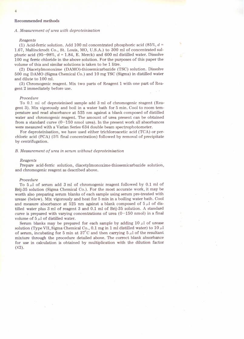
Fig. 1. Effect of sulphuric acid and phosphoric acid concentrations on colour formation. 0.1 ml of ureasolution (1 mmol/l) was boiled for 5 min with 1 ml DAMO-TSC solution (5 g DAMO and 50 mg TSC perlitre) and 2 ml acid solution containing varying concentrations (ml/l) of H2S04. (i) in absence of H3P04(*---*); and in presence of (H) 100 ml H3P04/1 (e---e); (Hi) 300 ml H3P04/1 (0---0); 500 mlH3P04/1 (.---.); and (iv) 700 ml H3P04/1 acid solution (*---*).
yields than phosphoric aci~, but chose the latter for simplicity in p'reparing thereagents. Fig. 1 shows that mixtures of these two acids give better colour yieldthan either alone, with an optimum in the region of 100 ml and 300 ml perlitre respectively of concentrated phosphoric and sulphuric acids.
Concentration of ferric, manganous and chloride ionsCatalytic acceleration by ferric salts was reported by Ceriotti and Spandrio
[4] and there is a report [6] of sensitisation by Cl- and colour stabilisation byMn2+.We found maximum colour development with 100 mg FeC13 per litre ofacid solution, slightly higher absorbances with the chloride than the sulphate,and some depression of absorbance above the optimum. Manganous chloridedepressed colour production at all concentrations tested.
Concentration of thiosemicarbazideThiosemicarbazide is essential for stability of colour. We experimented with
increasing concentrations and found a fairly sharp optimum at 100 J.1g per assaytube. Above this there was an increase in blank absorbance.
Concentration of diacetylmonoximeA sharp increase in absorbance was found up to 5 mg per assay tube. There
after there was no further change up to 10 mg per assay tube.
Heating cycleUsing the recommended reagents and procedure, absorbance reached its
maximum within 5 min in a boiling water bath and thereafter decreased slightlywith time. The water bath was covered to minimise evaporation and to excludelight.
Deproteinising agentsSolutions of urea were prepared in trichloroacetic acid (0-450 gjl) and per
chloric acid (0-540 gjl). O.l-mllots were then tested by the recommended procedure. There was no detectable variation in absorbance.
7
100
L:Io
ou
50
oo 2 4
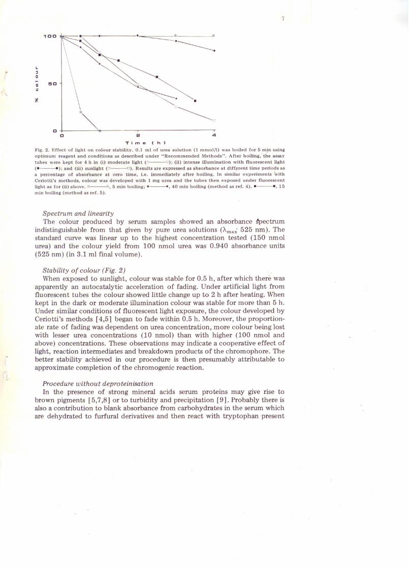
Time Ch)Fig. 2. Effect of light on colour stability. 0.1 ml of urea solution (1 mmolfl) was boiled for 5 m~n usingoptimum reagent and conditions as described under "Recommended Methods". After boiling,· the assaytubes were kept for 4 h in (i) moderate light (0---0); (ii) intense illumination with fluorescent light(e__ ); and (iii) sunlight (0---0). Results are expressed as absorbance at diff~rent time periods asa percentage of absorbance a~ zero time, i.e. immediately after boiling. In similar experiments withCeriotti's methods, colour was developed with 1 mg urea and the tubes then exposed under fluorescentlight as for (ii) above. <4---<4, 5 min boiling; *---*,40 min boiling (method as ref. 4) .• ---., 15min boiling (method as ref. 5).
Spectrum and linearityThe colour produced by serum samples showed an absorbance ~ectrum
indistinguishable from that given by pure urea solutions (A.ma~ 525 nm). Thestandard curve was linear up to the highest concentration tested (150 nmolurea) and the colour yield from 100 nmol urea was 0.940 absorbance units(525 nm) (in 3.1 ml final volume).
Stability of colour (Fig. 2) . ,When exposed to sunlight, colour was stable for 0.5 h, after which there was
apparently an autocatalytic acceleration of fading. Under artificial light fromfluorescent tubes the colour showed little change up to 2 h after heating. Whenkept in the dark or moderate illumination colour was stable for more than 5 h.Under similar condItions of fluorescent light exposure, the colour developed byCeriotti's methods [4,5] began to fade within 0.5 h. Moreover, the proportionate rate of fading was dependent on urea concentration, more colour being lostwith lesser urea concentrations (10 nmol) than with higher (100 nmol andabove) concentrations. These observations may indicate a cooperative effect oflight, reaction intermediates and breakdown products of the chromophore. Thebetter stability achieved in our procedure is then presumably attributable toapproximate completion of the chromogenic reaction.
Procedure without deproteinisationIn the presence of strong mineral acids serum proteins may give rise to
brown pigments [5,7,8] or to turbidity and precipitation [9]. Probably there isalso a contribution to blank absorbance from carbohydrates in the serum whichare dehydrated to furfural derivatives and then react with tryptophan present
8
in the serum proteins (Hopkins-Cole reaction) [10]. If a procedure is to be carried through without deproteinisation, these interferences must be minimisedand the approaches used in the past for determination of carbamido compounds like urea or citrulline have been to decrease sulphuric acid concentration (but with loss of sensitivity [5,8,9,11], and to use a smaller sample [5,8].We found non-ionic detergents very effective. They were without influence onthe colour reaction and perhaps act by solubilisation of acid-denatured protein.
Thirty normal serum samples were treated with urease, diluted with distilledwater and carried through procedure A (but omitting deproteinisation), givingan absorbance of 0.024 ± 0.003 (mean ± S.D.) for 5 ,ulserum. The same diluted,urea-free samples gave absorbances of 0.017 ± 0.010 (460 nm) by Ceriotti'sprocedure [5]. The same samples carried through procedure B gave 0.010 ±0.002 with 10 g Lubrox-WX (Sigma)/l substituted for the Brij solution, and0.004 ± 0.003 with the Brij solution specified. This is equivalent to 0.08 ± 0.06mmol ureall serum and can either be ignored, or corrected for by subtractinguniformly 0.08 mmolll from the result, or corrected for by applying individualserum blanks. For 25 serum samples with assay results in the normal range, thegreatest proportionate error introduced by ignoring the correction was 3.4%.
5,ul was chosen as the sample size purely as a matter of convenience and inview of the difficulty of accurate measurement of smaller volumes. The assay iscapable of measuring the urea present in 1-2 ,ul of normal serum, when theblank becomes quite negligible. The uniform correction suggested above is validfor a 5-,ulsample.
Reproducibility, etc.Procedure B gave a coefficient of variation of 3.02%, calculated from 15
determinations on a single sample divided between two batches. A comparisonwith Ceriotti's non-deproteinisation procedure [5] showed good agreement andconfirmed the much diminished serum blank correction required in the presentmethod.
InterferencesSeveral other compounds produce colour complexes with diacetyl moriox
ime but are of little importance when considering serum because present innegligible amount or yielding little colour (allantoin) [12]. The citrulline content of serum is usually too low to interfere, but the raised levels of arginosuccinate synthetase deficiency (citrullinaemia - 14 cases known up to 1978[13]) would give a significant non-urea response.
AdvantagesThe method is sensitive, simple and quick - especially simple when using the
method without deproteinisation and even then is amply accurate and reproducible for routine work. It should be easily adapted for automation. Further, thecolour produced is more stable to light than previously reported and the reagents are simple to prepare and stable on storage.
Acknowledgement
M.R. thanks the Hong Kong Government for the award of a CommonwealthScholarship.

9
References
1 Fearon. W.R. (1939) Biochem. J. 33,902-9072 Coulombe, J.J. and Favreau, L. (1963) Clin. Chem. 9, 102-1083 Ceriotti, G. and Spandrio, L. (1963) Clin. Chim. Acta 8, 295-2994 Ceriotti, G. and Spandrio, L. (1965) Clin. Chim. Acta 11, 519-5225 Ceriotti, G. (1971) Clin. Chem. 17,400-4026 Beale, R.N. and Croft, D. (1961) J. Clin. Pathol. 14,418-4247 Siest, G. and Vigneron, C. (1968) Clin. Chim. Acta 20, 373-3798 Ceriotti, G. (1973) Clin. Chim. Acta 47, 97-1059 Vassef, A.A. (1978) Clin. Chem. 24, 101-107
10 Holmes, E.J. (1968) J. Histochem. Cytochem. 16, 136-14611 Ohsita, M., Takeda, H., Kamiyama, Y., Ozawa, K. and Honjo, I. (1969) Clin. Chim. Acta 67, 145-15212 Mclean, P., Novello, F. and Gurney, N.W. (1965) Biochem. J. 94, 422-42613 Shih, V.E. (1978) in The Metabolic Basis of Inherited Disease, 4th Ed. (Stanbury, J.B., Wyngaarden,
J.B. and Fredrickson, S., Eds.), pp. 371-386, McGraw-Hill, New York