improving aquaculture: the impact of bacterial disease
TRANSCRIPT

Improving Aquaculture: the Impact of Bacterial Disease Treatments
on Salmonid Immune Performance
by
Shawna Leigh-Ann Semple
A thesis
presented to the University of Waterloo
in fulfilment of the
thesis requirement for the degree of
Doctor of Philosophy
in
Biology
Waterloo, Ontario, Canada, 2019
© Shawna Leigh-Ann Semple 2019

ii
Examining Committee Membership
The following served on the Examining Committee for this thesis. The decision of the Examining
Committee is by majority vote.
External Examiner MIKE CRISCITIELLO
Professor, Department of Veterinary Pathobiology
Texas A&M University
Supervisor(s) BRIAN DIXON
Professor, Canada Research Chair in Fish and
Environmental Immunology, Department of
Biology, University of Waterloo
DANIEL D. HEATH
Professor, Great Lakes Institute for Environmental
Research, Department of Biology,
University of Windsor
Internal Members CHRISTINE DUPONT
Continuing Lecturer, Biology Teaching Fellow,
Undergraduate Advisor, Department of Biology,
University of Waterloo
JOHN S. LUMSDEN
Professor, Department of Pathobiology
University of Guelph
Internal-external Member MARC AUCOIN
Academic Director WatPD – Engineering and
Associate Professor, Department of Chemical
Engineering, University of Waterloo

iii
Author’s Declaration
This thesis consists of material all of which I authored or co-authored: see Statement of Contributions
included in the thesis. This is a true copy of the thesis, including any required final revisions, as accepted
by my examiners.
I understand that my thesis may be made electronically available to the public.

iv
Statement of Contributions
In the case of published chapters, the numbering system of any published chapters has been updated to
fit this thesis.
Chapter 2 – L. Al-Hussinee performed the initial infection trials to determine resistant/susceptible
rainbow trout families. C.J. Kellendonk genotyped the MH class II β1 genes in six of the forty rainbow
trout families and analyzed these results. All other experiments, analyses, and the final manuscript
preparation was completed by S.L. Semple. This chapter was published in 2018 in Aquaculture, volume
483, pp. 131-140.
Chapter 3 – No additional contributors. This chapter was published online in 2019 in Microbial
Pathogenesis, doi: 10.1016/j.micpath.2019.103910.
Chapter 4 – T. Rodríguez-Ramos determined the minimum inhibitory concentration (MIC) of PACAP
for F. psychrophilum and helped with determining bacterial survival throughout live infection of
RTS11. All other experiments, analyses, and the final manuscript preparation was completed by S.L.
Semple. This chapter was published in 2019 in Frontiers in Immunology, volume 10, pp. 1-14.
Chapter 5 – G. Heath optimized and completed the subtractive indirect ELISA assay to determine
relative serum IgM levels against Vibrio anguillarum between the Chinook salmon crosses. C. Filice
helped to complete approximately 40% of the MH class II β1 genotyping. All other experiments,
analyses and the final manuscript preparation was completed by S.L. Semple. This chapter was
formatted for submission to Nature Heredity.
Chapter 6 – No additional contributors. This chapter is formatted for submission to Fish and Shellfish
Immunology.
Permission to use copyrighted material has been obtained from the publishers (Elsevier and
Frontiers in Immunology) and all joint authors.

v
Abstract
The farming of aquatic organisms, or aquaculture, is a global multibillion-dollar industry that
is continuously threatened by infectious diseases, including those of bacterial origin. Currently, the only
method to combat bacterial disease outbreaks as they occur is the use of antibiotics. Though effective,
this method increases both the prevalence and risk of generating antibiotic resistant bacteria. Any
prophylactic treatments available, such as vaccines, provide slightly increased protection but are in
need of improvements. To facilitate vaccine design and/or alternative treatment efforts, a deeper
understanding of the teleost immune system is essential. When considering finfish aquaculture
production in Canada, the industry is currently dominated by salmonids, which includes freshwater
rainbow trout (Oncorhynchus mykiss) and saltwater Chinook salmon (Oncorhynchus tshawytcha). The
contents of this thesis examine bacterial diseases that are relevant to the intensive culture of these two
salmonid species. More specifically, both in vitro and in vivo analyses were used to explore the immune
performance of salmonids when assessing breeding strategies, bacterial pathogenesis and alternative
treatment options.
The freshwater rainbow trout model was used to study infection with Flavobacterium
psychrophilum, the causative agent of bacterial coldwater disease (BCWD). Initially, forty full-sibling
families were created and assessed for resistance or susceptibility to F. psychrophilum following i.p.
injection with the pathogen. Based on survival, the immune performance of the highest and lowest
performing crosses was evaluated in regard to serum IgM production, MH class II β1 genotype and
respiratory burst activity (RBA) in replicated infection trials. Though there were no significant
differences observed between the high and low performers for any of the immune parameters analyzed,
RBA of head kidney leukocytes was observed to significantly decrease at the time when fish were
presenting clinical signs of disease. This observation led to the use of the monocyte/macrophage-like
cell line, RTS11, as a model system to learn more about the pathogenesis of the organism, an area of
limited understanding. Exposing RTS11 to live, heat-killed and the conditioned media of F.
psychrophilum (FpCM) revealed that the FpCM significantly reduced the phagocytic activity of

vi
RTS11. Furthermore, FpCM alone was able to stimulate cytokine transcript expression in a manner
that was similar to live bacteria but was short-lived comparatively. Meanwhile, heat-killed F.
psychrophilum resulted in little to no changes of RTS11, indicating that the common use of bacterins
for fish vaccines may not be optimal for all bacterial pathogens. Lastly, the efficacy of an antimicrobial
peptide, PACAP as an alternative treatment option for BCWD was assessed using the RTS11 infection
model system. PACAP was found to permeabilize the membrane of F. psychrophilum and, when
RTS11 received 24 hr pre-treatment with the peptide, could stimulate the immune cells to decrease the
number of viable bacteria in culture. Additionally, PACAP was able to significantly stimulate transcript
expression of pro-inflammatory cytokines both in the presence or absence of F. psychophilum.
Vibrio anguillarum, the causative agent of the hemorrhagic septicaemia known as vibriosis,
was used to study the immune function of the Chinook salmon saltwater model. In this system,
outbreeding was used to determine if the immune function of an inbred aquaculture stock (YIAL) could
be enhanced via hybrid vigor. The inbred stock was crossed with seven wild populations and, following
i.p. challenge with live V. anguillarum, various immune parameters were compared between high and
low performing crosses. More specifically, serum IgM production, MH class II β1 genotype, immune
transcript expression of spleen tissue and co-infection with Renibacterium salmoninarum was analyzed.
This revealed that the higher performing crosses had a greater percentage of individuals that were
heterozygous for MH class II β1 genotypes and also presented significantly reduced inflammatory
transcript expression when compared to the lower performing groups. Also, more individuals were co-
infected with R. salmoninarum in the low performing crosses providing further evidence that crosses
using less energetic resources had better outcomes when challenged with bacterial infection. To explore
V. anguillarum infection at the cellular level, a stromal cell line was created from Chinook salmon
spleen, CHST. Following the development and characterization of CHST, the cells were exposed to
heat-killed V. anguillarum and were observed to respond with both pro- and anti- inflammatory
cytokine production.

vii
The findings presented in this thesis were derived from both in vivo and in vitro approaches to
understand the salmonid immune response to bacterial pathogens. Thus, immune function was analyzed
at both the whole animal level, using large-scale infection trials, as well as at the cellular level, using
relevant cell lines. The resulting data was able to provide novel and functional information regarding
both the immune function of salmonids and the pathologic cycle of two relevant bacterial pathogens.
The knowledge presented within this thesis could aid in making some much needed improvements to
vaccine design to combat both F. psychrophilum and V. anguillarum infections. Additionally, a greater
understanding of the teleost immune system in response to bacterial infection was obtained and could
be used to enhance current and future treatment options for aquaculture. This will lead to less bacterial
disease outbreaks in aquaculture settings, translating into greater profits and more job opportunities
within this growing industry. Moreover, a steady and increasing supply of fish protein from responsible
aquaculture will reduce pressures on wild stocks, some of which are extensively exploited and in danger
of collapse.

viii
Acknowledgements
First and foremost, I would like to thank my supervisor Brian Dixon. I came to you with nothing
but an interest in immunology and a fascination with infectious disease. Despite my inexperience, you
believed in my abilities and gave me numerous responsibilities/projects that helped me thrive. Thank
you for always taking the time to give me excellent advice and suggestions. Very importantly, thank
you for always supporting my crazy ideas and giving me the freedom to develop my own research
questions and experiments so that I could hone these valuable skills. Your love of immunology is
contagious and I think that we both share a formidable scientific curiosity (which may have had
something to do with my continually growing list of side projects). Sometimes you just HAVE to know
the answer, right? Aside from all of the awesome science that was accomplished, you provided me with
many amazing opportunities to travel, collaborate, volunteer and network (I had never even been on a
plane before!). You made it so that I not only was able to do what I absolutely love, but also ensured
that my PhD experience was a very memorable and positive one.
After about one year of doing my MSc, I decided to transfer to a PhD and gained the wonderful
addition of Daniel Heath as my co-supervisor. It was invaluable to have another perspective for my
immunology/disease research, even though some population geneticists can seem a little intimidating
at first! You enabled several collaborations for me and also believed in my abilities even when I had
zero experience with some of your scary sequencing technologies. Thank you for all of your advice
throughout this endeavor, and also for giving me the opportunity to gain many new skills (including
becoming a supreme shitake mushroom inoculator).
It is quite possible that I never would have found Brian’s lab in the first place without the
influence of Christine Dupont. The courses that I took with her as an undergraduate student, along with
her passion for microbiology and infectious disease, were what initiated my interest in these subject
areas and immunology. You helped me progress from a shy/awkward student who was afraid of
professors, to someone who can now feel confident even in the face of those she admires. Thank you
for taking the time to have long discussions with me about infectious disease, career paths, teaching
and many other aspects of life.
When I began my graduate degree, I was probably the most excited about doing live bacterial
disease trials, even though I had NO idea how. Everything that I now know about fish pathology and
the execution of infection trials was learned through John Lumsden. As this has been a massive part of
my thesis and a primary research interest of mine, this meant that initially he had to deal with my
constant questions and lack of experience (all while being super busy as Chair of Pathobiology). Your
patience and reassurance were always very much appreciated and were also critical for me to gain more

ix
confidence. Thank you for welcoming me into your lab, for your blunt honesty and for all of your
valuable recommendations regarding my data, experimental design and future career goals.
As a whole, my committee was absolutely phenomenal. All four of you represent different
aspects of the scientist that I aspire to be. Each of you gave me individual opportunities so that I could
develop and improve as a researcher. It was uplifting to have so much support from all of you, even
when I wasn’t so sure of myself. I was incredibly fortunate to have so many amazing mentors to look
up to and work closely with. Apart from being great scientists, you are exemplary human beings and I
admire all of you.
It was an absolute pleasure to work with John and Ann Heath at YIAL. Thank you for all of
the amazing discussions/debates, your hospitality and most of all, for allowing me to use a live virulent
strain of bacteria on your farm. You were both very supportive and pushed me further to really think
about my experimental designs (and whether it was even possible to do all of the things that I had in
mind). It meant a lot that you trusted a crazy little PhD student to do so many large-scale infection
trials. I look forward to one day building the Path. Shack as promised so that we can start having some
truly infectious fun! A huge thank you also goes to the manager of YIAL, Elliot Haugen, for all of his
hard work and advice when it came to fish care. Thank you for collecting PIT tags when I could not be
at YIAL year round, and also for helping me transport a ridiculous number of fish from the sea pens. I
hope that one day I will be able to have as much sarcasm and sass that seems to flow effortlessly from
you.
Thank you to all of the members of the Dixon lab past and present for all of your valuable
advice and discussions. A huge thank you to Nguyen (Nathan) Vo for taking me on as a surprise BIOL
499 student and teaching me everything that I know about cell culture as well as giving me some
amazing guidance along the way. I also want to give a very special thank you to mi hermana cubana,
my kindred science spirit and good friend, Tanita Rodríguez-Ramos. I have learned SO much from
you. I can’t think of anyone else that could make those crazy long days not seem so bad while
continually exchanging random ideas for future experiments. You were always there to provide
amazing advice and some much needed laughter, especially when I was overthinking things. Thank
you for working with me and thank you for being a wonderful friend.
I have also been very fortunate to have the opportunity to collaborate with a number of
wonderful scientists throughout my degree. Dr. Yamila Carpio and Dr. Mario Estrada (CIGB); Dr.
Dennis Higgs, Dr. Mallory Wiper, Shelby Toews and Clare Venney (UWindsor); Dr. Mike Power and
Dr. Ingeborg Mulder (UW); Dr. Stephanie DeWitte-Orr and Dr. Sarah Poynter (WLU); Dr. Niels Bols,
Dr. Nathan Vo and Dr. John Pham (UW); Dr. Janet MacInnes and Dr. Lowia Al-Hussinee (UGuelph).

x
All of these experiences have helped me gain new and valuable skills so that I can become a better
scientist.
There have also been many friends that have helped me along the way. I could always depend
on Amy, Ashley and Brenda to make time for chats over bubble tea or sushi. Thanks to all of my crazy
pseudo-brothers (Kevin, John, Dave and Brandon) and their mob of friends that would somehow
convince me to chill a little and ride the ROFLcopter or just do some squats in case that ridiculous
steamroller showed up again. I also want to extend a very special thank you to my close friend Kayla
Robinson. I don’t even know if you realize how much you have helped me. During a time when I was
having difficulties, you befriended me and made me realize that it was okay to be different (and a little
compulsive). Our random road trips, shopping sprees, anime binges, baking fests and sporadic
conventions helped me to keep my sanity while having some much needed fun throughout my degrees.
I also want to give a massive acknowledgment to my partner, George. Your constant support
and encouragement were essential throughout this undertaking. Your laid back and calm demeanor
(unless we talk politics of course) perfectly complimented the ball of stress/neuroses that is me. You
helped me keep things in perspective so that I would stay positive (or at least try to) even when I was
at my most dramatic. You helped me to believe in myself. Thank you for listening to my problems,
celebrating my victories and sharing many wonderful experiences with me. You, and everything you
do, is very much appreciated.
Last but not least, I give my deepest gratitude to my family for their continual love and support.
To my mom, thank you for breaking the cycle so that I could be the first to demonstrate that the fate of
our family is not absolute. Through years of watching you support us on your own, you taught me the
importance of hard work because sometimes that is all that you have to prove yourself. Thank you to
my godfather, Doug Castles, who showed me that blood is only thicker than water in a chemical sense
and is certainly not required to be a father. You always made me feel valuable and reminded me that it
was important to relax and have fun despite my ambitious aspirations. I appreciate all that the two of
you did for me while I was growing up including the constant encouragement and acceptance that I
received for being my nerdy, weird self.

xi
Dedication
I dedicate this thesis to my father, James Elliot, and to my aunt, Sherry Semple.
Though our time together was cut short, you both played a massive role in shaping the person that I
am today.

xii
Table of Contents
Examining Committee Membership....................................................................................................... ii
Author’s Declaration ............................................................................................................................. iii
Statement of Contributions .................................................................................................................... iv
Abstract .................................................................................................................................................. v
Acknowledgements ............................................................................................................................. viii
Dedication ............................................................................................................................................. xi
Table of Contents ................................................................................................................................. xii
List of Figures ..................................................................................................................................... xvi
List of Tables ..................................................................................................................................... xviii
List of Abbreviations ........................................................................................................................... xix
Chapter 1: General Introduction .......................................................................................... 1
1.1 Canadian Aquaculture ................................................................................................................... 1
1.1.1 Freshwater Culture - Trout ................................................................................................... 2
1.1.2 Saltwater Culture - Salmon .................................................................................................. 3
1.2 Sources of economic loss in aquaculture ...................................................................................... 3
1.3 Bacterial pathogens of salmonid aquaculture .............................................................................. 4
1.4 Methods to combat bacterial infection ......................................................................................... 5
1.5 Teleost immune system .................................................................................................................. 6
1.6 Innate immunity of fish .................................................................................................................. 7
1.6.1 Cells of innate immunity ...................................................................................................... 7
1.6.2 Antimicrobial peptides ......................................................................................................... 9
1.6.3 Respiratory burst activity ................................................................................................... 10
1.7 Adaptive immunity of fish ........................................................................................................... 11
1.7.1 Cells of adaptive immunity ................................................................................................ 12
1.7.2 Major histocompatibility (MH) genes ................................................................................ 13
1.7.3 Antibody development ....................................................................................................... 15
1.8 Cytokines ....................................................................................................................................... 16
1.8.1 Pro-inflammatory ............................................................................................................... 16
1.8.2 Anti-inflammatory .............................................................................................................. 17
1.9 Thesis Objectives .......................................................................................................................... 18
Chapter 2: Serum IgM, MH class IIβ genotype and respiratory burst activity do not
differ between rainbow trout families displaying resistance or susceptibility to the
coldwater pathogen, Flavobacterium psychrophilum. ........................................................ 20
2.1 Overview........................................................................................................................................ 21
2.2 Introduction .................................................................................................................................. 22
2.3 Materials and Methods ................................................................................................................ 25
Fish ..................................................................................................................................... 25
Culture and quantification of F. psychrophilum strain 101 ................................................ 25
Determining susceptibility/resistance to F. psychrophilum (Preliminary Trials) ............... 26
MH class II β1 genotyping ................................................................................................. 26
2.3.4.1 DNA extraction ........................................................................................................... 26
2.3.4.2 Amplification and sequencing of MH class II β1 genotypes ...................................... 27

xiii
Induction of F. psychrophilum antibody responses ............................................................ 28
2.3.5.1 Infection ...................................................................................................................... 28
2.3.5.2 Isolating antigens from F. psychrophilum .................................................................. 28
2.3.5.3 Indirect ELISA assay to detect levels of F. psychrophilum antibodies in serum ....... 29
Measuring Respiratory Burst Activity (RBA) of blood and head kidney leukocytes ........ 30
2.3.6.1 Infection ...................................................................................................................... 30
2.3.6.2 Isolation of head kidney leukocytes............................................................................ 31
2.3.6.3 Real-time luminol-enhanced chemiluminescence assay for RBA .............................. 32
2.4 Results ........................................................................................................................................... 33
2.4.1 Preliminary trials to determine rainbow trout families resistant/susceptible to F.
psychrophilum .................................................................................................................... 33
2.4.2 Relating MH class IIβ genotype to disease susceptibility .................................................. 35
2.4.3 IgM antibody development in rainbow trout families resistant/susceptible to F.
psychrophilum .................................................................................................................... 37
2.4.4 The effect of head kidney and total blood RBA on resistance to F. psychrophilum .......... 39
2.5 Discussion ...................................................................................................................................... 41
2.6 Conclusions ................................................................................................................................... 45
Chapter 3: Understanding the pathogenesis of Flavobacterium psychrophilum using the
rainbow trout monocyte/macrophage-like cell line, RTS11, as an infection model. ...... 47
3.1 Overview........................................................................................................................................ 48
3.2 Introduction .................................................................................................................................. 49
3.3 Materials and Methods ................................................................................................................. 51
3.3.1 Maintenance of RTS11 ....................................................................................................... 51
3.3.2 F. psychrophilum growth in different media ...................................................................... 51
3.3.3 Bacterial growth for RTS11 exposure conditions .............................................................. 52
3.3.3.1 Live and heat-killed F. psychrophilum ....................................................................... 52
3.3.3.2 F. psychrophilum conditioned media (FpCM) ........................................................... 52
3.3.4 Exposure of RTS11 to live, heat-killed and the supernatant of F. psychrophilum ............. 53
3.3.5 Viability of RTS11 throughout exposure ........................................................................... 53
3.3.6 Measuring phagocytic function of RTS11 via flow cytometry .......................................... 54
3.3.7 qRT-PCR ............................................................................................................................ 54
3.3.7.1 RNA extraction and cDNA synthesis ......................................................................... 54
3.3.7.2 qRT-PCR Reactions.................................................................................................... 55
3.3.8 Statistical analyses ............................................................................................................... 56
3.4 Results ........................................................................................................................................... 56
3.4.1 Growth of F. psychrophilum in preferred and cell culture media ...................................... 56
3.4.2 Appearance of RTS11 when exposed to live, heat-killed and the supernatant of F.
psychrophilum .................................................................................................................... 57
3.4.3 Viability of RTS11 throughout bacterial exposure conditions ........................................... 59
3.4.4 Impact of live, killed and F. psychrophilum supernatant on phagocytic function ............. 60
3.4.5 Cytokine transcript expression throughout exposure conditions ........................................ 62
3.5 Discussion ...................................................................................................................................... 63
3.6 Conclusions ................................................................................................................................... 68
Chapter 4: PACAP is lethal to Flavobacterium psychrophilum through direct membrane
permeabilization and indirectly, by priming the immune response in RTS11. .............. 70
4.1 Overview........................................................................................................................................ 71
xiv
4.2 Introduction .................................................................................................................................. 72
4.3 Materials and Methods ................................................................................................................. 75
4.3.1 Maintenance of RTS11 ....................................................................................................... 75
4.3.2 Peptides .............................................................................................................................. 75
4.3.2.1 Synthetic PACAP from the teleost Clarias gariepinus .............................................. 75
4.3.2.2 Synthetic HSP70 peptide fragment from rainbow trout ............................................. 75
4.3.3 Growth of F. psychrophilum .............................................................................................. 75
4.3.4 Minimum inhibitory concentration of C. gariepinus PACAP-38 on F. psychrophilum .... 75
4.3.5 RTS11 Exposure Trials ...................................................................................................... 76
4.3.5.1 Exposure to PACAP ................................................................................................... 76
4.3.5.2 Simultaneous exposure to both PACAP and live F. psychrophilum .......................... 77
4.3.5.3 Pre-treatment with PACAP followed by infection with live F. psychrophilum ......... 77
4.3.6 Survival of RTS11 following PACAP exposure ................................................................ 77
4.3.7 Presence of live F. psychrophilum in RTS11 cell cultures following PACAP exposure ... 77
4.3.8 Permeabilization assay ....................................................................................................... 78
4.3.9 qRT-PCR ............................................................................................................................ 78
4.3.9.1 RNA extraction and cDNA synthesis ......................................................................... 78
4.3.9.2 qRT-PCR reactions ..................................................................................................... 79
4.3.10 Statistical analyses ............................................................................................................. 80
4.4 Results ........................................................................................................................................... 81
4.4.1 Minimum inhibitory concentration (MIC) ......................................................................... 81
4.4.2 Impact of PACAP concentrations on RTS11 survival ....................................................... 82
4.4.3 Effect of PACAP on F. psychrophilum growth throughout RTS11 infections .................. 83
4.4.4 Permeabilization of F. psychrophilum by PACAP ............................................................. 85
4.4.5 Influence of C. gariepinus PACAP-38 on RTS11 immune gene expression ..................... 86
4.4.5.1 Exposure to PACAP ................................................................................................... 86
4.4.5.2 Exposure to PACAP 24 hours before F. psychrophilum infection ............................. 88
4.5 Discussion ...................................................................................................................................... 90
4.6 Conclusions ................................................................................................................................... 96
Chapter 5: Revealing immune differences between eight outbred Chinook salmon
populations when challenged with live Vibrio anguillarum. ............................................. 97
5.1 Overview........................................................................................................................................ 98
5.2 Introduction .................................................................................................................................. 99
5.3 Materials and Methods .............................................................................................................. 102
5.3.1 Breeding design ................................................................................................................ 102
5.3.2 Rearing ............................................................................................................................. 103
5.3.3 Culture and quantification of V. anguillarum ................................................................... 103
5.3.4 Infection with live V. anguillarum and sample collection................................................ 104
5.3.5 Microsatellite analysis ...................................................................................................... 104
5.3.6 MH class II β1 genotyping ............................................................................................... 106
5.3.7 Measuring the serum antibody response to V. anguillarum ............................................. 107
5.3.7.1 Isolating antigens from V. anguillarum .................................................................... 107
5.3.7.2 Indirect subtractive ELISA to measure serum IgM between the crosses ................. 107
5.3.8 qRT-PCR of spleen transcripts throughout V. anguillarum infection .............................. 108
5.3.8.1 RNA extraction and cDNA synthesis ....................................................................... 108
5.3.8.2 Validatoin of qRT-PCR primers for Chinook salmon .............................................. 108
5.3.8.3 qRT-PCR reactions ................................................................................................... 109
xv
5.3.9 Measuring co-infection with bacterial kidney disease (R. salmoninarum) ...................... 110
5.3.10 Statistical analyses ........................................................................................................... 110
5.4 Results ......................................................................................................................................... 112
5.4.1 Survival of Chinook salmon crosses when challenged with live V. anguillarum or sham
injected ............................................................................................................................. 112
5.4.2 Comparing the heterozygosity between the eight populations ......................................... 114
5.4.3 MH class II β1 genotypes of the outbred Chinook salmon populations ........................... 114
5.4.4 IgM antibody development to V. anguillarum throughout infection ................................ 116
5.4.5 Induction of immune transcripts within spleen during V. anguillarum infection ............. 117
5.4.6 Co-infection with R. salmoninarum in highest and lowest performing crosses ............... 119
5.5 Discussion .................................................................................................................................... 120
5.6 Conclusions ................................................................................................................................. 125
Chapter 6: Development and characterization of a Chinook salmon spleen stromal cell
line to study the cellular immune response to Vibrio anguillarum. ................................ 127
6.1 Overview...................................................................................................................................... 128
6.2 Introduction ................................................................................................................................ 129
6.3 Materials and Methods ............................................................................................................... 131
6.3.1 Primary cultures ............................................................................................................... 131
6.3.2 Maintenance of CHST ...................................................................................................... 131
6.3.3 Cryopreservation of CHST ............................................................................................... 132
6.3.4 Optimal growth conditions of CHST ............................................................................... 132
6.3.5 Confirming species of origin ............................................................................................ 133
6.3.6 V. anguillarum growth in bacterial and animal cell media............................................... 133
6.3.7 Preparation of heat-killed V. anguillarum ........................................................................ 134
6.3.8 Exposure of CHST to heat-killed V. anguillarum ............................................................ 134
6.3.9 qRT-PCR .......................................................................................................................... 135
6.3.9.1 RNA extraction and cDNA synthesis ....................................................................... 135
6.3.9.2 qRT-PCR reactions ................................................................................................... 135
6.3.10 Statistical analyses ........................................................................................................... 136
6.4 Results ......................................................................................................................................... 136
6.4.1 Development of the CHST cell line ................................................................................. 136
6.4.2 Confirming the species of origin of CHST ....................................................................... 138
6.4.3 Determining optimal growth conditions for CHST .......................................................... 138
6.4.4 Growth of V. anguillarum in animal cell culture media ................................................... 140
6.4.5 Cytokine expression throughout exposure to heat-killed V. anguillarum ........................ 141
6.5 Discussion .................................................................................................................................... 142
6.6 Conclusions ................................................................................................................................. 145
Chapter 7: General Discussion and Future Directions ................................................... 146
7.1 The importance of understanding bony fish immunology ...................................................... 146
7.2 Heritable differences in selectively bred fish ........................................................................... 148
7.3 Vaccines for bacterial pathogens .............................................................................................. 151
7.4 AMPs as adjuvants/immunostimulants for aquaculture ........................................................ 154
7.5 The value of fish cell lines for immune analyses ...................................................................... 156
7.6 Final conclusions and future directions .................................................................................... 157
References ............................................................................................................................ 161
Appendices ........................................................................................................................... 198

xvi
List of Figures
Chapter 1
Figure 1.1. Schematic depiction of NADPH oxidase activation to enable the respiratory burst
activity of phagocytes. ..................................................................................................... 11
Figure 1.2. Structure of the MH class II molecule. ............................................................................ 14
Chapter 2
Figure 2.1. Survival curves throughout F. psychrophilum infection for the forty rainbow trout
families analyzed for resistance/susceptibility to BCWD. ............................................... 34
Figure 2.2. Indirect ELISA assay to compare serum IgM development against F. psychrophilum
between resistant and susceptible families throughout infection. .................................... 38
Figure 2.3. Respiratory burst activity of HKL and total blood in resistant and susceptible families of
rainbow trout. ................................................................................................................... 40
Chapter 3
Figure 3.1. A comparison of F. psychrophilium growth in culture media for bacterial cells
(cytophaga broth) and for animal cells (L15 with 2 % FBS). .......................................... 56
Figure 3.2. Influence of live, heat-killed and F. psychrophilum conditioned media (FpCM) on the
morphology of RTS11. .................................................................................................... 58
Figure 3.3. Survival of RTS11 when exposed to live, killed and the supernatant of F. psychrophilum
(FpCM). ........................................................................................................................... 59
Figure 3.4. Assessment of RTS11 phagocytic function following exposure to live, heat-killed and
the supernatant of F. psychrophilum (FpCM). ................................................................ 61
Figure 3.5. Influence of live, killed and the supernatant of F. psychrohilum (FpCM) on RTS11
cytokine mRNA expression 1-3 Days following exposure. ............................................. 63
Chapter 4
Figure 4.1. The minimum inhibitory concentration (MIC) of C. gariepinus PACAP-38 required to
prevent the growth of F. psychrophilum alone. ............................................................... 81
Figure 4.2. Impact of C. gariepinus PACAP-38 on RTS11 viability. ............................................... 82
Figure 4.3. Quantification of F. psychrophilum by standard plate count (SPC) of cell culture media
during live infection (MOI of 0.7-1.3) of PACAP-treated RTS11. ................................. 84
Figure 4.4. Disruption of F. psychrophilum membrane by PACAP-38 of C. gariepinus. ................ 85
Figure 4.5. Impact of C. gariepinus PACAP-38 on RTS11 pro-inflammatory cytokines and PACAP
receptors mRNA expression 1-4 days following peptide exposure. ................................ 87
Figure 4.5. RTS11 transcript expression when challenged with live F. psychrophilum following 24
hr pre-treatment with PACAP concentrations (0.002 µM, 0.02 µM and 0.1 µM). .......... 89
xvii
Chapter 5
Figure 5.1. Geographical location of the British Columbia rivers from which milt was collected to
produce the seven outbred Chinook salmon populations............................................... 103
Figure 5.2. Survival curves for the eight outbred Chinook salmon populations during exposure
challenges....................................................................................................................... 113
Figure 5.3. Differences in MH class II β1 genotypes between the Chinook salmon crosses. ......... 115
Figure 5.4. Serum IgM developed towards V. anguillarum in Chinook salmon crosses throughout
the exposure trial. ........................................................................................................... 116
Figure 5.5. Spleen immune transcript expression within the two highest and two lowest performing
populations throughout live V. anguillarum challenge. ................................................. 118
Figure 5.6. Determining co-infection with R. salmoninarum in the highest and lowest performing
Chinook salmon crosses. ................................................................................................ 119
Chapter 6
Figure 6.1. Development of the Chinook salmon spleen stromal cell line, CHST. ......................... 137
Figure 6.2. Optimal culture conditions for growth of CHST. .......................................................... 139
Figure 6.3. A comparison of V. anguillarum growth in culture media for bacterial cells and for
animal cells (L15 supplemented with 0%, 2%, 10% or 20% FBS). .............................. 140
Figure 6.4. Influence of heat-killed V. anguillarum on pro- and anti-inflammatory cytokine
expression in CHST throughout 96 h of exposure. ........................................................ 142

xviii
List of Tables
Chapter 1
Table 1.1. Common bacterial pathogens in salmonid aquaculture. .................................................... 5
Chapter 2
Table 2.1. MH Class IIβ genotype frequencies among the six families of Oncorhynchus mykiss. .. 35
Table 2.2. MH class IIβ genotype prevalence in mortalities and survivors to F. psychrophilum
infection. .......................................................................................................................... 36
Chapter 3
Table 3.1. Primers used for qRT-PCR analysis of cytokine genes in RTS11 cells. ......................... 55
Chapter 4
Table 4.1. Primers used for qRT-PCR analysis of immune and receptor genes in RTS11. ............. 80
Chapter 5
Table 5.1. Primer sequences used for qRT-PCR analysis of immune genes in Chinook salmon. .. 110
Table 5.2. Level of heterozygosity calculated between the eight Chinook salmon populations based
on microsatellite analysis. .............................................................................................. 114
Table 5.3. Record of shared and unique MH class II β1 alleles between the eight Chinook salmon
crosses. ........................................................................................................................... 115
Chapter 6
Table 6.1. Primer sequences used to amplify Chinook salmon genes. ............................................ 136
Table 6.2. Validating the species identity of CHST through cox1 barcoding. ................................ 138
xix
List of Abbreviations
AID activation-induced (Cytidine) deaminase
AIP56 apoptosis inducing protein of 56 kDa
AMP antimicrobial peptide
aMT alpha-methyltestosterone
ANOVA analysis of variance
APC antigen presenting cell
BC British Columbia
BCR B cell receptor
BCWD bacterial coldwater disease
BGD bacterial gill disease
BKD bacterial kidney disease
BLAST basic local alignment search tool
BMPA broth microdilution peptide assay
BQ Big Qualicum River
CA cytophaga agar
Cap Capilano River
CB cytophaga broth
CD cluster of differentiation
cDNA complementary deoxyribonucleic acid
CFU colony forming units
Chil Chillawack River
CHSE-214 Chinook salmon embryonic cell line
CHSS Chinook salmon spleen cell line
CHST Chinook salmon spleen stromal cell line
Cox1 cytochrome c oxidase subunit
CSR class switch recombination
CV coldwater vibriosis
DC dendritic cell
DEPC diethyl pyrocarbonate
DMSO dimethyl sulfoxide
DNA deoxyribonucleic acid
EDTA ethylenediaminetetraacetic acid
EF1α elongation factor 1α
ELISA enzyme linked immunosorbent assay
EUS epizootic ulcerative syndrome
FACS fluorescence-activated cell sorter
FAO Food and Agriculture Organization of the United Nations
FBS fetal bovine serum
Fcell bacterial cell fluorescence intensity
FITC fluorescein isothiocyanate
Fp Flavobacterium psychrophilum
FpCM F. psychrophilum conditioned media
FPG101 F. psychrophilum strain 101
FPG25 F. psychrophilum strain 25
GC germinal centre
GCPR G-coupled protein receptors
HDP host defense peptides
HK head kidney
HKL head kidney leukocytes

xx
HRP horseradish peroxidase
HSP70 heat shock protein of 70 kDa
HWE Hardy-Weinberg equilibrium
Ig immunoglobulin
IHNV infectious hematopoietic necrosis virus
IL interleukin
i.p. intraperitoneal
IPNV infectious pancreatic necrosis virus
IPTG isopropyl-β-D-thiogalactoside
L15 Leibovitz’s media
L15/FBS Leibovitz’s media supplemented with fetal bovine serum
LB lysogeny broth
LLPC long-lived plasma cell
LPS lipopolysaccharide
LSD least significant difference
MAS motile Aeromonas septicemia
MH major histocompatibility
MHC major histocompatibility complex
MIC minimum inhibitory concentration
MMC melanomacrophage centre
MOI multiplicity of infection
NADPH nicotinamide adenine dinucleotide phosphate
Nit Nitinat River
NK natural killer cell
NOX NADPH oxidase
OD optical density
PAC1 PACAP type 1 receptor
PACAP pituitary adenylate cyclase-activating polypeptide
PAMP pathogen associated molecular pattern
PBS phosphate buffered saline
PCR polymerase chain reaction
pH power of hydrogen
PI propidium iodide
PIT passive integrated transponder
PMA phorbol 12-myristate 13-acetate
p-NPP p-nitrophenyl phosphate
Poly I:C polyinosinic: polycytidylic acid
PRR pattern recognition receptor
Punt Puntledge River
qRT-PCR quantitative reverse transcriptase PCR
Quin Quinsam River
RAG recombination-activating gene
RBA respiratory burst activity
RBC red blood cells
RC Robertson Creek
RLU relative light units
RNA ribonucleic acid
ROI reactive oxygen intermediates
RT room temperature
RTS11 rainbow trout monocyte/macrophage-like cell line
RTSF rainbow trout fry syndrome
xxi
SD standard deviation
SEM standard error of the mean
SNP single nucleotide polymorphism
SPC standard plate count
TBS tris-buffered saline
TBS-T TBS with Tween 80
TCR T cell receptor
TdT terminal deoxynucleotidyl transferase
TLR toll-like receptor
TNF tumour necrosis factor
TSA tryptic soy agar
TSB tryptic soy broth
TSS rainbow trout spleen stromal cell line
TYESB tryptone yeast extract salts broth
USD United States dollar
UV ultraviolet
Va Vibrio anguillarum
VIP vasoactive intestinal polypeptide
VPAC1 VIP receptor 1
VPAC2 VIP receptor 2
WBC white blood cell
WGD whole genome duplication
WHO World Health Organization
X-gal 5-bromo-4-chloro-3-indolyl-β-D-galactopyranoside
YIAL Yellow Island Aquaculture Ltd.
1
Chapter 1: General Introduction
Given that both fresh- and saltwater account for 72% of Earth’s surface area, it was only a
matter of time before aquatic environments became the new frontier for agriculture. Because the
majority of food animals are currently raised on land, it is unsurprising that insights or advancements
in aquatic animal husbandry have lagged behind that of terrestrial species. As the global population
increases and with limited availability of productive land, the necessity of utilizing aquatic habitats for
animal food production is clear. Additionally, due to their high polyunsaturated fatty acid content
(Tasbozan & Gokce, 2017), many aquatic species provide an alternative and heart healthy protein
source in an age when cardiovascular disease is the leading cause of death worldwide (WHO, 2018).
For these reasons and more, global interest for fish protein is high. So high in fact that fisheries cannot
meet the global demand while also adhering to the harvesting restrictions that are currently in place
(Kvamsdal et al., 2016). This places a burden on wild populations because effective enforcement of
these restrictions is logistically difficult.
The culture of aquatic species, or aquaculture, can provide an alternative to alleviate some of
the pressure on wild populations. For many aquatic species, this culture production is in its nascent
form, meaning that time will be required to understand and optimize these industrial practises. One
manifestation of the issues faced by aquaculture is the increased prevalence of infectious disease,
including bacterial pathogens. Bacteria are able to take advantage of novel, high density farm
environments and thrive. This results in many of these prevalent microorganisms becoming
opportunistic pathogens in aquaculture settings. Obtaining a deeper understanding of bacterial diseases
that impact aquaculture, as well as what constitutes an effective immune response in relevant hosts, is
invaluable for the improvement of this industry.
1.1 Canadian Aquaculture
As the second largest country in the world (Statistics Canada, 2011), Canada is rich in a variety
of natural resources including water. The extensive access of this country to both fresh- and saltwater
coastlines means that Canada has the potential to play a significant role in the future of productive

2
aquaculture enterprises. Aquaculture is the farming of aquatic organisms such as fish, molluscs,
crustaceans and aquatic plants (de Silva & Anderson, 1994). Though known to have been practised for
over 2000 years in China (Ling, 1977), the first record of planned aquaculture activity in Canada
occurred in 1857 when the Superintendent of Fisheries studied the incubation and hatching of brook
trout eggs (MacCrimmon et al., 1974). Today, industrial aquaculture operations can be found in all
Canadian provinces as well as the Yukon which encompass over 45 species of finfish, shellfish and
marine plants (Statistics Canada, 2017). This continually growing industry has contributed significantly
to the Canadian economy by providing over 25,000 stable employment opportunities (CAIA, 2017) as
well as stimulating economic growth in remote, rural and coastal communities. Currently, the value of
aquaculture production in Canada is $1.39 billion, with salmonid production accounting for an
impressive 80% (Statistics Canada, 2017). Of the aquaculture species being produced, salmonids are
the most valuable worldwide with Canada ranking fourth among global salmon producers (DFO, 2016).
Given that salmonids can be produced in both fresh- and saltwater environments, Canada has ample
opportunity to make its mark in this expanding industry.
1.1.1 Freshwater Culture - Trout
Canada possesses roughly 20% of global surface freshwater (Statistics Canada, 2011), which
enables the culture production of trout in every province. Rainbow trout (Oncorhynchus mykiss) is
currently the third most valuable aquatic species farmed in Canada (Statistics Canada, 2017), however
they were not always found across the country. This highly versatile species was originally native to
the Western drainages of North America but due to its popularity as a sporting fish, was introduced to
water bodies all across Canada and the rest of the world (Ward & Post, 2014). Today, rainbow trout is
the most common species of trout in North America and can be found on every continent with the
exception of Antarctica (Ward & Post, 2014). The now broad geographical distribution of the species
highlights the variety of environments and water temperatures (ranging from 0°C to 29°C) that rainbow
trout can survive in (Currie et al., 1998). Because of their significance in global aquaculture and

3
resilience in laboratory settings, rainbow trout have become the gold standard research model for
salmonid immunity.
1.1.2 Saltwater Culture - Salmon
The majority of Canadian aquaculture facilities can be found on the East and West coasts as
saltwater operations. This reflects the high demand and monetary value of marine aquatic species,
particularly Atlantic salmon (Salmo salar). Depending on the coast of Canada, native salmon species
will vary. On the East coast, Atlantic salmon are the only salmon species found and are also the
predominant and most valuable finfish exported from Canada (DFO, 2017). In comparison, on the West
coast there are five main species of Pacific salmon: Coho salmon (Oncorhynchus kisutch), pink salmon
(Oncorhynchus gorbuscha), chum salmon (Oncorhynchus keta), Chinook salmon (Oncorhynchus
tshawytcha) and sockeye salmon (Oncorhynchus nerka). Of these Pacific species, solely Coho and
Chinook salmon are actively being farmed in Canada and this only occurs on the West coast (BCSFA,
2003). However, British Columbia is responsible for 71% of Canadian salmon production (Statistics
Canada, 2017), which means that Atlantic salmon are primarily farmed on the Pacific coast rather than
their native coast. Though this seems counterintuitive, Atlantic salmon are able to be grown at much
higher densities and have greater feed conversion rates when compared to their Pacific counterparts
(DFO, 1991). Despite these commercial benefits, there is great public concern regarding Atlantic
salmon as an invasive species on the West coast and the novel diseases the native species may be
exposed to. As such, this has made the development of high-performing Pacific salmon stocks a priority
for salmon culture in British Columbia.
1.2 Sources of economic loss in aquaculture
The utilization of aquatic environments means that some of the difficulties confronted by fish
farmers are very different when compared to their terrestrial counterparts. Common sources of financial
losses include environmental/husbandry (algal blooms, temperature oscillations, hypoxia,
supersaturation, etc.), chemical (nitrogen fluctuations, pH variation, etc.), predation, escapees and
infectious disease (Anrooy et al., 2006). Many of these problems can result in devastating financial

4
losses, but few compare to the consistent annual losses derived from infectious disease. In 2014, of the
$70 billion dollars of aquaculture product that was destined for human consumption, 10% of this was
lost due to infectious disease (World Bank, 2014; FAO, 2016). Though there are many different types
of infectious agents that contribute to these significant financial losses, this thesis will focus on bacterial
diseases of salmonid culture.
1.3 Bacterial pathogens of salmonid aquaculture
Cultured salmonids are susceptible to many bacterial pathogens (Table 1.1). The stress induced
by conditions such as overcrowding, temperature fluctuations and excessive handling can result in
normally benign microorganisms becoming opportunistic pathogens (Meyer, 1991). The aquatic
environments in which these animals reside are known to support the growth of bacteria for long periods
of time. Though not immediately causing infection, these opportunistic bacterial pathogens can survive
independently of their hosts (Wedemeyer & Nelson, 1977; Hoff, 1989; Madetoja et al., 2003). When
animals are stressed these microorganisms are well situated to become major impediments for
aquaculture. The poikilothermic nature of fish means that these animals have no control over their body
temperature as it is simply a representation of their surrounding environment (Lawrence, 2008). As a
result, there are different opportunistic bacterial pathogens that have taken advantage of the variety of
temperature niches. For fish farmers, this has made management and prevention strategies difficult as
there is a large degree of variability in route of entry, virulence factors, disease presentations and
pathologic cycle between the various bacterial pathogens (reviewed by Ringo et al., 2007; Defoirdt,
2013; Bentson-Tilla et al., 2016). The significance of bacterial pathogens in salmonid aquaculture
combined with the extensive gaps in knowledge regarding their pathogenesis means that further
investigation of these microorganisms could prove invaluable.

5
Table 1.1. Common bacterial pathogens in salmonid aquaculture. A list of bacterial pathogens that
affect aquaculture production of salmonids, their gram reaction and the diseases that they cause. The
organisms that are outlined in red will be the focus of this thesis.
Bacterial Species Gram Reaction Disease
Yersinia ruckeri - Enteric Redmouth Disease
Flavobacterium columnare - Columnaris Disease
Flavobacterium psychrophilum - Bacterial Coldwater Disease (BCWD)
Flavobacterium branchiophila - Bacterial Gill Disease (BGD)
Moritella viscosa - Winter Ulcer
Edwardsiella tarda - Edwardsiellosis
Piscirickettsia salmonis - Piscirickettsiosis
Aeromonas salmonicida - Furunculosis
Aeromonas hydrophila - Motile Aeromonas Septicemia (MAS)
Tenacibaculum maritimum - Mouth Rot
Vibrio salmonicida - Hitra Disease, Coldwater Vibriosis (CV)
Vibrio veronii - Epizootic Ulcerative Syndrome (EUS)
Vibrio anguillarum - Vibriosis
Renibacterium salmoninarum + Bacterial Kidney Disease (BKD)
Mycobacterium marinum + Mycobacteriosis
Streptococcus phocae + Streptococcosis
1.4 Methods to combat bacterial infection
Currently, fish farmers are limited in treatment options during disease outbreaks of any kind.
For bacterial pathogens, once an outbreak has occurred the main method of combat is the use of
antibiotics. This is usually administered to fish through medicated feed but is often expensive and can
be ineffective as sick fish often show reduced appetite. This use of antibiotics, many of which kill a
wide spectrum of bacteria, can have a negative impact on native microbial diversity. Additionally,
antibiotics may exacerbate difficulties with the disease by increasing the incidence of antibiotic
resistance in the pathogen of concern (reviewed by Watts et al., 2017). As a result, antibiotics for
aquaculture are tightly regulated in Canada with only four products (oxytetracycline, florfenicol,
trimethoprim/sulfadiazine or ormetoprim/sulfadimethoxine) registered for practise (DFO, 2018). Use
of antibiotics in aquaculture requires a prescription from a veterinarian, thereby limiting their use
further. Fortunately, with the development of vaccines for some aquatic bacterial diseases, the
administration of antibiotics has dramatically decreased (reviewed by Pridgeon & Klesius, 2012;
Assefa & Abunna, 2018).
6
Though unable to control outbreaks once they have occurred, prophylactic treatments have
become standard for dealing with bacterial pathogens in aquaculture. Historically, vaccines have been
used to prevent many different aquatic infectious diseases, including those caused by bacterial
pathogens. There are seven main types of vaccines: killed, attenuated, subunit, recombinant, synthetic
peptide, genetically modified and DNA vaccines (reviewed by Assefa & Abunna, 2018). The main
forms used to combat bacterial infections are killed or attenuated (live avirulent) vaccines (reviewed
by Pridgeon & Klesius, 2012). Presently, there are commercial vaccines available for some bacterial
diseases influencing aquaculture such as vibriosis, furunculosis and columnaris (reviewed by Pridgeon
& Klesius, 2012; Assefa & Abunna, 2018). Although their use has helped to reduce bacterial infection,
efficacy is not high, particularly when compared to terrestrial vaccines (reviewed by Adams, 2019).
This has resulted in relatively recent endeavors for other prophylactic treatments to supplement vaccine
use, such as probiotics (reviewed by Pandiyan et al., 2013), determining effective adjuvants (reviewed
by Adams, 2019) and antimicrobial peptides (reviewed by Keymanesh et al., 2009; Rajanbabu & Chen,
2011). However, ensuring appropriate stimulation of the fish immune system can be difficult due to
lack of knowledge regarding teleostean immune defenses. Thus, a deeper understanding of teleostean
immunity would be invaluable for making some much needed improvements to the current treatment
options for bacterial infections.
1.5 Teleost immune system
Bony fishes are divided into the Sarcopterygii (the lobe-finned fish) and the Actinopterygii (ray-
finned fish) to which Teleostei (Greek for “complete bone”) belongs (reviewed by Ravi & Venkatesh,
2018). Salmonids, the focus of this thesis, are members of Teleostei. The teleosts comprise 95% of
surviving fish species, which represents approximately half of all extant vertebrate species (Helfman et
al., 1997). The striking success of this class, along with their ability to thrive in a wide range of
environments, suggests that teleosts developed an impressive immune arsenal to counter pathogen
challenge. Much like the highly studied mammalian model, the immune system of teleosts can be

7
separated into two main branches: the innate and adaptive immune systems. The following information
represents a summary of teleostean immune defenses that are related to the work described in this thesis.
1.6 Innate immunity of fish
Due to the limitations of teleost adaptive immunity (i.e., slow initiation, limited antibody
repertoire, etc.), the burden of preventing and combatting infectious agents falls heavily to the innate
immune system. Fish have been shown to have all of the mammalian aspects generally associated with
innate immunity including physical barriers (skin and mucous membranes), humoral parameters
(complement, natural antibody, TLRs, etc.) and cellular components (phagocytosis, NK cells, etc.). As
the first line of defense, it is not surprising that the majority of the broad-spectrum parameters of innate
immunity are highly conserved across species and taxa. In all jawed vertebrates, the innate immune
system features a rapid defensive response towards invading pathogens and tissue damage. However,
it cannot provide well-directed, specific protection from individual pathogens or long-term
immunological memory.
1.6.1 Cells of innate immunity
All of the innate immune cells that are observed in mammalian blood are also present in the
blood of teleosts (monocytes, neutrophils, basophils and eosinophils), albeit at very different circulating
concentrations. Neutrophils, basophils and eosinophils are together referred to as granulocytes and are
aptly named due to the presence of cytoplasmic granules. These are filled with enzymes and host
defense peptides that can support immune responses during infections and/or allergic reactions
(reviewed by Shamri et al., 2010; Sheshachalam et al., 2014; Voehringer et al., 2017; Varricchi et al.,
2018). Monocytes patrol the blood contributing to inflammation, immune defenses and homeostasis by
clearing pathogens and cellular debris. Additionally, monocytes can enter tissues and differentiate into
macrophages or dendritic cells (DCs) to replenish these important immune cells (reviewed by Jakubzick
et al., 2017). Of these peripheral blood leukocytes, only monocytes, eosinophils and neutrophils are
phagocytic. When it comes to cell numbers, the granulocytes are the most prevalent circulating WBC
in mammals and represent 45-65% of this population, 92% of which are neutrophils. Meanwhile,

8
monocytes make up only 8% of total mammalian WBCs (reviewed by Houwen, 2001; Mathur et al.,
2013). In comparison, granulocytes comprise just 2-3% of teleostean WBCs while monocytes are
merely 0.1% (Blaxhall & Daisley, 1973; Niimi & Lowe-Jinde, 1984). Despite differences in circulating
concentrations, the function of these WBCs appears to be conserved between the two taxa.
As the principal phagocytic cell in fish, macrophages are considered one of the most important
contributors to the innate immune defenses of these animals. Though macrophages can derive from
monocytes (reviewed by Jakubzick et al., 2017), this happens relatively infrequently (Hashimoto et al.,
2013). Instead, recent evidence in mammals has shown that these cells are present in embryonic tissues
(yolk sac and fetal liver) prior to hematopoiesis and can then persist as self-maintaining populations to
perform organ specific functions (reviewed by Gordon & Pluddemann, 2017). Though further
investigation is required, recent work with zebrafish has shown that tissue macrophages are present
throughout adulthood even when adult hematopoiesis is absent (Soza-Reid et al., 2010). This indicates
that fish may also have tissue resident, self-maintaining macrophages. Functionally, macrophages are
armed with many pattern recognition receptors (PRRs) enabling these cells to detect a multitude of
pathogen associated molecular patterns (PAMPs) wherein strong binding will initiate phagocytosis of
the foreign entity (reviewed by Mogensen et al., 2009). Once ingested, macrophages can rapidly kill
foreign invaders through the production of toxic reactive intermediates and phagolysosomal
acidification (reviewed by Grayfer et al., 2018). Besides their antimicrobial function, these cells are
also able to present antigens to T cells and, depending on the surrounding stimuli, can orchestrate the
appropriate immune response via cytokine secretion (reviewed by Grayfer et al., 2018). Finally, once
an immune reaction has ceased, the phagocytic function of macrophages is critical for maintaining
tissue homeostasis by clearing cellular debris (reviewed by Wynn et al., 2013). The dynamic and
heterogeneous nature of macrophages means that these cells can be distinguished depending on the
source of activation and the resultant differences in cellular function, referred to as polarization
(reviewed by Zhou et al., 2014; Murray, 2017). In fish, the M1 macrophage polarization state is the

9
best characterized and appears to serve a vital role in host protection against bacterial pathogens
(reviewed by Hodgkinson et al., 2015; Grayfer et al., 2018).
1.6.2 Antimicrobial peptides
Antimicrobial peptides (AMPs) are a diverse class of highly conserved molecules that are
produced as a first line of defense in all multicellular organisms, including teleosts. These small
peptides (12-50 amino acids) are essential components of innate immunity capable of antimicrobial
activity against a broad range of microbial pathogens (reviewed by Zhang & Gallo, 2016), which
notably includes multi-drug resistant isolates (Lai & Gallo 2009; Lee et al., 2018). Quite often, AMPs
are produced at the constitutive level but are able to be induced upon exposure to pathogens or other
trauma (O’Neil et al., 1999; Schittek et al., 2001; reviewed by Katzenback, 2015). Most AMPs are
cationic amphipathic peptides that function by attacking the negatively charged membranes of
microorganisms (reviewed by Mahlapuu et al., 2016). Based on their secondary structures, AMPs can
be characterized as one of four types, β-sheet, α-helix, extended and loop. Of these four types, β-sheet
and α-helix are the most common (reviewed by Bahar and Ren, 2013). Functionally, they can be
characterized as either membrane disruptive AMPs, causing membrane permeabilization, or
nonmembrane disruptive AMPs, which directly passage into cells and act on intracellular targets
(reviewed by Kang et al., 2017). Besides direct destruction of pathogens, AMPS can perform
immunomodulatory functions in higher vertebrates (reviewed by Otvos, 2016) and as a result are also
called “host defense peptides” (HDPs) to emphasize these additional activities. The potential
immunomodulatory effects are diverse including stimulation of chemotaxis, immune cell
differentiation, initiation of adaptive immunity and stimulation of both pro- and anti- inflammatory
cytokines (Elssner et al, 2004; Yu et al, 2007; Mookherjee et al, 2006; Di Nardo et al, 2007). As many
AMPs have multiple functions that can be both bactericidal and immunostimulatory in nature, there
has been growing interest regarding their use in aquaculture as an alternative for antibiotics.

10
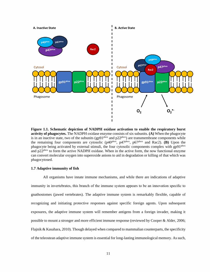
1.6.3 Respiratory burst activity
An essential immunological response to eliminate bacterial pathogens is the respiratory burst
activity (RBA) of phagocytes. Following ingestion of foreign particles, these immune cells can kill
most bacteria by producing reactive oxygen intermediates (ROI). The production of ROI requires
NADPH oxidase (NOX), which catalyzes the conversion of molecular oxygen into superoxide anions
(reviewed by Panday et al., 2015; Nguyen et al., 2017). Upon formation, the superoxide anion will then
transform into further ROIs such as hydrogen peroxide, hydroxyl radical, and hypochlorous acid, all of
which efficiently kill the phagocytosed microorganisms (DeLeo & Quinn, 1996). Inactive NOX
consists of six subunits wherein gp91phox and p22phox are membrane proteins (Figure 1.1A) that together
are known as flavocytochrome b558 (cyt b558, Nauseef, 2018). The remaining four regulatory subunits,
(p40phox, p47phox, p67phox and Rac2) normally exist in the cytosol (Figure 1.1A) but upon the activation
of leukocytes by particulate stimuli, will translocate to the membrane and associate with cyt b558
(Figure 1.1B) to form the active oxidase (reviewed by Panday et al., 2015; Nauseef, 2018). It is well
established that fish phagocytes possess all of these NADAPH oxidase components as well as an RBA
response comparable to that of mammals (Stafford et al., 2002; Sepulcre et al., 2007a; Boltana et al.,
2009). Additionally, this immune defense has been extensively studied in relation to bacterial pathogens
of fish (Sharp et al., 1993; Ardo et al., 2010; Hodgkinson et al., 2012; Havixbeck et al., 2017). Given
that the RBA in fish is not markedly influenced by temperature (reviewed by Le Morvan et al., 1998;
Collazos et al., 1994a; Collazos et al., 1994b; Nikoskelainen et al., 2004), this innate immune response
is an essential defensive mechanism for these poikilothermic organisms.
11
Figure 1.1. Schematic depiction of NADPH oxidase activation to enable the respiratory burst
activity of phagocytes. The NADPH oxidase enzyme consists of six subunits. (A) When the phagocyte
is in an inactive state, two of the subunits (gp91phox and p22phox) are transmembrane components while
the remaining four components are cytosolic (p40phox, p47phox, p67phox and Rac2). (B) Upon the
phagocyte being activated by external stimuli, the four cytosolic components complex with gp91phox
and p22phox to form the active NADPH oxidase. When in the active form, the now functional enzyme
can convert molecular oxygen into superoxide anions to aid in degradation or killing of that which was
phagocytosed.
1.7 Adaptive immunity of fish
All organisms have innate immune mechanisms, and while there are indications of adaptive
immunity in invertebrates, this branch of the immune system appears to be an innovation specific to
gnathostomes (jawed vertebrates). The adaptive immune system is remarkably flexible, capable of
recognizing and initiating protective responses against specific foreign agents. Upon subsequent
exposures, the adaptive immune system will remember antigens from a foreign invader, making it
possible to mount a stronger and more efficient immune response (reviewed by Cooper & Alder, 2006;
Flajnik & Kasahara, 2010). Though delayed when compared to mammalian counterparts, the specificity
of the teleostean adaptive immune system is essential for long-lasting immunological memory. As such,

12
this branch of the immune system is critical for vaccine design, an enterprise in need of improvement
for the aquaculture industry.
1.7.1 Cells of adaptive immunity
Much like in mammalian models, lymphocytes are considered the adaptive immune cells of
fish. The two types of lymphocytes, T and B lymphocytes, represent the only cells capable of
recognizing and responding specifically to an antigenic epitope. This antigen detection is based on
compatibility with either the surface T cell receptor (TCR) or B cell receptor (BCR) depending on the
lymphocyte. The genes that encode for these receptors undergo a series of DNA recombinations,
providing them with immense phenotypic diversity to improve the likelihood of antigen recognition
(reviewed by Nemazee, 2000). Depending on the type of activation, T lymphocytes can produce
cytokines to direct immune responses (CD4+ T cell, reviewed by Abbas et al., 1996) or induce
programmed cell death in virally infected cells (CD8+ T cell, reviewed by Barry & Bleackley, 2002).
In comparison, B cells will transform into plasma cells following activation to produce antigen specific
antibodies (reviewed by LeBien & Tedder, 2008). Though participating in a variety of different
activities, the proficient function of both T and B lymphocytes are crucial for the success of the adaptive
immune system.
In mammals, lymphocytes represent the largest blood cell in diameter (8-10 µm) and account
for approximately 20-40% of WBCs (reviewed by Houwen, 2001; Mathur et al., 2013). This is quite
different from teleost species where lymphocytes are smaller in size (5-8 µm) and represent the
dominant circulating leukocyte at 83-90% of total WBCs (Pitombeira & Martins, 1970; Blaxhall &
Daisley, 1973; Niimi & Lowe-Jinde, 1984). Regardless of the model system used, T and B lymphocytes
appear identical when observed under a microscope, making it impossible to discern between the two
without identifying cell surface markers. Though studied in detail for mammalian models, this was not
possible in teleosts due to the absence of appropriate antibodies. Fortunately, with the recent
development of antibodies specific for some of the cell surface markers on salmonid lymphocytes

13
(Zhang et al., 2010; Maisey et al., 2016; Takizawa et al., 2016), comparative immunologists are able to
finally start understanding adaptive immune functions in teleosts.
1.7.2 Major histocompatibility (MH) genes
To effectively combat the inevitable interaction with foreign entities, vertebrates have evolved
two distinct antigen presentation pathways that, when paired with effective innate immune system
activation, can stimulate long-term immunological memory. The major histocompatibility complex
(MHC) molecules are critical in this important immune process yet the MHC gene equivalents in
teleosts are not clustered on a single chromosome as they are in mammals so they are not considered
to be a “complex”. Instead these genes can be found on more than one chromosome and as a result are
simply referred to as Major Histocompatibility (MH) genes (reviewed by Dixon & Stet, 2001). In the
mammalian model, the endogenous antigen presentation pathway includes MHC class I molecules
which are found within all nucleated cells. This pathway involves the processing of antigens from
intracellular pathogens and their presentation via MHC class I to CD8+ cytotoxic T lymphocytes
(reviewed by Neefjes et al., 2011). In comparison, the exogenous antigen presentation pathway uses
MHC class II dimers which are generally only found on specific cell types, namely antigen presenting
cells (APCs) that are capable of phagocytosis (reviewed by Landsverk et al., 2009). Once phagocytosed
and processed, the antigens from extracellular sources are then loaded onto the MHC class II dimer and
presented at the cell surface to CD4+ T lymphocytes (reviewed by Blum et al., 2013). In fish, the MHC
equivalents function in an identical matter to what has been observed in mammals. Because this thesis
focuses on bacterial pathogens which are typically extracellular in nature, more emphasis will be placed
on MH class II (Figure 1.2).
Per individual, the MH molecules play an important role by binding to and presenting well-
matched peptides to appropriate T lymphocytes. The compatibility of these pathogen derived antigens
to MH molecules is controlled by allelic variation at the peptide binding groove. There are many
possible alleles for the peptide binding region and every individual within a species has a limited
repertoire inherited from their parents in a Mendelian fashion (reviewed by Choo, 2007; Garcia et al.,

14
2012). The genetic polymorphism at the MH loci can provide more or less protection to pathogens and
thus these genes are believed to be under strong selection pressure that is often governed by the
surrounding habitat (reviewed by Yamaguchi & Dijkstra, 2019). Regardless of the species, individuals
that are heterozygous at MH loci are believed to be better protected as the resulting molecules should
be able to bind to and present a more extensive collection of antigens (Lens, 2011; Pierini & Lens,
2018). In fish, this is supported by heterozygous individuals presenting less infection and/or mortality
when challenged with an infectious agent (Evans & Neff, 2009; Becker et al., 2014). Unlike what has
been observed in some terrestrial species (Pazderka et al., 1975; Hill et al., 1992; Shen et al., 2014),
specific MH alleles have not yet been shown to consistently predict resistance or susceptibility towards
specific pathogens in fish, but perhaps more research is required
Figure 1.2. Structure of the MH class II molecule. The structure and function of the teleostean MH
class II dimer is very similar to mammalian MHC class II. The molecule is a dimer, consisting of an
alpha chain and beta chain, both of which have transmembrane regions. As outlined in red, both the α1
and β1 domains of this molecule have a hypervariable region that is part of the peptide binding groove.
It is this region that is capable of binding to compatible antigens and presenting it to T cells. In this
thesis, the hypervariable region of the β1 domain is used to genotype individuals and determine whether
certain alleles at the peptide binding groove will infer resistance or susceptibility to bacterial pathogens.

15
1.7.3 Antibody development
Antibody development and production is of paramount importance in the humoral immune
response of all jawed vertebrates, including bony fishes. This defense is particularly important when
dealing with extracellular threats, as is the case with most bacterial pathogens. Antibodies prevent the
growth and colonization of bacterial pathogens by neutralization, complement activation and/or
opsonization to enhance phagocytosis (reviewed by Forthal, 2014). To date, there are three known
antibody classes in teleosts based on differences in their constant region: IgM, IgD, and IgT (reviewed
by Sunyer, 2013). IgM was the first isotype discovered in teleosts and can be found on B cells as well
as secreted in the serum or mucus as a tetramer (reviewed by Mashoof & Criscitiello, 2016). The
secreted form of teleost IgM is by far the most prevalent immunoglobulin in the serum and is
responsible for systemic immunity of bony fishes (reviewed by Parra et al., 2015). At much lower
concentrations, IgM is also present in the gut and skin mucosa. Similar to mammals, all mature IgM B
cells in teleosts also express IgD, a class of antibody whose function is still not fully understood
regardless of the model system used (reviewed by Sunyer, 2013). However, there have been slight
improvements in elucidating mammalian IgD function recently (reviewed by Gutzeit et al., 2018).
Lastly, IgT is a recently discovered antibody isotype exclusive to bony fishes (Hansen et al., 2005). IgT
is present within the serum as monomers, while forming tetramers in the gut mucosa. With
concentrations of IgT in the gut mucosa being double that observed in the serum (Zhang et al., 2010),
it was believed that this Ig class likely had a vital role in mucosal immunity. This has been heavily
supported since then with numerous studies demonstrating the role of IgT in teleostean mucosal
immunity (Xu et al., 2013; Piazzon et al., 2016; Yu et al., 2018). When considering adaptive immune
defenses that are important for combatting extracellular bacterial pathogens, effective antibody
production and development is invaluable.
Although teleosts contain all of the components necessary for adaptive immune responses,
opinion is divided as to whether they are capable of immunological memory. Rather than developing
memory B cells, it has been proposed that the most mature stage of B lymphocytes in teleosts are long-

16
lived plasma cells (LLPCs). These cells reside within the head kidney, a primary immune organ of fish
that is considered to be analogous to mammalian bone marrow. These LLPCs have been observed to
secrete high amounts of antibody and appear to be the only detectable source of prolonged, high-titered
antibody (Bromage et al., 2004). However, the idea of ‘immunological memory’ is called into question
because the antibody response in fish shows poor affinity maturation and slow development of the
secondary immune response, taking 3-4 weeks to initiate in fish (Ye et al., 2011a). This is a stark
contrast to the rapid development anticipated based on the mammalian paradigm of immunological
memory.
1.8 Cytokines
As described above, the immune system of vertebrates is a complex network connecting
numerous cell types, barriers and specialized systems to prevent the entry and/or colonization of foreign
entities within the host. The successful function of this multifaceted system depends on the ability of
immune cells to migrate to and communicate with one another, a role fulfilled by extracellular
mediators known as cytokines. Cytokines are a large family of small glycoproteins that are capable of
acting in an autocrine, paracrine or endocrine fashion (reviewed by Zhang & An, 2009). These soluble
proteins play crucial roles in regulating inflammation, haematopoiesis, cellular movements and immune
cell activation (reviewed by Metcalf, 2008; Grayfer & Belosevic, 2012; Newton & Dixit, 2012; Hughes
& Nibbs, 2018). As such, cytokines act as an essential link between the innate and adaptive arms of the
immune system.
1.8.1 Pro-inflammatory
Inflammation is an essential response to combat tissue damage of any type whether it be from
infection, trauma, foreign objects or toxins. Though there are many important components involved in
an inflammatory response (reviewed by Martin & Leibovich, 2005, Newton & Dixit, 2012), the
initiation and perpetuation of inflammation is governed primarily by pro-inflammatory cytokines
produced by damaged cells and/or responding immune cells. In mammalian models, the three classical
pro-inflammatory cytokines are interleukin (IL)-1β, IL-6 and tumour necrosis factor (TNF)α. Upon

17
tissue damage, keratinocytes and fibroblasts release IL-1β inducing fever, T cell proliferation and
increasing vascular permeability (Feghali & Wright, 1997). Meanwhile resident mast cells (MCs)
degranulate in response to the mechanical trauma (reviewed by Watkins et al, 1995) releasing a wide
variety of inflammatory mediators including, but not limited to, TNFα and IL-6 (reviewed by
Theoharides et al, 2012, Wulff & Wilgus, 2013). The released cache of inflammatory mediators also
aids in vascular permeability as well as the activation/recruitment of circulating immune cells which
produce more pro-inflammatory cytokines. Leukocytes that are normally restricted to blood vessels
will then gain access to the site of tissue injury and attempt to eliminate any invading targets (reviewed
by Medzhitov, 2008). If the inflammatory response is successful in clearing the threat, it is followed by
a resolution and repair phase mediated mainly by tissue-resident and recruited macrophages that shift
the response from pro-inflammatory to anti-inflammatory (reviewed by Serhan & Savill, 2005). While
fish immune systems are not as well characterized as mammalian equivalents, they possess all of the
major pro-inflammatory cell types and cytokines (reviewed by Grayfer & Belosevic, 2012).
1.8.2 Anti-inflammatory
Though necessary for homeostatic maintenance, inflammatory responses can be quite
damaging to surrounding tissues. This makes the control of such reactions essential for the day-to-day
welfare of the host. Regulation and control of inflammatory responses requires a constant and ever-
changing balance between pro-inflammatory and anti-inflammatory cytokines. Under normal
circumstances, as the source of tissue damage is cleared, there is less stimuli available to induce a strong
inflammatory response. This enables the constitutively produced anti-inflammatory cytokines to skew
the reaction towards tissue repair mechanisms. Though there are several cytokines that are considered
to have anti-inflammatory properties (reviewed by Zhang & An, 2009), IL-10 represents one that is
believed to function solely as a potent anti-inflammatory cytokine. Produced by almost all leukocyte
subsets including T cells, B cells, macrophages and mast cells (reviewed by Ouyang & O’Garra), IL-
10 plays a vital role in ensuring that both innate and adaptive immune responses cannot have a strong
reaction unless a true threat is present. The importance of controlling inflammation through endogenous

18
levels of IL-10 was made clear by Kuhn et al. (1993) when they generated IL-10 knockout mice. The
IL-10 deficient mice spontaneously developed inflammatory enteritis, indicating that the mice were
unable to prevent the inflammatory response towards their own commensal gut-associated bacteria
(Kuhn et al., 1993). Presently, antibodies specific for teleostean cytokines are absent, thus current data
pertaining to IL-10 and other cytokines are based solely on transcriptional analyses (Chettri et al.,
2011). As with most components of the immune system, inflammatory responses must be tightly
regulated.
1.9 Thesis Objectives
The overarching goal of my thesis was to enhance our understanding of salmonid immunity
with regard to industrially relevant bacterial pathogens. More specifically, the outcomes of my research
will help to understand salmonid immunity in response to Flavobacterium psychrophilum and Vibrio
anguillarum with the potential to improve therapies for these bacterial infections. The three specific
objectives of my PhD thesis were to:
i. Determine whether selected salmonid immune markers could predict resistance and/or
susceptibility to bacterial pathogens in whole animals (Chapters 2 and 5).
ii. Understand the immunity to, and pathogenesis of, aquatic bacterial pathogens in relevant in vitro
models (Chapters 3 and 6).
iii. Assess the caliber of an alternative treatment option for a bacterial disease in aquaculture
(Chapter 4).
To accomplish these objectives, five independent hypotheses were generated:
i. Selectively breeding rainbow trout for resistance and susceptibility to BCWD will result in
observable, family-specific differences in functional immune markers (MH class II β1 genotype,
RBA or serum anti-F. psychrophilum IgM).
ii. Exposing rainbow trout immune cells to the extracellular products of F. psychrophilum will
stimulate and influence their function.

19
iii. Teleostean PACAP will be directly bactericidal to F. psychrophilum and will also display
immunomodulatory effects on salmonid immune cells following exposure.
iv. Outbreeding of an inbred Chinook salmon population will confer hybrid vigor, as evidenced by
survival to V. anguillarum infection and functional differences in immune performance.
v. Exposing a Chinook salmon spleen stromal cell line to heat-killed V. anguillarum, will stimulate
a pro-inflammatory cytokine response.

20
Chapter 2: Serum IgM, MH class IIβ genotype and respiratory burst activity do not differ
between rainbow trout families displaying resistance or susceptibility to the coldwater pathogen,
Flavobacterium psychrophilum.
Shawna L. Semple1, Calvin J. Kellendonk1, Lowia Al-Hussinee2, Janet I. MacInnes2, John S.
Lumsden2, Brian Dixon1
1University of Waterloo, Department of Biology, Waterloo, Canada 2University of Guelph, Ontario Veterinary College, Department of Pathobiology, Guelph, Canada
Aquaculture, 483, pp. 131-140. 2018.

21
2.1 Overview
Flavobacterium psychrophilum, the causative agent of bacterial coldwater disease (BCWD) is a
significant threat to global aquaculture. True to its name, BCWD tends to occur at temperatures between
8 and 12°C and presents as a systemic disease with characteristic skin ulcerations. Juvenile rainbow
trout are particularly susceptible and in these fish the condition is referred to as rainbow trout fry
syndrome (RTFS). Resistance to F. psychrophilum is heritable and is not adversely correlated with the
growth of fish, thus selective breeding appears to be an achievable approach to its control. The current
study explores the connection between resistance to BCWD and several immunological markers. After
determining resistance/susceptibility to F. psychrophilum following experimental infection in 40 full-
sibling families of rainbow trout, selected families were experimentally infected with F. psychrophilum
and differences in antibody production, major histocompatibility (MH) class IIβ genotype and
respiratory burst activity (RBA) throughout infection were compared. Serum IgM production increased
over time but significant differences between resistant and susceptible families were not observed at
either 28 days or 120 days. Of the six families that were genotyped for MH class IIβ, there did not
appear to be specific genotypes that conferred resistance or susceptibility to F. psychrophilum. Further,
the RBA of both head kidney leukocytes and whole blood was not significantly different between the
resistant and susceptible rainbow trout families. Although the selected immune markers did not differ
based on resistance status, the RBA of head kidney leukocytes in all families studied dramatically
decreased seven days after infection while total blood RBA remained constant. Day seven was also
when severe symptoms and/or mortality due to BCWD was first observed, thus these results may reveal
information regarding the pathogenesis of the organism. A better understanding of appropriate immune
defenses could provide the basis for breeding programs to effectively combat this costly pathogen, but
further study of functional immune markers particularly during the fry stage of development is required.
22
2.2 Introduction
A large obstacle to overcome when dealing with aquaculture production of any type is the
vulnerability to disease outbreaks. Like other forms of agriculture, aquaculture places animals in a
setting that deviates significantly from that of their natural environment. The stress induced by
conditions such as overcrowding, temperature fluctuations and handling can result in normally benign
microorganisms becoming opportunistic pathogens (reviewed by Meyer, 1991). One such pathogen
causing significant losses in the farming of rainbow trout (Oncorhynchus mykiss) is Flavobacterium
psychrophilum, the etiological agent of bacterial cold-water disease (BCWD). Rainbow trout, a
coldwater salmonid, is an important species of choice in global aquaculture, which has enabled BCWD
to become a worldwide concern.
F. psychrophilum is widespread in freshwater aquatic environments but typically only causes
infection in intensively reared salmonids at temperatures lower than 16°C (Holt, 1987). Although F.
psychrophilum can infect many salmonid species and some non-salmonid species, juveniles of coho
salmon (Oncorhynchus kisutch) and rainbow trout are particularly susceptible (reviewed by
Nematollahi et al., 2003; Starliper, 2011). Clinical signs and manifestations vary depending on location
as there appears to be geographic variability between isolates (Lumsden et al., 2004). In North America,
the most common presentation of BCWD in fish larger than fingerling is an ulcerative dermatitis known
as peduncle disease (Lumsden et al., 2004). The nature of this presentation means that the financial
losses associated with BCWD are not due to mortalities alone as surviving fish often produce fillets
that are less valuable or not marketable (Lumsden et al., 2004). In fry, the most important presentation
is a systemic disease, often with exopthalmia and anemia, known as rainbow trout fry syndrome (RTFS)
(Bebak et al., 2007). With no commercial vaccine available and mortality, although variable, has been
reported to be as high as 50-85% (Brown et al., 1997; Cipriano & Holt, 2005), the financial losses due
to F. psychrophilum infection can be devastating.
To combat the significant losses due to infectious disease, there has been increasing interest in
selectively breeding aquaculture stocks for disease resistance. As has been demonstrated with many

23
other aquatic diseases (Gjedrem et al., 1991; Henryon et al., 2005; Kjoglum et al., 2006, 2008),
resistance to BCWD does appear to be heritable (Silverstein et al., 2009), but there has been little
research to identify the basis for this resistance. In addition, a method to consistently produce resistant
crosses is yet to be developed for the vast majority of aquatic organisms. Creating and challenging
families of rainbow trout takes a great deal of time and finances, thus identifying a marker for the
observed resistance to BCWD would decrease costs and expedite this process immensely. With the
immune system playing a vital role in the prevention and clearance of infection, it is reasonable to
hypothesise that the heritable resistance to BCWD may be associated with teleostean immune function.
Like mammals, teleosts have both innate and adaptive immune systems. The innate immune
response constitutes the first line of defense against infection and is activated through recognition of
conserved molecular patterns common to pathogenic organisms (Uribe et al., 2011). Phagocytosis, or
the engulfment of particles, is a major component of immunity in fish (Uribe et al., 2011). Circulating
phagocytes will take up foreign bodies such as bacteria and eradicate them through the production of
reactive oxygen intermediates (ROI) during a respiratory burst (Nikoskelainen et al., 2004).
Phagocytosis, and the killing activity associated with it, is reported to be the mechanism of innate
immunity that is the least influenced by low temperatures (Collazos et al., 1994a; Collazos et al., 1994b;
Nikoskelainen et al., 2004), thus this process may be critical when dealing with a cold-water pathogen
such as F. psychrophilum.
While the innate immune system is fast-acting, adaptive immunity requires more time to
develop and has greater specificity. A key component of vertebrate adaptive immunity is the generation
of antigen-specific antibodies from B cells. Antibodies promote the control and clearance of infection
through such processes as opsonisation for phagocytes, complement activation and neutralization of
microbes and toxins (Forthal, 2014). To date, three classes of immunoglobulins have been discovered
in teleosts: IgM, IgT and IgD (reviewed by Sunyer, 2013). The most abundant immunoglobulin is a
tetramer of the IgM class (Fillatreau et al., 2013). IgM is the first antibody class to appear after initial
exposure to an antigen and may be part of an effective humoral response against invading pathogens

24
(Fillatreau et al., 2013; Reyes-Cerpa et al, 2012). With these protective properties in mind, there have
been numerous attempts to identify antigens capable of inducing an effective antibody response to
combat F. psychrophilum (Crump et al., 2005; Dumetz et al., 2006; LaFrentz et al., 2002; Sudheesh et
al. 2007). Although antibody titres have been observed to increase after immunization with many of
these antigens and whole-cell lysates, similar increases were often observed with adjuvant alone
(LaFrentz et al., 2002). As there is limited information regarding consistent infection and vaccination
models for F. psychrophilum (Gomez et al., 2014), there is a critical need to better understand the serum
IgM response in fish presenting resistance/susceptibility to BCWD.
Although fish farmers can benefit from breeding programs based solely on survival phenotype,
identifying genes (markers) correlated with enhanced immune performance could aid in the more rapid
development of optimum stocks. One possible marker, and a focus of this study, is the major
histocompatibility (MH) class II genes. As the class of molecules that recognize and present antigens
from extracellular pathogens to tailor the resulting immune responses (Neefjes et al., 2011), there has
been a great deal of interest in determining whether specific MH class II genotypes are linked to
bacterial resistance/susceptibility in fish species (Dionne et al., 2009; Kjoglum et al., 2008; Rakus et
al., 2009; Xu et al., 2010). Conversely, MH class I is associated with recognition and presentation of
antigens from intracellular pathogens such as viruses (Neefjes et al., 2011). Because F. psychrophilum
appears to have both intracellular and extracellular tendencies throughout its infection cycle (Decostere
et al., 2001; Nematollahi et al., 2005; Nilsen et al., 2011b; Wood & Yasutake, 1956), it is not fully
understood whether MH class I or MH class II genes would provide more protection towards BCWD.
To date there has been some evidence of an association between MH class II variability and resistance
to F. psychrophilum (Johnson et al., 2008), but there has been minimal research since then to relate
specific alleles with BCWD resistance. As such, one component of this study aims to link MH class IIβ
alleles with observed resistance to BCWD and determine whether this association goes further.
In the present study, full-sibling families of rainbow trout displaying resistance/susceptibility
to BCWD were experimentally infected with F. psychrophilum so that differences in MH class IIβ
25
genotype, respiratory burst activity and antibody production could be observed and compared. As there
is limited knowledge regarding both the pathogenesis of F. psychrophilum and what constitutes an
effective immune response against the organism, this study aims to analyze functional immune
differences in fish that are resistant/susceptible to provide information that could be used for the
development of effective breeding programs.
2.3 Materials and Methods
Fish
Forty full-sibling families of rainbow trout were produced at Lyndon Fish Hatcheries (Dundee,
Ont.). These families were transported to the Hagen Aqua Lab or the Alma Aquaculture Facility at the
University of Guelph as eyed eggs. Susceptibility to F. psychrophilum experimental infection was
compared among the 40 family groups. Six to ten families per experiment were divided into four 60 L
tanks containing 40 fish (40 g each) per tank, which were supplied with single pass well-water at 11°C.
Fish were fed a commercial pellet diet (Martin Mills Inc.). All maintenance and procedures were done
in accordance with the University of Guelph Animal Care Guidelines.
Culture and quantification of F. psychrophilum strain 101
Frozen stocks of F. psychrophilum strain 101 (FPG 101) were sub-cultured onto cytophaga agar
(CA) then following incubation at 15°C for 72 h, cells were harvested and suspended in cytophaga
broth (CB) (Hesami et al., 2011). Cell suspensions were adjusted to an optical density of 0.6 at 600 nm
using a Novaspec Plus spectrophotometer (GE Healthcare Life Sciences) and the suspension was then
diluted to obtain a final concentration of ~1 x 10⁹ colony forming units (CFU)/mL. The concentration
was then adjusted so that the desired CFU/mL for each trial could be obtained. To confirm the cell
numbers, suspensions were enumerated by plating 0.1 mL of ten-fold serial dilutions on CA plates in
triplicate. After incubation at 15°C for 4 d, plates containing 25-250 colonies were counted and the
number of CFU/mL calculated.

26
Determining susceptibility/resistance to F. psychrophilum (Preliminary Trials)
The Ontario F. psychrophilum isolate, FPG101, was used for all experimental infections as it had
been shown to produce ~50% mortality in experimental trials of 30-40 g rainbow trout at doses similar
to those used in the current study (JS Lumsden pers. comm.). After sedation with ~50 ppm benzocaine
(Millipore Sigma) in well-aerated water, triplicate tanks (n = 40) of each family received intraperitoneal
(i.p.) 0.1 mL injections ranging in concentration between 1.13 and 6.60 x 10⁷ CFU/mL. With every
family, a fourth tank of fish was sham-injected with 0.1 mL of sterile CB as a negative control.
Following injection, all fish were observed three times a day and any mortalities were collected and
saved at -20°C. In addition, fish with marked exopthalmia, hemorrhage or skin ulcerations were
removed and euthanized using an overdose of benzocaine. These fish were counted as mortalities.
After the predetermined trial period (15 d), any surviving fish were euthanized with an overdose
of benzocaine and were recorded as survivors. The caudal fins of mortalities and final survivors were
collected, submerged in RNAlater and stored at -20°C for later use. Mortality was the measure used to
rank the rainbow trout families for their resistance to F. psychrophilum infection. For subsequent
experiments described below (sections 2.3.4 and 2.3.5), resistant and susceptible families were selected
based on this resistance ranking.
MH class II β1 genotyping
2.3.4.1 DNA extraction
Based on the susceptibility results (section 2.3), caudal fins from survivors and mortalities of
families 3, 4, 6, 7, 9, and 10 were collected for DNA extraction. DNA was extracted from all fin clips
as described by Kellendonk, 2014. The final concentration of DNA was determined using the Take3
plate in a Synergy H1 plate reader (BioTek Instruments) before samples were moved to the -80°C for
storage.

27
2.3.4.2 Amplification and sequencing of MH class II β1 genotypes
Primers were designed to amplify the MH class IIβ region for this study. The forward primer,
OMYF (5'-TGCCAATTGCCTTCTACATTTGCCTG-3') was designed to a conserved region of MH
class IIβ at the beginning of exon 1, while the reverse primer, OMYR (5'-
TGGGGGCTCAACTGTCTTGTCCAGT-3') was designed to a conserved region at the end of exon 2
(Hodgins, 2011). These primers ensured the amplification of a portion of the peptide binding groove,
the region that binds specifically to foreign antigens such as extracellular pathogens. For MH class IIβ
amplification, the following PCR parameters were used: denaturation at 95°C for 2 min, followed by
24 cycles of denaturation at 94°C for 30 sec, annealing at 55°C for 30 sec and extension at 72°C for 1.5
min. A final extension at 72°C was carried out for 8 min. Two PCR reactions were performed per
individual. The amplified PCR products were separated on 1% agarose gels containing 3% Gel Red
(Biotium Inc.). Bands of the desired size were cut out of the gel and extracted using the QIAquick Gel
extraction kit (Qiagen). The resultant fragments were cloned into pGEM®-T Easy as per the
manufacturer’s instructions (Promega Corporation, Madison, WI). Competent E. coli XL1–blue cells
were transformed using the Inoue procedure as described in Green and Sambrook, (2012) and grown
on LB plates with 100 μg/mL of ampicillin, 240 ng/mL IPTG and 100 μg/mL X-gal. Ten colonies per
PCR reaction were selected and grown in LB medium. Plasmids were extracted from the transformants
using the GenElute Plasmid Miniprep Kit (Millipore Sigma) according to the manufacturer’s
instructions. Plasmids were sequenced using both T7 and SP6 primers at the TCAG sequencing facility
(Sick Kids Hospital, Toronto, Ont.). The sequences were analyzed using both GENEious and the Basic
Local Alignment Search Tool (BLAST) software (http://blast.ncbi.nlm.nih.gov/Blast.cgi). A G-test
was performed on the resulting contingency table using the SPSS statistical software. Significant
differences were established at p < 0.05.
28
Induction of F. psychrophilum antibody responses
2.3.5.1 Infection
Two trials were performed to measure the levels of IgM antibody developed towards F.
psychrophilum throughout infection. These trials were conducted in triplicate tanks (n = 40) when the
fish were, on average, 108 g. The first trial (referred to as Trial 1) included two resistant families (F21
and F30), as well as two susceptible families (F11 and F17). After sedation with benzocaine, all fish
received 100 μL injections i.p. of 1.10 x 10⁷ CFU/mL of FPG101 suspended in CB. As a control, fish
from the four families analyzed were mixed, then received 100 μL i.p. injections of sterile CB and
separated into triplicate tanks. On days 0, 1, 7, 14, 21 and 28, blood was drawn from the caudal vein
after anesthesia with ~50 ppm benzocaine. Fish were then euthanized using an overdose of benzocaine
and whole-body weights and lengths were measured. Blood samples were left to clot overnight at 4°C
before centrifugation at 1000 g for 10 min. The serum was then removed, aliquoted into 1.5 mL tubes
and immediately stored at -20°C.
Because it has been reported that rainbow trout require up to 12 weeks to reach peak serum IgM
levels when injected with killed F. psychrophilum (LaFrentz et al., 2002), a second trial (referred to as
Trial 2) was conducted wherein fish were injected i.p. with 100 μL of a lower dose of live FPG101
(3.25 x 10⁶ CFU/mL) and sampled over 140 days. In this trial, one resistant family (F32) and one
susceptible family (F38) was used and all experiments were done in triplicate tanks (n = 40) where the
fish were, on average, 85 g. For both families, triplicate control tanks were set up where all control fish
received 100 μL i.p. injections of sterile CB broth. Serum samples were collected as described above.
In Trial 2, the anterior head kidney was also removed from sampled individuals so that respiratory burst
activity of head kidney leukocytes could be measured (described further in 2.3.6).
2.3.5.2 Isolating antigens from F. psychrophilum
Subcultures of glycerol stocks of FPG101 were grown on CA at 14°C and carefully checked for
purity. An isolated colony was then used to inoculate 50 mL of CB, which was subsequently used to

29
inoculate 200 mL of CB after three days of incubation at 14°C. When the culture reached an optical
density of approximately 0.8 at 525 nm, indicating a concentration of ~1 x 10⁸ CFU/mL (Holt 1987),
0.5% formalin was added to the flask and cells were incubated at 14°C for 24 h with gentle stirring.
The killed bacterial cells were then washed twice with 4 mL of phosphate buffered saline (PBS) then
harvested by centrifugation (9,500 g at 4°C for 15 min) and the resultant pellets resuspended in 2 mL
of PBS.
Six hundred microliters of 0.1 mm zirconia/silica beads (BioSpec Products) was added to a 1.5
mL screw cap tube followed by the addition of 600 μL of the inactivated F. psychrophilum culture. The
tubes were placed in a BioSpec mini-beadbeater and agitated on full speed for 45 sec followed by 1
min of incubation on ice. This procedure was repeated five times for each tube before the samples were
centrifuged at 9,500 g for 15 min. Following centrifugation, the supernatants were removed and
combined in a sterile 15 mL Falcon tube. The protein concentration of the bacterial lysate was then
quantified using a bicinchoninic acid assay (ThermoFisher Scientific) following the manufacturer’s
protocol. The bacterial lysate was then diluted to a final concentration of 0.5 µg/mL in coating buffer
(0.015 M Na2CO3, 0.035 M NaHCO3, pH 9.6).
2.3.5.3 Indirect ELISA assay to detect levels of F. psychrophilum antibodies in serum
One-hundred microliters of the whole-cell bacterial lysate preparation was used to coat the test
wells of NuncTM MicroWellTM 96-well microplates (ThermoFisher Scientific). Hyperimmune serum
from rainbow trout injected with F. psychrophilum FPG25 (Hesami et al., 2008) was also used at a
dilution of 1:50 as a positive control. To obtain hyperimmune serum, 200 g rainbow trout were injected
in the dorsal sinus with 100 μl of formalin-killed FPG25. Three months later, fish received a 100 μl
booster of formalin-killed F. psychrophilum FPG25. Serum samples were obtained at 5 months and 7
months following the initial exposure. Negative control wells were coated with the whole cell bacterial
lysate but were incubated with 100 µl Tris-buffered saline supplemented with 0.5% Tween 80 (TBS-
T) rather than rainbow trout serum to account for any non-specific binding of the detecting antibodies.
The coated plates were incubated overnight at 4°C to enable adherence of the antigens.
30
The following day, all wells were washed three times with TBS-T. Three hundred microliters of
blocking solution (2% laboratory grade gelatin (ThermoFisher Scientific) dissolved in TBS-T) was
added to each well and incubated for 1 h at 37oC. The plate was then washed three times with TBS-T
and 100 μL of each trout serum (diluted 1:50) was added to triplicate test wells. After incubation for 1
h at room temperature (RT) the wells were washed thrice with TBS-T and 100 μL of mouse anti-
rainbow trout IgM monoclonal antibody (Aquatic Diagnostics) at a dilution of 1:50 was added to each
test well and hyperimmune serum control wells. The plate was incubated for 1 h at RT after which all
wells were washed thrice with TBS-T. All test and the hyperimmune serum positive control wells
received 100 µl of goat anti-mouse IgG conjugated to alkaline phosphatase (Millipore Sigma) at a
dilution of 1:30 000. The plate was incubated for 1 h at RT and then washed three times with TBS-T.
Fifty microliters of substrate, p-nitrophenyl phosphate (p-NPP), was added to each well and the plate
was incubated in the dark for 30 min. The reaction was then stopped by the addition of 50 µl of 0.03M
of NaOH to each well. The plate was read immediately at an absorbance of 405 nm in a Synergy H1
plate reader (BioTek). The negative control wells, which received TBS-T rather than trout serum, were
averaged and subtracted from all other absorbance values. Plate variation was accounted for by
normalizing each plate to the hyperimmune serum positive control. Statistical analysis was conducted
using a two-way ANOVA through the GraphPad Prism software (v7.0, GraphPad Software, Inc. USA)
where each of the triplicate tanks was considered an experimental unit (n = 3), and variation among
tanks of each family receiving the same treatment was used as experimental error. This was then
followed by Sidak’s Multiple Comparisons Test with significant differences established at p < 0.05.
Measuring Respiratory Burst Activity (RBA) of blood and head kidney leukocytes
2.3.6.1 Infection
To determine whether RBA was associated with the observed resistance or susceptibility to F.
psychrophilum, two infection trials were conducted. The families used for the first RBA trial are
described above in section 2.3.5 (Trial 2). On days 0, 1, 7, 14 and 28, three fish per tank were euthanized
with an overdose of benzocaine and the anterior head kidneys were collected and transferred to sterile
31
plasticware containing approximately 20 mL of L15 medium (Lonza) supplemented with 5% FBS, 100
U/mL of penicillin and 100 U/ml of streptomycin. These samples were stored on ice until the HKL
could be isolated.
To validate the results observed for RBA in Trial 2, a repeat trial (Trial 3) was completed using
the resistant family, F36 and the susceptible family, F38. Challenged fish received 100 μL i.p. injections
of FPG101 (3.50 x 10⁶ CFU/mL). This trial was conducted in triplicate tanks (n = 40) when the fish
were, on average, 200 g. As a control, fish from the two families analyzed were mixed, received 100
μL i.p. injections of sterile CB, and were separated into triplicate tanks. On days 0, 3, 7, 14 and 20,
three fish per tank were euthanized with an overdose of benzocaine so that the anterior head kidney of
each individual could be removed and processed as described.
2.3.6.2 Isolation of head kidney leukocytes
The head kidney samples were vigorously vortexed 5 times at maximum speed for 30-60 sec to
dissociate the cells from the underlying connective tissues. The dissociated cells were passed through
a 40 μm sterile nylon mesh (ThermoFisher Scientific) then rinsed with 5 mL of sterile media. The
resulting cell suspensions were brought up to a final volume of 28 mL and, after thorough mixing,
divided into four 7 mL aliquots. The 7 mL cell suspensions were then layered on top of 3 mL of
Histopaque 1077 (Millipore Sigma) and centrifuged for 500 x g for 30 min at 4°C. The leukocyte layer
located at the medium:histopaque interface was collected and cells were pelleted by centrifugation at
500 x g for 5 min at 4°C. The supernatant was aspirated and the pellets were washed using 5 mL of
PBS and again, harvested by centrifugation. The resultant cell pellets were resuspended in 1-3 mL of
fresh 5% FBS/L15 so that cell concentration and viability could be determined using a trypan blue
exclusion assay. After cell viability was determined to be above 95%, cell concentrations were adjusted
to 7.0 x 10⁶ cells/mL.

32
2.3.6.3 Real-time luminol-enhanced chemiluminescence assay for RBA
The RBA of both the whole blood samples and the head kidney leukocytes (HKL) was measured
following the protocol of Jimenez-Vera et al. (2013) with minor variations. Briefly, CorningTM 96-well
solid white polystyrene microplates were prepared with 6 wells containing either 100 μL of HKL (7.0
x 106 cells/mL) or 75 μL of blood sample from each individual. The first three wells per individual
received 40 μL of luminol (10 mM, Millipore Sigma), 100 μL of zymosan A (20 mg/mL, Millipore
Sigma), and were brought to a final volume of 300 μL using Hank’s Balanced Salt Solution (HBSS,
Millipore Sigma). To ensure that the increase in luminescence was due to stimulation, the remaining
three wells per individual were used as unstimulated controls and received 100 μL of HBSS in place of
zymosan A. The chemiluminescence of each well was measured with a Synergy H1 luminometer
(Biotek) every 2 min for 90 min. The chemiluminescence emission of the HKL and blood samples was
expressed as the integral of the relative light units (RLU) between 0 and 90 min. Statistical analysis
was conducted using a two-way ANOVA through the GraphPad Prism software (v7.0, GraphPad
Software, Inc. USA) where each of the triplicate tanks was considered an experimental unit (n = 3),
and variation among tanks of each family receiving the same treatment was used as experimental error.
This was followed by Sidak’s Multiple Comparisons Test and significant differences were established
at p < 0.05.

33
2.4 Results
2.4.1 Preliminary trials to determine rainbow trout families resistant/susceptible to F. psychrophilum
Fish from 40 families were infected with F. psychrophilum and survival was monitored. As
Figure 2.1A displays, there was a broad range of survival between the families, with the most resistant
family (F8) having 90.9% survival while the most susceptible family (F36) had only 2.5% survival.
From the results presented in this study, families were chosen to conduct repeat infection trials so that
both genetic (families presented in Figure 2.1B) and functional immune markers (families presented
Figure 2.1C) could be assessed. To ensure that the observed resistance/susceptibility was consistent,
the infection trial was repeated for a resistant family (F7) and a susceptible family (F10). In the initial
trial F7 and F10 displayed 90.5% and 14.3% survival respectively. These results were confirmed to be
robust as F7 and F10 displayed 86.0% and 19.9% survival respectively when the infection trial was
repeated (data not shown). As the fish were approximately the same size (~30-50 g) during this repeat
trial, these results provide evidence that susceptibility to FPG101 was consistent at this size/age.

34
Figure 2.1. Survival curves throughout F. psychrophilum infection for the forty rainbow trout
families analyzed for resistance/susceptibility to BCWD. Panel A: Survival curves for all rainbow
trout families analyzed. Panel B: Survival curves for the six families used for MH class II β1
genotyping. Panel C: The survival curves for the eight families selected for the functional immune trials
(Trials 1–3).

35
2.4.2 Relating MH class IIβ genotype to disease susceptibility
To explore whether there is a link between BCWD resistance and MH class IIβ alleles, a
minimum of 18 individuals from six of the rainbow trout families studied were genotyped. The families
selected (F3, F4, F6, F7, F9 and F10) had variable survival rates when challenged with F.
psychrophilum (see Figure 2.1B) and thus were hypothesized to have variability in their MH genotypes.
Table 2.1 presents the MH class IIβ genotype frequencies observed between the six different families.
Overall, there were five alleles identified which created a total of twelve MH class IIβ genotypes in the
six families studied. When relating the allele prevalence to the observed number of survivors per family,
as presented in Table 2.2, both DAB*1001/DAB*1001 and DAB*1001/DAB*0801 were observed at
the highest frequency in both survivors and mortalities.
Table 2.1. MH Class IIβ genotype frequencies among the six families of Oncorhynchus mykiss. A
total of 19 individuals were sequenced for families 4 and 9, and 18 individuals were sequenced for the
other four families. Frequency was calculated using the formula f=x/n, where x is the number of times
the genotype was observed and n is the number of individuals.
Families
3 4 6 7 9 10
MH Class IIβ Genotype
DAB*1001/DAB*1001 - 0.74 0.39 0.67 0.26 -
DAB*1001/DAB*1101 0.22 0.16 - - 0.11 0.44
DAB*0801/DAB*0801 - - - - 0.11 0.06
DAB*1001/DAB*0801 - 0.05 0.56 0.33 0.21 0.39
DAB*1101/DAB*0801 0.22 - 0.06 - - -
DAB*1201/DAB*1001 0.17 - - - - -
DAB*1201/DAB*0801 0.17 - - - - -
DAB*0401/DAB*1001 - - - - 0.05 -
DAB*0801/1001/1101 0.11 0.05 - - 0.05 0.11
DAB*0401/0801/1001 - - - - 0.16 -
DAB*1001/1101/1201 0.06 - - - - -
DAB*0401/0801/1001/1101 - - - - 0.05 -

36
Table 2.2. MH class IIβ genotype prevalence in mortalities and survivors to F. psychrophilum
infection. A G-Test was performed on this contingency table using SPSS statistical software to
identify any significant differences between genotypes.
Genotype Percent of Mortalities Percent of Survivors
DAB*1001/DAB*1001 32 40
DAB*1001/DAB*1101 18 13
DAB*0801/DAB*0801 3 2
DAB*1001/DAB*0801 19 31
DAB*1101/DAB*0801 6 2
DAB*1201/DAB*1001 3 2
DAB*1201/DAB*0801 5 2
DAB*0401/DAB*1001 2 0
DAB*0401/DAB*0801 2 0
DAB*0801/1001/1101 6 4
DAB*0404/0801/1001 3 0
DAB*1001/1101/1201 0 2
DAB*0401/0801/1001/1101 0 2

37
2.4.3 IgM antibody development in rainbow trout families resistant/susceptible to F. psychrophilum
The production of rainbow trout serum IgM against F. psychrophilum FPG101 appeared to
increase over time regardless of the previously observed resistance in the six families studied. In Trial
1, anti-F. psychrophilum IgM levels increased over the 28-day infection but there was no significant
difference in antibody production between the resistant (F21 and F30) and the susceptible (F11 and
F17) families (Figure 2.2A). A similar trend was also seen in the 140-day infection trial, Trial 2 (Figure
2.2B), where serum IgM levels were also seen to increase over time with no significant difference
between the resistant (F39) and susceptible (F32) families studied. As expected, the saline-injected
controls in Trial 2 showed no increase in anti-F. psychrophilum serum IgM levels over time (Figure
2.2B).
The survival curves of families in both Trial 1 (Figure 2.2C) and Trial 2 (Figure 2.2D) did not
reveal significant differences between resistant and susceptible families in contrast to the preliminary
trials (Figure 2.1C). This can be seen in Figure 2.2E and 2.2F where the percent mortality of families
in the preliminary trial is compared to the percent mortality in Trial 1 (Figure 2.2E) and Trial 2 (Figure
2.2F). In the preliminary trials, the resistant families had low mortality rates (5 to 42%) while the rates
in susceptible families were much higher (57 to 85%). However, upon completion of Trial 1 and Trial
2, the percent mortality was more similar regardless of whether the family was previously observed to
be resistant or susceptible to F. psychrophilum (Figure 2.2E and 2.2F). In Trial 1, the four families
studied had observed mortalities between 70 and 80% while in Trial 2, the two families studied had
mortalities of 25 and 30%.

38
Figure 2.2. Indirect ELISA assay to compare serum IgM development against F. psychrophilum
between resistant and susceptible families throughout infection. Indirect ELISA results for serum
IgM levels developed against F. psychrophilum between two resistant (F21 and F30) and two
susceptible (F11 and F17) rainbow trout families throughout a 28-day infection trial (A). Indirect
ELISA results for serum IgM levels developed against F. psychrophilum between one resistant (F39)
and one susceptible (F32) rainbow trout families throughout a 140-day infection trial. Serum from
saline injected controls was also tested for antibody development to F. psychrophilum throughout the
infection trial (B). Survival curve for the two resistant (F21 and F30) and two susceptible (F11 and
F17) families during the 28-day infection trial (C). Survival curve for the resistant (39) and the
susceptible (F32) families during the 140-day infection trial. Survival is only shown up to day 90 as
there was no further mortality observed in this trial after this time (D). Comparison of the observed
mortality during the preliminary trial to determine resistance/susceptibility and the trials used to
measure serum IgM against F. psychrophilum (E-F). Error bars represent the standard error of the mean
(SEM).

39
2.4.4 The effect of head kidney and total blood RBA on resistance to F. psychrophilum
The RBA of head kidney leukocytes from the infected resistant (F39) and susceptible (F32)
families of Trial 2 were observed to differ significantly on day 14 (Figure 2.3A). Otherwise there was
no significant difference in RBA between the groups. Throughout infection, the two families had very
similar trends in RBA with high activity observed on days 0 and 1, followed by a significant decrease
in activity on day 7 of the trial. For the final two weeks of the trial, RBA was observed to slowly recover
until it was restored to pre-infection levels on day 28 (Figure 2.3A). The large drop in RBA on Day 7
was interesting as this was also the time when mortalities were first observed in Trial 2. On day 28 and
beyond, when RBA activity appeared to have fully recovered, mortalities were rarely observed. The
observed decrease in RBA of HKL in infected fish appears to be a consequence of F. psychrophilum
infection as the sterile CB infected control fish had no significant difference in HKL RBA across all
time points (Figure 2.3A). Much like what was observed in Trial 2, the RBA of the HKL did not differ
significantly between the resistant (F36) and susceptible (F38) families studied. A significant decrease
in RBA was observed at day 3 of Trial 3 (Figure 2.3B) and was maintained until trial completion on
day 20. Further, there was no difference in total blood RBA between the two families studied
throughout infection in Trial 3 (Figure 2.3C).
Although there was little to no difference in the RBA between the resistant and susceptible
families studied, it is important to relate these results to the survival of each group throughout F.
psychrophilum infection. In the preliminary trial, low mortality was observed in the resistant families,
F32 and F36 (2-5%), while high mortality was noted in the susceptible families, F38 and F39 (42-55%)
(Figure 2.1C). Despite these initial large differences in survival, upon completion of the RBA trials,
all four of the families had consistent mortalities ranging between 1-10% (Figure 2.3D).

40
Figure 2.3. Respiratory burst activity of HKL and total blood in resistant and susceptible families
of rainbow trout. RBA of HKL in one resistant (F39) and one susceptible (F32) rainbow trout family
throughout F. psychrophilum infection. Control fish received i.p. injections of sterile CB (A). RBA of
HKL in another resistant (F38) and susceptible (F36) rainbow trout family throughout F.
psychrophilum infection (B). Impact of F. psychrophilum on the RBA of total blood in a resistant (F38)
and a susceptible (F36) rainbow trout family throughout F. psychrophilum infection (C). Comparison
of the observed mortality during the preliminary trial to determine resistance/susceptibility and the
observed mortality in the trials used to measure RBA throughout F. psychrophilum infection (D). Error
bars represent the SEM.

41
2.5 Discussion
The purpose of this study was to determine whether both functional and/or genetic immune
markers could be used to predict resistance/susceptibility to the coldwater bacterial pathogen, F.
psychrophilum. To assess this, forty full-sibling families of rainbow trout were created and when
subsets of each were infected with F. psychrophilum, some families performed extremely well (90%
survival), while others had very low survival (3% survival). This broad range in BCWD susceptibility
has been observed in previous studies aimed at understanding and consistently standardizing the
production of resistant rainbow trout (Hadidi et al., 2008; Wiens et al., 2013). This observed
resistance/susceptibility to BCWD has been shown to be heritable through multiple generations of
rainbow trout (Leeds et al., 2010; Silverstein et al., 2009; Wiens et al., 2013). Though promising, a
great deal of time and resources is required to first identify resistant strains and then to subsequently
propagate appropriate crosses until you have highly resistant stocks of fish. As a result, there is an
undeniable benefit to identifying specific immune markers that are able to predict resistant or
susceptible phenotypes towards BCWD.
Because of its role in the exogenous antigen presentation pathway, MH class II alleles are a
genetic candidate of interest when attempting to select for resistance towards bacterial pathogens such
as F. psychrophilum. In this study, when families shown to be resistant/susceptible to BCWD were
genotyped for MH class II β1, only five alleles were observed resulting in a total of 12 different possible
genotypes. This is a lower number than previously observed when wild fish populations were analyzed
(Conejeros et al., 2008; Dorschner et al., 2000; Conejeros et al. 2012). However, because the rainbow
trout families used in the current study were not from wild populations and thus had likely been selected
for faster growth, better feed to weight conversion and better flesh quality, this greater degree of
homogeneity was not unexpected. Increasing the variability of MH genotypes has not been a breeding
focus for these families, therefore low diversity in MH genes may simply be a by-product of selection
for other desirable traits. This has been reported in many other studies of production (Croisetiere et al.,

42
2008; Gomez et al., 2011; Grimholt et al., 2003; Kjoglum et al., 2008), thus the low allele frequencies
presented in this study are likely representative of the commercial origin of the families that were used.
Remarkably, individuals with three or four allele genotypes for MH class IIβ were observed in
this study but these generally occurred at very low frequencies in the families analyzed. The occurrence
of two or more alleles per individual has been observed in previous MH studies (Conejeros et al., 2012;
Conejeros et al., 2008; Dixon et al., 1996; Kruiswijk et al., 2004; Noakes et al., 2003) but this was
usually attributed to high MH polymorphism. Although PCR artifacts and false alleles can arise due to
the nature of MH sequences and the cloning process, precautions were taken in this study by modifying
the PCR protocol to minimize their formation (Lenz & Becker, 2008). Accordingly, we have high
confidence that the sequences presented in this study represent real genotypes, even those animals
containing three to four alleles. Another explanation for these interesting genotypes is that the alleles
come from two different loci. If this is the case, after phylogenetic tree analysis, researchers would
anticipate seeing two major branches with clusters of alleles in each. Instead, researchers that have
analyzed this phenomenon have observed a subdivided tree with many small clusters of alleles
(Dorschner et al., 2000). As this has not been the central focus of MH analysis in teleost species, further
research is required.
When relating the allele prevalence to the observed number of survivors per family, both
DAB*1001/DAB*1001 and DAB*1001/DAB*0801 were observed at the highest frequency in both
survivors and mortalities. These results are consistent with what has been previously observed in
microsatellite and SNP analysis studies wherein classical MH class II genotypes were not found to be
linked to BCWD resistance (Johnson et al., 2008) or only a weak correlation (p < 0.15) has been noted
(Overturf et al., 2010). Despite these similar results, the MH genotypes presented in the current study
represent full allele sequences rather than correlating a measure of MH genetic diversity with BCWD
resistance. As a result, this is the first study in which specific amino acid sequences of the MH class
IIβ peptide binding groove have been analyzed in rainbow trout to determine whether they are
associated with resistance/susceptibility to F. psychrophilum infection. Based on the analysis of 110
43
rainbow trout from six different full-sibling families, the MH class II genotypes of these fish do not
appear to be associated with resistance/susceptibility towards FPG101.
Although the pathogenesis of F. psychrophilum is not fully understood, the organism is known
to have a systemic phase during infection (reviewed by Nilsen et al., 2011b; Starliper, 2011).
Accordingly, it is reasonable to hypothesize that the humoral immune response, including antibody
production, may be important for the control of BCWD. In a recent study by Marancik et al. (2014)
comparing whole body transcriptomes between resistant and susceptible rainbow trout groups, higher
igm gene transcript levels were observed in rainbow trout lines that were resistant when compared to
their susceptible counterparts. Thus, serum IgM could play a role in the observed resistance to BCWD.
Infection with F. psychrophilum was shown here to induce an antibody response, but the relative levels
were not significantly different when comparing between resistant and susceptible fish. Although there
has been an increasing interest in understanding the mucosal and systemic immunity in response to F.
psychrophilum (Makesh et al., 2015), this is the first study in which serum IgM levels have been
compared between families with established F. psychrophilum resistance and susceptibility
phenotypes.
Depending on the organism in question, bacterial pathogens influence the respiratory burst
activity of fish leukocytes in a variety of ways. The intracellular pathogen, Aeromonas hydrophila has
been shown to increase the RBA of host leukocytes 7 days following infection (Biller-Takahashi et al.,
2013). In contrast, both intracellular Renibacterium salmoninarum and extracellular Vibrio
anguillarum have been shown to reduce the spleen leukocyte and HKL RBA of their hosts (Densmore
et al., 1998; Sepulcre et al., 2007b). This is not surprising considering that R. salmoninarum and V.
anguillarum both produce virulence factors that negatively impact fish leukocytes, p57 and RTX
proteins respectively (Li et al., 2008; Wiens & Kaattari, 1991). Despite this, a comparable virulence
factor of F. psychrophilum has not yet been discovered. To date there is little to no data regarding the
effect of F. psychrophilum on total leukocyte populations but it has been previously shown that the
organism can survive within, and be cytotoxic to, rainbow trout phagocytes (Decostere et al., 2001;
44
Lammens et al., 2000; Nematollahi et al., 2005; Wiklund & Dalsgaard, 2003). As HKL would contain
phagocytes and the RBA of these cells was dramatically reduced in this study, perhaps this cytotoxic
effect to phagocytes extends to other leukocytes. Further research is necessary to understand what
aspect of F. psychrophilum is responsible for the decrease in HKL RBA that was observed in this study.
To ensure that the observed reduction in RBA was due to bacterial challenge, rather than
leukocytes leaving the head kidney to monitor the periphery, a repeat trial (Trial 3) was conducted in
which the respiratory burst activity of the HKL as well as in total blood from each fish was measured.
If leukocytes had left the head kidney to monitor the periphery, an increase in the RBA of total blood
would be anticipated. It is tempting to speculate that the HKL are not travelling to the bloodstream
because the total blood RBA did not significantly change throughout infection. This is a possibility, but
it is also feasible that immune cells were travelling to inflammatory sites such as the spleen or external
lesions, and that their transit was not monitored at the time points selected for this trial. Although there
is limited research on immune cell transit in salmonids, it has been shown in the embryonic zebrafish
model that it can take as little as 4 hours for neutrophils to travel to the injection site of live
Pseudomonas aeruginosa while no neutrophil transport is observed following sterile PBS injection
(Deng et al., 2012). Similarly, phagocytes also migrate rapidly to sites of inflammation and bacterial
infection (Davis et al., 2002; Herbomel et al., 1999; Zakrzewska et al., 2010) thus the time points
selected for this study may not have been early enough to observe leukocyte migration. The results of
the current study do suggest that F. psychrophilum may reduce the respiratory burst activity of head
kidney leukocytes but this finding merits further research to validate whether the leukocytes are truly
influenced by F. psychrophilum, or simply in transit to deal with the infection at other locations.
Though there were little to no differences in the immune parameters studied here between
resistant and susceptible rainbow trout families, it is important to consider the massive variability in
survival between each of the three trials. Furthermore, how these trials compare to the preliminary trial
used to rank family resistance. The difference in mortality between Trial 1 and Trial 2 can be explained
by the fact that the infectious dose used in Trial 2 was lower by a factor of ~10 which was used to

45
ensure enough fish survived for sampling throughout the longer (4 mo.) period of this trial. The large
differences in mortality between Trials 1 and 2 also reflected differences in the size and age of the fish.
In the preliminary trials, all fish were approximately 40 g in size, versus the 108 g or 85 g average size
of the fish for Trials 1 and 2 respectively. This was also seen in the RBA trials where the susceptibility
of F39 and F38 that was observed in the preliminary trial was almost nonexistent in Trial 3, when the
fish had grown to an average size of 205 g. These results further support previous findings that the age
of the host is very important in F. psychrophilum infection. For example, this age-related resistance has
been observed in RTFS wherein macrophages obtained from susceptible 10-week-old rainbow trout fry
were shown to contain viable, potentially replicating F. psychrophilum, while resilient fish aged 5
months and older did not (Decostere et al., 2001). Age-related disease susceptibility in salmonids has
been observed for several other pathogens including the parasite Hexamita salmonis (Tojo &
Santamarina, 1998), infectious pancreatic necrosis virus (IPNV, (Espinoza & Kuznar, 2002; Roberts &
Pearson, 2005) and Flavobacterium branchiophilum (Good, Thorburn, & Stevenson, 2008). Thus,
age/size differences have been shown to influence susceptibility to several aquatic diseases including
F. psychrophilum, which may provide an explanation as to why the observed serum IgM for Trials 1
and 2 and the RBA for Trials 2 and 3 did not correlate with the resistance/susceptibility observed in the
preliminary trial.
2.6 Conclusions
In this study, we showed that the resistance of rainbow trout fry to F. psychrophilum may be
influenced by age/size as differences in susceptibility were not consistent at later life stages. Breeding
for resistance in fry/fingerlings could still be beneficial for fish farmers as BCWD tends to be a disease
of young salmonids. As such, losses of even young stock can be costly in terms of increased
management costs, reduced feed conversion, etc. No clear genetic markers were identified that could
be used for informed breeding strategies aimed at combatting BCWD. More specifically, MH class IIβ
genotypes do not appear to correlate with the observed resistance/susceptibility to F. psychrophilum.
In addition, neither anti-F. psychrophilum IgM levels nor the RBA of HKL and total blood were found

46
to differ between families previously observed to be resistant or susceptible to the organism. It is
possible that the early observed differences in susceptibility would correlate with differences in these
selected immune markers if tested during that earlier life stage. Accordingly, future research should
emphasize the study of functional immune markers during the fry stage of development and additional
immune/host markers should be evaluated. As RBA of HKL was significantly reduced in response to
live infection with F. psychrophilum, future studies should also focus on understanding the
pathogenesis of Flavobacterium psychrophilum FPG101 and its interaction with rainbow trout
leukocytes.
Acknowledgements
Lyndon Hatcheries for creating the rainbow trout families and Alma Research Station at the University
of Guelph for maintaining the crosses as they grew. Matt Cornish and Mike Davies of the University
of Guelph’s Hagen Aqualab for their helpful advice and technical insights when planning and executing
the infection trials. And very importantly, thank-you to the many individuals who helped with infections
and fish care throughout the numerous trials, including Paul Huber, Ehab Misk, Maureen Jarau, Juan-
Ting Liu, Ryan Horricks and Ryan Eagleson. This work was supported by a grant from the Natural
Sciences and Engineering Council entitled “Immunity, vaccination and rainbow trout family
susceptibility to Flavobacterium psychrophilum” STPGP 413317-2011.
47
Chapter 3: Understanding the pathogenesis of Flavobacterium psychrophilum using the rainbow
trout monocyte/macrophage-like cell line, RTS11, as an infection model.
Shawna L. Semple1, Niels C. Bols1, John S. Lumsden2 and Brian Dixon1
1University of Waterloo, Department of Biology, Waterloo, Canada 2University of Guelph, Ontario Veterinary College, Department of Pathobiology, Guelph, Canada
Microbial Pathogenesis, doi: 10.1016/j.micpath.2019.103910. 2019.

48
3.1 Overview
The life cycle of Flavobacterium psychrophilum, the causative agent of bacterial coldwater disease
(BCWD) and rainbow trout fry syndrome (RTFS), appears to involve interactions with spleen and head
kidney macrophages. To develop an in vitro model for studying this, F. psychrophilum was incubated
with a rainbow trout monocyte/macrophage-like cell line (RTS11) in different ways and fundamental
macrophage functions were evaluated. The animal cell basal medium, L15, supplemented with bovine
serum (FBS) supports RTS11 maintenance, and surprisingly, L15 with 2% FBS (L15/FBS) also
supported F. psychrophilum growth. L15/FBS in which the bacteria had been grown is referred to as
F. psychrophilum conditioned medium (FpCM). Adding FpCM to RTS11 cultures caused a small
percentage of cells to die, many cells to become more spread, and phagocytosis to be temporarily
reduced. FpCM also stimulated transcript expression for pro-inflammatory cytokines (IL-1β, TNFα
and IL-6) and the anti-inflammatory cytokine (IL-10). Adding live F. psychrophilum to RTS11 cultures
also altered cell shape, stimulated cytokine expression more profoundly than FpCM, but phagocytosis
was not impaired. Adding heat-killed bacteria to RTS11 cultures elicited few changes. These
bacteria/RTS11 co-cultures should be useful for gaining a deeper understanding of the pathogenesis of
F. psychrophilum which may aid in the development of effective measures to prevent infection and
spread of this troublesome disease.

49
3.2 Introduction
The relatively recent development of salmonid aquaculture means that there are still many gaps
in our knowledge regarding the challenges that this enterprise faces. Several of these obstacles can
cause significant financial losses, but none are as severe as infectious disease. In 2014 the global loss
due to infectious disease totalled approximately $6 billion dollars USD (World Bank, 2014), a
significant deficit to the $70 billion dollars of product destined for human consumption (FAO, 2016).
This has made both disease prevention and control a primary concern for aquaculture production. Yet
when compared with our understanding of terrestrial pathogens, little is known about the life cycle and
pathogenesis of the aquatic microorganisms that plague aquaculture. Furthermore, advances in
salmonid immunology have called into question mammalian notions of what constitutes an effective
immune response. This has left farmers with relatively few options outside of antibiotics to combat
infectious agents, often exacerbating the problem by enhancing antibiotic resistance (reviewed by
Santos & Ramos, 2018). Until we can advance our understanding of aquatic pathogens, both the
development and improvement of disease control programs may be limited.
The bacterium, Flavobacterium psychrophilum, is one example of an aquatic pathogen causing
financial losses for global rainbow trout aquaculture. But despite this, there is still very little known
regarding its’ infection process. Interestingly, differences in geographic location, bacterial isolate and
host age can result in two separate disease presentations: bacterial coldwater disease (BCWD) or
rainbow trout fry syndrome (RTFS, Lumsden et al., 2004). These conditions present as either an acute
bacteremia primarily in small fish (RTFS) or as a more chronic disease characterized by an ulcerative
dermatitis (BCWD) in larger fish (Lumsden et al., 2004; Starliper, 2011). Financial losses due to this
organism can reach as high as 50-90% (Brown et al., 1997; Cipriano & Holt 2005; Nilsen et al., 2011b)
resulting in several unsuccessful attempts to develop a vaccine (Hoare et al., 2017; Makesh et al., 2015).
Because the infectious route of F. psychrophilum is unresolved, confirming the efficacy of vaccine
candidates using methods that mimic natural infection has not been possible and has hindered vaccine
design (reviewed by Gomez et al., 2014; Henriksen et al., 2013). As a result, antibiotic resistance has

50
increased in F. psychrophilum isolates and there are currently few other modes of combat or prevention
(Hesami et al., 2010; Miranda et al., 2016; Ngo et al., 2017). When taking this all into consideration, it
is clear that additional study of F. psychrophilum-host interactions within a relevant model system
would prove invaluable.
In most animal models, it is well established that monocytes patrol the blood and blood
associated tissues as immune effector cells. Upon damage or infiltration, monocytes are rapidly
recruited to affected tissues where they can then differentiate into macrophages, the tissue resident
phagocytic cells. Macrophages play many important roles in homeostatic maintenance but are also
equipped with an arsenal of pattern recognition receptors (PRRs) to recognize any abnormalities or
foreign entities and respond accordingly (reviewed by Geissman et al., 2010; Yang et al., 2014). Though
its pathogenesis is not a common study focus, there is evidence to support that the infection cycle of F.
psychrophilum is intricately involved with rainbow trout macrophages (Nematollahi et al., 2005;
Semple et al., 2018a). As such, the rainbow trout splenic monocyte/macrophage-like cell line, RTS11
(Ganassin & Bols, 1998), would be an ideal model system to study this bacterial pathogen and confirm
its interaction with these cells. To date, the contributions of RTS11 to teleostean immunology have
been monumental in advancing our knowledge of salmonid gene expression (Alvarez et al., 2016;
Semple et al., 2019), phagocytic activity (Montero et al., 2008; Leal et al., 2017), apoptosis (Rojas et
al., 2010; Rojas et al., 2012), chemotaxis (Montero et al., 2008; Semple et al., 2018c) and more. Given
all that has been accomplished with this cell line, RTS11 is likely to reveal more information regarding
the pathologic process of F. psychrophilum as well as its impact on salmonid immune cells.
In an effort to combat F. psychrophilum infections, several studies have focused on breeding
for resistance (Silverstein et al., 2009; Marancik et al., 2015), phage therapy (Castillo et al., 2012;
Madsen et al., 2013) and/or vaccine development (Madetoja et al., 2005; LaFrentz et al., 2008; Hoare
et al., 2017). Most of these methods have proven efficacious in examples of terrestrial disease (Bouters
et al., 1973; Shurig et al., 1975; Briles et al., 1977) but as of yet, none have been successful in providing
viable treatment options for BCWD/RTFS. The purpose of this study was to optimize and ultimately
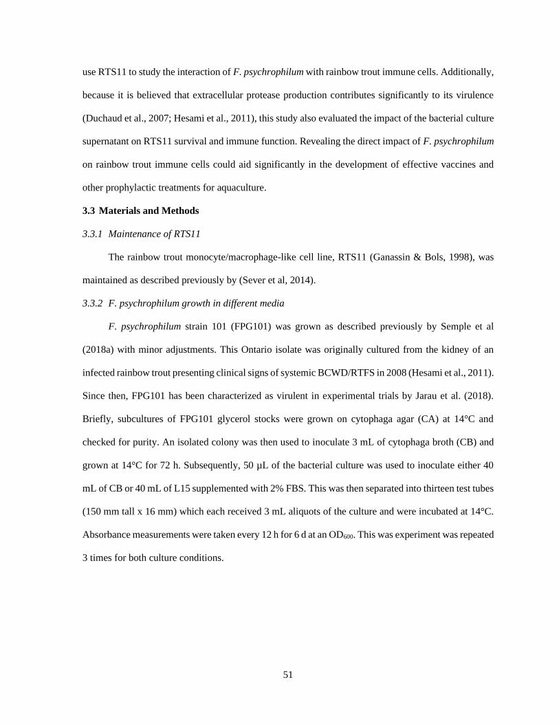
51
use RTS11 to study the interaction of F. psychrophilum with rainbow trout immune cells. Additionally,
because it is believed that extracellular protease production contributes significantly to its virulence
(Duchaud et al., 2007; Hesami et al., 2011), this study also evaluated the impact of the bacterial culture
supernatant on RTS11 survival and immune function. Revealing the direct impact of F. psychrophilum
on rainbow trout immune cells could aid significantly in the development of effective vaccines and
other prophylactic treatments for aquaculture.
3.3 Materials and Methods
3.3.1 Maintenance of RTS11
The rainbow trout monocyte/macrophage-like cell line, RTS11 (Ganassin & Bols, 1998), was
maintained as described previously by (Sever et al, 2014).
3.3.2 F. psychrophilum growth in different media
F. psychrophilum strain 101 (FPG101) was grown as described previously by Semple et al
(2018a) with minor adjustments. This Ontario isolate was originally cultured from the kidney of an
infected rainbow trout presenting clinical signs of systemic BCWD/RTFS in 2008 (Hesami et al., 2011).
Since then, FPG101 has been characterized as virulent in experimental trials by Jarau et al. (2018).
Briefly, subcultures of FPG101 glycerol stocks were grown on cytophaga agar (CA) at 14°C and
checked for purity. An isolated colony was then used to inoculate 3 mL of cytophaga broth (CB) and
grown at 14°C for 72 h. Subsequently, 50 µL of the bacterial culture was used to inoculate either 40
mL of CB or 40 mL of L15 supplemented with 2% FBS. This was then separated into thirteen test tubes
(150 mm tall x 16 mm) which each received 3 mL aliquots of the culture and were incubated at 14°C.
Absorbance measurements were taken every 12 h for 6 d at an OD600. This was experiment was repeated
3 times for both culture conditions.

52
3.3.3 Bacterial growth for RTS11 exposure conditions
3.3.3.1 Live and heat-killed F. psychrophilum
An isolated colony of F. psychrophilum strain 101 (FPG101) was used to inoculate 3 mL of
cytophaga broth and was grown as described in section 3.3.2. After this time, the OD600 of the bacterial
growth was consistently between 0.4-0.5, indicating a viable bacterial count of 2-5 x 10⁸ CFU/mL. For
every culture of FPG101, a standard plate count (SPC) was completed to confirm the anticipated
bacterial concentration. Two 0.5 mL aliquots were removed from the 3 mL culture and centrifuged for
8 min at 8000 rpm. The resulting cell pellets were then washed twice with phosphate buffered saline
(PBS). One cell pellet was resuspended in 0.5 mL of L15 supplemented with 2% fetal bovine serum
(FBS) which was to be used to expose RTS11 to live bacteria.
The remaining cell pellet was resuspended in PBS and boiled for 20 minutes to kill any live
bacteria. After boiling, 100 µL of the heat-killed FPG101 was spread onto a CA plate to confirm the
absence of viable bacteria. Subsequently, the killed bacterial culture was centrifuged for 8 min at 8000
rpm and resuspended in 0.5 mL of L15 supplemented with 2% FBS which was to be used to expose
RTS11 to heat-killed F. psychrophilum.
3.3.3.2 F. psychrophilum conditioned media (FpCM)
Following 72 h of growth at 14°C on CA, isolated colonies of FPG101 were used to inoculate
multiple test tubes containing 3 mL of L15 media supplemented with 2% FBS. These cultures were
grown at 14°C for 72 h after which a streak plate was performed for each culture to ensure purity. The
cultures were then centrifuged for 8 min at 8000 rpm at 4°C so that supernatants could be removed and
passed through a 0.22 µm filter to remove any residual bacteria. The isolated supernatants were
combined and used to expose RTS11 to a final concentration of 50% supernatant (referred to as FpCM).

53
3.3.4 Exposure of RTS11 to live, heat-killed and the supernatant of F. psychrophilum
In 6-well tissue culture plates (ThermoFisher), RTS11 was seeded at 1.5 x 10⁶ cells/well in 2 mL
of L15 media supplemented with 2% FBS and maintained overnight at 14oC. Cells were exposed to a
single dose of either live F. psychrophilum (MOI of 1), heat-killed F. psychrophilum (MOI of 1), F.
psychrophilum supernatant (FpCM) or a media alone control to a final volume of 4 mL per well.
Following this single exposure to the appropriate condition, all experimental plates were returned to
the 14oC incubator. Unless otherwise described, cell pellets were collected for analyses. On Days 1, 2
and 3, the supernatant was collected from experimental wells and adherent cells were mechanically
dislodged using a sterile 23 cm cell scraper (ThermoFisher) and added to the supernatant of respective
wells. All wells were then washed with 1 mL of phosphate buffered saline (PBS, Gibco) which was
also added to the appropriate supernatant/cell mixture. The cells were centrifuged (5 min, 500 x g, 4°C),
washed three times with 5 mL of PBS, and the resulting cell pellets were stored at -80°C for future use.
3.3.5 Viability of RTS11 throughout exposure
To determine the impact that the organism has on RTS11 survival, the cells were exposed to a
single dose of live F. psychrophilum (MOI of 1), killed F. psychrophilum (MOI of 1) or FpCM
(prepared as described in section 2.4). On Days 1, 2 and 3 following this exposure, the media was
collected from experimental wells and any adherent cells were detached using 400 µL of 0.25% trypsin-
EDTA (Gibco). All wells were then washed with 1 mL of PBS. The trypsinized cells were combined
with the collected supernatant which was then centrifuged 500 x g for 5 min at 4oC. The cell pellet was
washed twice with 1 mL of PBS before resuspending in 200 µL of PBS. To determine RTS11 cell
viability after the exposure conditions, a trypan blue (Sigma) exclusion test was performed using a
haemocytometer under a phase contrast microscope (Leica). This experiment was repeated three times.

54
3.3.6 Measuring phagocytic function of RTS11 via flow cytometry
RTS11 was exposed to a single dose of live F. psychrophilum (MOI of 1), killed F.
psychrophilum (MOI of 1), FpCM or no treatment as described above in section 2.4. Immediately
following exposure, each well received 4.6 x 10⁷ FITC 2.0 µm latex beads (L4530, Sigma) after which,
all plates were returned to a 14°C incubator. On 1, 2 and 3 d following exposure, the RTS11 cells were
collected as described above in section 2.4. The final cell pellet was resuspended in 500 µL of PBS
before transfer to a 5 mL polystyrene round-bottom tube (ThermoFisher).
The fluorescence of the beads within RTS11 cells was analyzed by flow cytometry using the
FACSAria Fusion Cell Sorter with an integrated 3-laser and 9-color detection unit (BD Biosciences).
The RTS11 cells that had phagocytosed the beads were gated to exclude the count of free FITC-
conjugated beads from the measurement. Data is expressed as the percentage of RTS11 cells containing
FITC-conjugated beads (10,000 events) after standardizing to the media only control. Data analysis
was completed using the FlowJo® software (https://www.flowjo.com).
3.3.7 qRT-PCR
3.3.7.1 RNA extraction and cDNA synthesis
RNA was extracted from RTS11 cell pellets (1.5 x 10⁶ cells) using an RNeasy RNA Extraction
Kit (Qiagen) as described by the manufacturer. To remove any contaminating genomic DNA, all RNA
samples were treated with DNase I (Thermo Scientific). RNA samples were then quantified using the
Take3 plate of a Synergy H1 plate reader (BioTek Instruments) and were stored at -80oC until further
use. Complementary DNA (cDNA) was synthesized from 500 ng of total RNA using the qScript cDNA
Supermix (Quanta Biosciences) in accordance to the manufacturer’s instructions. For a no template
control, 500 ng of RNA suspended in 20 uL of DEPC water was included in the cDNA synthesis
reaction without reverse transcriptase.

55
3.3.7.2 qRT-PCR Reactions
To assess transcript levels of IL-1β, TNFα, IL-6, and IL-10 in RTS11 cells, qRT-PCR analysis
was completed. All PCR reactions were 10 µl and contained: 2.5 μl of cDNA (25 ng/μl diluted 1:10 in
Rnase free water), 2x WISENT ADVANCEDTM qPCR mastermix (Wisent), and forward and reverse
primers (Sigma Aldrich) to a final working concentration of 0.25 μM. All qPCR reactions were
completed on the LightCycler® 480 II (Roche). The sequences for all primer sets are outlined in Table
3.1. Each experimental sample was run in triplicate. For each plate, triplicate wells of a calibrator, no
template control and RNA only control were also present. The program used for all qRT-PCR reactions
was as follows: pre-incubation at 95°C for 10 min followed by 40 cycles of denaturation at 95°C for
10 sec, annealing at 60°C for 5 sec and extension at 72°C for 8 sec. A melting curve was completed for
every run from 65°C to 97°C with a read every 5 sec. Product specificity was determined through single
PCR melting peaks. All qRT-PCR data was analyzed using the ΔΔCt method and is presented as the
average of 3 experimental replicates with the standard deviation. Specifically, gene expression was
normalized to the reference gene (EF1α) and expressed as fold change over the day 0 control group
where control expression was set to 1.
Table 3.1. Primers used for qRT-PCR analysis of cytokine genes in RTS11 cells. Forward (F) and
reverse (R) primer sequences for the genes analyzed are presented along with the base pair length for
each amplicon and the original references or Genbank accession numbers for primer sets.
Primer
Name
Sequence (5’ – 3’) Length Reference or
Accession No.
IL-1β F: CCACAAAGTGCATTTGAAC
R: GCAACCTCCTCTAGGTGC
155 Semple et al,
2018b
TNFα F: GTGCAAAAGATACCCACC
R: CACTGCACGGTGTCAG
108 Semple et al,
2018b
IL-6 F: CTTCTACACGCTATCTCTCACTC
R: CGTCTGTCCCGAGCT
128 Semple et al,
2018b
IL-10 F: GCCTTCTCCACCATCAGAGAC
R: GATGCTGTCCATAGCGTGAC
120 NM_001245099.1
EF1α F: CGCACAGTAACACCGAAACTAATTAAGC
R: GCCTCCGCACTTGTAGATCAGATG
134 Semple et al,
2018b

56
3.3.8 Statistical analyses
After confirming a normal distribution and homogeneity of variance, statistical analyses were
performed using a two-way ANOVA. This was then followed by a Tukey’s posthoc test to determine
significant differences between the exposure conditions at each individual timepoint. A p-value of less
than 0.05 was considered statistically significant. All statistical analyses were completed using the
software Statistica version 7 (StatSoft, Tulsa, OK).
3.4 Results
3.4.1 Growth of F. psychrophilum in preferred and cell culture media
F. psychrophilum strain 101 was found to grow well in its preferred culture media, cytophaga
broth with the logarithmic phase beginning at 24 h and ending at approximately 84 h (Figure 3.1). In
comparison, although F. psychrophilum was capable of growth in L15 supplemented with 2% FBS,
this growth was much slower and absorbance readings were significantly lower when compared to
growth in cytophaga broth. Further, what would be considered the logarithmic phase during growth in
2% FBS was much more prolonged beginning at 36 h and reaching a plateau at 120 h (Figure 1).
Figure 3.1. A comparison of F. psychrophilum growth in culture media for bacterial cells
(cytophaga broth) and for animal cells (L15 with 2 % FBS). Growth was at 14°C and was monitored
every 12 h for 144 h by measuring optical density at 600 nm. This figure represents three independent
experiments for each culture condition. Data are presented as the mean with error bars representing
±SD.
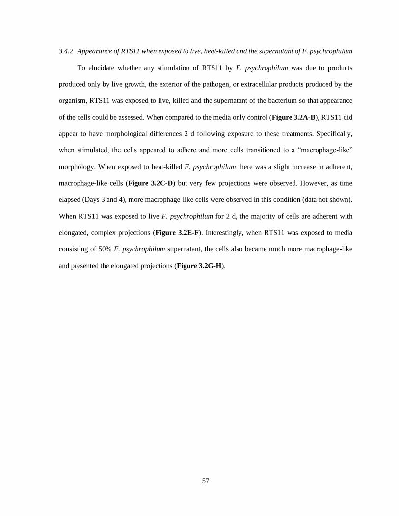
57
3.4.2 Appearance of RTS11 when exposed to live, heat-killed and the supernatant of F. psychrophilum
To elucidate whether any stimulation of RTS11 by F. psychrophilum was due to products
produced only by live growth, the exterior of the pathogen, or extracellular products produced by the
organism, RTS11 was exposed to live, killed and the supernatant of the bacterium so that appearance
of the cells could be assessed. When compared to the media only control (Figure 3.2A-B), RTS11 did
appear to have morphological differences 2 d following exposure to these treatments. Specifically,
when stimulated, the cells appeared to adhere and more cells transitioned to a “macrophage-like”
morphology. When exposed to heat-killed F. psychrophilum there was a slight increase in adherent,
macrophage-like cells (Figure 3.2C-D) but very few projections were observed. However, as time
elapsed (Days 3 and 4), more macrophage-like cells were observed in this condition (data not shown).
When RTS11 was exposed to live F. psychrophilum for 2 d, the majority of cells are adherent with
elongated, complex projections (Figure 3.2E-F). Interestingly, when RTS11 was exposed to media
consisting of 50% F. psychrophilum supernatant, the cells also became much more macrophage-like
and presented the elongated projections (Figure 3.2G-H).

58
Figure 3.2. Influence of live, heat-killed and F. psychrophilum conditioned media (FpCM) on the
morphology of RTS11. RTS11 was exposed to either cell culture media alone (A, B), heat-killed F.
psychrophilum at a MOI of 1 (C, D), live F. psychrophilum at an MOI of 1 (E, F), or FpCM which
refers to 50% media conditioned with 3 days of F. psychrophilum growth in L15/FBS (G, H). Black
arrowheads indicate the F. psychrophilum bacteria. Representative pictures were collected to observe
morphological differences in RTS11 at Day 2 following these exposure conditions.
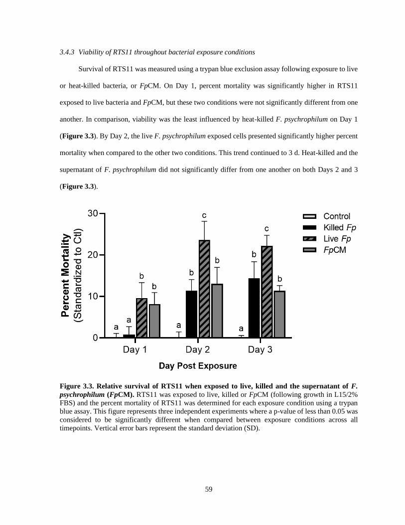
59
3.4.3 Viability of RTS11 throughout bacterial exposure conditions
Survival of RTS11 was measured using a trypan blue exclusion assay following exposure to live
or heat-killed bacteria, or FpCM. On Day 1, percent mortality was significantly higher in RTS11
exposed to live bacteria and FpCM, but these two conditions were not significantly different from one
another. In comparison, viability was the least influenced by heat-killed F. psychrophilum on Day 1
(Figure 3.3). By Day 2, the live F. psychrophilum exposed cells presented significantly higher percent
mortality when compared to the other two conditions. This trend continued to 3 d. Heat-killed and the
supernatant of F. psychrophilum did not significantly differ from one another on both Days 2 and 3
(Figure 3.3).
Figure 3.3. Relative survival of RTS11 when exposed to live, killed and the supernatant of F.
psychrophilum (FpCM). RTS11 was exposed to live, killed or FpCM (following growth in L15/2%
FBS) and the percent mortality of RTS11 was determined for each exposure condition using a trypan
blue assay. This figure represents three independent experiments where a p-value of less than 0.05 was
considered to be significantly different when compared between exposure conditions across all
timepoints. Vertical error bars represent the standard deviation (SD).

60
3.4.4 Impact of live, killed and F. psychrophilum supernatant on phagocytic function
Following exposure to both FITC labelled beads and one of either live or killed bacteria or the
supernatant of F. psychrophilum, flow cytometric analysis was conducted to determine the impact that
these exposure conditions had on the phagocytic activity of RTS11. When treated with live and heat-
killed F. psychrophilum, RTS11 did not differ from each other at all timepoints analyzed (Figure 3.4A).
Interestingly, when RTS11 was exposed the FpCM condition (50% of the F. psychrophilum
conditioned media), phagocytic function was significantly suppressed at Day 1 (Figure 3.4A).
Phagocytic function recovered in the FpCM exposure condition by Day 2 and this condition, along with
live F. psychrophilum exposure, actually increased phagocytic activity by Day 3. Representative
images of RTS11 in the various exposure conditions reveal that some cells became engorged with the
FITC labelled beads while others appeared to phagocytose very few (Figure 3.4B).
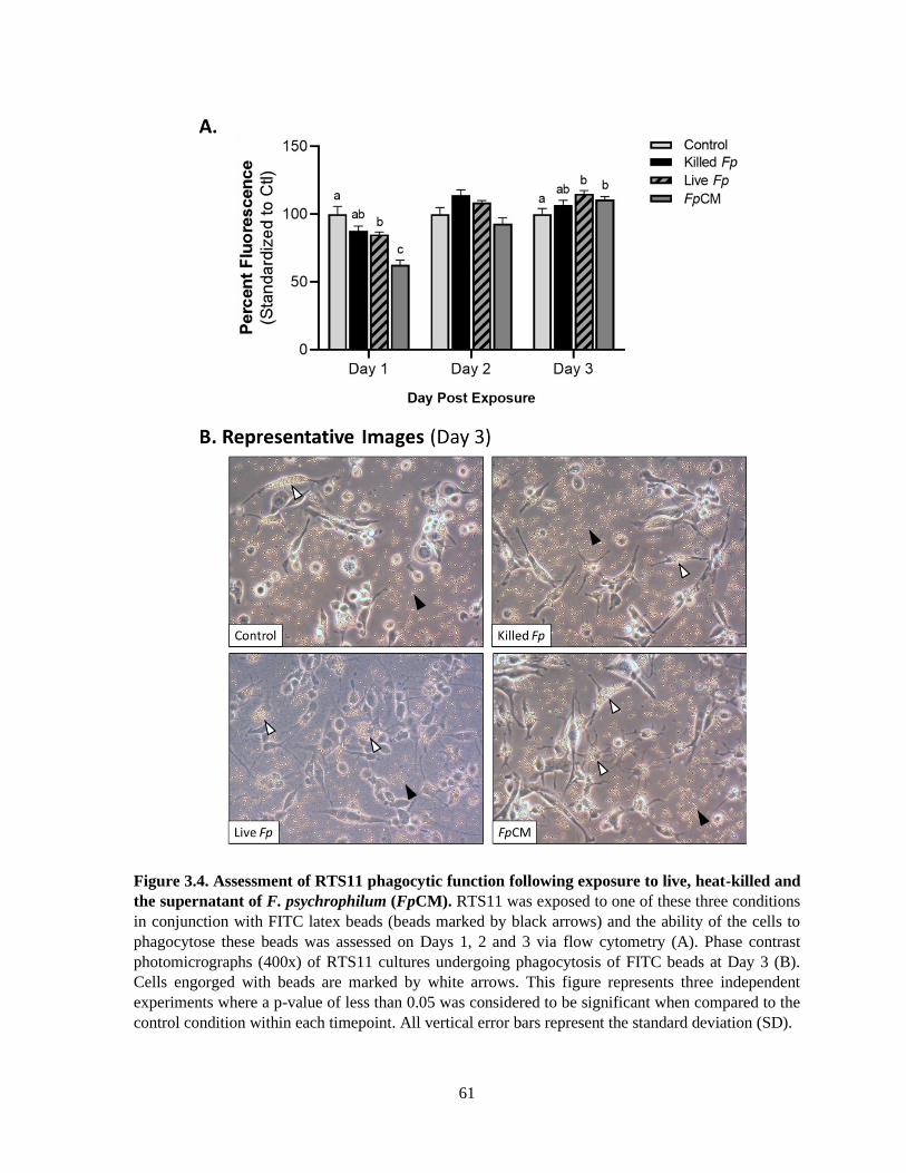
61
Figure 3.4. Assessment of RTS11 phagocytic function following exposure to live, heat-killed and
the supernatant of F. psychrophilum (FpCM). RTS11 was exposed to one of these three conditions
in conjunction with FITC latex beads (beads marked by black arrows) and the ability of the cells to
phagocytose these beads was assessed on Days 1, 2 and 3 via flow cytometry (A). Phase contrast
photomicrographs (400x) of RTS11 cultures undergoing phagocytosis of FITC beads at Day 3 (B).
Cells engorged with beads are marked by white arrows. This figure represents three independent
experiments where a p-value of less than 0.05 was considered to be significant when compared to the
control condition within each timepoint. All vertical error bars represent the standard deviation (SD).
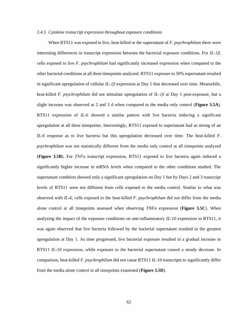
62
3.4.5 Cytokine transcript expression throughout exposure conditions
When RTS11 was exposed to live, heat-killed or the supernatant of F. psychrophilum there were
interesting differences in transcript expression between the bacterial exposure conditions. For IL-1β,
cells exposed to live F. psychrophilum had significantly increased expression when compared to the
other bacterial conditions at all three timepoints analyzed. RTS11 exposure to 50% supernatant resulted
in significant upregulation of cellular IL-1β expression at Day 1 that decreased over time. Meanwhile,
heat-killed F. psychrophilum did not stimulate upregulation of IL-1β at Day 1 post-exposure, but a
slight increase was observed at 2 and 3 d when compared to the media only control (Figure 3.5A).
RTS11 expression of IL-6 showed a similar pattern with live bacteria inducing a significant
upregulation at all three timepoints. Interestingly, RTS11 exposed to supernatant had as strong of an
IL-6 response as to live bacteria but this upregulation decreased over time. The heat-killed F.
psychrophilum was not statistically different from the media only control at all timepoints analyzed
(Figure 3.5B). For TNFα transcript expression, RTS11 exposed to live bacteria again induced a
significantly higher increase in mRNA levels when compared to the other conditions studied. The
supernatant condition showed only a significant upregulation on Day 1 but by Days 2 and 3 transcript
levels of RTS11 were not different from cells exposed to the media control. Similar to what was
observed with IL-6, cells exposed to the heat-killed F. psychrophilum did not differ from the media
alone control at all timepoints assessed when observing TNFa expression (Figure 3.5C). When
analyzing the impact of the exposure conditions on anti-inflammatory IL-10 expression in RTS11, it
was again observed that live bacteria followed by the bacterial supernatant resulted in the greatest
upregulation at Day 1. As time progressed, live bacterial exposure resulted in a gradual increase in
RTS11 IL-10 expression, while exposure to the bacterial supernatant caused a steady decrease. In
comparison, heat-killed F. psychrophilum did not cause RTS11 IL-10 transcripts to significantly differ
from the media alone control in all timepoints examined (Figure 3.5D).
63
Figure 3.5. Influence of live, killed and the supernatant of F. psychrophilum (FpCM) on RTS11
cytokine mRNA expression 1-3 Days following exposure. Transcript expression of pro-inflammatory
cytokines IL-1β (A), TNFα (B), IL-6 (C) as well as the anti-inflammatory cytokine IL-10 (D) was
assessed so that the effect of the different bacterial exposure conditions on immune function could be
analyzed. All data was normalized to the reference gene (EF1α) and expressed as a fold change over
the Day 0 control group where control expression was set to 1. All panels represent three independent
experiments and are presented as means + SD. A p-value of less than 0.05 was considered to be
statistically significant when compared to the media alone control for each timepoint.
3.5 Discussion
There has been a growing interest in the study of F. psychrophilum due to its harmful influence
on global rainbow trout aquaculture. Initially, simply isolating and culturing the organism in vitro was
problematic due to the absence of effective media (reviewed by Nematollahi et al., 2003; Starliper,
2011). Since then, there have been several media formulations that have dramatically improved
experimental work with the bacterium including Shieh (Shieh, 1980), cytophaga (Wakabayashi &
Egusa, 1974) and tryptone yeast extract salts broth (TYESB, Holt, 1987). But despite past
complications surrounding the culture of this fastidious organism, the present study has revealed that
F. psychrophilum is capable of considerable growth in cell culture media supplemented with 2% FBS.

64
This was unexpected as isolates of F. psychrophilum normally do not grow, or grow poorly, in high
nutrient media (reviewed by Starliper, 2011). Though several types of animal sera have been used for
cell culture work, fetal bovine serum is notoriously nutrient dense, contains many valuable growth
factors and also presents low levels of antibodies (Bettger & McKeehan, 1986; Fang et al., 2017). These
characteristics allow for considerable growth in essentially any cell culture system, which has made
FBS the most commonly used media supplement (Fang et al., 2017). Because infection with F.
psychrophilum can present as a septicaemia (Ekman & Norrgren, 2003), perhaps FBS provides a
physiological environment that mimics blood and enables the organism to grow. Though only
supplementation with 2% FBS was used here, further analyses should be completed to explore the
maximum and minimum nutrient requirements for F. psychrophilum growth in this system. The ability
of F. psychrophilum to grow in cell culture media means that live exposure of the pathogen is a
possibility for in vitro cell culture systems. Additionally, further study of the bacterial supernatant
(FpCM) as well as its impact on eukaryotic cells can now be achieved.
The structure of any bacterium represents a complex mixture of pathogen associated molecular
patterns (PAMPs) that can alert the host immune system of foreign invasion. To avoid this detection,
many bacterial pathogens can generate exotoxins, cytolysins, proteases, and other extracellular
substances that suppress and/or confuse immune responses (Esteve et al., 1995; Ruckdeschel et al.,
1997; Hava et al., 2008). These factors prolong pathogen survival thus enhancing its virulence and
fitness. When RTS11 was exposed to F. psychrophilum supernatant the cells became more
macrophage-like, similar to when they were exposed to live pathogen alone. Therefore, it appears that
both the bacterium, and something produced by the organism, are capable of stimulating RTS11. Under
normal conditions, the composition of RTS11 is expected to be roughly 90% monocyte-like with the
remaining 10% of cells presenting an adherent, macrophage-like morphology (Ganassin & Bols, 1998).
But when RTS11 cells were stimulated with phorbol 12-myristate 13-acetate (PMA), these proportions
changed so that a higher percentage of cells became macrophage-like (Ganassin & Bols, 1998),
comparable to what was observed in the current study. Based on these collective results it seems that
65
the monocyte cells of RTS11 will differentiate in a manner analogous to what has been noted in whole
animal models following appropriate stimulation (reviewed by Shi & Pamer, 2011). In the present
study, it appears that live F. psychrophilum, the bacterial supernatant and, to a lesser extent, the heat-
killed pathogen are able to provide this stimulatory effect. As a host model system for the study of F.
psychrophilum infection, RTS11 provides the potential to learn more about this bacterial pathogen in
relation to immune cells from a suitable host.
Several mammalian pathogens actively kill immune cells thereby preventing the host from
mounting an effective response to control infection (reviewed by Menestrina et al., 1995; Narayanan et
al., 2002; Alonzo & Torres, 2013). Quite often this strategy proves effective for the pathogen but there
has been little observation of similar virulence factors in aquatic diseases. Using the in vitro model
developed here, only live F. psychrophilum was found to consistently reduce the survival of RTS11 at
all timepoints analyzed. Interestingly at Day 1, the supernatant alone was observed to induce RTS11
mortality akin to that of the live F. psychrophilum condition. Yet by Days 2 and 3, only live bacterial
exposure significantly reduced RTS11 survival. Numerous bacterial extracellular products have been
found to be cytotoxic to mammalian macrophages. The supernatant of Pseudomonas aeruginosa as
well as an invasin produced by Salmonella typhimurium both induce apoptosis in murine macrophages
(Hersh et al., 1999; Zhang et al., 2003). Because live F. psychrophilum would be expected to
continually replenish the extracellular products when compared to supernatant exposure alone, it is
possible that the aquatic pathogen secretes a product that reduces survival of rainbow trout immune
cells. If this secreted substance was used up or degraded over time, it would explain why the supernatant
alone condition only increased mortality at Day 1 while the live bacterial condition continued to have
a greater impact on RTS11 survival throughout the trial. Further exploration regarding the type of cell
death observed here would be required to fully understand the impact of F. psychrophilum on rainbow
trout immune cells.
The defining feature of a macrophage is its ability to engulf and subsequently digest apoptotic
cells as well as any abnormal or foreign entities (reviewed by Gordon & Pluddemann, 2017).

66
Depending on the constituents of that which was consumed, these highly versatile cells will then direct
immune responses to ensure healing, homeostatic maintenance or pathogen clearance (reviewed by
Flannagan et al., 2011). Given the importance of phagocytosis for averting microbial colonization
within a host, it is not surprising that many bacterial pathogens develop virulence factors to inhibit this
ability (reviewed by Ernst, 2001; Celli & Finlay, 2002; Sarantis & Grinstein, 2012). In the present
study, it was observed that the supernatant of F. psychrophilum significantly suppressed the phagocytic
activity of RTS11 at 1 d post-exposure when compared to the control condition. By Day 3, phagocytic
activity of RTS11 recovered in the supernatant exposure, but perhaps it would have continued to impede
had fresh supernatant been replaced over time. There are many ways that bacterial pathogens can
suppress the phagocytic activity of macrophages. Virulent strains of the aquatic pathogen
Photobacterium damselae piscicida produce a toxin called AIP56 (apoptosis inducing protein of 56
kDa) which causes apoptotic destruction of macrophages and neutrophils in infected sea bass (do Vale
et al., 2003; do Vale et al., 2007; Costa-Ramos et al., 2011). This prevents phagocytosis by eliminating
cell types capable of this function. Alternatively, some bacteria secrete products that inactivate the
cellular machinery required to form phagosomes around extracellular pathogens. An emerging human
pathogen, Photorhabdus asymbiotica, produces a toxin that causes actin disassembly so that phagocytes
are unable to migrate and/or phagocytose effectively (Jank et al., 2013). Yet another strategy used to
decrease the efficacy of phagocytosis is inhibition of opsonization. A secreted metalloprotease of
Staphylococcus aureus, aureolysin, acts as an alternative C3 convertase. Aureolysin cleaves C3 into
forms of C3a and C3b that degrade quickly without binding to bacteria, thus preventing the anticipated
opsonization by these complement proteins (Laarman et al., 2011). F. psychrophilum is known to
produce several extracellular enzymes (Duchaud et al., 2007; Hesami et al., 2011; Castillo et al., 2016)
so it is possible that some may act as virulence factors to inhibit leukocyte function as noted here and
in previous studies (Lammens et al., 2000; Semple et al., 2018a). Although reagents for teleost proteins
and immune cells are not readily available for further validation, the results presented here connect the
virulence of F. psychrophilum to impairment of phagocytosis in rainbow trout macrophages.
67
As a first line of defense in host tissues, macrophages have the responsibility of processing
foreign substances and ensuring that the immune system responds accordingly. This task is mediated
through cytokines produced by the macrophages to properly guide and activate immune cells (reviewed
by Zhang & Wang, 2014). Quite often for bacterial pathogens, such as F. psychrophilum, it is
anticipated that the initial reaction will be pro-inflammatory to combat the pathogen and inhibit its
growth, followed by an anti-inflammatory response during the resolution of infection (reviewed by
Wilson et al., 1998; Lauw et al., 2002). Because inflammatory responses can be quite damaging to the
surrounding tissues, it is very important to have these reactions well controlled (reviewed by Lauw et
al., 2002; Chen et al., 2018). When RTS11 was exposed to live F. psychrophilum the cytokine levels
fit this described expression profile, and at Day 1 the bacterial supernatant was found to stimulate a
similar, yet less intense, response. Unexpectedly, heat-killed F. psychrophilum resulted in very little to
no significant differences in gene expression when compared to control RTS11 cells. Similar
transcriptional trends have been observed in head kidney leukocytes isolated from rainbow trout
susceptible to F. psychrophilum infection (Langevin et al., 2012). When measuring transcript
expression 5 d following exposure to live F. psychrophilum, these primary leukocytes upregulated IL-
1β, TNFα and IL-10 (Langevin et al., 2012). Stimulation of pro-inflammatory cytokines was also
observed in whole spleen tissue, where both TNFα and IL-1β transcript expression was induced upon
live F. psychrophilum challenge in resistant and susceptible rainbow trout lines (Kutyrev et al., 2016).
In fact, IL-1β gene expression has been observed to be overexpressed in multiple rainbow trout tissues
(gills, liver, spleen and kidney) when naturally infected with F. psychrophilum (Orieux et al., 2013).
These previous studies support the cytokine response that was observed here when co-culturing RTS11
with F. psychrophilum. Thus RTS11 is an affordable, reliable model for understanding both the immune
response to, and the pathogenesis of, F. psychrophilum.
Many attempts to create an effective vaccine for BCWD/RTFS have focused on the use of
killed whole-cell bacteria (Kirici et al., 2014; Hoare et al., 2017), likely because this vaccination method

68
has proven effective for other aquatic diseases such as vibriosis and its corresponding Vibrogen 2
vaccine (reviewed by Frans et al., 2011). Interestingly, though the killed F. psychrophilum used here
could not stimulate a response in RTS11, our group has previously shown that the killed whole-cell
bacteria of Vibrogen 2 can increase pro-inflammatory cytokine expression in the same cell line
(Soulliere, 2017). In terrestrial pathogens, it has been shown that different species of heat-killed
bacteria, even between strains of the same bacterial species, can result in very diverse gene expression
profiles (Sela et al., 2018). This is likely due to the components of the bacterial membrane, thus F.
psychrophilum cells alone may not be as immunogenic as those of Vibrio anguillarum. Perhaps future
vaccine regimes for F. psychrophilum should incorporate the extracellular products of the organism to
provide protection against its secreted virulence factors as well as the pathogen itself. This vaccination
technique has proven protective for farmed sole when injected with sublethal doses of Vibrio
extracellular products (Zorrilla et al., 2003). Based on the results presented here, it is evident that killed
F. psychrophilum alone does not stimulate a strong response in rainbow trout immune cells, thus more
research should be devoted to understanding the pathogen to streamline efforts for vaccine design.
3.6 Conclusions
Despite the damaging role that F. psychrophilum plays in the global rainbow trout aquaculture
industry, there is very little known regarding the pathogenesis of this opportunistic pathogen. In the
present study, a model system was developed using the rainbow trout monocyte/macrophage-like cell
line, RTS11, to study the infection cycle of F. psychrophilum. The results presented show that the
organism can grow in media used for successful culture of RTS11, making it possible to expose these
cells to live, heat-killed and the supernatant of F. psychrophilum. When exposed to these different
conditions, RTS11 responded by making a pronounced morphological change to become primarily
macrophage-like. The live F. psychrophilum condition was shown to significantly decrease the survival
of RTS11 with the bacterial supernatant having a negative impact only at earlier timepoints. Regarding
cellular function, phagocytosis was inhibited primarily by the supernatant, indicating that the organism
may produce virulence factors to evade the host immune cells. Finally, it was found that only live

69
bacteria and the supernatant of F. psychrophilum were able to stimulate a significant cytokine response
in RTS11, which revealed that perhaps the use of killed whole-cell bacteria for vaccine development
may not induce the protective response that is desired. Though further study is required, it is clear that
gaining a deeper understanding of F. psychrophilum pathogenesis will aid massively in vaccine design
and the development of other treatments options for BCWD/RTFS.
Acknowledgements
Terence Tang for his technical assistance with flow cytometry.

70
Chapter 4: PACAP is lethal to Flavobacterium psychrophilum through direct membrane
permeabilization and indirectly, by priming the immune response in RTS11.
Shawna L. Semple1, Tania Rodríguez-Ramos1, Yamila Carpio2, John S. Lumsden3, Mario P. Estrada2
and Brian Dixon1
1University of Waterloo, Department of Biology, Waterloo, Canada 2Center for Genetic Engineering and Biotechnology, Havana, Cuba 3University of Guelph, Ontario Veterinary College, Department of Pathobiology, Guelph, Canada
Frontiers in Immunology, 10, pp. 1-14. 2019.

71
4.1 Overview
Pituitary adenylate cyclase-activating polypeptide (PACAP) is a multifunctional neuropeptide that is
widely distributed in mammals and is capable of performing roles as a neurotransmitter,
neuromodulator and vasodilator. This polypeptide belongs to the glucagon/secretin superfamily, of
which some members have been shown to act as antimicrobial peptides in both mammalian and aquatic
organisms. In teleosts, PACAP has been demonstrated to have direct antimicrobial activity against
several aquatic pathogens, yet this phenomenon has never been studied throughout a live bacterial
challenge. The present study focuses on the influence of synthetic Clarias gariepinus 38 amino acid
PACAP on the rainbow trout monocyte/macrophage-like cell line, RTS11, when exposed to the
coldwater bacterial pathogen Flavobacterium psychrophilum. PACAP was shown to have direct
antimicrobial activity on F. psychrophilum when grown in both cytophaga broth and cell culture media
(L15). Further, the ability of teleostean PACAP to permeabilize the membrane of an aquatic pathogen,
F. psychrophilum, was demonstrated for the first time. The viability of RTS11 when exposed to PACAP
was also observed using a trypan blue exclusion assay to determine optimal experimental doses of the
antimicrobial peptide. This displayed that only concentrations lower than 0.1 µM did not have a
negative impact on RTS11 survival. Interestingly, when RTS11 was pre-treated with PACAP for 24 h
before experiencing infection with live F. psychrophilum, growth of the pathogen was severely
inhibited in a dose-dependent manner when compared to cells receiving no pre-treatment with the
polypeptide. Relative expression of pro-inflammatory cytokines (IL-1β, TNFα and IL-6) and PACAP
receptors (VPAC1 and PAC1) was also analyzed in RTS11 following PACAP exposure alone and in
conjunction with live F. psychrophilum challenge. These qRT-PCR findings revealed that PACAP may
have a synergistic effect on RTS11 immune function. The results of this study provide evidence that
PACAP has immunostimulatory activity on rainbow trout immune cells as well as antimicrobial activity
against aquatic bacterial pathogens such as F. psychrophilum. As there are numerous pathogens that
plague the aquaculture industry, PACAP may stimulate the teleost immune system while also providing
an efficacious alternative to antibiotic use.

72
4.2 Introduction
Due to the rising demand for fish protein (FAO, 2018), aquaculture has become a necessary
means to protect wild populations from irreversible overfishing. As such, it is imperative that these
culture systems have a minimal impact on the environment while still being able to provide the high-
quality product for market. To attain this goal, alternative methods must be developed to combat
infectious disease as this is one of the greatest sources of instability and financial cost in aquaculture.
Global losses due to aquatic infections total approximately $6 billion USD (World Bank, 2014) and
currently, fish farmers have few methods outside of antibiotics to prevent/control outbreaks. With
multi-drug resistance continually rising (reviewed by Santos & Ramos, 2018; Watts et al., 2017),
antibiotic use in aquaculture is tightly regulated which often leaves farmers with few options when
outbreaks do occur. This problem has led to an increased interest in the development of alternative
approaches for disease prevention, including the use of naturally occurring antimicrobial peptides
(AMPs). Though novel AMPs and their activities are continuously being discovered, one that has
gained a lot of interest as a result of its vast pleiotropic effects is pituitary adenylate cyclase activating
polypeptide (PACAP).
Initially, PACAP was discovered as a neuropeptide due to its ability to stimulate adenylate
cyclase activity in ovine pituitary cell cultures (Miyata et al., 1989). Derived from a 175 amino acid
precursor, functional PACAP has two molecular forms. The first has 38 amino acids (PACAP-38) while
the other form is truncated containing only 27 residues (PACAP-27, Kimura et al., 1990; Ogi et al.,
1990). Of the two, PACAP-38 is considered to be more bioactive as it has been shown to display 100-
1000 times greater potency in stimulating cell proliferation, DNA synthesis and inositol phospholipid
turnover in cells (Deutsch & Sun 1992; Matsumoto et al., 1993). Further analysis of PACAP-38
revealed that this peptide shared 68% sequence similarity with vasoactive intestinal polypeptide (VIP),
thereby classifying PACAP as a member of the secretin/glucagon/growth hormone-releasing
hormone/vasoactive intestinal peptide superfamily (Miyata et al., 1989). As this was the case, it is not
surprising that PACAP-38 is able to bind with equal affinity to the same G-coupled protein receptors
73
(GCPRs) as VIP, vasoactive intestinal polypeptide receptor 1 (VPAC1) and vasoactive intestinal
polypeptide receptor 2 (VPAC2), while also binding to its own receptor, pituitary adenylate cyclase-
activating polypeptide type I receptor (PAC1, Pisegna & Wank 1993; Shivers et al., 1991). All three of
these receptors have a wide tissue distribution much like the neuropeptides themselves (Zhu et al.,
2015). PACAP-38 in particular, displays a broad range of functions in multiple tissue types, including
antimicrobial activity, growth, immunomodulation, neural development, anti-tumor activity and
metabolism to name a few (Adams et al., 2008; Gray et al., 2001; Leyton et al,. 1999; Murakami et al.,
1995; Starr et al. 2018). From an evolutionary perspective, the amino acid sequence of PACAP-38 is
identical in all mammals with only a few amino acid substitutions when comparing to other species
(e.g. frog, salmon, tunicate, etc.). PACAP-38 therefore must play a vital role in physiological function
as it has remained essentially unchanged for ~700 million years (Arimura, 1998). This broad functional
profile as well as its highly conserved nature has made PACAP-38 an attractive candidate for disease
control and therapeutic use in aquaculture.
Though there are numerous pathogens that impact the aquaculture industry, Flavobacterium
psychrophilum has proven to be a global threat in the culture of freshwater rainbow trout
(Oncorhynchus mykiss). This gram-negative bacterial pathogen is the causative agent of bacterial
coldwater disease (BCWD) and rainbow trout fry syndrome (RTFS), two separate conditions that can
occur depending on the bacterial isolate, geographical location and age of the host (Lumsden et al.,
2004). These conditions present as either an acute bacteremia primarily in small fish (RTSF) or as a
more chronic disease most commonly characterized by an ulcerative dermatitis (BCWD) in larger fish
(Lumsden et al., 2004; Starliper, 2011). Though variable, mortality resulting from these conditions
without intervention generally ranges from 2-30% (Wiens et al., 2018) but in extreme cases can be as
high as 50-90% (Brown et al., 1997; Cipriano & Holt 2005; Nilsen et al., 2011b). Despite concerted
efforts to selectively breed for resistance to F. psychrophilum (Marancik et al., 2014; Silverstein et al.,
2009; Vallejo et al., 2014; Wiens et al., 2018) and multiple attempts to develop an effective vaccine
(reviewed by Gomez et al., 2014; Hoare et al., 2017; Makesh et al., 2015), there is little information

74
regarding the pathogenesis of the organism. Based on the data presented thus far, it appears that F.
psychrophilum has an intricate relationship with the spleen and head kidney macrophages of rainbow
trout (Nematollahi et al., 2005; Semple et al., 2018). As such, the spleen monocyte/macrophage-like
cell line, RTS11 (Ganassin & Bols, 1998), is an ideal model system for studying F. psychrophilum
infections. As a relevant immune cell line, RTS11 could provide further insight regarding the
immunomodulatory effects of PACAP-38 as well as its antimicrobial function within an appropriate
infection model.
Previous research involving teleostean PACAP-38 has focused on assessing its antimicrobial
activity when directly dosing aquatic pathogens (Lugo et al., 2019), the growth/immunomodulatory
effects of the peptide alone (Carpio et al., 2008; Lugo et al., 2013; Lugo et al., 2010a), or how
viral/bacterial infection can influence gene expression of the peptide and its associated receptors
(Gorgoglione et al., 2015). Though these results were promising, there is yet to be a study evaluating
the activity of PACAP-38 in a live infection model. Furthermore, the effect of PACAP-38 has never
been explored with respect to the industrially relevant pathogen, F. psychrophilum. The purpose of this
study was to measure and understand the antimicrobial activity of PACAP-38 on F. psychrophilum as
well as to determine whether PACAP could stimulate a protective immune response in RTS11 cells.
Confirming the efficacy of PACAP in an in vitro infection model will provide further evidence to
support its use in in vivo experiments. Additionally, the results of this work could provide valuable
insights regarding the efficacy of PACAP-38 during live infections and thus aid in the development of
a potential alternative for antibiotic use in aquaculture.

75
4.3 Materials and Methods
4.3.1 Maintenance of RTS11
The rainbow trout monocyte/macrophage-like cell line, RTS11 (Ganassin & Bols 1998), was
maintained as described previously by (Sever et al., 2014).
4.3.2 Peptides
4.3.2.1 Synthetic PACAP from the teleost Clarias gariepinus
Clarias gariepinus synthetic PACAP-38 (amino acid sequence of
HSDGIFTDSYSRYRKQMAVKKYLAAVLGRRYRQRFRNK, MW of 4.7 kDa) was purchased from
CS Bio (Shanghai) Ltd, China with 85% purity.
4.3.2.2 Synthetic HSP70 peptide fragment from rainbow trout
A synthetic peptide fragment of rainbow trout HSP70 (amino acid sequence of
CGDQARTSSGASSQ, MW of 1.3 kDa) was purchased from Biomatik with 98% purity.
4.3.3 Growth of F. psychrophilum
F. psychrophilum strain 101 (FPG101) was grown as described previously by (Semple et al.,
2018) with minor adjustments. This bacterial isolate has been characterized as virulent in experimental
trials by Jarau and colleagues (Jarau et al., 2018). Briefly, subcultures of FPG101 glycerol stocks were
grown on cytophaga agar (CA) at 14°C and checked for purity. An isolated colony was then used to
inoculate 3 mL of cytophaga broth (CB) and grown at 14°C for 72 h. After this time, the OD600 of the
bacterial growth was consistently between 0.4-0.5, indicating a viable bacterial count of 2 -5 x 10⁸
CFU/mL. For every culture of FPG101, a standard plate count (SPC) was completed to confirm the
anticipated bacterial concentration.
4.3.4 Minimum inhibitory concentration of C. gariepinus PACAP-38 on F. psychrophilum
The minimum inhibitory concentration (MIC) of C. gariepinus PACAP-38 on FPG101 was
assessed by a broth microdilution peptide assay (BMPA) (Otvos & Cudic 2007). To prepare FPG101
for this assay, 3 mL of CB was inoculated with a single colony and allowed to grow overnight at 14°C.

76
After this time, 1 mL of the growth was centrifuged at 5,000 rpm for 5 min, the supernatant removed,
and the pellet resuspended in 4 mL of fresh CB, to have an OD600 of 0.1-0.4. Finally, the bacterial
suspension was diluted in CB to obtain a final OD600 of 0.001.
The BMPA was made using a flat-bottom 96-well plate (Fisher Scientific). The plate set up
consisted of wells containing 90 μL of bacterial suspension and 10 μL of PACAP at 10 different final
concentrations from 5 to 50 μM. In the positive control wells, PACAP was substituted with 10 μL of
CB while the negative control wells contained 100 μL of CB only. All PACAP concentrations and
controls were tested in triplicates. The bacterial growth was monitored after three days of incubation at
14°C, by measuring the change in the absorbance at 600 nm using a microplate reader (BioTek). The
growth inhibition curves were generated by plotting the OD at 600 nm and the peptide concentration.
The MIC was considered as the lowest concentration of PACAP at which no bacterial growth was
detected (an OD600 of 0).
4.3.5 RTS11 Exposure Trials
4.3.5.1 Exposure to PACAP
In 6-well tissue culture plates (ThermoFisher), RTS11 was seeded at 1.5 x 10⁶ cells/well in 1.5
mL of L15 media with no antibiotics and maintained overnight at 14°C. Cells were exposed to PACAP
concentrations of either 0.0002 µM, 0.002 µM, 0.02 µM, 0.1 µM, 0.2 µM, 2 µM, 20 µM or a no PACAP
control to a final volume of 4 mL per well. Following this single exposure to PACAP, all experimental
plates were returned to the 14°C incubator. On days 1, 2 and 3, the supernatant was collected from
experimental wells and adherent cells were mechanically dislodged using a sterile 23 cm cell scraper
(ThermoFisher) and added to the supernatant of respective wells. All wells were then washed with 1
mL of phosphate buffered saline (PBS, Gibco) which was also added to the appropriate supernatant/cell
mixture. The cells were centrifuged (5 min, 500 x g, 4°C), washed once with 5 mL of PBS, and the
resulting cell pellets were stored at -80°C for future use.
77
4.3.5.2 Simultaneous exposure to both PACAP and live F. psychrophilum
In a second experiment, RTS11 was exposed to PACAP concentrations of 0.0002 µM, 0.002
µM, 0.02 µM and 0.1 µM in similar conditions as described above in the first PACAP trial (section
4.3.5.1). Prior to the single addition of PACAP, 0.5 mL of FPG101 was added to each well at bacterial
concentrations ranging from 1.3-2.0 x 10⁶ CFU/mL (multiplicity of infection [MOI] of 0.7-1.3).
Sampling was completed as described above (section 4.3.5.1).
4.3.5.3 Pre-treatment with PACAP followed by infection with live F. psychrophilum
It was observed that F. psychrophilum grew rapidly in wells when exposed to RTS11
simultaneously with PACAP. Because it was possible that PACAP might not be able to influence either
RTS11 alone, the F. psychrophilum alone, or both due to this rapid growth, PACAP was added to
RTS11 wells 24 h before the addition of live F. psychrophilum. Otherwise all procedures for sample
collection and exposure were identical to those described above in the first PACAP experiment (section
4.3.5.1).
4.3.6 Survival of RTS11 following PACAP exposure
To determine whether PACAP negatively influenced RTS11 viability, the cells were exposed to
a single dose of 0.002 µM, 0.02 µM, 0.1 µM, 0.2 µM, 2 µM, 20 µM or a no PACAP control as described
above. On days 1, 2 and 3 following this exposure, the supernatant was collected from experimental
wells and any adherent cells were detached using 400 µL of 0.25% trypsin-EDTA (Gibco) and the wells
washed with 1 mL of PBS. These trypsinized cells were combined with the collected supernatant which
was then centrifuged 500 x g for 5 min at 4°C. The cell pellet was washed twice with 1 mL of PBS
before resuspending in 200 µL of PBS. To determine RTS11 cell viability after exposure to PACAP, a
trypan blue (Sigma) exclusion test was performed using a haemocytometer under a phase contrast
microscope (Leica). This experiment was repeated three times.
4.3.7 Presence of live F. psychrophilum in RTS11 cell cultures following PACAP exposure
In six 6-well plates, quadruple wells of RTS11 cells were exposed to either PACAP and F.
psychrophilum simultaneously or to 24 h pre-treatment of PACAP prior to the addition of F.

78
psychrophilum as described above. All experiments had a MOI of 1. On days 2 and 3 post-infection
with live F. psychrophilum, 500 µL of the supernatant from each well was removed and serially diluted
for an SPC assay to determine the number of viable bacterial cells in the supernatant. Otherwise the
RTS11 cells for each day were collected as described above and pellets were frozen at -80°C for future
RNA extraction.
4.3.8 Permeabilization assay
FPG101 was grown as described above for the MIC assay in section 2.4. One milliliter of the
final bacterial culture was removed and boiled for 20 min to act as a heat-killed control. After boiling,
100 µL of the heat-killed FPG101 was spread onto a CA plate to confirm the absence of viable bacteria.
In a sterile, 96-well BioLite plate (ThermoFisher), 90 µL of live bacterial culture was added to
all experimental wells. In triplicate, 10 µL of either PACAP or the synthetic HSP70 peptide fragment
to reach final concentrations of 50 µM PACAP, 30 µM PACAP, 0.1 µM PACAP, and 50 µM HSP70.
As a live bacteria control, 10 µL of cytophaga broth alone was added to triplicate wells of the live
FPG101 culture. As a negative control, 90 µL of heat-killed FPG101 was added to triplicate wells and
filled to 100 µL with CB. As a blank, triplicate wells received 100 µL of CB. The assay plate received
gentle shaking to mix well contents and was incubated at 14°C for 72 h. Following incubation, each
well received 100 µL of 2X BacLight solution (ThermoFisher, L13152) and was incubated in the dark
for 15 min. Because the BacLight solution consists of both SYTO 9 (6 µM) and propidium iodide (30
µM), the plate was read at an excitation of 485 nm and an emission of 530 nm for SYTO 9 (green) as
well as an excitation of 485 nm and an emission of 630 nm for propidium iodide (red). The reads were
completed using a Synergy H1 plate reader (BioTek Instruments). The bacterial fluorescent intensities
(Fcell) were calculated as a ratio of Fcell530/Fcell630 and presented as the green/red fluorescence ratio.
4.3.9 qRT-PCR
4.3.9.1 RNA extraction and cDNA synthesis
RNA was extracted from RTS11 cell pellets (1.5 x 10⁶ cells) using an RNeasy RNA Extraction
Kit (Qiagen) as described by the manufacturer. To remove any contaminating genomic DNA, all RNA

79
samples were treated with DNase I (Thermo Scientific). RNA samples were then quantified using the
Take3 plate of a Synergy H1 plate reader (BioTek Instruments) and were stored at -80oC until further
use. Complementary DNA (cDNA) was synthesized from 500 ng of total RNA using the qScript cDNA
Supermix (Quanta Biosciences) in accordance to the manufacturer’s instructions. For a no template
control, 500 ng of RNA suspended in 20 uL of DEPC water was included in the cDNA synthesis
reaction without reverse transcriptase.
4.3.9.2 qRT-PCR reactions
To assess transcript levels of IL-1β, TNFα, IL-6, PAC1 and VPAC1 in RTS11 cells, qRT-PCR
analysis was completed. All PCR reactions were 10 µl and contained: 2.5 μl of cDNA (25 ng/μl diluted
1:10 in RNase free water), 2x WISENT ADVANCEDTM qPCR mastermix (Wisent), and forward and
reverse primers (Sigma Aldrich) to a final working concentration of 0.25 μM. All qRT-PCR reactions
were completed on the LightCycler® 480 II (Roche). The sequences for all primer sets are outlined in
Table 4.1. Each experimental sample was run in triplicate. For each plate, triplicate wells of a
calibrator, no template control and RNA only control were also present. The program used for all qRT-
PCR reactions was as follows: pre-incubation at 95°C for 10 min followed by 40 cycles of denaturation
at 95°C for 10 sec, annealing at 60°C for 5 sec and extension at 72°C for 8 sec. A melting curve was
completed for every run from 65°C to 97°C with a read every 5 sec. Product specificity was determined
through single PCR melting peaks. All qRT-PCR data was analyzed using the ΔΔCt method and is
presented as the average of 3 experimental replicates with the standard deviation. Specifically, gene
expression was normalized to the reference gene (EF1α) and expressed as fold change over the day 0
control group where control expression was set to 1.
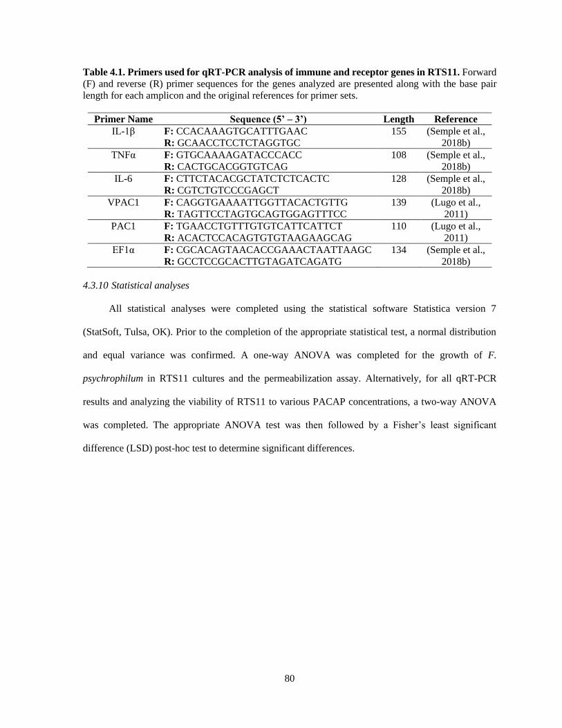
80
Table 4.1. Primers used for qRT-PCR analysis of immune and receptor genes in RTS11. Forward
(F) and reverse (R) primer sequences for the genes analyzed are presented along with the base pair
length for each amplicon and the original references for primer sets.
Primer Name Sequence (5’ – 3’) Length Reference
IL-1β F: CCACAAAGTGCATTTGAAC
R: GCAACCTCCTCTAGGTGC
155 (Semple et al.,
2018b)
TNFα F: GTGCAAAAGATACCCACC
R: CACTGCACGGTGTCAG
108 (Semple et al.,
2018b)
IL-6 F: CTTCTACACGCTATCTCTCACTC
R: CGTCTGTCCCGAGCT
128 (Semple et al.,
2018b)
VPAC1 F: CAGGTGAAAATTGGTTACACTGTTG
R: TAGTTCCTAGTGCAGTGGAGTTTCC
139 (Lugo et al.,
2011)
PAC1 F: TGAACCTGTTTGTGTCATTCATTCT
R: ACACTCCACAGTGTGTAAGAAGCAG
110 (Lugo et al.,
2011)
EF1α F: CGCACAGTAACACCGAAACTAATTAAGC
R: GCCTCCGCACTTGTAGATCAGATG
134 (Semple et al.,
2018b)
4.3.10 Statistical analyses
All statistical analyses were completed using the statistical software Statistica version 7
(StatSoft, Tulsa, OK). Prior to the completion of the appropriate statistical test, a normal distribution
and equal variance was confirmed. A one-way ANOVA was completed for the growth of F.
psychrophilum in RTS11 cultures and the permeabilization assay. Alternatively, for all qRT-PCR
results and analyzing the viability of RTS11 to various PACAP concentrations, a two-way ANOVA
was completed. The appropriate ANOVA test was then followed by a Fisher’s least significant
difference (LSD) post-hoc test to determine significant differences.

81
4.4 Results
4.4.1 Minimum inhibitory concentration (MIC)
The MIC was analyzed using both the preferred growth medium of F. psychrophilum, cytophaga
broth (CB), and the L15 cell culture media used to sustain the RTS11 cultures (Figure 4.1). For both
CB (Figure 4.1A) and L15 (Figure 4.1B), the MIC was found to be 30 µM. It appears that PACAP
can maintain its antimicrobial function in both media types, thus the function of this peptide could be
assessed during in vitro live infection experiments with RTS11.
Figure 4.1. The minimum inhibitory concentration (MIC) of C. gariepinus PACAP-38 required
to prevent the growth of F. psychrophilum alone. The MIC was determined following three days of
growth in both cytophaga broth (A) and in Leibovitz-15 (L15) cell culture media (B). Each panel
represents the results of three independent experiments.

82
4.4.2 Impact of PACAP concentrations on RTS11 survival
Even if PACAP is capable of killing aquatic bacterial pathogens, this ability has reduced value
if the peptide negatively impacts the survival of rainbow trout immune cells as well. Based on the six
concentrations of PACAP analyzed here (ranging from 0.002 µM – 20 µM), only PACAP
concentrations of 0.2 µM and higher significantly decreased the viability of RTS11 (Figure 4.2).
Furthermore, cell death was only observed on day 2 of exposure. Cell viability was not significantly
different between PACAP concentrations on both days 1 and 3 of exposure.
Figure 4.2. Impact of C. gariepinus PACAP-38 on RTS11 viability. RTS11 was exposed to varying
concentrations of PACAP ranging from 0 µM – 20 µM and the percent mortality of RTS11 was
determined for each concentration using a trypan blue assay. This figure represents three independent
experiments where a p-value of less than 0.05 was considered to be significantly different when
compared to the no PACAP control for individual timepoints. Significant differences at each timepoint
are denoted by different letters (a, b, c). All vertical error bars represent the standard deviation (SD).

83
4.4.3 Effect of PACAP on F. psychrophilum growth throughout RTS11 infections
When RTS11 was exposed to live F. psychrophilum simultaneously with various PACAP
concentrations, the number of viable bacteria present in the supernatant was not significantly different
when compared to that of the no PACAP control on day 2 (Figure 4.3A). As it was possible that
PACAP required time to stimulate a defensive immune state in RTS11, this experiment was repeated
but this time the cells were exposed to PACAP concentrations 24 hours prior to receiving the infectious
dose of F. psychrophilum. When using this experimental design, all three concentrations of PACAP
(0.002 µM, 0.02 µM and 0.1 µM) were shown to significantly reduce the number of viable bacteria in
the RTS11 on day 2 (Figure 4.3B). This reduction was still observed on day 3 but was only found to
be significant in the two higher concentrations of PACAP at 0.02 µM and 0.1 µM (Figure 4.3C).

84
Figure 4.3. Quantification of F. psychrophilum by standard plate count (SPC) of cell culture
media during live infection (MOI of 0.7-1.3) of PACAP-treated RTS11. RTS11 was exposed to live
F. psychrophilum either alone or in combination with C. gariepinus PACAP-38 concentrations (0.0002
µM – 0.02 µM) and CFU/mL was calculated on day 3 (A). RTS11 pre-treated with PACAP-38
concentrations (0.002 µM – 0.1 µM) 24 h before exposure to live F. psychrophilum and the CFU/mL
was calculated on day 2 post-infection (B) and day 3 post-infection (C). Each panel represents the
results of three independent experiments where a p-value of less than 0.05 was considered to be
significantly different when compared to the no PACAP control (i.e. RTS11 exposed to only live F.
psychrophilum). All vertical error bars represent the SD.
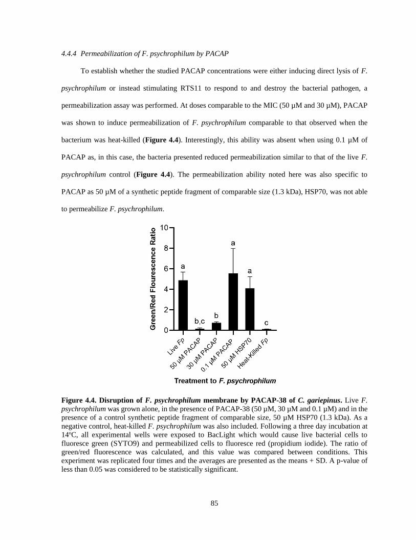
85
4.4.4 Permeabilization of F. psychrophilum by PACAP
To establish whether the studied PACAP concentrations were either inducing direct lysis of F.
psychrophilum or instead stimulating RTS11 to respond to and destroy the bacterial pathogen, a
permeabilization assay was performed. At doses comparable to the MIC (50 µM and 30 µM), PACAP
was shown to induce permeabilization of F. psychrophilum comparable to that observed when the
bacterium was heat-killed (Figure 4.4). Interestingly, this ability was absent when using 0.1 µM of
PACAP as, in this case, the bacteria presented reduced permeabilization similar to that of the live F.
psychrophilum control (Figure 4.4). The permeabilization ability noted here was also specific to
PACAP as 50 µM of a synthetic peptide fragment of comparable size (1.3 kDa), HSP70, was not able
to permeabilize F. psychrophilum.
Figure 4.4. Disruption of F. psychrophilum membrane by PACAP-38 of C. gariepinus. Live F.
psychrophilum was grown alone, in the presence of PACAP-38 (50 µM, 30 µM and 0.1 µM) and in the
presence of a control synthetic peptide fragment of comparable size, 50 µM HSP70 (1.3 kDa). As a
negative control, heat-killed F. psychrophilum was also included. Following a three day incubation at
14oC, all experimental wells were exposed to BacLight which would cause live bacterial cells to
fluoresce green (SYTO9) and permeabilized cells to fluoresce red (propidium iodide). The ratio of
green/red fluorescence was calculated, and this value was compared between conditions. This
experiment was replicated four times and the averages are presented as the means + SD. A p-value of
less than 0.05 was considered to be statistically significant.

86
4.4.5 Influence of C. gariepinus PACAP-38 on RTS11 immune gene expression
4.4.5.1 Exposure to PACAP
To determine whether PACAP alone could stimulate a response in RTS11, the cells were exposed
to various concentrations of the peptide (0.002 µM, 0.02 µM and 0.1 µM) over four days. Following
this exposure, gene expression of pro-inflammatory cytokines (IL-1β, TNFα and IL-6) and PACAP
receptors (PAC1 and VPAC1) were measured using qRT-PCR. For all three of the pro-inflammatory
cytokines measured, a significant difference was only seen on day 2 (Figure 4.5A-C). Furthermore, for
TNFα and IL-6, this significant increase was only observed at the highest PACAP concentration of 0.1
µM. Meanwhile for IL-1β, a significant increase occurred at both 0.02 µM and 0.1 µM of PACAP.
When regarding the PACAP receptors, there were no significant increases observed for PAC1 but by
day 2, VPAC1 significantly increased at all of the concentrations studied (Figure 4.5D-E). To confirm
that this response was specific to PACAP and not just a property of synthetic peptides in general, RTS11
was also exposed to 0.1 µM of a synthetic peptide fragment of rainbow trout HSP70, which was unable
to induce significant expression differences in all of the genes selected for this study (Figure 4.5F).

87
Figure 4.5. Impact of C. gariepinus PACAP-38 on RTS11 pro-inflammatory cytokines and
PACAP receptor mRNA expression 1-4 days following peptide exposure. Transcript expression of
pro-inflammatory cytokines IL-1β (A), TNFα (B) and IL-6 (C) was measured so that the influence of
PACAP alone on immune function could be assessed. The present PACAP receptors PAC1 (D) and
VPAC1 (E) were also measured to determine whether PACAP could prime RTS11 cells to bind more
PACAP. Because one day after the pre-exposure appeared to be the only timepoint with significant
upregulation due to PACAP, RTS11 was also exposed to 0.1 µM of a synthetic HSP70 peptide fragment
control of comparable size (1.3 kDa), for 48 hours to confirm that this stimulation was due to PACAP
and not a property of synthetic peptides alone (F). All panels represent three independent experiments
and are presented as means + SD. A p-value of less than 0.05 was considered to be statistically
significant when compared to the no PACAP control for each timepoint.
88
4.4.5.2 Exposure to PACAP 24 hours before F. psychrophilum infection
When RTS11 was challenged with live F. psychrophilum infection 24 hours after exposure to
PACAP, there were some interesting differences in transcript expression that were not observed during
PACAP exposure alone (Figure 4.5). For IL-1β expression, there were no significant differences at 1
and 2 days post-infection when compared to the RTS11 cells exposed to live pathogen alone. However,
by day 3 of infection, a significant increase in IL-1β transcripts was observed for all three concentrations
of PACAP (Figure 4.6A). In comparison, TNFα expression was significantly upregulated at day 1, 2
and 3 post-infection but only at 0.1 µM of PACAP, the highest concentration of the AMP (Figure
4.6B). Interestingly, all three concentrations of PACAP showed a significant increase in IL-6
expression on day 1 post-infection but by day 2 this upregulation was either lost at 0.002 µM or was
significantly reduced in the two higher concentrations of PACAP (Figure 4.6C). When compared to
the PACAP only expression (Figure 4.5), the PACAP receptors were also influenced differently during
pathogen challenge. On day 1 post-infection, PAC1 showed a significant increase only 0.1 µM (Figure
4.6D). Meanwhile, VPAC1 showed a significant increase at day 2 in the two higher PACAP
concentrations (0.02 µM and 0.1 µM) and on day 3, all three concentrations of PACAP presented a
significant upregulation when compared to the control cells that were exposed to F. psychrophilum
alone (Figure 4.6E).

89
Figure 4.5. RTS11 transcript expression when challenged with live F. psychrophilum following 24
hr pre-treatment with PACAP concentrations (0.002 µM, 0.02 µM and 0.1 µM). RTS11 that was
not exposed to PACAP or live F. psychrophilum was also included as a no stimuli control. To determine
whether PACAP has immunomodulatory effects during live infection, transcripts of the pro-
inflammatory cytokines IL-1β (A), TNFα (B) and IL-6 (C) were measured in RTS11 on days 1, 2 and
3 of infection. The PACAP receptors that are found within RTS11, PAC1 (D) and VPAC1 (E) were
also assessed throughout the live infection challenge. All panels represent three independent
experiments and are presented as means + SD. A p-value of less than 0.05 was considered to be
statistically significant when samples were compared within each timepoint.

90
4.5 Discussion
Currently aquaculture facilities have very few options outside of antibiotics to combat disease
outbreaks. When this is combined with the rising incidence of multi-drug resistance, AMPs such as
PACAP are promising alternatives for disease control/prevention. The purpose of this study was to
evaluate the antimicrobial activity and immunomodulatory function of PACAP within a live infection
model consisting of RTS11 and the coldwater pathogen, F. psychrophilum. To assess the efficacy of
this proposed system, it was critical to determine the impact that PACAP alone had on both components
of the infection model: the host and the bacterium. For several bacterial pathogens, PACAP has been
shown to have a direct antimicrobial effect (Lugo et al., 2019; Starr et al., 2018) including those of
aquatic origin (Lugo et al.. 2019). Thus it was not surprising that PACAP presented a similar result
when F. psychrophilum was exposed to various concentrations in a preferred growth medium,
cytophaga broth. Additionally, the MIC of PACAP was not influenced when F. psychrophilum was
grown in cell culture media, a substance meant to mimic physiological conditions (reviewed by Yao &
Asayama, 2017). This suggests that synthetic PACAP may maintain its antimicrobial effects in some
physiological conditions, as may be the case when administered to live organisms. But despite this
promising observation, it is important to remember that disease outbreaks in aquaculture settings would
differ significantly from microbial culture settings in important ways. Namely the assumption of
sterility and the resulting absence of competing microorganisms. As such, concentrations surrounding
the observed MIC may not represent an effective therapeutic dose to control/prevent live infection and
prove to be suboptimal for the host organism if not properly evaluated.
Aside from their potential as antimicrobials and immunomodulators, AMPs are also believed to
have minimal negative effects on mammalian host cells (Deslouches et al., 2005) including those of
immune origin (Zhu et al., 2015). Quite often this “cell selectivity” is based on the concentration
required for the AMP to induce 50% hemolysis in host red blood cells (RBCs). If this concentration is
much higher than what is required for the MIC, the peptide is considered to be essentially nontoxic to
host cells (reviewed by Matsuzaki, 2009). This has been shown with C. gariepinus PACAP-38 when

91
both human and fish RBCs were exposed to the peptide, revealing only RBC lysis at extremely high
concentrations (Lugo et al., 2019). Unfortunately, these methods of measurement are not always
directly comparable as antimicrobial assays generally use a bacterial concentration of ~5 x 10⁵ CFU/mL
while hemolysis assays use what corresponds to be 6 x 10⁸ cells/mL (reviewed by Matsuzaki 2009). In
fact, when Imura and colleagues (2008) corrected for this concentration difference during their analyses
of the antimicrobial peptide magainin, the MIC concentration of 10 µM was enough to completely lyse
the host RBCs. In the present study, RTS11 was exposed to PACAP wherein higher concentrations (0.2
µM, 2 µM and 20 µM) had a significantly negative impact on RTS11 viability. Some studies have
alluded to the idea that AMPs may be toxic to mammalian cells in the absence of microorganisms
(Imura et al., 2008), thus it is possible that this may also be observed in fish cells. There are important
differences between prokaryotic and eukaryotic membranes that may improve the chance that AMPs
will preferentially bind to the membrane of microorganisms before host cells (reviewed by Huang et
al., 2010). Prokaryotic membranes have a high negative charge due to being predominantly composed
of phosphatidylglycerol, cardiolipin or phosphatidylserine, thus have a greater chance of attracting the
cationic peptides that are AMPs. In comparison, mammalian cells may be less attractive for AMP
penetration as they are enriched in zwitterionic phospholipids resulting in an overall neutral charge
(reviewed by Huang et al., 2010; Matsuzaki, 1999). Furthermore, mammalian cell membranes contain
cholesterol, something that is absent in prokaryotic membranes. Interestingly, it has been shown that
cholesterol can dramatically reduce the activity of AMPs (Matsuzaki et al., 1995) providing another
potential layer of protection for mammalian cells. But despite these important differences that may help
with membrane selection of AMPs, complete protection of the host cells from AMP-induced
cytotoxicity may not be possible at higher AMP concentrations. To even consider PACAP for use as a
therapeutic agent in aquaculture settings, the therapeutic dose must not be cytotoxic to the host, whether
infected or microbe-free.
In spite of the value that can be obtained from determining the MIC of PACAP in various culture
media, as well as the ideal concentration for survival of host cells, the observed antimicrobial activity
92
is meaningless if it is lost or not effective during live pathogen challenge. The current study is the first
of its kind that has demonstrated the antimicrobial activity of PACAP during an in vitro live infection
model with an aquatic pathogen. But interestingly, the teleostean version of PACAP was only able to
reduce the viable bacterial count of F. psychrophilum when RTS11 cells were pre-treated with PACAP
for 24 hours. It appears that rainbow trout macrophages require time to respond and activate an effective
immune response when exposed to F. psychrophilum. As obligate poikilotherms, metabolic rates in
fish are heavily influenced by their environmental temperature (Gillooly et al., 2002; Patterson et al.,
2013). Because cells derived from rainbow trout, a coldwater salmonid, are grown at much lower
temperatures than their mammalian counterparts, it may take more time for these cells to respond to
stimuli. Indeed this has been shown with both RTS11 and rainbow trout B cells where the
chemoattractant ability of the chemokine CK9 strongly increased when the cells were pre-treated with
T-independent antigen (Aquilino et al., 2016). Likewise, in rainbow trout primary head kidney culture,
the cells sometimes required 48 hours before an increase in respiratory burst activity was observed
(Novoa et al., 1996). In both of these examples, the immune cells were maintained at 18oC, but in the
current study, RTS11 was held at 14oC as this is a relevant temperature at which BCWD occurs (Holt
1987). Thus, a longer pre-treatment time at this lower temperature may be required depending on the
response that is being measured. Though a temperature between 8-14oC would be optimal for testing
the efficacy of PACAP in protecting rainbow trout from infection with F. psychrophilum, there are
many infectious diseases that influence the culture of numerous aquatic organisms. As a result,
experimental doses with therapeutic AMPs must be tested in vivo to ensure that they will provide
protection and effective immune stimulation towards relevant pathogens within an applicable
temperature range.
Cell culture systems provide a controlled, cost-effective method for exploring numerous
biological phenomena, but it is important to recognize the limits of these models. In a cell culture
setting, individual cells are directly exposed to the experimental stimulant, without physiological
barriers or a complicated cellular milieu to overcome. As a result, in vitro systems often have much
93
lower doses than what is appropriate within the whole organism. This was displayed when Gotlieb et
al (2015) used several methods to isolate and stimulate NK cells revealing that the cells were 10-30
times more susceptible to stress hormones in vitro than what was observed when stimulated in plasma,
a much more biologically relevant medium. Though the effects of PACAP on fish infections in vivo
have not yet been explored, there have been several studies to determine the impact of this AMP on
growth, immunomodulation and physiology (Cardoso et al., 2015; Lugo et al., 2010a; Lugo et al.,
2010b; Wang et al., 2013). One study by Lugo et al (2010a) exposed juvenile fish to an average of 4
µg of PACAP per fish, which significantly enhanced tilapia growth. In comparison, with the RTS11
infection model presented here, each well received 1.8 µg or less and antimicrobial activity was still
observed. Though the PACAP doses optimized for the current in vitro study were very effective, an in
vivo model would require further optimization to develop an efficacious exposure range.
When it comes to bacterial pathogens, the consensus regarding AMP function is that they are
either membrane disrupting, or nonmembrane disrupting (reviewed by Bahar & Ren, 2013). Though
PACAP was able to lower the number of viable bacteria when grown alone in media (for the MIC) and
in RTS11 cultures throughout live infection, it was unclear whether this was due to the direct
antimicrobial activity of PACAP acting on F. psychrophilum. Previous work with mammalian PACAP
confirmed for the first time that the peptide was capable of disrupting membranes of relevant terrestrial
bacterial pathogens (Starr et al., 2018). This permeabilization is a common mode of action for α-helical
AMPs (Sato & Feix, 2006), including PACAP, but until now this ability has not been confirmed for the
version of PACAP produced by fish. Previous bioinformatic analysis of PACAP-38 from C. garipinus
provided evidence that this peptide was very likely to have cell penetrating properties (Lugo et al.,
2019). The present study was able to functionally validate the ability of C. gariepinus PACAP-38 to
permeabilize the membrane of F. psychrophilum at concentrations surrounding the MIC. Furthermore,
this was an ability specific to PACAP as another synthetic peptide fragment of comparable size from a
teleost, HSP70, did not induce permeabilization. Interestingly, at the highest concentration that reduced

94
the viable bacterial count during live infection, 0.1 µM, PACAP was unable to permeabilize the
membrane of F. psychrophilum. This finding confirmed that at 0.1 µM, one of the many other effects
that PACAP may have on teleostean immunity must have been responsible for stimulating RTS11 to
destroy and/or slow the growth of the coldwater pathogen.
PACAP has been shown to have immunomodulatory effects on whole fish and in fish cells
(Kasica-Jarosz et al., 2018; Lugo et al., 2010b; Lugo et al., 2019; Wang et al., 2013) but this has only
been studied in the absence of live infection. Furthermore, there has been limited research regarding
the activity of PACAP directly on teleost immune cells. Specifically with RTS11, the
immunostimulatory effect of three other α-helical AMPs has been reported (Chiou et al., 2006) but not
when dealing with a live bacterial challenge. The present study explores, for the first time, the impact
of relevant doses of PACAP on the immune function of RTS11 in both the presence and absence of F.
psychrophilum. In the absence of bacterial infection, PACAP was shown to stimulate pro-inflammatory
cytokine expression 48 hours following PACAP treatment as well increasing the expression of one of
the PACAP receptors, VPAC1. When analyzing PACAP receptor expression in RTS11, it is important
to note that only VPAC1 and PAC1 receptor genes were measured as it has previously been shown by
Lugo et al (Lugo et al., 2011) that these cells do not express the third receptor gene, VPAC2. Lugo and
colleagues also showed that despite PAC1 presenting the highest constitutive expression in all rainbow
trout lymphoid tissues in vivo, this was not observed in RTS11 where VPAC1 presents the greatest
expression (Lugo et al., 2011). As PAC1 has been found to be a fundamental type I receptor for PACAP
this result was unexpected. Nonetheless, the current study validated this finding when VPAC1 presented
significant upregulation following RTS11 stimulation with PACAP while PAC1 did not. As PACAP
was shown to stimulate a slight increase in pro-inflammatory cytokines and upregulate the expression
of VPAC1, pre-treatment with this AMP may stimulate a protective state within the rainbow trout
immune cells.
In itself, the presence of live bacteria would be capable of inducing pro-inflammatory cytokine
expression in RTS11. But when this was combined with 24-hour pre-treatments with PACAP, infection

95
with F. psychrophilum had an effect on transcript expression that was quite different from PACAP
alone. Rather than all three of the studied pro-inflammatory cytokines increasing their expression at the
same time, each one showed significant upregulation at different time points post-infection when
compared to RTS11 exposed to the bacteria alone. The pattern of enhancing inflammatory cytokine
production following pre-treatment is similar to that of trained immunity that has been observed in
mammalian monocytes and macrophages (Quintin et al., 2012). Quintin and colleagues showed that
when primed with β-glucan prior to exposure to LPS, monocytes and macrophages were able to induce
a greater pro-inflammatory response than unprimed cells (Quintin et al., 2012). Perhaps PACAP has a
similar function and is able to prime immune cells to produce a faster, more damaging response when
they come into contact with a live pathogen. This would provide an explanation for the observed
decrease in viable bacteria following PACAP exposure at doses that were not able to directly
permeabilize the membrane of F. psychrophilum.
Despite the similarity to trained immunity, the actual function of PACAP in mammalian models
appears to be contrary to what has been observed in bony fish. The vast majority of mammalian studies
discuss the anti-inflammatory role of PACAP during experimental bacterial infection. These
experiments often involve exposure to bacterial products (such as LPS) simultaneously with PACAP,
after which various immune parameters are observed (Delgado et al., 1999a; Delgado & Ganea, 2001;
Martinez et al., 2002). This has led to the belief that many AMPs, including PACAP, play important
anti-inflammatory roles to protect the host from dangerous, over-reactive inflammatory responses
(Delgado et al., 1999b). Though very valuable, these mammalian studies are not directly comparable
to a live, growing infection within an organism or cell culture. Additionally, aside from zebrafish,
teleosts appear to be lacking TLR4, which binds and responds to LPS (Ali et al., 2014; Sepulcre et al.,
2009). As a result, observations in mammalian study systems may not be directly transferrable to those
of fish. The immunostimulatory effect of PACAP on rainbow trout immune cells as observed in this
study has been previously reported in head kidney leukocytes derived from another bony fish, the grass
carp (Wang et al., 2013). Wang and colleagues found that when these immune cells were exposed to

96
bacterial products, PACAP induced inflammatory cytokine expression while having no impact on the
expression of the anti-inflammatory cytokine, IL-10 (Wang et al., 2013). When all of this information
is taken together, it appears that PACAP may play a different, yet valuable role in the
immunomodulation of teleosts when compared to what has been observed in mammals.
4.6 Conclusions
Antimicrobial peptides (AMPs) are promising alternatives to antibiotics in the ongoing battle
between aquaculture facilities and infectious agents. One AMP that has received a lot of attention due
to its pleiotropic effects in aquatic species is PACAP. The results of the present study revealed that
PACAP derived from the teleost Clarias gariepinus acts as a potent antimicrobial peptide against the
causative agent of BCWD, F. psychrophilum. Furthermore, its mode of action was confirmed to be
permeabilization of the bacterial membrane. When a live infection model was developed with this
pathogen and the monocyte/macrophage-like cell line, RTS11, 24 h pre-exposure of PACAP appeared
to protect RTS11 by significantly reducing the number of viable bacteria in the culture system. Based
on transcript levels of pro-inflammatory cytokines and receptors for the AMP, PACAP was also shown
to have an immunostimulatory effect on RTS11 whether exposed to the AMP alone or exposed to both
PACAP and live F. psychrophilum challenge. Overall, this study was able to provide further validation
regarding the antimicrobial effect of PACAP on aquatic pathogens as well as its immunomodulatory
activity on teleost immune cells. As a promising candidate for use in aquatic models, future studies
should focus on confirming these valuable functions of PACAP throughout live infection models in
vivo.
Acknowledgements
Dr. Niels Bols for kindly allowing the use of his cell culture facility.

97
Chapter 5: Revealing immune differences between eight outbred Chinook salmon populations
when challenged with live Vibrio anguillarum.
Shawna L. Semple1, George Heath1, Carina Filice1, Daniel D. Heath2 and Brian Dixon1
1University of Waterloo, Department of Biology, Waterloo, Canada 2Great Lakes Institute for Environmental Research, University of Windsor, Windsor, Canada
Formatted for submission to: Nature Heredity

98
5.1 Overview
Aquaculture in North America is currently dominated by Atlantic salmon, but there has been an
increasing interest in the production of species native to the Pacific coast such as Chinook salmon
(Oncorhynchus tshawytcha). Chinook salmon is relatively new to culture production; therefore, the
selection of appropriate stocks is critical for the success of this developing species. In many cases genes
from wild populations are incorporated into farmed stocks to avoid the decrease in performance
associated with inbreeding. The current study focuses on assessing the immunological performance of
eight outbred Chinook salmon stocks after challenge with the marine pathogen, Vibrio anguillarum.
Throughout exposure challenge to both the pathogen and sterile PBS, significant differences in
mortality were observed. Fish were also assessed for MH class II β1 genotype to determine whether
there were specific alleles that could predict resistance/susceptibility. Although unique MH alleles did
not appear to confer resistance, the crosses with better survival presented a higher percentage of
individuals that possessed a heterozygous MH genotype. The stress induced during infection resulted
in several individuals presenting signs of Bacterial Kidney Disease (Renibacterium salmoninarum)
indicating that chronic co-infection may have played a role in susceptibility. This was supported by the
highest and lowest performing populations presenting the lowest and highest prevalence of BKD
respectively as validated through an ELISA-based assay of head kidney samples. When spleen samples
from the highest and lowest performing populations were assessed for cytokine and respiratory burst
gene expression throughout bacterial challenge, the high surviving stocks presented lower expression
of inflammatory cytokine transcripts (IL-1β, IL-6 and TNFα) when compared to the low performing
stocks. Interestingly, microsatellite analysis revealed that there were little to no differences in
heterozygosity among these eight populations despite the variations observed regarding immune
performance. Understanding the impact of outbreeding on the immune function of farmed, and often
inbred, Chinook salmon could aid in the future development of high-quality aquaculture stocks for this
species.

99
5.2 Introduction
The high demand for alternative and healthy protein options has resulted in aquaculture
becoming the fastest growing animal food sector for the past 40 years (FAO, 2018). North American
aquaculture is currently dominated by Atlantic salmon, resulting in production of this species on both
the East and West coasts of Canada. However, because Atlantic salmon are not indigenous to the West
coast, there is public concern regarding the introduction of foreign diseases to wild populations as well
as the exposure of native salmon species to ecological niche competition through farmed escapees
(reviewed by Noakes et al., 2000). Despite the negative publicity associated with farming Atlantic
salmon on the West coast, a staggering 71% of Canadian salmon production occurs in British Columbia
(Statistics Canada, 2017). Thus, the majority of Atlantic salmon are actually grown on the Pacific coast
rather than their native coast in Canada. To alleviate some of the public apprehension, there have been
numerous attempts to domesticate and farm Pacific salmon species for aquaculture production.
While there are five major Pacific salmon species, Chinook salmon (Oncorhynchus
tshawytscha) is the largest and most highly valued in North America (Christensen et al., 2018;
Ohlberger et al., 2018), making it a leading candidate for use in aquaculture. The farming of Chinook
salmon on the West coast offers the benefit of potentially higher disease resistance resulting from
accumulated adaptations to resident pathogens (Evans & Neff, 2009). Unfortunately, when compared
to the highly domesticated Atlantic salmon species, Chinook salmon are currently unable to tolerate the
high densities used for commercial salmon production (DFO, 1991). So, from an economic perspective,
the culture of Chinook salmon would not appear profitable enough to convince farmers to convert to
such low-density farming despite a more environmentally friendly product. Given that Atlantic salmon
underwent over 50 years of domestication in aquaculture (Bicskei et al., 2014), it is reasonable that
both time and concerted efforts would be required to prepare Chinook salmon for comparable large-
scale production.
A common method used to generate domesticated stocks for any species of interest is selective
breeding. This involves identifying individuals whose phenotype is optimal (high growth, disease
100
resistance, feed conversion, etc.) and using these animals to breed the next generation. Over successive
iterations of this process, traits of interest can be enhanced however this strategy can also result in a
phenomenon known as inbreeding depression. Inbreeding depression refers to the reduced fitness
observed in the progeny of individuals that are closely related (Conner & Hartl, 2004). For agricultural
species this can present itself as a reduction in overall yields, reproduction and survival (reviewed by
Charlesworth & Willis, 2009). These negative effects occur because inbreeding increases
homozygosity at all loci, thus enhancing the probability of unmasking deleterious alleles that would
not present themselves within heterozygous individuals (reviewed by Paige, 2010). Fortunately,
inbreeding depression can be countered by the introduction of outside genetic material through a
practise known as outbreeding.
When it comes to farming native species on the Pacific coast, attempts to domesticate Chinook
salmon have resulted in farmed stocks becoming increasingly inbred. It is possible that these stocks are
suffering inbreeding depression, thus impeding their survival at higher densities. Though there are
many factors that can contribute to this reduction in fitness, susceptibility to disease is a significant
challenge continuously faced by fish farmers (reviewed by Assefa & Abunna, 2018). The negative
impact associated with infectious disease is high and is often considered to be the leading cause of
financial loss for several aquaculture sectors (Leung & Bates, 2012; reviewed by Stentiford et al., 2012;
Stentiford et al., 2017). Moreover, there is limited understanding of aquatic pathogens and minimal
success in selecting for disease resistance when compared to terrestrial counterparts (Pazderka et al.,
1975; Snowder et al., 2005; Odegard et al., 2011). This may reflect a general lack of insight regarding
aquatic environments including the many microbial pathogens that influence the aquaculture industry.
One particularly costly infectious agent is the gram-negative bacterium, Vibrio anguillarum.
This saltwater pathogen is the causative agent of a highly fatal hemorrhagic septicaemia known as
vibriosis (reviewed by Frans et al., 2011). As an opportunistic pathogen, outbreaks tend to coincide
with environmental and/or other stressors (Austin & Austin, 2007). Though symptoms of vibriosis are
typical of a hemorrhagic septicemia (bleeding vent, swollen ulcerated lesions, bleeding eyes,

101
exopthalmia, etc.), acute outbreaks can result in extremely high mortality without the presentation of
any clinical signs (reviewed by Frans et al., 2011; Austin & Austin, 2007). Given that the organism can
infect over 90 different aquatic species (reviewed by Hickey & Lee, 2018), V. anguillarum has
historically been, and continues to be, responsible for significant financial losses to the aquaculture
industry. In Canada, the first reported case of vibriosis in a Pacific salmon species was in 1971 (Evelyn,
1971). Since then, the pathogen has plagued Canadian salmon culture making vibriosis a highly relevant
disease model for evaluating Chinook salmon stock quality.
The present study describes the crossing of an inbred Chinook salmon stock with seven wild
populations and the subsequent evaluation of offspring immune performance when challenged with
live V. anguillarum. The seven outbred populations and the control inbred stock had variable survival
to V. anguillarum, indicating that there may be identifiable immune components used to cope with
infection. Aspects of both innate and adaptive immunity were explored at functional and genetic levels.
More specifically, differences in major histocompatibility (MH) class II β1 genotypes, serum IgM, co-
infection with Renibacterium salmoninarum and immune gene expression were compared between
crosses. Additionally, microsatellite analysis was completed to determine the level of heterozygosity
between the seven outbred crosses and the control inbred stock. It is possible that the differences
observed here indicate hybrid vigor and could potentially be targets of selection to improve the
performance of Chinook salmon stocks for future production measures.

102
5.3 Materials and Methods
5.3.1 Breeding design
Breeding and rearing occurred at Yellow Island Aquaculture Ltd. (YIAL), an organic Chinook
salmon farm located on Quadra Island, British Columbia, Canada. The YIAL Chinook salmon stock
has been in production since 1985 and originated from the Robertson Creek and Big Qualicum River
(Vancouver Island, British Columbia, Canada) Chinook salmon stocks. To generate outbred crosses
with the YIAL production stock, milt from males taken from seven wild populations was collected from
across Vancouver Island and lower mainland British Columbia (Figure 5.1). The target populations
used to provide the milt were: Big Qualicum River (BQ), Capilano River (Cap), Chilliwack River
(Chil), Nitinat River (Nit), Puntledge River (Punt), Robertson Creek (RC) and Quinsam River (Quin).
The domestic production cross (YIAL x YIAL) would serve as an internal control for comparative
assessment between the stocks. Milt collection and all associated procedures are as described in
Semeniuk et al. (2019).
To minimize maternal effects (Wellband et al., 2017), eggs from 17 highly inbred female
offspring of one self-fertilizing hermaphrodite Chinook salmon from YIAL were used (see Komsa,
2012). Briefly, hermaphrodite fish (genetically female, but phenotypically both male and female) were
generated from incomplete sex reversal by exposing female larvae to 17-alpha-methyltestosterone
(17aMT) treatments. Female offspring from the hermaphrodite salmon had an average inbreeding
coefficient of 0.50. Fertilization of all crosses took place on November 1, 2013 and was performed as
described by Semeniuk et al. (2019).

103
Figure 5.1. Geographical location of the British Columbia rivers from which milt was collected
to produce the seven outbred Chinook salmon populations. The location denoted as YIAL (Yellow
Island Aquaculture Ltd.) indicates the source of the farmed salmon milt used as the inbred population
control. This Chinook salmon farm was also the location for the live V. anguillarum infection trial used
to produce samples for this study.
5.3.2 Rearing
Rearing of the eight different crosses during both fresh- and saltwater phases of growth were as
described by Semeniuk et al. (2019). Prior to sea transfer, a subset of each cross received passive
integrated transponders (PIT) identification tags.
5.3.3 Culture and quantification of V. anguillarum
V. anguillarum serotype O1 was kindly provided by Simon Jones of Pacific Biological Station
(Nanaimo, BC). This particular strain of V. anguillarum was isolated from a diseased winter steelhead
trout obtained from Little Campbell River, BC. Frozen stocks of the V. anguillarum were subcultured
onto tryptic soy agar (TSA) supplemented with 2% NaCl. Following incubation at room temperature
(RT) for 48 h, a single colony was used to inoculate 2.5 mL of tryptic soy broth (TSB) supplemented
with 2% NaCl and incubated for 17 h at RT and 200 rpm. Following this incubation, 100 µL of this
culture was used to inoculate 100 mL of TSB with 2% NaCl and incubated at RT for 24 h while shaking
at 200 rpm. The resulting culture had an optical density of 0.25 which was confirmed to be 2.0 x 10⁹

104
CFU/mL by a standard plate count (SPC). Specifically, bacterial numbers were enumerated by plating
0.05 mL of ten-fold serial dilutions on TSA with 2% NaCl plates in triplicate. After incubation at RT
for 30 hours, plates containing 25-250 colonies were counted and the number of CFU/mL calculated.
5.3.4 Infection with live V. anguillarum and sample collection
On July 16, 2015, two weeks before the start date of the infection trial, 160 fish from each of the
8 different populations were transferred from net pens to 2000 L troughs. Because each fish was PIT
tagged, 40 fish from four of the populations were in each of the eight troughs (n = 160). Each trough
was replicated four times, with two of the trough replicates being used for live V. anguillarum infection,
and the remaining two being used for sham injection with sterile phosphate buffered saline (PBS,
Lonza). Two days prior to challenge (day 0), blood, gill, spleen, head kidney, tail fin, and muscle
samples were collected from two individuals per family per trough to act as Day 0 unstimulated
controls.
Following sedation with clove oil, all fish received 100 µL intraperitoneal (i.p.) injections of 8.0
x 104 CFU/mL of V. anguillarum suspended in sterile PBS. In comparison, all fish in the control troughs
received i.p. injections of 100 µL sterile PBS. The water outflow from all challenged troughs underwent
UV sterilization (493.5 mJ/cm2) which ensured the elimination of the infectious agent. On days -2, 1,
7, 12, and 28, three fish from every population within each trough were sampled and blood was drawn
from the caudal vein after anesthesia with clove oil. Fish were then euthanized with an overdose of
clove oil and whole-body weights and lengths were measured. Gill, spleen, head kidney (HK), tail fin,
and muscle samples were collected for each individual and stored in RNA Later at -20°C for future use.
Blood samples were left to clot overnight at 4°C before centrifugation at 1000 g for 10 min. The serum
was then removed, aliquoted into 1.5 mL low protein binding tubes (Costar) and immediately stored at
-20°C.
5.3.5 Microsatellite analysis
To further explore the differences between the single hatchery (YIAL) and the seven outcross
populations, estimates of percent heterozygosity were obtained using DNA extracted from fin clips.

105
Fin clips were collected and preserved from the seven outbred and the YIAL populations. DNA was
extracted from fin clips using the DNeasy Blood and Tissue Kit (Qiagen) according to the
manufacturer’s instructions. The DNA samples were then quantified using the Take3 plate of a Synergy
H1 plate reader (BioTek Instruments) and were stored at -80°C until further use. Individual genotypes
were determined through polymerase chain reactions (PCR) at 9 previously described microsatellite
loci, specifically Oneu3, Oneu8 (Scribner et al., 1996), Omm1135 (Rexroad et al., 2001), Omy325
(O’Connell et al., 1997), OtsG432, OtsG474 (Williamson et al., 2002), Ots1, Ots4 (Banks et al. 1999)
and Ogo4 (Olsen et al., 1998). PCR conditions included: a 5-min denaturation step (94°C), followed
by 30 cycles of a 20-s denaturation step (94°C), a 20-s annealing step (64.6oC – Omy325, Ots4; 63.5oC
– Oneu8, Ogo4; 54.3oC – Oneu3; 58.3oC – Omm1135, Ots1; 60.2oC – OtsG474, OtsG432) and a 30-s
extension step (72 °C), followed by a final extension step of 3 min. PCR products at all 9 loci were
pooled by individual, cleaned by precipitation with isopropanol, resuspended in milliQ water and then
individually barcoded. The barcoding PCR included: a 2-min denaturation step (94 °C), followed by 8
cycles of a 30-s denaturation step (94 °C), a 30-s annealing step (60 °C) and a 1-min extension step (72
°C), followed by a final extension of 5 min. The barcoded amplicons were then pooled and gel-extracted
using a Qiaquick Gel Purification Kit (Qiagen).
The library was assessed and quantified using the High Sensitivity DNA Reagents Kit (Agilent
Technologies) on a Bioanalyser 2100 (Agilent Technologies) and was then diluted to 60 pM for
template preparation using the 400 bp Hi-Q View Kit (Life Technologies). Sequencing of the library
was performed on the Ion Torrent Personal Genome Machine (Life Technologies) using a 318 v2TM
chip (Life Technologies). Truncated and low-quality sequences were removed from the data and the
remaining sequences were separated based on individual and microsatellite loci using Mothur (Schloss
et al., 2009). Fragment sizes (alleles) were then identified using an R based bioinformatics framework
developed to score microsatellites generated from next generation sequencing platforms (R Core Team,
2014; Roy et al., 2017).

106
5.3.6 MH class II β1 genotyping
DNA was extracted from fin clip samples as described above in section 5.3.5. Primers that were
previously validated by Becker et al (Becker et al., 2014) to amplify the hypervariable region of MHC
class II β1 (exon 2) of Chinook salmon were used for this study. Specifically, the forward primer used
was 5’-CTTGGTCTTGACTTGMTCAGTCA-3’ and the reverse primer was
5’CCCGAGAAGCTTCCGATACTCCTCAAAGGACCTGCA-3’. For amplification, the following
PCR parameters were used: denaturation at 95°C for 5 min, followed by 25 cycles of denaturation at
95°C for 45 s, annealing at 57°C for 30 s and extension at 72°C for 45 s. A final extension at 72°C was
carried out for 5 min. Two PCR reactions were performed per individual. The amplified PCR products
were separated on 1% agarose gels containing 1% GelGreen (Biotium Inc.) and bands of the desired
size (310 bp) were cut out of the gel and extracted using the QIAquick Gel extraction kit (Qiagen). The
resultant fragments were cloned into pGEM®-T Easy and transformed into competent Escherichia coli
XL1-blue cells as described previously by Semple and colleagues (Semple et al., 2018a). Following
extraction from a minimum of 10 different clones per PCR reaction, plasmids were sequenced using
SP6 primers at the TCAG sequencing facility (Sick Kids Hospital, Toronto, Ont.). The sequences were
analyzed using both Clustal Omega (www.ebi.ac.uk/Tools/msa/clustalo/) and the Basic Local
Alignment Search Tool (BLAST) software (blast.ncbi.nlm.nih. gov/Blast.cgi). All DNA sequences
were then converted to amino acid sequences using the ExPASy Bioinformatics Resource Portal
(web.expasy.org/translate/). Only functional amino acid sequences that were observed in both PCR
reactions for each individual were considered to be representative of true MHC class II β1 alleles.

107
5.3.7 Measuring the serum antibody response to V. anguillarum
5.3.7.1 Isolating antigens from V. anguillarum
Subcultures from glycerol stocks of V. anguillarum were grown on TSA supplemented with 2%
NaCl. An isolated colony was used to inoculate 50 mL of TSB with 2% NaCl and grown for 24 hr at
RT while shaking at 200 rpm. When the culture reached an optical density of approximately 0.24 at
600 nm, indicating a concentration of approximately 3.6 x 10⁹ CFU/mL, 0.5% formalin was added to
the flask and the cells were incubated at RT for 24 hr while shaking. The killed bacterial cells were
then washed three times with PBS, harvested by centrifugation (10,000 rpm at 4°C for 10 min) and the
subsequent pellets were resuspended in 2 mL of PBS. The resulting formalin-killed V. anguillarum
culture was then bead-beaten to liberate whole cell antigens as described previously by Semple et al.,
2018a. The bacterial lysate was then diluted to a final concentration of 5 μg/mL in coating buffer (0.015
M Na2CO3, 0.035 M NaHCO3, pH 9.6).
5.3.7.2 Indirect subtractive ELISA to measure serum IgM between the crosses
For each sample being tested, six wells of an Immulon HBX 96-well polystyrene plate
(ThermoFisher) were prepared wherein three wells were coated with 100 µL of whole cell bacterial
lysate (5 µg/mL) and the remaining triplicate wells received 100 µL of coating buffer alone. The plates
were then incubated overnight at 4°C. Following coating and in between all subsequent steps, solutions
were removed and all wells on the plate were washed three times with 300 µL of tris-buffered saline
with 1% Tween 80 (TBS-T). Prior to samples being added, all wells on the plate were blocked with
300 µL of blocking solution (5% skim milk dissolved in TBS) for 1 hr at RT. After blocking, Chinook
serum samples were prepared as a 1:50 dilution in sterile-filtered phosphate buffered saline (PBS).
Serum samples were added to the plate in triplicates of 100 µL for both the bacterial antigen coated and
uncoated wells. Plates were then incubated at RT for 1 hr. Following removal of samples and washing,
100 µL of rabbit anti-salmonid immunoglobulin primary detection antibody (1:2500 dilution in
blocking solution; Cedarlane) was added to all wells and incubated at RT for 1 hr. The primary antibody

108
was removed and the plate was washed followed by a 1 hr incubation at RT with 100 µL of goat anti-
rabbit secondary detection antibody conjugated to biotin (1:2000 dilution in blocking buffer;
Cedarlane). After removal of the secondary antibody and washing, 100 µL of streptavidin conjugated
to horse-radish peroxidase (HRP, Biolegend) at a 1:1000 dilution in blocking buffer was added and
incubated at RT in the dark for 1 hr. Following washing, 100 µL of TMB SENSTM (Cedarlane) was
added to all wells and the plate was incubated at RT in the dark for 1 hr. At the end of this incubation,
100 µl of stop solution (0.3M H2SO4) was added to each well to prevent further development of colored
product. The resulting color development was measured at 450 nm using a BioTek Synergy H1 Hybrid
Plate reader (BioTek). For each sample, raw absorbance values from wells with no antigen were
subtracted from raw absorbance values of wells coated with bacterial antigen. This value was taken as
a relative measure of individual serum IgM antibody developed against V. anguillarum.
5.3.8 qRT-PCR of spleen transcripts throughout V. anguillarum infection
5.3.8.1 RNA extraction and cDNA synthesis
RNA was extracted from 20-40 mg of spleen tissue using 1 mL of Trizol (Life Technologies) as
described by the manufacturer. To remove any contaminating genomic DNA, RNA samples were
treated with DNase I (Thermo Scientific). RNA samples were then quantified using the Take3 plate of
a Synergy H1 plate reader (BioTek Instruments) and were stored at -80°C until further use.
Complementary DNA (cDNA) was synthesized from 500 ng of total RNA using the qScript cDNA
Supermix (Quanta Biosciences) in accordance to the manufacturer’s instructions. For a no template
control, 500 ng of RNA suspended in 20 uL of diethyl pyrocarbonate (DEPC) water was included in
the cDNA synthesis reaction without reverse transcriptase.
5.3.8.2 Validatoin of qRT-PCR primers for Chinook salmon
Many of the qRT-PCR primers used for this study had only been validated in rainbow trout
(Semple et al., 2018b) making it necessary to confirm their specificity when using a cDNA template
obtained from Chinook salmon. Specificity of each primer set was confirmed using cDNA obtained
109
from the spleen of Chinook salmon challenged with V. anguillarum. Sequence validation was
confirmed as described above in section 5.3.6.
5.3.8.3 qRT-PCR reactions
To assess transcript levels of IL-1β, TNFα, IL-6, IL-10, NADPH p22phox and NADPH p40phox in
Chinook salmon spleen samples, qRT-PCR analysis was completed. All PCR reactions were 10 µl and
contained: 2.5 μl of cDNA (25 ng/μl diluted 1:10 in RNase free water), 2x WISENT ADVANCED™
qPCR mastermix (Wisent), and forward and reverse primers (Sigma Aldrich) to a final working
concentration of 0.25 μM. All qRT-PCR reactions were completed on the LightCycler® 480 II (Roche).
The sequences for all primer sets are outlined in Table 5.1. Each experimental sample was run in
triplicate. For each plate, triplicate wells of a calibrator, no template control and RNA only control were
also present. The program used for all qRT-PCR reactions was as follows: pre-incubation at 95°C for
10 min followed by 40 cycles of denaturation at 95°C for 10 sec, annealing at 60°C for 5 sec and
extension at 72°C for 8 sec. A melting curve was completed for every run from 65°C to 97°C with a
read every 5 sec. Product specificity was determined through single PCR melting peaks. All qRT-PCR
data was analyzed using the ΔΔCt method while incorporating individual primer efficiencies as
described by Pfaffl (2001) and is presented as the average of 3 experimental replicates for each
individual (n = 6) with the standard error of the mean (SEM). All primer efficiencies were above 1.87
and below 2.12. Specifically, gene expression was normalized to the reference gene (EF1α) and
expressed as fold change over the day -2 control group where control expression was set to 1. Multiple
reference gene primer sets were developed but EF1α was selected as it was observed to have consistent
Cp values across a subset of samples (data not shown).

110
Table 5.1. Primer sequences used for qRT-PCR analysis of immune genes in Chinook salmon.
Forward (F) and reverse (R) primer sequences for the genes analyzed are presented along with the
original references/accession numbers for the primer sets.
Primer Name Sequence (5’ – 3’) Accession No.
NADPH p22phox F: TGGGAGTTTGCTGCTTATGCT
R: TGGGCCGAATGACTTCACAC
XM_024440320.1
NADPH p40phox F: GTCTTACAGCTGCCTGCGC
R: CACATCTCTCATGCGTGCCAAT
XM_024409180.1
IL-1β F: CCACAAAGTGCATTTGAAC
R: GCAACCTCCTCTAGGTGC
XM_024418276.1
TNFα F: GTGCAAAAGATACCCACC
R: CACTGCACGGTGTCAG
XM_024396390.1
IL-6 F: CTTCTACACGCTATCTCTCACTC
R: CGTCTGTCCCGAGCT
XM_024404411.1
IL-10 F: GCCTTCTCCACCATCAGAGAC
R: GATGCTGTCCATAGCGTGAC
XM_024405117.1
EF1α F: CGCACAGTAACACCGAAACTAATTAAGC
R: GCCTCCGCACTTGTAGATCAGATG
XM_024396038.1
5.3.9 Measuring co-infection with bacterial kidney disease (R. salmoninarum)
Infection status with R. salmoninarum, the causative agent of bacterial kidney disease (BKD),
was determined for the highest performing (YIAL) and the lowest performing (Punt) Chinook salmon
crosses. Head kidney samples from these populations were processed for use in a commercial R.
salmoninarum ELISA assay (Ango) as described by the manufacturer. Head kidney samples that had
an absorbance equal to or higher than that of the provided positive control were considered to represent
individuals infected with R. salmoninarum.
5.3.10 Statistical analyses
For exposure trials to live V. anguillarum and sterile PBS, statistical analyses of survival curves
was conducted. To determine significant differences in survival between the eight different crosses for
each exposure condition, a Mantel-Cox test was completed with an adjusted alpha level of 0.0018 (p =
0.05/28) given the multiple pairwise comparisons among the eight populations. These statistical
analyses were completed using GraphPad Prism 8.1.0.
For the microsatellite analysis, individuals that were genotyped at fewer than 6 loci were
removed from subsequent analyses. As a result, all microsatellite genetic analyses included 19 to 28

111
individuals for each of the eight populations. At all loci, significant deviations from Hardy-Weinberg
Equilibrium (HWE) were tested using Genepop version 4.2 (Raymond & Rousset, 1995; Rousset,
2008). Significant linkage disequilibrium was also analyzed using Genepop version 4.2 (Raymond &
Rousset, 1995; Rousset, 2008) with an adjusted alpha level of 0.006 (p = 0.05/9) given multiple pairwise
comparisons among the 9 loci. The mean observed (Ho) and mean expected (He) heterozygosity across
all loci were calculated using GenAlEx version 6.5 (Peakall & Smouse 2012). After confirming normal
distributions and homogeneity of variance in each of the populations, heterozygosity estimates were
then compared among the eight groups using a one-way ANOVA in the statistical software, Statistica
version 7 (StatSoft, Tulsa, OK).
When analyzing the data for serum IgM and immune transcript expression, after confirming a
normal distribution and homogeneity of variance, statistical analyses were performed using a two-way
ANOVA. This was then followed by a Tukey’s posthoc test to determine significant differences
between the exposure conditions at each individual timepoint. A p-value of less than 0.05 was
considered statistically significant. All statistical analyses were completed using the software Statistica
version 7 (StatSoft, Tulsa, OK).

112
5.4 Results
5.4.1 Survival of Chinook salmon crosses when challenged with live V. anguillarum or sham injected
When experiencing live V. anguillarum challenge, the eight populations analyzed showed high
variability in survival (Figure 5.2A). The two highest performing crosses were YIAL (63% survival)
and Nitinat (60% survival), while the two lowest performing crosses consisted of Big Qualicum (22%
survival) and Puntledge (18% survival). The two highest and lowest performing crosses were
significantly different from one another (see Appendix A1). From days 0 to 2, a reduction in survival
was observed in several of the populations likely due to the handling stress when receiving the i.p.
injections. A much more dramatic decrease in survival was observed between day 4 and day 7, this was
also the time when symptoms of vibriosis were first observed. Similar to the live infection, the PBS
sham injected fish also presented variability in survival (Figure 5.2B). The highest performing crosses
in the sham exposed individuals were Nitinat (69% survival) and Chilliwack (67% survival) while the
lowest performing were Capilano (26% survival) and Quinsam (10% survival). Mortality due to the
initial handling stress was also observed in the sham injected controls from days 0 to 2. For significance
between the crosses of the shame injected exposure treatment, see Appendix A2.

113
Figure 5.2. Survival curves for the eight outbred Chinook salmon populations during exposure
challenges. Individuals within the eight Chinook salmon crosses received either 100 µL of Vibrio
anguillarum at a concentration of 8.0 x 10⁴ CFU/mL (A) or 100 µL of sterile PBS to act as a sham
injected control (B). Both exposure conditions were completed in duplicate troughs (2000 L) that
housed a mixture of four of the populations in each trough (n = 40 per population). Error bars represent
± SD.

114
5.4.2 Comparing the heterozygosity between the eight populations
None of the loci had significant deviations from HWE in all eight groups and no pairs of loci had
significant linkage disequilibrium. Observed heterozygosity ranged from 53.9% to 63.6% but there
were no significant differences between any of the eight populations (Table 5.2; p = 0.8137). Expected
heterozygosity was also not significantly different between the groups (Table 5.2, p = 0.9883).
Table 5.2. Level of heterozygosity calculated between the eight Chinook salmon populations
based on microsatellite analysis. The values for observed (Ho) and expected (He) heterozygosity as
well as the number of individuals genotyped (N) are presented.
Group N Ho He
Big Qualicum 27 0.636 0.624
Puntledge 28 0.631 0.613
Robertson Creek 24 0.634 0.609
Nitinat 26 0.580 0.561
Quinsam 27 0.602 0.591
Chilliwack 27 0.614 0.600
Capilano 19 0.586 0.584
YIAL 22 0.539 0.566
5.4.3 MH class II β1 genotypes of the outbred Chinook salmon populations
Following MH class II β1 genotyping of 30 individuals per cross, the eight populations did not
present large differences in the number of MH class II β1 alleles. All crosses had 2-3 alleles with the
exception of the lowest performing cross, Punt, which presented 5 individual alleles (Figure 5.3A).
When comparing the percentage of fish that were heterozygous for multiple MHC class II β1 alleles, it
was observed that the two highest performing populations, YIAL and Nit, also had the highest
percentage of heterozygous fish. Meanwhile, the two lowest performing populations, BQ and Punt, had
very few fish that were heterozygous for this gene (Figure 5.3B). Following the conversion of
nucleotide sequence to amino acid sequence, a total of nine individual alleles were identified in the 240
fish that were assessed. When comparing the number of unique alleles observed between the eight
different crosses, most of the nine alleles were shared with only Punt, Chil and YIAL presenting unique
alleles (Table 5.3).

115
Figure 5.3. Differences in MH class II β1 genotypes between the Chinook salmon crosses.
Following the genotyping of 30 individuals from each of the 8 crosses studied, nucleotide sequences
were converted to amino acids and the number of alleles observed in each population was reported (A).
In the two highest and two lowest performing populations when subjected to live V. anguillarum
challenge, the percent of individuals that were heterozygous for MH class II β1 genotype is presented
(B).
Table 5.3. Record of shared and unique MH class II β1 alleles between the eight Chinook salmon
crosses. Following conversion to amino acid sequences, a total of 9 alleles were observed in the 240
fish assessed (n=30 per population).
Population Onts1 Onts2 Onts7 Onts9 Unique
Big Qualicum ✓ ✓ 0
Puntledge ✓ ✓ 3
Capilano ✓ ✓ 0
Robertson Creek ✓ ✓ ✓ 0
Quinsam ✓ ✓ 0
Chillawack ✓ ✓ 1
Nitinat ✓ ✓ ✓ 0
YIAL ✓ ✓ 1

116
5.4.4 IgM antibody development to V. anguillarum throughout infection
When observing specific anti-Vibrio antibody titres throughout the live exposure challenge, most
of the populations had an increase in serum IgM towards V. anguillarum over time (Figure 5.4A). At
the final timepoint of 28 d, only Nit was observed to be significantly different from the outbred Quin
and BQ crosses (Figure 5.4A). In comparison, when each stock received i.p. injections of sterile PBS,
there were no significant differences in serum anti-V. anguillarum IgM between any populations at the
timepoints analyzed (Figure 5.4B).
Figure 5.4. Serum IgM developed towards V. anguillarum in Chinook salmon crosses throughout
the exposure trial. Levels of serum IgM were determined using a subtractive indirect ELISA assay for
the Chinook salmon populations when receiving i.p. injections of either live V. anguillarum (A) or
sterile PBS which acted as sham injected controls (B). Error bars represent +SEM and each data point
is the average of 6 individuals. Significant differences were only reported if they were observed
between populations at the same timepoint. A p-value of less than 0.06 is represented by * while a p-
value of less than 0.01 is represented by a ** symbol.
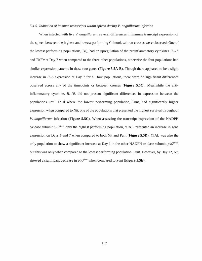
117
5.4.5 Induction of immune transcripts within spleen during V. anguillarum infection
When infected with live V. anguillarum, several differences in immune transcript expression of
the spleen between the highest and lowest performing Chinook salmon crosses were observed. One of
the lowest performing populations, BQ, had an upregulation of the proinflammatory cytokines IL-1β
and TNFα at Day 7 when compared to the three other populations, otherwise the four populations had
similar expression patterns in these two genes (Figure 5.5A-B). Though there appeared to be a slight
increase in IL-6 expression at Day 7 for all four populations, there were no significant differences
observed across any of the timepoints or between crosses (Figure 5.5C). Meanwhile the anti-
inflammatory cytokine, IL-10, did not present significant differences in expression between the
populations until 12 d where the lowest performing population, Punt, had significantly higher
expression when compared to Nit, one of the populations that presented the highest survival throughout
V. anguillarum infection (Figure 5.5C). When assessing the transcript expression of the NADPH
oxidase subunit p22phox, only the highest performing population, YIAL, presented an increase in gene
expression on Days 1 and 7 when compared to both Nit and Punt (Figure 5.5D). YIAL was also the
only population to show a significant increase at Day 1 in the other NADPH oxidase subunit, p40phox,
but this was only when compared to the lowest performing population, Punt. However, by Day 12, Nit
showed a significant decrease in p40phox when compared to Punt (Figure 5.5E).

118
Figure 5.5. Spleen immune transcript expression within the two highest and two lowest
performing populations throughout live V. anguillarum challenge. Transcript expression of
cytokines IL-1β (A), TNFα (B), IL-6 (C) and IL-10 (D) as well as the NADPH oxidase subunits p22phox
(E) and p40phox (F) was assessed via qRT-PCR analysis on Days 1, 7 and 12 following injection with
live V. anguillarum. Only individuals from the two highest performing crosses (YIAL and Nit) and the
two lowest performing crosses (BQ and Punt) was evaluated. All data was normalized to the reference
gene (EF1α) and expressed as a fold change over the Day 0 control group where control expression
was set to 1. Error bars represent +SEM and each data point is the average of 6 individuals. A p-value
of less than 0.05 was considered to be statistically significant.

119
5.4.6 Co-infection with R. salmoninarum in highest and lowest performing crosses
Because R. salmoninarum is another common pathogen in Chinook salmon aquaculture, an
ELISA-based assay was used to determine whether the highest performing (YIAL) and lowest
performing (Punt) crosses had differences in the number of fish that were positive for BKD infection.
Punt had the lowest survival following challenge with live V. anguillarum and this 85% of the fish
tested (n = 20) in this cross were also positive for BKD (Figure 5.6). In comparison, YIAL had the
highest survival throughout V. anguillarum infection and only 55% (n = 20) of the fish from this
population were positive for BKD (Figure 5.6).
Figure 5.6. Determining co-infection with R. salmoninarum in the highest and lowest performing
Chinook salmon crosses. Head kidney samples of crosses that presented the highest and lowest
survival to V. anguillarum infection (YIAL and Punt respectively) were tested for co-infection of R.
salmoninarum, the causative agent of BKD.

120
5.5 Discussion
Reintroducing genetic diversity into domesticated or captive populations has been shown to
enhance immune performance in several species (Pinard-Van der Laan et al., 1997; Spielman et al.,
2004; Smallbone et al., 2016), yet precise improvements for desired immune traits are not guaranteed
by this strategy. Thus, even though outbreeding is a common technique for the improvement of inbred
animals, there has been limited application of this practise in aquaculture. The primary goal of the
current study was to improve immune performance of the domesticated YIAL Chinook salmon stock
by outbreeding and identify potential genetic or functional immune markers that could predict any
resulting immune differences. Given that the YIAL production stock has been bred for over 30 years
without outside genetic input, it was our prediction that some outbred crosses would maintain the
desired phenotypes of domestication while also exhibiting improved responses to disease challenge
indicative of hybrid vigor.
When challenged with live V. anguillarum, there was variable survival across all eight groups
studied with the pure domesticated cross (YIAL) presenting the highest survival. Chinook salmon
production at YIAL follows organic standards and consequently, through the avoidance of antibiotic
use, has selected for disease resistance over multiple generations. It was anticipated that the YIAL stock
would contribute valuable traits for disease resistance, however, it was unknown whether the
introduction of wild genes would further enhance or inhibit this. The results presented here reveal that
only the Punt and BQ outbred crosses were significantly more susceptible to V. anguillarum infection
when compared to YIAL or Nit. Differences in susceptibility to vibriosis have been observed in BC
Pacific salmon populations previously (Beacham & Evelyn, 1991; Beacham & Evelyn, 1992a; Balfry
et al., 2001; Balfry et al., 2008). One study by Beacham & Evelyn (1991) showed significant variation
in mortality between three populations of Chinook salmon originating from the Kitimat, Nitinat and
Quinsam rivers of BC after receiving i.p. injection of live V. anguillarum. Surprisingly, this study
reported that Nitinat mortality was more than double that of Quinsam and Kitimat (Beacham & Evelyn,
1991). This is contrary to the results presented here wherein the outbred Nitinat was one of the highest

121
surviving crosses when challenged with V. anguillarum. This contradictory result for Nitinat could
reflect that a high proportion of this population died when transferred to saltwater (Semeniuk et al.,
2019). As a result, the outbred Nitinat fish used in this study may have actually represented the most
resilient of that cross thus providing an explanation for the unexpected high performance. Overall, the
variability in survival to V. anguillarum challenge that was observed between the eight crosses could
reflect interesting immune differences for discovery.
Though the focus of this study was to compare differences in immune performance throughout
infection challenge, it was observed that even when injected with sterile PBS there was disparity in
survival among the eight crosses. When comparing the effect of handling stress between wild and
hatchery-reared Chinook salmon, Mazur & Iwama (1993) found that hatchery-reared salmon had lower
plasma cortisol levels and lower hematocrits when compared to their wild counterparts. Similar trends
were shown in other salmonids such as sea trout (Salmo trutta), Atlantic salmon (Salmo salar) and
rainbow trout (Oncorynchus mykiss, Woodward & Strange, 1987; Johnsson et al., 2001; Lepage et al.,
2001) indicating that domesticated stocks may be better able to cope with and overcome stressful
situations. Despite these observations, the highly domesticated YIAL stock presented lower survival in
the sham injected controls than three of the outbred populations. Though YIAL was only significantly
different from one outbred cross, it was anticipated that the more domesticated group would outperform
the other crosses as these fish spent the longest duration in captivity. Because the outbred populations
would maintain some traits from the captive YIAL population parent, it is possible that this unexpected
result was an example of heterosis.
When the host encounters bacterial pathogens, like V. anguillarum, the major histocompatibility
(MH) class II molecules are responsible for the recognition and presentation of antigens to helper T
lymphocytes. In doing so, this class of MH directs the immune response to effectively combat particular
extracellular threats (reviewed by Grimholt, 2016). The binding of pathogen derived antigens to MH
molecules is governed by allelic variation at the peptide binding groove. There are many such alleles
and every individual within a species has a limited repertoire. The genetic polymorphism at the MH

122
loci can provide more or less protection towards relevant pathogens and thus these genes are believed
to be under strong selection pressure (reviewed by Yamaguchi & Dijkstra, 2019). Despite this, the
results of the current study revealed that outbreeding a domesticated stock with seven wild populations
did not increase the allelic diversity of MH class II β1 genotypes. Across all eight groups, only 9 alleles
were detected in the 240 individuals assessed. This level of allelic variation is similar to what has been
reported previously for wild Chinook salmon populations (Kim et al., 1999; Evans et al., 2010; Lehnert
et al., 2016). Interestingly, the lowest performing cross, Punt, presented the greatest number of unique
alleles. This is contradictory to the belief that more allelic diversity in MH genotypes will better protect
a population as a whole (reviewed by Bernatchez & Landry, 2003; Spurgin & Richardson, 2010), but
this could be pathogen dependent. It was also observed that a greater percentage of individuals were
heterozygous for MH class II β1 genotype in the two highest performing crosses when compared to the
two lowest performing crosses. This may support the notion that heterozygotes are able to detect a
broader range of pathogen-driven antigens because they have greater variability in their MH molecules,
a form of balancing selection referred to as heterozygote advantage (reviewed by Sommer, 2005;
Spurgin & Richardson, 2010). Indeed, heterozygosity in MH genotype has been associated with better
survival in Chinook salmon when challenged with infectious hematopoietic necrosis virus (IHNV,
Arkush et al., 2001) and V. anguillarum (Becker et al., 2014) as well as heterozygous fry exhibiting
fewer bacterial infections overall when compared to their homozygous counterparts (Evans & Neff,
2009). As of yet, specific alleles at the MH class II loci have not been associated with resistance or
susceptibility to individual pathogens in salmonids. This was also observed here wherein individual
MHC class II alleles could not be attributed to the variable survival to V. anguillarum infection, but
further support for heterozygote advantage hypothesis in Chinook salmon was observed.
Because vibriosis is a bacterial septicaemia, host development of pathogen-specific antibodies
should be crucial for limiting the spread of infection. To date, teleosts are known to have three classes
of antibody, with IgM being the dominant class within the blood (reviewed by Fillatreau et al., 2013;
Sunyer, 2013). In the current study, a subtractive indirect ELISA assay was developed so that serum

123
IgM levels could be compared between the seven outbred crosses and the domesticated control group.
When serum IgM was measured throughout V. anguillarum challenge, only the outbred Nit group was
observed to have significantly higher levels of serum IgM when compared to outbred Quin, Cap and
BQ at Day 28. Given that survival between these four outbred crosses was variable, it does not seem
that higher levels of serum IgM conferred protection from this isolate of V. anguillarum. Serum IgM
levels in salmonids have been studied previously in response to bacterial pathogens using ELISA-based
assays (Boesen et al., 1997; Johnson et al., 2004; Becker et al., 2014; Semple et al., 2018a). Though
significant differences have been observed previously in Chinook salmon serum IgM (Becker et al.,
2014), but quite often the large variability between individuals masks any obvious trends between
groups. With regard to the results presented here, it does not appear that serum IgM levels corresponded
to or could predict survival with regards to V. anguillarum, despite some significant differences being
observed.
The teleost spleen is responsible for filtering blood and initiating immune reactions to blood-
borne antigens/threats (reviewed by Secombes & Wang, 2012). Consequently, this organ can be a major
reservoir for extracellular pathogens that replicate in the blood, such as V. anguillarum. As a result,
transcript expression of the spleen may reflect the immune status of infected fish. When spleen tissues
from the two highest and two lowest performing crosses were assessed for gene expression of pro-
inflammatory cytokines, the only significant difference was observed in the low performing BQ
population which had a significant upregulation in IL-1β and TNFα at 7 d. Additionally, though this
was not significant, it does appear that the lower surviving crosses had an enhanced pro-inflammatory
response in all three genes at Day 12. Given this trend, it is possible that the higher inflammatory
response observed in the lower performing crosses depleted energy resources, making these fish more
susceptible to vibriosis. As has been shown with septic shock in mammals, a greater cytokine response
is not always beneficial for the host (Waage et al., 1989; Pinsky et al., 1993; Li et al., 1995). Findings
like these are absent from teleost literature but there has been a large body of research devoted to
understanding the antagonistic mechanisms controlling inflammation in fish (reviewed by Rebl &

124
Goldammer, 2018). These studies provide evidence that inflammatory responses in teleosts also must
be tightly regulated. Thus, it is reasonable to suggest that the cytokine dysregulation observed in the
present trial could be related to poor survival during bacterial infection. Additionally, IL-10 has been
shown to protect both mice and humans from lethal sepsis (Howard et al., 1993; Li et al., 2017). Yet
the results presented here show that higher IL-10 expression in Punt did not protect this cross as it was
the lowest performing in the V. anguillarum challenge condition. It seems that when it comes to
cytokine levels for Chinook salmon infected with V. anguillarum, a lower response may provide greater
protection.
The respiratory burst activity of phagocytes is an essential immunological response to eliminate
bacterial pathogens in all vertebrate species. Following ingestion, phagocytic leukocytes can kill most
bacteria by producing reactive oxygen intermediates (ROI). This process requires NADPH oxidase
(NOX), which catalyzes the conversion of molecular oxygen into superoxide anions (reviewed by
Panday et al., 2015; Nguyen et al., 2017). Previous work with sea bass (Dicentrarchus labrax L.) has
shown that V. anguillarum inhibits the respiratory burst of head kidney leukocytes when measured by
a functional chemiluminescent assay (Sepulcre et al., 2007b). Sepulcre and colleagues (2007b) were
then able to link this functional decrease in RBA to downregulation in gene expression of NOX
subunits, p22phox and p40phox, during live V. anguillarum infection. In our study, transcript expression
of p22phox and p40phox was analyzed and compared between the two highest and two lowest performing
crosses. YIAL was observed to have significantly higher expression of these genes at earlier timepoints
when compared to the outbred groups. Decreases in circulating leukocytes has been reported previously
in rainbow trout with vibriosis (Lamas et al., 1994). Thus, reduced expression of NOX genes could be
a sign of immune cells succumbing to the associated virulence factors. Because Chinook salmon from
the YIAL cross presented the greatest survival to V. anguillarum challenge and this group has
undergone over 30 years of selection without antibiotics, it is possible that these animals are able to
mount a more effective and rapid ROI response. If this was the case, it may help explain the greater
survival of YIAL when challenged with such a fast-acting pathogen.
125
The results of this study thus far have focused on linking the immune response of Chinook
salmon crosses to their survival throughout V. anguillarum infection. However, when the animals are
grown in sea-pens, V. anguillarum is not the only microorganism that they will come into contact with.
Another common pathogen in salmon culture is R. salmoninarum, the causative agent of bacterial
kidney disease (BKD). Because R. salmoninarum is an intracellular pathogen (Gutenberger et al.,
1997), the requirements of the immune system to clear and/or inhibit disease presentation are different
than for that of the extracellular V. anguillarum. This means that if the animals were already dealing
with R. salmoninarum infection, they may have had less energetic resources remaining to combat the
live V. anguillarum administered in this study. Indeed, it was observed in the present study that the
highest performing group (YIAL) had a much lower percentage of fish positive for BKD when
compared to the lowest surviving cross (Punt). This would not be the first time that co-infection has
been seen to inhibit the survival of salmonid species to infection (Johansen et al., 2009; Roon et al.,
2015; Figueroa et al., 2017) and has been studied extensively in fish (reviewed by Kotob et al., 2016).
When Chinook salmon are infected with the parasite, Nanophyetus salmincola, significantly greater
susceptibility to V. anguillarum has been observed (Jacobson et al., 2003). Given the consistently
negative impact of co-infection on survival in fish, it was not surprising that high co-infection coincided
with reduced performance in the current study.
5.6 Conclusions
In this study, a domesticated stock of Chinook salmon was crossed with seven wild population
so that immunological performance could be assessed when challenged with the marine pathogen V.
anguillarum. Significant differences in survival were observed with the domesticated YIAL population
and the outbred Nitinat group having the highest survival while the lowest performance was observed
in the outbred Puntledge and Big Qualicam crosses. Specific MH class II β1 alleles were not found to
predict survival to V. anguillarum infection but crosses that had a higher percentage of fish presenting
a heterozygous genotype had greater survival. When assessing serum IgM levels between the crosses,
only Nitinat showed a significant increase when compared to Quinsam, Big Qualicum and Capilano,

126
thus this did not seem to coincide with the observed survival. Notable differences were observed in
spleen transcript levels between the two highest and two lowest performing where a stronger cytokine
response predicted lower survival to V. anguillarum infection, possibly due to depletion of energetic
resources. Meanwhile, upregulation of RBA genes at earlier timepoints was observed in the highest
performing cross, YIAL. Lastly, it was observed that the lowest performing cross had a higher
percentage of fish infected with R. salmoninarum when compared to the highest surviving cross. Thus,
co-infection may have impacted the observed survival to V. anguillarum exposure. This study
represents the first of its kind to outbreed a domesticated stock with wild populations of Chinook
salmon and compare the immune function of resulting offspring. Though clear immune markers for
resistance/susceptibility to vibriosis were not determined, there were obvious differences between the
crosses regarding their immune function. However, the inbred, domesticated stock had the highest
performance overall. As a result, it appears possible that Chinook salmon stocks could be enhanced for
commercial culture, but outbreeding may not be the method of choice.
Acknowledgements
John and Ann Heath for their continual support and advice needed for planning and executing the
infection trial. Elliot Haugen for his much-needed assistance with fish transport and care. Marije
Booman and Simon Jones for providing the virulent strain of V. anguillarum and valuable advice
regarding the successful culture of the pathogen. Andrew Doxey for his help identifying intron and
exon junctions of Chinook salmon RBA genes. Clare Venney, Sarah Lehnert, Shelby Toews and Denis
Roy for their assistance with sample preparation and calculating heterozygosity estimates for the
microsatellite analysis.
127
Chapter 6: Development and characterization of a Chinook salmon spleen stromal cell line to
study the cellular immune response to Vibrio anguillarum.
Shawna L. Semple1, Daniel D. Heath2 and Brian Dixon1
1University of Waterloo, Department of Biology, Waterloo, Canada 2Great Lakes Institute for Environmental Research, University of Windsor, Windsor, Canada
Formatted for Submission to: Fish and Shellfish Immunology

128
6.1 Overview
Despite increased interest in Chinook salmon aquaculture, there is inadequate understanding regarding
the bacterial immune defenses of this species. This study describes the establishment and
characterization of a continuous stromal cell line derived from Chinook salmon spleen, CHST, and its
response to heat-killed bacterial exposure challenge. Optimal growth of CHST was seen at 18°C when
grown in Leibovitz’s L15 media supplemented with 20% fetal bovine serum. DNA analyses confirmed
that CHST did originate from Chinook salmon tissue. The bacterial species used for exposure studies
was Vibrio anguillarum, a common pathogen of marine aquaculture and the causative agent of an acute
hemorrhagic septicaemia known as vibriosis. V. anguillarum was observed to grow rapidly in cell
culture media and is known to produce a plethora of extracellular toxins, thus live bacterial challenge
was not used for this study. Instead, V. anguillarum was heat-killed and CHST was challenged so that
pro-inflammatory (IL-1β, IL-6 and TNFα) as well as anti-inflammatory (IL-10) cytokine transcript
levels could be measured at 1, 4, 12, 24 and 96 hr post-exposure. The heat-killed bacteria was observed
to significantly stimulate the expression of all three pro-inflammatory cytokines at 4, 12, 24 and 96 hr
post-exposure with the peak in expression occurring at 12 and 24 hr. Meanwhile, IL-10 was not
observed to significantly increase until 96 hr post-stimulation, which was also the time when the
inflammatory cytokine expression was decreasing. The establishment and characterization of CHST
provides a valuable model for studying the immune response of Chinook salmon stromal cells in
response to stimuli, including those of bacterial origin.
129
6.2 Introduction
Due to their large size and high market value, Chinook salmon (Oncorhynchus tshawytcha)
have historically been a focus for capture fishery production on the West coast of Canada (Christensen
et al., 2018, Ohlberger et al., 2018). This is rapidly changing though, because declining numbers in
native populations have resulted in the inability of fisheries to meet the rising demands for Chinook
salmon consumption (Johnson et al., 2017). Additionally, since the majority of salmon farmed in British
Columbia are Atlantic (Statistics Canada, 2017), there is growing apprehension that escapees and the
introduction of novel diseases could be implicated in the diminishing numbers of wild Chinook salmon
(reviewed by Noakes et al., 2000; Naylor et al., 2003; Ayer & Tyedmers, 2009). All of these concerns
have resulted in increased public awareness promoting the culture of Chinook salmon on the West coast
to hopefully ease some of the pressure experienced by wild populations. Unfortunately, because this
species is not as well domesticated as their Atlantic counterparts, there are many gaps in knowledge
regarding their efficient culture which makes this endeavor difficult. Obtaining and applying a deeper
understanding Chinook salmon immunity is one way to help make this species more robust in intensive
culture situations.
Mortality due to infectious disease translates into one of the greatest sources of financial loss
for global aquaculture (World Bank, 2014; reviewed by Stentiford et al., 2017). With countless bacterial
species inhabiting aquatic environments, some are able to take advantage of host stress and/or immune
suppression to become opportunistic pathogens (Sundberg et al., 2016). Though there are numerous
bacterial infectious agents in marine environments (reviewed by Toranzo et al., 2005), Vibrio
anguillarum is one responsible for significant financial losses to salmon aquaculture (reviewed by Frans
et al., 2011; Hickey & Lee, 2018). This gram negative bacterium is the causative agent of vibriosis, a
fatal hemorrhagic septicaemia capable of infecting over 90 different aquatic species (reviewed by
Hickey & Lee, 2018). Acute outbreaks of the disease can be particularly problematic as these can result
in high mortalities without the presentation of any clinical signs (reviewed by Frans et al., 2011).
Despite the presence of several commercial vaccines (reviewed by Hickey & Lee, 2018), vibriosis is
130
still a prevalent disease in salmon aquaculture, outlining that there is more to learn of this pathogen and
what constitutes an effective immune response towards it.
As a bacterial septicaemia, V. anguillarum can be found throughout the blood of infected
individuals, making the spleen an important site for immune defenses. Like mammals, the spleen of
teleosts is an important haematopoietic organ that can be separated into ellipsoids, red pulp and white
pulp (reviewed by Fange & Nilsson, 1985; Espenes et al., 1995; Genten et al., 2009). However, fish
also have the additional element of melanomacrophage centres (MMCs) which are believed to be
primitive analogues of mammalian germinal centres (GCs, reviewed by Stosik et al., 2019). The red
pulp is a macrophage and erythrocyte dense region that plays an important role in iron recycling and
surveillance of blood-borne pathogens. In comparison the white pulp is a lymphocyte-rich area that
supports antigen specific immune responses (reviewed by Klei et al., 2017; Golub et al., 2018). As
observed in all tissues, the functional cells of the spleen are supported by an intricate network of stromal
cells. More specifically, the immune cells within the spleen require the signals that are produced by
stromal cells in response to their surroundings. As a result, splenic stromal cells provide a crucial
microenvironment to support haematopoiesis along with the proliferation and development of immune
cells (Wilson et al., 2000; O’Neill et al., 2011). Thus, splenic stromal cells enable the establishment
and continuation of effective immune responses, particularly when dealing with a bacterial septicaemia
like V. anguillarum.
The present study describes the establishment and characterization of CHST, a stromal cell line
derived from the spleen of an adult Chinook salmon. The species of origin for CHST was confirmed to
be Chinook salmon and optimal growth conditions were determined. Additionally, various animal cell
culture media formulations were assessed to determine if they could support the growth of V.
anguillarum for potential live challenges in vitro. Following a 96 hr exposure challenge to heat-killed
V. anguillarum, significant induction of CHST pro-inflammatory (IL-1β, IL-6, TNFα) and anti-
inflammatory (IL-10) cytokine expression was observed. As CHST is the only known stromal cell line

131
created from Chinook salmon, these cells are very relevant for understanding species specific aspects
of immunity and cellular function.
6.3 Materials and Methods
6.3.1 Primary cultures
In September of 2016, a 200 g Chinook salmon was retrieved from a net-pen that was produced
and maintained at Yellow Island Aquaculture Ltd (YIAL, Quadra Island, BC, Canada). The fish was
euthanized with an overdose of clove oil prior to tissue collection. All procedures were performed
following the guidelines of the Animal Care Committee at the University of Waterloo. The spleen,
caudal fin and gills were collected and stored in L15 media supplemented with 20% fetal bovine serum
(FBS, Gibco), 200 U/mL of penicillin and 200 U/mL streptomycin (Thermo Scientific). Upon arrival
to the University of Waterloo, each tissue type was diced into small pieces in a laminar flow hood and
rinsed three times with buffered saline solution (PBS, Lonza) containing the same antibiotics as noted
above. Tissue pieces were then transferred to 25 cm2 flasks (BD Falcon) where approximately 1–2 mL
of L15 supplemented with 20% FBS and the previously described antibiotic concentrations. Primary
cultures were established by the explant outgrowth method as previously demonstrated with walleye
spleens and fins (Vo et al., 2015, Vo et al., 2016). Flasks were then incubated at 18°C with media
changes occurring every 2–3 days. It was eventually observed that some flasks were developing cell
populations that were hematopoietic in nature. The media supplementation to these flasks was changed
to L15 with 30% FBS to promote these cell populations. In an effort to isolate the immune cells, only
the floating cells were transferred to new cell culture flasks. Eventually the floating cells adhered to the
flask and took on a fibroblastic-like morphology. These fibroblastic-like cells would eventually develop
into the CHST cell line.
6.3.2 Maintenance of CHST
Initially, CHST was routinely grown in L15 supplemented with 30% FBS at 18°C to maintain
its hematopoietic cell populations. Following the first passage, the adherent CHST cells were
subcultured at a 1:2 ratio on a weekly or bi-weekly basis using trypsin (Lonza) and maintained in

132
L15/20% FBS. CHST has been maintained for over three years and has undergone more than 30
passages.
6.3.3 Cryopreservation of CHST
Approximately 4 x 10⁶ CHST cells at multiple passages were cryogenically frozen in L15
containing 20% FBS and 10% dimethyl sulphoxide (DMSO, Sigma). The cells were frozen at -60°C
overnight and subsequently immersed in liquid nitrogen (-196°C) for long-term storage. To determine
cell viability upon thawing, a trypan blue (Sigma) exclusion assay was performed using a
haemocytometer under a phase contrast microscope (Leica).
6.3.4 Optimal growth conditions of CHST
Optimal temperature of growth for CHST was analyzed between 4 and 26°C. CHST cells were
seeded into five 6-well plates (Fisher Scientific) at a concentration of 2×10⁵ cells/well and incubated
overnight at 18°C. Three wells of one plate were used to provide the day 0 cell counts. Two 6-well
plates were incubated at each of the four temperatures studied (4, 14, 18 and 26°C) for 6 days. On days
0, 3 and 6, triplicate wells from each temperature were washed with 1 mL of PBS and cells were
dissociated using 300 μL of trypsin (Gibco). Cell counts for each well were determined using a
hemocytometer under a phase contrast microscope (Leica). Cell counts for each time interval were
averaged and calculated as a percentage of the day 0 growth to determine the percent growth.
To determine the optimal FBS concentration for maintenance of CHST, five 6-well plates were
seeded with cells as described above. Following overnight adherence at 18°C, media was removed from
all plates and one 6-well plate received 2 mL of either 5% FBS, 10% FBS, 15% FBS or 20% FBS
media. All plates were then returned to the 18°C incubator. On days 0, 3 and 6, triplicate wells from
each FBS concentration were washed with 1 mL of PBS so that cells could be dissociated and counted
as described above.

133
6.3.5 Confirming species of origin
Genomic DNA was extracted from CHST cell pellets using the DNeasy Blood and Tissue Kit
(Qiagen) as described by the manufacturer. To validate the origin of the CHST cells, primers previously
shown to amplify a segment of the Chinook salmon mitochondrial cytochrome c oxidase subunit (cox1,
Table 6.1) were used as described previously by Semple et al (2018d). The PCR product was then run
on a 1.5% agarose gel with 1X GelGreen (Biotium). Bands of the desired size were cut out of the gel
and extracted using the QIAquick Gel extraction kit (Qiagen). The resultant fragments were cloned into
pGEM®-T Easy plasmids (Promega Corporation) and transformed as previously described by Semple
et al. (2018a). Five colonies per PCR reaction were selected and grown in LB medium. Plasmids were
extracted from the transformants using the GenElute Plasmid Miniprep Kit (Millipore Sigma)
according to the manufacturer's instructions. Plasmids were sequenced using SP6 primers at the TCAG
sequencing facility (Sick Kids Hospital, Toronto, Ont.). The sequences were analyzed using the Basic
Local Alignment Search Tool (BLAST) software (http://blast.ncbi.nlm.nih.gov/Blast.cgi).
6.3.6 V. anguillarum growth in bacterial and animal cell media
Frozen stocks of the V. anguillarum were subcultured onto tryptic soy agar (TSA) with 2% NaCl.
Following incubation at room temperature (RT) for 48 h, a single colony was used to inoculate 3 mL
of tryptic soy broth (TSB) supplemented with 2% NaCl and incubated for 10 h at RT and 200 rpm.
After this incubation, 1 mL of the bacterial culture was centrifuged (8 min at 8000 rpm) and was washed
twice with 1 mL of PBS. The bacterial pellet was then resuspended in 1 mL of PBS.
To determine the growth of V. anguillarum in its preferred culture media (TSB with 2% NaCl)
as well as in animal culture media formulations (L15 with no FBS, 2% FBS, 10% FBS or 20% FBS),
1 µL of the bacterial growth was added to 1 mL of the different media types. In a sterile, 96-well BioLite
plate (ThermoFisher), 100 µL of each inoculum was added to 6 wells of the plate. For each media type,
four wells received 100 µL of the media alone to act as blanks. All perimeter wells received 300 µL of
sterile water to prevent evaporation of experimental wells. Once all wells were prepared, the absorbance
134
at 600 nm was read for each well every hour for 25 hr using a Synergy H1 plate reader (BioTek
instruments). For every absorbance reading, the blank culture absorbance value was subtracted from
the six bacterial growth wells for that respective media type.
6.3.7 Preparation of heat-killed V. anguillarum
Three milliliters of V. anguillarum bacterial culture was prepared as described above in section
6.3.6. The resulting culture had an optical density of 0.9 which was confirmed to be 1.08 x 10¹⁰ CFU/mL
by a standard plate count (SPC). Specifically, bacterial numbers were enumerated by plating 0.1 mL of
ten-fold serial dilutions on TSA with 2% NaCl plates in triplicate. After incubation at RT for 30 hours,
plates containing 25-250 colonies were counted and the number of CFU/mL calculated. One 1 mL
aliquot was removed from the V. anguillarum growth and centrifuged for 8 min at 8000 rpm. The
resulting cell pellet was then washed twice with 1 mL of PBS before being resuspended in 1 mL of
PBS. The V. anguillarum culture was then boiled for 25 min to kill any live bacteria. After boiling, 100
µL of the heat-killed V. anguillarum was spread onto a TSA with 2% NaCl plate to confirm the absence
of viable bacteria. Subsequently, the killed bacterial culture was centrifuged for 8 min at 8000 rpm and
resuspended in 900 µL of L15 supplemented with 20% FBS which was to be used to expose CHST to
the heat-killed bacterium.
6.3.8 Exposure of CHST to heat-killed V. anguillarum
In 6-well tissue culture plates (ThermoFisher), CHST was seeded at 4 x 10⁵ cells/well and
maintained overnight at 18°C. The cells were then exposed to either heat-killed V. anguillarum at an
MOI of 50, or media alone to act as unstimulated controls. Following this single exposure, all
experimental plates were returned to the 18°C incubator. At 1 hr, 4 hr, 12 hr, 24 hr and 96 hr following
stimulation, CHST cells were mechanically dislodged using sterile 23 cm cell scrapers (ThermoFisher)
and transferred to a sterile, 15 mL conical tube (Falcon). The cells were centrifuged (5 min, 500 x g,
4°C), washed once with 5 mL of PBS, and resulting cell pellets were stored at -80°C.
135
6.3.9 qRT-PCR
6.3.9.1 RNA extraction and cDNA synthesis
RNA was extracted from CHST cell pellets (4 x 10⁵ cells) using an RNeasy RNA Extraction Kit
(Qiagen) as described by the manufacturer. To remove any contaminating genomic DNA, all RNA
samples were treated with DNase I (Thermo Scientific). RNA samples were then quantified using the
Take3 plate of a Synergy H1 plate reader (BioTek Instruments) and were stored at -80oC until further
use. Complementary DNA (cDNA) was synthesized from 500 ng of total RNA using the qScript cDNA
Supermix (Quanta Biosciences) in accordance to the manufacturer’s instructions. For a no template
control, 500 ng of RNA suspended in 20 uL of DEPC water was included in the cDNA synthesis
reaction without reverse transcriptase
6.3.9.2 qRT-PCR reactions
To assess transcript levels of IL-1β, TNFα, IL-6, and IL-10 in CHST cells, qRT-PCR analysis
was completed. All PCR reactions were 10 µl and contained: 2.5 μl of cDNA (25 ng/μl diluted 1:10 in
RNase free water), 2x WISENT ADVANCEDTM qRT-PCR mastermix (Wisent), and forward and
reverse primers (Sigma Aldrich) to a final working concentration of 0.25 μM. All qPCR reactions were
completed on the LightCycler® 480 II (Roche). The sequences for all primer sets are outlined in Table
6.1. Each experimental sample was run in triplicate. For each plate, triplicate wells of a calibrator, no
template control and RNA only control were also present. The program used for all qRT-PCR reactions
was as follows: pre-incubation at 95°C for 10 min followed by 40 cycles of denaturation at 95°C for
10 sec, annealing at 60°C for 5 sec and extension at 72°C for 8 sec. A melting curve was completed for
every run from 65°C to 97°C with a read every 5 sec. Product specificity was determined through single
PCR melting peaks. All qRT-PCR data was analyzed using the ΔΔCt method while incorporating
individual primer efficiencies into the equation as described by Pfaffl (2001) and is presented as the
average of 3 experimental replicates with the standard deviation. Specifically, gene expression was
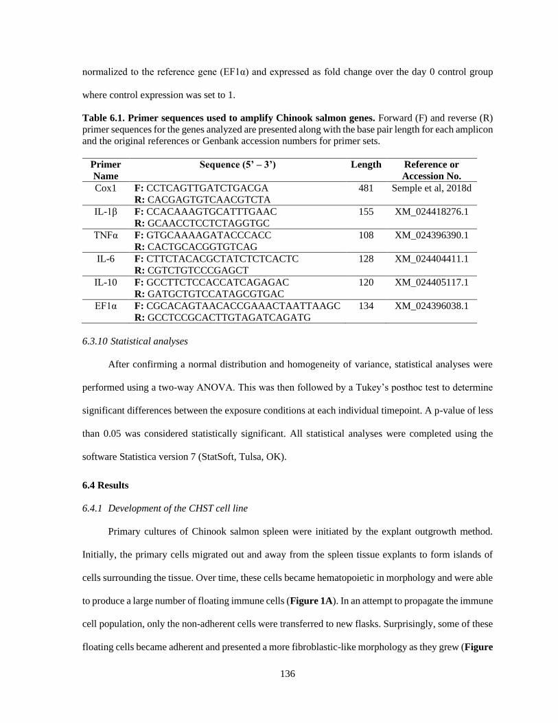
136
normalized to the reference gene (EF1α) and expressed as fold change over the day 0 control group
where control expression was set to 1.
Table 6.1. Primer sequences used to amplify Chinook salmon genes. Forward (F) and reverse (R)
primer sequences for the genes analyzed are presented along with the base pair length for each amplicon
and the original references or Genbank accession numbers for primer sets.
Primer
Name
Sequence (5’ – 3’) Length Reference or
Accession No.
Cox1 F: CCTCAGTTGATCTGACGA
R: CACGAGTGTCAACGTCTA
481 Semple et al, 2018d
IL-1β F: CCACAAAGTGCATTTGAAC
R: GCAACCTCCTCTAGGTGC
155 XM_024418276.1
TNFα F: GTGCAAAAGATACCCACC
R: CACTGCACGGTGTCAG
108 XM_024396390.1
IL-6 F: CTTCTACACGCTATCTCTCACTC
R: CGTCTGTCCCGAGCT
128 XM_024404411.1
IL-10 F: GCCTTCTCCACCATCAGAGAC
R: GATGCTGTCCATAGCGTGAC
120 XM_024405117.1
EF1α F: CGCACAGTAACACCGAAACTAATTAAGC
R: GCCTCCGCACTTGTAGATCAGATG
134 XM_024396038.1
6.3.10 Statistical analyses
After confirming a normal distribution and homogeneity of variance, statistical analyses were
performed using a two-way ANOVA. This was then followed by a Tukey’s posthoc test to determine
significant differences between the exposure conditions at each individual timepoint. A p-value of less
than 0.05 was considered statistically significant. All statistical analyses were completed using the
software Statistica version 7 (StatSoft, Tulsa, OK).
6.4 Results
6.4.1 Development of the CHST cell line
Primary cultures of Chinook salmon spleen were initiated by the explant outgrowth method.
Initially, the primary cells migrated out and away from the spleen tissue explants to form islands of
cells surrounding the tissue. Over time, these cells became hematopoietic in morphology and were able
to produce a large number of floating immune cells (Figure 1A). In an attempt to propagate the immune
cell population, only the non-adherent cells were transferred to new flasks. Surprisingly, some of these
floating cells became adherent and presented a more fibroblastic-like morphology as they grew (Figure
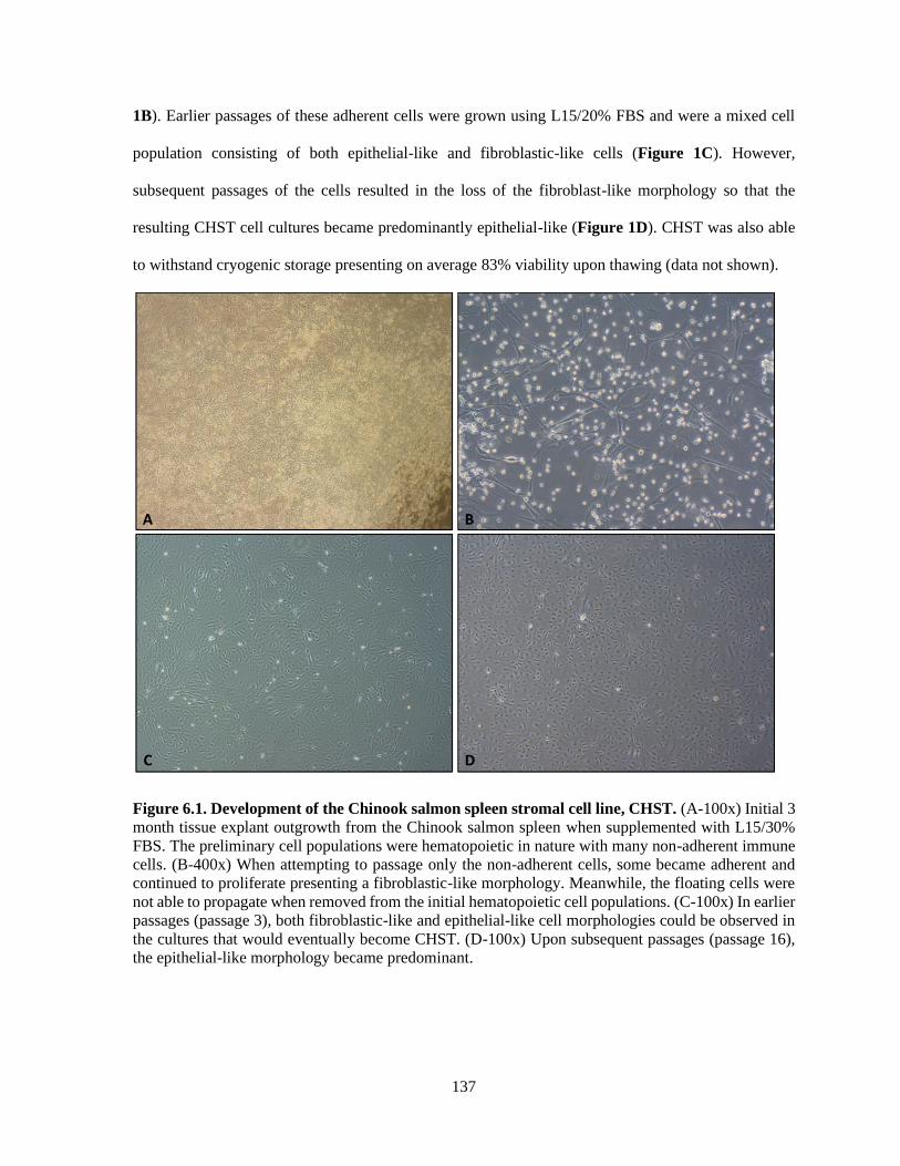
137
1B). Earlier passages of these adherent cells were grown using L15/20% FBS and were a mixed cell
population consisting of both epithelial-like and fibroblastic-like cells (Figure 1C). However,
subsequent passages of the cells resulted in the loss of the fibroblast-like morphology so that the
resulting CHST cell cultures became predominantly epithelial-like (Figure 1D). CHST was also able
to withstand cryogenic storage presenting on average 83% viability upon thawing (data not shown).
Figure 6.1. Development of the Chinook salmon spleen stromal cell line, CHST. (A-100x) Initial 3
month tissue explant outgrowth from the Chinook salmon spleen when supplemented with L15/30%
FBS. The preliminary cell populations were hematopoietic in nature with many non-adherent immune
cells. (B-400x) When attempting to passage only the non-adherent cells, some became adherent and
continued to proliferate presenting a fibroblastic-like morphology. Meanwhile, the floating cells were
not able to propagate when removed from the initial hematopoietic cell populations. (C-100x) In earlier
passages (passage 3), both fibroblastic-like and epithelial-like cell morphologies could be observed in
the cultures that would eventually become CHST. (D-100x) Upon subsequent passages (passage 16),
the epithelial-like morphology became predominant.

138
6.4.2 Confirming the species of origin of CHST
Confirming the species identity of CHST was accomplished through the amplification of the
cox1 gene. Using BLAST, the sequence obtained from CHST were searched and found to have the
highest identity and query cover with Chinook salmon sequences (Table 6.2).
Table 6.2. Validating the species identity of CHST through cox1 barcoding. The cox1 gene was
amplified and sequenced to confirm the species of origin for CHST. The four known sequences that
had the highest similarity to the CHST cox1 sequence are presented.
Gene Length (bp) % Identity to Chinook salmon
Sequences
Cytochrome C Oxidase Subunit 1
(Cox1)
478
100%, JX960927.1
99.79%, HQ167683.1
99.79%, AF392054.1
99.58%, KX958414.1
6.4.3 Determining optimal growth conditions for CHST
To assess optimal growth conditions of the newly established CHST, effect of temperature and
FBS supplementation on growth was explored. Over a six day period, CHST displayed the highest
growth rate at 18°C with significantly less growth being observed at 4°C, 14°C and 18°C (Figure
6.2A). When characterizing media supplementation, CHST displayed the greatest proliferation in L15
media supplemented with 20% FBS at 18°C (Figure 6.2B). As there was no significant difference in
cellular growth between 20% and 15% FBS, growing CHST at the lower FBS concentration would be
a cost-effective alternative pending consistent passaging of the culture.
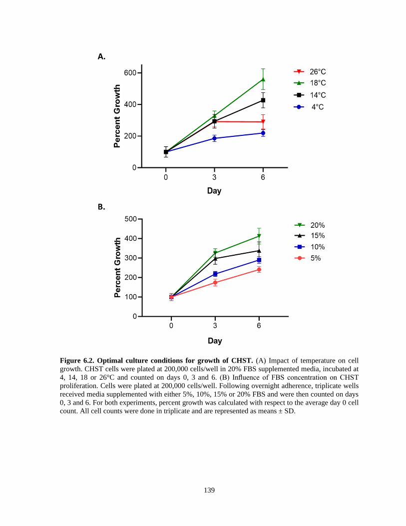
139
Figure 6.2. Optimal culture conditions for growth of CHST. (A) Impact of temperature on cell
growth. CHST cells were plated at 200,000 cells/well in 20% FBS supplemented media, incubated at
4, 14, 18 or 26°C and counted on days 0, 3 and 6. (B) Influence of FBS concentration on CHST
proliferation. Cells were plated at 200,000 cells/well. Following overnight adherence, triplicate wells
received media supplemented with either 5%, 10%, 15% or 20% FBS and were then counted on days
0, 3 and 6. For both experiments, percent growth was calculated with respect to the average day 0 cell
count. All cell counts were done in triplicate and are represented as means ± SD.
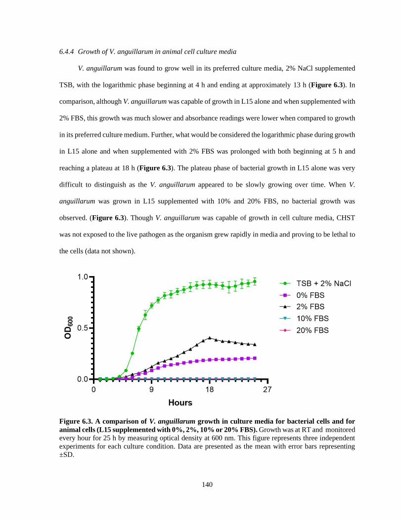
140
6.4.4 Growth of V. anguillarum in animal cell culture media
V. anguillarum was found to grow well in its preferred culture media, 2% NaCl supplemented
TSB, with the logarithmic phase beginning at 4 h and ending at approximately 13 h (Figure 6.3). In
comparison, although V. anguillarum was capable of growth in L15 alone and when supplemented with
2% FBS, this growth was much slower and absorbance readings were lower when compared to growth
in its preferred culture medium. Further, what would be considered the logarithmic phase during growth
in L15 alone and when supplemented with 2% FBS was prolonged with both beginning at 5 h and
reaching a plateau at 18 h (Figure 6.3). The plateau phase of bacterial growth in L15 alone was very
difficult to distinguish as the V. anguillarum appeared to be slowly growing over time. When V.
anguillarum was grown in L15 supplemented with 10% and 20% FBS, no bacterial growth was
observed. (Figure 6.3). Though V. anguillarum was capable of growth in cell culture media, CHST
was not exposed to the live pathogen as the organism grew rapidly in media and proving to be lethal to
the cells (data not shown).
Figure 6.3. A comparison of V. anguillarum growth in culture media for bacterial cells and for
animal cells (L15 supplemented with 0%, 2%, 10% or 20% FBS). Growth was at RT and monitored
every hour for 25 h by measuring optical density at 600 nm. This figure represents three independent
experiments for each culture condition. Data are presented as the mean with error bars representing
±SD.
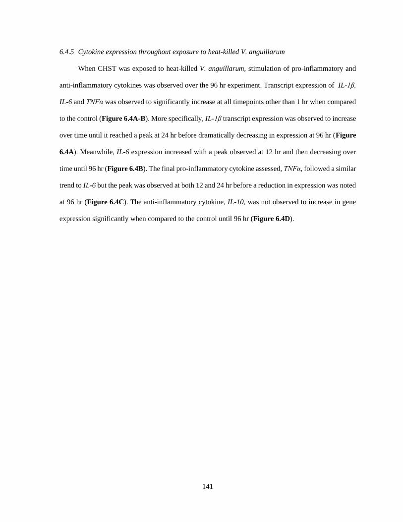
141
6.4.5 Cytokine expression throughout exposure to heat-killed V. anguillarum
When CHST was exposed to heat-killed V. anguillarum, stimulation of pro-inflammatory and
anti-inflammatory cytokines was observed over the 96 hr experiment. Transcript expression of IL-1β,
IL-6 and TNFα was observed to significantly increase at all timepoints other than 1 hr when compared
to the control (Figure 6.4A-B). More specifically, IL-1β transcript expression was observed to increase
over time until it reached a peak at 24 hr before dramatically decreasing in expression at 96 hr (Figure
6.4A). Meanwhile, IL-6 expression increased with a peak observed at 12 hr and then decreasing over
time until 96 hr (Figure 6.4B). The final pro-inflammatory cytokine assessed, TNFα, followed a similar
trend to IL-6 but the peak was observed at both 12 and 24 hr before a reduction in expression was noted
at 96 hr (Figure 6.4C). The anti-inflammatory cytokine, IL-10, was not observed to increase in gene
expression significantly when compared to the control until 96 hr (Figure 6.4D).

142
Figure 6.4. Influence of heat-killed V. anguillarum on pro- and anti-inflammatory cytokine
expression in CHST throughout 96 h of exposure. Transcript expression of pro-inflammatory
cytokines IL-1β (A), TNFα (B), IL-6 (C) as well as the anti-inflammatory cytokine IL-10 (D) was
assessed so that the effect of killed V. anguillarum on immune function could be analyzed. All data was
normalized to the reference gene (EF1α) and expressed as a fold change over the day 0 control group
where control expression was set to 1. All panels represent three independent experiments and are
presented as means + SD. A p-value of less than 0.05 was considered to be statistically significant when
compared to the media alone control for each timepoint.
6.5 Discussion
In the present study, a Chinook salmon-derived spleen stromal cell line, CHST, was
characterized and its transcriptional response to heat-killed V. anguillarum was assessed. Currently
there are only two Chinook salmon cell lines available, the embryonic epithelial cell line CHSE-214
(McCain, 1970) and an epithelial spleen cell line derived from a triploid Chinook salmon, CHSS
(Semple et al., 2018d). Though both of these are excellent tools for understanding Pacific salmonid
immunity, CHST originated from non-adherent immune cells of a haematopoietic culture. These cells
were surrounded with a microenvironment meant to support haematopoiesis, thereby increasing the
likelihood that when these cells differentiated, they would become stromal in nature. As such, CHST

143
can further the understanding of Chinook salmon immunity in regards to the cells that are meant to
support immune function and differentiation, as these have been shown to play meaningful roles in the
immunity of other species (reviewed by Roozendaal & Mebius, 2011; Crowley et al., 2018).
Following the establishment of novel cell lines, it is important to report the optimal growth
conditions so that future experiments using these cell cultures can be repeated reliably. Ideally, cell
lines will have thermal temperature ranges that reflect their species of origin but because both the
culture system and external growth factors differ significantly from that of the whole animal, this is not
always the case. Adult Chinook salmon are reported to have a preferred temperature range between 12
and 15°C (Richter and Kolmes, 2005) but optimal growth of this species is seen at 15-19°C depending
on feeding rates (Brett et al., 1982; Marine, 1997). As CHST was derived from an adult Chinook
salmon, an optimal growth temperature of 18°C fits within the anticipated thermal optimum for this
species and is comparable to other salmonid in vitro models (Semple et al., 2018d).
The strain of V. anguillarum used for this study was observed to grow in both its preferred
culture media, and in formulations of animal cell basal media that had less supplementation with FBS.
As a bacterial septicaemia that flourishes in the blood of teleosts, greater growth in low serum solutions
was unexpected. However, given that mammals and fish and the vast differences in their habitats, the
components of serum between these two taxa is likely to differ significantly. It has been shown that
total plasma protein and albumin content of mammalian serum is twice and four times that of fish
respectively (Zhang et al., 2013). Though variable, lower values of albumin and plasma protein when
compared to mammals have been consistently shown in sera from fish species (Metcalf et al., 1998;
Manera & Britti, 2008; Silva et al., 2015). Thus, V. anguillarum may be better able to grow at lower
concentrations of FBS as this would be comparable to both its native marine environment and the blood
of potential teleostean host species. Additionally, V. anguillarum is considered an opportunistic
pathogen capable of long-term growth in saltwater (Enger et al., 1990; Eguchi et al., 2003), so the
organism does not have a strict requirement for the added nutritional composition of serum to guarantee
its survival. This means that the salt and nutrient content of L15 media alone (reviewed by Bols & Lee,
144
1991) is enough to support of V. anguillarum, though not to a degree that is comparable to its preferred
culture media.
When an individual is infected with a bacterial pathogen the stromal cells of the spleen interact
with blood borne lymphocytes through cytokine signalling. In our study, when challenged with heat-
killed V. anguillarum, CHST was observed to increase expression of pro-inflammatory cytokines (IL-
1β, IL-6 and TNFα) significantly when compared to unstimulated controls. Over time, this induction
was countered by an increase in IL-10 cytokine expression. The cytokine response of rainbow trout
stromal cells has also been explored using the stromal cell line, TSS, when exposed to the stimulants
LPS, polyinosinic polycytidylic acid (poly I:C) or levamisole (Fierro-Castro et al., 2012; Fierro-Castro
et al., 2013). Fierro-Castro and colleagues (2013) found that the stromal cells responded to poly I:C
and levamisole in a manner that was comparable to rainbow trout macrophage cells for TNFα, IL-10
and IL-1β. However, when exposed to LPS the TSS cells showed little to no induction of transcripts.
This may reflect the fact that LPS is often considered an imperfect stimuli for some fish species as the
toll-like receptor that binds and responds to LPS, TLR4, has only been found in zebrafish and no other
teleostean species (Sepulcre et al., 2009; Ali et al., 2014). This outlines the importance of using whole-
killed or live bacteria stimuli when possible. IL-6 is often not the focus of studies using fish stromal
cells but this cytokine has been shown to be produced in splenic stromal cells isolated from mice with
expression increasing as the animals age (Park et al., 2014). When cells were isolated from the
peritoneal membrane of gilthead seabream they were shown to upregulate gene expression of IL-6 when
stimulated with genomic DNA isolated from V. anguillarum (Roca et al., 2008). When all of this is
taken together, it appears that the structural stromal cells necessary to support immune cell function
can sometimes play vital roles in the continuation and regulation of immune function.
145
6.6 Conclusions
CHST is a valuable tool for understanding the cellular physiology and, as described in this
study, the immune function of Chinook salmon. The results presented here describe the characterization
of a stromal cell line derived from the spleen of an adult Chinook salmon. Though CHST was not able
to be exposed to live pathogen, heat-killed V. anguillarum was able to be used as a stimuli and was
much more effective than previous studies using LPS for stromal cell stimulation. Further, CHST was
found to respond significantly to heat-killed V. anguillarum when analyzing transcript expression of
both pro- and anti-inflammatory cytokines over time. When it comes to understanding the stromal cells
necessary to induce the development and differentiation of immune cells in response to stimuli, CHST
provides an excellent model system.
Acknowledgements
Dr. Niels Bols for kindly allowing the use of his cell culture facility and Dr. Nguyen (Nathan) Vo for
his technical insights regarding the initiation of salmonid cell lines.

146
Chapter 7: General Discussion and Future Directions
7.1 The importance of understanding bony fish immunology
Human beings have relied on fish populations for sustenance throughout history. In regard to
the environment, fish play a vital role in natural food webs supporting the growth and survival of
numerous species (Pauly et al., 1998; Wipfli & Baxter, 2011). From an economic perspective, fish
represent a valuable commercial product providing many employment options while also ensuring that
people have access to healthy protein sources worldwide. Aquaculture production also has the added
benefit of ensuring that wild populations will not be dangerously overfished in order to meet the rising
demand for this food source. As the intensive culture of fish is still in its infancy when compared to
terrestrial agriculture, there are still many developments to be made. One way to help enhance the
productivity of aquaculture efforts is to obtain a deeper understanding of teleostean immunity.
Advances here will lead to improved vaccine design and therapeutic options, thereby augmenting yields
and strengthening fish health in these facilities.
Though there are many similarities between the immune system of fish and mammals, there
are also notable differences. Despite the highly studied mammalian model providing a solid baseline,
comparative immunologists must experimentally confirm whether mammalian immune responses and
tissues are analogous in fish. As an example, the bone marrow of mammals is the site of haematopoiesis
and where B cells develop (reviewed by Mercier et al, 2012). However, fish do not have bone marrow,
so instead this essential process occurs in the anterior portion of their kidney (Secombes & Wang,
2012). When it comes to specific immune receptors, fish have many similarities to those found in
mammals (reviewed by Uribe et al., 2011). Nevertheless, teleostean equivalents quite often vary in
number and ligand specificity. This can be seen when comparing toll-like receptors (TLRs) between
these two taxa. One mammalian example, humans, are known to have 10 different TLRs while the
majority of fish have at least 16 (reviewed by Palti, 2011; Nie et al., 2018). Some of these human TLRs
can be found in fish, while others have been lost in many bony fish species, such as TLR6 and TLR10
(reviewed by Palti, 2011). Yet despite these interesting observations between fish and mammalian

147
TLRs, direct evidence of ligand specificity has only been shown in five of the sixteen TLRs found in
fish (Tsujita et al., 2004; Phelan et al., 2005; Matsuo et al., 2008; Ribeiro et al., 2010). Given the large
differences in environment, combined with the fact that fish have undergone at least one, if not two,
whole genome duplications (WGD) not experienced by mammalian species (reviewed by Glasauer &
Neuhauss, 2014), it is not surprising that there are significant differences both functionally and
genetically between fish and mammals. This emphasizes the importance of validating immune
paradigms in teleosts before basing therapies on concepts that have only been confirmed in mammalian
models.
Teleosts represent approximately half of all surviving vertebrate species, making them the
largest and most diverse group of this subphylum (Volff, 2005). Just as there are differences between
species within the taxa Mammalia (reviewed by Zschaler et al., 2014; Tao & Reese, 2017), there are
also distinct differences between species of fish, indeed these differences are bigger given fish emerged
and diversified close to 400 million years ago (reviewed by Brazeau & Friedman, 2015). As an
example, the majority of fish studied have genes for both MH class I and MH class II molecules but
the Atlantic cod (Gadus morhua) has lost the genes for MH class II and its accessory molecules (Star
et al., 2011). Furthermore, the cod-like fish, broadnosed pipefish (Syngnathus typhle), has lost MH class
II function while still maintaining some of the genetic information (Haase et al., 2013). When the whole
genome of cod was compared to other sequenced fish species, it was observed that cod has large gene
expansions and several gene losses in the TLR repertoire (Solbakken et al., 2016). It is possible that the
loss of such a large component of adaptive immunity resulted in a greater dependence on the innate
immune system in this particular species. Understanding the interspecies differences in immunity such
as these will help increase the efficacy of future therapies and vaccine initiatives. As this thesis focused
strictly on salmonid immune function in response to bacterial pathogens, reactions of rainbow trout and
Chinook salmon were explored at both the innate and adaptive levels of immunity. This has provided
valuable information regarding these industrially relevant species and their response to bacterial
pathogens.
148
7.2 Heritable differences in selectively bred fish
There are a number of ways in which the breeding of fish differs from other livestock, and this
is often due to the high fecundity of aquatic species. This allows for strong selection intensity and
results in large families, which can facilitate the extensive collection of phenotypic records of close
relatives for selection candidates within breeding programs (reviewed by Gjedrem & Robinson, 2014).
As it relates directly to the economic potential of an aquaculture facility, growth is often the initial
focus of selective breeding endeavors. Heritability of growth has been observed in several fish species
including tilapia (Oreochromis niloticus), rainbow trout, Atlantic salmon, Asian seabass (Lates
calcarifer), as well as many others (Thodensen et al., 1999; Perry et al., 2005; Bentsen et al., 2017; Ye
et al., 2017). Though now well established, the first breeding programs for aquatic species used mass
selection and were generally unsuccessful (Moav & Wohlfarth, 1976; Hulata et al., 1986). It is now
understood that this was likely due to the accumulated effects of inbreeding as they are known to
influence several economic traits such as growth (Bentsen et al., 1998; Bentsen & Oleson, 2002;
Rodgveller et al., 2005; Bentsen et al., 2017). Fortunately, with the development of creative breeding
programs, a reduction in inbreeding can be consistently predicted for some aquatic species
(D’Ambrosio et al., 2019). Yet, because the effects of heterosis tends to vary between species and
individual stocks, confirmation of genetic improvements should be analyzed on a case-by-case basis.
Despite being extremely valuable, growth certainly is not the only factor selected for in
aquaculture. Given the large losses due to infectious disease (World Bank, 2014), many breeding
programs have been adopted to maintain improved growth while also selecting for resistance to
problematic pathogens (reviewed by Yanez et al., 2014). In some cases, heritability of disease resistance
is observed without negatively influencing animal growth (Imsland et al., 2002), such as has been
observed in rainbow trout with resistance towards F. psychrophilum (Silverstein et al., 2009) or F.
columnare (Evenhuis et al., 2015). However, it has also been shown that selecting for resistance to a
single pathogen can sometimes result in increased susceptibility to another or decreased growth
(Fevolden et al., 1992; Gjoen et al., 1997; Grimholt et al., 2003; Yanez et al., 2016; Trang et al., 2019).

149
Additionally, the gold standard for determining disease resistance is survival comparisons throughout
live infection challenges (reviewed in Odegard et al., 2011). However, the subsequent results may not
be representative as experimental infection models are often not comparable to what would be observed
in a natural infection. For some aquatic pathogens, experimental models of infection that appropriately
mimic live infection have still not been established. This has led to several alternative approaches for
mimicking the outbreaks that are observed in aquaculture facilities such as co-habitation (Murray et al.,
1992; Munang’andu et al., 2016), waterborne (Long et al., 2014; Zhang et al., 2016) and stress induction
challenges (Taksdal et al., 1998; Henriksen et al., 2013). The major issue with these approaches is that
they often are not repeatable if they are successful in producing disease symptoms. Intraperitoneal
injection, such as that used in this thesis, will result in a much more consistent infection status for
disease challenge trials. However, this method is not representative of a natural outbreak as the
integument and mucous barriers are bypassed. Lack of appropriate challenge models has been a
confounding factor for determining whether disease resistance observed in a lab setting will translate
to disease resistance in an intensive culture situation. Understandably, this has also made it difficult to
determine the true efficacy of vaccines and other therapeutics.
In an attempt to make salmonid stocks that were more robust and/or resilient in the face of
bacterial disease, two different breeding strategies were used within this thesis. Initially, selective
breeding to determine functional immune parameters that could predict resistance to BCWD was
completed for rainbow trout. Though there was large variability in survival observed, this was not the
case as the fish grew larger in size. This phenomenon of age-related disease susceptibility has been
reported with other aquatic diseases including F. branchiophilum (Good et al., 2008) and infectious
pancreatic necrosis virus (IPNV, Roberts & Pearson, 2005). Several long-term breeding studies have
shown that resistance to BCWD is heritable and also that breeding over multiple generations can
achieve consistently high resistance (Weins et al., 2013; Marancik et al., 2014; Wiens et al., 2018). The
specific immune function components responsible for this resistance have not yet been identified,
though there is some evidence to support the hypotheses that MH class IB (Johnson et al., 2008), spleen
150
size (Hadidi et al., 2008), IgT+ B cells (Zwollo et al., 2017) and the microbiome of mucosal tissues
(Brown et al., 2019) could be implicated. But because correlation is not always causation, some of these
examples have already been debunked (Wiens et al., 2015), emphasizing the importance of trial
repetition and design in future experiments. The data presented in this thesis does not support the idea
that resistance to BCWD is related to MH class II β1 genotype, serum IgM production or respiratory
burst activity. However, this may be due to the age of the fish or the fact that the families used here
were only of the first generation. Thus, to confidently conclude that these immune components have
no influence on the heritable resistance to BCWD, repeated experiments would need to be conducted
wherein the immune parameters are measured immediately following assessment of resistance. The
results presented here provide data important to the understanding of host response to F. psychrophilum
throughout live infection. Ideally, once immune factors are identified that can predict resistance to
BCWD, selective breeding programs will then be simplified, and the rainbow trout industry will have
one less pathogen to be concerned about.
Although selective breeding for resistance has shown promise, this technique can be difficult
to accomplish when aquaculture practises have propagated inbred stocks over time. Another breeding
strategy used in this thesis was outbreeding to improve the biological fitness of a highly inbred Chinook
salmon stock. There are multiple ways to evaluate the fitness of a group, but because this thesis focuses
on bacterial pathogens, survival and immune performance following V. anguillarum challenge was
assessed between seven outbred crosses and the control inbred stock. Significant differences between
the eight populations in survival, immune transcript expression and serum IgM antibody to V.
anguillarum were observed. Interestingly, it was the control inbred and one outbred population that
presented the highest performance but overall there was a large degree of variability in survival across
the groups. For aquaculture purposes it would be important to determine if these immunological
differences are heritable in subsequent generations. Historically, heritability estimates for vibriosis have
been low in Atlantic salmon but significant differences have been reported specifically for V.
anguillarum (Gjedrem & Aulstad, 1973; Gjoen et al., 1997) and other hemorrhagic bacterial diseases
151
(Standal & Gjerde, 1987). This indicates that selection against such conditions could result in genetic
changes in salmonid species. Yet despite this, a comparable study was completed using several Pacific
salmon species wherein these claims of heritability to vibriosis were contradicted (Beacham & Evelyn,
1992b). Following analyses of specific immune parameters, it was observed that specific MH class II
β1 alleles could not predict resistance to V. anguillarum, similarly to the genotyping results obtained
from resistant/susceptible rainbow trout infected with BCWD. This is contrary to what has been
observed in another teleost species, such as Japanese flounder (Paralichthys olivaceus), wherein certain
MH class IIβ alleles displayed higher resistance to V. anguillarum infection and were also heritable
(Xu et al., 2008). Though serum IgM was not observed to predict resistance to F. psychrophilum in
rainbow trout, some significant differences between the outbred crosses were observed in Chinook
salmon challenged with V. anguillarum. Unfortunately, serum IgM response to V. anguillarum does
not appear to be very heritable based on previous studies in Atlantic salmon (Stromsheim et al., 1994;
Fjalestad et al., 1996). These studies only analyzed antibody development to one antigen, the O-antigen,
thus it is possible that serum IgM developed to the entire pathogen could be heritable in Pacific salmon
species such as Chinook salmon. Although significant immune differences were observed here between
the eight outbred crosses, much like what was observed with selectively breeding rainbow trout, future
studies and more generations of offspring would be required to determine the heritability of these traits
in Chinook salmon.
7.3 Vaccines for bacterial pathogens
Though there are several different types of vaccines produced for aquaculture (Assefa &
Abunna, 2018), the majority used for bacterial pathogens are killed whole-cell preparations. These do
provide some protection but when compared to the successes of terrestrial vaccine formulations, are
limited at best. This lower efficacy is likely due to fundamental differences between the teleostean and
mammalian adaptive immune response and how it translates into immunological memory. Because fish
do not have IgG as an antibody isotype (reviewed by Sunyer, 2013), any secondary antibody response
observed is slight and uses a different approach than the canonical mammalian definition of
152
immunological memory. This means that fish depend only on the low affinity but high avidity of IgM
for repeated exposure to antigen, and thus a less intense secondary serum antibody response is observed
in these animals (Kaattari et al., 2002; reviewed by Solem & Stenvik, 2006). Additionally, fish do not
appear to go through class switch recombination (CSR), despite having all of the necessary components
and enzymes (i.e. AID, RAG1/2, Ikaros, TdT, etc.) required to complete this process (reviewed by
Dickerson & Findly, 2017), due to the structure of their heavy chain genes. Interestingly, even though
the catalytic domain of activation-induced (Cytidine) deaminase (AID) differs from tetrapods, when
this enzyme is transfected into murine B cells it is still able to catalyze CSR (Barreto et al., 2005). Since
fish do not appear capable of CSR, this finding revealed that the actual process of CSR must have
evolved separately from the AID enzyme itself (Barreto et al., 2005; Wake et al., 2006). Though
protective responses have been observed in aquatic species years following initial exposure to antigen
(Findly et al., 2013), the requirements to consistently stimulate immunological memory/protection to
specific pathogens must still be scientifically confirmed.
Rather than the rapid isotype switching following initial exposure to a foreign entity, fish rely
heavily on both IgM and IgT antibody responses (reviewed by Sunyer, 2013). This reveals the
importance of stimulating mucosal immunity for future vaccine design regimes, something that was
not a central focus historically (reviewed by Munang’andu et al., 2015). For certain bacterial pathogens,
immersion vaccination of fish has been found to be effective in inducing a mucosal response (Liu et
al., 2014; Hoare et al., 2017). However, in many of these cases boosters are required and such
application routines for cultured fish have not been developed (reviewed by Sudheesh & Cain, 2017).
In mammals, high antibody levels can be observed as early as three weeks following antigen exposure
in what is known as the primary antibody response (Punt et al., 2019). Yet based on the data presented
in this thesis, even after four weeks and/or four months following live infection there was large
variability in response wherein many individuals present low antibody production. Additionally, in the
Chinook salmon model, the fish were vaccinated against V. anguillarum and were then maintained in
net pens for multiple years where they would have likely been exposed to environmental V.
153
anguillarum. Despite this, they still did not present a strong memory response. This low level of
antibody production is consistent with what has been observed in other salmonid models where
antibody titers can require 12 weeks or longer to reach peak levels in rainbow trout (LaFrentz et al.,
2002, Ye et al., 2011b). Given that delayed antibody production is a consistent finding in salmonid
species, perhaps alternative approaches must be made to successfully stimulate protective responses
towards bacterial pathogens.
Surprisingly, despite the negative impact that bacterial pathogens have on aquaculture
facilities, there is limited research devoted to understanding the pathogenesis of these organisms.
Elucidating the pathologic cycle has been instrumental in developing effective vaccines for several
terrestrial pathogens. The vaccines for both tetanus (Clostridium tetani) and diphtheria
(Corynebacterium diphtheriae) are toxoids as it is the toxin produced by the bacterium that causes
fatalities, not the organism itself (Kretsinger et al., 2006). The influence of extracellular products on
disease state has been observed with aquatic pathogens both as described here for F. psychrophilum,
and as observed previously when conducting in vivo studies with Moritella viscosa in Atlantic salmon
(MacKinnon et al., 2019). Here, the conditioned media of F. psychrophilum alone was shown to
stimulate cytokine pro-inflammatory transcript levels in immune cells and significantly inhibit their
phagocytic activity. These extracellular products of F. psychrophilum have historically been a focus
regarding the pathogenesis of the organism. Ostland and colleagues (2000) previously demonstrated
that when a crude extracellular preparation of F. psychrophilum was injected directly into the muscle
of rainbow trout, muscle necrosis was observed. This led to further studies attempting to determine
virulence factors of F. psychrophilum, including the identification of extracellular proteases and
mutation analyses using the identified secreted proteases (Secades et al., 2001; Secades et al., 2003;
Perez-Pascual et al., 2011; Perez-Pascual et al., 2017). Unfortunately identification of individual
virulence factors has had limited success, but loss of virulence has been observed in some cases (Perez-
Pascual et al., 2015; Perez-Pascual et al., 2017). Most of these experiments looked directly at the
resulting virulence of the pathogen. Some analyses have observed the immune function of the host, but

154
these represent very few in the field. Therefore, studies such as those presented in this thesis will
eventually explain the pathogenesis of F. psychrophilum, and better treatment options will be
developed.
Like many pathogens, it appears that F. psychrophilum has both intracellular and extracellular
components of its pathologic cycle (Ekman & Norrgren, 2003; Nematollahi et al., 2005; Nilsen et al.,
2011b). This may further impede design of BCWD therapies as vaccines for intracellular bacterial
pathogens, such as Mycobacterium tuberculosis, have shown variable efficacy (reviewed by Henao-
Tamayo et al., 2014; de la Maza et al., 2017). In these situations, a typical humoral response will not
successfully eliminate the pathogen of interest so alternative methods of immune stimulation must be
made (reviewed by Titball, 2008; Griffiths & Khader, 2014). This mirrors the difficulties and variable
success observed when attempting to develop a fish vaccine for R. salmoninarum, an intracellular
bacterial pathogen (Burnley et al., 2010). Despite all that is unknown regarding F. psychrophilum, there
have been some promising vaccine candidates developed recently for BCWD (Fredriksen et al., 2013;
Ma et al., 2019; Hoare et al., 2019) but these studies must be repeated and tested during natural
outbreak/exposure conditions. For many bacterial pathogens, developing an effective vaccine or
improving current formulations has required an understanding of the bacterial pathogenesis. This is
precisely the information that is lacking with regard to many salmonid pathogens afflicting aquaculture
and also why one aim of this thesis was to understand the pathologic cycle of F. psychrophilum.
7.4 AMPs as adjuvants/immunostimulants for aquaculture
As seen in successful mammalian vaccine preparations, the presence of the antigen itself is
important driving the specificity of immunological memory, but appropriate adjuvants are key to ensure
that the resulting immune response is protective. If a vaccine contained purified protein antigens alone,
this would result in only a slight antibody response with little to no T cell activation. Thus, multiple
immunizations would likely be required to stimulate a sufficient antibody memory response (reviewed
by Reed et al., 2013). For terrestrial animals, there are a variety of different and effective adjuvants
used for vaccination programs (reviewed by Di Pasquale et al., 2015). Presently for fish, commercial
155
vaccines consist mainly of purified antigens for the pathogen of interest as well as an emulsifying agent
(reviewed by Tafalla et al., 2013). However, there has been significant research regarding the use of
PAMPs, cytokines and other immunostimulants to enhance vaccine efficacy. For many of these studies,
the protective response has been improved during experimental challenge with the pathogen of interest.
The addition of flagellin along with Hsp60 and Hsp70 chaperonins to a subunit vaccine for P. salmonis
resulted in a high protective response in Atlantic salmon (Wilhelm et al., 2006). One study used
aluminum sulphate (alum), a common adjuvant of mammalian vaccines, in conjunction with an
Escherichia coli mutant to be used to vaccinate for Edwardsiella ictaluri in catfish (Ictalurus
punctatus). Following disease challenge, the alum adjuvant resulted in 92% survival when compared
to the 54% survival observed no adjuvant control (Tyler & Klesius, 1994). Recombinant cytokines such
as IL-8 and IL-1β have also shown promise as adjuvants for fish vaccines (Wang et al., 2016; Cao et
al., 2017). Adjuvants have been an essential component for the successes of mammalian vaccines. Their
addition to vaccine candidates for fish have had promising results but further study is required to
determine whether these results are reliable or repeatable.
The production of AMPs by fish species has been extensively studied as an alternative
treatment for antibiotics (reviewed by Chaturvedi et al., 2018). Aside from their direct impact on
pathogens, AMPs have been shown to significantly influence the immune system of fish, making them
prime candidates as indirect therapeutic agents and/or adjuvants. In fact, co-administration of AMPs
with antigens from pathogens has been shown to boost immunogenicity in tilapia (Acosta et al., 2014).
In salmonids, there have been few studies involving co-administration with vaccine candidates, but
exposure to AMPs alone has been consistently shown to stimulate pro-inflammatory responses in vitro
and in vivo (Chiou et al., 2006; Bridle et al., 2011; Zhang et al., 2017). When fish are challenged with
bacterial pathogens or relevant antigens, significant induction of antimicrobial peptides in response to
these stimuli has been reliably observed (Ruangsri et al., 2013; Kitani et al., 2015; Furlan et al., 2018;
Kitani et al., 2019). Additionally, in several cases fish AMPs have had synergistic effects when used in
conjunction with other AMPs (Lauth et al., 2005) or when used with therapeutic drugs (Zahran & Noga,

156
2010) in laboratory settings. This is similar to what was observed in this thesis with PACAP when the
presence of both bacteria and the AMP significantly enhanced the stimulation of pro-inflammatory
transcripts. When used alone, PACAP was able to significantly stimulate pro-inflammatory transcripts
of rainbow trout immune cells, indicating that this peptide has immunostimulatory activity similar to
other AMPs (Chaly et al., 2000; Zhang et al., 2001; Niyonsaba et al., 2007). Interestingly, these
stimulatory effects are contrary to what has been observed with PACAP in mammalian studies wherein
anti-inflammatory responses have been primarily observed (Delgado et al., 1999a; Delgado et al.,
1999b; Martinez et al., 2002).This emphasizes the importance of confirming the activity of these
molecules in each model organism before assigning a canonical function. Additionally, as a member of
secratin/glucagon/growth hormone-releasing hormone/vasoactive intestinal peptide superfamily
(Miyata et al., 1989), PACAP has also been linked to improved growth in some fish species (Tao &
Boulding, 2003; Lugo et al., 2008; Lugo et al., 2010a), thereby adding another potential benefit for
farmers if used as an adjuvant and/or a therapeutic agent. Given the need for appropriate adjuvants in
fish vaccines, the use of AMPs may provide an effective, environmentally friendly alternative that is
naturally produced by the host species. Nevertheless, it is necessary to test the efficacy these small
peptides, as described in the present thesis, because they may not always have the desired
immunomodulatory effect.
7.5 The value of fish cell lines for immune analyses
Considering the diversity of teleostean species and their utility all over the world, there are
relatively few cell lines available to represent the many members of this infraclass, especially when
compared to mammals. The first permanent fish cell line, RTG-2, was established in 1962 from rainbow
trout gonadal tissue (Wolf & Quimby, 1962). Since then, more fish cell lines have been established
with greater than 300 developed from approximately 110 species of teleosts (reviewed by Fryer &
Lannan, 1994; Lakra et al., 2011). This only accounts for 0.4% of the known 26,000 teleostean species.
Many of these cell lines are derived from industrially relevant species but as aquaculture practises
change to meet market demands/preferences, establishment of appropriate eukaryotic cell lines is

157
necessary. With the rising interest in Chinook salmon culture on the Pacific coast, the development of
cell lines from relevant tissues will ensure that researchers have the necessary tools to explore various
aspects of cellular function, physiology and immunity in this species.
Historically, the primary purpose of fish cell lines has been to isolate, propagate and study
aquatic viruses. Though less common, these cultures can also provide a controlled, cost-effective
environment to explore both the pathogenesis and host cellular immune response associated with
bacterial pathogens. This has been observed with several aquatic bacterial pathogens including Yersinia
ruckeri (Menanteau-Ledouble et al., 2018), Francisella noatunensis (Soto et al., 2017) and
Photobacterium damselae (Lopez-Doriga et al., 2000) to name a few. These studies were able to
significantly improve the understanding of the associated pathogens and helped to improve future in
vivo trials. The data presented in this thesis focused on the establishment of a stromal cell line from the
spleen of an adult Chinook salmon and its use to understand the host cytokine transcript response when
exposed to V. anguillarum. There has been previous work with this bacterial pathogen and the
embryonic Chinook salmon cell line, CHSE-214 (Krovacek et al., 1987; Ormonde et al., 2000) but
these studies primarily looked at adhesion properties of the bacterium itself. With the establishment of
CHST, it is now possible to study host immunity in response to many bacterial pathogens in cells
obtained from an adult Chinook salmon. As previously shown by Semple and colleagues (2018d),
developmental age of the animal can alter the immune defenses observed in isolated cell cultures.
Considering that these cells were developed to help improve knowledge of host immunity for Pacific
salmon aquaculture, using cells from an adult fish is much more applicable than the Pacific salmon cell
lines currently available.
7.6 Final conclusions and future directions
The results of this thesis provide evidence that selective breeding or outbreeding will result in
variable survival when challenged with live bacterial pathogens. Though there were interesting trends,
identification of specific immune markers that could be linked to increased survival were not identified
in either of the model systems explored. When analyzing rainbow trout that were resistant/susceptible
158
to F. psychrophilum, this may have been due to the age of the fish as they were assessed for
resistance/susceptibility when they were much smaller in size. Future studies should focus on repeating
these results in younger fish to determine if more obvious functional trends can be observed. When
exploring the pathogenesis of F. psychrophilum, extracellular products produced by the pathogen
significantly inhibited the functionality of immune cells in vitro. As it appears that something produced
by the pathogen is necessary for its pathologic cycle, an interesting experiment would be to inject fish
with the FpCM to determine whether (a) an immune response to these extracellular products could
protect the animals from future re-infection and (b) if the FpCM alone would be enough to produce the
muscle degradation as well as immune activation that is characteristic of F. psychrophilum infection.
Finally, the antimicrobial peptide PACAP was shown to stimulate immune function and have direct
bactericidal activity on F. psychrophilum. As this was completed in an in vitro model, an obvious next
step would be to expose rainbow trout to PACAP via different routes and determine whether this could
protect the animals during a live infection challenge with F. psychrophilum. Though there is still much
to learn regarding BCWD in rainbow trout aquaculture, the data presented in this thesis was able to
provide important information that could be used to enhance current vaccination and/or therapeutic
efforts.
Despite the increasing demand for Chinook salmon aquaculture, there is still limited research
devoted to understanding their immune function. In an effort to make domesticated stocks more
resilient, outbred Chinook salmon were challenged with live V. anguillarum so that their immune
responses could be assessed and compared. Significant differences were observed between the groups
in survival, immune gene expression and serum IgM. Follow-up research should use the highest and
lowest performing crosses to make a second generation and assess whether the observed immune
differences are a consistent trend/are heritable. To make further study of the cellular immune response
of this species possible, a novel stromal cell line derived from the spleen of an adult Chinook salmon
was created and characterized. This cell line was exposed to heat-killed V. anguillarum so that cytokine
transcripts could be assessed. Similar studies have been performed in other salmonid cell lines, but this

159
was the first using a cell line derived from an adult Chinook salmon. As a stromal cell line, future
studies should utilize this tool to determine if cytokine production can be altered and influence the
immune response of primary leukocytes from Chinook salmon. Additionally, the impact of various
bacterial and viral pathogens can be analyzed using this cell culture to identify the permissiveness of
Chinook salmon to certain pathogens. Answers regarding immune function at both the whole animal
and cellular level will help to make well-informed choices regarding stock improvements for Chinook
salmon. This will hopefully improve domestication efforts of this species.
Currently, the most valuable and prevalent salmonid species grown for global aquaculture is
Atlantic salmon (FAO, 2018). Like all anadromous salmonids, Atlantic salmon has two distinct
phases throughout development: the freshwater phase and the saltwater phase. For the three to five
months following hatch (depending on light cycle and temperature manipulations), Atlantic salmon
develop in freshwater (FAO, 2009). During this period, any pathogens that the fry/parr are exposed to
will be of freshwater origin. Because BCWD/RTFS is largely a condition of young salmonids
(reviewed by Cipriano & Holt, 2005), this disease has had a significant impact on the Atlantic salmon
industry during freshwater development of this species (Nilsen et al., 2011a; Fredriksen et al., 2016).
Before entering the saltwater phase of their life parr undergo a process called smoltification. This
entails diverse physiological changes necessary to survive in marine environments, after which the
fish can be transported to saltwater net pens (Johansson et al., 2016; FAO, 2009). The new marine
environment exposes the fish to a variety of saltwater pathogens. One such bacterial pathogen, V.
anguillarum, has historically been problematic for Atlantic salmon culture and can result in large
financial losses during outbreaks (Myhr et al., 1991; reviewed by Toranzo et al., 2005; Frans et al.,
2011). Thus, aside from directly improving both rainbow trout and Chinook salmon aquaculture, the
data presented in this thesis can also help to improve treatment options for pertinent bacterial diseases
throughout all developmental stages of Atlantic salmon culture.

160
With the demand for salmonid aquaculture continually increasing, the results derived from this
thesis provide valuable information regarding host immunity and how it relates to the pathogenesis of
relevant bacteria. Based on the data presented, breeding strategies and future experimental design can
be streamlined by focusing on, or disregarding, some of the immune markers that were explored here.
By learning more about problematic bacterial pathogens and also what constitutes an effective immune
response in the host, the results of this thesis can act as the basis of work that will enable the
development of new and improved vaccines. This has the potential to significantly reduce a large
portion of the $6 billion dollars of disease losses in the global aquaculture industry. Furthermore, with
the negative atmosphere surrounding the culture of Atlantic salmon on the West coast of Canada, the
Chinook salmon outbreeding results obtained here will improve the tenacity of this species for culture
production. This will provide a much more environmentally friendly option for West coast aquaculture,
regardless of whether BC fish farms are forced to move to land given the current political climate
(Withers, 2019). Aquaculture is an important industry and as a result, studies such as these are
invaluable for helping to determine the future direction of this expanding industry.

161
References
Abbas A.K., Murphy K.M., Sher A. (1996) Functional diversity of helper T lymphocytes. Nature,
doi: 10.1038/383787a0
Acosta J., Carpio Y., Valdes I., Velazquez J., Zamora Y., Morales R., Morales A., Rodriguez E.,
Estrada M.P. (2014) Co-administration of tilapia alpha-helical antimicrobial peptides with
subunit antigens boost immunogenicity in mice and tilapia (Oreochromis niloticus). Vaccine,
doi: 10.1016/j.vaccine.2013.11.009
Adams A. (2019) Progress, challenges and opportunities in fish vaccine development. Fish and
Shellfish Immunology, doi: 10.1016/j.fsi.2019.04.066
Adams B.A., Gray S.L., Isaac E.R., Biaco A.C., Vidal-Puig A.J., Sherwood N.M. (2008) Feeding and
metabolism in mice lacking pituitary adenylate cyclase-activating polypeptide. Endocrinology,
doi: 10.1210/en.2007-0515
Ali A., Rexroad III C.E., Thorgaard G.H., Yao J., Salem M. (2014) Characterization of the rainbow
trout spleen transcriptome and identification of immune-related genes. Frontiers in Genetics,
doi: 10.3389/fgene.2014.00348
Alonzo F. III, Torres V.J. (2013) Bacterial survival amidst an immune onslaught: the contribution of
the Staphylococcus aureus leukotoxins. PLoS Pathogens, doi: 10.1371/journal.ppat.1003143
Alvarez C.A., Gomez F.A., Mercado L., Ramirez R., Marshall S.H. (2016) Piscirickettsia salmonis
imbalances the innate immune response to succeed in a productive infection in a salmonid cell
line model. PLoS One, doi: 10.1371/journal.pone.0163943
Aquilino C., Granja A.G., Castro R., Wang T., Abos B., Parra D., Secombes C.J., Tafalla C. (2016)
Rainbow trout CK9, a CCL25-like ancient chemokine that attracts and regulates B cells and
macrophages, the main antigen presenting cells in fish. Oncotarget, doi:
10.18632/oncotarget.8163
Ardo L., Jeney Z., Adams A., Jeney G. (2010) Immune responses of resistant and susceptible
common carp families following experimental challenge with Aeromonas hydrophila. Fish and
Shellfish Immunology, doi: 10.1016/j.fsi.2010.02.029
Arimura A. (1998) Perspectives on Pituitary Adenylate Cyclase Activating Polypeptide (PACAP) in
the neuroendocrine, endocrine, and nervous systems. Japanese Journal of Physiology, 48:301-
331
Arimura A., Somogyvari-Vigh A., Miyata A., Mizuno K., Coy D.H., Kitada C. (1991) Tissue
distribution of PACAP as determined by RIA: highly abundant in rat brain and testes.
Endocrinology, 129:2787-2789
Arkush K.D., Giese A.R., Mendonca H.L., McBride A.M., Marty G.D., Hedrick P.W. (2001)
Resistance to three pathogens in the endangered winter-run Chinook salmon (Oncorhynchus
tshawytscha): effects of inbreeding and major histocompatibility complex genotypes. Canadian
Journal of Fisheries and Aquatic Sciences, doi: 10.1139/F02-066
Assefa A., Abunna F. (2018) Maintenance of fish health in aquaculture: review of epidemiological
approaches for prevention and control of infectious diseases of fish. Veterinary Medicine
International, doi: 10.1155/2018/5432497

162
Austin B., Austin D.A. (2007) Bacterial Fish Pathogens: Diseases of Farmed and Wild Fish, 4th Ed.
Springer-Praxis Publishing, New York-Chichester.
Ayer N.W., Tyedmers P.H. (2009) Assessing alternative aquaculture technologies: life cycle
assessment of salmonid culture systems in Canada. Journal of Cleaner Production, doi:
10.1016/j.jclepro.2008.08.002
Bahar A.A., Ren D. (2013) Antimicrobial peptides. Pharmaceuticals, doi: 10.3390/ph6121543
Balfry S.K., Heath D.D., Iwama G.K. (2008) Genetic analysis of lysozyme activity and resistance to
vibriosis in farmed Chinook salmon, Oncorhynchus tshawytscha (Walbaum). Aquaculture
Research, doi: 10.1046/j.1365-2109.1997.00920.x
Balfry S.K., Maule A.G., Iwama G.K. (2001) Coho salmon Oncorhynchus kisutch strain differences
in disease resistance and non-specific immunity, following immersion challenges with Vibrio
anguillarum. Diseases of Aquatic Organisms, 47:39-48
Banks M.A., Blouin M.S., Baldwin B.A., Rashbrook V.K., Fitzgerald H.A., Blankenship S.M.,
Hedgecock D. (1999) Isolation and inheritance of novel microsatellites in Chinook salmon
(Oncorhynchus tshawytscha). The American Genetic Association, 90:281-288
Barreto V.M., Pan-Hammarstrom Q., Zhao Y., Hammarstrom L., Misulovin Z., Nussenzweig M.C.
(2005) AID from bony fish catalyzes class switch recombination. Journal of Experimental
Medicine, doi: 10.1084/jem.20051378
Barry M., Bleackley R.C. (2002) Cytoxic T lymphocytes: all roads lead to death. Nature Reviews
Immunology, doi: doi.org/10.1038/nri819
BCSFA (2003) Salmon Aquaculture in BC Today. Campbell River, BC: BC Salmon Farmers
Association.
Beacham T.D., Evelyn T.P.T. (1991) Population and genetic variation in resistance of Chinook
salmon to vibriosis, furunculosis and bacterial kidney disease. Journal of Aquatic Animal
Health, 4:153-167
Beacham T.D., Evelyn T.P.T. (1992a) Population variation in resistance of pink salmon to vibriosis
and furunculosis. Journal of Aquatic Animal Health, 4:168-173
Beacham T.D., Evelyn T.P.T. (1992b) Genetic variation in disease resistance and growth of Chinook,
coho, and chum salmon with respect to vibriosis, furunculosis, and bacterial kidney disease.
Transactions of the American Fisheries Society, doi: 10.1577/1548-
8659(1992)121<0456:GVIDRA>2.3.CO;2
Bebak J.A., Welch, T.J., Starliper, C.E., Baya, A.M., Garner M.M. (2007) Improved husbandry to
control a outbreak of rainbow trout fry syndrom caused by infection with Flavobacterium
psychrophilum. Journal of the American Veterinary Association, 231, 114-116.
Becker L.A., Kirkland M., Heath J.W., Heath D.D., Dixon B. (2014) Breeding strategy and rearing
environment effects on the disease resistance of cultured Chinook salmon (Oncorhynchus
tshawytscha). Aquaculture, doi: 10.1016/j.aquaculture.2013.12.010
Bentsen H.B., Eknath A.E., Pallada-de Vera M.S., Danting J.C., Bolivar H.L., Reyes R.A., Dionisio
E.E., Longalong F.M., Circa A.V., Tayamen M.M., Gjerde B. (1998) Genetic improvement of
163
farmed tilapias: growth performance in a complete diallel cross experiment with eight strains of
Oreochromis niloticus. Aquaculture, doi: 10.1016/S0044-8486(97)00230-5
Bentsen H.B., Gjerde B., Eknath A.E., de Vera M.S.P., Velasco R.R., Danting J.C., Dionisio E.E.,
Longalong F.M., Reyes R.A., Abella T.A., Tayamen M.M., Ponzoni R.W. (2017) Genetic
improvement of farmed tilapias: response to five generations of selection for increased body
weight at harvest in Oreochromis niloticus and the further impact of the project. Aquaculture,
doi: 10.1016/j.aquaculture.2016.10.018
Bentsen H.B., Olesen I. (2002) Designing aquaculture mass selection programs to avoid high
inbreeding rates. Aquaculture, doi: 10.1016/S0044-8486(01)00846-8
Bentzon-Tilia M., Sonnenschein E.C., Gram L. (2016) Monitoring and managing microbes in
aquaculture – towards a sustainable industry. Microbial Biotechnology, doi: 10.1111/1751-
7915.12392
Bernatchez L., Landry C. (2003) MHC studies in nonmodel vertebrates: what have we learned about
natural selection in 15 years? Journal of Evolutionary Biology, 16:363-377
Bettger W.J., McKeehan W.L. (1986) Mechanisms of cellular nutrition. Physiological Reviews, 66:1-
35
Bicskei B., Bron J.E., Glover K.A., Taggart J.B. (2014) A comparison of gene transcription profiles
of domesticated and wild Atlantic salmon (Salmo salar L.) at early life stages, reared under
controlled conditions. BMC Genomics, doi: 10.1186/1471-2164-15-884
Biller-Takahashi J.D., Takahashi L.S., Saita M.V., Gimbo R.Y., Urbinati E.C. (2013) Leukocytes
respiratory burst activity as indicator of innate immunity of pacu Piaractus mesopotamicus.
Brazilian Journal of Biology, doi: 10.1590/S1519-69842013000200026
Blaxhall P.C., Daisley K.W. (1973) Routine haematological methods for use with fish blood. Journal
of Fish Biology, 5:771-781
Blum J.S., Wearsch P.A., Cresswell P. (2013) Pathways of antigen processing. Annual Review of
Immunology, doi: 10.1146/annurev-immunol-032712-095910
Boesen H.T., Pedersen K., Koch C., Larsen J.L. (1997) Immune response of rainbow trout
(Oncorhynchus mykiss) to antigenic preparations from Vibrio anguillarum serogroup O1. Fish
and Shellfish Immunology, 7:543-553
Bols N.C., Lee L.E.J. (1991) Technology and uses of cell cultures from the tissues and organs of
bony fish. Cytotechnology, 6:163-187
Boltana S., Donate C., Goetz F.W., MacKenzie S., Balasch J.C. (2009) Characterization and
expression of NADPH oxidase in LPS-, poly(I:C)- and zymosan-stimulated trout
(Oncorhynchus mykiss W.) macrophages. Fish and Shellfish Immunology, doi:
10.1016/j.fsi.2008.11.011
Bouters R., De Keyser J., Vanderplassche M., Van Aert A., Brone E., Bonte P. (1973) Vibrio fetus
infection in bulls: curative and preventive vaccination. British Veterinary Journal, 129:52-57
Brazeau M.D., Friedman M. (2015) The origin and early phylogenetic history of jawed vertebrates.
Nature, doi: 10.1038/nature14438

164
Brett J.R., Clarke W.C., Shelbourn J.E. (1982) Experiments on thermal requirements for growth and
food conversion efficiency of juvenile Chinook salmon Oncorhynchus tshawytsha. In:
Canadian Technican Report Aquatic Science No. 1127. Nanaimo, BC: Pacific Biological
Station
Bridle A., Nosworthy E., Polinski M., Nowak B. (2011) Evidence of an antimicrobial-
immunomodulatory role of Atlantic salmon cathelicidins during infection with Yersina ruckeri.
PLoS One, doi: 10.1371/journal.pone.0023417
Briles W.E., Stone H.A., Cole R.K. (1977) Marek’s Disease: Effects of B histocompatibility
alloalleles in resistant and susceptible chicken lines. Science, 195:193-195
Bromage E.S., Kaattari I.M., Zwollo P., Kattari S.L. (2004) Plasmablasts and plasma cell production
and distribution in trout immune tissues. The Journal of Immunology, doi:
10.4049/jimmunol.173.12.7317
Brown L.L., Cox W.T., Levine R.P. (1997) Evidence that the causal agent of bacterial cold-water
disease Flavobacterium psychrophilum is transmitted within salmonid eggs. Diseases of
Aquatic Organisms, 29:213-218
Brown R.M., Wiens G.D., Salinas I. (2019) Analysis of the gut and gill microbiome of resistant and
susceptible lines of rainbow trout (Oncorhynchus mykiss). Fish and Shellfish Immunology, doi:
10.1016/j.fsi.2018.11.079
Burnley T.A., Stryhn H., Burnley H.J., Hammell K.L. (2010) Randomized clinical field trial of a
bacterial kidney disease vaccine in Atlantic salmon, Salmo salar L. Journal of Fish Diseases,
doi: 10.1111/j.1365-2761.2010.01151.x
CAIA (2017) Sustainable, Diverse and Growing: The State of Farmed Seafood in Canada 2017.
Ottawa, ON: Canadian Aquaculture Industry Alliance
Cao Y., Zhang Q., Xu L., Li S., Wang D., Zhao J., Liu H., Feng J., Lu T. (2017) Effects of different
cytokines on immune responses of rainbow trout in a virus DNA vaccination model.
Oncotarget, doi: 10.18632/oncotarget.23095
Cardoso J.C.R., Felix R.C., Martins R.S.T., Trindade M., Fonseca V.G., Fuentes J. (2015) PACAP
system evolution and its role in melanophore function in teleost fish skin. Molecular and
Cellular Enodcrinology, doi: 10.1016/j.mce.2015.04.020
Carpio Y., Lugo J.M., Leon K., Morales R., Estrada M.P. (2008) Novel function of recombinant
pituitary adenylate cyclase-activating polypeptide as stimulator of innate immunity in African
catfish (Clarias gariepinus) fry. Fish and Shellfish Immunology, doi: 10.1016/j.fsi.2008.06.004
Castillo D., Christiansen R.H., Dalsgaard I., Madsen L., Espejo R., Middelboe M. (2016)
Comparative genome analysis provides insights into the pathogenicity of Flavobacterium
psychrophilum. PLoS One, doi: 10.1371/journal.pone.0152515
Castillo D., Higuera G., Villa M., Middelboe M., Dalsgaard I., Madsen L., Espejo R.T. (2012)
Diversity of Flavobacterium psychrophilum and the potential use of its phages for protection
against bacterial coldwater disease in salmonids. Journal of Fish Diseases, doi:
10.1111/j.1365-2761.2011.01336.x
Celli J., Finlay B.B. (2002) Bacterial avoidance of phagocytosis. Trends in Microbiology, 10:232-237

165
Chaly Y.V., Paleolog E.M., Kolesnikova T.S., Tikhonov I.I., Petratchenko E.V., Voitenok N.N.
(2000) Neutrophil alpha-defensin human neutrophil peptide modulates cytokine production in
human monocytes and adhesion molecule expression in endothelial cells. European Cytokine
Network, 11:257-266
Charlesworth D., Willis J.H. (2009) The genetics of inbreeding depression. Nature Reviews Genetics,
10:783-796
Chaturvedi P., Bhat R.A.H., Pande A. (2018) Antimicrobial peptides of fish: innocuous alternatives to
antibiotics. Reviews in Aquaculture, doi: 10.1111/raq.12306
Chen L., Deng H., Cui H., Fang J., Zuo Z., Deng J., Li Y., Wang X., Zhao L. (2018) Inflammatory
responses and inflammation-associated diseases in organs. Oncotarget, doi:
10.18632/oncotarget.23208
Chettri J.K., Raida M.K., Holten-Anderson L., Kania P.W., Buchmann K. (2011) PAMP induced
expression of immune relevant genes in head kidney leukocytes of rainbow trout
(Oncorhynchus mykiss). Developmental and Comparative Immunology, doi:
10.1016/j.dci.2010.12.001
Chiou P.P., Khoo J., Bols N.C., Douglas S., Chen T.T. (2006) Effects of linear cationic α-helical
antimicrobial peptides on immune-relevant genes in trout macrophages. Developmental and
Comparative Immunology, doi: 10.1016/j.dci.2005.10.011
Choo S.Y. (2007) The HLA system: Genetics, immunology, clinical testing and clinical implications.
Yonsei Medical Journal, doi: 10.3349/ymj.2007.48.1.11
Christensen K.A., Leong J.S., Sakhrani D., Biagi C.A., Minkley D.R., Withler R.E., Rondeau E.B.,
Koop B.F., Devlin R.H. (2018) Chinook salmon (Oncorhynchus tshawytscha) genome and
transcriptome. PLoS One, doi: 10.1371/journal.pone.0195461
Cipriano R.C., Holt R.A. (2005) Flavobacterium psychrophilum, cause of bacterial cold-water
disease and rainbow trout fry syndrome. Fish Disease Leaflet No. 86. United States Dept. of
the Interior. U.S. Geological Service, National Fish Health Research Laboratory, Leetown, WV
Collazos M.E., Barriga C., Ortega E. (1994a) Enhanced granulocyte phagocytosis at low winter
temperature and high summer temperature in tench (Tinca tinca L.). Comparative Biochemistry
and Physiology, 109:643-648
Collazos M.E., Ortega E., Barriga C. (1994b) Effect of Temperature on the immune system of a
cyprinid fish (Tinca tinca, L.). Blood phagocyte function at low temperature. Fish and Shellfish
Immunology, doi: 10.1006/fsim.1994.1021
Conejeros P., Phan A., Power M., Alekseyev S., O’Connell M., Dempson B., Dixon B. (2008). MH
class II alpha polymorphism in local and global adaptation of Arctic charr (Salvelinus alpinus
L.). Immunogenetics, doi: 10.1007/s00251-008-0290-6
Conejeros P., Power M., Alekseyev S., Dixon B. (2012) Global major histocompatibility class II β
(MH-IIβ)-polymorphism in Arctic charr Salvelinus alpinus. Journal of Fish Biology, doi:
10.1111/j.1095-8649.2012.03350.x
Conner J.K., Hartl D.L. (2004) A primer of ecological genetics. Sunderland, MA. Sinauer Associates
Inc.
166
Cooper M.D., Alder M.N. (2006) The evolution of adaptive immune systems. Cell, doi:
10.1016/j.cell.2006.02.001
Costa-Ramos C., Vale A., Ludovico P., dos Santos N.M., Silva M.T. (2011) The bacterial exotoxin
AIP56 induces fish macrophage and neutrophil apoptosis using mechanisms of the extrinsic
and intrinsic pathways. Fish and Shellfish Immunology, doi: 10.1016/j.fsi.2010.10.007
Croisetiere S., Tarte P.D., Bernatchez L., Belhumeur P. (2008) Identification of MHC class IIβ
resistance/susceptibility alleles to Aeromonas salmonicida in brook charr (Salvelinus
fontinalis). Molecular Immunology, doi: 10.1016/j.molimm.2008.03.007
Crowley T., Buckley C.D., Clark A.R. (2018) Stroma: the forgotten cells of innate immune memory.
Clinical and Experimental Immunology, doi: 10.1111/cei.13149
Crump E.M., Burian J., Allen P.D., Kay W.W. (2005) Identification and expression of a host-
recognized antigen, FspA, from Flavobacterium psychrophilum. Microbiology, doi:
10.1099/mic.0.28140-0
Currie R.J., Bennett W.A., Beitinger T.L. (1998) Critical thermal minima and maxima of three
freshwater game-fish species acclimated to constant temperatures. Environmental Biology of
Fishes, 51:187-200
D’Ambrosio J., Phocas F., Haffray P., Bestin A., Brard-Fudulea S., Poncet C., Quillet E., Dechamp
N., Fraslin C., Charles M., Dupont-Nivet M. (2019) Genome-wide estimates of genetic
diversity, inbreeding and effect size of experimental and commercial rainbow trout lines
undergoing selective breeding. Genetics Selection Evolution, doi: 10.1186/s12711-019-0468-4
Davis J.M., Clay H., Lewis J.L., Ghori N., Herbomel P., Ramakrishnan L. (2002) Real-time
visualization of mycobacterium-macrophage interactions leading to initiation of granuloma
formation in zebrafish embryos. Immunity, doi: 10.1016/s1074-7613(02)00475-2
de Silva S.S., Anderson T.A. (1994) Fish Nutrition in Aquaculture. Boundary Row, London:
Chapman & Hall.
Decostere A., D’Haese E., Lammens M., Nelis H., Haesebrouk F. (2001) In vivo study of
phagocytosis, intracellular survival and multiplication of Flavobacterium psychrophilum in
rainbow trout, Oncorhynchus mykiss (Walbaum), spleen phagocytes. Journal of Fish Diseases,
doi: 10.1046/j.1365-2761.2001.00322.x
Defoirdt T. (2013) Virulence mechanisms of bacterial aquaculture pathogens and antivirulence
therapy for aquaculture. Reviews in Aquaculture, doi: 10.1111/raq.12030
de la Maza L.M., Zhong G., Brunham R.C. (2017) Update on Chlamydia trachomatis vaccinology.
American Society for Microbiology, doi: 10.1128/CVI.00543-16
DeLeo F.R., Quinn M.T. (1996) Assembly of the phagocyte NADPH oxidase: molecular interaction
of oxidase proteins. Journal of Leukocyte Biology, 60:677-691
Delgado M., Ganea D. (2001) Inhibition of endotoxin-induced macrophage chemokine production by
vasoactive intestinal peptide and pituitary adenylate cyclase-activating polypeptide in vitro and
in vivo. The Journal of Immunology, doi: 10.4049/jimmunol.167.2.966

167
Delgado M., Munoz-Elias E.J., Gomariz R.P., Ganea D. (1999a) VIP and PACAP inhibit IL-12
production in LPS-stimulated macrophages. Subsequent effect on IFNγ synthesis by T cells.
Journal of Neuroimmunology, doi: 10.1016/S0165-5728(99)00023-5
Delgado M., Munoz-Elias E.J., Gomariz R.P., Ganea D. (1999b) Vasoactive intestinal peptide and
pituitary adenylate cyclase-activating polypeptide enhance IL-10 production by murine
macrophages: in vitro and in vivo studies. The Journal of Immunology, 162:1707-1716
Deng Q., Harvie E.A., Huttenlocher A. (2012) Distinct signalling mechanisms mediate neutrophil
attraction to bacterial infection and tissue injury. Cellular Microbiology, doi: 10.1111/j.1462-
5822.2011.01738.x
Densmore C.L., Smith S.A., Holladay S.D. (1998) In vitro effects of the extracellular protein of
Renibacterium salmoninarumon phagocyte function in brook trout (Salvelinus fontinalis).
Veterinary Immunology and Immunopathology, doi: 10.1016/s0165-2427(98)00101-9
Deslouches B., Phadke S.M., Lazarevic V., Cascio M., Islam K., Montelaro R.C., Mietzner T.A.
(2005) De novo generation of cationic antimicrobial peptides: influence in length and
tryptophan substitution on antimicrobial activity. Antimicrobial Agents and Chemotherapy, doi:
10.1128/AAC.49.1.316-322.2005
Deutsch P.J., Sun Y. (1992) The 38 amino acid form of pituitary adenylate cyclase activating
polypeptide stimulates dual signaling cascadein PC12 cells and promotes neurite growth.
Journal of Biological Chemistry, 267:5108-5113
DFO (1991) The financial structure and performance of the salmon farming industry in Canada.
Vancouver, BC. Price Waterhouse Management Consultants, contract no. FP 802-92675.
DFO (2016) Canadian Council of Fisheries and Aquaculture Ministers (CCFAM): Aquaculture
Development Strategy 2016-2019. Ottawa, ON: Fisheries and Oceans Canada.
DFO (2017) Economic analysis and statistics of international trade. In: Canadian Fisheries Fast
Facts 2017. Ottawa, ON: Fisheries and Oceans Canada
DFO (2018) Fish Health Products Master List: Substances Glossary. https://dfo-
mpo.gc.ca/aquaculture/management-gestion/glossary-glossaire-eng.htm. Accessed 15 Sept
2019.
Dickerson H.W., Findly R.C. (2017) Vertebrate adaptive immunity – comparative insights from a
teleost model. Frontiers in Immunology, doi: 10.3389/fimmu.2017.01379
Di Nardo A., Braff M.H., Taylor K.R., Na C., Granstein R.D., McInturff J.E., Krutzik S., Modlin
R.L., Gallo R.L. (2007) Cathelicidin antimicrobial peptides block dendritic cell TLR4
activation and allergic contact sensitization. The Journal of Immunology, doi:
10.4049/jimmunol.178.3.1829
Dionne M., Miller K.M., Dodson J.J., Bernatchez L. (2009) MHC standing genetic variation and
pathogen resistance in wild Atlantic salmon. Philosophical Transactions: Biological Sciences,
doi: 10.1098/rstb.2009.0011
Di Pasquale A., Preiss S., Da Silva F.T., Garcon N. (2015) Vaccine adjuvants: from 1920 to 2015 and
beyond. Vaccines, doi: 10.3390/vaccines3020320

168
Dixon B., Stet R.J.M. (2001) The relationship between major histocompatibility receptors and innate
immunity in teleost fish. Developmental and Comparative Immunology, 25:683-699
Dixon B., Nagelkerke L., Sibbing F., Egberts E., Stet R.J. (1996) Evolution of MHC class II beta
chain-encoding genes in the Lake Tana barbel species of flock (Barbus intermedius complex).
Immunogenetics, 44:419-431
do Vale A., Costa-Ramos C., Silva A., Silva D.S., Gartner F., dos Santos N.M., Silva M.T. (2007)
Systemic macrophage and neutrophil destruction by secondary necrosis induced by a bacterial
exotoxin in a Gram-negative septicaemia. Cellular Microbiology, doi: 10.1111/j.1462-
5822.2006.00846.x
do Vale A., Marques F., Silva M.T. (2003) Apoptosis of sea bass (Dicentrarchus labrax L.)
neutrophils and macrophages induced by experimental infection with Photobacterium
damselae subsp. Piscicida. Fish and Shellfish Immunology, doi: 10.1016/s1050-
4648(02)00144-4
Dorschner M.O., Duris T., Bronte C.R., Burnham-Curtis M.K., Phillips R.B. (2000) High levels of
MHC class II allelic diversity in lake trout from Lake Superior. The Journal of Heredity,
91:359–63
Duchaud E., Boussaha M., Loux V., Berardet J-F., Michel C., Kerouault B., Mondot S., Nicolas P.,
Bossy R., Caron C., Bessieres P., Gibrat J-F., Claverol S., Dumetz F., Le Henaff M.,
Benmansour A. (2007) Complete genome sequence of the fish pathogen Flavobacterium
psychrophilum. Nature Biotechnology, 25:763-769
Dumetz F., Duchaud E., LaPatra S.E., Le Marrec C., Claverol S., Urdaci M., Le Henaff M. (2006) A
protective immune response is generated in rainbow trout by an OmpH-like surface antigen
(P18) of Flavobacterium psychrophilum. Applied Environmental Microbiology, doi:
10.1128/AEM.00279-06
Eguchi M., Fujiwara-Nagata E., Miyamoto N. (2003) Physiological state of Vibrio anguillarum, a
fish pathogen, under starved and low-osmotic environments. Microbes and Environments, doi:
10.1264/jsme2.18.160
Ekman E., Norrgren L. (2003) Pathology and immunohistochemistry in three species of salmonids
after experimental infection with Flavobacterium psychrophilum. Journal of Fish Diseases,
26:529-538
Elssner A., Duncan M., Gavrilin M., Wewers M.D. (2004) A novel P2X7 receptor activator, the
human cathelicidin-derived peptide LL37, induces IL-1β processing and release. The Journal
of Immunology, doi: 10.4049/jimmunol.172.8.4987
Enger O., Hoff K.A., Schei G.H., Dundas I. (1990) Starvation survival of the fish pathogenic bacteria
Vibrio anguillarum and Vibrio salmonicida in marine environments. FEMS Microbiology,
74:215-220
Ernst J.D. (2001) Bacterial inhibition of phagocytosis. Cellular Microbiology, doi: 10.1046/j.1462-
5822.2000.00075.x
Espenes A., Press C.M., Dannevig B.H., Landsverk T. (1995) Investigation of the structural and
functional features of splenic ellipsoids in rainbow trout (Oncorhynchus mykiss). Cell and
Tissue Research, 279:469-474

169
Espinoza J.C., Kuznar J. (2002) Rapid simultaneous detection and quantitation of infectious
pancreatic necrosis virus (IPNV). Journal of Virological Methods, doi: 10.1016/s0166-
0934(02)00083-6
Esteve C., Amaro C., Garay E., Santos Y., Toranzo A.E. (1995) Pathogenicity of live bacteria and
extracellular products of motile Aeromonas isolated from eels. Journal of Applied
Bacteriology, 78:555-562
Evans M.L., Neff B.D. (2009) Major histocompatibility complex heterozygote advantage and
widespread bacterial infections in populations of Chinook salmon (Oncorhynchus
tshawytscha). Molecular Ecology, 18:4716-4729
Evans M.L., Neff B.D., Heath D.D. (2010) MHC genetic structure and divergence across populations
of Chinook salmon (Oncorhynchus tshawytscha). Heredity, doi: 0.1038/hdy.2009.121
Evelyn T.P.T. (1971) First records of vibriosis in Pacific salmon cultured in Canada, and taxonomic
status of the responsible bacterium, Vibrio anguillarum. Journal of Fisheries Board of Canada,
28:517-525
Evenhuis J.P., Leeds T.D., Marancik D.P., LaPatra S.E., Wiens G.D. (2015) Rainbow trout
(Oncorhynchus mykiss) resistance to columnaris disease is heritable and favorably correlated
with bacterial cold water disease resistance. Journal of Animal Science, doi: 10.2527/jas.2014-
8566
Fang C-Y., Wu C-C., Fang C-L., Chen W-Y., Chen C-L. (2017) Long-term growth comparison
studies of FBS and FBS alternatives in six head and neck cell lines. PLoS One, doi:
10.1371/journal.pone.0178960
Fange R., Nilsson S. (1985) The fish spleen: structure and function. Experientia, 41:152-158
FAO (2009) Salmo salar. In: Cultured aquatic species fact sheets. Rome, Italy. 7 pp.
FAO (2016) The State of World Fisheries and Aquaculture 2016 - Contributing to food security and
nutrition for all. Rome, Italy. 200 pp.
FAO (2018) The State of World Fisheries and Aquaculture 2018 - Meeting the sustainable
development goals. Rome, Italy. Licence: CC BY-NC-SA 3.0 IGO.
Feghali C.A., Wright T.M. (1997) Cytokines in acute and chronic inflammation. Frontiers in
Bioscience, 2:12-26
Fevolden S.E., Refstie T., Roed K.H. (1992) Disease resistance in rainbow trout (Oncorhynchus
mykiss) selected for stress response. Aquaculture, 104:19-29
Fierro-Castro C., Barrioluengo L., Lopez-Fierro P., Razquin B.E., Carracedo B., Villena A.J. (2012)
Fish cell cultures as in vitro models of pro-inflammatory responses elicited by
immunostimulants. Fish and Shellfish Immunology, doi: 10.1016/j.fsi.2012.05.019
Fierro-Castro C., Barrioluengo L., Lopez-Fierro P., Razquin B.E., Villena A.J. (2013) Fish cell
cultures as in vitro models of inflammatory responses elicited by immunostimulants.
Expression of regulatory genes of the innate immune response. Fish and Shellfish Immunology,
doi: 10.1016/j.fsi.2013.07.015
170
Figueroa C., Bustos P., Torrealba D., Dixon B., Soto C., Conejeros P., Gallardo J.A. (2017)
Coinfection takes its toll: sea lice override the protective effects of vaccination against a
bacterial pathogen in Atlantic salmon. Scientific Reports, doi: 10.1038/s41598-017-18180-6
Fillatreau S., Six A., Magadan S., Castro R., Sunyer J.O., Boudinot P. (2013) The astonishing
diversity of Ig classes and B cell repertoired in teleost fish. Frontiers in Immunology, doi:
10.3389/fimmu.2013.00028
Findly R.C., Zhao X., Noe J., Camus A.C., Dickerson H.W. (2013) B cell memory following
infection and challenge of channel catfish with Ichthyophthirius multifiliis. Developmental and
Comparative Immunology, doi: 10.1016/j.dci.2012.08.007
Fjalestad K.T., Larsen H.J.S., Roed K.H. (1996) Antibody response in Atlantic salmon (Salmo salar)
against Vibrio anguillarum and Vibrio salmonicida O-antigens: heritabilities, genetic
correlations and correlations with survival. Aquaculture, 145:77-89
Flajnik M.F., Kasahara M. (2010) Origin and evolution of the adaptive immune system: genetic
events and selection pressures. Nature Reviews Genetics, doi: 10.1038/nrg2703
Flannagan R.S., Jaumouille V., Grinstein S. (2011) The cell biology of phagocytosis. Annual Review
of Pathology, doi: 10.1146/annurev-pathol-011811-132445
Forthal D.N. (2014) Functions of antibodies. Microbiology Spectrum, 2:1-17
Frans I., Michiels C.W., Bossier P., Willems K.A., Lievens B., Rediers H. (2011) Vibrio anguillarum
as a fish pathogens: virulence factors, diagnosis and prevention. Journal of Fish Diseases,
34:643-661
Fredriksen B.N., Furevik A., Olsen R.H., Gauthier D., Mendoza J., Norderhus E-A. (2016) Virulence
of Chilean field isolates of Flavobacterium psychrophilum in Atlantic salmon (Salmo salar L.)
parr. Bulletin of the European Association of Fish Pathologists, 36:67-75
Fredriksen B.N., Olsen R.H., Furevik A., Souhoka R.A., Gauthier D., Brudeseth B. (2013) Efficacy
of a divalent and multivalent water-in-oil formulated vaccine against a highly virulent strain of
Flavobacterium psychrophilum after intramuscular challenge of rainbow trout (Oncorhynchus
mykiss). Vaccine, doi: 10.1016/j.vaccine.2013.01.016
Fryer J.L., Lannan C.N. (1994) Three decades of fish cell culture: a current listing of cell lines
derived from fishes. Journal of Tissue Culture Methods, 16:87-94
Furlan M., Rosani U., Gambato S., Irato P., Manfrin A., Mardirossian M., Venier P., Pallavicini A.,
Scocchi M. (2018) Induced expression of cathelicidins in trout (Oncorhynchus mykiss)
challenged with four different bacterial pathogens. Journal of Peptide Science, doi:
10.1002/psc.3089
Ganassin R.C., Bols N.C. (1998) Development of a monocyte/macrophage-like cell line, RTS11,
from rainbow trout spleen. Fish and Shellfish Immunology, 8:457-476
Garcia M.A.A., Yebra B.G., Flores A.L.L., Guerra E.G. (2012) The major histocompatibility
complex in transplantation. Journal of Transplantation, doi: 10.1155/2012/842141
Geissman F., Manz M.G., Jung S., Sieweke M.H., Merad M., Ley K. (2010) Development of
monocytes, macrophages, and dendritic cells. Science, doi: 10.1126/science.1178331
171
Genten F., Terwinghe E., Danguy A. (2009) Atlas of Fish Histology. Engield, NH: Science
Publishers.
Gillooly J.F., Charnov E.L., West G.B., Savage V.M., Brown J.H. (2002) Effects of size and
temperature on developmental time. Nature, doi: 10.1038/417070a
Gjedrem T., Aulstad D. (1974) Selection experiments with salmon. I. Differences in resistance to
vibrio disease of salmon parr (Salmo salar). Aquaculture, 3:51-59
Gjedrem T., Robinson N. (2014) Advances by selective breeding for aquatic species: a review.
Agricultural Sciences, doi: 10.4236/as.2014.512125
Gjedrem T., Salte R., Gjoen H.M. (1991) Genetic variation in susceptibility of Atlantic salmon to
furunculosis. Aquaculture, doi: 10.1016/0044-8486(91)90274-B
Gjoen H.M., Refstie T., Ulla O., Gjerde B. (1997) Genetic correlations between survival of Atlantic
salmon in challenge and field tests. Aquaculture, 158:277-288
Glasauer S.M.K., Neuhauss S.C.F (2014) Whole-genome duplication in teleost fishes and its
evolutionary consequences. Molecular Genetics and Genomics, doi: 10.1007/s00438-014-
0889-2
Golub R., Tan J., Watanabe T., Brendolan A. (2018) Origin and immunological functions of spleen
stromal cells. Trends in Immunology, doi: 10.1016/j.it.2018.02.007
Gomez D., Conejeros P., Consuegra S., Marshall S.H. (2011) MHC mediated resistance to
Piscirickettsia salmonis in salmonids farmed in Chile. Aquaculture, doi:
10.1016/j.aquaculture.2011.04.023
Gomez E., Mendez J., Cascales D., Guijarro J.A. (2014) Flavobacterium psychrophilum vaccine
development: a difficult task. Microbial Biotechnology, doi: 10.1111/1751-7915.12099
Good C.M., Thorburn M.A., Stevenson M.W. (2008) Factors associated with the incidence of
bacterial gill disease in salmonid lots reared in Ontario, Canada government hatcheries.
Preventive Veterinary Medicine, doi: 10.1016/j.prevetmed.2007.08.007
Gordon S., Pluddemann A. (2017) Tissue macrophages: heterogeneity and functions. BMC Biology,
doi: 10.1186/s12915-017-0392-4
Gorgoglione B., Carpio Y., Secombes C.J., Taylor N.G.H., Lugo J.M., Estrada M.P. (2015) Viral and
bacterial septicaemic infections modulate the expression of PACAP splicing variants and
VIP/PACAP receptors in brown trout immune organs. Fish and Shellfish Immunology, doi:
10.1016/j.fsi.2015.10.014
Gotlieb N., Rosenne E., Matzner P., Shaashua L., Sorski L., Ben-Eliyahu S. (2015) The misleading
nature of in vitro and ex-vivo findings in studying the impact of stress hormones on NK cell
cytotoxicity. Brain, Behavior, and Immunity, doi: 10.1016/j.bbi.2014.12.020
Gray S.L., Cummings K.J., Jirik F.R., Sherwood N.M. (2001) Targeted disruption of the pituitary
adenylate cyclase-activating polypeptide gene results in early postnatal death associated with
dysfunction of lipid and carbohydrate metabolism. Molecular Endocrinology, doi:
10.1210/mend.15.10.0705

172
Grayfer L., Belosevic M. (2012) Cytokine regulation of teleost inflammatory responses. In: New
Advances and Contributions to Fish Biology. Rijeka, Croatia: InTech
Grayfer L., Kerimoglu B., Yaparla A., Hodgkinson J.W., Xie J., Belosevic M. (2018) Mechanisms of
fish macrophage antimicrobial immunity. Frontiers in Immunology, doi:
10.3389/fimmu.2018.01105
Green M.R., Sambrook J. (2012) “The Inoue method for preparation and transformation of competent
E. coli: Ultracompetent cells” in Molecular Cloning: A laboratory manual 4th Ed. Cold Spring
Harbour Laboratory Press, Cold Spring Harbour, New York, USA.
Griffiths K.L., Khader S.A. (2014) Novel vaccine approaches for protection against intracellular
pathogens. Current Opinions in Immunology, doi: 10.1016/j.coi.2014.02.003
Grimholt U. (2016) MHC and evolution in teleosts. Biology, doi: 10.3390/biology5010006
Grimholt U., Larsen S., Nordmo R., Midtlyng P., Kjoeglum S., Storset A., Saebo S., Stet R.J. (2003)
MHC polymorphism and disease resistance in Atlantic salmon (Salmo salar); facing pathogens
with single expressed major histocompatibility class I and class II loci. Immunogenetics, doi:
10.1007/s00251-003-0567-8
Gutenberger S.K., Duimstra J.R., Rohovec J.S., Fryer J.L. (1997) Intracellular survival of
Renibacterium salmoninarum in trough mononuclear phagocytes. Diseases of Aquatic
Organisms, 28:93-106
Gutzeit C., Chen K., Gerutti A. (2018) The enigmatic function of IgD: some answers at last.
European Journal of Immunology, doi: 10.1002/eji.201646547
Hadidi S., Glenney G.W., Welch T.J., Silverstein J.T., Wiens G.D. (2008) Spleen size predicts
resistance of rainbow trout to Flavobacterium psychrophilum challenge. The Journal of
Immunology, doi: 10.4049/jimmunol.180.6.4156
Hansen J.D., Landis E.D., Phillips R.B. (2005) Discovery of a unique Ig heavy-chain isotype (IgT) in
rainbow trout: implications for a distinctive B cell development pathway in teleost fish. PNAS,
doi: 10.1073/pnas.0500027102
Hashimoto D., Chow A., Noizat C., Teo P., Beasley M.B., Leboeuf M., Becker C.D., See P., Price J.,
Lucas D., Greter M., Mortha A., Boyer S.W., Forsberg E.C., Tanaka M., van Rooijen N.,
Garcia-Sastre A., Stanley E.R., Ginhoux F., Frenette P.S., Merad M. (2013) Tissue-resident
macrophages self-maintain locally throughout adult life with minimal contribution from
circulating monocytes. Immunity, doi: 10.1016/j.immuni.2013.04.004
Hashimoto H., Ishihara T., Shigemoto R., Mori K., Nagata S. (1993) Molecular cloning and tissue
distribution of a receptor for pituitary adenylate cyclase-activating polypeptide. Neuron, doi:
10.1016/0896-6273(93)90188-W
Haase D., Roth O., Kalbe M., Schmiedeskamp G., Scharsack J.P., Rosenstiel P., Reusch T.B. (2013)
Absence of major histocompatibility complex class II mediated immunity in pipefish,
Syngnathus typhle: evidence from deep transcriptome sequencing. Biology Letters, doi:
10.1098/rsbl.2013.0044
Hava D.L., van der Wel N., Cohen N., Dascher C.C., Houben D., Leon L., Agarwal S., Sugita M.,
van Zon M., Kent S.C., Shams H., Peters P.J., Brenner M.B. (2008) Evasion of peptide, but not

173
lipid antigen presentation, through pathogen-induced dendritic cell maturation. Proceedings of
the National Academy of Sciences, 105:11281-11286
Havixbeck J.J., Rieger A.M., Churchill L.J., Barreda D.R. (2017) Neutrophils exert protection in
early Aeromonas veronii infections through the clearance of both bacteria and dying
macrophages. Fish and Shellfish Immunology, doi: 10.1016/j.fsi.2017.02.001.
Helfman G.S., Collette B.B., Facey D.E. (1997) The Diversity of Fishes. Oxford, England: Blackwell
Henao-Tamayo M., Ordway D.J., Orme I.M. (2014) Memory T cell subsets in tuberculosis: what
should we be targeting? Tuberculosis, doi: 10.1016/j.tube.2014.05.001
Henriksen M.M.M., Madsen L., Dalsgaard I. (2013) Effect of hydrogen peroxide on immersion
challenge of rainbow trout fry with Flavobacterium psychrophilum. PLoS One, doi:
10.1371/journal.pone.0062590
Henryon M., Berg P., Oleson N.J., Kjae T.E., Slierendrecht W.J., Jokumsen A., Lund I. (2005)
Selective breeding provides an approach to increase resistance of rainbow trout (Oncorhynchus
mykiss) to the diseases, enteric redmouth disease, rainbow trout fry syndrome and viral
haemorrhagic septicaemia. Aquaculture, doi: 10.1016/j.aquaculture.2004.12.022
Herbomel P., Thisse B., Thisse C. (1999) Ontogeny and behaviour of early macrophages in the
zebrafish embryo. Development, 126:3735-3745
Hersh D., Monack D.M., Smith M.R., Ghori N., Falkow S., Zychlinsky A. (1999) The Salmonella
invasin SipB induces macrophage apoptosis by binding to caspase-1. PNAS, doi:
10.1073/pnas.96.5.2396
Hesami S., Parkman J., MacInnes J.I., Gray J.T., Gyles C.L., Lumsden J.S. (2010) Antimicrobial
susceptibility of Flavobacterium psychrophilum isolates from Ontario. Journal of Aquatic
Animal Health, 22:39-49
Hesami S., Allen K.J., Metcalf D., Ostland V.E., MacInnes J.I., Lumsden J.S. (2008) Phenotypic and
genotypic analysis of Flavobacterium psychrophilum from Ontario salmonids with bacterial
coldwater disease. Canadian Journal of Microbiology, 54:619-629
Hesami S., Metcalf D.S., Lumsden J.S., MacInnes J.I. (2011) Identification of cold-temperature-
regulated genes in Flavobacterium psychrophilum. Applied and Environmental Microbiology,
77:1593-1600
Hickey M.E., Lee J-L. (2018) A comprehensive review of Vibrio (Listonella) anguillarum: ecology,
pathology and prevention. Reviews in Aquaculture, doi: 10.1111/raq.12188
Hill A.V.S., Elvin J., Willis A.C., Aidoo M., Allsopp C.E.M., Gotch F.M., Gao X.M., Takiguchi M.,
Greenwood B.M., Townsend A.R.M., McMichael A.J., Whittle H.C. (1992) Molecular analysis
of the association of HLA-B53 and resistance to severe malaria. Nature, 360:434-439
Hoare R., Jung S-J., Ngo T.P.H., Bartie K.L., Thompson K.D., Adams A. (2019) Efficacy of a
polyvalent injectable vaccine against Flavobacterium psychrophilum administered to rainbow
trout (Oncorhynchus mykiss L.). Journal of Fish Diseases, doi: 10.1111/jfd.12919
Hoare R., Ngo T.P.H., Bartie K.L., Adams A. (2017) Efficacy of a polyvalent immersion vaccine
against Flavobacterium psychrophilum and evaluation of immune response to vaccination in
174
rainbow trout fry (Oncorhynchus mykiss L.). Veterinary Research, doi: 10.1186/s13567-017-
0448-z
Hodgins S. (2011) Characterizing the immune response of Flavobacterium psychrophilum, and
possible resistance/susceptibility conferring major histocompatibility alleles in Oncorhynchus
mykiss. MSc Thesis, Waterloo, ON: University of Waterloo.
Hodgkinson J.W., Ge J.Q., Grayfer L., Stafford J., Belosevic M. (2012) Analysis of the immune
response in infections of the goldfish (Carassius auratus L.) with Mycobacterium marinum.
Developmental and Comparative Immunology, doi: 10.1016/j.dci.2012.07.006
Hodgkinson J.W., Grayfer L., Belosevic M. (2015) Biology of bony fish macrophages. Biology, doi:
10.3390/biology4040881
Hoff K.A. (1989) Survival of Vibrio anguillarum and Vibrio salmonicida at different salinities.
Applied and Environmental Microbiology, 55:1775-1786
Holt R.A. (1987) Cytophaga psychrophila the causative agent of bacterial cold-water disease in
salmonid fish. PhD Thesis, Corvalis, OR: Oregon State University.
Houwen B. (2001) The differential cell count. Laboratory Hematology, 7:89-100
Howard M., Muchamuel T., Andrade S., Menon S. (1993) Interleukin 10 protects mice from lethal
endotoxemia. Journal of Experimental Medicine, doi: 10.1084/jem.177.4.1205
Huang Y., Huang J., Chen Y. (2010) Alpha-helical cationic antimicrobial peptides: relationships of
structure and function. Protein and Cell, doi: 10.1007/s13238-010-0004-3
Hughes C.E., Nibbs R.J.B. (2018) A guide to chemokines and their receptors. The FEBS Journal, doi:
10.1111/febs.14466
Hulata G., Wohlfarth G.W., Halevy A. (1986) Mass selection for growth rate in the Nile tilapia
(Oreochromis niloticus). Aquaculture, doi: 10.1016/0044-8486(86)90195-X
Imsland A.K., Jonassen T.M., Langston A., Hoare R., Wergeland H., FitzGerald R., Mulcahy M.,
Stefansson S.O. (2002) The interrelation of growth and disease resistance of different
populations of juvenile Atlantic halibut (Hippoglossus hippoglossus L.). Aquaculture, doi:
10.1016/S0044-8486(01)00656-1
Imura Y., Choda N., Matsuzaki K. (2008) Magainin 2 in action: distinct modes of membrane
permeabilization in living bacterial and mammalian cells. Biophysical Journal, doi:
10.1529/biophysj.108.133488
Jacobson K.C., Arkoosh M.R., Kagley A.N., Clemons E.R., Collier T.K., Casillas E. (2003)
Cumulative effects of natural and anthropogenic stress on immune function and disease
resistance in juvenile Chinook salmon. Journal of Aquatic Animal Health, 15:1-12
Jakubzick C.V., Randolph G.J., Henson P.M. (2017) Monocyte differentiation and antigen-presenting
functions. Nature Reviews Immunology, doi: 10.1038/nri.2017.28
Jank T., Bogdanovix X., Wirth C., Haaf E., Spoerner M., Bohmer K.E., Steinemann M., Orth J.H.,
Kalbitzer H.R., Warscjheid B., Hunte C., Aktories K. (2013) A bacterial toxin catalyzing
tyrosine glycosylation of Rho and deamidation of Gq and Gi proteins. Nature Structural &
Molecular Biology, doi: 10.1038/nsmb.2688

175
Jarau M., Di Natale A., Huber P., MacInnes J., Lumsden J.S. (2018) Virulence potential of
Flavobacterium psychrophilum isolates from Ontario, Canada in rainbow trout Oncorhynchus
mykiss (Walbaum). Journal of Fish Diseases, doi:10.1111/jfd.12861.
Jimenez-Vera N.I., Pietretti D., Wiegertjes G.F., Nielsen M.E. (2013) Comparative study of β-glucan
induced respiratory burst measured by nitroblue tetrazolium assay and real-time luminol-
enhanced chemiluminescence assay in common carp (Cyprinus carpio L.). Fish and Shellfish
Immunology, 34:1216-1222.
Johansen L.H., Eggset G., Sommer A. (2009) Experimental IPN virus infection of Atlantic salmon
parr; recurrence of IPN and effects on secondary bacterial infections in post-smolts.
Aquaculture, doi: 10.1016/j.aquaculture.2009.02.002
Johansson L-H., Timmerhaus G., Afanasyev S., Jorgensen S.M., Krasnov A. (2016) Smoltification
and seawater transfer of Atlantic salmon (Salmo salar L.) is associated with systemic
repression of the immune transcriptome. Fish and Shellfish Immunology, doi:
10.1016/j.fsi.2016.09.026
Johnson R.M., Shrimpton J.M., Heath J.W., Heath D.D. (2004) Family, induction methodology and
interaction effects on the performance of diploid and triploid Chinook salmon (Oncorhynchus
tshawytscha). Aquaculture, doi: 10.1016/j.aquaculture.2004.01.024
Johnson N.A., Vallejo R.L., Silverstein J.T., Welch T.J., Wiens G.D., Hallerman E.M., Palti Y.
(2008) Suggestive association of major histocompatability IB genetic markers with resistance
to bacterial cold water disease in rainbow trout (Oncorhynchus mykiss). Marine Biotechnology,
10:429-437
Johnsson J.I., Hojesjo J., Fleming I.A. (2001) Behavioural and heart rate responses to predation risk
in wild and domesticated Atlantic salmon. Canadian Journal of Fisheries and Aquatic
Sciences, 58:788-794
Kang H., Kim C., Seo C.H., Park Y. (2017) The therapeutic applications of antimicrobial peptides
(AMPs): a patent review. Journal of Microbiology, doi: 10.1007/s12275-017-6452-1
Kasica-Jarosz N., Podlasz P., Kaleczyc J. (2018) Pituitary adenylate cyclase-activating polypeptide
(PACAP-38) plays an inhibitory role against inflammation induced by chemical damage to
zebrafish hair cells. PLoS One, doi: 10.1371/journal.pone.0198180
Kaattari S.L., Zhang H.L., Khor I.W., Kaattari I.M., Shapiro D.A. (2002) Affinity maturation in trout:
clonal dominance of high affinity antibodies late in the immune response. Developmental and
Comparative Immunology, doi: 10.1016/S0145-305X(01)00064-7
Katzenback B.A. (2015) Antimicrobial peptides as mediators of innate immunity in teleosts. Biology,
doi: 10.3390/biology4040607
Kellendonk C. (2014) Identification of MH class IIβ alleles conferring resistance/susceptibility to
Flavobacterium psychrophilum in Oncorhynchus mykiss families. MSc Thesis, Waterloo, ON:
University of Waterloo.
Keymanesh K., Soltani S., Sardari S. (2009) Application of antimicrobial peptides in agriculture and
food industry. World Journal of Microbiology and Biotechnology, 25:933-944

176
Kim T.J., Parker K.M., Hedrick P.W. (1999) Major histocompatibility complex differentiation in
Sacramento River Chinook salmon. Genetics, 151:1115-1122
Kimura C., Ohkubo S., Ogi K., Hosoya M., Itoh Y., Onda H. (1990) A novel peptide which
stimulates adenylate cyclase: Molecular cloning and characterization of the ovine and human
cDNAs. Biochemical and Biophysical Research Communications, 1:81-89
Kirici M., Ispir U., Atamanalp M. (2014) Efficacy of a formalin-killed vaccine against
Flavobacterium psychrophilum in rainbow trout (Oncorhynchus mykiss). Turkish Journal of
Agricultural and Natural Sciences, 1:215-218
Kitani Y., Hieu D.Q., Kiron V. (2019) Cloning of selected body surface antimicrobial peptide/protein
genes of Atlantic salmon and their responses to Aeromonas salmonicida. Fisheries Science,
doi: 10.1007/s12562-019-01331-1
Kitani Y., Fernandes J.M.O., Kiron V. (2015) Identification of the Atlantic cod l-amino acid oxidase
and its alterations following bacterial exposure. Developmental and Comparative Immunology,
doi: 10.1016/j.dci.2015.02.007
Kjoglum S., Henryon M., Aasmundstad T., Korsgaard I. (2008) Selective breeding can increase
resistance of Atlantic salmon to furunculosis, infectious salmon anaemia and infectious
pancreatic necrosis. Aquaculture Research, 39:498-505
Kjoglum S., Larsen S., Bakke H.G., Grimholt U. (2006) How specific MHC class I and class II
combinations affect disease resistance against infectious salmon anaemia in Atlantic salmon
(Salmo salar). Fish and Shellfish Immunology 21:431-441
Kjoglum S., Larsen S., Bakke H.G., Grimholt U. (2007) The effect of specific MHC class I and class
II combinations on resistance to furunculosis in Atlantic salmon (Salmo salar). Scandanavian
Journal of Immunology, 67:160-168
Klei T.R.L., Meinderts S.M., van den Berg T.K., van Bruggen R. (2017) From the cradle to the grave:
the role of macrophages in erythropoiesis and erythrophagocytosis. Frontiers in Immunology,
doi: 10.3389/fimmu.2017.00073
Komsa K. (2012) Inbreeding depression and segregation distortion in Chinook salmon: conservation
implications of genetic load. University of Windsor, Windsor, pp. 1-88
Kotob M.H., Menanteau-Ledouble S., Kumar G., Abdelzaher M., El-Matbouli M. (2016) The impacts
of co-infections on fish: a review. Veterinary Research, doi: 10.1186/s13567-016-0383-4
Kretsinger K., Broder K.R., Cortese M.M., Joyce M.P., Ortega-Sanchez I., Lee G.M., Tiwari T.,
Cohn A.C., Slade B.A., Iskander J.K., Mijalski C.M., Brown K.H., Murphy T.V. (2006)
Preventing tetanus, diptheria and pertussis among adults: use of tetanus toxoid, reduced
diphtheria toxoid and acellular pertussis vaccine. In: Recommendations and reports: morbidity
and mortality weekly report. Atlanta, GA: CDC.
Krovacek K., Faris A., Ahne W., Mansson I. (1987) Adhesion of Aeromonas hydrophila and Vibrio
anguillarum to fish cell and to mucus-coated glass slides. FEMS Microbiology Letters, 42:85-
89

177
Kruiswijk C.P., Hermsen T., Fujiki K., Dixon B., Savelkoul H.F.J., Stet R.J.M. (2004) Analysis of
genomic and expressed major histocompatibility class Ia and class II genes in a hexaploid Lake
Tana African “large” barb individual (Barbus intermedius). Immunogenetics, 55:770-781
Kuhn R., Lohler J., Rennick D., Rajewsky K., Muller W. (1993) Interleukin-10-deficient mice
develop chronic entercolitis. Cell, 75:263-274
Kutyrev I., Cleveland B., Leeds T., Wiens G.D. (2016) Proinflammatory cytokine and cytokine
receptor gene expression kinetics following challenge with Flavobacterium psychrophilum in
resistant and susceptible lines of rainbow trout (Oncorhynchus mykiss). Fish and Shellfish
Immunology, doi: 10.1016/j.fsi.2016.09.053
Kvamsdal S.F., Eide A., Ekerhovd N-A., Enberg K., Gudmundsdottir A., Hoel A.H., Mills K.E.,
Mueter F.J., Ravn-Jonsen L., Sandal L.K., Stiansen J.E., Vestergaard N. (2016) Harvest control
rules in modern fisheries management. Elementa: Science of the Anthropocene, doi:
10.12952/journal.elementa.000114
Laarman A.J., Ruyken M., Malone C.L., van Strijp J.A., Horswill A.R., Rooijakkers S.H. (2011)
Staphylococcus aureus metalloprotease aureolysin cleaves complement C3 to mediate immune
evasion. The Journal of Immunology, doi: 10.4049/jimmunol.1002948.
LaFrentz B.R., LaPatra S.E., Jones G.R., Congleton J.L., Sun B., Cain K.D. (2002) Characterization
of serum and mucosal antibody responses and relative percent survival in rainbow trout,
Oncorhynchus mykiss (Walbaum), following immunization and challenge with Flavobacterium
psychrophilum. Journal of Fish Diseases, 25, 703-713
Lai Y., Gallo R.L. (2009) AMPed up immunity: how antimicrobial peptides have multiple roles in
immune defense. Trends in Immunology, doi: 10.1016/j.it.2008.12.003
Lakra W.S., Swaminathan T.R., Joy K.P. (2011) Development, characterization, conservation and
storage of fish cell lines: a review. Fish Physiology and Biochemistry, doi: 10.1007/s10695-
010-9411-x
Lamas J., Santos Y., Bruno D.W., Toranzo A.E., Anadon R. (1994) Non-specific cellular responses
of rainbow trout to Vibrio anguillarum and its extracellular products (ECPs). Journal of Fish
Biology, 45:839-854
Lammens M., Decostere A., Haesebrouk F. (2000) Effect of Flavobacterium psychrophilum strains
and their metabolites on the oxidative activity of rainbow trout Oncorhynchus mykiss
phagocytes. Diseases of Aquatic Organisms, doi: 10.3354/dao041173
Landsverk OJB, Bakke O, Gregers TF (2009) MHC II and the endocytic pathways: regulation by
invariant chain. Scandinavian Journal of Immunology, 70:184-193.
Langevin C., Blanco M., Martin S.A.M., Jouneau L., Bernardet J-F., Houel A., Lunazzi A., Duchaud
E., Michel C., Quillet E., Boudinot P. (2012) Transcriptional responses of resistant and
susceptible clones to the bacterial pathogen Flavobacterium psychrophilum. PLoS One, doi:
10.1371/journal.pone.0039126
Lauth X., Babon J.J., Stannard J.A., Singh S., Nizet V., Carlberg J.M., Ostland V.E., Pennington
M.W., Norton R.S., Westerman M.E. (2005) Bass hepcidin synthesis, solution structure,
antimicrobial activities and synergism, and in vivo hepatic response to bacterial infections.
Journal of Biological Chemistry, doi: 10.1074/jbc.M411154200
178
Lauw F.N., van Deventer S.J.H., van der Poll T. (2002) The Role of Interleukin-10 During Systemic
Inflammation and Bacterial Infection. In: Sepsis and Organ Dysfunction. Milano, Italy:
Springer.
Lawrence E. (2008) Henderson’s Dictionary of Biology, 14th Ed. Edinburg, Scotland: Pearson
Education Limited.
Le Morvan C., Troutaud D., Deschaux P. (1998) Differential effects of temperature on specific and
nonspecific immune defences in fish. The Journal of Experimental Biology, 201:165-168
Leal E., Zarza C., Tafalla C. (2017) Effect of vitamin C on innate immune responses of rainbow trout
(Oncorhynchus mykiss) leukocytes. Fish and Shellfish Immunology, 67:179-188
LeBien T.W., Tedder T.F. (2008) B lymphocytes: how they develop and function. Blood, doi:
10.1182/blood-2008-02-078071
Lee J., Luchian T., Park Y. (2018) New antimicrobial peptide kills drug-resistant pathogens without
detectable resistance. Oncotarget, doi: 10.18632/oncotarget.24582
Lehnert S.J., Pitcher T.E., Devlin R.H., Heath D.D. (2016) Red and white Chinook salmon: genetic
divergence and mate choice. Molecular Ecology, doi: 10.1111/mec.13560
Lens T.L. (2011) Computational prediction of MHC II-antigen binding supports divergent allele
advantage and explains trans-species polymorphism. Evolution, doi: 10.1111/j.1558-
5646.2011.01288.x
Lenz T.L., Becker S. (2008) Simple approach to reduce PCR artefact formation leads to reliable
genotyping of MHC and other highly polymorphic loci—implications for evolutionary
analysis. Gene, doi: 10.1016/j.gene.2008.09.013
Lepage O., Overli O., Petersson E., Jarvi T., Winberg S. (2001) Differential stress coping in wild and
domesticated sea trout. Brain, Behaviour and Evolution, doi: 10.1159/000047209
Leung T.L.F., Bates A.E. (2012) More rapid and severe disease outbreaks for aquaculture at the
tropics: implications for food security. Journal of Applied Ecology, doi: 10.1111/1365-
2644.12017
Leyton J., Gozes Y., Pisegna J., Coy D., Purdom S., Casibang M., Zia F., Moody T.W. (1999)
PACAP(6-38) is a PACAP receptor antagonist for breast cancer cells. Breast Cancer Research
and Treatment, 56:177-186
Li P., Allen H., Banerjee S., Franklin S., Herzog L., Johnston C., McDowell J., Paskind M., Rodman
L., Salfeld J., Towne E., Tracey D., Wardwell S., Wei F-Y., Wong W., Kamen R., Seshadri T.
(1995) Mice deficient in IL-1β-converting enzyme are defective in production of mature IL-1β
and resistant to endotoxic shock. Cell, 80:401-411
Li X., Xu Z., Pang X., Huang Y., Yang B., Yang Y., Chen K., Liu X., Mao P., Li Y. (2017)
Interleuking-10/lymphocyte ratio predicts mortality in severe septic patients. PLoS One, doi:
10.1371/journal.pone.0179050
Li L., Rock J.L., Nelson D.R. (2008) Identification and characterization of a Repeat-in-Toxin gene
cluster in Vibrio anguillarum. Infection and Immunity, doi: 10.1128/IAI.01308-07

179
Ling S.W. (1977) Aquaculture in Southeast Asia: A Historical Review. Seattle, WA: University of
Washington Press.
Liu X., Wu H., Chang X., Tang Y., Liu Q., Zhang Y. (2014) Notable mucosal immune responses
induced in the intestine of zebrafish (Danio rerio) bath-vaccinated with a live attenuated Vibrio
anguillarum vaccine. Fish and Shellfish Immunology, doi: 10.1016/j.fsi.2014.06.030.
Long A., Fehringer T.R., LaFrentz B.R., Call D.R., Cain K.D. (2014) Development of a waterborne
challenge model for Flavobacterium psychrophilum. FEMS Microbiology Letters, doi:
10.1111/1574-6968.12563
Lopez-Doriga M.V., Barnes A.C., dos Santos N.M.S., Ellis A.E. (2000) Invasion of fish epithelial cell
by Photobacterium damselae subsp. piscicida: evidence for receptor specificity, and effect of
capsule and serum. Microbiology, 146:21-30
Lugo J.M., Carpio Y., Morales R., Roriguez-Ramos T., Ramos L., Estrada M.P. (2013) First report of
the pituitary adenylate cyclase activating polypeptide (PACAP) in crustaceans: conservation of
its functions as growth promoting factor and immunomodulator in the white shrimp
Litopenaeus vannamei. Fish and Shellfish Immunology, doi: 10.1016/j.fsi.2013.08.028
Lugo J.M., Oliva A., Morales A., Reyes O., Garay H.E., Herrera F., Cabrales A., Perez E., Estrada
M.P. (2010a) The biological role of pituitary adenylate cyclase-activating polypeptide
(PACAP) in growth and feeding behavior in juvenile fish. Journal of Peptide Science, doi:
10.1002/psc.1275
Lugo J.M., Carpio Y., Oliva A., Morales A., Estrada M.P. (2010b) Pituitary adenylate cyclase-
activating polypeptide (PACAP): a regulator of the innate and acquired immune functions in
juvenile fish. Fish and Shellfish Immunology, doi: 10.1016/j.fsi.2010.05.004
Lugo J.M., Rodriguez A., Helguera Y., Morales R., Gonzalez O., Acosta J., Besada V., Sanchez A.,
Estrada M.P. (2008) Recombinant novel pituitary adenylate cyclase-activating polypeptide
from African catfish (Clarias gariepinus) authenticates its biological function as a growth-
promoting factor in low vertebrates. Journal of Endocrinology, doi: 10.1677/JOE-07-0555
Lugo J.M., Tafalla C., Leceta J., Gomariz R.P., Estrada M.P. (2011) Differential expression pattern of
pituitary adenylate cyclase-activating polypeptide (PACAP) alternative splicing variants and its
receptors in the immune system of rainbow trout (Oncorhynchus mykiss). Fish and Shellfish
Immunology, doi: 10.1016/j.fsi.2010.12.008
Lugo J.M., Tafalla C., Oliva A., Pons T., Oliva B., Aquilino C., Morales R., Estrada M.P. (2019)
Evidence for antimicrobial and anticancer activity of pituitary adenylate cyclase-activating
polypeptide (PACAP) from North African catfish (Clarias gariepinus): Its potential use as a
novel therapeutic agent in fish and humans. Fish and Shellfish Immunology, doi:
10.1016/j.fsi.2018.11.056
Lumsden J.S., Young K., Welsh K., MacInnes J., Russell S., Hesami S. (2004) Management
Approaches for Coldwater Diseases caused by Flavobacterium psychrophilum. Proceedings of
the Canadian Freshwater Aquaculture Symposium. Aquaculture Canada, 11:111-116
Ma J., Bruce T.J., Sudheesh P.S., Knupp C., Loch T.P., Faisal M., Cain K.D. (2019) Assessment of
cross-protection to heterologous strains of Flavobacterium psychrophilum following
vaccination with a live-attenuated coldwater disease immersion vaccine. Journal of Fish
Diseases, doi: 10.1111/jfd.12902

180
MacKinnon B., Groman D., Fast M.D., Manning A.J., Jones P., St-Hilaire S. (2019) Atlantic salmon
challenged with extracellular products from Moritella viscosa. Diseases of Aquatic Organisms,
doi: 10.3354/dao03337
Madetoja J., Lonnstrom L.G., Bjorkblom C., Ulukoy G., Bylund G., Syvertsen C., Gravningen K.,
Norderhus E.A., Wiklund T. (2005) Efficacy of injection vaccines against Flavobacterium
psychrophilum in rainbow trout, Oncorhynchus mykiss (Walbaum). Journal of Fish Diseases,
doi: 10.1111/j.1365-2761.2005.00676.x
Madsen L., Bertelsen S.K., Dalsgaard I., Middelboe M. (2013) Dispersal and survival of
Flavobacterium psychrophilum phage in vivo in rainbow trout and in vitro under laboratory
conditions: implications for their use in phage therapy. Applied and Environmental
Microbiology, 79:4853-4861.
Mahlapuu M., Hakansson J., Ringstad L., Bjorn C. (2016) Antimicrobial peptides: an emerging
category of therapeutic agents. Frontiers in Cellular and Infection Microbiology, doi:
10.3389/fcimb.2016.00194
Makesh M., Sudheesh P.S., Cain K.D. (2015) Systemic and mucosal immune response of rainbow
trout to immunization with an attenuated Flavobacterium psychrophilum vaccine strain by
different routes. Fish and Shellfish Immunology, doi: 10.1016/j.fsi.2015.02.003
Manera M., Britti D. (2008) Assessment of serum protein fractions in rainbow trout using automated
electrophoresis and densitometry. Veterinary Clinical Pathology, doi: 10.1111/j.1939-
165X.2008.00070.x
Marancik D., Gao G., Paneru B., Ma H., Hernandez A.G., Salem M., Yao J., Palti Y., Wiens G.D.
(2015) Whole-body transcriptome of selectivecly bred, resistant-, control- and susceptible-line
rainbow trout following experimental challenge with Flavobacterium psychrophilum. Frontiers
in Genetics, doi:10.3389/fgene.2014.00453
Marancik D.P., Leeds T.D., Wiens G.D. (2014) Histopathologic changes in disease-resistant-line and
disease-susceptible-line juvenile rainbow trout experimentally infected with Flavobacterium
psychrophilum. Journal of Aquatic Animal Health, doi: 10.1080/08997659.2014.920735
Marine K.R. (1997) Effects of elevated water temperature on some aspects of the physiological and
ecological performance of juvenile chinook salmon (Oncorhynchus tshawytsha): Implications
for management of California’s Central Valley salmon stocks. MSc thesis. University of
California, Davis.
Martinez C., Abad C., Delgado M., Arranz A., Juarranz M.G., Rodriquez-Hench N., Brabet P., Leceta
J., Gomariz R.P. (2002) Anti-inflammatory role of septic shock of pituitary adenylate cyclase-
activating polypeptide receptor. Proceedings of the National Academy of Sciences of the United
States of America, doi: 10.1073/pnas.012367999
Matsuo A., Oshiumi H., Tsujita T., Mitani H., Kasai H., Yoshimizu M., Matsumoto M., Seya T.
(2008) Teleost TLR22 recognizes RNA duplex to induce IFN and protect cells from
birnaviruses. The Journal of Immunology, doi: 10.4049/jimmunol.181.5.3474
Matsumoto H., Koyama C., Sawada T., Koike K., Hirota K., Miyake A. (1993) Pituitary folliculo-
stallate-like cell line (TtT/GF) responds to novel hypophysiotrophic peptide (pituitary
adenylate cyclase-activating peptide), showing increased adenosine 3’,5’-monophosphate and
interleukin-6 secretion and cell proliferation. Endocrinology, 133:2150-2155

181
Matsuzaki K. (1999) Why and how are peptide-lipid interactions utilized for self-defense? Magainins
and tachyplesins as archetypes. Biochimica et Biophysica Acta (BBA) – Biomembranes, doi:
10.1016/S0005-2736(99)00197-2
Matsuzaki K. (2009) Control of cell selectivity of antimicrobial peptides. Biochimica et Biophysica
Acta, doi:10.1016/j.bbamem.2008.09.013Meyer, F.P. (1991) Aquaculture disease and health
management. Journal of Animal Science 69, 4201-4208
Matsuzaki K., Sugishita K.I., Fujini N., Miyajima K. (1995) Molecular basis for membrane
selectivity of an antimicrobial peptide, Magainin 2. Biochemicstry, doi: 10.1021/bi00010a034
McCain B.B. (1970) The Oregon Sockeye Salmon Virus: A. Biophysical Biochemical Characteristics
B. Antigenic Relationship to Two Other Salmonid Viruses. PhD Thesis, Oregon State
University, Oregon.
Menanteau-Ledouble S., Lawrence M.L., El-Matbouli M. (2018) Invasion and replication of Yersina
ruckeri in fish cell cultures. BMC Veterinary Research, doi: 10.1186/s12917-018-1408-1
Menestrina G., Serra M.D., Pederzolli C., Bregante M., Gambale F. (1995) Bacterial hemolysins and
leukotoxins affect target cells by forming large exogenous pores into their plasma membrane;
Escherichia coli hemolysin as a case example. Bioscience Reports, 15:543-551
Mercier F.E., Ragu C., Scadden D.T. (2012) The bone marrow at the crossroads of blood and
immunity. Nature Reviews Immunology, doi: 10.1038/nri3132
Metcalf V., Brennan S., Chambers G., George P. (1998) The albumins of Chinook salmon
(Oncorhynchus tshawytscha) and brown trout (Salmo trutta) appear to lack a propeptide.
Archives of Biochemistry and Biophysics, doi: 10.1006/abbi.1997.0509
Miranda C.D., Smith P., Rojas R., Contreras-Lynch S., Vega J.M. (2016) Antimicrobial susceptibility
of Flavobacterium psychrophilum from Chilean salmon farms and their epidemiological cut-
off values using agar dilution and disk diffusion methods. Frontiers in Microbiology, doi:
10.3389/fmicb.2016.01880
Miyata A., Arimura A., Dahl R.R., Minamino N., Uehara A., Jiang L., Culler M.D., Coy D.H. (1989)
Isolation of a novel 38 residue-hypothalamic polypeptide which stimulates adenylate cyclase in
pituitary cells. Biochemical and Biophysical Research Communications, doi: 10.1016/0006-
291x(89)91757-9
Moav R., Wohlfarth G.W. (1976) Two way selection for growth rate in the common carp (Cyprinus
carpio L.). Genetics, 82:83-101
Montero J., Coll J., Sevilla N., Cuesta A., Bols N.C., Tafalla C. (2008) Interleukin 8 and CK-6
chemokines specifically attract rainbow trout (Oncorhynchus mykiss) RTS11 monocyte-
macrophage cells and have variable effects on their immune functions. Developmental and
Comparative Immunology, 32:1374-1384
Mookherjee N., Brown K.L., Bowdish D.M.E., Doria S., Falsafi R., Hokamp K., Roche F.M., Mu R.,
Doho G.H., Pistolic J., Powers J.P., Bryan J., Brinkman F.S., Hancock R.E. (2006) Modulation
of the TLR-mediated inflammatory response by the endogenous human host defense peptide
LL-37. The Journal of Immunology, doi: 10.4049/jimmunol.176.4.2455

182
Munang’andu H.M., Mutoloki S., Evensen O. (2015) An overview of challenges limiting the design
of protective mucosal vaccines for finfish. Frontiers in Immunology, doi:
10.3389/fimmu.2015.00542
Munang’andu H.M., Santi N., Fredriksen B.N., Lokling K-E., Evensen O. (2016) A systematic
approach towards optimizing a cohabitation challenge model for infectious pancreatic necrosis
virus in Atlantic salmon (Salmo salar L.). PLoS One, doi: 10.1371/journal.pone.0148467
Murakami Y., Koshimura K., Yamauchi K., Nishiki M., Tanaka J., Furuya H., Miyake T., Kato Y.
(1995) Pituitary adenylate cyclase activating polypeptide (PACAP) stimulates growth hormone
release from GH3 cells through type II PACAP receptor. Regulatory Peptides, 56:35-40
Murray C.B., Evelyn T.P.T., Beacham T.D., Barner L.W., Ketcheson J.E., Prosperi-Porta L. (1992)
Experimental induction of bacterial kidney disease in Chinook salmon by immersion and
cohabitation challenges. Diseases of Aquatic Organisms, 12:91-96
Myhr E., Larsen J.L., Lillehaug A., Gudding R., Heum M., Hastein T. (1991) Characterization of
Vibrio anguillarum and closely related species isolated from farmed fish in Norway. Applied
and Environmental Microbiology, 57:2750-2757
Namazee D. (2000) Receptor selection in B and T lymphocytes. Annual Review of Immunology, doi:
10.1146/annurev.immunol.18.1.19
Narayanan S.K., Nagaraja T.G., Chengappa M., Stewart G.C. (2002) Leukotoxins of gram-negative
bacteria. Veterinary Microbiology, 84:337-356
Nauseef W.M. (2018) The phagocyte NOX2 NADPH oxidase in microbial killing and cell signalling.
Current Opinion in Immunology, doi: 10.1016/j.coi.2019.05.006
Naylor R.L., Eagle J., Smith W.L. (2003) Salmon aquaculture in the Pacific Northwest: a global
industry with local impacts. Environment: Science and Policy for Sustainable Development,
doi: 10.1080/00139150309604562
Neefjes J., Jongsma M.L.M., Paul P., Bakke O. (2011) Towards a systems understanding of MHC
class I and MHC class II antigen presentation. Nature Review Immunology, doi:
10.1038/nri3084
Nematollahi A., Decostere A., Pasmans F., Haesebrouck F. (2003) Flavobacterium psychrophilum
infections in salmonid fish. Journal of Fish Diseases, doi: 10.1046/j.1365-2761.2003.00488.x
Nematollahi A., Pasmans F., Haesebrouck F., Decostere A. (2005) Early interactions of
Flavobacterium psychrophilum with macrophages of rainbow trout Oncorhynchus mykiss.
Diseases of Aquatic Organisms, doi: 10.3354/dao064023
Newton K., Dixit V.M. (2012) Signaling in innate immunity and inflammation. Cold Spring Harbor
Perspectives in Biology, doi: 10.1101/cshperspect.a006049
Ngo T.P.H., Smith P., Bartie K.L., Thompson K.D., Verner-Jeffreys D.W., Hoare R., Adams A.
(2017) Antimicrobial susceptibility of Flavobacterium psychrophilum isolates from the United
Kingdom. Journal of Fish Diseases, doi: 10.1111/jfd.12730
Nguyen G.T., Green E.R., Mecsas J. (2017) Neutrophils to the ROScue: mechanisms of NADPH
oxidase activation and bacterial resistance. Frontiers in Cellular and Infection Biology, doi:
10.3389/fcimb.2017.00373
183
Nie L., Cai S-Y., Shao J-Z., Chen J. (2018) Toll-like receptors, associated biological roles, and
signaling networks in non-mammals. Frontiers in Immunology, doi:
10.3389/fimmu.2018.01523
Niimi A.J., Lowe-Jinde L. (1984) Differential blood cell ratios of rainbow trout (Salmo gairdneri)
exposed to methylmercury and chlorobenzenes. Archives of Environmental Contamination and
Toxicology, 13:303-311
Nikoskelainen S., Bylund G., Lilius E. (2004) Effect of environmental temperature on rainbow trout
(Oncorhynchus mykiss) innate immunity. Developmental and Comparative Immunology, doi:
10.1016/j.dci.2003.10.003
Nilsen H., Johansen R., Colquhoun D.J., Kaada I., Bottolfsen K., Vagnes O., Olsen A.B. (2011a)
Flavobacterium psychrophilum associated with septicaemia and necrotic myositis in Atlantic
salmon Salmo salar: a case report. Diseases of Aquatic Organisms, doi: 10.3354/dao02390
Nilsen H., Olsen A.B., Vaagnes O., Hellberg H., Bottolfsen K., Skjelstad H., Colquhoun D.J. (2011b)
Systemic Flavobacterium psychrophilum infection in rainbow trout, Oncorhynchus mykiss
(Walbaum), farmed in fresh and brackish water in Norway. Journal of Fish Diseases, doi:
10.1111/j.1365-2761.2011.01249.x
Niyonsaba F., Ushio H., Nakano N., Ng W., Sayama K., Hashimoto K., Nagaoka I., Okumura K.,
Ogawa H. (2007) Antimicrobial peptides human β-defensins stimulate epidermal keratinocyte
migration, proliferation and production of proinflammatory cytokines and chemokines. Journal
of Investigative Dermatology, doi: 10.1038/sj.jid.5700599
Noakes D.J., Beamish R.J., Kent M.L. (2000) On the decline of Pacific salmon and speculative links
to salmon farming in British Columbia. Aquaculture, 183:363-386
Noakes M., Reimer T., Phillips R.B. (2003) Genotypic characterization of an MHC class II locus in
lake trout (Salvelinus namaycush) from Lake Superior by single-stranded conformational
polymorphism analysis and reference strand-mediated conformational analysis. Marine
Biotechnology (New York, N.Y.), doi: 10.1007/s10126-002-0079-9
Novoa B., Figueras A., Ashton I., Secombes C.J. (1996) In vitro studies on the regulation of rainbow
trout (Oncorhynchus mykiss) macrophage respiratory burst activity. Developmental and
Comparative Immunology, 20:207-216
O’Connell M., Danzmann R.G., Cornuet J., Wright J.M., Ferguson M.M. (1997) Differentiation of
rainbow trout (Oncorhynchus mykiss) populations in Lake Ontario and the evaluation of the
stepwise mutation and infinite allele mutation models using microsatellite variability.
Canadian Journal of Fisheries and Aquatic Sciences, 54:1391-1399
Odegard J., Baranski M., Gjerde B., Gjedrem T. (2011) Methodology for genetic evaluation of
disease resistance in aquaculture species: challenges and future prospects. Aquaculture
Research, doi: 10.1111/j.1365-2109.2010.02669.x
O’Neill H.C., Griffiths K.L., Periasamy P., Hinton R.A., Hey Y.Y., Petvises S., Tan J.K. (2011)
Spleen as a site for hematopoiesis of a distinct antigen presenting cell type. Stem Cells
International, doi: 10.4061/2011/954275

184
O’Neil D.A., Porter E.M., Elewaut D., Anderson G.M., Eckmann L., Ganz T., Kagnoff M.F. (1999)
Expression and regulation of the human beta-defensins hBD-1 and hBD-2 in intestinal
epithelium. The Journal of Immunology, 15:6718-6724
Ogi K., Kimura C., Onda H., Arimura A., Fujino M. (1990) Molecular cloning and characterization of
cDNA for the precursor of rat pituitary adenylate cyclase activating polypeptide (PACAP).
Biochemical and Biophysical Research Communications, 173:1271-1279
Ohlberger J., Ward E.J., Schindler D.E., Lewis B. (2018) Demographic changes in chinook salmon
across the Northeast Pacific ocean. Fish and Fisheries, 19:533–546
Olsen J.B., Bentzen P., Seeb J.E. (1998) Characterization of seven microsatellite loci derived from
pink salmon. Molecular Ecology, 7:1087-1089
Orieux N., Dout D-G., Le Henaff M., Bourdineaud J-P. (2013) Prevalence of Flavobacterium
psychrophilum bacterial cells in farmed rainbow trout: characterization of metallothionein A
and interleukin1-β genes as markers overexpressed in spleen and kidney of diseased fish.
Veterinary Microbiology, doi: 10.1016/j.vetmic.2012.08.015
Ormonde P., Horstedt P., O’Toole R., Milton D.L. (2000) Role of motility in adherence to and
invasion of a fish cell line by Vibrio anguillarum. Journal of Bacteriology, 182:2326-2328
Otvos L. (2016) Immunomodulatory effects of anti-microbial peptides. Acta Microbiologica et
Immunologica Hungarica, doi: 10.1556/030.63.2016.005
Otvos L., Cudic M. (2007) Broth microdilution antibacterial assay of peptides. Methods in Molecular
Biology, doi: 10.1007/978-1-59745-430-8_12.
Ouyang W., O’Garra A. (2019) IL-10 family cytokines IL-10 and IL-22: from basic science to
clinical translation. Immunity, doi: 10.1016/j.immuni.2019.03.020
Overturf K., LaPatra S., Towner R., Campbell N., Narum S. (2010) Relationships between growth
and disease resistance in rainbow trout, Oncorhynchus mykiss (Walbaum). Journal of Fish
Diseases, doi: 10.1111/j.1365-2761.2009.01124.x
Paige K.N. (2010) The functional genomics of inbreeding depression: a new approach to an old
problem. BioScience, 60:267-277
Panday A., Sahoo M.K., Osorio D., Batra S. (2015) NADPH oxidases: an overview from structure to
innate immunity-associated pathologies. Cellular and Molecular Immunology, doi:
10.1038/cmi.2014.89
Pandiyan P., Balaraman D., Thirunavukkarasu R., George E.G.J., Subaramaniyan K., Manikkam S.,
Sadayappan B. (2013) Probiotics in aquaculture. Drug Invention Today, doi:
10.1016/j.dit.2013.03.003
Park J., Miyakawa T., Shiokawa A., Nakajima-Adachi H., Tanokura M., Hachimura S. (2014)
Splenic stromal cells from aged mice produce higher levels of IL-6 compared to young mice.
Mediators of Inflammation, doi: 10.1155/2014/826987
Parra D., Reyes-Lopez F.E., Tort L. (2015) Mucosal immunity and B cells in teleosts: effect of
vaccination and stress. Frontiers Immunology, doi: 10.3389/fimmu.2015.00354

185
Patterson J.T., Mims S.D., Wright R.A. (2013) Effects of body mass and water temperature on
routine metabolism of American paddlefish Polyoden spathula. Journal of Fish Biology, doi:
10.1111/jfb.12066
Pauly D., Christensen V., Dalsgaard J., Froese R., Torres F.J. (1998) Fishing down marine food webs.
Science, doi: 10.1126/science.279.5352.860
Pazderka F., Longenecker B.M., Law G.R.J., Stone H.A., Ruth R.F. (1975) Histocompatibility of
chicken populations selected for resistance to Marek’s disease. Immunogenetics, 2:93-100
Peakall R., Smouse P. (2012) GenAlEx 6.5: genetic analysis in Excel. Population genetic software for
teaching and research. Bioinformatics, 28:2537-2539
Perez-Pascual D., Gomez E., Guijarro J.A. (2015) Lack of a type-2 glycosyltransferase in the fish
pathogen Flavobacterium psychrophilum determines pleiotropic changes and loss of virulence.
Veterinary Research, doi: 10.1186/s13567-014-0124-5
Perez-Pascual D., Gomez E., Alvarez B., Mendez J., Reimundo P., Navais R., Duchaud E., Guijarro
J.A. (2011) Comparative analysis and mutation effects of fpp2-fpp1 tandem genes encoding
proteolytic extracellular enzymes of Flavobacterium psychrophilum. Microbiology, doi:
10.1099/mic.0.046938-0
Perez-Pascual D., Rochat T., Kerouault B., Gomez E., Neulat-Ripoll F., Henry C., Quillet E.,
Guijarro J.A., Bernardet J.F., Duchaud E. (2017) More than gliding: involvement of GldD and
GldG in the virulence of Flavobacterium psychrophilum. Frontiers in Microbiology, doi:
10.3389/fmicb.2017.02168
Perry G.M.L., Martyniuk C.M., Ferguson M.M., Danzmann R.G. (2005) Genetic parameters for
upper thermal tolerance and growth-related traits in rainbow trout (Oncorhynchus mykiss).
Aquaculture, doi: 10.1016/j.aquaculture.2005.04.042
Pfaffl M.W. (2001) A new mathematical model for relative quantification in real-time RTPCR.
Nucleic Acids Research, 29:2002–2007
Phelan P.E., Mello M.T., Kim C.H. (2005) Functional characterization of full-length TLR3, IRAK-4
and TRAF6 in zebrafish (Danio rerio). Molecular Immunology, doi:
10.1016/j.molimm.2004.11.005
Piazzon M.C., Galindo-Villegas J., Pereiro P., Estensoro I., Calduch-Giner J.A., Gomez-Casado E.,
Novoa B., Mulero V., Sitja-Bobadilla A., Perez-Sanchez J. (2016) Differential modulation of
IgT and IgM upon parasitic, bacterial, viral and dietary challenges in a Perciform fish.
Frontiers Immunology, doi: 10.3389/fimmu.2016.00637
Pierini F., Lenz T.L. (2018) Divergent allele advantage at human MHC genes: signatures of past and
ongoing selection. Molecular Biology and Evolution, doi: 10.1093/molbev/msy116
Pinard-Van der Laan M-H., Monvoisin J-L., Pery P., Hamet N., Thomas M. (1997) Comparison of
outbred lines of chickens for resistance to experimental infection with coccidiosis (Elmeria
tenella). Poultry Science, 77:185-191.
Pinsky M.R., Vincent J-L., Deviere J., Alegre M., Kahn R.J., Dupont E. (1993) Serum cytokine levels
in human septic shock: relation to multiple-system organ failure and mortality. Chest, doi:
10.1378/chest.103.2.565

186
Pisegna J.R., Wank S.A. (1993) Molecular cloning and functional expression of the pituitary
adenylate cyclase-activating polypeptide type I receptor. Biochemistry, 90:6345-6349
Pitombeira M.S., Martins J.M. (1970) Hematology of the Spanish mackerel, Scomberomorus
maculatus. American Society of Ichthyologists and Herpetologists, 1:182-186
Pridgeon J.W., Klesius P.H. (2012) Major bacterial diseases in aquaculture and their vaccine
development. CAB Reviews, doi: 10.1079/PAVSNNR20127048
Punt J., Stranford S., Jones P., Owen J. (2019) Kuby Immunology. New York, NY: W.H. Freeman
Quintin J., Saeed S., Martens J.H.A., Giamarellos-Bourboulis E.J., Ifrim D.C., Logie C., Jacobs L.,
Jansen T., Kullberg B.J., Wijmenga C., Joosten L.A.B., Xavier R.J., van der Meer J.W.M.,
Stunnenberg H.G., Netea M.G. (2012) Candida albicans infection affords protection against
reinfection via functional reprogramming of monocytes. Cell Host and Microbe, doi:
10.1016/j.chom.2012.06.006
R Core Team, (2014) R: A language and environment for statistical computing. R Foundation for
Statistical Computing, Vienna, Austria. URL https://www.R-project.org/
Rajanbabu V., Chen J-Y. (2011) Applications of antimicrobial peptides from fish and perspectives for
the future. Peptides, doi: 10.1016/j.peptides.2010.11.005
Rakus K.L., Wiegertjes G.F., Jurecka P., Walker P.D., Pilarczyk A., Irnazarow I. (2009) Major
histocompatibility (MH) class II Β gene polymorphism influences disease resistance of
common carp (Cyprinus carpio L.). Aquaculture, doi: 10.1016/j.aquaculture.2008.11.016
Ravi V., Venkatesh B. (2018) The divergent genomes of teleosts. Annual Review of Animal
Biosciences, doi: 10.1146/annurev-animal-030117-014821
Raymond M., Rousset F. (1995) GENEPOP (version 1.2): population genetics software for exact tests
and ecumenicism. Journal of Heredity, 86:248-249
Rebl A., Goldammer T. (2018) Under control: the innate immunity of fish from the inhibitors’
perspective. Fish and Shellfish Immunology, doi: 10.1016/j.fsi.2018.04.016
Reed S.G., Orr M.T., Fox C.B. (2013) Key roles of adjuvants in modern vaccines. Nature Medicine,
doi: 10.1038/nm.3409
Rexroad III C.E., Coleman R.L., Martin A.M., Hershberger W.K., Killefer J. (2001) Thirty-five
polymorphic microsatellite markers for rainbow trout (Oncorhynchus mykiss). Animal
Genetics, 32:317-319
Reyes-Cerpa S., Maisey K., Reyes-Lopez F., Toro-Ascuy D., Sandino A.M., Imarai M. (2013) Fish
cytokines and immune response. In New Advances and Contributions to Fish Immunology, by
H. Turker, pp 3-58. InTech.
Ribeiro C.M.S., Hermsen T., Taverne-Thiele A.J., Savelkoul H.F.J., Wiegertjes G.F. (2010)
Evolution of recognition of ligands from gram-positive bacteria: similarities and differences in
the TLR2-mediated response between mammalian vertebrates and teleost fish. The Journal of
Immunology, doi: 10.4049/jimmunol.0900990
Richter A., Kolmes S.A., 2005. Maximum temperature thermal limits for Chinook, coho, and chum
salmon, and steelhead trout in the Pacific Northwest. Reviews in Fisheries Science, 13, 23-49.

187
Ringo E., Myklebust R., Mayhew T.M., Olsen R.E. (2007) Bacterial translocation and pathogenesis
in the digestive tract of larvae and fry. Aquaculture, doi: 10.1016/j.aquaculture.2007.04.047
Roberts R.J., Pearson M.D. (2005) Infectious pancreatic necrosis in Atlantic salmon Salmo salar L.
Journal of Fish Diseases, doi: 10.1111/j.1365-2761.2005.00642.x
Roca F.J., Mulero I., Lopez-Munoz A., Sepulcre M.P., Renshaw S.A., Meseguer J., Mulero V. (2008)
Evolution of the inflammatory response in vertebrates: fish TNF-α is a powerful activator of
endothelial cells but hardly activates phagocytes. The Journal of Immunology, doi:
10.4049/jimmunol.181.7.5071
Rodgveller C.J., Smoker W.W., Gray A.K., Joyce J.E., Gharrett A.J. (2005) Effects of inbreeding and
family origin on variation size of Chinook salmon Oncorhynchus tshawytscha fry. Alaska
Fishery Research Bulletin, 11:73-81
Rojas V., Galanti N., Bols N.C., Jimenez V., Paredes R., Marshall S.H. (2010) Piscirickettsia
salmonis induces apoptosis in macrophages and monocyte-like cells from rainbow trout.
Journal of Cellular Biochemistry, doi: 10.1002/jcb.22560
Rojas V., Guzman F., Valenzuela C., Marshall S.H., Mercado L. (2012) Development of caspase-3
antibody as a tool for detecting apoptosis in cells from rainbow trout (Oncorhynchus mykiss).
Electronic Journal of Biotechnology, doi: 10.2225/vol15-issue5-fulltext-16
Roozendaal R., Mebius R.E. (2011) Stromal cell-immune interactions. Annual Review of
Immunology, doi: 10.1146/annurev-immunol-031210-101357.
Rousset F. (2008) Genepop'007: a complete reimplementation of the Genepop software for Windows
and Linux. Molecular Ecology Resources, 8:103-106
Roy D., Lehnert S., Heath D.D. (2017) NGS-μsat: An R-based bioinformatics framework supporting
high throughput population and parentage microsatellite genotyping from next generation
sequencing platforms. https://github.com/denisroy1/NGS-usat.
Ruangsri J., Kitani Y., Kiron V., Lokesh J., Brinchmann M.F., Karsen B.O., Fernandes J.M. (2013) A
novel beta-defensin antimicrobial peptide in Atlantic cod with stimulatory effect on phagocytic
activity. PLoS One, doi: 10.1371/journal.pone.0062302
Ruckdeschel R., Roggenkamp A., Lafont V., Mangeat P., Heesemann J., Rouot B. (1997) Interaction
of Yersinia enterocolitica with macrophages leads to macrophage cell death through apoptosis.
Infection and Immunity, 65:4813-4821
Santos L., Ramos F. (2018) Antimicrobial resistance in aquaculture: current knowledge and
alternatives to tackle the problem. International Journal of Antimicrobial Agents, doi:
10.1016/j.ijantimicag.2018.03.010
Sarantis H., Grinstein S. (2012) Subversion of phagocytosis for pathogen survival. Cell Host &
Microbe, 12:419-431
Sato H., Feix J.B. (2006) Peptide-membrane interactions and mechanisms of membrane destruction
by amphipathic α-helical antimicrobial peptides. Biochimica et Biophysica Acta (BBA) –
Biomembranes, doi: 10.1016/j.bbamem.2006.02.021

188
Schittek B., Hipfel R., Sauer B., Bauer J., Kalbacher H., Stevanovic S., Schirle M., Schroeder K.,
Blin N., Meier F., Rassner G., Garbe C. (2001) Dermcidin: a novel human antibiotic peptide
secreted by sweat glands. Nature Immunology, doi: 10.1038/ni732
Schloss P.D., Westcott S.L., Ryabin T., Hall J.R., Hartmann M., Hollister E.B., Lesniewski R.A.,
Oakley B.B., Parks D.H., Robinson C.J., Sahl J.W., Stres B., Thallinger G.G., Van Horn D.J.,
Weber C.F. (2009) Introducing mothur: open-source, platform-independent, community-
supported software for describing and comparing microbial communities. Applied and
Environmental Microbiology, 75:7537-7541
Scribner K.T., Gust J.R., Fields R.L. (1996) Isolation and characterization of novel salmon
microsatellite loci: cross-species amplification and population genetic applications. Canadian
Journal of Fisheries and Aquatic Sciences, 53:833-841
Secades P., Alvarez B., Guijarro J.A. (2001) Purification and characterization of a psychrophilic,
calcium-induced, growth-phase-dependent metalloprotease from the fish pathogen
Flavobacterium psychrophilum. Applied and Environmental Microbiology, doi:
10.1128/AEM.67.6.2436–2444.2001
Secades P., Alvarez B., Guijarro J.A. (2003) Purification and properties of a new psychrophilic
metalloprotease (Fpp2) in the fish pathogen Flavobacterium psychrophilum. FEMS
Microbiology Letters, doi: 10.1016/S0378-1097(03)00599-8
Secombes C.J., Wang T. (2012) The innate and adaptive immune system of fish. In: Infectious
disease in aquaculture: prevention and control. Cambridge, UK: Woodhead Publishing Ltd.
Sela U., Euler C.W., Correa da Rosa J., Fischetti V.A. (2018) Strains of bacterial species induce a
greatly varied acute adaptive immune response: the contribution of the accessory genome.
PLoS Patogens, doi: 10.1371/journal.ppat.1006726
Semeniuk C.A.D., Capelle P.A., Dender M.G.E., Devlin R., Dixon B., Drown J., Heath J., Hepburn
R., Higgs D.M., Janisse K., Lehnert S.J., Love O.P., Mayrand J., Mickle M., Pitcher T.E., Neff
B., Semple S.L., Smith J.L., Toews S., Wellband K., Venney C.J., Heath D. (2019) Domestic-
wild hybridization to improve aquaculture performance in Chinook salmon. Aquaculture, doi:
10.1016/j.aquaculture.2019.734255
Semple S.L., Kellendonk C.J., Al-Hussinee L., MacInnes J.I., Lumsden J.S., Dixon B. (2018a) Serum
IgM, MH class IIβ genotype and respiratory burst activity do not differ between rainbow trout
families displaying resistance or susceptibility to the coldwater pathogen, Flavobacterium
psychrophilum. Aquaculture, doi: 10.1016/j.aquaculture.2017.10.020
Semple S.L., Mulder I.M., Rodríguez-Ramos T., Power M. and Dixon B. (2018b) Long-term
implantation of acoustic transmitters induces chronic inflammatory cytokine expression in
adult rainbow trout (Oncorhynchus mykiss). Veterinary Immunology and Immunopathology,
doi: 10.1016/j.vetimm.2018.10.003
Semple S.L., Eshaque S., Fujiki K., Mitchell L., Bols N.C., Dixon B. (2018c) CK-2 of rainbow trout
(Oncorhynchus mykiss) has two differentially regulated alleles that encode a functional
chemokine. Veterinary Immunology and Immunopathology, 198:26-36
Semple S.L., Vo N.T.K., Poynter S.J., Heath D.D., DeWitte-Orr S.J., Dixon B. (2018d) Extracellular
dsRNA induces a type I interferon response mediated via class A scavenger receptors in a

189
novel Chinook salmon derived spleen cell line. Developmental and Comparative Immunology,
89:93-101
Semple S.L., Rodríguez-Ramos T., Carpio Y., Lumsden J.S., Estrada M.P., Dixon B. (2019) PACAP
is lethal to Flavobacterium psychrophilum through either direct membrane permeabilization or
indirectly, by priming the immune response in rainbow trout macrophages. Frontiers in
Immunology, doi: 10.3389/fimmu.2019.00926.
Sepulcre M.P., Alcaraz-Perez F., Lopez-Munoz A., Rocz F.J., Meseguer J., Cayuela M.L. (2009)
Evolution of lipopolysaccharide (LPS) recognition and signaling: fish TLR4 does not recognize
LPS and negatively regulates NF-kappaB activation. The Journal of Immunology, doi:
10.4049/jimmunol.0801755.
Sepulcre M.P., Lopez-Castejon G., Meseguer J., Mulero V. (2007a) The activation of gilthead
seabream professional phagocytes by different PAMPs underlines behavioural diversity of the
main innate immune cells by bony fish. Molecular Immunology, doi:
10.1016/j.molimm.2006.09.022
Sepulcre M.P., Sarropoulou E., Kotoulas G., Meseguer J., Mulero V. (2007b) Vibrio anguillarum
evades the immune response of the bony fish sea bass (Dicentrarchus labrax L.) through the
inhibition of leukocyte respiratory burst and down-regulation of apoptotic caspases. Molecular
Immunology, doi: 10.1016/j.molimm.2007.03.021
Serhan C.N., Savill J. (2005) Resolution of inflammation: the beginning programs the end. Nature
Immunology, 6:1191-1197
Sever L., Vo N.T.K., Bols N.C., Dixon B. (2014) Expression of tapasin in rainbow trout tissues and
cell lines and up regulation in a monocyte/macrophage cell line (RTS11) by a viral mimic and
viral infection. Developmental and Comparative Immunology, doi: 10.1016/j.dci.2013.11.019
Shamri R., Xenakis J.J., Spencer L.A. (2011) Eosinophils in innate immunity: an evolving story. Cell
and Tissue Research, doi: 10.1007/s00441-010-1049-6
Shen H., Han G., Jia B., Jiang S., Du Y. (2014) MHC-DRB1/DQB1 gene polymorphism and its
association with resistance/susceptibility to cystic echinococcosis in Chinese Merino sheep.
Journal of Parasitology Research, doi: 10.13039/501100001809
Sheshachalam A., Srivastava N., Mitchell T., Lacy P., Eitzen G. (2014) Granule protein processing
and regulated secretion in neutrophils. Frontiers in Immunology, doi:
10.3389/fimmu.2014.00448
Shi C., Pamer E.G. (2011) Monocyte recruitment during infection and inflammation. Nature Reviews
Immunology, doi: 10.1038/nri3070
Shieh H.S. (1980) Studies on the nutrition of a fish pathogen, Flexibacter columnaris. Microbios
Letters, 13:129-133
Shivers B.D., Gorcs T.J., Gottschall P.E., Arimura A. (1991) Two high affinity binding sites for
pituitary adenylate cyclase-activating polypeptide have different tissue distributions.
Endocrinology, doi: 10.1210/endo-128-6-3055

190
Shurig G.G.D., Hall C.E., Corbeil L.B., Duncan J.R., Winter A.J. (1975) Bovine venereal vibriosis:
cure of genital infection in females by systemic immunization. Infection and Immunity, 11:245-
251
Silva D., Cortez C.M., Nogueira N.P., Bastos F.F.C. (2015) Purification and physico-chemical study
of serum albumins of two neotropical fish species from the Sao Francisco River Basin, Brazil.
Biota Neotropica, doi: 10.1590/1676-0611-BN-2014-0179
Silverstein J.T., Vallejo R.L., Palti Y., Leeds T.D., Rexroad III C.E., Welch T.J., Wiens G.D.,
Ducrocq V. (2009) Rainbow trout resistance to bacterial cold-water disease is moderately
heritable and is not adversely correlated with growth. Journal of Animal Science, doi:
10.2527/jas.2008-1157
Smallbone W., van Oosterhout C., Cable J. (2016) The effects of inbreeding on disease susceptibility:
Gyrodactylus turnbulli infection of guppies, Poecilia reticulata. Experimental Parasitology,
167:32-37
Snowder G.D., Van Vleck L.D., Cundiff L.V., Bennett G.L. (2005) Genetic and environmental
factors associated with incidence of infectious bovine keratoconjunctivitis in preweaned beef
calves. Journal of Animal Science, 83:507-518
Solbakken M.H., Torresen O.K., Nederbragt A.J., Seppola M., Gregers T.F., Jakobsen K.S., Jentoft S.
(2016) Evolutionary redesign of the Atlantic cod (Gadus morhua L.) toll-like receptor
repertoire by gene losses and expansions. Scientific Reports, doi: 10.1038/srep25211
Solem S.T., Stenvik J. (2006) Antibody repertoire development in teleosts – a review with emphasis
on salmonids and Gadus morhua L. Developmental and Comparative Immunology, doi:
10.1016/j.dci.2005.06.007
Soto E., Yun S., Lewis J., Kearney M.T., Hansen J. (2017) Interaction of Francisella noatunensis
subsp. orientalis with Oreochromis mossambicus bulbus arteriosus cell line. Microbial
Pathogenesis, doi: 10.1016/j.micpath.2017.03.003
Soulliere C. (2017) Regulation of rainbow trout (Oncorhynchus mykiss) mRNA at the innate/adaptive
immunity interface in response to Vibrogen 2. University of Waterloo, Waterloo, pp. 1-125
Soza-Reid C., Hess I., Netuschil N., Schorpp M., Boehm T. (2010) Essential role of c-myb in
definitive hematopoiesis is evolutionarily conserved. PNAS, doi: 10.1073/pnas.1004640107
Spielman D., Brook B.W., Briscoe D.A., Frankham R. (2004) Does inbreeding and loss of genetic
diversity decrease disease resistance? Conservation Genetics, 5:439-448
Spurgin L.G., Richardson D.S. (2010) How pathogens drive genetic diversity: MHC, mechanisms and
misunderstandings. Proceedings of the Royal Society B, doi: 10.1098/rspb.2009.2084
Stafford J.L., Galvez F., Goss G.G., Belosevic M. (2002) Induction of nitric oxide and respiratory
burst response in activated goldfish macrophages requires potassium channel activity.
Developmental and Comparative Immunology, doi: 10.1016/S0145-305X(01)00087-8
Standal M., Gjerde B. (1987) Genetic variation in survival of Atlantic salmon during the sea-rearing
period. Aquaculture, 66:197-207
Star B., Nederbragt A.J., Jentoft S., Grimholt U., Malmstrom M., Gregers T.B., Paulsen J., Solbakken
M.H., Sharma A., Wetten O.F., Lanzen A., Winer R., Knight J., Vogel J-H., Aken B.,

191
Anderson O., Lagesen K., Tooming-Klunderud A., Edvardsen R.B., Tina K.G., Espelund M.,
Nepal C., Previti C., Karlsen B.O., Moum T., Skage M., Berg P.R., Gjoen T., Kuhl H., Thorsen
J., Malde K., Reinhardt R., Du L., Johansen S.D., Searle S., Lien S., Nilsen F., Jonassen I.,
Omholt S.W., Stenset N.C., Jakobsen K.S. (2011) The genome sequence of Atlantic cod
reveals a unique immune system. Nature, doi: 10.1038/nature10342
Starliper C.E. (2011) Bacterial coldwater disease of fishes caused by Flavobacterium psychrophilum.
Journal of Advanced Research, doi: 10.1016/j.jare.2010.04.001
Starr C.G., Maderdrut J.L., He J., Coy D.H., Wimley W.C. (2018) Pituitary adenylate cyclase-
activating polypeptide is a potent broad-spectrum antimicrobial peptide: Structure-activity
relationships. Peptides, doi: 10.1016/j.peptides.2018.04.006
Statistics Canada (2011) Canada Year Book 2011: International Perspective. Government of Canada.
Catalogue No. 11-402-X
Statistics Canada (2017) Canadian aquaculture, production and value. Government of Canada. Table
32-10-0107-01
Stentiford G.D., Neil D.M., Peeler E.J., Shields J.D., Small H.J., Flegel T.W., Vlak J.M., Jones B.,
Morado F., Moss S., Lotz J., Bartholomay L., Behringer D.C., Hauton C., Lightner D.V. (2012)
Disease will limit future food supply from the global crustacean fishery and aquaculture
sectors. Journal of Invertebrate Pathology, 110:141-157.
Stentiford G.D., Sritunyalucksana K., Flegel T.W., Williams B.A.P., Withyachumnarnkul B.,
Itsathitphaisarn O., Bass D. (2017) New paradigms to help solve the global aquaculture disease
crisis. PLoS Pathogens, doi: 10.1371/journal.ppat.1006160
Stosik M.P., Tokarz-Deptula B., Deptula W. (2019) Melanomacrophages and melanomacrophage
centres in Osteichthyes. Central European Journal of Immunology, doi:
10.5114/ceji.2019.87072
Stromsheim A., Eide D.M., Hofgaarde P.O., Larsen H.J.S., Refstie T., Roed K.H. (1994) Genetic
variation in the humoral immune response against Vibrio salmonicida and in antibody titre
against Vibrio anguillarum and total IgM in Atlantic salmon (Salmo salar). Veterinary
Immunology and Immunopathology, 44:85-95
Sundberg L-R., Ketola T., Laanto E., Kinnula H., Bamford J.K.H., Penttinen R., Mappes J. (2016)
Intensive aquaculture selects for increased virulence and interference competition in bacteria.
Proceedings of the Royal Society B, doi: 10.1098/rspb.2015.3069
Sudheesh P.S., Cain K.D. (2017) Prospects and challenges of developing and commercializing
immersion vaccines for aquaculture. International Biology Review, 1:1-19
Sudheesh P.S., LaFrentz B.R., Call D.R., Siems W.F., LaPatra S.E., Wiens G.D., Cain K.D. (2007)
Identification of potential vaccine target antigens by immunoproteomic analysis of a virulent
and non-virulent strain of the fish pathogen Flavobacterium psychrophilum. Diseases of
Aquatic Organisms, doi: 10.3354/dao074037
Sunyer J.O. (2013) Fishing for mammalian paradigms in the teleost immune system. Nature
Immunology, doi: 10.1038/ni.2549

192
Tafalla C., Bogwald J., Dalmo R.A. (2013) Adjuvants and immunostimulants in fish vaccines: current
knowledge and future perspectives. Fish and Shellfish Immunology, doi:
10.1016/j.fsi.2013.02.029
Takizawa F., Magadan S., Parra D., Xu Z., Korytar T., Boudinot P., Sunyer J.O. (2016) Novel teleost
CD4-bearing cell populations provide insights into the evolutionary origins and primordial
roles of CD4+ lymphocytes and CD4+ macrophages. The Journal of Immunology, doi:
10.4049/jimmunol.1600222
Taksdal T., Ramstad A., Stangeland K., Dannevig B.H. (1998) Induction of infectious pancreatic
necrosis (IPN) in covertly infected Atlantic salmon, Salmo salar L., post-smolts by stress
exposure, by injection of IPN virus (IPNV) and by cohabitation. Journal of Fish Diseases,
21:193-204
Tao L., Reese T.A. (2017) Making mouse models that reflect human immune responses. Trends in
Immunology, doi: 10.1016/j.it.2016.12.007 1
Tao W.J., Boulding E.G. (2003) Associations between single nucleotide polymorphisms in candidate
genes and growth rate in Arctic charr (Salvelinus alpinus L.). Heredity, doi:
10.1038/sj.hdy.6800281
Tasbozan O., Gokce M.A. (2017) Fatty acids in fish. In: Fatty Acids. Rijeka, Croatia: InTech
Theoharides T.C., Alysandratos K., Angelidou A., Delivanis D., Sismanopoulos N., Zhang B., Asadi
S., Vasiadi M., Weng Z., Miniati A., Kalogeromitros D. (2012) Mast cells and inflammation.
Biochimica et Biophysica Acta, 1822:21-33.
Thodesen J., Grisdale-Helland B., Helland S.J., Gjerde B. (1999) Feed intake, growth and feed
utilization of offspring from wild and selected Atlantic salmon (Salmo salar). Aquaculture, doi:
10.1016/S0044-8486(99)00204-5
Titball R.W. (2008) Vaccines against intracellular bacterial pathogens. Drug Discovery Today, doi:
10.1016/j.drudis.2008.04.010
Tojo J.L., Santamarina M.T. (1998) Oral pharmacological treatments for parasitic diseases of rainbow
trout Oncorhynchus mykiss. I: Hexamita salmonis. Diseases of Aquatic Organisms, doi:
10.3354/dao033051
Toranzo A.E., Magarinos B., Romalde J.L. (2005) A review of the main bacterial fish diseases in
mariculture systems. Aquaculture, doi: 10.1016/j.aquaculture.2005.01.002
Trang T.T., Hung N.H., Ninh N.H., Knibb W., Nguyen N.H. (2019) Genetic variation in disease
resistance against white spot syndrome virus (WSSV) in Liptopenaeus vannamei. Frontiers in
Genetics, doi: 10.3389/fgene.2019.00264
Tsujita T., Tsukada H., Nakao M., Oshiumi H., Matsumoto M., Seya T. (2004) Sensing bacterial
flagellin by membrane and soluble orthologs of toll-like receptor 5 in rainbow trout
(Oncorhynchus mykiss). The Journal of Biological Chemistry, doi: 10.1074/jbc.M407634200
Tyler J.W., Klesius P.H. (1994) Protection against enteric septicemia of catfish (Ictalurus punctatus)
by immunization with the R-mutant, Escherichia coli (J5). American Journal of Veterinary
Research, 55:1256-1260
193
Uribe C., Folch H., Enriquez R., Moran G. (2011) Innate and adaptive immunity in teleost fish: A
review. Veterinarni Medicina, 56:486-503
Vallejo R.L., Palti Y., Liu S., Evenhuis J.P., Gao G., Rexroad III C.E., Wiens G.D. (2013) Detection
of QTL in rainbow trout affecting survival when challenged with Flavobacterium
psychrophilum. Marine Biotechnology, doi: 10.1007/s10126-013-9553-9
van Anrooy R., Secretan P.A.D., Lou Y., Roberts R., Upare M. (2006) Review of the current state of
world aquaculture insurance. Rome, Italy: Food and Agriculture Organization of the United
Nations
Varricchi G., Raap U., Rivellese F., Marone G., Gibbs B.F. (2018) Human mast cells and basophils –
How are they similar how are they different? Immunological Reviews, doi: 10.1111/imr.12627
Vo NTK, Bender AW, Ammendolia DA, Lumsden JS, Dixon B, Bols NC (2015) Development of a
walleye spleen stromal cell line sensitive to viral hemorrhagic septicemia virus (VHSV IVb)
and to protection by synthetic dsRNA. Fish Shellfish Immunology, 45:83–93.
Vo NTK, Bender AW, Lumsden JS, Dixon B, Bols NC (2016). Differential viral haemorrhagic
septicemia virus genotype IVb infection in fibroblast and epithelial cell lines from walleye
caudal fin at cold temperatures. Journal of Fish Diseases, 39:175–188
Voehringer D. (2017) Recent advances in understanding basophil functions in vivo. F1000 Research,
doi: 10.12688/f1000research.11697.1
Volff J-N. (2005) Genome evolution and biodiversity in teleost fish. Heredity, doi:
10.1038/sj.hdy.6800635
Waage A., Brandtzaeg P., Halstensen A., Kierfulf P., Espevik T. (1989) The complex pattern of
cytokines in serum from patients with meningococcal septic shock. Association between
interleukin 6, interleukin 1 and fatal outcome. Journal of Experimental Medicine, doi:
10.1084/jem.169.1.333
Wakabayashi H., Egusa S. (1974) Characteristics of myxobacteria associated with some freshwater
fish diseases in Japan. Bulletin of the Japanese Society for the Science of Fish, 40:751 – 757.
Wakae K., Magor B.G., Saunders H., Nagaoka H., Kawamura A., Kinoshita K., Honjo T., Muramatsu
M. (2006) Evolution of class switch recombination function in fish activation-induced cytidine
deaminase, AID. International Immunology, doi: 10.1093/intimm/dxh347
Wang E., Long B., Wang K., Wang J., Yang H., Wang X., Yang Q., Liu T., Chen D., Geng Y.,
Huang X., Ouyang P., Lai W. (2016) Interleukin-8 holds promise to serve as a molecular
adjuvant in DNA vaccination model against Streptococcus iniae infection in fish. Oncotarget,
doi: 10.18632/oncotarget.13728
Wang X., Wei H., Zhao T., Zhu X., Yang X., Chen D. (2013) Evidence for pituitary adenylate
cyclase-activating peptide as a direct immunoregulator in teleost head kidney. Fish and
Shellfish Immunology, doi: 10.1016/j.fsi.2012.11.002)
Ward H., Post J. (2014) Assessment and Status Report on the Rainbow trout (Oncorhynchus mykiss)
Athabasca River populations in Canada. Ottawa, ON: Canadian Wildlife Service, Environment
Canada.

194
Watkins L.R., Maier S.F., Goehler L.E. (1995) Immune activation: the role of pro-inflammatory
cytokines in inflammation, illness responses and pathological pain states. Pain, 63:289-302.
Watts J.E.M., Schreier H.J., Lanska L., Hale M.S. (2017) The rising tide of antimicrobial resistance in
aquaculture: sources, sinks and solutions. Marine Drugs, doi: 10.3390/md150601580
Wedemeyer G.A., Nelson N.C. (1977) Survival of two bacterial fish pathogens (Aeromonas
salmonicida and the enteric redmouth bacterium) in ozonated, chlorinated and untreated
waters. Journal of the Fisheries Research Board of Canada, doi: 10.1139/f77-070
Wellband K.W., Heath J.W., Heath D.D. (2017) Environmental and genetic determinants of
transcriptional plasticity in Chinook salmon. Heredity, doi: 10.1038/s41437-017-0009-2.
WHO (2018) Global Health Estimates 2016: Deaths by cause, age, sex, by country and by region,
2000-2016. Geneva, Switzerland: World Health Organization.
Wiens G.D., Kaattari S.L. (1991) Monoclonal antibody characterization of a leucoagglutinin
produced by Renibacterium salmoninarum. Infection and Immunity, 59:631-637.
Wiens G.D., Marancik D.P., Zwollo P., Kaattari S.L. (2015) Reduction of rainbow trout spleen size
by splenectomy does not alter resistance against bacterial coldwater disease. Developmental
and Comparative Immunology, doi: 10.1016/j.dci.2014.11.003.
Wiens G.D., Palti Y., Leeds T.D. (2018) Three generations of selective breeding improved rainbow
trout (Oncorhynchus mykiss) disease resistance against natural challenge with Flavobacterium
psychrophilum during early life-stage rearing. Aquaculture, doi:
10.1016/j.aquaculture.2018.07.064
Wiens G.D., LaPatra S.E., Welch T.J., Evenhuis J.P., Rexroad III C.E., Leeds T.D. (2013) On-farm
performance of rainbow trout (Oncorhynchus mykiss) selectively bred for resistance to
bacterial coldwater disease: Effect of rearing environment on survival phenotype. Aquaculture,
doi: 10.1016/j.aquaculture.2013.01.018
Wiklund T., Dalsgaard I. (2003) Association of Flavobacterium psychrophilum with rainbow trout
(Oncorhynchus mykiss) kidney phagocytes in vitro. Fish and Shellfish Immunology, doi:
10.1016/S1050-4648(03)00009-3
Wilhelm V., Miquel A., Burzio L.O., Rozemblatt M., Engel E., Valenzuela S., Parada G., Valenzuela
P.D.T. (2006) A vaccine against the salmonid pathogen Piscirickettsia salmonis based on
recombinant proteins. Vaccine, doi: 10.1016/j.vaccine.2006.03.027
Williamson K.S., Cordes J.F., May B. (2002) Characterization of microsatellite loci in chinook
salmon (Oncorhynchus tshawytscha) and cross-species amplification in other salmonids.
Molecular Ecology Notes, 2:17-19.
Wilson H.L., Ni K., O’Neill H.C. (2000) Identification of progenitor cells in long-term spleen stromal
cultures that produce immature dendritic cells. PNAS, doi: 10.1073/pnas.080278897
Wilson M., Seymour R., Henderson B. (1998) Bacterial perturbation of cytokine networks. Infection
and Immunity, 66:2401-2409.
Wipfli M.S., Baxter C.V. (2011) Linking ecosystems, food webs, and fish production: subsidies in
salmonid watersheds. Fisheries, doi: 10.1577/1548-8446-35.8.373

195
Withers P. (2019) Liberal promise to end open-pen salmon farms in B.C. making waves on East
Coast. https:// https://www.cbc.ca/news/canada/nova-scotia/liberal-promise-open-pen-salmon-
farms-bc-canada-1.5303565 (Accessed Oct. 4, 2019)
Wolf K, Quimby M.C. (1962) Established eurythermic line of fish cells in vitro. Science, 135:1065-
1066.
Wood E.M., Yasutake W.T. (1956) Histopathology of fish III. Peduncle (‘coldwater’) disease.
Progressive Fish-Culturist, 18:58-61
Woodward C.C., Strange R.J. (1987) Physiological stress responses in wild and hatchery-reared
rainbow trout. Transactions of the American Fisheries Society, 116:574-579
World Bank, (2014) Reducing disease risks in aquaculture. World Bank Report #88257-GLB.
Wulff B.C., Wilgus T.A. (2013) Mast cell activity in the healing wound: more than meets the eye?
Experimental Dermatology, doi: 10.1111/exd.12169
Wynn T.A., Chawla A., Pollard J.W. (2013) Macrophage biology in development, homeostasis and
disease. Nature, doi: 10.1038/nature12034
Xu Z., Parra D., Gomez D., Salinas I., Zhang Y.A., von Gersdorff J.L., Heinecke R.D., Buchmann K.,
LaPatra S., Sunyer J.O. (2013) Teleost skin, an ancient mucosal surface that elicits gut-like
immune responses. PNAS, doi: 10.1073/pnas.1304319110
Xu T., Chen S., Zhang Y. (2010) MHC class IIα gene polymorphism and its association with
resistance/susceptibility to Vibrio anguillarum in Japanese flounder (Paralichthys olivaceus).
Developmental and Comparative Immunology, doi: 10.1016/j.dci.2010.05.008
Xu T-J., Chen S.L., Ji X-S., Tian Y-S. (2008) MHC polymorphism and disease resistance to Vibrio
anguillarum in 12 selective Japanese flounder (Paralichthys olivaceus) families. Fish and
Shellfish Immunology, doi: 10.1016/j.fsi.2008.05.007
Yamaguchi T., Dijkstra J.M. (2019) Major Histocompatibility Complex genes and disease resistance
in fish. Cells, doi: 10.3390/cells8040378
Yanez J.M., Bangera R., Lhorente J.P., Barria A., Oyarzun M., Neira R., Newman S. (2016) Negative
genetic correlation between resistance against Piscirickettsia salmonis and harvest weight in
coho salmon (Oncorhynchus kisutch). Aquaculture, doi: 10.1016/j.aquaculture.2016.03.020
Yanez J.M., Houston R.D., Newman S. (2014) Genetics and genomics of disease resistance in
salmonid species. Frontiers in Genetics, doi: 10.3389/fgene.2014.00415
Yang J., Zhange L., Yu C., Yang X-F., Wang H. (2014) Monocyte and macrophage differentiation:
circulation of inflammatory monocyte as biomarker for inflammatory diseases. Biomarker
Research, doi: 10.1186/2050-7771-2-1
Yao T., Asayama Y. (2017) Animal-cell culture media: history, characteristics and current issues.
Reproductive Medicine Biology, doi: 10.1002/rmb2.12024
Ye B., Wan Z., Wang L., Pang H., Wen Y., Liu H., Liang B., Lim H.S., Jiang J., Yue G. (2017)
Heritability of growth traits in the Asian seabass (Lates calcarifer). Aquaculture and Fisheries,
doi: 10.1016/j.aaf.2017.06.001
196
Ye J., Kattari I., Kattari S. (2011a) Plasmablasts and plasma cells: reconsidering teleost immune
system organization. Developmental and Comparative Immunology, doi:
10.1016/j.dci.2011.03.005
Ye J., Kaattari I.M., Kaattari S.L. (2011b) The differential dynamics of antibody subpopulation
expression during affinity maturation in a teleost. Fish and Shellfish Immunology, doi:
10.1016/j.fsi.2010.11.013
Yu J., Mookerjee N., Wee K., Bowdish D.M.E., Pistolic J., Li Y., Rehaume L., Hancock R.E. (2007)
Host defense peptide LL-37, in synergy with inflammatory mediator IL-1β, augments immune
responses by multiple pathways. The Journal of Immunology, doi:
10.4049/jimmunol.179.11.7684
Yu Y-Y., Kong W., Yin Y-X., Dong F., Huang Z-Y., Yin G-M., Dong S., Salinas I., Zhang Y-A., Xu
Z. (2018) Mucosal immunoglobulins protect the olfactory organ of teleost fish against parasitic
infection. PLoS Pathogens, doi: 10.1371/journal.ppat.1007251
Zahran E., Noga E.J. (2010) Evidence of synergism of the antimicrobial peptide piscidin 2 with
antiparasitic and antioomycete drugs. Journal of Fish Diseases, doi: 10.1111/j.1365-
2761.2010.01205.x
Zakrzewska A., Cui C., Stockhammer O.W., Benard E.L., Spaink H.P., Meijer A.H. (2010)
Macrophage-specific gene functions in Spi1-directed innate immunity. Blood, doi:
10.1182/blood-2010-01-262873
Zhang D., Xu D-H., Shoemaker C. (2016) Experimental induction of motile Aeromonas septicemia in
channel catfish (Ictalurus punctatus) by waterborne challenge with virulent Aeromonas
hydrophila. Aquaculture Reports, doi: 10.1016/j.aqrep.2015.11.003
Zhang H., Porro G., Orzech N., Mullen B., Liu M., Slutsky A.S. (2001) Neutrophil defensins mediate
acute inflammatory response and lung dysfunction in dose-related fashion. American Journal
of Physiology-Lung Cellular and Molecular Physiology, doi: 10.1152/ajplung.2001.280.5.L947
Zhang J., Takayama H., Matsuba T., Jiang R., Tanaka Y. (2003) Induction of apoptosis in
macrophage cell line, J774, by the cell-free supernatant from Pseudomonas aeruginosa.
Microbiology and Immunology, 47:199-206.
Zhang J-M., An J. (2009) Cytokines, inflammation and pain. International Anesthesiology Clinics,
doi: 10.1097/AIA.0b013e318034194e
Zhang L., Gallo R.L. (2016) Antimicrobial peptides. Current Biology, doi: 10.1016/j.cub.2015.11.017
Zhang L., Wang C-C. (2014) Inflammatory response of macrophages in infection. Hepatobiliary &
Pancreatic Diseases International, 13:138-152
Zhang W., Liang G., Wu L., Tuo X., Wang W., Chen J., Xie P. (2013) Why mammals are more
susceptible to the hepatotoxic microcystins than fish: evidence from plasma and albumin
protein binding through equilibrium dialysis. Ecotoxicology, doi: 10.1007/s10646-013-1086-5
Zhang X-J., Wang P., Zhang N., Chen D-D., Nie P., Li J-L., Zhang Y-A. (2017) B cell functions can
be modulated by antimicrobial peptides in rainbow trout Oncorhynchus mykiss: novel insights
into the innate nature of B cells in fish. Frontiers in Immunology, doi:
10.3389/fimmu.2017.00388

197
Zhang Y-A., Salinas I., Li J., Parra D., Bjork S., Xu Z., LaPatra S.E., Bartholomew J., Sunyer J.O.
(2010) IgT, a primitive immunoglobulin class specialized in mucosal immunity. Nature
Immunology, doi: 10.1038/ni.1913
Zhou D., Huang C., Lin Z., Zhan S., Kong L., Fang C., Li J. (2014) Macrophage polarization and
function with emphasis on the evolving roles of coordinated regulation of cellular signalling
pathways. Cellular Signalling, doi: 10.1016/j.cellsig.2013.11.004
Zhu X., Shan A., Ma Z., Xu W., Wang J., Chou S., Cheng B. (2015) Bactericidal efficiency and
modes of action of the novel antimicrobial peptide T9W against Pseudomonas aeruginosa.
Antimicrobial Agents and Chemotherapy, doi: 10.1128/AAC.04830-14
Zorrilla I., Arijo S., Chabrillon M., Diaz P., Martinez-Manzanares E., Balebona M.C., Morinigo M.A.
(2003) Vibrio species isolated from diseased farmed sole, Solea senegalensis (Kaup), and
evaluation of the potential virulence role of their extracellular products. Journal of Fish
Diseases, 26:103-108.
Zschaler J., Schlorke D., Arnhold J. (2014) Differences in innate immune response between man and
mouse. Critical Reviews in Immunology, 34:433-454
Zwollo P., Hennessey E., Moore C., Marancik D.P., Wiens G.D., Epp L. (2017) A BCWD-resistant
line of rainbow trout exhibits higher abundance of IgT+ B cells and heavy chain tau transcripts
compared to a susceptible line following challenge with Flavobacterium psychrophilum.
Developmental and Comparative Immunology, doi: 10.1016/j.dci.2017.04.019.
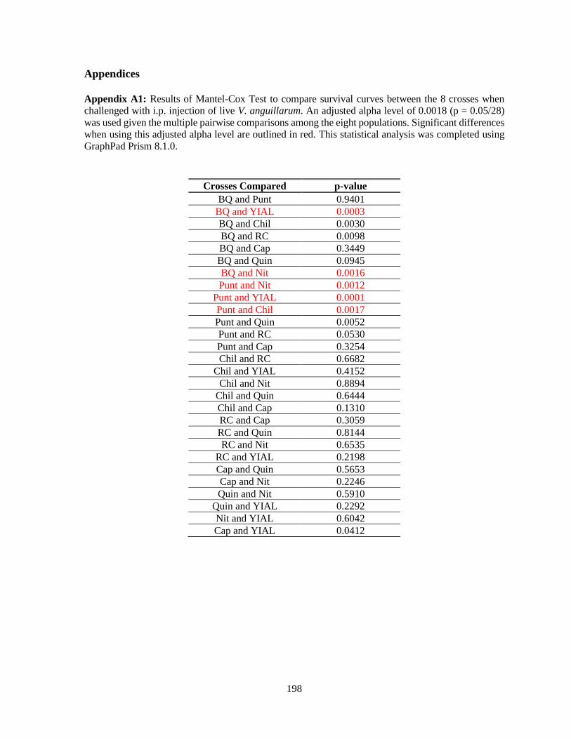
198
Appendices
Appendix A1: Results of Mantel-Cox Test to compare survival curves between the 8 crosses when
challenged with i.p. injection of live V. anguillarum. An adjusted alpha level of 0.0018 (p = 0.05/28)
was used given the multiple pairwise comparisons among the eight populations. Significant differences
when using this adjusted alpha level are outlined in red. This statistical analysis was completed using
GraphPad Prism 8.1.0.
Crosses Compared p-value
BQ and Punt 0.9401
BQ and YIAL 0.0003
BQ and Chil 0.0030
BQ and RC 0.0098
BQ and Cap 0.3449
BQ and Quin 0.0945
BQ and Nit 0.0016
Punt and Nit 0.0012
Punt and YIAL 0.0001
Punt and Chil 0.0017
Punt and Quin 0.0052
Punt and RC 0.0530
Punt and Cap 0.3254
Chil and RC 0.6682
Chil and YIAL 0.4152
Chil and Nit 0.8894
Chil and Quin 0.6444
Chil and Cap 0.1310
RC and Cap 0.3059
RC and Quin 0.8144
RC and Nit 0.6535
RC and YIAL 0.2198
Cap and Quin 0.5653
Cap and Nit 0.2246
Quin and Nit 0.5910
Quin and YIAL 0.2292
Nit and YIAL 0.6042
Cap and YIAL 0.0412

199
Appendix A2: Results of Mantel-Cox Test to compare survival curves between the 8 crosses when
challenged with i.p. injection of sterile PBS. An adjusted alpha level of 0.0018 (p = 0.05/28) was used
given the multiple pairwise comparisons among the eight populations. Significant differences are
outlined in red. This statistical analysis were completed using GraphPad Prism 8.1.0.
Crosses Compared p-value
BQ and Punt 0.2662
BQ and YIAL 0.4350
BQ and Chil 0.0544
BQ and RC 0.7146
BQ and Cap 0.1888
BQ and Quin 0.0119
BQ and Nit 0.0951
Punt and Nit 0.5983
Punt and YIAL 0.7741
Punt and Chil 0.3991
Punt and Quin 0.0003
Punt and RC 0.3813
Punt and Cap 0.0123
Chil and RC 0.0775
Chil and YIAL 0.2413
Chil and Nit 0.7498
Chil and Quin <0.0001
Chil and Cap 0.0005
RC and Cap 0.0578
RC and Quin 0.0011
RC and Nit 0.1501
RC and YIAL 0.6344
Cap and Quin 0.1375
Cap and Nit 0.0024
Quin and Nit <0.0001
Quin and YIAL 0.0010
Nit and YIAL 0.4213
Cap and YIAL 0.0319