in cretin as
TRANSCRIPT
The Role of Incretins in Glucose Homeostasisand Diabetes Treatment
WOOK KIM AND JOSEPHINE M. EGAN
National Institute on Aging/National Institutes of Health, Baltimore, Maryland
Abstract . . . . . . . . . . . . . . . . . . . . . . . . . . . . . . . . . . . . . . . . . . . . . . . . . . . . . . . . . . . . . . . . . . . . . . . . . . . . . . 470I. Background and introduction . . . . . . . . . . . . . . . . . . . . . . . . . . . . . . . . . . . . . . . . . . . . . . . . . . . . . . . . . . . 470
II. General description of incretins . . . . . . . . . . . . . . . . . . . . . . . . . . . . . . . . . . . . . . . . . . . . . . . . . . . . . . . . . 473A. Glucose-dependent insulinotropic peptide . . . . . . . . . . . . . . . . . . . . . . . . . . . . . . . . . . . . . . . . . . . . . 473B. Glucagon-like peptide-1 . . . . . . . . . . . . . . . . . . . . . . . . . . . . . . . . . . . . . . . . . . . . . . . . . . . . . . . . . . . . . 473
III. Synthesis, secretion, and degradation of incretins . . . . . . . . . . . . . . . . . . . . . . . . . . . . . . . . . . . . . . . . . 474A. Synthesis, secretion, and degradation of glucose-dependent insulinotropic peptide . . . . . . . . . 474
1. Pro-glucose-dependent insulinotropic peptide gene structure, expression andpost-translational processing. . . . . . . . . . . . . . . . . . . . . . . . . . . . . . . . . . . . . . . . . . . . . . . . . . . . . . 474
2. Glucose-dependent insulinotropic peptide secretion and degradation . . . . . . . . . . . . . . . . . . 475B. Synthesis, secretion and degradation of glucagon-like peptide-1 . . . . . . . . . . . . . . . . . . . . . . . . . 476
1. Proglucagon gene structure and expression . . . . . . . . . . . . . . . . . . . . . . . . . . . . . . . . . . . . . . . . . 4762. Tissue-specific post-translational processing of proglucagon, product secretion,
and degradation . . . . . . . . . . . . . . . . . . . . . . . . . . . . . . . . . . . . . . . . . . . . . . . . . . . . . . . . . . . . . . . . . 477C. Incretin secretion in type 2 diabetes . . . . . . . . . . . . . . . . . . . . . . . . . . . . . . . . . . . . . . . . . . . . . . . . . . 479
IV. Incretin receptors . . . . . . . . . . . . . . . . . . . . . . . . . . . . . . . . . . . . . . . . . . . . . . . . . . . . . . . . . . . . . . . . . . . . . . 480A. Glucose-dependent insulinotropic peptide receptor . . . . . . . . . . . . . . . . . . . . . . . . . . . . . . . . . . . . . 481B. Glucagon-like peptide-1 receptor . . . . . . . . . . . . . . . . . . . . . . . . . . . . . . . . . . . . . . . . . . . . . . . . . . . . . 481
V. The incretin effect . . . . . . . . . . . . . . . . . . . . . . . . . . . . . . . . . . . . . . . . . . . . . . . . . . . . . . . . . . . . . . . . . . . . . 482A. The incretin effect of glucose-dependent insulinotropic peptide and its impact in type 2
diabetes . . . . . . . . . . . . . . . . . . . . . . . . . . . . . . . . . . . . . . . . . . . . . . . . . . . . . . . . . . . . . . . . . . . . . . . . . . . 482B. The incretin effect of glucagon-like peptide-1 and its impact in type 2 diabetes. . . . . . . . . . . . 484
VI. Pleiotropic effects of incretins in pancreas . . . . . . . . . . . . . . . . . . . . . . . . . . . . . . . . . . . . . . . . . . . . . . . . 485A. Pleiotropic effects of glucose-dependent insulinotropic peptide in pancreas. . . . . . . . . . . . . . . . 485
1. Effects on � cells. . . . . . . . . . . . . . . . . . . . . . . . . . . . . . . . . . . . . . . . . . . . . . . . . . . . . . . . . . . . . . . . . 4852. Effects on glucagon secretion. . . . . . . . . . . . . . . . . . . . . . . . . . . . . . . . . . . . . . . . . . . . . . . . . . . . . . 488
B. Pleiotropic effects of glucagon-like peptide-1 in pancreas. . . . . . . . . . . . . . . . . . . . . . . . . . . . . . . . 4881. Effects on � cells. . . . . . . . . . . . . . . . . . . . . . . . . . . . . . . . . . . . . . . . . . . . . . . . . . . . . . . . . . . . . . . . . 4882. Effects on glucagon secretion. . . . . . . . . . . . . . . . . . . . . . . . . . . . . . . . . . . . . . . . . . . . . . . . . . . . . . 4913. Effects on pancreatic exocrine and ductal cells . . . . . . . . . . . . . . . . . . . . . . . . . . . . . . . . . . . . . . 492
VII. Extrapancreatic effects of incretins . . . . . . . . . . . . . . . . . . . . . . . . . . . . . . . . . . . . . . . . . . . . . . . . . . . . . . 492A. Extrapancreatic effects of glucose-dependent insulinotropic peptide . . . . . . . . . . . . . . . . . . . . . . 492
1. Central nervous system . . . . . . . . . . . . . . . . . . . . . . . . . . . . . . . . . . . . . . . . . . . . . . . . . . . . . . . . . . 4922. Gastrointestinal tract . . . . . . . . . . . . . . . . . . . . . . . . . . . . . . . . . . . . . . . . . . . . . . . . . . . . . . . . . . . . 4923. Adipose tissue . . . . . . . . . . . . . . . . . . . . . . . . . . . . . . . . . . . . . . . . . . . . . . . . . . . . . . . . . . . . . . . . . . . 4924. Bone. . . . . . . . . . . . . . . . . . . . . . . . . . . . . . . . . . . . . . . . . . . . . . . . . . . . . . . . . . . . . . . . . . . . . . . . . . . . 493
B. Extrapancreatic effects of glucagon-like peptide-1 . . . . . . . . . . . . . . . . . . . . . . . . . . . . . . . . . . . . . . 4941. Central and peripheral nervous system effects on food intake and glucose homeostasis . . . . . 4942. Gastrointestinal tract and gastric emptying . . . . . . . . . . . . . . . . . . . . . . . . . . . . . . . . . . . . . . . . 4953. Muscle, adipose tissue, and liver . . . . . . . . . . . . . . . . . . . . . . . . . . . . . . . . . . . . . . . . . . . . . . . . . . 4954. Bone. . . . . . . . . . . . . . . . . . . . . . . . . . . . . . . . . . . . . . . . . . . . . . . . . . . . . . . . . . . . . . . . . . . . . . . . . . . . 4965. Cardiovascular system . . . . . . . . . . . . . . . . . . . . . . . . . . . . . . . . . . . . . . . . . . . . . . . . . . . . . . . . . . . 4966. Hypothalamic-pituitary axis . . . . . . . . . . . . . . . . . . . . . . . . . . . . . . . . . . . . . . . . . . . . . . . . . . . . . . 497
Address correspondence to: Dr. Josephine M. Egan, National Institute on Aging, Intramural Research Program, 5600 Nathan Shock Drive,Baltimore, MD 21224. E-mail: [email protected]
This article is available online at http://pharmrev.aspetjournals.org.doi:10.1124/pr.108.000604.
0031-6997/08/6004-470–512$20.00PHARMACOLOGICAL REVIEWS Vol. 60, No. 4U.S. Government work not protected by U.S. copyright 604/3428101Pharmacol Rev 60:470–512, 2008 Printed in U.S.A.
470
by guest on April 15, 2014
pharmrev.aspetjournals.org
Dow
nloaded from
VIII. The development of therapies for diabetes based on incretin actions. . . . . . . . . . . . . . . . . . . . . . . . . 497A. Exenatide . . . . . . . . . . . . . . . . . . . . . . . . . . . . . . . . . . . . . . . . . . . . . . . . . . . . . . . . . . . . . . . . . . . . . . . . . . 497
1. Relevant clinical studies . . . . . . . . . . . . . . . . . . . . . . . . . . . . . . . . . . . . . . . . . . . . . . . . . . . . . . . . . . 498B. Sitagliptin . . . . . . . . . . . . . . . . . . . . . . . . . . . . . . . . . . . . . . . . . . . . . . . . . . . . . . . . . . . . . . . . . . . . . . . . . 499
1. Relevant clinical studies . . . . . . . . . . . . . . . . . . . . . . . . . . . . . . . . . . . . . . . . . . . . . . . . . . . . . . . . . . 499C. Therapies under development . . . . . . . . . . . . . . . . . . . . . . . . . . . . . . . . . . . . . . . . . . . . . . . . . . . . . . . . 500
1. Vildagliptin . . . . . . . . . . . . . . . . . . . . . . . . . . . . . . . . . . . . . . . . . . . . . . . . . . . . . . . . . . . . . . . . . . . . . 5002. Liraglutide . . . . . . . . . . . . . . . . . . . . . . . . . . . . . . . . . . . . . . . . . . . . . . . . . . . . . . . . . . . . . . . . . . . . . . 500
a. Selected clinical trials . . . . . . . . . . . . . . . . . . . . . . . . . . . . . . . . . . . . . . . . . . . . . . . . . . . . . . . . . 501b. Side effects . . . . . . . . . . . . . . . . . . . . . . . . . . . . . . . . . . . . . . . . . . . . . . . . . . . . . . . . . . . . . . . . . . . 501
IX. Potential disease-modifying effects of incretin-based therapies. . . . . . . . . . . . . . . . . . . . . . . . . . . . . . 501X. Concluding remarks . . . . . . . . . . . . . . . . . . . . . . . . . . . . . . . . . . . . . . . . . . . . . . . . . . . . . . . . . . . . . . . . . . . . 502
Acknowledgments . . . . . . . . . . . . . . . . . . . . . . . . . . . . . . . . . . . . . . . . . . . . . . . . . . . . . . . . . . . . . . . . . . . . . . 502References . . . . . . . . . . . . . . . . . . . . . . . . . . . . . . . . . . . . . . . . . . . . . . . . . . . . . . . . . . . . . . . . . . . . . . . . . . . . 502
Abstract——Incretins are gut hormones that are se-creted from enteroendocrine cells into the blood withinminutes after eating. One of their many physiologicalroles is to regulate the amount of insulin that is secretedafter eating. In this manner, as well as others to be de-scribed in this review, their final common raison d’etreis to aid in disposal of the products of digestion. Thereare two incretins, known as glucose-dependent insuli-notropic peptide (GIP) and glucagon-like peptide-1(GLP-1), that share many common actions in the pan-creas but have distinct actions outside of the pan-creas. Both incretins are rapidly deactivated by anenzyme called dipeptidyl peptidase 4 (DPP4). A lack ofsecretion of incretins or an increase in their clearanceare not pathogenic factors in diabetes. However, intype 2 diabetes (T2DM), GIP no longer modulatesglucose-dependent insulin secretion, even at supra-physiological (pharmacological) plasma levels, andtherefore GIP incompetence is detrimental to �-cellfunction, especially after eating. GLP-1, on the other
hand, is still insulinotropic in T2DM, and this has ledto the development of compounds that activate theGLP-1 receptor with a view to improving insulin se-cretion. Since 2005, two new classes of drugs based onincretin action have been approved for lowering bloodglucose levels in T2DM: an incretin mimetic (ex-enatide, which is a potent long-acting agonist of theGLP-1 receptor) and an incretin enhancer (sitagliptin,which is a DPP4 inhibitor). Exenatide is injected sub-cutaneously twice daily and its use leads to lowerblood glucose and higher insulin levels, especially inthe fed state. There is glucose-dependency to its insu-lin secretory capacity, making it unlikely to cause lowblood sugars (hypoglycemia). DPP4 inhibitors areorally active and they increase endogenous blood lev-els of active incretins, thus leading to prolonged incre-tin action. The elevated levels of GLP-1 are thought tobe the mechanism underlying their blood glucose-low-ering effects.
I. Background and Introduction
Incretins are hormones that are released from the gutinto the bloodstream in response to ingestion of food, andthey then modulate the insulin secretory response to theproducts within the nutrients in the food. The insulinsecretory response of incretins, called the incretin effect,accounts for at least 50% of the total insulin secretedafter oral glucose. Therefore, by definition, incretin hor-mones are insulinotropic (i.e., they induce insulin secre-tion) at usual physiological concentrations seen in theplasma after ingestion. The concept of incretins is atleast a century old (Table 1). In 1902, Bayliss and Star-ling published their landmark manuscript, “The Mech-anism of Pancreatic Secretion.” The authors found thatacid infused into the digestive system caused pancreaticsecretion of juices through the pancreatic duct from thepancreas, even after they cut the ennervation to theintestine. Until that time, it was thought that nervoussystem signals controlled secretion of pancreatic juices.They carried out ground-breaking studies that led them
to conclude that the nature of the signal to the pancreaswas most likely a chemical stimulus: they removed ex-tracts from the intestinal wall after it had been stimu-lated by acid, injected the extracts into the bloodstream,and once again they could see juices coming from thepancreatic duct of the animal that had been injected.Therefore, they proved that the extracts must have con-tained a substance that must normally be secreted fromthe intestinal wall into the bloodstream to stimulate theflow of pancreatic juice. They called the substance “se-cretin.” In his “Cronian Lectures,” Starling introducedthe word “hormone” (derived from the Greek word mean-ing “impetus”) for clinical factors that are released fromone site and act on another (Starling, 1905). The exam-ple of this was that the intestinal extracts containedsecretin, which induced obvious “exocrine” secretion ofpancreatic juices. Moore wrote in 1906 that Bayliss andStarling considered the possibility that the duodenumalso supplied a chemical excitant for the “internal” se-cretion of the pancreas. They wrote: “This line of argu-ment seems to have occurred to the discoverers of secre-
INCRETINS AND INCRETIN-BASED THERAPIES 471
tin themselves, for Starling mentions a case of diabeteswhich was tested by Spriggs by injections of secretinsolutions but with negative results.” Moore (1906) alsodescribed experiments carried out on individual youngdiabetic patients to whom he gave, by mouth, extractsof intestinal mucosa. This is therefore the first attemptat “incretin-based” therapies for treating diabetes, al-though, of course, the investigator did not call it that. Hereported achieving some success, but his experimentswere essentially doomed, because we now know that thechemical excitants are peptides that would have beendegraded when given orally. After World War I endedand insulin was extracted from pancreatic islets byBanting and Best in 1921, there was further work on thepossibility of food entering the gut leading to secretion ofan excitant into the bloodstream that would ultimatelylead to insulin secretion and lowering of blood glucose.Different groups published various and often contradic-tory results from experiments on the effects of extractsof duodenal mucosa on fasting blood glucose and/or onhyperglycemia induced by injection or ingestion of glu-cose. In 1932, La Barre used the word “incretin” to referto an extract from upper gut mucosa that produces hy-poglycemia but does not induce exocrine secretion, al-
though he did not prove incontrovertibly that incretinsexisted. Progress on the incretin concept was rapidlymade once radioimmunoassays for insulin became avail-able. Between 1964 and 1967, at least three groupsshowed independently that glucose, given orally, in-duced a greater insulin response (by radioimmunoassay)than i.v. glucose injection even if the blood glucose levelsattained were higher because of the i.v. glucose (Elricket al., 1964; McIntyre et al., 1964; Perley and Kipnis,1967). The three groups therefore knew that the oralglucose was indeed inducing release of “incretins” intothe bloodstream that subsequently increased insulin se-cretion, more than did glucose itself. In 1971, John C.Brown isolated and deduced the amino acid structure ofa peptide he had isolated from intestinal mucosa (Brownand Dryburgh, 1971). Exogenous administration of thepeptide inhibited gastric acid secretion in dogs, so hecalled it gastric inhibitory polypeptide (GIP)1 (Brownand Dryburgh, 1971). Brown and colleagues subse-quently found that it had insulinotropic properties andsuggested that it be called glucose-dependent insulino-tropic peptide, retaining the acronym GIP (Dupre et al.,1973). They not only demonstrated GIP to be insulino-tropic but also demonstrated the glucose dependence ofthe insulinotropic activity; i.e., plasma glucose must beelevated in order for GIP to induce insulin secretion. GIPwas therefore the first incretin to be isolated and itsproperties characterized.
1 Abbreviations: 8-pT-2�-O-Me-cAMP, 8-(4-chlorophenylthio)-2�-O-methyladenosine-3�,5�-cyclic monophosphate; AA, arachidonic acid;AP, area postrema; AR231453, 2-fluoro-4-methanesulfonyl-phenyl)-{6-[4-(3-isopropyl-[1,2,4]oxadiazol-5-yl)-piperidin-1-yl]-5-nitro-pyrimidin-4-yl}-amine; BBB, blood-brain barrier; bp, base pair(s);CNS, central nervous system; CRE, cAMP responsive element; CT,COOH-terminal tail; DPP4, dipeptidyl peptidase 4; DUSP14, dual-specificity phosphatase 14; EGFR, epidermal growth factor receptor;ER, endoplasmic reticulum; Ex-4, exendin-4; FDA, Food and DrugAdministration; FPG, fasting plasma glucose; GI, gastrointestinal;GIP, glucose-dependent insulinotropic peptide; GIPR, glucose-dependent insulinotropic peptide receptor; GLP-1, glucagon-likepeptide-1; GLP-1R, glucagon-like peptide-1 receptor; GLP-2, gluca-gon-like peptide-2; GLUT2, glucose transporter2; GPCR, G protein-coupled receptor; H89, N-[2-(4-bromocinnamylamino)ethyl]-5-iso-quinoline; HbA1c, hemoglobin A1c; IP3R, inositol 1,4,5 triphosphatereceptor; kb, kilobase pair(s); LY294002, 2-(4-morpholinyl)-8-phenyl-4H-1-benzopyran-4-one; MAPK, mitogen-activated protein kinase;NEP, neutral endopeptidase; NFAT, nuclear factor of activated Tcells; NTS, nucleus of the solitary tract; PC, proconvertase; PDX-1,pancreatic duodenal homeobox-1; PI-3K, phosphatidyl inositol-3 ki-nase; PKA, protein kinase A; PKB, protein kinase B; PKC, proteinkinase C; PLA2, phospholipase A2; PPG, postprandial plasma glu-cose; RyR, ryanodine receptor; STZ, streptozotocin; T2DM, type 2diabetes; TCF7L2, T-cell factor-like 2; TNF, tumor necrosis factor;TZD, thiazolidinedione; U0126, 1,4-diamino-2,3-dicyano-1,4-bis-(methylthio)butadiene; ZDF, diabetic-fatty Zucker.
TABLE 1Time-line to use of incretin-based therapies in the treatment of diabetes
Year The Development of Incretin-Based Therapies
1869 Islets of Langerhans (nests of cells that appeareddifferent from the surrounding pancreatictissue) in the pancreas were described.
1901 The role of islets of Langerhans (what ultimatelybecame known as an endocrine function) indiabetes was described.
1902 The role of a substance (called secretin) secretedby gut cells that stimulates the digestive juicesfor the pancreas (what ultimately becameknown as exocrine function) was described.
1905 This type of substance, a presumed “chemicalmessenger,” was now called a “hormone.”
1906 The role of a gut-derived hormone to treatdiabetes was first alluded to.
1921–1922 Extraction of insulin from pancreas and itspotential to treat type 1 diabetes was shown.
1932 The term “incretin” was used for the first time torefer to a substance derived from the gut,presumably a hormone, that regulates insulinsecretion after eating.
1960 Radioimmunoassay was developed formeasurement of plasma insulin levels.
1964–1967 Clinical proof that a gut-derived factor positivelymodulated insulin secretion.
1971 The first incretin, GIP, was isolated andsequenced.
1985 The second incretin, GLP-1, was described.1992–1994 Studies show that exogenous GIP does not lower
blood glucose in T2DM, but exogenous GLP-1does so.
2002 Exendin-4, a GLP-1 receptor agonist extractedfrom Gila monster lizard saliva, was shown topowerfully stimulate insulin secretion in aglucose-dependent manner in subjects withand without T2DM.
2005 Exenatide (synthetic exendin-4) came intoclinical use for T2DM.
2006 Sitagliptin, an orally active dipeptidyl peptidase4 inhibitor, came into use in T2DM.
472 KIM AND EGAN
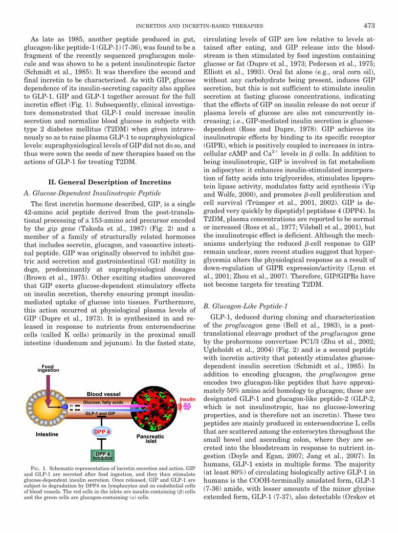
As late as 1985, another peptide produced in gut,glucagon-like peptide-1 (GLP-1) (7-36), was found to be afragment of the recently sequenced proglucagon mole-cule and was shown to be a potent insulinotropic factor(Schmidt et al., 1985). It was therefore the second andfinal incretin to be characterized. As with GIP, glucosedependence of its insulin-secreting capacity also appliesto GLP-1. GIP and GLP-1 together account for the fullincretin effect (Fig. 1). Subsequently, clinical investiga-tors demonstrated that GLP-1 could increase insulinsecretion and normalize blood glucose in subjects withtype 2 diabetes mellitus (T2DM) when given intrave-nously so as to raise plasma GLP-1 to supraphysiologicallevels: supraphysiological levels of GIP did not do so, andthus were sown the seeds of new therapies based on theactions of GLP-1 for treating T2DM.
II. General Description of Incretins
A. Glucose-Dependent Insulinotropic Peptide
The first incretin hormone described, GIP, is a single42-amino acid peptide derived from the post-transla-tional processing of a 153-amino acid precursor encodedby the gip gene (Takeda et al., 1987) (Fig. 2) and amember of a family of structurally related hormonesthat includes secretin, glucagon, and vasoactive intesti-nal peptide. GIP was originally observed to inhibit gas-tric acid secretion and gastrointestinal (GI) motility indogs, predominantly at supraphysiological dosages(Brown et al., 1975). Other exciting studies uncoveredthat GIP exerts glucose-dependent stimulatory effectson insulin secretion, thereby ensuring prompt insulin-mediated uptake of glucose into tissues. Furthermore,this action occurred at physiological plasma levels ofGIP (Dupre et al., 1973). It is synthesized in and re-leased in response to nutrients from enteroendocrinecells (called K cells) primarily in the proximal smallintestine (duodenum and jejunum). In the fasted state,
circulating levels of GIP are low relative to levels at-tained after eating, and GIP release into the blood-stream is then stimulated by food ingestion containingglucose or fat (Dupre et al., 1973; Pederson et al., 1975;Elliott et al., 1993). Oral fat alone (e.g., oral corn oil),without any carbohydrate being present, induces GIPsecretion, but this is not sufficient to stimulate insulinsecretion at fasting glucose concentrations, indicatingthat the effects of GIP on insulin release do not occur ifplasma levels of glucose are also not concurrently in-creasing; i.e., GIP-mediated insulin secretion is glucose-dependent (Ross and Dupre, 1978). GIP achieves itsinsulinotropic effects by binding to its specific receptor(GIPR), which is positively coupled to increases in intra-cellular cAMP and Ca2� levels in � cells. In addition tobeing insulinotropic, GIP is involved in fat metabolismin adipocytes: it enhances insulin-stimulated incorpora-tion of fatty acids into triglycerides, stimulates lipopro-tein lipase activity, modulates fatty acid synthesis (Yipand Wolfe, 2000), and promotes �-cell proliferation andcell survival (Trumper et al., 2001, 2002). GIP is de-graded very quickly by dipeptidyl peptidase 4 (DPP4). InT2DM, plasma concentrations are reported to be normalor increased (Ross et al., 1977; Vilsbøll et al., 2001), butthe insulinotropic effect is deficient. Although the mech-anisms underlying the reduced �-cell response to GIPremain unclear, more recent studies suggest that hyper-glycemia alters the physiological response as a result ofdown-regulation of GIPR expression/activity (Lynn etal., 2001; Zhou et al., 2007). Therefore, GIP/GIPRs havenot become targets for treating T2DM.
B. Glucagon-Like Peptide-1
GLP-1, deduced during cloning and characterizationof the proglucagon gene (Bell et al., 1983), is a post-translational cleavage product of the proglucagon geneby the prohormone convertase PC1/3 (Zhu et al., 2002;Ugleholdt et al., 2004) (Fig. 2) and is a second peptidewith incretin activity that potently stimulates glucose-dependent insulin secretion (Schmidt et al., 1985). Inaddition to encoding glucagon, the proglucagon geneencodes two glucagon-like peptides that have approxi-mately 50% amino acid homology to glucagon; these aredesignated GLP-1 and glucagon-like peptide-2 (GLP-2,which is not insulinotropic, has no glucose-loweringproperties, and is therefore not an incretin). These twopeptides are mainly produced in enteroendocrine L cellsthat are scattered among the enterocytes throughout thesmall bowel and ascending colon, where they are se-creted into the bloodstream in response to nutrient in-gestion (Doyle and Egan, 2007; Jang et al., 2007). Inhumans, GLP-1 exists in multiple forms. The majority(at least 80%) of circulating biologically active GLP-1 inhumans is the COOH-terminally amidated form, GLP-1(7-36) amide, with lesser amounts of the minor glycineextended form, GLP-1 (7-37), also detectable (Orskov et
Insulin
Pancreatic Pancreatic isletislet
IntestineIntestine
Food ingestion
GLP-1 and GIP
Glucose, fatty acids
Blood vesselBlood vessel
DPP 4
DPP 4inhibitor
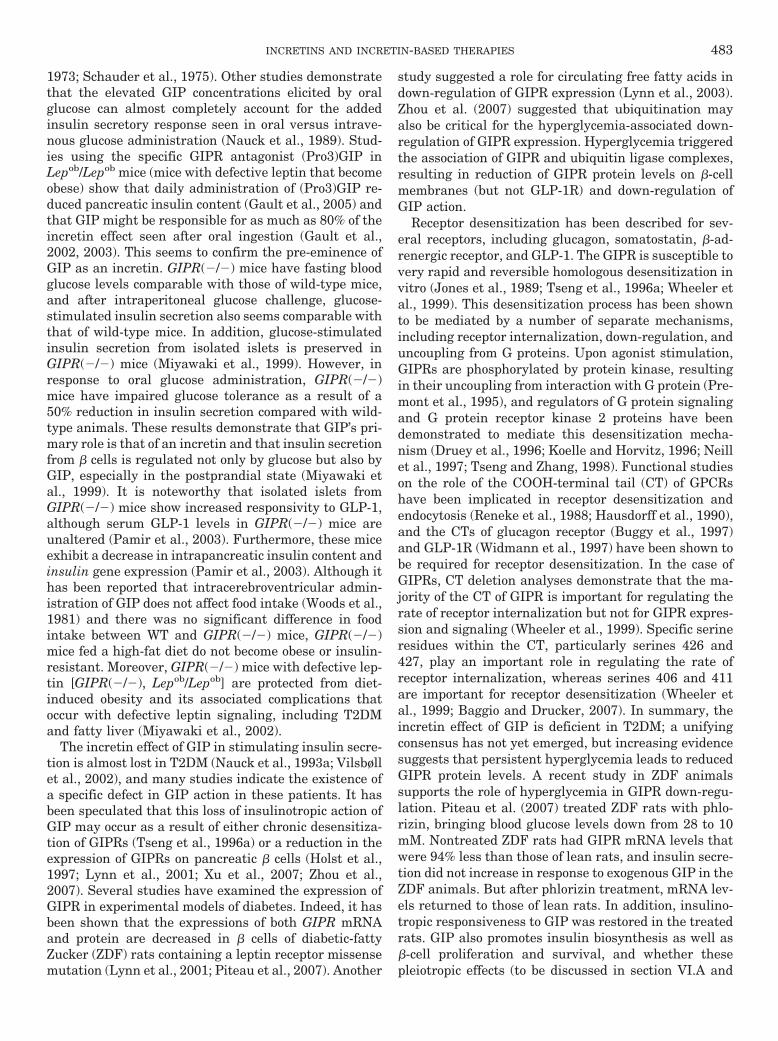
FIG. 1. Schematic representation of incretin secretion and action. GIPand GLP-1 are secreted after food ingestion, and they then stimulateglucose-dependent insulin secretion. Once released, GIP and GLP-1 aresubject to degradation by DPP4 on lymphocytes and on endothelial cellsof blood vessels. The red cells in the islets are insulin-containing (�) cellsand the green cells are glucagon-containing (�) cells.
INCRETINS AND INCRETIN-BASED THERAPIES 473
al., 1986). Like GIP, GLP-1 achieves its insulinotropiceffects by binding to its specific receptor (GLP-1R) thatis positively coupled to increases in intracellular cAMPand Ca2� levels in � cells. In addition to its insulino-tropic effects, it inhibits gastric emptying, decreases foodintake (Willms et al., 1996), inhibits glucagon secretion(Komatsu et al., 1989), and slows the rate of endogenousglucose production (Prigeon et al., 2003), all of whichshould help to lower blood glucose in T2DM. It has alsobeen shown to protect � cells from apoptosis (Farilla etal., 2002) and to stimulate �-cell proliferation by up-regulation of the �-cell transcription factor pancreaticduodenal homeobox-1 protein (PDX-1) (Perfetti et al.,2000; Stoffers et al., 2000), which is known to augmentinsulin gene transcription and up-regulate glucokinaseand glucose transporter2 (GLUT2) (Wang et al., 1999).There is no evidence that defective secretion of GLP-1 isa cause of T2DM (further discussed in depth in sectionIII.C). Continuous GLP-1 treatment in T2DM can nor-malize blood glucose, improve �-cell function, and re-store first-phase insulin secretion and “glucose compe-tence” to � cells (Holz et al., 1993; Zander et al., 2002);hence, GLP-1/GLP-1Rs are therapeutic targets for treat-ing T2DM. Continuous GLP-1 administration is re-quired for maintenance of glucose homeostasis becauseof its short half-life (1.5–2 min); similar to GIP, it isdegraded by DPP4. The following sections provide ourcurrent understanding of the physiology, the therapeu-tic potential, and the targets of incretin-based therapies.
III. Synthesis, Secretion, and Degradationof Incretins
A. Synthesis, Secretion, and Degradation of Glucose-Dependent Insulinotropic Peptide
1. Pro-Glucose-Dependent Insulinotropic Peptide GeneStructure, Expression and Post-Translational Process-ing. As already stated, GIP is synthesized within andsecreted from K cells in the small intestine, the highestdensity of K cells being in duodenum and jejunum; few,if any, K cells are present in distal ileum (Buffa et al.,1975; Buchan et al., 1978). Human GIP is a single 42-amino acid peptide derived from the processing by PC1/3of proGIP, a 153-amino acid precursor (Fig. 2) that isencoded by a 459-bp open reading frame and whose geneis localized to chromosome 17q. It is composed of sixexons, and the majority of GIP-encoding sequences arein exon 3 (reviewed extensively in Fehmann et al., 1995).This sequence includes a 51-amino acid N-terminal pep-tide containing a signal peptide with a cleavage site atglycine and a 60-amino acid C-terminal peptide flankingthe 42-amino acid GIP hormone, which presently seemsto be the only biologically active peptide derived fromproGIP (Fig. 2). The rat GIP cDNA has a 432-bp openreading frame encoding a 144-amino acid polypeptide,and the figure is similar but not identical to the humangene (Higashimoto et al., 1992). There is a �90% aminoacid homology of GIP between human, porcine, bovine,mouse, and rat. Expression of the gip gene in the gut is
S N-terminal peptide
C-terminal peptideGIP
42 amino acids
YAEGTFISDYSIAMDKIHQQDFVNWLLAQKGKKNDWKHNITQ
Posttranslational cleavage by PC1/3
in endocrine K cells
DPP 4
1 42
S GRPP GLP-1IP-1Glucagon IP-2 GLP-2
30 amino acids
Posttranslational cleavage by PC1/3
in endocrine L cells
HAEGTFTSDVSSYLEGQAAKEFIAWLVKGR37 6
PC1/3 PC1/3
PC1/3 PC1/3
33 amino acids
proGIP
proglucagon
GIP
GLP-1
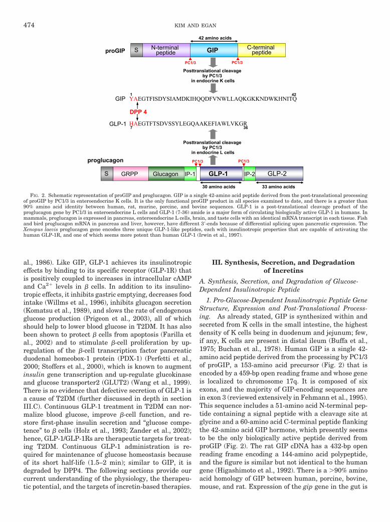
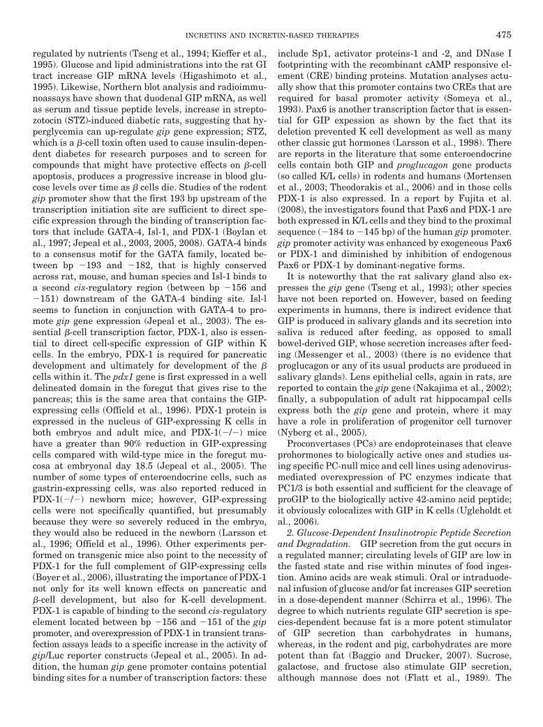
FIG. 2. Schematic representation of proGIP and proglucagon. GIP is a single 42-amino acid peptide derived from the post-translational processingof proGIP by PC1/3 in enteroendocrine K cells. It is the only functional proGIP product in all species examined to date, and there is a greater than90% amino acid identity between human, rat, murine, porcine, and bovine sequences. GLP-1 is a post-translational cleavage product of theproglucagon gene by PC1/3 in enteroendocrine L cells and GLP-1 (7-36) amide is a major form of circulating biologically active GLP-1 in humans. Inmammals, proglucagon is expressed in pancreas, enteroendocrine L cells, brain, and taste cells with an identical mRNA transcript in each tissue. Fishand bird proglucagon mRNA in pancreas and liver, however, have different 3�-ends because of differential splicing upon pancreatic expression. TheXenopus laevis proglucagon gene encodes three unique GLP-1-like peptides, each with insulinotropic properties that are capable of activating thehuman GLP-1R, and one of which seems more potent than human GLP-1 (Irwin et al., 1997).
474 KIM AND EGAN
regulated by nutrients (Tseng et al., 1994; Kieffer et al.,1995). Glucose and lipid administrations into the rat GItract increase GIP mRNA levels (Higashimoto et al.,1995). Likewise, Northern blot analysis and radioimmu-noassays have shown that duodenal GIP mRNA, as wellas serum and tissue peptide levels, increase in strepto-zotocin (STZ)-induced diabetic rats, suggesting that hy-perglycemia can up-regulate gip gene expression; STZ,which is a �-cell toxin often used to cause insulin-depen-dent diabetes for research purposes and to screen forcompounds that might have protective effects on �-cellapoptosis, produces a progressive increase in blood glu-cose levels over time as � cells die. Studies of the rodentgip promoter show that the first 193 bp upstream of thetranscription initiation site are sufficient to direct spe-cific expression through the binding of transcription fac-tors that include GATA-4, Isl-1, and PDX-1 (Boylan etal., 1997; Jepeal et al., 2003, 2005, 2008). GATA-4 bindsto a consensus motif for the GATA family, located be-tween bp �193 and �182, that is highly conservedacross rat, mouse, and human species and Isl-1 binds toa second cis-regulatory region (between bp �156 and�151) downstream of the GATA-4 binding site. Isl-lseems to function in conjunction with GATA-4 to pro-mote gip gene expression (Jepeal et al., 2003). The es-sential �-cell transcription factor, PDX-1, also is essen-tial to direct cell-specific expression of GIP within Kcells. In the embryo, PDX-1 is required for pancreaticdevelopment and ultimately for development of the �cells within it. The pdx1 gene is first expressed in a welldelineated domain in the foregut that gives rise to thepancreas; this is the same area that contains the GIP-expressing cells (Offield et al., 1996). PDX-1 protein isexpressed in the nucleus of GIP-expressing K cells inboth embryos and adult mice, and PDX-1(�/�) micehave a greater than 90% reduction in GIP-expressingcells compared with wild-type mice in the foregut mu-cosa at embryonal day 18.5 (Jepeal et al., 2005). Thenumber of some types of enteroendocrine cells, such asgastrin-expressing cells, was also reported reduced inPDX-1(�/�) newborn mice; however, GIP-expressingcells were not specifically quantified, but presumablybecause they were so severely reduced in the embryo,they would also be reduced in the newborn (Larsson etal., 1996; Offield et al., 1996). Other experiments per-formed on transgenic mice also point to the necessity ofPDX-1 for the full complement of GIP-expressing cells(Boyer et al., 2006), illustrating the importance of PDX-1not only for its well known effects on pancreatic and�-cell development, but also for K-cell development.PDX-1 is capable of binding to the second cis-regulatoryelement located between bp �156 and �151 of the gippromoter, and overexpression of PDX-1 in transient trans-fection assays leads to a specific increase in the activity ofgip/Luc reporter constructs (Jepeal et al., 2005). In ad-dition, the human gip gene promoter contains potentialbinding sites for a number of transcription factors: these
include Sp1, activator proteins-1 and -2, and DNase Ifootprinting with the recombinant cAMP responsive el-ement (CRE) binding proteins. Mutation analyses actu-ally show that this promoter contains two CREs that arerequired for basal promoter activity (Someya et al.,1993). Pax6 is another transcription factor that is essen-tial for GIP expession as shown by the fact that itsdeletion prevented K cell development as well as manyother classic gut hormones (Larsson et al., 1998). Thereare reports in the literature that some enteroendocrinecells contain both GIP and proglucagon gene products(so called K/L cells) in rodents and humans (Mortensenet al., 2003; Theodorakis et al., 2006) and in those cellsPDX-1 is also expressed. In a report by Fujita et al.(2008), the investigators found that Pax6 and PDX-1 areboth expressed in K/L cells and they bind to the proximalsequence (�184 to �145 bp) of the human gip promoter.gip promoter activity was enhanced by exogeneous Pax6or PDX-1 and diminished by inhibition of endogenousPax6 or PDX-1 by dominant-negative forms.
It is noteworthy that the rat salivary gland also ex-presses the gip gene (Tseng et al., 1993); other specieshave not been reported on. However, based on feedingexperiments in humans, there is indirect evidence thatGIP is produced in salivary glands and its secretion intosaliva is reduced after feeding, as opposed to smallbowel-derived GIP, whose secretion increases after feed-ing (Messenger et al., 2003) (there is no evidence thatproglucagon or any of its usual products are produced insalivary glands). Lens epithelial cells, again in rats, arereported to contain the gip gene (Nakajima et al., 2002);finally, a subpopulation of adult rat hippocampal cellsexpress both the gip gene and protein, where it mayhave a role in proliferation of progenitor cell turnover(Nyberg et al., 2005).
Proconvertases (PCs) are endoproteinases that cleaveprohormones to biologically active ones and studies us-ing specific PC-null mice and cell lines using adenovirus-mediated overexpression of PC enzymes indicate thatPC1/3 is both essential and sufficient for the cleavage ofproGIP to the biologically active 42-amino acid peptide;it obviously colocalizes with GIP in K cells (Ugleholdt etal., 2006).
2. Glucose-Dependent Insulinotropic Peptide Secretionand Degradation. GIP secretion from the gut occurs ina regulated manner; circulating levels of GIP are low inthe fasted state and rise within minutes of food inges-tion. Amino acids are weak stimuli. Oral or intraduode-nal infusion of glucose and/or fat increases GIP secretionin a dose-dependent manner (Schirra et al., 1996). Thedegree to which nutrients regulate GIP secretion is spe-cies-dependent because fat is a more potent stimulatorof GIP secretion than carbohydrates in humans,whereas, in the rodent and pig, carbohydrates are morepotent than fat (Baggio and Drucker, 2007). Sucrose,galactose, and fructose also stimulate GIP secretion,although mannose does not (Flatt et al., 1989). The
INCRETINS AND INCRETIN-BASED THERAPIES 475
postprandial level of circulating GIP is dependent onmeal size (Hampton et al., 1986; Vilsbøll et al., 2003c).GIP secretion is reduced in patients with intestinal mal-absorption (Besterman et al., 1978, 1979), presumablyas a result of insufficient K cells, as well as in chronicpancreatitis (Ebert et al., 1976). GIP secretion couplingpathways are poorly understood and have not been stud-ied to the same extent as has secretion from L cells,because GIP has not become a therapeutic target forT2DM, but presumably elevation of intracellular Ca2�
levels is required. In addition, some K cells containsweet receptors, which are G protein-coupled (GPCRs),and their activation by sugars and sweeteners may leadto GIP secretion (Jang et al., 2007; Egan and Margols-kee, 2008). Other GPCRs that are activated by fats andmay be responsible for initiating GIP secretion are beingdescribed as mechanisms by which GLP-1 is secreted arebeing studied, and these will be discussed below in sec-tion III.B.2.
GIP secretion in response to oral glucose ingestion ordifferent test meals has been quantified in numerousstudies. In humans, fasting plasma GIP levels, assayedfrom peripheral veins, are approximately 9 to 11 pM(total GIP) and peak plasma concentrations of 50 to 120pM are achieved after eating, depending on health sta-tus of the subject and the amount and quality of the foodconsumed (Vilsbøll et al., 2001).
Once released, GIP is subject to degradation by DPP4,which is bound to lymphocytes (where it is called CD-26)and endothelial cells of blood vessels of gut and liver aswell as being present in soluble form in the circulation.The first two amino acids (Tyr Ala) at the N terminus offull-length GIP (1-42) are cleaved in 1 to 2 min in ro-dents and 5 to 7 min in humans by DPP4 and convertedto GIP (3-42), which has insignificant, if any, insulino-tropic activity (Kieffer et al., 1995; Deacon et al., 2000).It is then excreted by the kidney. The elimination ratesof GIP (1-42) and GIP (3-42) are similar in subjects withT2DM and those without (Vilsbøll et al., 2006); hence,more rapid degradation/elimination of GIP is unlikely tobe a factor in defective insulinotropic effects seen inT2DM.
B. Synthesis, Secretion and Degradation of Glucagon-Like Peptide-1
1. Proglucagon Gene Structure and Expression. Thehuman proglucagon gene located on the long arm ofchromosome 2 has six exons, of which exons 2 to 5encode distinct functional domains (Bell, 1986), and fiveintrons. It spans approximately 9.4 kb (White and Saun-ders, 1986). Just a single gene encodes the proglucagonsequence in mammals and proglucagon, 180 amino acidslong, is very similar in all mammalian species (greaterthan 90% amino acid sequence homology). Glucagon isencoded in exon 3, and GLP-1 and -2 are encoded inexons 4 and 5, respectively (White and Saunders, 1986).A single size structurally identical mRNA transcript is
produced in all tissues that contain proglucagon (Novaket al., 1987; Drucker and Asa, 1988). These tissues arethe L cells of the intestine, the � cells of islets of Lang-erhans, some taste cells in the tongue, and some neuronsin the brainstem and hypothalamus (Drucker and Asa,1988; Eissele et al., 1992; Fehmann et al., 1995; Shin etal., 2008).
It seems that the 5�-flanking regions of human and ratproglucagon genes are essential for their tissue-specificexpression in the pancreas, brain, and intestine (Whiteand Saunders, 1986; Lee et al., 1992; Jin and Drucker,1995; Nian et al., 1999). Indeed, the DNA sequencelocated between �1.3 and �2.3 kb of rat proglucagonpromoter, the proglucagon gene upstream enhancer el-ement (GUE), contains multiple cis-acting positive andnegative transcriptional elements contributing to thetranscriptional control of proglucagon gene expressionin intestinal L cells (Jin and Drucker, 1995), and initialstudies of proglucagon gene expression using transgeniemice have shown that �1.3 kb of 5�-flanking sequencesare responsible for the pancreatic � cell- and brain-specific expression of rat proglucagon gene (Efrat et al.,1988). Further studies identified the first 300 bp of the5�-flanking region of the proglucagon gene as containinga minimum promoter region (G1) and four enhancerelements (G2–G5) contributing to the pancreatic � cellspecificity of glucagon gene expression (Philippe et al.,1988; Herzig et al., 2000), via the binding to these ele-ments to a cocktail of transcription factors, includingIsl-1, Pax6, Cdx2/3, Brn4, Pbx, Foxa1, and c-Maf. Studyof the human proglucagon gene transcription usingtransfected reporter genes and cell lines in vitro andtransgenic mice in vivo has indicated that �1.6 kb of the5�-flanking region of human proglucagon promoter isrequired for proglucagon gene transcription in the brainand intestine, but not in pancreatic islets, whereas �6kb of human proglucagon promoter are required for ex-pression in islet cell lines (Nian et al., 1999).
A typical CRE also exists in the 5�-flanking region ofthe rat proglucagon gene (Philippe et al., 1988), andincreased levels of cAMP stimulate proglucagon geneexpression in both pancreatic � cells and intestinal Lcells via protein kinase A (PKA)- or Epac/MAPK-depen-dent pathway (Knepel et al., 1990; Drucker et al., 1994;Brubaker et al., 1998; Lotfi et al., 2006; Jin, 2008). Inhumans, however, whether cAMP signaling is capable ofstimulating human proglucagon gene expression is un-known. Pax6, a critical determinant of islet cell devel-opment (Habener et al., 2005), as well as K cell devel-opment, as described in section III.A.1, also activatesproglucagon gene transcription in the intestine and pan-creas, via binding to the G1, G3, and G5 elements in theproglucagon promoter (Jin, 2008). This is supported bythe following two observations: 1) the dominant-nega-tive (SEYNeu) form of Pax6 in mice leads to markedreduction of proglucagon mRNA transcripts in the intes-tine and pancreatic � cells (Sander et al., 1997), and
476 KIM AND EGAN
neither GLP-1 nor GLP-2-immunopositive enteroendo-crine cells are detected in the intestinal mucosa (Hill etal., 1999); 2) adenoviral-expressed Pax6 activated bothproglucagon promoter-luciferase activity and expressionof the endogenous proglucagon gene in enteroendocrinecell lines, and furthermore, increased levels of endoge-nous proglucagon gene expression were observed in pri-mary rat intestinal cell cultures in vitro, and in ratcolonic epithelium in vivo, after adenoviral-mediatedoverexpression of Pax-6 (Trinh et al., 2003). Similar to �cells in islets, L cells do not express PDX-1: only the K/Lcells require it, as well as Pax6 (Fujita et al., 2008).Recent studies have indicated that the Wnt signalingpathway via �-catenin/T-cell factor-4, the major effectorof the Wnt signaling pathway, is a potent mediator ofproglucagon gene expression and GLP-1 production in Lcells but not in islets (Ni et al., 2003; Yi et al., 2005). Inthese studies, proglucagon gene expression and GLP-1production were activated by inhibition of glycogen syn-thase kinase-3�, a major negative modulator of the Wntpathway, and by �-catenin overexpression in L cells butnot in islets, and this effect was mediated by binding ofthe transcription factor TCF-4 to the proglucagon genepromoter G2 enhancer that is abundantly expressed inthe gut but not in pancreatic islets. More recently, Yi etal. (2008) suggested the existence of cross-talk betweenthe insulin and Wnt signaling pathways and the regu-lation of proglucagon gene expression and GLP-1 pro-duction by this cross-talk in the L cells. Insulin has beenshown to repress proglucagon gene transcription in thepancreatic islets by regulation of binding of FoxO1 to theG3 element of the proglucagon gene promoter (Philippe,1989, 1991; McKinnon et al., 2006). It is noteworthy thatthe opposite may be true in enteroendocrine cells of thegut. Insulin was shown to actually activate proglucagongene expression and GLP-1 production in a murine L cellline and in primary rat L cells as well as in the mouseintestine in vivo, and this effect of insulin was thought tobe mediated by nuclear �-catenin accumulation andbinding of �-catenin/T-cell factor-4 to the proglucagongene promoter, suggesting a potential novel function forinsulin—namely up-regulation of GLP-1 (and GLP-2)production, via cross-talk with Wnt signaling pathway(Yi et al., 2008). This dichotomy of insulin effect has notbeen looked for in humans. It also seems that differentdownstream signaling pathways are in action in the Lcells and pancreatic � cells, because protein kinase B(PKB) activity is involved in proglucagon gene expres-sion in the � cells (Schinner et al., 2005), but not in theL cell lines (Yi et al., 2008). In an � cell line, orexin-A, aneuropeptide hormone that regulates food intake andenergy homeostasis, inhibits proglucagon gene expres-sion via activation of the PKB/PI-3K/FoxO1-dependentpathway, and also inhibits glucagon secretion via de-creases of intracellular � cell cAMP and Ca2� concen-tration (Goncz et al., 2008). However, the effect oforexin-A, if any, in the L cells has not been determined.
(The players in the regulation of proglucagon expressionin brain and taste cells are also virtually unknown.)
2. Tissue-Specific Post-Translational Processing of Pro-glucagon, Product Secretion, and Degradation. Peptideproducts with different physiological properties in tissuesarise as a consequence of tissue-specific, differential, post-translational processing of proglucagon, and this allowsone protein to serve a variety of functions (Mojsov et al.,1986; Orskov et al., 1986). Seven PCs have been identified(von Eggelkraut-Gottanka and Beck-Sickinger, 2004); two,PC1/3 and PC2, seem localized to only neuroendocrinecells, whereas a third, furin, is found in both neuroendo-crine and nonendocrine cells. Recent findings have shownthat the expression level of PC2 is high in the pancreatic �cells and absent in the gut, whereas PC1/3 is definitelyexpressed in gut endocrine cells (Rouille et al., 1994; Scopsiet al., 1995). The activity of the specific PC ultimatelydetermines entirely different peptide products. It seemsthat of all the PCs, only PC1/3 and PC2 are essential forproglucagon processing (Rothenberg et al., 1995; Rouille etal., 1995). PC2, in conjunction with 7B2, a chaperone pro-tein that is responsible for maturation of PC2 as well as itsenzymatic activity, is responsible for the pancreatic � cell-specific processing of proglucagon to glucagon (Rouille etal., 1995). Consistent with this, mice lacking active PC2exhibit multiple endocrine disorders, have severely im-paired processing of proglucagon to glucagon, and conse-quently become severely hypoglycemic (Furuta et al.,1997). The proglucagon-derived peptides from the pancre-atic � cells include glucagon, glicentin-related pancreaticpeptide, intervening peptide-1, and the major proglucagonfragment (Fig. 3). In addition to being present in K cells,where, as stated above, it is required for the cleavage ofproGIP, PC1/3 is also present in intestinal L cells and isresponsible for the processing of proglucagon to GLP-1(and GLP-2) (Rothenberg et al., 1995; Rouille et al., 1995)(Fig. 2). In agreement with this, mice with a targeteddeletion of the PC1/3 gene cannot process proglucagon toGLP-1 and GLP-2 (Zhu et al., 2002; Ugleholdt et al., 2004).
S GRPP GLP-1IP-1Glucagon IP-2 GLP-2
GRPP Oxyntomodulin
GRPP Glucagon IP-1
Glicentin MPGF
GLP-1 IP-2 GLP-2
L cell (PC1/3) α cell (PC2, 7B2)
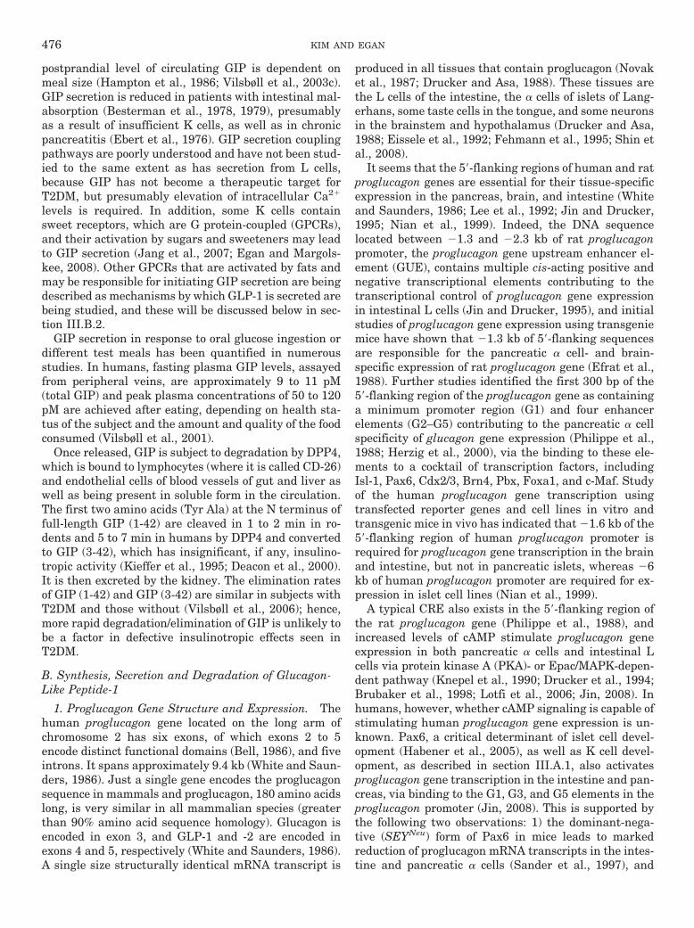
FIG. 3. Schematic representation of tissue-specific post-translationalprocessing of proglucagon in the intestinal L cell and the pancreatic �cell. PC1/3 is responsible for the processing of proglucagon in the L cell torelease GLP-1 (and GLP-2), and PC2, in conjunction with 7B2, is respon-sible for the pancreatic � cell-specific processing of proglucagon. Process-ing of proglucagon also occurs in brain. In taste cells of the tongue, bothPC1/3 and PC2 (as well as 7B2) are present, and consequently GLP-1,GLP-2, and glucagon are all present.
INCRETINS AND INCRETIN-BASED THERAPIES 477
In a human case of PC1/3 deficiency, however (of whichthere are just three cases described in the literature), thepatient suffered from a plethora of endocrinopathies andhad some mature GLP-1 in her plasma, indicating a lack ofabsolute dependence for PC1/3 in humans (Jackson et al.,2003). It is noteworthy that Wideman et al. (2006) recentlydemonstrated that up-regulation of PC1/3 expression us-ing adenovirus system in pancreatic � cells leads to in-crease of islet GLP-1 secretion, resulting in improved glu-cose-stimulated insulin secretion and enhanced survival ofislets in response to cytokine treatment. Similar effectshave been observed also in PC1/3-expressing � cells de-rived from mice lacking active PC2 (Wideman et al., 2007).Moreover, this ability of PC1/3-expressing � cells was at-tenuated in GLP-1R(�/�) mice and transplantation ofPC1/3-expressing � cells prevented STZ-induced hypergly-cemia by preserving �-cell area and islet morphology viaincreased �-cell proliferation (Wideman et al., 2007). Be-sides GLP-1 and GLP-2, the other proglucagon-derivedpeptides from the L cells include glicentin, oxyntomodulin,and intervening peptide-2 (Fig. 3). Although proglucagonexists in the brain and processing of proglucagon also oc-curs (Drucker and Asa, 1988), the PC enzymes have notbeen specifically studied in brain. In taste cells of thetongue, however, both PC1/3 and PC2 (as well as 7B2) arepresent; consequently, GLP-1, GLP-2, and glucagon are allpresent (Shin et al., 2008).
The primary physiological stimuli for the secretion ofGLP-1 are fat- and carbohydrate-rich meals, but mixedmeals or individual nutrients, including glucose andother sugars, sweeteners, fatty acids, amino acids, anddietary fiber, also can stimulate GLP-1 secretion (Baggioand Drucker, 2007). Although functional GLP-1 hasbeen found in taste buds and brain, the majority if notall of the GLP-1 measured in peripheral blood is synthe-sized in L cells, stored in granules, and released aftereating from the L cells that are distributed throughoutthe small and large intestine (Eissele et al., 1992). MoreL cells are located in the distal ileum and colon than inthe duodenum and jejunum, in contrast to GIP-secretingK cells. K/L cells seem most abundant in terminal ileum(Fujita et al., 2008). Plasma levels of GLP-1 increaserapidly within just a few minutes after oral glucose inrodents and humans. Meal ingestion results in a bipha-sic pattern of GLP-1 secretion, with an early phase be-ginning within 5 to 15 min and a prolonged second phasefollowing within 30 to 60 min (Herrmann et al., 1995).Thus, the early phase and the prolonged second phase ofGLP-1 secretion may be due to both direct nutrientcontact with the L cells and K/L cells in upper smallintestine and by nutrient (most likely fat, as nutrient-derived carbohydrate should have been absorbed beforearriving in the ileum), neural, and other gut-derived(and even non–gut-derived) endocrine factors activatingL cells in the distal bowel (Roberge and Brubaker, 1991;Rocca and Brubaker, 1999; De Leon et al., 2006). It nowseems probable that the mechanism by which the early
phase of GLP-1 secretion occurs is that sugars activatesweet taste receptors on L and or K/L cells (Theodorakiset al., 2006; Jang et al., 2007). The L cell (as well as theK cell) is an open-type intestinal epithelial endocrine cellthat is in direct contact with nutrients (Eissele et al.,1992) through its microvilli on the luminal surface, incontact with the enteric nervous system as well as thecentral nervous system (CNS) via the vagus, and incontact with the microvasculature through its basolat-eral membrane (Hansen et al., 1999; Anini et al., 2002).This allows GLP-1 secretion from L cells, as well as GIPfrom K cells, to be regulated by a variety of nutrient,neural, and endocrine signals. Indeed, several studieshave postulated that GLP-1 secretion from the L cells isregulated by a complex proximal-distal loop that in-volves both endocrine and neural factors with the vagusnerves having an essential role in this loop (Balks et al.,1997; Rocca and Brubaker, 1999; Anini and Brubaker,2003a). In this loop also, GIP, acetylcholine, and gastrin-releasing peptide act as mediators: the afferent vagusnerve is activated by GIP, which subsequently stimu-lates GLP-1 secretion through the efferent vagus nerveand enteric neurons that release acetylcholine and gas-trin-releasing peptide (Roberge et al., 1996; Rocca andBrubaker, 1999; Persson et al., 2000; Anini et al., 2002;Anini and Brubaker, 2003a). It seems that GLP-1secretion is also affected by other neurotransmittersand peptides, including GABA and calcitonin gene-related peptide (Brubaker, 1991; Gameiro et al., 2005).Furthermore, non-nutrient factors, including leptin(Anini and Brubaker, 2003b) and insulin (Lim andBrubaker, 2006), have also been identified as stimula-tors of GLP-1 secretion. Conversely, somatostatin,which is produced from the intestinal enteroendocrine Dcells (as well as from � cells in islets of Langerhans) andwhose secretion is increased by GLP-1, has been identi-fied as the inhibitor of GLP-1 secretion, implicating theexistence of a negative local feedback loop in the gut(Brubaker, 1991; Hansen et al., 2000; Chisholm andGreenberg, 2002).
Recent studies have uncovered some of the intracellu-lar signaling pathways that mediate nutrient-inducedGLP-1 secretion from L cells, which is then most likelymodified by the neural and endocrine factors mentionedabove. Several studies show that specific GPCRs presenton L cells are necessary for GLP-1 secretion. In partic-ular, long-chain free fatty acids and lipids stimulateGLP-1 secretion through interaction with GPCRs, in-cluding GPR120 (Hirasawa et al., 2005), GPR119 (Chuet al., 2008), and GPR40 (Edfalk et al., 2008). GPR120 ishighly expressed in the intestine and the stimulation ofGPR120 by free fatty acids promotes the secretion ofGLP-1 via increase of intracellular Ca2� levels and ac-tivation of p42/44 MAPK (Hirasawa et al., 2005).GPR119 mRNA is found in intestinal subregions of hu-mans and mouse and in cultured L cell lines, and in situhybridization studies also show that most GLP-1-pro-
478 KIM AND EGAN
ducing cells express GPR119 mRNA. The engagement ofGPR119 by phospholipids and fatty acid amides stimu-lates GLP-1 secretion (Chu et al., 2008). AR231453, apotent GPR119 agonist, stimulated intracellular cAMPaccumulation and GLP-1 release from the GLUTag Lcell line and, when given by oral gavage before oralglucose, plasma levels of GLP-1 were higher than inthose animals not given AR231453. It also increasedplasma levels of GIP even though GPR119 was unde-tectable by in situ hybridization in K cells. Furthermore,the effects of AR231453 on incretin secretion were ab-sent in GPR119-deficient mice (Chu et al., 2008). Morerecently, Edfalk et al. (2008) suggested that GPR40modulated FFA-stimulated insulin secretion from � cellsnot only directly but also indirectly via regulation ofincretin secretion. In that study, GPR40 expression wasuncovered in endocrine cells of the GI tract as well as in� cells, and PDX-1 was required for its expression in �cells, stomach, and duodenum. However, in distal GI,such as ileum, where PDX-1 is not expressed and wherethe greatest number of L cells reside, GPR40 expressionwas also present, indicating that PDX-1 expression isnot absolutely necessary for its expression. Secretion ofGLP-1 and GIP were both diminished, though not ab-sent, in GPR40-null mutant mice in response to a fatdiet (Edfalk et al., 2008). Therefore, it seems likely thatmany different GPRs present on both K and L cellscomplement one another to bring about fatty acid-medi-ated incretin secretion. It is also likely that differentGPRs reside in the various parts of the bowel so that fat,in various stages of digestion as it moves though thebowel, can continue to bring about enteroendocrine hor-mone secretion.
With regard to glucose stimulation of GLP-1 secretion,and analogous to � cells, expression of glucokinase, theglucose sensor in � cells, has been found in the mouseintestinal L cells (Jetton et al., 1994), and GLUT2, themost abundant glucose transporter in � cells, seems tobe required for GLP-1 secretion (Cani et al., 2007). Inaddition, it is likely that both GIPR and GLP-1R arerequired for the full regulation of oral glucose-inducedGLP-1 and GIP secretion (Cani et al., 2007). The regu-lation of glucose-induced GLP-1 secretion by taste trans-duction elements was identified by our recent studies.Jang et al. (2007) demonstrated that glucose (and thesucrose analog, sucralose, commonly used as a nonca-loric sweetener) led to secretion from L cells via a sig-naling pathway quite similar to that used by taste cellsin the tongue. The human L cell line NCI-H716 andduodenal L cells expressed taste transduction elements,including sweet taste receptors, the taste G protein gust-ducin, and several other signaling elements. Ingestion ofglucose by �-gustducin-null mice revealed deficiencies insecretion of GLP-1 and isolated small bowel and intes-tinal villi from these mice showed markedly defectiveGLP-1 secretion in response to glucose. Furthermore,GLP-1 release from NCI-H716 cells was promoted by the
sugar sucralose and blocked by the sweet receptor an-tagonist lactisole. Down-regulation of �-gustducin ex-pression by small interfering RNA also lessened glucose-induced GLP-1 secretion. In conclusion, the L cells of thegut “taste” glucose through the same mechanisms usedby taste cells of the tongue, and this has an essential rolein glucose-induced GLP-1 secretion (Jang et al., 2007).
Typical basal (fasting) levels of bioactive GLP-1, mea-sured from peripheral veins, are in the range of 5 to 10pM and increase by 2- to 3-fold after meal ingestion,depending on the size and composition of meal (Elliott etal., 1993). The first two N-terminal amino acids (HisAla) of native GLP-1 are rapidly cleaved by DPP4, andthe resulting GLP-1 (9-36) fragment is not insulinotropic(Hansen et al., 1999). GLP-1 is also degraded by neutralendopeptidase 24.11 (NEP-24.11) (Plamboeck et al.,2005), which is a membrane-bound zinc metallopepti-dase (Turner et al., 2001), and six potential cleavagesites in GLP-1 have been identified (Hupe-Sodmann etal., 1995). High levels of this enzyme are found in thekidney and GLP-1, and its metabolites are rapidlycleared through the kidneys (Ruiz-Grande et al., 1993),implying the involvement of NEP-24.11 in renal clear-ance of GLP-1. Therefore, many modifications have beenmade to synthetic GLP-1 so as to increase its biologicalhalf-life and consequently its efficacy in vivo, and ther-apeutic strategies based on modulating GLP-1 levelsand GLP-1 activity through administration of GLP-1and its analogs or by inhibiting its degradation havebeen tested and/or are under development for treatingT2DM. The elimination rates of GLP-1 are similar inT2DM and nondiabetic subjects, and, as with GIP, morerapid degradation of GLP-1 is unlikely to be a contribu-tory cause for glucose-responsiveness in T2DM (Vilsbøllet al., 2003a).
Exenatide, a synthetic form of exendin-4 (Ex-4, aGLP-1R agonist that is naturally synthesized in thesalivary glands of the Gila monster lizard and does notpossess a DPP4 recognition site), and sitagliptin, a small,orally active DPP4 inhibitor, are now FDA-approved andare being used for lowering blood glucose in T2DM. Forexample, in the ACCORD study, 12.1% of the intensivelytreated subjects with diabetes received exenatide (Ger-stein et al., 2008).
C. Incretin Secretion in Type 2 Diabetes
T2DM is characterized by a severely impaired or ab-sent GIP insulinotropic effect (Nauck et al., 1986) (to befurther discussed in section V) that most likely results inworsening insulin secretion. However, T2DM seems un-likely to result from deficient incretin secretion. One ofthe reasons frequently given for using exenatide or DPP4inhibitors is that they lead to “normalization” of incretinlevels that are supposedly reduced compared with nondi-abetic subjects (Drucker and Nauck, 2006). However, oncloser analysis of all the data in print, it is far from certainthat incretin secretion is reduced in this condition. For
INCRETINS AND INCRETIN-BASED THERAPIES 479
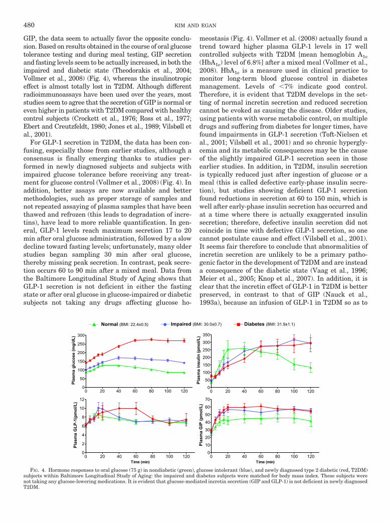
GIP, the data seem to actually favor the opposite conclu-sion. Based on results obtained in the course of oral glucosetolerance testing and during meal testing, GIP secretionand fasting levels seem to be actually increased, in both theimpaired and diabetic state (Theodorakis et al., 2004;Vollmer et al., 2008) (Fig. 4), whereas the insulinotropiceffect is almost totally lost in T2DM. Although differentradioimmunoassays have been used over the years, moststudies seem to agree that the secretion of GIP is normal oreven higher in patients with T2DM compared with healthycontrol subjects (Crockett et al., 1976; Ross et al., 1977;Ebert and Creutzfeldt, 1980; Jones et al., 1989; Vilsbøll etal., 2001).
For GLP-1 secretion in T2DM, the data has been con-fusing, especially those from earlier studies, although aconsensus is finally emerging thanks to studies per-formed in newly diagnosed subjects and subjects withimpaired glucose tolerance before receiving any treat-ment for glucose control (Vollmer et al., 2008) (Fig. 4). Inaddition, better assays are now available and bettermethodologies, such as proper storage of samples andnot repeated assaying of plasma samples that have beenthawed and refrozen (this leads to degradation of incre-tins), have lead to more reliable quantification. In gen-eral, GLP-1 levels reach maximum secretion 17 to 20min after oral glucose administration, followed by a slowdecline toward fasting levels; unfortunately, many olderstudies began sampling 30 min after oral glucose,thereby missing peak secretion. In contrast, peak secre-tion occurs 60 to 90 min after a mixed meal. Data fromthe Baltimore Longitudinal Study of Aging shows thatGLP-1 secretion is not deficient in either the fastingstate or after oral glucose in glucose-impaired or diabeticsubjects not taking any drugs affecting glucose ho-
meostasis (Fig. 4). Vollmer et al. (2008) actually found atrend toward higher plasma GLP-1 levels in 17 wellcontrolled subjects with T2DM [mean hemoglobin A1c(HbA1c) level of 6.8%] after a mixed meal (Vollmer et al.,2008). HbA1c is a measure used in clinical practice tomonitor long-term blood glucose control in diabetesmanagement. Levels of �7% indicate good control.Therefore, it is evident that T2DM develops in the set-ting of normal incretin secretion and reduced secretioncannot be evoked as causing the disease. Older studies,using patients with worse metabolic control, on multipledrugs and suffering from diabetes for longer times, havefound impairments in GLP-1 secretion (Toft-Nielsen etal., 2001; Vilsbøll et al., 2001) and so chronic hypergly-cemia and its metabolic consequences may be the causeof the slightly impaired GLP-1 secretion seen in thoseearlier studies. In addition, in T2DM, insulin secretionis typically reduced just after ingestion of glucose or ameal (this is called defective early-phase insulin secre-tion), but studies showing deficient GLP-1 secretionfound reductions in secretion at 60 to 150 min, which iswell after early-phase insulin secretion has occurred andat a time where there is actually exaggerated insulinsecretion; therefore, defective insulin secretion did notcoincide in time with defective GLP-1 secretion, so onecannot postulate cause and effect (Vilsbøll et al., 2001).It seems fair therefore to conclude that abnormalities ofincretin secretion are unlikely to be a primary patho-genic factor in the development of T2DM and are insteada consequence of the diabetic state (Vaag et al., 1996;Meier et al., 2005; Knop et al., 2007). In addition, it isclear that the incretin effect of GLP-1 in T2DM is betterpreserved, in contrast to that of GIP (Nauck et al.,1993a), because an infusion of GLP-1 in T2DM so as to
Normal (BMI: 22.4±0.5) Diabetes (BMI: 31.9±1.1)Impaired (BMI: 30.0±0.7)
0 20 40 60 80 100 1200
50
100
150
200
250
300
Pla
sma
glu
cose
(m
g/d
L)
0 20 40 60 80 100 1200
50
100
150
200
250
300
350
Pla
sma
insu
lin (
pm
ol/L
)
0 20 40 60 80 100 1200
2
4
6
8
10
12
Time (min)
Pla
sma
GL
P-1
(pm
ol/L
)
0 20 40 60 80 100 1200
10
20
30
40
50
60
70
Time (min)
Pla
sma
GIP
(p
mo
l/L)
FIG. 4. Hormone responses to oral glucose (75 g) in nondiabetic (green), glucose intolerant (blue), and newly diagnosed type 2 diabetic (red, T2DM)subjects within Baltimore Longitudinal Study of Aging: the impaired and diabetes subjects were matched for body mass index. These subjects werenot taking any glucose-lowering medications. It is evident that glucose-mediated incretin secretion (GIP and GLP-1) is not deficient in newly diagnosedT2DM.
480 KIM AND EGAN

reach pharmacologic concentrations in plasma can nor-malize fasting (Nauck et al., 1993b; Gutniak et al., 1997;Rachman et al., 1997) and postprandial (Gutniak et al.,1997; Rachman et al., 1997; Meier et al., 2003a) glucoseconcentrations, resulting from increase of glucose-stim-ulated insulin secretion, decrease of glucagon secretionand slowing of gastric emptying (Gutniak et al., 1992;Nauck et al., 1993b; Holst and Gromada, 2004). Contin-uous i.v. infusion of GLP-1 also lowers postprandialplasma glucose (PPG) levels in subjects with type 1diabetes by delaying gastric emptying (Gutniak et al.,1992). These effects of GLP-1 have been consistentlyshown in a number of human studies (Nathan et al.,1992; Nauck et al., 1993a,b; Elahi et al., 1994). In par-ticular, continuous subcutaneous infusion (4.8 pmol/kg/min) of GLP-1 for 6 weeks in T2DM subjects was asso-ciated with significant reductions in both fasting plasmaglucose (FPG) and PPG as well as HbA1c with a slightdecrease of body weight (Zander et al., 2002). Prolongingthe GLP-1 infusion (3.2 pmol/kg/min) for 3 months inpatients with T2DM resulted in a restoration of first-phase insulin secretion as well as an improvement oflate-phase secretion during a glucose clamp, but no sig-nificant changes in body weight and plasma glucagonlevels were noted (Meneilly et al., 2003). In anotherstudy, repeated i.v. infusion (1�1.2 pmol/kg/min) ofGLP-1 also normalized FPG in patients with T2DM(Nauck et al., 1997). Thus, there has been considerableinterest in an incretin-based therapeutic approach fortreating T2DM. However, continuous GLP-1 infusion orrepeated GLP-1 injections are impractical and expensiveways to lower blood glucose and so the strategies men-tioned above have been developed.
IV. Incretin Receptors
A. Glucose-Dependent Insulinotropic Peptide Receptor
The presence of GIPRs was first demonstrated in atransplantable hamster insulinoma (Maletti et al., 1984)and an insulin-secreting hamster �-cell line In111(Amiranoff et al., 1984, 1985), followed by cloning fromrat cerebral cortex cDNA library (Usdin et al., 1993),and subsequently hamster (Yasuda et al., 1994) andhuman GIPRs (Yamada et al., 1995) were cloned. Thegene encoding rat and mouse GIPRs contains 15 exons(Boylan et al., 1999). The human gipr gene comprises 14exons that span approximately 14 kb (Yamada et al.,1995) and is localized to chromosome 19, band q13.3. Itsprotein exists as two isoforms of 466 and 493 aminoacids. Northern blot, reverse transcription/polymerasechain reaction, and in situ hybridization studies haveshown that the gipr gene is expressed in both � and �cells in pancreatic islets (Moens et al., 1996), GI tract,adipose tissue, adrenal cortex, pituitary, heart, testis,endothelium of major blood vessels, bone, trachea,spleen, thymus, lung, kidney, thyroid, and several brainareas (Usdin et al., 1993; Yasuda et al., 1994; McIntosh
et al., 1996; Yip et al., 1998). Both GIP (1-42) and severalGIP fragments, truncated at the N and C termini, bindto the GIPR with high affinity, but none of the relatedpeptides from the glucagon family do so (Wheeler et al.,1995). The GIPR is a glycoprotein belonging to the classII G protein-coupled receptor superfamily that includesreceptors for glucagon, GLP-1, secretin, vasoactive in-testinal polypeptide, and pituitary adenylyl cyclase-ac-tivating protein. As with other GPCRs of this class,GIPR comprises an N-terminal extracellular domainthat is essential for high-affinity GIP binding and recep-tor activation; a central transmembrane domain (thefirst transmembrane domain of which is important forreceptor activation and cAMP coupling); and a C-termi-nal cytoplasmic domain that mediates intracellular sig-naling by physical association with Gs protein (Usdin etal., 1993; Gelling et al., 1997; Wheeler et al., 1999).Although the majority of the cytoplasmic domain of theGIPR mediates intracellular signal transduction, a min-imum length of approximately 405 amino acids is re-quired for efficient transport and plasma membrane in-sertion (Baggio and Drucker, 2007). Ligand binding tothe GIPR activates a heterotrimeric Gs protein that inturn activates adenylate cyclase, elevates intracellularcAMP and Ca2� levels, and activates PKA, as well as ahost of other signaling pathways, including PI-3K, PKB,MAPK, and phospholipase A2, which result in a cascadeof intracellular events that mediate the potentiation ofglucose- and depolarization-stimulated exocytosis of in-sulin-containing granules.
The regulators of GIPR expression are not all known.Rat gipr gene contains a TATA-less promoter, and thefirst 100 bp of this promoter directed high levels of geneexpression (Boylan et al., 1999). The 5�-flanking regionof the gipr gene is sufficient to direct transcription in arat insulinoma cell line 2 (RIN38), and this region con-tains negative regulatory sequences distal to the tran-scription start site controlling the cell-specific expres-sion of the gipr gene (Boylan et al., 1999). In addition,the 5�-flanking region of gipr gene contains the bindingsites for Oct-1, Sp1, and Sp3 transcription factors; bind-ing of these transcription factors is critical not only forhigh levels of gipr transcription but also for cell-specificexpression of the GIPR (Boylan et al., 2006).
B. Glucagon-Like Peptide-1 Receptor
GLP-1R is in the same class of receptors as GIPR.Although, as stated above, GLP-1, GLP-2, and glucagonresult from post-translational modifications of the pro-glucagon molecule encoded by one gene, ligand bindingof the three hormones to their unique receptors is highlyspecific with no relevant cross-reactivity to receptors forthe other two peptides. Glucagon, for example, bindsGLP-1R with 100- to 1000-fold less affinity than doesGLP-1 (Thorens, 1992; Fehmann et al., 1994; Doyle andEgan, 2007). Ex-4 and its N-terminally truncated pep-tide exendin (9-39) (Ex-9-39) bind GLP-1R and act as
INCRETINS AND INCRETIN-BASED THERAPIES 481
potent and specific GLP-1R agonists and antagonists,respectively (Goke et al., 1993). Thus Ex-9-39 is alsoused as an investigational tool to uncover the physiolog-ical effects of GLP-1 signaling. The glp-1r gene was firstcloned from rat pancreatic islets cDNA library in 1992(Thorens, 1992), and the highly homologous human re-ceptor was next cloned from a human pancreatic insuli-noma (Dillon et al., 1993; Thorens et al., 1993) and a guttumor cell line (Graziano et al., 1993). The human glp-1rgene is located on chromosome 6p21.1 (Stoffel et al.,1993). The rat and human GLP-1Rs are 463 amino acidsin length and are 90% identical (Thorens, 1992; Thorenset al., 1993), differing at 42 amino acid positions(Tibaduiza et al., 2001). Although alternate splicing re-sults in two different transcripts for GLP-1R in rat andhuman (Thorens, 1992; Dillon et al., 1993), and numer-ous attempts have been made to identify alternativeGLP-1R, only one structurally and functionally identicalGLP-1R has been described. Ex-4 is also a ligand for theknown GLP-1R and yet the lizard also synthesizesGLP-1 in its gut, but it does not have a unique Ex-4receptor. GLP-1R has, over the years, been reported tobe expressed in a variety of tissues: pancreatic ducts,many cell types within pancreatic islets, thyroid C cells,kidney, lung, heart, gastrointestinal track, skin, pitu-itary, and multiple regions of the peripheral and centralnervous system, including hypothalamus, hippocampusand cortex. In islets, it is clear that GLP-1Rs are ex-pressed in all � cells, whereas much contradictory infor-mation has been published with regard to their presencein the other islet cell types (Fehmann and Habener,1991; Heller and Aponte, 1995; Heller et al., 1997). Arecent study using in situ hybridization and double andtriple fluorescence microscopy in mouse, rat, and humanpancreas seems to have cleared the waters regardingwhich islet cell types express GLP-1Rs (Tornehave et al.,2008). Judging from those data, GLP-1R may be almostexclusively restricted to the � cells, because it was notobserved in � cells and was rarely observed in � (soma-tostatin-secreting) cells, and is present in cells lining thepancreatic ducts. Therefore, additional studies are re-quired to determine whether GLP-1R actually exists in �cells [The presence of GLP-1Rs (or GIPRs for that mat-ter) on pancreatic polypeptide- or ghrelin-expressingcells in islets was not investigated.] It is noteworthy thatfunctional GLP-1Rs are also detected on intragemmalnerve fibers of taste buds of rodent and monkey (Shin etal., 2008). There is considerable controversy with respectto the presence of functional GLP-1Rs in human androdent muscle, adipose tissue, and liver, althoughGLP-1R expression was detected in muscle and adiposetissue of dog (Sandhu et al., 1999) and in muscle, liver,and fat of rodents (Campos et al., 1994; Egan et al.,1994). GLP-1R mRNA transcripts have also been de-tected in spleen, thymus, and lymph nodes from nondi-abetic and diabetic mice (Hadjiyanni et al., 2008). Inbrain, activated GLP-1Rs are involved in regulation of
satiety and food intake, memory and learning, and, sim-ilar to GIP, hippocampal cell turnover.
Like other GIPRs in its superfamily, GLP-1R com-prises an N-terminal extracellular region that is impor-tant for GLP-1 recognition and binding, seven �-helicaltransmembrane domains (the second and fourth trans-membrane domains is also important for GLP-1 bind-ing), and a C-terminal, cytoplasmic region that containsthe major determinants required for specific G proteincoupling. GLP-1R is capable of signaling through G�ssubunit as well as additional G protein subunits such asG�q, G�o, and G�i (Montrose-Rafizadeh et al., 1999;Hallbrink et al., 2001). Under physiological conditionsstudied to date, however, it seems that GLP-1R activa-tion leads to increased intracellular cAMP and Ca2�
concentrations and activation of downstream pathways,including PKA, PKC, PI-3K, Epac2, and MAPK signal-ing pathway (Drucker et al., 1987; Thorens, 1992;Wheeler et al., 1993; Holz et al., 1995; Montrose-Rafiza-deh et al., 1999). Distinct domains within the third in-tracellular loop of GLP-1R are responsible for the acti-vation of the different G proteins (Hallbrink et al., 2001).However, the coupling of additional G proteins toGLP-1R in vivo remains to be clarified, although thecoupling of the receptor to specific G proteins could beexplained by the existence of subtypes of GLP-1R (to bediscussed in section VII.B.3) (which have not beenshown to be present in humans), or could result from aconsequence of tissue-specific distribution of G proteins(Hallbrink et al., 2001). It is likely that glycosylation ofGLP-1R also regulates and optimizes its function byfacilitating its correct insertion into the cell membrane.Treatment with tunicamycin, which prevents glycosyla-tion, resulted in a decrease in the number of GLP-1binding sites in the membrane of rat insulinomaRINm5F cells without an inhibition of transcription ofmRNA but a reduction in cAMP production occurred inresponse to GLP-1 stimulation (Goke et al., 1994). Thesignal transduction activity of GLP-1R is also regulatedby palmitoylation at the cysteine 438 residue (Vazquezet al., 2005b). However, the significance of these effectsin vivo is unknown.
V. The Incretin Effect
A. The Incretin Effect of Glucose-DependentInsulinotropic Peptide and Its Impact in Type 2Diabetes
The incretin function of GIP has been identified instudies using GIP antagonists (Tseng et al., 1996b;Lewis et al., 2000) and GIPR antisera (“immunoneutral-ization studies”) (Ebert and Creutzfeldt, 1982). Treat-ment of animals with these compounds caused a reduc-tion in the insulin response to oral glucose that resultedin impaired glucose tolerance. In vitro studies of treat-ment of isolated islets or perfused pancreas with GIPindicate that it increases insulin secretion (Dupre et al.,
482 KIM AND EGAN
1973; Schauder et al., 1975). Other studies demonstratethat the elevated GIP concentrations elicited by oralglucose can almost completely account for the addedinsulin secretory response seen in oral versus intrave-nous glucose administration (Nauck et al., 1989). Stud-ies using the specific GIPR antagonist (Pro3)GIP inLepob/Lepob mice (mice with defective leptin that becomeobese) show that daily administration of (Pro3)GIP re-duced pancreatic insulin content (Gault et al., 2005) andthat GIP might be responsible for as much as 80% of theincretin effect seen after oral ingestion (Gault et al.,2002, 2003). This seems to confirm the pre-eminence ofGIP as an incretin. GIPR(�/�) mice have fasting bloodglucose levels comparable with those of wild-type mice,and after intraperitoneal glucose challenge, glucose-stimulated insulin secretion also seems comparable withthat of wild-type mice. In addition, glucose-stimulatedinsulin secretion from isolated islets is preserved inGIPR(�/�) mice (Miyawaki et al., 1999). However, inresponse to oral glucose administration, GIPR(�/�)mice have impaired glucose tolerance as a result of a50% reduction in insulin secretion compared with wild-type animals. These results demonstrate that GIP’s pri-mary role is that of an incretin and that insulin secretionfrom � cells is regulated not only by glucose but also byGIP, especially in the postprandial state (Miyawaki etal., 1999). It is noteworthy that isolated islets fromGIPR(�/�) mice show increased responsivity to GLP-1,although serum GLP-1 levels in GIPR(�/�) mice areunaltered (Pamir et al., 2003). Furthermore, these miceexhibit a decrease in intrapancreatic insulin content andinsulin gene expression (Pamir et al., 2003). Although ithas been reported that intracerebroventricular admin-istration of GIP does not affect food intake (Woods et al.,1981) and there was no significant difference in foodintake between WT and GIPR(�/�) mice, GIPR(�/�)mice fed a high-fat diet do not become obese or insulin-resistant. Moreover, GIPR(�/�) mice with defective lep-tin [GIPR(�/�), Lepob/Lepob] are protected from diet-induced obesity and its associated complications thatoccur with defective leptin signaling, including T2DMand fatty liver (Miyawaki et al., 2002).
The incretin effect of GIP in stimulating insulin secre-tion is almost lost in T2DM (Nauck et al., 1993a; Vilsbøllet al., 2002), and many studies indicate the existence ofa specific defect in GIP action in these patients. It hasbeen speculated that this loss of insulinotropic action ofGIP may occur as a result of either chronic desensitiza-tion of GIPRs (Tseng et al., 1996a) or a reduction in theexpression of GIPRs on pancreatic � cells (Holst et al.,1997; Lynn et al., 2001; Xu et al., 2007; Zhou et al.,2007). Several studies have examined the expression ofGIPR in experimental models of diabetes. Indeed, it hasbeen shown that the expressions of both GIPR mRNAand protein are decreased in � cells of diabetic-fattyZucker (ZDF) rats containing a leptin receptor missensemutation (Lynn et al., 2001; Piteau et al., 2007). Another
study suggested a role for circulating free fatty acids indown-regulation of GIPR expression (Lynn et al., 2003).Zhou et al. (2007) suggested that ubiquitination mayalso be critical for the hyperglycemia-associated down-regulation of GIPR expression. Hyperglycemia triggeredthe association of GIPR and ubiquitin ligase complexes,resulting in reduction of GIPR protein levels on �-cellmembranes (but not GLP-1R) and down-regulation ofGIP action.
Receptor desensitization has been described for sev-eral receptors, including glucagon, somatostatin, �-ad-renergic receptor, and GLP-1. The GIPR is susceptible tovery rapid and reversible homologous desensitization invitro (Jones et al., 1989; Tseng et al., 1996a; Wheeler etal., 1999). This desensitization process has been shownto be mediated by a number of separate mechanisms,including receptor internalization, down-regulation, anduncoupling from G proteins. Upon agonist stimulation,GIPRs are phosphorylated by protein kinase, resultingin their uncoupling from interaction with G protein (Pre-mont et al., 1995), and regulators of G protein signalingand G protein receptor kinase 2 proteins have beendemonstrated to mediate this desensitization mecha-nism (Druey et al., 1996; Koelle and Horvitz, 1996; Neillet al., 1997; Tseng and Zhang, 1998). Functional studieson the role of the COOH-terminal tail (CT) of GPCRshave been implicated in receptor desensitization andendocytosis (Reneke et al., 1988; Hausdorff et al., 1990),and the CTs of glucagon receptor (Buggy et al., 1997)and GLP-1R (Widmann et al., 1997) have been shown tobe required for receptor desensitization. In the case ofGIPRs, CT deletion analyses demonstrate that the ma-jority of the CT of GIPR is important for regulating therate of receptor internalization but not for GIPR expres-sion and signaling (Wheeler et al., 1999). Specific serineresidues within the CT, particularly serines 426 and427, play an important role in regulating the rate ofreceptor internalization, whereas serines 406 and 411are important for receptor desensitization (Wheeler etal., 1999; Baggio and Drucker, 2007). In summary, theincretin effect of GIP is deficient in T2DM; a unifyingconsensus has not yet emerged, but increasing evidencesuggests that persistent hyperglycemia leads to reducedGIPR protein levels. A recent study in ZDF animalssupports the role of hyperglycemia in GIPR down-regu-lation. Piteau et al. (2007) treated ZDF rats with phlo-rizin, bringing blood glucose levels down from 28 to 10mM. Nontreated ZDF rats had GIPR mRNA levels thatwere 94% less than those of lean rats, and insulin secre-tion did not increase in response to exogenous GIP in theZDF animals. But after phlorizin treatment, mRNA lev-els returned to those of lean rats. In addition, insulino-tropic responsiveness to GIP was restored in the treatedrats. GIP also promotes insulin biosynthesis as well as�-cell proliferation and survival, and whether thesepleiotropic effects (to be discussed in section VI.A and
INCRETINS AND INCRETIN-BASED THERAPIES 483
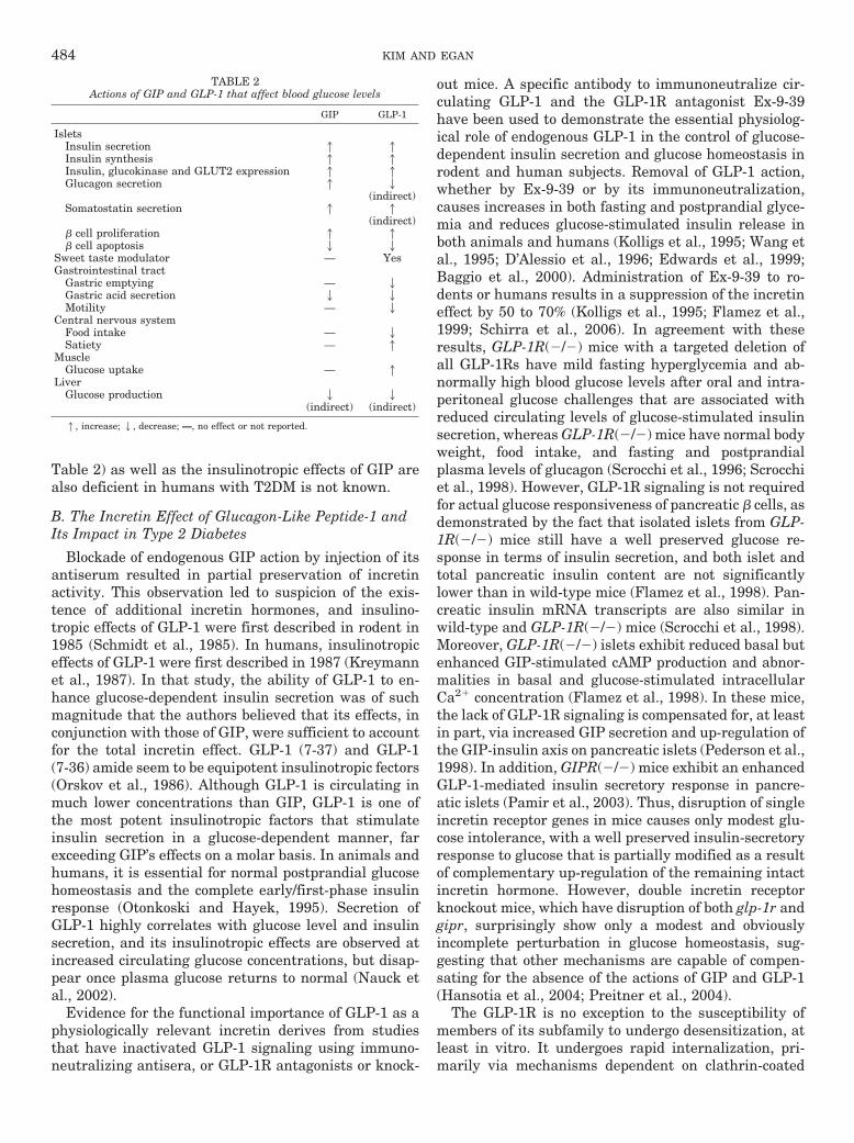
Table 2) as well as the insulinotropic effects of GIP arealso deficient in humans with T2DM is not known.
B. The Incretin Effect of Glucagon-Like Peptide-1 andIts Impact in Type 2 Diabetes
Blockade of endogenous GIP action by injection of itsantiserum resulted in partial preservation of incretinactivity. This observation led to suspicion of the exis-tence of additional incretin hormones, and insulino-tropic effects of GLP-1 were first described in rodent in1985 (Schmidt et al., 1985). In humans, insulinotropiceffects of GLP-1 were first described in 1987 (Kreymannet al., 1987). In that study, the ability of GLP-1 to en-hance glucose-dependent insulin secretion was of suchmagnitude that the authors believed that its effects, inconjunction with those of GIP, were sufficient to accountfor the total incretin effect. GLP-1 (7-37) and GLP-1(7-36) amide seem to be equipotent insulinotropic fectors(Orskov et al., 1986). Although GLP-1 is circulating inmuch lower concentrations than GIP, GLP-1 is one ofthe most potent insulinotropic factors that stimulateinsulin secretion in a glucose-dependent manner, farexceeding GIP’s effects on a molar basis. In animals andhumans, it is essential for normal postprandial glucosehomeostasis and the complete early/first-phase insulinresponse (Otonkoski and Hayek, 1995). Secretion ofGLP-1 highly correlates with glucose level and insulinsecretion, and its insulinotropic effects are observed atincreased circulating glucose concentrations, but disap-pear once plasma glucose returns to normal (Nauck etal., 2002).
Evidence for the functional importance of GLP-1 as aphysiologically relevant incretin derives from studiesthat have inactivated GLP-1 signaling using immuno-neutralizing antisera, or GLP-1R antagonists or knock-
out mice. A specific antibody to immunoneutralize cir-culating GLP-1 and the GLP-1R antagonist Ex-9-39have been used to demonstrate the essential physiolog-ical role of endogenous GLP-1 in the control of glucose-dependent insulin secretion and glucose homeostasis inrodent and human subjects. Removal of GLP-1 action,whether by Ex-9-39 or by its immunoneutralization,causes increases in both fasting and postprandial glyce-mia and reduces glucose-stimulated insulin release inboth animals and humans (Kolligs et al., 1995; Wang etal., 1995; D’Alessio et al., 1996; Edwards et al., 1999;Baggio et al., 2000). Administration of Ex-9-39 to ro-dents or humans results in a suppression of the incretineffect by 50 to 70% (Kolligs et al., 1995; Flamez et al.,1999; Schirra et al., 2006). In agreement with theseresults, GLP-1R(�/�) mice with a targeted deletion ofall GLP-1Rs have mild fasting hyperglycemia and ab-normally high blood glucose levels after oral and intra-peritoneal glucose challenges that are associated withreduced circulating levels of glucose-stimulated insulinsecretion, whereas GLP-1R(�/�) mice have normal bodyweight, food intake, and fasting and postprandialplasma levels of glucagon (Scrocchi et al., 1996; Scrocchiet al., 1998). However, GLP-1R signaling is not requiredfor actual glucose responsiveness of pancreatic � cells, asdemonstrated by the fact that isolated islets from GLP-1R(�/�) mice still have a well preserved glucose re-sponse in terms of insulin secretion, and both islet andtotal pancreatic insulin content are not significantlylower than in wild-type mice (Flamez et al., 1998). Pan-creatic insulin mRNA transcripts are also similar inwild-type and GLP-1R(�/�) mice (Scrocchi et al., 1998).Moreover, GLP-1R(�/�) islets exhibit reduced basal butenhanced GIP-stimulated cAMP production and abnor-malities in basal and glucose-stimulated intracellularCa2� concentration (Flamez et al., 1998). In these mice,the lack of GLP-1R signaling is compensated for, at leastin part, via increased GIP secretion and up-regulation ofthe GIP-insulin axis on pancreatic islets (Pederson et al.,1998). In addition, GIPR(�/�) mice exhibit an enhancedGLP-1-mediated insulin secretory response in pancre-atic islets (Pamir et al., 2003). Thus, disruption of singleincretin receptor genes in mice causes only modest glu-cose intolerance, with a well preserved insulin-secretoryresponse to glucose that is partially modified as a resultof complementary up-regulation of the remaining intactincretin hormone. However, double incretin receptorknockout mice, which have disruption of both glp-1r andgipr, surprisingly show only a modest and obviouslyincomplete perturbation in glucose homeostasis, sug-gesting that other mechanisms are capable of compen-sating for the absence of the actions of GIP and GLP-1(Hansotia et al., 2004; Preitner et al., 2004).
The GLP-1R is no exception to the susceptibility ofmembers of its subfamily to undergo desensitization, atleast in vitro. It undergoes rapid internalization, pri-marily via mechanisms dependent on clathrin-coated
TABLE 2Actions of GIP and GLP-1 that affect blood glucose levels
GIP GLP-1
IsletsInsulin secretion 1 1Insulin synthesis 1 1Insulin, glucokinase and GLUT2 expression 1 1Glucagon secretion 1 2
(indirect)Somatostatin secretion 1 1
(indirect)� cell proliferation 1 1� cell apoptosis 2 2
Sweet taste modulator — YesGastrointestinal tract
Gastric emptying — 2Gastric acid secretion 2 2Motility — 2
Central nervous systemFood intake — 2Satiety — 1
MuscleGlucose uptake — 1
LiverGlucose production 2
(indirect)2
(indirect)
1, increase; 2, decrease; —, no effect or not reported.
484 KIM AND EGAN
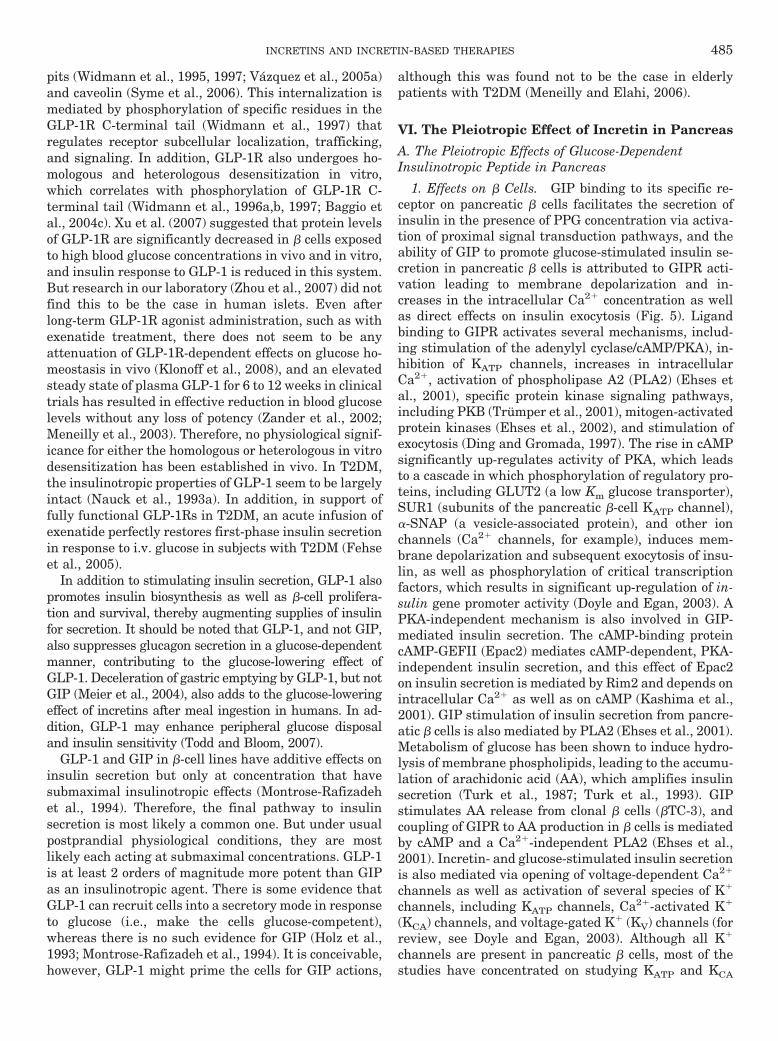
pits (Widmann et al., 1995, 1997; Vazquez et al., 2005a)and caveolin (Syme et al., 2006). This internalization ismediated by phosphorylation of specific residues in theGLP-1R C-terminal tail (Widmann et al., 1997) thatregulates receptor subcellular localization, trafficking,and signaling. In addition, GLP-1R also undergoes ho-mologous and heterologous desensitization in vitro,which correlates with phosphorylation of GLP-1R C-terminal tail (Widmann et al., 1996a,b, 1997; Baggio etal., 2004c). Xu et al. (2007) suggested that protein levelsof GLP-1R are significantly decreased in � cells exposedto high blood glucose concentrations in vivo and in vitro,and insulin response to GLP-1 is reduced in this system.But research in our laboratory (Zhou et al., 2007) did notfind this to be the case in human islets. Even afterlong-term GLP-1R agonist administration, such as withexenatide treatment, there does not seem to be anyattenuation of GLP-1R-dependent effects on glucose ho-meostasis in vivo (Klonoff et al., 2008), and an elevatedsteady state of plasma GLP-1 for 6 to 12 weeks in clinicaltrials has resulted in effective reduction in blood glucoselevels without any loss of potency (Zander et al., 2002;Meneilly et al., 2003). Therefore, no physiological signif-icance for either the homologous or heterologous in vitrodesensitization has been established in vivo. In T2DM,the insulinotropic properties of GLP-1 seem to be largelyintact (Nauck et al., 1993a). In addition, in support offully functional GLP-1Rs in T2DM, an acute infusion ofexenatide perfectly restores first-phase insulin secretionin response to i.v. glucose in subjects with T2DM (Fehseet al., 2005).
In addition to stimulating insulin secretion, GLP-1 alsopromotes insulin biosynthesis as well as �-cell prolifera-tion and survival, thereby augmenting supplies of insulinfor secretion. It should be noted that GLP-1, and not GIP,also suppresses glucagon secretion in a glucose-dependentmanner, contributing to the glucose-lowering effect ofGLP-1. Deceleration of gastric emptying by GLP-1, but notGIP (Meier et al., 2004), also adds to the glucose-loweringeffect of incretins after meal ingestion in humans. In ad-dition, GLP-1 may enhance peripheral glucose disposaland insulin sensitivity (Todd and Bloom, 2007).
GLP-1 and GIP in �-cell lines have additive effects oninsulin secretion but only at concentration that havesubmaximal insulinotropic effects (Montrose-Rafizadehet al., 1994). Therefore, the final pathway to insulinsecretion is most likely a common one. But under usualpostprandial physiological conditions, they are mostlikely each acting at submaximal concentrations. GLP-1is at least 2 orders of magnitude more potent than GIPas an insulinotropic agent. There is some evidence thatGLP-1 can recruit cells into a secretory mode in responseto glucose (i.e., make the cells glucose-competent),whereas there is no such evidence for GIP (Holz et al.,1993; Montrose-Rafizadeh et al., 1994). It is conceivable,however, GLP-1 might prime the cells for GIP actions,
although this was found not to be the case in elderlypatients with T2DM (Meneilly and Elahi, 2006).
VI. The Pleiotropic Effect of Incretin in Pancreas
A. The Pleiotropic Effects of Glucose-DependentInsulinotropic Peptide in Pancreas
1. Effects on � Cells. GIP binding to its specific re-ceptor on pancreatic � cells facilitates the secretion ofinsulin in the presence of PPG concentration via activa-tion of proximal signal transduction pathways, and theability of GIP to promote glucose-stimulated insulin se-cretion in pancreatic � cells is attributed to GIPR acti-vation leading to membrane depolarization and in-creases in the intracellular Ca2� concentration as wellas direct effects on insulin exocytosis (Fig. 5). Ligandbinding to GIPR activates several mechanisms, includ-ing stimulation of the adenylyl cyclase/cAMP/PKA), in-hibition of KATP channels, increases in intracellularCa2�, activation of phospholipase A2 (PLA2) (Ehses etal., 2001), specific protein kinase signaling pathways,including PKB (Trumper et al., 2001), mitogen-activatedprotein kinases (Ehses et al., 2002), and stimulation ofexocytosis (Ding and Gromada, 1997). The rise in cAMPsignificantly up-regulates activity of PKA, which leadsto a cascade in which phosphorylation of regulatory pro-teins, including GLUT2 (a low Km glucose transporter),SUR1 (subunits of the pancreatic �-cell KATP channel),�-SNAP (a vesicle-associated protein), and other ionchannels (Ca2� channels, for example), induces mem-brane depolarization and subsequent exocytosis of insu-lin, as well as phosphorylation of critical transcriptionfactors, which results in significant up-regulation of in-sulin gene promoter activity (Doyle and Egan, 2003). APKA-independent mechanism is also involved in GIP-mediated insulin secretion. The cAMP-binding proteincAMP-GEFII (Epac2) mediates cAMP-dependent, PKA-independent insulin secretion, and this effect of Epac2on insulin secretion is mediated by Rim2 and depends onintracellular Ca2� as well as on cAMP (Kashima et al.,2001). GIP stimulation of insulin secretion from pancre-atic � cells is also mediated by PLA2 (Ehses et al., 2001).Metabolism of glucose has been shown to induce hydro-lysis of membrane phospholipids, leading to the accumu-lation of arachidonic acid (AA), which amplifies insulinsecretion (Turk et al., 1987; Turk et al., 1993). GIPstimulates AA release from clonal � cells (�TC-3), andcoupling of GIPR to AA production in � cells is mediatedby cAMP and a Ca2�-independent PLA2 (Ehses et al.,2001). Incretin- and glucose-stimulated insulin secretionis also mediated via opening of voltage-dependent Ca2�
channels as well as activation of several species of K�
channels, including KATP channels, Ca2�-activated K�
(KCA) channels, and voltage-gated K� (KV) channels (forreview, see Doyle and Egan, 2003). Although all K�
channels are present in pancreatic � cells, most of thestudies have concentrated on studying KATP and KCA
INCRETINS AND INCRETIN-BASED THERAPIES 485
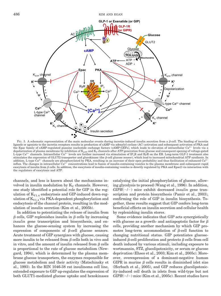
channels, and less is known about the mechanisms in-volved in insulin modulation by KV channels. However,one study identified a potential role for GIP in the reg-ulation of KV1.4 endocytosis and GIP-induced down-reg-ulation of KV1.4 via PKA-dependent phosphorylation andendocytosis of the channel protein, resulting in the mod-ulation of insulin secretion (Kim et al., 2005b).
In addition to potentiating the release of insulin from� cells, GIP replenishes insulin in � cells by increasinginsulin gene transcription and biosynthesis, and en-hances the glucose-sensing system by increasing theexpression of components of �-cell glucose sensors.Acute treatment of GIP synergizes with glucose, causingmore insulin to be released from � cells both in vivo andin vitro, and the amount of insulin released from � cellsis proportional to the rate of glucose metabolism (New-gard, 1994), which is determined by the plasma mem-brane glucose transporters, the enzymes responsible forglucose metabolism and their activity (Matschinsky etal., 1993). In the RIN 1046-38 rat insulinoma cell line,extended exposure to GIP up-regulates the expression ofboth GLUT1-mediated glucose uptake and hexokinases
catalyzing the initial phosphorylation of glucose, allow-ing glycolysis to proceed (Wang et al., 1996). In addition,GIPR(�/�) mice exhibit decreased insulin gene tran-scription and protein biosynthesis (Pamir et al., 2003),confirming the role of GIP in insulin biosynthesis. To-gether, these results suggest that GIP confers long-termbeneficial effects on insulin secretion and cellular healthby replenishing insulin stores.
Some evidence indicates that GIP acts synergisticallywith glucose as a growth- and antiapoptotic factor for �cells, providing another mechanism by which GIP pro-motes long-term accommodation of �-cell function tochanging nutritional status. GIP potentiates glucose-induced �-cell proliferation and protects � cells from celldeath induced by various stimuli, including exposure towortmannin, STZ, glucolipotoxicity, or serum or glucosedeprivation (Ehses et al., 2003; Kim et al., 2005c). More-over, overexpression of a dominant-negative humanGIPR in murine � cells results in diminished islet size(Herbach et al., 2005), and GIP reduces glucolipotoxic-ity-induced cell death in islets from wild-type but notGIPR(�/�) mice (Kim et al., 2005c). Recent studies have
FIG. 5. A schematic representation of the main molecular events during incretin-induced insulin secretion from a �-cell. The binding of incretinligands or agonists to the incretin receptors results in production of cAMP via adenylyl cyclase (AC) activation and subsequent activation of PKA andthe Epac family of cAMP-regulated guanine nucleotide exchange factors (cAMP-GEFs), which leads to elevation of intracellular Ca2� levels via adepolarization of plasma membrane by inhibition of KATP and KV channels after ATP generation from glucose and consequent opening of voltage-gatedL-type Ca2� channels. Intracellular Ca2� levels are further increased via stimulation of IP3R and RyR on the ER. Long-term GLP-1 treatment alsostimulates the expression of GLUT2 transporter and glucokinase (the �-cell glucose sensor), which lead to increased mitochondrial ATP synthesis. Inaddition, L-type Ca2� channels are phosphorylated by PKA, resulting in an increase of their open probability and thus facilitation of enhanced Ca2�
influx. The changes in intracellular Ca2� concentrations lead to fusion of insulin-containing vesicles to the plasma membrane and subsequent rapidexocytosis of insulin from � cells. In addition, the exocytosis of insulin-containing vesicles is directly regulated by PKA and Epac2 via interaction withthe regulators of exocytosis and ATP.
486 KIM AND EGAN

begun to unravel the molecular signaling pathways thatmediate the proliferative and antiapoptotic actions ofGIP in the �-cell, and it is clear that growth and anti-apoptotic pathways rely on similar pathways, includingactivation of cAMP/PKA, PKA/CREB, MAPK, and PI-3K-dependent activation of PKB (Trumper et al., 2001,2002; Ehses et al., 2002, 2003; Kim et al., 2005c) (Fig. 6).These signaling modules are dependent on glucose me-tabolism and Ca2� influx, and cAMP-PKA-Rab1-extra-cellular signal-regulated kinase 1/2 pathway may alsobe one way in which GIP regulates �-cell proliferation(Ehses et al., 2002). In particular, GIP phosphorylatesseveral transcription factors such as FoxO1 and p70S6K,crucial regulators of translation known to be involved inglucose-induced �-cell mitogenesis, suggesting possiblemediators of GIP-mediated �-cell growth (Ehses et al.,2002). FoxO1 has been shown to play a critical role inGIP-mediated �-cell survival and apoptosis. GIP im-proved cell survival in INS-1 (clone 832/13) cells andmurine islets via GIP-induced phosphorylation ofFoxO1, resulting in inactivation of the pro-apoptotic baxgene (Kim et al., 2005c). In addition, a 2-week infusion ofGIP into diabetic (ZDF) rats was shown to significantlyreduce �-cell apoptosis because of down-regulation of thebax gene and up-regulation of the antiapoptotic bcl-2gene: these effects were mediated by activation of PI-3K/PKB, and subsequent phosphorylation and nuclear ex-
clusion of FoxO1 (Kim et al., 2005c). The protectiveeffects of GIP on �-cell apoptosis in the diabetic state arevery intriguing to us. It is clear from many studies thatGIP is not insulinotropic in T2DM and that in animalmodels, such as the ZDF rat discussed above (sectionV.A), GIP does not stimulate insulin secretion, and itsreceptor is severely down-regulated (94% reduction com-pared with lean nondiabetic rats) (Piteau et al., 2007).So, if the GIP receptor is so severely down-regulated inthe ZDF rats, how did 2 weeks of GIP infusion have itsantiapoptotic effects? Unlike the more recent work, inwhich the authors found that reduction in hyperglyemiaby phlorizin treatment reversed GIPR down-regulation(Piteau et al., 2007), Kim et al. (2005c) did not first lowerhyperglycemia before giving exogenous GIP. Is there acase to be made that the antiapoptotic effect, in vivo, isnot through the known GIPR on � cells? Could this meanthat by not developing GIPR agonists to treat T2DM, weare missing the other potential effect of GIPR stimula-tion? Is there a case to be made that GLP-1R and GIPRstimulation, together, might be beneficial in T2DM? In�-cell lines at least, a direct antiapoptotic effect of GIPseems to hold up, because another study from the sameresearch group corroborated that GIP has antiapoptoticproperties and that the antiapoptotic effect of GIP is, atthe very least, partially due to GIP-stimulated increasesin bcl-2 gene expression (Kim et al., 2008b). In that
FIG. 6. Schema outlining the incretin downstream signal transduction pathways in a � cell. GLP-1R activation (and GIPR activation, to someextent, but this has not been as well studied as GLP-1R activation) recruits signaling mechanisms that considerably overlap, leading to promotion of�-cell proliferation and prevention of �-cell apoptosis. Dashed line indicates mechanism that is not fully delineated.
INCRETINS AND INCRETIN-BASED THERAPIES 487
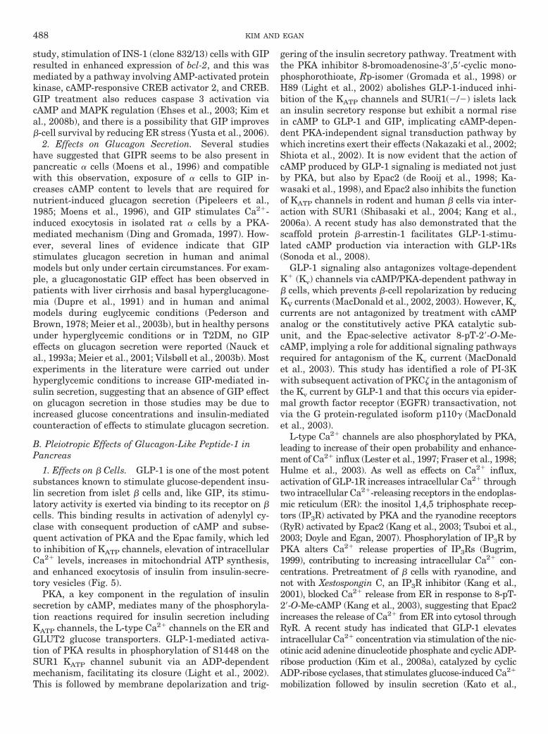
study, stimulation of INS-1 (clone 832/13) cells with GIPresulted in enhanced expression of bcl-2, and this wasmediated by a pathway involving AMP-activated proteinkinase, cAMP-responsive CREB activator 2, and CREB.GIP treatment also reduces caspase 3 activation viacAMP and MAPK regulation (Ehses et al., 2003; Kim etal., 2008b), and there is a possibility that GIP improves�-cell survival by reducing ER stress (Yusta et al., 2006).
2. Effects on Glucagon Secretion. Several studieshave suggested that GIPR seems to be also present inpancreatic � cells (Moens et al., 1996) and compatiblewith this observation, exposure of � cells to GIP in-creases cAMP content to levels that are required fornutrient-induced glucagon secretion (Pipeleers et al.,1985; Moens et al., 1996), and GIP stimulates Ca2�-induced exocytosis in isolated rat � cells by a PKA-mediated mechanism (Ding and Gromada, 1997). How-ever, several lines of evidence indicate that GIPstimulates glucagon secretion in human and animalmodels but only under certain circumstances. For exam-ple, a glucagonostatic GIP effect has been observed inpatients with liver cirrhosis and basal hyperglucagone-mia (Dupre et al., 1991) and in human and animalmodels during euglycemic conditions (Pederson andBrown, 1978; Meier et al., 2003b), but in healthy personsunder hyperglycemic conditions or in T2DM, no GIPeffects on glucagon secretion were reported (Nauck etal., 1993a; Meier et al., 2001; Vilsbøll et al., 2003b). Mostexperiments in the literature were carried out underhyperglycemic conditions to increase GIP-mediated in-sulin secretion, suggesting that an absence of GIP effecton glucagon secretion in those studies may be due toincreased glucose concentrations and insulin-mediatedcounteraction of effects to stimulate glucagon secretion.
B. Pleiotropic Effects of Glucagon-Like Peptide-1 inPancreas
1. Effects on � Cells. GLP-1 is one of the most potentsubstances known to stimulate glucose-dependent insu-lin secretion from islet � cells and, like GIP, its stimu-latory activity is exerted via binding to its receptor on �cells. This binding results in activation of adenylyl cy-clase with consequent production of cAMP and subse-quent activation of PKA and the Epac family, which ledto inhibition of KATP channels, elevation of intracellularCa2� levels, increases in mitochondrial ATP synthesis,and enhanced exocytosis of insulin from insulin-secre-tory vesicles (Fig. 5).
PKA, a key component in the regulation of insulinsecretion by cAMP, mediates many of the phosphoryla-tion reactions required for insulin secretion includingKATP channels, the L-type Ca2� channels on the ER andGLUT2 glucose transporters. GLP-1-mediated activa-tion of PKA results in phosphorylation of S1448 on theSUR1 KATP channel subunit via an ADP-dependentmechanism, facilitating its closure (Light et al., 2002).This is followed by membrane depolarization and trig-
gering of the insulin secretory pathway. Treatment withthe PKA inhibitor 8-bromoadenosine-3�,5�-cyclic mono-phosphorothioate, Rp-isomer (Gromada et al., 1998) orH89 (Light et al., 2002) abolishes GLP-1-induced inhi-bition of the KATP channels and SUR1(�/�) islets lackan insulin secretory response but exhibit a normal risein cAMP to GLP-1 and GIP, implicating cAMP-depen-dent PKA-independent signal transduction pathway bywhich incretins exert their effects (Nakazaki et al., 2002;Shiota et al., 2002). It is now evident that the action ofcAMP produced by GLP-1 signaling is mediated not justby PKA, but also by Epac2 (de Rooij et al., 1998; Ka-wasaki et al., 1998), and Epac2 also inhibits the functionof KATP channels in rodent and human � cells via inter-action with SUR1 (Shibasaki et al., 2004; Kang et al.,2006a). A recent study has also demonstrated that thescaffold protein �-arrestin-1 facilitates GLP-1-stimu-lated cAMP production via interaction with GLP-1Rs(Sonoda et al., 2008).
GLP-1 signaling also antagonizes voltage-dependentK� (Kv) channels via cAMP/PKA-dependent pathway in� cells, which prevents �-cell repolarization by reducingKV currents (MacDonald et al., 2002, 2003). However, Kvcurrents are not antagonized by treatment with cAMPanalog or the constitutively active PKA catalytic sub-unit, and the Epac-selective activator 8-pT-2�-O-Me-cAMP, implying a role for additional signaling pathwaysrequired for antagonism of the Kv current (MacDonaldet al., 2003). This study has identified a role of PI-3Kwith subsequent activation of PKC� in the antagonism ofthe Kv current by GLP-1 and that this occurs via epider-mal growth factor receptor (EGFR) transactivation, notvia the G protein-regulated isoform p110� (MacDonaldet al., 2003).
L-type Ca2� channels are also phosphorylated by PKA,leading to increase of their open probability and enhance-ment of Ca2� influx (Lester et al., 1997; Fraser et al., 1998;Hulme et al., 2003). As well as effects on Ca2� influx,activation of GLP-1R increases intracellular Ca2� throughtwo intracellular Ca2�-releasing receptors in the endoplas-mic reticulum (ER): the inositol 1,4,5 triphosphate recep-tors (IP3R) activated by PKA and the ryanodine receptors(RyR) activated by Epac2 (Kang et al., 2003; Tsuboi et al.,2003; Doyle and Egan, 2007). Phosphorylation of IP3R byPKA alters Ca2� release properties of IP3Rs (Bugrim,1999), contributing to increasing intracellular Ca2� con-centrations. Pretreatment of � cells with ryanodine, andnot with Xestospongin C, an IP3R inhibitor (Kang et al.,2001), blocked Ca2� release from ER in response to 8-pT-2�-O-Me-cAMP (Kang et al., 2003), suggesting that Epac2increases the release of Ca2� from ER into cytosol throughRyR. A recent study has indicated that GLP-1 elevatesintracellular Ca2� concentration via stimulation of the nic-otinic acid adenine dinucleotide phosphate and cyclic ADP-ribose production (Kim et al., 2008a), catalyzed by cyclicADP-ribose cyclases, that stimulates glucose-induced Ca2�
mobilization followed by insulin secretion (Kato et al.,
488 KIM AND EGAN
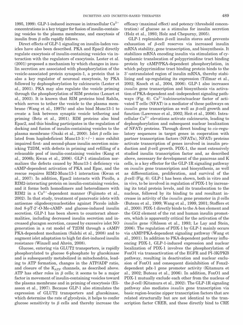
1995, 1999). GLP-1-induced increase in intracellular Ca2�
concentrations is a key trigger for fusion of insulin-contain-ing vesicles to the plasma membrane, and exocytosis ofinsulin from � cells rapidly follows.
Direct effects of GLP-1 signaling on insulin-laden ves-icles have also been described. PKA and Epac2 directlyregulate exocytosis of insulin-containing vesicles via in-teraction with the regulators of exocytosis. Lester et al.(2001) proposed a mechanism by which changes in insu-lin secretion are associated with phosphorylation of thevesicle-associated protein synapsin-1, a protein that isalso a key regulator of neuronal exocytosis, by PKAfollowed by dephosphorylation by calcineurin (Lester etal., 2001). PKA may also regulate the vesicle primingthrough the phosphorylation of RIM proteins (Lonart etal., 2003). It is known that RIM proteins bind Rab3a,which serves to tether the vesicle to the plasma mem-brane (Wang et al., 1997b) and also bind Munc13-1 tocreate a link between synaptic vesicle tethering andpriming (Betz et al., 2001). RIM proteins also bindEpac2, and this binding participates in the regulation ofdocking and fusion of insulin-containing vesicles to theplasma membrane (Ozaki et al., 2000). Islet � cells iso-lated from haplodeficient Munc13-1(�/�) mice exhibitimpaired first- and second-phase insulin secretion mim-icking T2DM, with defects in priming and refilling of areleasable pool of insulin-containing vesicles (Kang etal., 2006b; Kwan et al., 2006). GLP-1 stimulation nor-malizes the defects caused by Munc13-1 deficiency viacAMP-dependent activation of PKA and Epac, and therescue requires RIM2-Munc13-1 interaction (Kwan etal., 2007). In addition, Epac2 interacts with Picollo, aRIM2-interacting protein on insulin-containing vesicles,and it forms both homodimers and heterodimers withRIM2 in a Ca2�-dependent manner (Fujimoto et al.,2002). In that study, treatment of pancreatic islets withantisense oligodeoxynucleotides against Piccolo inhib-ited 8-pT-2�-O-Me-cAMP- and glucose-induced insulinsecretion. GLP-1 has been shown to counteract abnor-malities, including decreased insulin secretion and in-creased glucagon secretion, caused by excessive islet NOgeneration in a rat model of T2DM through a cAMP/PKA-dependent mechanism (Salehi et al., 2008) and toaugment islet adaptation to high fat diet-induced insulinresistance (Winzell and Ahren, 2008).
Glucose, entering via GLUT2 transporters, is rapidlyphosphorylated to glucose 6-phosphate by glucokinaseand is subsequently metabolized in mitochondria, lead-ing to ATP formation, changes in the ATP/ADP ratio,and closure of the KATP channels, as described above.ATP has other roles in � cells; it seems to be a majorfactor in movement of insulin-containing vesicles towardthe plasma membrane and in priming of exocytosis (Eli-asson et al., 1997). Because GLP-1 also stimulates theexpression of GLUT2 transporters and glucokinase,which determine the rate of glycolysis, it helps to conferglucose sensitivity to � cells and thereby increase the
efficacy (maximal effect) and potency (threshold concen-tration) of glucose as a stimulus for insulin secretion(Holz et al., 1993; Holz and Chepurny, 2005).
GLP-1 replenishes �-cell insulin stores and preventsexhaustion of �-cell reserves via increased insulinmRNA stability, gene transcription, and biosynthesis. Itstabilizes mRNA encoding insulin via stimulation of cy-toplasmic translocation of polypyrimidine tract bindingprotein by cAMP/PKA-dependent phosphorylation, inwhich polypyrimidine tract binding protein binds to the3�-untranslated region of insulin mRNA, thereby stabi-lizing and up-regulating its expression (Tillmar et al.,2002; Knoch et al., 2004, 2006). GLP-1 also increasesinsulin gene transcription and biosynthesis via activa-tion of PKA-dependent and -independent signaling path-ways (Fig. 6). Ca2�/calcineurin/nuclear factor of acti-vated T cells (NFAT) is a mediator of these pathways oninsulin gene transcription as well as �-cell growth andfunction (Lawrence et al., 2002; Heit et al., 2006). Intra-cellular Ca2� elevations activate calcineurin, leading todephosphorylation and subsequent nuclear localizationof NFATc proteins. Through direct binding to cis-regu-latory sequences in target genes in cooperation withpartner transcription factors (NFATn), NFATc proteinsactivate transcription of genes involved in insulin pro-duction and �-cell growth. PDX-1, the most extensivelystudied insulin transcription factor and, as describedabove, necessary for development of the pancreas and Kcells, is a key effector for the GLP-1R signaling pathwayon insulin gene transcription and biosynthesis, as wellas differentiation, proliferation, and survival of the�-cell (Fig. 6). GLP-1 has been shown, both in vitro andin vivo, to be involved in regulation of PDX-1 by increas-ing its total protein levels, and its translocation to thenucleus, followed by its binding to and resultant in-crease in activity of the insulin gene promoter in � cells(Buteau et al., 1999; Wang et al., 1999, 2001; Stoffers etal., 2000). PDX-1 directly binds to the A-box element andthe GG2 element of the rat and human insulin promot-ers, which is apparently critical for the activation of theinsulin gene (Ohlsson et al., 1993; Le Lay and Stein,2006). The regulation of PDX-1 by GLP-1 mainly occursvia cAMP/PKA-dependent signaling pathway (Wang etal., 2001). In addition to PKA-dependent pathway influ-encing PDX-1, GLP-1-induced expression and nuclearlocalization of PDX-1 involves the phosphorylation ofFoxO1 via transactivation of the EGFR and PI-3K/PKBpathway, resulting in deactivation and nuclear exclu-sion of FoxO1 and consequent disinhibition of Foxa2-dependent pdx-1 gene promoter activity (Kitamura etal., 2002; Buteau et al., 2006). In addition, FoxO1 andPDX-1 mutually exclude each other from the nucleus ofthe �-cell (Kitamura et al., 2002). The GLP-1R signalingpathway also mediates insulin gene transcription viabasic region-leucine zipper transcription factors that arerelated structurally but are not identical to the tran-scription factor CREB, and these directly bind to CRE
INCRETINS AND INCRETIN-BASED THERAPIES 489
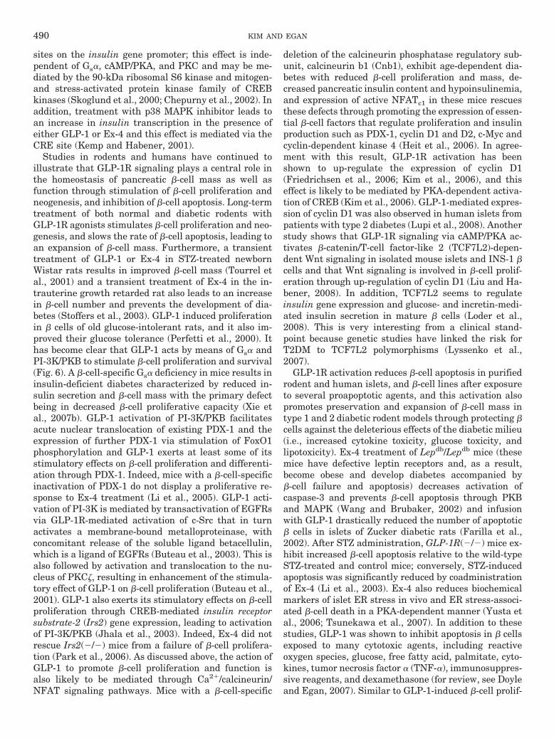
sites on the insulin gene promoter; this effect is inde-pendent of Gs�, cAMP/PKA, and PKC and may be me-diated by the 90-kDa ribosomal S6 kinase and mitogen-and stress-activated protein kinase family of CREBkinases (Skoglund et al., 2000; Chepurny et al., 2002). Inaddition, treatment with p38 MAPK inhibitor leads toan increase in insulin transcription in the presence ofeither GLP-1 or Ex-4 and this effect is mediated via theCRE site (Kemp and Habener, 2001).
Studies in rodents and humans have continued toillustrate that GLP-1R signaling plays a central role inthe homeostasis of pancreatic �-cell mass as well asfunction through stimulation of �-cell proliferation andneogenesis, and inhibition of �-cell apoptosis. Long-termtreatment of both normal and diabetic rodents withGLP-1R agonists stimulates �-cell proliferation and neo-genesis, and slows the rate of �-cell apoptosis, leading toan expansion of �-cell mass. Furthermore, a transienttreatment of GLP-1 or Ex-4 in STZ-treated newbornWistar rats results in improved �-cell mass (Tourrel etal., 2001) and a transient treatment of Ex-4 in the in-trauterine growth retarded rat also leads to an increasein �-cell number and prevents the development of dia-betes (Stoffers et al., 2003). GLP-1 induced proliferationin � cells of old glucose-intolerant rats, and it also im-proved their glucose tolerance (Perfetti et al., 2000). Ithas become clear that GLP-1 acts by means of Gs� andPI-3K/PKB to stimulate �-cell proliferation and survival(Fig. 6). A �-cell-specific Gs� deficiency in mice results ininsulin-deficient diabetes characterized by reduced in-sulin secretion and �-cell mass with the primary defectbeing in decreased �-cell proliferative capacity (Xie etal., 2007b). GLP-1 activation of PI-3K/PKB facilitatesacute nuclear translocation of existing PDX-1 and theexpression of further PDX-1 via stimulation of FoxO1phosphorylation and GLP-1 exerts at least some of itsstimulatory effects on �-cell proliferation and differenti-ation through PDX-1. Indeed, mice with a �-cell-specificinactivation of PDX-1 do not display a proliferative re-sponse to Ex-4 treatment (Li et al., 2005). GLP-1 acti-vation of PI-3K is mediated by transactivation of EGFRsvia GLP-1R-mediated activation of c-Src that in turnactivates a membrane-bound metalloproteinase, withconcomitant release of the soluble ligand betacellulin,which is a ligand of EGFRs (Buteau et al., 2003). This isalso followed by activation and translocation to the nu-cleus of PKC�, resulting in enhancement of the stimula-tory effect of GLP-1 on �-cell proliferation (Buteau et al.,2001). GLP-1 also exerts its stimulatory effects on �-cellproliferation through CREB-mediated insulin receptorsubstrate-2 (Irs2) gene expression, leading to activationof PI-3K/PKB (Jhala et al., 2003). Indeed, Ex-4 did notrescue Irs2(�/�) mice from a failure of �-cell prolifera-tion (Park et al., 2006). As discussed above, the action ofGLP-1 to promote �-cell proliferation and function isalso likely to be mediated through Ca2�/calcineurin/NFAT signaling pathways. Mice with a �-cell-specific
deletion of the calcineurin phosphatase regulatory sub-unit, calcineurin b1 (Cnb1), exhibit age-dependent dia-betes with reduced �-cell proliferation and mass, de-creased pancreatic insulin content and hypoinsulinemia,and expression of active NFATc1 in these mice rescuesthese defects through promoting the expression of essen-tial �-cell factors that regulate proliferation and insulinproduction such as PDX-1, cyclin D1 and D2, c-Myc andcyclin-dependent kinase 4 (Heit et al., 2006). In agree-ment with this result, GLP-1R activation has beenshown to up-regulate the expression of cyclin D1(Friedrichsen et al., 2006; Kim et al., 2006), and thiseffect is likely to be mediated by PKA-dependent activa-tion of CREB (Kim et al., 2006). GLP-1-mediated expres-sion of cyclin D1 was also observed in human islets frompatients with type 2 diabetes (Lupi et al., 2008). Anotherstudy shows that GLP-1R signaling via cAMP/PKA ac-tivates �-catenin/T-cell factor-like 2 (TCF7L2)-depen-dent Wnt signaling in isolated mouse islets and INS-1 �cells and that Wnt signaling is involved in �-cell prolif-eration through up-regulation of cyclin D1 (Liu and Ha-bener, 2008). In addition, TCF7L2 seems to regulateinsulin gene expression and glucose- and incretin-medi-ated insulin secretion in mature � cells (Loder et al.,2008). This is very interesting from a clinical stand-point because genetic studies have linked the risk forT2DM to TCF7L2 polymorphisms (Lyssenko et al.,2007).
GLP-1R activation reduces �-cell apoptosis in purifiedrodent and human islets, and �-cell lines after exposureto several proapoptotic agents, and this activation alsopromotes preservation and expansion of �-cell mass intype 1 and 2 diabetic rodent models through protecting �cells against the deleterious effects of the diabetic milieu(i.e., increased cytokine toxicity, glucose toxicity, andlipotoxicity). Ex-4 treatment of Lepdb/Lepdb mice (thesemice have defective leptin receptors and, as a result,become obese and develop diabetes accompanied by�-cell failure and apoptosis) decreases activation ofcaspase-3 and prevents �-cell apoptosis through PKBand MAPK (Wang and Brubaker, 2002) and infusionwith GLP-1 drastically reduced the number of apoptotic� cells in islets of Zucker diabetic rats (Farilla et al.,2002). After STZ administration, GLP-1R(�/�) mice ex-hibit increased �-cell apoptosis relative to the wild-typeSTZ-treated and control mice; conversely, STZ-inducedapoptosis was significantly reduced by coadministrationof Ex-4 (Li et al., 2003). Ex-4 also reduces biochemicalmarkers of islet ER stress in vivo and ER stress-associ-ated �-cell death in a PKA-dependent manner (Yusta etal., 2006; Tsunekawa et al., 2007). In addition to thesestudies, GLP-1 was shown to inhibit apoptosis in � cellsexposed to many cytotoxic agents, including reactiveoxygen species, glucose, free fatty acid, palmitate, cyto-kines, tumor necrosis factor � (TNF-�), immunosuppres-sive reagents, and dexamethasone (for review, see Doyleand Egan, 2007). Similar to GLP-1-induced �-cell prolif-
490 KIM AND EGAN
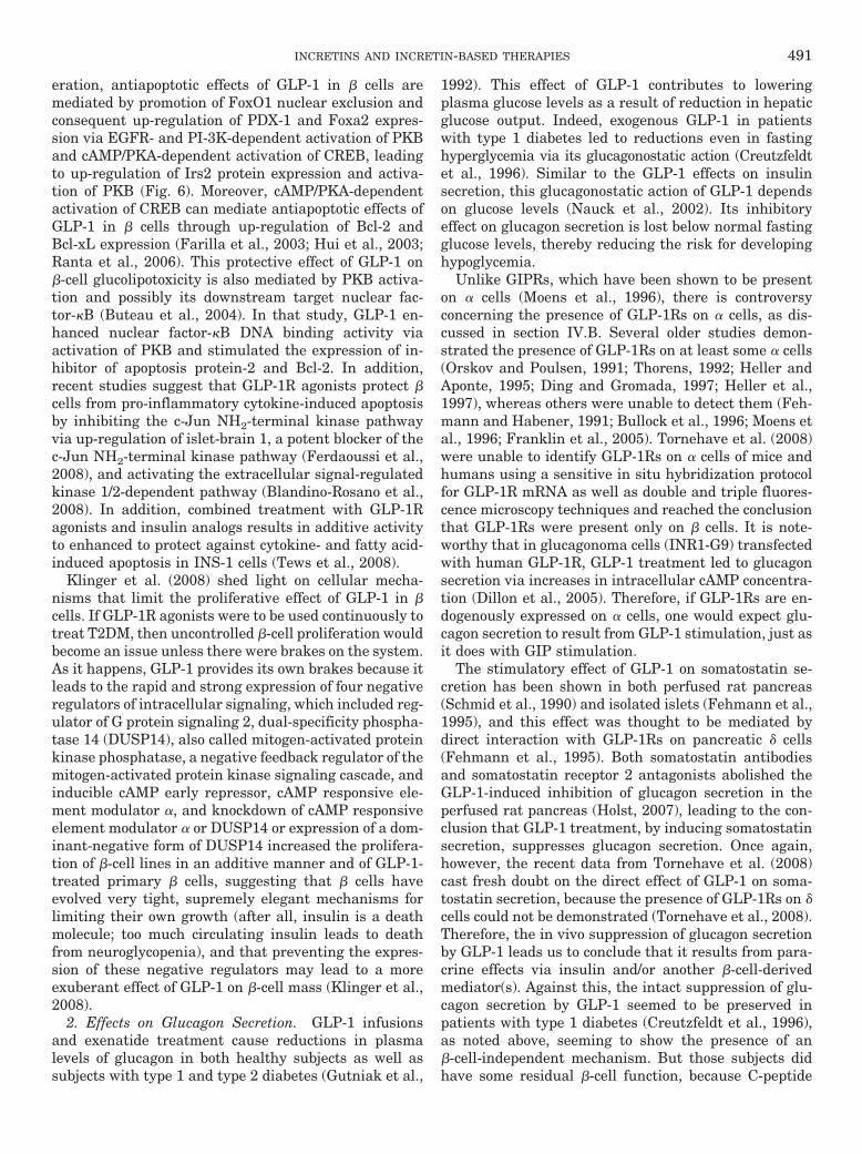
eration, antiapoptotic effects of GLP-1 in � cells aremediated by promotion of FoxO1 nuclear exclusion andconsequent up-regulation of PDX-1 and Foxa2 expres-sion via EGFR- and PI-3K-dependent activation of PKBand cAMP/PKA-dependent activation of CREB, leadingto up-regulation of Irs2 protein expression and activa-tion of PKB (Fig. 6). Moreover, cAMP/PKA-dependentactivation of CREB can mediate antiapoptotic effects ofGLP-1 in � cells through up-regulation of Bcl-2 andBcl-xL expression (Farilla et al., 2003; Hui et al., 2003;Ranta et al., 2006). This protective effect of GLP-1 on�-cell glucolipotoxicity is also mediated by PKB activa-tion and possibly its downstream target nuclear fac-tor-�B (Buteau et al., 2004). In that study, GLP-1 en-hanced nuclear factor-�B DNA binding activity viaactivation of PKB and stimulated the expression of in-hibitor of apoptosis protein-2 and Bcl-2. In addition,recent studies suggest that GLP-1R agonists protect �cells from pro-inflammatory cytokine-induced apoptosisby inhibiting the c-Jun NH2-terminal kinase pathwayvia up-regulation of islet-brain 1, a potent blocker of thec-Jun NH2-terminal kinase pathway (Ferdaoussi et al.,2008), and activating the extracellular signal-regulatedkinase 1/2-dependent pathway (Blandino-Rosano et al.,2008). In addition, combined treatment with GLP-1Ragonists and insulin analogs results in additive activityto enhanced to protect against cytokine- and fatty acid-induced apoptosis in INS-1 cells (Tews et al., 2008).
Klinger et al. (2008) shed light on cellular mecha-nisms that limit the proliferative effect of GLP-1 in �cells. If GLP-1R agonists were to be used continuously totreat T2DM, then uncontrolled �-cell proliferation wouldbecome an issue unless there were brakes on the system.As it happens, GLP-1 provides its own brakes because itleads to the rapid and strong expression of four negativeregulators of intracellular signaling, which included reg-ulator of G protein signaling 2, dual-specificity phospha-tase 14 (DUSP14), also called mitogen-activated proteinkinase phosphatase, a negative feedback regulator of themitogen-activated protein kinase signaling cascade, andinducible cAMP early repressor, cAMP responsive ele-ment modulator �, and knockdown of cAMP responsiveelement modulator � or DUSP14 or expression of a dom-inant-negative form of DUSP14 increased the prolifera-tion of �-cell lines in an additive manner and of GLP-1-treated primary � cells, suggesting that � cells haveevolved very tight, supremely elegant mechanisms forlimiting their own growth (after all, insulin is a deathmolecule; too much circulating insulin leads to deathfrom neuroglycopenia), and that preventing the expres-sion of these negative regulators may lead to a moreexuberant effect of GLP-1 on �-cell mass (Klinger et al.,2008).
2. Effects on Glucagon Secretion. GLP-1 infusionsand exenatide treatment cause reductions in plasmalevels of glucagon in both healthy subjects as well assubjects with type 1 and type 2 diabetes (Gutniak et al.,
1992). This effect of GLP-1 contributes to loweringplasma glucose levels as a result of reduction in hepaticglucose output. Indeed, exogenous GLP-1 in patientswith type 1 diabetes led to reductions even in fastinghyperglycemia via its glucagonostatic action (Creutzfeldtet al., 1996). Similar to the GLP-1 effects on insulinsecretion, this glucagonostatic action of GLP-1 dependson glucose levels (Nauck et al., 2002). Its inhibitoryeffect on glucagon secretion is lost below normal fastingglucose levels, thereby reducing the risk for developinghypoglycemia.
Unlike GIPRs, which have been shown to be presenton � cells (Moens et al., 1996), there is controversyconcerning the presence of GLP-1Rs on � cells, as dis-cussed in section IV.B. Several older studies demon-strated the presence of GLP-1Rs on at least some � cells(Orskov and Poulsen, 1991; Thorens, 1992; Heller andAponte, 1995; Ding and Gromada, 1997; Heller et al.,1997), whereas others were unable to detect them (Feh-mann and Habener, 1991; Bullock et al., 1996; Moens etal., 1996; Franklin et al., 2005). Tornehave et al. (2008)were unable to identify GLP-1Rs on � cells of mice andhumans using a sensitive in situ hybridization protocolfor GLP-1R mRNA as well as double and triple fluores-cence microscopy techniques and reached the conclusionthat GLP-1Rs were present only on � cells. It is note-worthy that in glucagonoma cells (INR1-G9) transfectedwith human GLP-1R, GLP-1 treatment led to glucagonsecretion via increases in intracellular cAMP concentra-tion (Dillon et al., 2005). Therefore, if GLP-1Rs are en-dogenously expressed on � cells, one would expect glu-cagon secretion to result from GLP-1 stimulation, just asit does with GIP stimulation.
The stimulatory effect of GLP-1 on somatostatin se-cretion has been shown in both perfused rat pancreas(Schmid et al., 1990) and isolated islets (Fehmann et al.,1995), and this effect was thought to be mediated bydirect interaction with GLP-1Rs on pancreatic � cells(Fehmann et al., 1995). Both somatostatin antibodiesand somatostatin receptor 2 antagonists abolished theGLP-1-induced inhibition of glucagon secretion in theperfused rat pancreas (Holst, 2007), leading to the con-clusion that GLP-1 treatment, by inducing somatostatinsecretion, suppresses glucagon secretion. Once again,however, the recent data from Tornehave et al. (2008)cast fresh doubt on the direct effect of GLP-1 on soma-tostatin secretion, because the presence of GLP-1Rs on �cells could not be demonstrated (Tornehave et al., 2008).Therefore, the in vivo suppression of glucagon secretionby GLP-1 leads us to conclude that it results from para-crine effects via insulin and/or another �-cell-derivedmediator(s). Against this, the intact suppression of glu-cagon secretion by GLP-1 seemed to be preserved inpatients with type 1 diabetes (Creutzfeldt et al., 1996),as noted above, seeming to show the presence of an�-cell-independent mechanism. But those subjects didhave some residual �-cell function, because C-peptide
INCRETINS AND INCRETIN-BASED THERAPIES 491
levels increased after GLP-1 treatment, so a �-cell me-diator still cannot be totally ruled out as a cause ofglucagon suppression. In addition, �-cell-specific inacti-vation of the pdx-1 gene led to loss of Ex-4-inducedinhibition of glucagon secretion (PDX-1 is not expressedin � cells, as discussed above, and is not necessary fortheir development in the embryo), which also favors theconclusion that glucagon suppression requires a �-cellproduct (Li et al., 2005).
3. Effects on Pancreatic Exocrine and Ductal Cells. Ourstudy using the AR42J acinar cell line derived from a ratpancreatic tumor has shown that GLP-1Rs exist on thesecells and that treatment with GLP-1 and Ex-4 causes in-creases in both intracellular cAMP and Ca2� levels (Zhouet al., 1999a). GLP-1Rs were recently confirmed to bepresent in pancreatic ducts in mouse, rat, and human bythe same group that found GLP-1Rs to be present in only� cells within islets (Tornehave et al., 2008).
Activation of GLP-1R signaling either in ductal oracinar cell lines or in vivo in rodents has resulted indifferentiation of a fraction of these cells toward anislet-like phenotype, in association with activation ofPKC and MAPK, and transcription factors necessary foran endocrine phenotype such as PDX-1, as well as theglucose-sensing factors glucokinase and GLUT2 (Zhouet al., 1999b, 2002; Hui et al., 2001). GLP-1R activationof those cells also affects transforming growth factor-�signaling pathways, resulting in reduced Smad activitythat is conducive to endocrine phenotypes (Yew et al.,2004, 2005; Tei et al., 2005). AR42J cells, even withoutGLP-1R activation, have the potential to be convertedinto endocrine (Mashima et al., 1996) or exocrine cells(Christophe, 1994) but are negative for islet hormonesand their transcripts under usual culture conditions(Zhou et al., 1999b). When these cells were exposed toGLP-1 or Ex-4, approximately 20% of the cells containedinsulin protein and were capable of releasing insulin ina glucose-mediated fashion (Zhou et al., 1999b). ThisGLP-1 effect was also observed in Capan-1 cell line(Zhou et al., 2002) and rat ARIP and human PANC-1 celllines; similar to the AR42J cells, GLUT2 and glucoki-nase transcripts were induced in these cell lines (Hui etal., 2001). In particular, the differentiation-promotingactivity of GLP-1 requires the expression of PDX-1, be-cause PANC-1 cells, which lack endogenous PDX-1, dif-ferentiate only when transfected with PDX-1, whereasrat ARIP cells that express PDX-1 are susceptible toundergoing differentiation into insulin-secreting cells(Hui et al., 2001). In the Capan-1 cell line, differentia-tion to insulin-producing cells was also seen when theywere transfected with PDX-1, and PDX-1 antisense to-tally inhibited such conversion (Zhou et al., 2002). More-over, both nondiabetic and diabetic Lepdb/Lepdb micetreated with Ex-4 for 2 weeks show enhanced expressionof PDX-1 in the ducts (favoring the presence of GLP-1Rsin ductal cells, as referenced above) and the exocrinetissue (Stoffers et al., 2000), which means that GLP-1R
agonists aid in islet neogenesis, because ductal cellshave been thought to be the main source for endocrineneogenesis (Egan et al., 2003).
VII. Extrapancreatic Effects of Incretins
A. Extrapancreatic Effects of Glucose-DependentInsulinotropic Peptide
Several studies have demonstrated that GIPRs arepresent in the central nervous system, adipose tissues,and osteoblasts, but GIP actions, unlike those of GLP-1on extrapancreatic tissues, have not received muchattention.
1. Central Nervous System. GIPRs have been detectedin many areas of the brain, including the hippocampus andhippocampal progenitor cells (Usdin et al., 1993; Kaplanand Vigna, 1994; Nyberg et al., 2005). Although for years itwas assumed that GIP was expressed only in K cells andsalivary glands, GIP mRNA and protein are also expressedin rat retina and the olfactory system (Cho et al., 2002;Nyberg et al., 2005). Hippocampus, Purkinje cells, amyg-dala, substantia nigra, striatum, and several areas of thethalamus, hypothalamus and brainstem (Nyberg et al.,2005, 2007; Sondhi et al., 2006) are all positive for GIPmRNA and/or protein. Exogenous administration of GIPincreased hippocampal progenitor cell proliferation in vivoand in vitro (Nyberg et al., 2005). Conversely, GIPR(�/�)mice exhibit significantly reduced numbers of new prolif-erating cells in the hippocampal dentate gyrus comparedwith wild-type mice, implying a role for GIP in neurogen-esis (Nyberg et al., 2005). Moreover, transgenic mice thatoverexpress the GIPR display enhanced exploratory be-havior in the open-field locomotor activity test and alsoincreased performance in some of the motor function testscompared with wild-type mice, suggesting that GIPR playsa role in the regulation of locomotor activity and explora-tion (Ding et al., 2006). Thus, GIP and GIPR are indeedexpressed in the CNS and seem to play a role in neuralprogenitor cell proliferation and behavior modification(Nyberg et al., 2005, 2007; Ding et al., 2006). Whether thisis true for humans is unknown.
2. Gastrointestinal Tract. ”Gastric inhibitory polypep-tide,” as already stated, was originally characterized for itsinhibition of gastric acid secretion (Brown and Dryburgh,1971; Pederson and Brown, 1972). However, although itsinhibitory effect on gastric acid secretion is seen underphysiologic conditions in dogs (Wolfe et al., 1983), humansrequire supraphysiologic plasma concentrations to bringabout gastric acid inhibition (Nauck et al., 1992). Upperand lower GI motility is inhibited by GIP at supraphysi-ologic plasma concentrations in healthy human subjects(Thor et al., 1987), but the rate of gastric emptying isunaffected by GIP, even at highly supraphysiologic plasmalevels (Meier et al., 2004).
3. Adipose Tissue. GIP seems to have effects on in-sulin sensitivity of adipocytes as well as being involvedin fat metabolism and the development of obesity. Fat
492 KIM AND EGAN

intake stimulates GIP secretion in humans, and plasmaGIP concentrations have been shown to be elevated inobese persons (Falko et al., 1975; Creutzfeldt et al.,1978; Ross and Dupre, 1978; Flatt et al., 1983). Func-tional GIPRs are present on adipocytes, and their num-bers increase when preadipocyte 3T3-L1 cells undergodifferentiation to adipocytes (Yip et al., 1998). Thus, GIPpossibly plays a role as an anabolic regulator of fatmetabolism (Yip and Wolfe, 2000). GIP also inhibitsglucagon-stimulated lipolysis and enhances lipoproteinlipase activity. In isolated adipocytes or adipocyte celllines, GIP potentiates the effects of insulin by promotingglucose and fatty acid uptake and fatty acid storage intriglycerides. More recent work has confirmed earlierfindings and identified some GIP-stimulated signalingpathways involved in increasing fat storage in adipo-cytes (Kim et al., 2007b,c; Song et al., 2007). Kim et al.(2007b,c) show that GIP increases lipoprotein lipase ac-tivity and triglyceride accumulation through a pathwayinvolving increased PKB phosphorylation and decreasesin LKB1 and AMP-activated protein kinase phosphory-lation in differentiated 3T3-L1, cultured subcutaneoushuman adipocytes, and epidermal fat tissue of the VDFZucker rat (Kim et al., 2007b) and these effects of GIPare mediated through the direct regulation of adipokineresistin release (Kim et al., 2007c). Moreover, GIPRexpression is enhanced during the differentiation pro-cess of adipocytes, similar to that described in 3T3-L1cells (Yip et al., 1998), and activation of GIPR up-regu-lates the expression of aP2, a fat cell marker, and en-hances adipocyte glucose uptake through PKB activa-tion and subsequent membrane translocation of GLUT4(Song et al., 2007).
Of great interest to diabetes and obesity researchersare data generated using GIPR(�/�) mice suggestingthat GIP is an obesity-promoting factor linking overnu-trition to obesity (Miyawaki et al., 2002). In normalmice, high-fat diet leads to increased adipocyte mass andinsulin resistance, whereas GIPR(�/�) mice fed high-fatdiets do not become obese or develop insulin resistance.The mice seem to have a higher energy expenditurerather than lower energy intake compared with wild-type mice. Moreover, double homozygous [GIPR(�/�),Lepob/Lepob] mice, as stated earlier, have a lower adipo-cyte mass and do not become obese (Miyawaki et al.,2002). Consistent with these results, daily administra-tion of the GIPR antagonist (Pro3) GIP to Lepob/Lepob
mice improves glucose tolerance, prevents onset of insu-lin resistance, and prevents abnormalities of islet struc-ture and function, suggesting that antagonizing GIP’sactions might serve as an effective means of controllingbody weight (Gault et al., 2005). In addition, normalmice given a high-fat diet for 160 days and then given(Pro3) GIP for an additional 50 days while being main-tained on the high-fat diet demonstrate amelioration oftheir insulin resistance accompanied by a drop in liverand muscle levels of triglycerides, a drop in circulating
levels of triglycerides, and a significant drop in theirbody weight, without a decrease in food intake (McCleanet al., 2007).
Some of these effects resulting from absence of GIPRin adipose tissue are similar to what occurs with perox-isome proliferator-activated receptor � activation bythiazolidinediones (TZDs). TZDs cause a decrease invisceral fat, increased insulin sensitivity, increased tri-glyceride storage in the subcutaneous space, and de-creased plasma triglyceride levels (Rizos et al., 2008).TZDs, however, lead to weight gain, most likely becauseof increased subcutaneous fat and fluid retention. But itis interesting to speculate that GIPR signaling influ-ences the actions of adipose peroxisome proliferator-activated receptors, and this area of interaction might beinteresting to study.
On assimilation all of the in vitro and in vivo work,and to understand how GIPR blockage might lead toincreased energy expenditure, one can hypothesize thatwhen GIPRs are blocked in adipocytes, uptake and feed-forward oxidation of fatty acids by liver and muscle isfavored, because these tissues, lacking GIPRs, are notdirectly affected by the antagonist. However, one mustbe aware of how whole-body glucose homeostasis may beaffected, because antagonism of GIP action also leads toelevated blood glucose levels, as reported from severalanimal studies (Tseng et al., 1996b; Miyawaki et al.,1999) as a result of the inhibition of the insulinotropicaction of GIP in � cells. In addition, no direct link be-tween obesity and GIP in humans has been reported,and no studies of GIPR antagonism have been per-formed in humans. Thus, additional studies are requiredto fully delineate and characterize the biological signif-icance and the complex nature of GIP or its analogs.
4. Bone. Functional GIPRs are present on bone cells,including osteoblasts and osteocytes in bone proper aswell as in established osteoblast-like cell lines, and GIPseems to have a function in these cells (Bollag et al.,2000). GIPRs have also been detected in osteoclasts, andadministration of GIP inhibited bone resorption and theresorptive activity of mature osteoclasts in an organculture system (Zhong et al., 2007). Addition of GIP toosteoblast-like cells also leads to an increase in cAMPcontent, intracellular Ca2�, collagen type I expression,and alkaline phosphatase activity, all of which are asso-ciated with bone formation (Bollag et al., 2000). Consis-tent with an anabolic effect of GIP on bone, administra-tion of GIP prevents bone loss in an ovariectomized ratmodel (Bollag et al., 2001), and GIP-overexpressingtransgenic mice have a significant increase in bonemass, decreased biochemical markers of bone resorptionand increased markers of bone formation, all once againsuggesting that GIP inhibits bone resorption and stim-ulates bone formation (Xie et al., 2007a). Conversely,absence of GIPR results in decreased bone size andmass, increased bone breakdown, and reduced boneturnover (Xie et al., 2005; Tsukiyama et al., 2006). In
INCRETINS AND INCRETIN-BASED THERAPIES 493
addition, GIPR(�/�) mice show increased plasma Ca2�
concentration after meal ingestion, suggesting thatwhen GIP is not present, efficient storage in bone of themeal-derived Ca2� does not occur (Tsukiyama et al.,2006). However, these effects of GIP have not been de-scribed in human studies.
B. Extrapancreatic Effects of Glucagon-Like Peptide-1
1. Central and Peripheral Nervous System Effects onFood Intake and Glucose Homeostasis. A number ofstudies have proposed the reciprocal systems, “the gut-brain axis” (as opposed to brain-gut signaling), betweenthe GI tract and the CNS to regulate short- and long-term energy homeostasis, such as the appetite, foodintake, and body weight, in which the GI tract senses thepresence of food and signals to the hypothalamus andbrainstem, key brain regions involved in feeding control,via neural and endocrine mechanisms. It is clear thatGLP-1 is a key regulator of the appetite, food intake, andbody weight in this axis. Rodent studies indicate thatGLP-1R agonists reduce short-term food intake wheninjected either peripherally or into the CNS, and theirrepeated injection significantly inhibits not only foodintake but also weight gain (Tang-Christensen et al.,1996; Turton et al., 1996; Meeran et al., 1999; Baggio etal., 2004a; Abbott et al., 2005), implying that both pe-ripheral GLP-1 from the intestinal L cells and centralGLP-1 from neurons of the nucleus of the solitary tract(NTS) in the brainstem (Jin et al., 1988) regulate foodintake and body weight. These effects of GLP-1 are ob-served in lean and obese as well as diabetic humans(Flint et al., 1998; Naslund et al., 1999; Verdich et al.,2001; Zander et al., 2002). Conversely, antagonist ofGLP-1R, such as with Ex-9-39, inhibits the effect ofGLP-1 (Turton et al., 1996; Meeran et al., 1999) andexenatide has no effect on feeding in GLP-1R(�/�) mice(Baggio et al., 2004a). In addition, GLP-1R is present inthe nodose ganglion neurons of the vagus nerve andseveral regions of CNS, including the NTS of brainstemand hypothalamus (Goke et al., 1995a; Larsen et al.,1997; Nakagawa et al., 2004). Thus, these suggest thatGLP-1 has direct effects through GLP-1Rs in the brain.
Peripherally administered GLP-1 regulation of appe-tite and food intake seems to involve both the brainstem-vagus complex and the hypothalamus. As stated above,GLP-1R is expressed in the nodose ganglion neurons ofthe vagus nerve, brainstem, and hypothalamus. Like cen-tral GLP-1, peripherally injected GLP-1 activates neuralc-Fos expression, a marker of neural activation (Sagar etal., 1988), in the paraventricular nucleus (PVN) of thehypothalamus (Baggio et al., 2004a; Dakin et al., 2004;Abbott et al., 2005). Feeding inhibition of GLP-1 is abol-ished by ablation of vagal-brainstem-hypothalamic path-way through either subdiaphragmatic total truncal vagot-omy or surgical transection of brainstem-hypothalamicpathway in rodents (Abbott et al., 2005), and also by cap-saicin, which causes selective degeneration of small diam-
eter unmyelinated sensory neurons, including the nodoseganglion and the vagus nerve (Talsania et al., 2005). Inaddition, recombinant GLP-1-albumin (albugon) (Baggioet al., 2004b) and Ex-4-albumin (Baggio et al., 2008) fusionproteins that are unable to cross the blood-brain barrier(BBB) activate c-Fos expression in the multiple regions ofCNS, including the area postrema (AP), NTS, and PVN,and inhibit food intake, gastric emptying, and weight gainwhen peripherally administered. It has been also demon-strated that circulating GLP-l in the portal vein is con-verted to neural information by the hepatic vagus, imply-ing that this information may be conveyed to various partsof the CNS so as to further distribute a variety of informa-tion to target organs, including the pancreas (Nishizawa etal., 1996). This is substantiated by the observation thatdirect intraportal GLP-1 injections facilitate the activationof the pancreatic vagal efferents in normal rats (Nakaba-yashi et al., 1996). Consistent with these studies, and fur-ther substantiating indirect effects of GLP-1 even on �cells, GLP-1-dependent insulin secretion was blocked bythe ganglionic blocker chlorisondamine (Balkan and Li,2000) and by capsaicin (Ahren, 2004). Central GLP-1 andEx-4 infusion, but not Ex-9-39, also promote glucose-stim-ulated insulin secretion (Knauf et al., 2005; Sandoval et al.,2008). Moreover, GLP-1 injections into the portal veinstimulate peripheral glucose disposal through activation ofthe gut-glucose sensors, and this pathway is mediated bycentral GLP-1R signaling because increased glucose clear-ance after glucose infusion into the portal vein is abrogatedby coinfusion with Ex-9-39 and absent in GLP-1R(�/�)mice (Burcelin et al., 2001). It is also possible that periph-eral GLP-1 directly interacts with central GLP-1Rs, be-cause GLP-1 is a small molecule that can cross the BBB.Indeed, several regulatory peptides from the peripheryaccess the brain via diffusion (Larsen and Holst, 2005), andcirculating GLP-1 is accessible to GLP-1Rs in the subfor-nical organ and the AP (Orskov et al., 1996). In addition,ghrelin (Chelikani et al., 2006) and leptin (Goldstone et al.,1997; Goldstone et al., 2000) may exert their effects on foodintake at least in part through attenuating or enhancingthe effects of GLP-1, respectively.
It is noteworthy that Knauf et al. (2005) have sug-gested a novel function of central GLP-1 in the control ofwhole-body and tissue-specific glucose homeostasis thatis distinct and independent from that of peripheralGLP-1 and GLP-1Rs. They demonstrate that during hy-perglycemia, blockade of the central GLP-1R by admin-istration of Ex-9-39 into the CNS increased muscle gly-cogen deposition independent of muscle insulin action,whereas insulin secretion and hepatic glycogen levelswere reduced. Therefore, centrally acting GLP-1 favorsglucose uptake into liver at the expense of muscle up-take. In addition, central exenatide infusion inducedmuscle/adipose insulin resistance and increased insulinsecretion and liver glycogen storage. More recently, theysuggest that control of whole-body glucose utilizationand muscle glycogen synthesis are regulated by enteric
494 KIM AND EGAN
glucose sensors and that the glucose sensors requireGLP-1R signaling in the brain. Low-rate intragastricinfusion of glucose leads to c-Fos activation in bothbrainstem and hypothalamus in wild-type mice but notin GLP-1R(�/�) mice (Knauf et al., 2008).
2. Gastrointestinal Tract and Gastric Emptying. Un-like GIP, which does not affect to gastric emptying inhumans (Meier et al., 2004), GLP-1 has been identifiedas a potent inhibitor of several gastrointestinal func-tions, such as gastric acid secretion, gastric emptying,and motility, thereby slowing the entry of nutrients intothe circulation and contributing to the normalization ofblood glucose levels. Given its inhibitory function of gas-tric acid secretion and motility, GLP-1 is thought to bean important mediator of “ileal brake effect”—the endo-crine inhibition of upper gastrointestinal functions acti-vated by the presence of nutrients in the ileum (Layer etal., 1992; Holst, 1997; Wettergren et al., 1997a,b). De-celeration of gastric emptying by GLP-1 is observed inboth rodents and humans (Wettergren et al., 1993;Wishart et al., 1998; Chelikani et al., 2005), and thiseffect is blocked by administration of the Ex-9-39(Imeryuz et al., 1997). Moreover, inhibition of gastricemptying also lowers blood glucose levels in patientswith type 1 or type 2 diabetes (Dupre et al., 1995; Willmset al., 1996; Meier et al., 2003a).
The mechanism(s) whereby GLP-1R activation causesslowing of gastric emptying is not fully worked out butare most likely neurally mediated. Peripherally admin-istered Albugon and Ex-4-albumin activate c-Fos ex-pression in the multiple regions of CNS and inhibitgastric emptying (Baggio et al., 2004b, 2008). In addi-tion, Wettergren et al. (1994) have demonstrated thatGLP-1 in physiological concentrations inhibits vagallystimulated acid secretion in humans during modifiedsham feeding, and the inhibitory effect is impaired insubjects that were vagotomized for duodenal ulcer dis-ease (Wettergren et al., 1997b). Further studies alsoshow that vagal afferent denervation abolished the cen-tral and peripheral action of GLP-1 on gastric emptying,whereas intracerebroventricular administration of Ex-9-39 was ineffective (Imeryuz et al., 1997). Thus, themechanism by which GLP-1 inhibits gastric emptying isthought to involve the gut-brain axis mediated by neuralmechanisms via vagal nerves dependent on GLP-1Rs.However, after vagal deafferentation, GLP-1 still inhib-its antral motility in pigs (Nagell et al., 2006), andGLP-1 causes contraction of smooth muscle cells fromthe human colon, which is blocked by Ex-9-39 (Ayachi etal., 2005). Moreover, GLP-1Rs are present in the GItract, including stomach and intestine, and recent stud-ies show that genetic variation in stomach GLP-1R ex-pression is associated with altered rates of gastric emp-tying in mice (Kumar et al., 2008). Thus, it remainspossible that GLP-1 also regulates gastric emptying atleast in part via a direct mechanism in the gut.
3. Muscle, Adipose Tissue, and Liver. GLP-1 seemsto have insulin-like effects in major extrapancreatic tis-sues, participating in glucose homeostasis and lipid me-tabolism in tissues such as muscle, liver, and adiposetissues. However, GLP-1 treatment did not seem to beassociated with intracellular cAMP generation in thosetissues but with inositol phosphoglycan generation.Therefore, GLP-1 actions seem different from that inthe pancreas (Galera et al., 1996; Trapote et al., 1996;Marquez et al., 1998; Yang et al., 1998; Luque et al.,2002). In L6 myotubes transfected with pancreatic GLP-1R, GLP-1 inhibits glycogen synthesis, whereas, in pa-rental L6 myotubes, GLP-1 enhances insulin-stimulatedglycogen synthesis (Yang et al., 1998). However, there isconsiderable controversy with respect to the presence offunctional GLP-1Rs in these tissues, and it is unclearwhether or not GLP-1 acts independently of the islethormones, such as insulin, glucagon, and somatostatin.
In human skeletal muscle strips and primary culturemyotubes, GLP-1 and Ex-4, but not Ex-9-39, stimulatedglycogen synthase a activity and glycogen synthesis inassociation with activation of PI-3K/PKB and p42/44MAPK pathways (Acitores et al., 2004; Gonzalez et al.,2005), and they also stimulated glucose oxidation andutilization, as did insulin (Alcantara et al., 1997;Luque et al., 2002). These effects are preserved inSTZ-treated diabetic rats (Morales et al., 1997).GLP-1R agonists also stimulate glucose uptake alongwith increases in glucose transporter levels via path-ways that involve PI-3K activation in normal anddiabetic rats (Villanueva-Penacarrillo et al., 2001; Id-ris et al., 2002; Gonzalez et al., 2005). It is noteworthythat Ex-9-39 also has effects similar to GLP-1 in nor-mal human myocytes (Luque et al., 2002; Gonzalez etal., 2005) and L6 myotubes (Yang et al., 1998).
GLP-1 also stimulates glucose uptake, lipogenesis,and lipolysis in adipocytes and has the ability to stimu-late fatty acid synthesis in omental adipose tissue cul-ture. Several studies show that injection of a recombi-nant adenovirus expressing GLP-1 into obese diabeticmice increases insulin-stimulated glucose uptake in adi-pocytes (Lee et al., 2007), and GLP-1 also augmentsinsulin-stimulated glucose uptake along with an in-crease in glucose transporter levels in 3T3-L1 adipocytes(Wang et al., 1997a; Gao et al., 2007). In normal humanadipocytes, GLP-1 and Ex-4, but not Ex-9-39 increaseglucose uptake in association with activation of PI-3Kand MAPK, and this effect is impaired in obese patients(Sancho et al., 2007). PI-3K and MAPK pathways arealso involved in GLP-1-mediated lipogenesis and lipoly-sis in rat adipocytes (Sancho et al., 2005). GLP-1 infu-sion in rat also reduces intestinal lymph flow, triglycer-ide absorption, and apolipoprotein production, althoughit does not affect cholesterol absorption, suggesting theeffect of GLP-1 to limit the excursion of lipids, like glu-cose, after meals (Qin et al., 2005).
INCRETINS AND INCRETIN-BASED THERAPIES 495
Stimulating effects of GLP-1 on glycogen synthase aactivity and glycogen synthesis have been shown inhepatocytes isolated from normal and diabetic rats (Mo-rales et al., 1997; Redondo et al., 2003), and these effectsseem to occur through a cellular signaling pathway thatinvolves activation of PI-3K, PKB and PKC (Redondo etal., 2003). Recent studies have shown that transductionof GLP-1 using adenovirus system into obese diabeticmice reduces hepatic gluconeogenesis and hepatic ex-pression of phosphoenolpyruvate carboxykinase, glu-cose-6-phosphatase, and fatty acid synthase and im-proves insulin signaling and insulin sensitivity (Lee etal., 2007).
4. Bone. In contrast to GIPRs, GLP-1Rs has not beenfound in bone. However, recent studies have shown thatGLP-1R(�/�) mice have cortical osteopenia and en-hanced bone fragility that is likely to be attributable toincreased bone resorption by osteoclasts (Yamada et al.,2008). Unlike GIP, GLP-1 has no direct effects on oste-oclasts and osteoblasts in in vitro experiments, so theGLP-1R(�/�) mice may have osteopenia because of re-duced levels of calcitonin, which is a known inhibitor ofbone resorption that is produced by thyroid C cells (Zaidiet al., 2002); before common usage of bisphosphonates totreat Paget’s disease, it was the drug of chose in inhib-iting osteoclast activity in that disease. It has been re-ported that GLP-1Rs are present in rat and murinethyroid C cells and that GLP-1 stimulates the secretionof calcitonin via a cAMP-dependent mechanism in cul-tured murine C cells (Crespel et al., 1996; Lamari et al.,1996). Consistent with these results, Ex-4 administra-tion into normal mice increases calcitonin gene expres-sion in thyroid glands. Thus, the authors suggest thatGLP-1R signaling may regulate bone resorption andturnover through a calcitonin-dependent pathway(Yamada et al., 2008).
5. Cardiovascular System. GLP-1Rs are present onrodent and human cardiac myocytes and endothelialcells and vascular smooth muscle cells (Richter et al.,1993; Wei and Mojsov, 1995; Bullock et al., 1996; Nys-trom et al., 2004; Ban et al., 2008) and the deducedamino acid sequences of the GLP-1Rs from heart are thesame as those from pancreas (Wei and Mojsov, 1995).GLP-1R(�/�) mice have structural and functional car-diac abnormalities, including cardiac contractility anddiastolic dysfunction and changes in heart weight, wallthickness, and resting heart rate (Gros et al., 2003). Inaddition, activation of GLP-1Rs in brain may modulatecardiovascular functions. Centrally and/or peripherallyadministrated GLP-1R agonists increase blood pressureand heart rate (Barragan et al., 1994, 1999; Yamamotoet al., 2002), and these effects are blocked by Ex-9-39(Barragan et al., 1999). In rats, bilateral vagotomyblocks the stimulating effect of central GLP-1 adminis-tration on blood pressure and heart rate and also pre-vents the blocking effect of central but not peripheralEx-9-39 (Barragan et al., 1999). Administration of
GLP-1 activates GLP-1R-expressing neurons in auto-nomic regulatory sites in the rat brain that modulatecardiovascular function (Yamamoto et al., 2002), andexperiments show that GLP-1R-expressing catechol-amine neurons in the AP link peripheral GLP-1 andcentral autonomic control sites that mediate diverseneuroendocrine and autonomic actions of peripheralGLP-1 (Yamamoto et al., 2003). Based on these studies,the effects of GLP-1 on heart rate and blood pressure aremediated by neuroendocrine and autonomic control viavagal nerves dependent on GLP-1Rs. Besides acute ef-fects on cardiac hemodynamics, GLP-1 seems to havebeneficial effects on cardiac function. A 72-h infusion ofGLP-1 improves regional and global left ventricularfunction in patients with acute myocardial infarctionand severe systolic dysfunction after successful primaryangioplasty (Nikolaidis et al., 2004b). The same authorsalso demonstrate that a 48-h infusion of GLP-1 in con-scious dogs with advanced dilated cardiomyopathy im-proves left ventricular function and systemic hemody-namics in association with increase in myocardialglucose uptake and decreases in plasma norepinephrineand glucagon levels, despite similar insulin levels in thesaline control group, suggesting that GLP-1 exerts myo-cardial metabolic effects that are independent of insuli-notropic effects of GLP-1 (Nikolaidis et al., 2004a).
Besides cardiac contractility effects, GLP-1 has beendemonstrated to improve endothelial dysfunction. Inparticular, GLP-1 infusion significantly ameliorates en-dothelial dysfunction in patients with T2DM with anestablished coronary artery disease (Nystrom et al.,2004). Recent studies have identified potential molecu-lar mechanisms responsible for this effect on endothelialfunction (Liu et al., 2008). GLP-1 attenuates TNF-�-induced expression of plasminogen activator inhibitor-1,a regulator of plasminogen activation implicated in en-dothelial cell dysfunction (Norata et al., 2006), in a spon-taneously transformed vascular endothelial cell line(C11-STH) and in primary human vascular endothelialcells. In addition, GLP-1 inhibits TNF-�-mediated ex-pression of Nur77, an orphan nuclear receptor, and PKBphosphorylation. The authors suggest that GLP-1 mayimprove endothelial dysfunction through inhibition ofTNF-�-mediated plasminogen activator inhibitor-1 in-duction in vascular endothelial cells, and this effect mayinvolve PKB-mediated signaling pathways and the mod-ulation of Nur77 expression (Liu et al., 2008). However,recent studies also show that GLP-1 agonists have car-diovascular effects independent of the autonomic ner-vous system (Gardiner et al., 2008) and even indepen-dent of known GLP-1R-linked pathways (Ban et al.,2008).
Besides affecting myocardial performance, GLP-1Rsignaling has been also shown to exert cardioprotectiveeffects against ischemic damage or heart failure. Instudies using both isolated perfused rat heart andwhole-animal models of ischemia/reperfusion, GLP-1
496 KIM AND EGAN
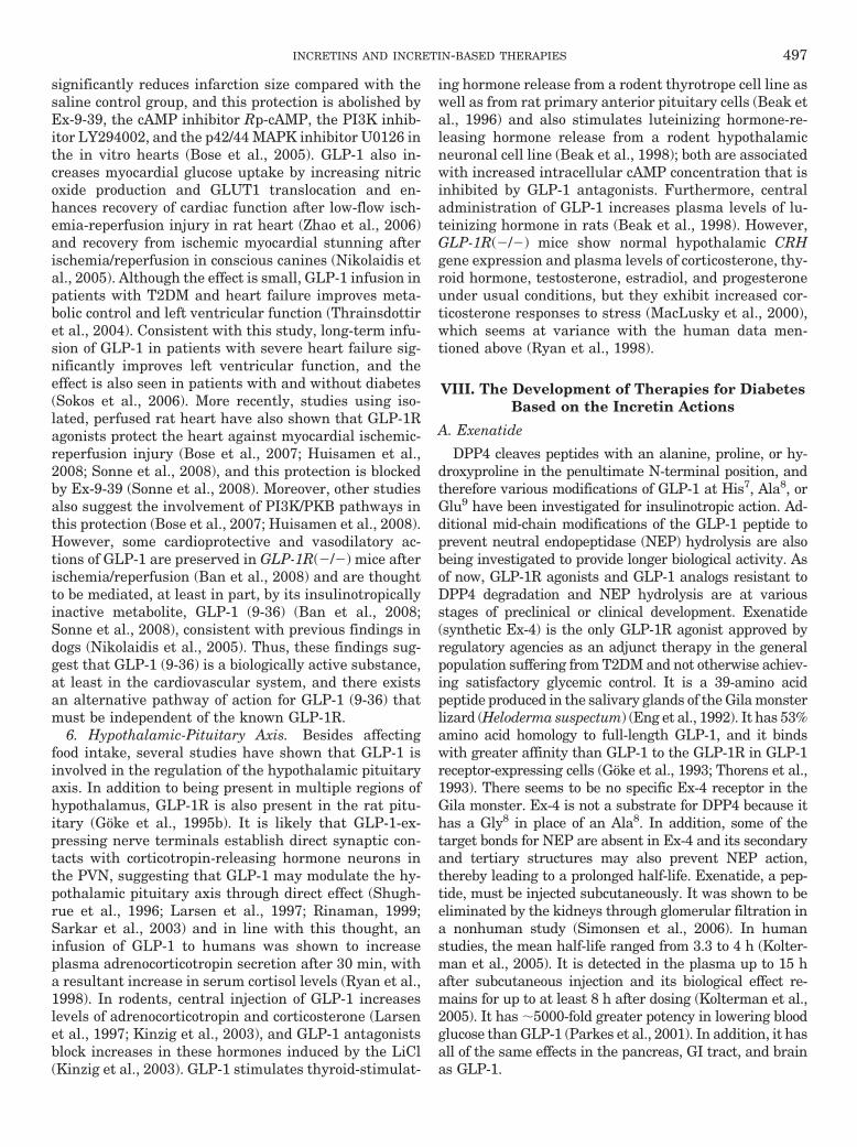
significantly reduces infarction size compared with thesaline control group, and this protection is abolished byEx-9-39, the cAMP inhibitor Rp-cAMP, the PI3K inhib-itor LY294002, and the p42/44 MAPK inhibitor U0126 inthe in vitro hearts (Bose et al., 2005). GLP-1 also in-creases myocardial glucose uptake by increasing nitricoxide production and GLUT1 translocation and en-hances recovery of cardiac function after low-flow isch-emia-reperfusion injury in rat heart (Zhao et al., 2006)and recovery from ischemic myocardial stunning afterischemia/reperfusion in conscious canines (Nikolaidis etal., 2005). Although the effect is small, GLP-1 infusion inpatients with T2DM and heart failure improves meta-bolic control and left ventricular function (Thrainsdottiret al., 2004). Consistent with this study, long-term infu-sion of GLP-1 in patients with severe heart failure sig-nificantly improves left ventricular function, and theeffect is also seen in patients with and without diabetes(Sokos et al., 2006). More recently, studies using iso-lated, perfused rat heart have also shown that GLP-1Ragonists protect the heart against myocardial ischemic-reperfusion injury (Bose et al., 2007; Huisamen et al.,2008; Sonne et al., 2008), and this protection is blockedby Ex-9-39 (Sonne et al., 2008). Moreover, other studiesalso suggest the involvement of PI3K/PKB pathways inthis protection (Bose et al., 2007; Huisamen et al., 2008).However, some cardioprotective and vasodilatory ac-tions of GLP-1 are preserved in GLP-1R(�/�) mice afterischemia/reperfusion (Ban et al., 2008) and are thoughtto be mediated, at least in part, by its insulinotropicallyinactive metabolite, GLP-1 (9-36) (Ban et al., 2008;Sonne et al., 2008), consistent with previous findings indogs (Nikolaidis et al., 2005). Thus, these findings sug-gest that GLP-1 (9-36) is a biologically active substance,at least in the cardiovascular system, and there existsan alternative pathway of action for GLP-1 (9-36) thatmust be independent of the known GLP-1R.
6. Hypothalamic-Pituitary Axis. Besides affectingfood intake, several studies have shown that GLP-1 isinvolved in the regulation of the hypothalamic pituitaryaxis. In addition to being present in multiple regions ofhypothalamus, GLP-1R is also present in the rat pitu-itary (Goke et al., 1995b). It is likely that GLP-1-ex-pressing nerve terminals establish direct synaptic con-tacts with corticotropin-releasing hormone neurons inthe PVN, suggesting that GLP-1 may modulate the hy-pothalamic pituitary axis through direct effect (Shugh-rue et al., 1996; Larsen et al., 1997; Rinaman, 1999;Sarkar et al., 2003) and in line with this thought, aninfusion of GLP-1 to humans was shown to increaseplasma adrenocorticotropin secretion after 30 min, witha resultant increase in serum cortisol levels (Ryan et al.,1998). In rodents, central injection of GLP-1 increaseslevels of adrenocorticotropin and corticosterone (Larsenet al., 1997; Kinzig et al., 2003), and GLP-1 antagonistsblock increases in these hormones induced by the LiCl(Kinzig et al., 2003). GLP-1 stimulates thyroid-stimulat-
ing hormone release from a rodent thyrotrope cell line aswell as from rat primary anterior pituitary cells (Beak etal., 1996) and also stimulates luteinizing hormone-re-leasing hormone release from a rodent hypothalamicneuronal cell line (Beak et al., 1998); both are associatedwith increased intracellular cAMP concentration that isinhibited by GLP-1 antagonists. Furthermore, centraladministration of GLP-1 increases plasma levels of lu-teinizing hormone in rats (Beak et al., 1998). However,GLP-1R(�/�) mice show normal hypothalamic CRHgene expression and plasma levels of corticosterone, thy-roid hormone, testosterone, estradiol, and progesteroneunder usual conditions, but they exhibit increased cor-ticosterone responses to stress (MacLusky et al., 2000),which seems at variance with the human data men-tioned above (Ryan et al., 1998).
VIII. The Development of Therapies for DiabetesBased on the Incretin Actions
A. Exenatide
DPP4 cleaves peptides with an alanine, proline, or hy-droxyproline in the penultimate N-terminal position, andtherefore various modifications of GLP-1 at His7, Ala8, orGlu9 have been investigated for insulinotropic action. Ad-ditional mid-chain modifications of the GLP-1 peptide toprevent neutral endopeptidase (NEP) hydrolysis are alsobeing investigated to provide longer biological activity. Asof now, GLP-1R agonists and GLP-1 analogs resistant toDPP4 degradation and NEP hydrolysis are at variousstages of preclinical or clinical development. Exenatide(synthetic Ex-4) is the only GLP-1R agonist approved byregulatory agencies as an adjunct therapy in the generalpopulation suffering from T2DM and not otherwise achiev-ing satisfactory glycemic control. It is a 39-amino acidpeptide produced in the salivary glands of the Gila monsterlizard (Heloderma suspectum) (Eng et al., 1992). It has 53%amino acid homology to full-length GLP-1, and it bindswith greater affinity than GLP-1 to the GLP-1R in GLP-1receptor-expressing cells (Goke et al., 1993; Thorens et al.,1993). There seems to be no specific Ex-4 receptor in theGila monster. Ex-4 is not a substrate for DPP4 because ithas a Gly8 in place of an Ala8. In addition, some of thetarget bonds for NEP are absent in Ex-4 and its secondaryand tertiary structures may also prevent NEP action,thereby leading to a prolonged half-life. Exenatide, a pep-tide, must be injected subcutaneously. It was shown to beeliminated by the kidneys through glomerular filtration ina nonhuman study (Simonsen et al., 2006). In humanstudies, the mean half-life ranged from 3.3 to 4 h (Kolter-man et al., 2005). It is detected in the plasma up to 15 hafter subcutaneous injection and its biological effect re-mains for up to at least 8 h after dosing (Kolterman et al.,2005). It has �5000-fold greater potency in lowering bloodglucose than GLP-1 (Parkes et al., 2001). In addition, it hasall of the same effects in the pancreas, GI tract, and brainas GLP-1.
INCRETINS AND INCRETIN-BASED THERAPIES 497
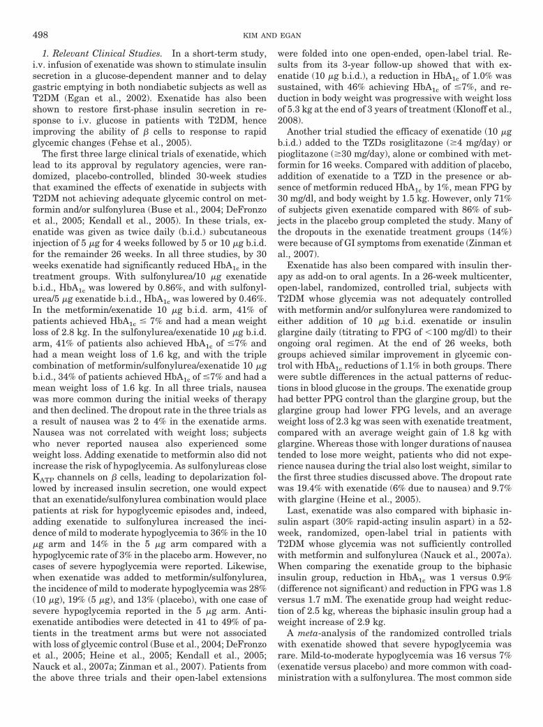
1. Relevant Clinical Studies. In a short-term study,i.v. infusion of exenatide was shown to stimulate insulinsecretion in a glucose-dependent manner and to delaygastric emptying in both nondiabetic subjects as well asT2DM (Egan et al., 2002). Exenatide has also beenshown to restore first-phase insulin secretion in re-sponse to i.v. glucose in patients with T2DM, henceimproving the ability of � cells to response to rapidglycemic changes (Fehse et al., 2005).
The first three large clinical trials of exenatide, whichlead to its approval by regulatory agencies, were ran-domized, placebo-controlled, blinded 30-week studiesthat examined the effects of exenatide in subjects withT2DM not achieving adequate glycemic control on met-formin and/or sulfonylurea (Buse et al., 2004; DeFronzoet al., 2005; Kendall et al., 2005). In these trials, ex-enatide was given as twice daily (b.i.d.) subcutaneousinjection of 5 �g for 4 weeks followed by 5 or 10 �g b.i.d.for the remainder 26 weeks. In all three studies, by 30weeks exenatide had significantly reduced HbA1c in thetreatment groups. With sulfonylurea/10 �g exenatideb.i.d., HbA1c was lowered by 0.86%, and with sulfonyl-urea/5 �g exenatide b.i.d., HbA1c was lowered by 0.46%.In the metformin/exenatide 10 �g b.i.d. arm, 41% ofpatients achieved HbA1c � 7% and had a mean weightloss of 2.8 kg. In the sulfonylurea/exenatide 10 �g b.i.d.arm, 41% of patients also achieved HbA1c of �7% andhad a mean weight loss of 1.6 kg, and with the triplecombination of metformin/sulfonylurea/exenatide 10 �gb.i.d., 34% of patients achieved HbA1c of �7% and had amean weight loss of 1.6 kg. In all three trials, nauseawas more common during the initial weeks of therapyand then declined. The dropout rate in the three trials asa result of nausea was 2 to 4% in the exenatide arms.Nausea was not correlated with weight loss; subjectswho never reported nausea also experienced someweight loss. Adding exenatide to metformin also did notincrease the risk of hypoglycemia. As sulfonylureas closeKATP channels on � cells, leading to depolarization fol-lowed by increased insulin secretion, one would expectthat an exenatide/sulfonylurea combination would placepatients at risk for hypoglycemic episodes and, indeed,adding exenatide to sulfonylurea increased the inci-dence of mild to moderate hypoglycemia to 36% in the 10�g arm and 14% in the 5 �g arm compared with ahypoglycemic rate of 3% in the placebo arm. However, nocases of severe hypoglycemia were reported. Likewise,when exenatide was added to metformin/sulfonylurea,the incidence of mild to moderate hypoglycemia was 28%(10 �g), 19% (5 �g), and 13% (placebo), with one case ofsevere hypoglycemia reported in the 5 �g arm. Anti-exenatide antibodies were detected in 41 to 49% of pa-tients in the treatment arms but were not associatedwith loss of glycemic control (Buse et al., 2004; DeFronzoet al., 2005; Heine et al., 2005; Kendall et al., 2005;Nauck et al., 2007a; Zinman et al., 2007). Patients fromthe above three trials and their open-label extensions
were folded into one open-ended, open-label trial. Re-sults from its 3-year follow-up showed that with ex-enatide (10 �g b.i.d.), a reduction in HbA1c of 1.0% wassustained, with 46% achieving HbA1c of �7%, and re-duction in body weight was progressive with weight lossof 5.3 kg at the end of 3 years of treatment (Klonoff et al.,2008).
Another trial studied the efficacy of exenatide (10 �gb.i.d.) added to the TZDs rosiglitazone (4 mg/day) orpioglitazone (30 mg/day), alone or combined with met-formin for 16 weeks. Compared with addition of placebo,addition of exenatide to a TZD in the presence or ab-sence of metformin reduced HbA1c by 1%, mean FPG by30 mg/dl, and body weight by 1.5 kg. However, only 71%of subjects given exenatide compared with 86% of sub-jects in the placebo group completed the study. Many ofthe dropouts in the exenatide treatment groups (14%)were because of GI symptoms from exenatide (Zinman etal., 2007).
Exenatide has also been compared with insulin ther-apy as add-on to oral agents. In a 26-week multicenter,open-label, randomized, controlled trial, subjects withT2DM whose glycemia was not adequately controlledwith metformin and/or sulfonylurea were randomized toeither addition of 10 �g b.i.d. exenatide or insulinglargine daily (titrating to FPG of �100 mg/dl) to theirongoing oral regimen. At the end of 26 weeks, bothgroups achieved similar improvement in glycemic con-trol with HbA1c reductions of 1.1% in both groups. Therewere subtle differences in the actual patterns of reduc-tions in blood glucose in the groups. The exenatide grouphad better PPG control than the glargine group, but theglargine group had lower FPG levels, and an averageweight loss of 2.3 kg was seen with exenatide treatment,compared with an average weight gain of 1.8 kg withglargine. Whereas those with longer durations of nauseatended to lose more weight, patients who did not expe-rience nausea during the trial also lost weight, similar tothe first three studies discussed above. The dropout ratewas 19.4% with exenatide (6% due to nausea) and 9.7%with glargine (Heine et al., 2005).
Last, exenatide was also compared with biphasic in-sulin aspart (30% rapid-acting insulin aspart) in a 52-week, randomized, open-label trial in patients withT2DM whose glycemia was not sufficiently controlledwith metformin and sulfonylurea (Nauck et al., 2007a).When comparing the exenatide group to the biphasicinsulin group, reduction in HbA1c was 1 versus 0.9%(difference not significant) and reduction in FPG was 1.8versus 1.7 mM. The exenatide group had weight reduc-tion of 2.5 kg, whereas the biphasic insulin group had aweight increase of 2.9 kg.
A meta-analysis of the randomized controlled trialswith exenatide showed that severe hypoglycemia wasrare. Mild-to-moderate hypoglycemia was 16 versus 7%(exenatide versus placebo) and more common with coad-ministration with a sulfonylurea. The most common side
498 KIM AND EGAN
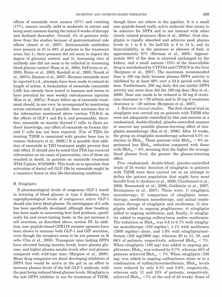
effects of exenatide were nausea (57%) and vomiting(17%), nausea usually mild to moderate in nature andbeing most common during the initial 8 weeks of therapyand declined thereafter. Overall, 4% of patients with-drew from the studies because of gastrointestinal sideeffects (Amori et al., 2007). Antiexenatide antibodieswere present in 41 to 49% of patients in the treatmentarms, but 1), their presence did not seem to impact thedegree of glycemic control, and 2), increasing titer ofantibody also did not seem to be reflected in worseningblood glucose control (Buse et al., 2004; DeFronzo et al.,2005; Heine et al., 2005; Kendall et al., 2005; Nauck etal., 2007a; Zinman et al., 2007). Because exenatide mustbe injected b.i.d., attempts have been made to extend itslength of action. A formulation of exenatide (exenatideLAR) has already been tested in humans and seems tohave potential for once weekly subcutaneous injection(Kim et al., 2007a). Future follow-up of exenatide treat-ment should, in our view, be accompanied by monitoringserum calcitonin and, if possible, bone outcomes due tothe information mentioned above (section VII.B.4) onthe effects of GLP-1 and Ex-4, and presumably, there-fore, exenatide on murine thyroid C cells: to these au-thors’ knowledge, any effect of exenatide on human thy-roid C cells has not been reported. [Use of TZDs fortreating T2DM is associated with greater bone loss inwomen (Schwartz et al., 2006).] It is possible that addi-tion of exenatide to TZD treatment might prevent thatside effect. It should also be noted that FDA has receivedinformation on six cases of pancreatitis, two of those sixresulted in death, in patients on exenatide treatment(FDA Update, 8/18/2008). This leads us to speculate thatactivation of ductal cell GLP-1Rs by exenatide might bea causative factor in this life-threatening condition.
B. Sitagliptin
If pharmacological levels of exogenous GLP-1 resultin lowering of blood glucose in type 2 diabetes, thensupraphysiological levels of endogenous active GLP-1should also lower blood glucose. No secretagogue of L cellshas been specifically developed, although clear headwayhas been made in uncovering how food products, specif-ically fat and sweet-tasting foods, in the gut increase Lcell secretion, as described above, section III. In addi-tion, non–peptide-based GPR119 receptor agonists havebeen shown to increase both GLP-1 and GIP secretion,even though the receptors seem to be not present on Kcells (Chu et al., 2008). Transgenic mice lacking DPP4have elevated fasting incretin levels, lower plasma glu-cose, and higher plasma insulin levels after oral glucosecompared with wild-type mice (Marguet et al., 2000).Many drug companies set about developing inhibitors ofDPP4 that would be active in the gut in an effort toincrease plasma levels of the full GLP-1 molecule, withthe goal being reduced blood glucose levels. Sitagliptin isthe sole DPP4 inhibitor in use for treatment of T2DM,
though there are others in the pipeline. It is a smallnon–peptide-based orally active molecule that seems tobe selective for DPP4 and to not interact with otherclosely related proteases (Kim et al., 2005a). Oral sita-gliptin is rapidly absorbed and achieves peak plasmalevels in 1 to 6 h. Its half-life is 8 to 14 h, and itsbioavailability, in the presence or absence of food, isapproximately 87% (Herman et al., 2005). Approxi-mately 80% of the dose is excreted unchanged by thekidney, and a small amount (15%) of the bioavailabledrug is metabolized by CYP3A4 and CYP2C8 in the liver(Bergman et al., 2007). The maximum recommendeddose is 100 mg daily because plasma DPP4 activity isinhibited by at least 80% over a 24-h period with thisdose. Furthermore, 200 mg daily did not inhibit DPP4activity any more than did the 100-mg dose (Raz et al.,2006). Dose size needs to be reduced to 50 mg if creati-nine clearance is �50 ml/min and to 25 mg if creatinineclearance is �30 ml/min (Bergman et al., 2007).
1. Relevant clinical studies. The first clinical trial onsitagliptin was carried out on patients with T2DM whowere not adequately controlled by diet and exercise in arandomized, double-blinded, placebo-controlled mannerto uncover any possible glucose-lowering effect of sita-gliptin monotherapy (Raz et al., 2006). After 18 weeks,the group on sitagliptin monotherapy achieved 0.5% re-duction in HbA1c. Those patients with HbA1c � 8% ex-perienced less HbA1c reduction compared with thosewith HbA1c � 8%, meaning that the higher the averageblood glucose level, the greater the glucose-loweringeffect.
Five randomized, double-blind, placebo-controlledtrials of 24 weeks’ duration of sitagliptin in patientswith T2DM were then carried out in an attempt todefine the patient population that might have mostbenefit from it (Aschner et al., 2006; Charbonnel et al.,2006; Rosenstock et al., 2006; Goldstein et al., 2007;Hermansen et al., 2007). These were: 1) sitagliptinmonotherapy; 2) comparison of sitagliptin mono-therapy, metformin monotherapy, and initial combi-nation therapy of sitagliptin and metformin; 3) sita-gliptin added to ongoing pioglitazone; 4) sitagliptinadded to ongoing metformin; and, finally, 5) sitaglip-tin added to ongoing sulfonylurea and/or metformin.The reduction in HbA1c was 0.6 to 0.7% with sitaglip-tin monotherapy (100 mg/day), 1.1% with metformin(2000 mg/day) alone, and 1.9% with sitagliptin/met-formin (100 mg/2000 mg), whereas 20 to 41, 38, and66% of patients, respectively, achieved HbA1c � 7%.When sitagliptin (100 mg) was added to ongoing pio-glitazone, HbA1c was reduced by 0.7%, whereas 45% ofpatients achieved HbA1c � 7%. When sitagliptin (100mg) was added to ongoing sulfonylurea alone or to acombination of sulfonylurea/metformin, HbA1c levelswere reduced by only 0.3% and 0.6%, respectively,whereas only 11 and 23% of patients, respectively,achieved HbA1c �7% at the end of 24 weeks. Some of
INCRETINS AND INCRETIN-BASED THERAPIES 499
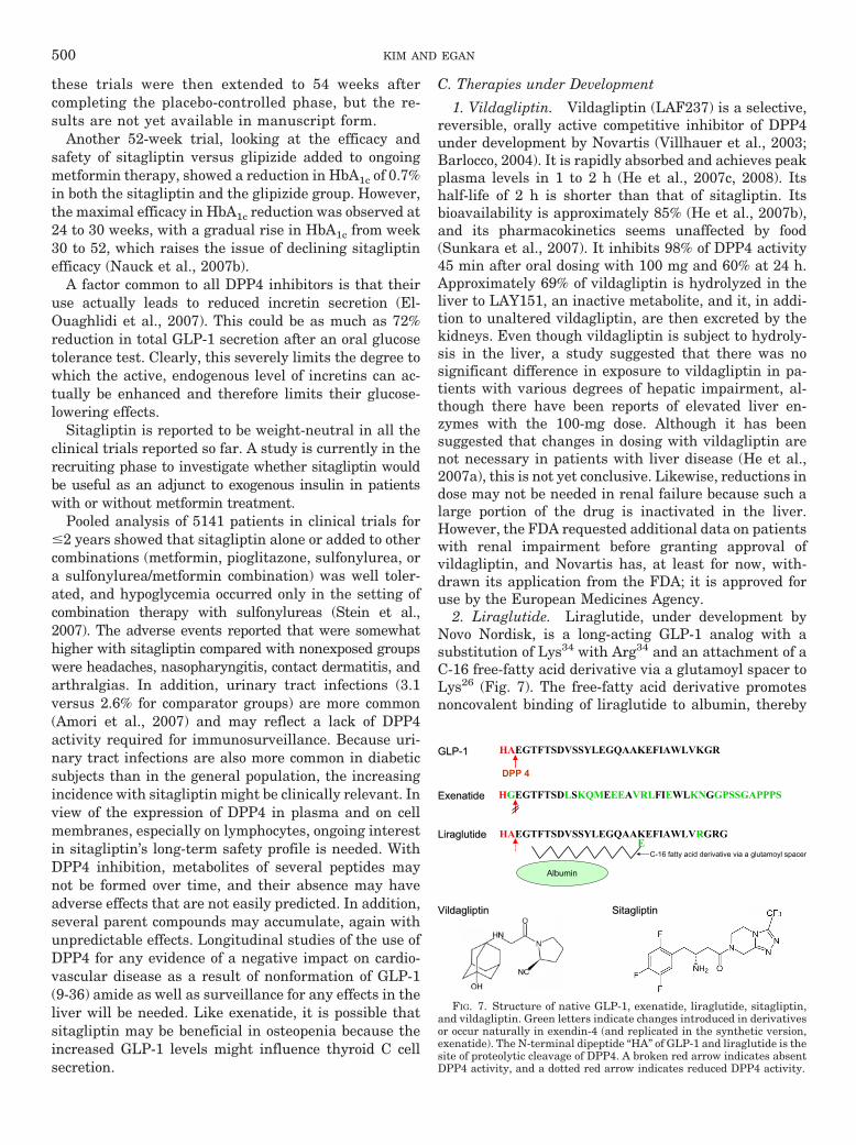
these trials were then extended to 54 weeks aftercompleting the placebo-controlled phase, but the re-sults are not yet available in manuscript form.
Another 52-week trial, looking at the efficacy andsafety of sitagliptin versus glipizide added to ongoingmetformin therapy, showed a reduction in HbA1c of 0.7%in both the sitagliptin and the glipizide group. However,the maximal efficacy in HbA1c reduction was observed at24 to 30 weeks, with a gradual rise in HbA1c from week30 to 52, which raises the issue of declining sitagliptinefficacy (Nauck et al., 2007b).
A factor common to all DPP4 inhibitors is that theiruse actually leads to reduced incretin secretion (El-Ouaghlidi et al., 2007). This could be as much as 72%reduction in total GLP-1 secretion after an oral glucosetolerance test. Clearly, this severely limits the degree towhich the active, endogenous level of incretins can ac-tually be enhanced and therefore limits their glucose-lowering effects.
Sitagliptin is reported to be weight-neutral in all theclinical trials reported so far. A study is currently in therecruiting phase to investigate whether sitagliptin wouldbe useful as an adjunct to exogenous insulin in patientswith or without metformin treatment.
Pooled analysis of 5141 patients in clinical trials for�2 years showed that sitagliptin alone or added to othercombinations (metformin, pioglitazone, sulfonylurea, ora sulfonylurea/metformin combination) was well toler-ated, and hypoglycemia occurred only in the setting ofcombination therapy with sulfonylureas (Stein et al.,2007). The adverse events reported that were somewhathigher with sitagliptin compared with nonexposed groupswere headaches, nasopharyngitis, contact dermatitis, andarthralgias. In addition, urinary tract infections (3.1versus 2.6% for comparator groups) are more common(Amori et al., 2007) and may reflect a lack of DPP4activity required for immunosurveillance. Because uri-nary tract infections are also more common in diabeticsubjects than in the general population, the increasingincidence with sitagliptin might be clinically relevant. Inview of the expression of DPP4 in plasma and on cellmembranes, especially on lymphocytes, ongoing interestin sitagliptin’s long-term safety profile is needed. WithDPP4 inhibition, metabolites of several peptides maynot be formed over time, and their absence may haveadverse effects that are not easily predicted. In addition,several parent compounds may accumulate, again withunpredictable effects. Longitudinal studies of the use ofDPP4 for any evidence of a negative impact on cardio-vascular disease as a result of nonformation of GLP-1(9-36) amide as well as surveillance for any effects in theliver will be needed. Like exenatide, it is possible thatsitagliptin may be beneficial in osteopenia because theincreased GLP-1 levels might influence thyroid C cellsecretion.
C. Therapies under Development
1. Vildagliptin. Vildagliptin (LAF237) is a selective,reversible, orally active competitive inhibitor of DPP4under development by Novartis (Villhauer et al., 2003;Barlocco, 2004). It is rapidly absorbed and achieves peakplasma levels in 1 to 2 h (He et al., 2007c, 2008). Itshalf-life of 2 h is shorter than that of sitagliptin. Itsbioavailability is approximately 85% (He et al., 2007b),and its pharmacokinetics seems unaffected by food(Sunkara et al., 2007). It inhibits 98% of DPP4 activity45 min after oral dosing with 100 mg and 60% at 24 h.Approximately 69% of vildagliptin is hydrolyzed in theliver to LAY151, an inactive metabolite, and it, in addi-tion to unaltered vildagliptin, are then excreted by thekidneys. Even though vildagliptin is subject to hydroly-sis in the liver, a study suggested that there was nosignificant difference in exposure to vildagliptin in pa-tients with various degrees of hepatic impairment, al-though there have been reports of elevated liver en-zymes with the 100-mg dose. Although it has beensuggested that changes in dosing with vildagliptin arenot necessary in patients with liver disease (He et al.,2007a), this is not yet conclusive. Likewise, reductions indose may not be needed in renal failure because such alarge portion of the drug is inactivated in the liver.However, the FDA requested additional data on patientswith renal impairment before granting approval ofvildagliptin, and Novartis has, at least for now, with-drawn its application from the FDA; it is approved foruse by the European Medicines Agency.
2. Liraglutide. Liraglutide, under development byNovo Nordisk, is a long-acting GLP-1 analog with asubstitution of Lys34 with Arg34 and an attachment of aC-16 free-fatty acid derivative via a glutamoyl spacer toLys26 (Fig. 7). The free-fatty acid derivative promotesnoncovalent binding of liraglutide to albumin, thereby
HAEGTFTSDVSSYLEGQAAKEFIAWLVKGRGLPGLP--11
HGEGTFTSDLSKQMEEEAVRLFIEWLKNGGPSSGAPPPSExenatideExenatide
VildagliptinVildagliptin SitagliptinSitagliptin
LiraglutideLiraglutide HAEGTFTSDVSSYLEGQAAKEFIAWLVRGRGE
C-16 fatty acid derivative via a glutamoyl spacer
Albumin
DPP 4
FIG. 7. Structure of native GLP-1, exenatide, liraglutide, sitagliptin,and vildagliptin. Green letters indicate changes introduced in derivativesor occur naturally in exendin-4 (and replicated in the synthetic version,exenatide). The N-terminal dipeptide “HA” of GLP-1 and liraglutide is thesite of proteolytic cleavage of DPP4. A broken red arrow indicates absentDPP4 activity, and a dotted red arrow indicates reduced DPP4 activity.
500 KIM AND EGAN
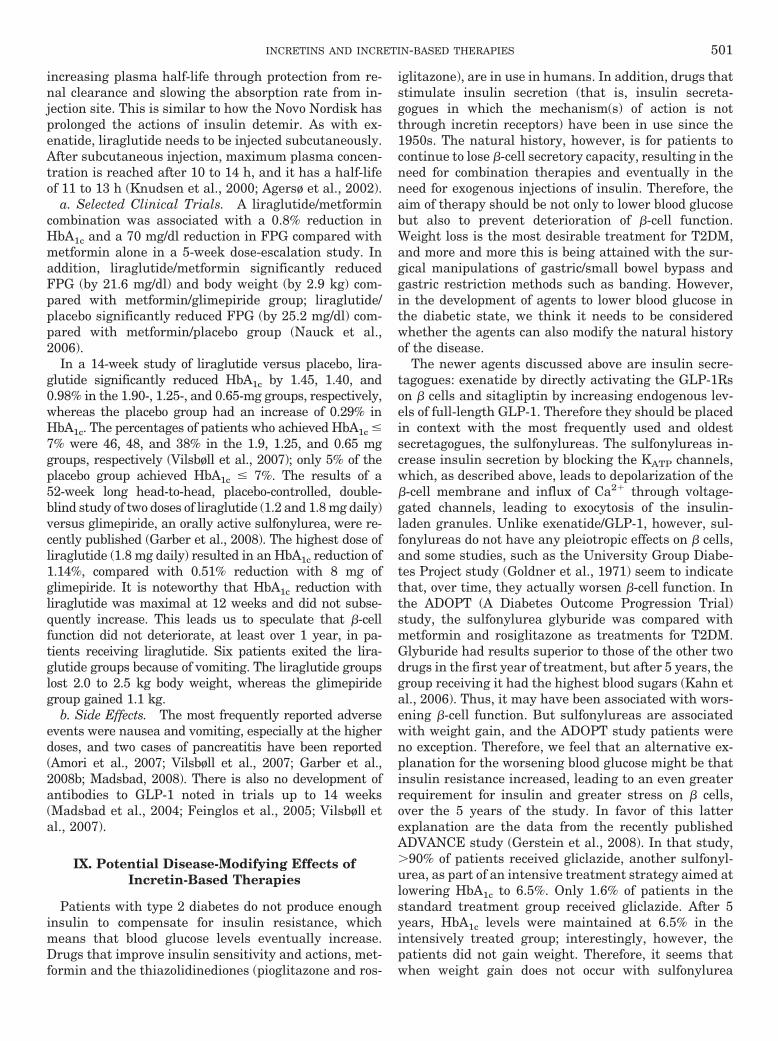
increasing plasma half-life through protection from re-nal clearance and slowing the absorption rate from in-jection site. This is similar to how the Novo Nordisk hasprolonged the actions of insulin detemir. As with ex-enatide, liraglutide needs to be injected subcutaneously.After subcutaneous injection, maximum plasma concen-tration is reached after 10 to 14 h, and it has a half-lifeof 11 to 13 h (Knudsen et al., 2000; Agersø et al., 2002).
a. Selected Clinical Trials. A liraglutide/metformincombination was associated with a 0.8% reduction inHbA1c and a 70 mg/dl reduction in FPG compared withmetformin alone in a 5-week dose-escalation study. Inaddition, liraglutide/metformin significantly reducedFPG (by 21.6 mg/dl) and body weight (by 2.9 kg) com-pared with metformin/glimepiride group; liraglutide/placebo significantly reduced FPG (by 25.2 mg/dl) com-pared with metformin/placebo group (Nauck et al.,2006).
In a 14-week study of liraglutide versus placebo, lira-glutide significantly reduced HbA1c by 1.45, 1.40, and0.98% in the 1.90-, 1.25-, and 0.65-mg groups, respectively,whereas the placebo group had an increase of 0.29% inHbA1c. The percentages of patients who achieved HbA1c �7% were 46, 48, and 38% in the 1.9, 1.25, and 0.65 mggroups, respectively (Vilsbøll et al., 2007); only 5% of theplacebo group achieved HbA1c � 7%. The results of a52-week long head-to-head, placebo-controlled, double-blind study of two doses of liraglutide (1.2 and 1.8 mg daily)versus glimepiride, an orally active sulfonylurea, were re-cently published (Garber et al., 2008). The highest dose ofliraglutide (1.8 mg daily) resulted in an HbA1c reduction of1.14%, compared with 0.51% reduction with 8 mg ofglimepiride. It is noteworthy that HbA1c reduction withliraglutide was maximal at 12 weeks and did not subse-quently increase. This leads us to speculate that �-cellfunction did not deteriorate, at least over 1 year, in pa-tients receiving liraglutide. Six patients exited the lira-glutide groups because of vomiting. The liraglutide groupslost 2.0 to 2.5 kg body weight, whereas the glimepiridegroup gained 1.1 kg.
b. Side Effects. The most frequently reported adverseevents were nausea and vomiting, especially at the higherdoses, and two cases of pancreatitis have been reported(Amori et al., 2007; Vilsbøll et al., 2007; Garber et al.,2008b; Madsbad, 2008). There is also no development ofantibodies to GLP-1 noted in trials up to 14 weeks(Madsbad et al., 2004; Feinglos et al., 2005; Vilsbøll etal., 2007).
IX. Potential Disease-Modifying Effects ofIncretin-Based Therapies
Patients with type 2 diabetes do not produce enoughinsulin to compensate for insulin resistance, whichmeans that blood glucose levels eventually increase.Drugs that improve insulin sensitivity and actions, met-formin and the thiazolidinediones (pioglitazone and ros-
iglitazone), are in use in humans. In addition, drugs thatstimulate insulin secretion (that is, insulin secreta-gogues in which the mechanism(s) of action is notthrough incretin receptors) have been in use since the1950s. The natural history, however, is for patients tocontinue to lose �-cell secretory capacity, resulting in theneed for combination therapies and eventually in theneed for exogenous injections of insulin. Therefore, theaim of therapy should be not only to lower blood glucosebut also to prevent deterioration of �-cell function.Weight loss is the most desirable treatment for T2DM,and more and more this is being attained with the sur-gical manipulations of gastric/small bowel bypass andgastric restriction methods such as banding. However,in the development of agents to lower blood glucose inthe diabetic state, we think it needs to be consideredwhether the agents can also modify the natural historyof the disease.
The newer agents discussed above are insulin secre-tagogues: exenatide by directly activating the GLP-1Rson � cells and sitagliptin by increasing endogenous lev-els of full-length GLP-1. Therefore they should be placedin context with the most frequently used and oldestsecretagogues, the sulfonylureas. The sulfonylureas in-crease insulin secretion by blocking the KATP channels,which, as described above, leads to depolarization of the�-cell membrane and influx of Ca2� through voltage-gated channels, leading to exocytosis of the insulin-laden granules. Unlike exenatide/GLP-1, however, sul-fonylureas do not have any pleiotropic effects on � cells,and some studies, such as the University Group Diabe-tes Project study (Goldner et al., 1971) seem to indicatethat, over time, they actually worsen �-cell function. Inthe ADOPT (A Diabetes Outcome Progression Trial)study, the sulfonylurea glyburide was compared withmetformin and rosiglitazone as treatments for T2DM.Glyburide had results superior to those of the other twodrugs in the first year of treatment, but after 5 years, thegroup receiving it had the highest blood sugars (Kahn etal., 2006). Thus, it may have been associated with wors-ening �-cell function. But sulfonylureas are associatedwith weight gain, and the ADOPT study patients wereno exception. Therefore, we feel that an alternative ex-planation for the worsening blood glucose might be thatinsulin resistance increased, leading to an even greaterrequirement for insulin and greater stress on � cells,over the 5 years of the study. In favor of this latterexplanation are the data from the recently publishedADVANCE study (Gerstein et al., 2008). In that study,�90% of patients received gliclazide, another sulfonyl-urea, as part of an intensive treatment strategy aimed atlowering HbA1c to 6.5%. Only 1.6% of patients in thestandard treatment group received gliclazide. After 5years, HbA1c levels were maintained at 6.5% in theintensively treated group; interestingly, however, thepatients did not gain weight. Therefore, it seems thatwhen weight gain does not occur with sulfonylurea
INCRETINS AND INCRETIN-BASED THERAPIES 501
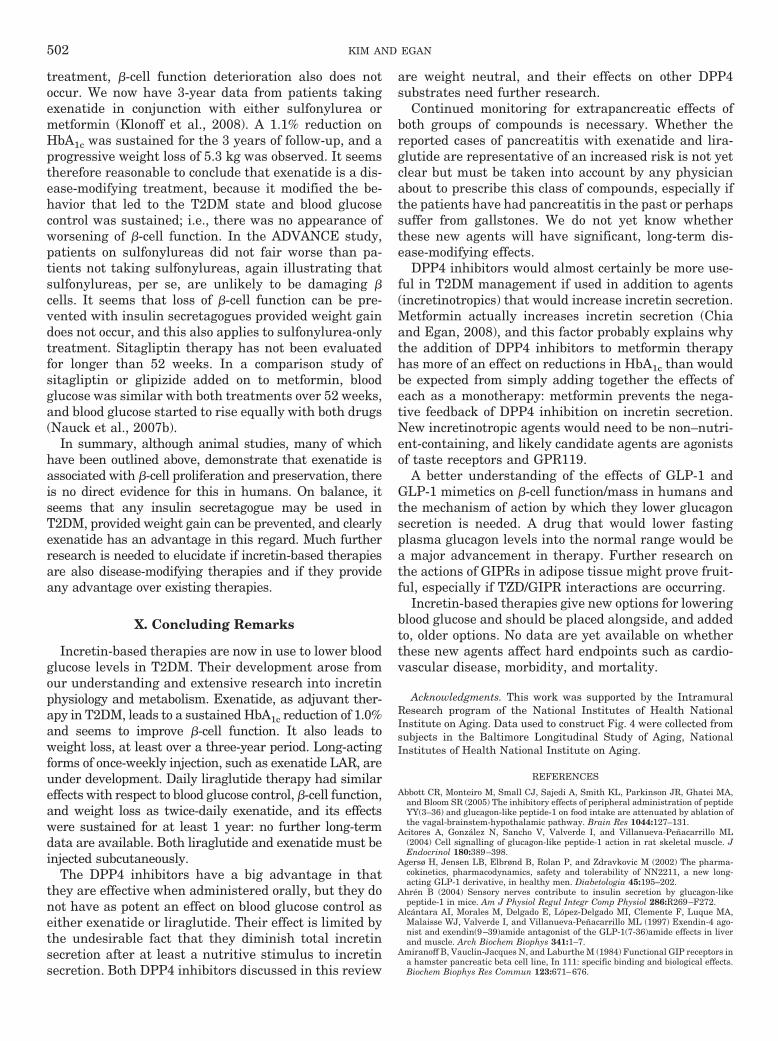
treatment, �-cell function deterioration also does notoccur. We now have 3-year data from patients takingexenatide in conjunction with either sulfonylurea ormetformin (Klonoff et al., 2008). A 1.1% reduction onHbA1c was sustained for the 3 years of follow-up, and aprogressive weight loss of 5.3 kg was observed. It seemstherefore reasonable to conclude that exenatide is a dis-ease-modifying treatment, because it modified the be-havior that led to the T2DM state and blood glucosecontrol was sustained; i.e., there was no appearance ofworsening of �-cell function. In the ADVANCE study,patients on sulfonylureas did not fair worse than pa-tients not taking sulfonylureas, again illustrating thatsulfonylureas, per se, are unlikely to be damaging �cells. It seems that loss of �-cell function can be pre-vented with insulin secretagogues provided weight gaindoes not occur, and this also applies to sulfonylurea-onlytreatment. Sitagliptin therapy has not been evaluatedfor longer than 52 weeks. In a comparison study ofsitagliptin or glipizide added on to metformin, bloodglucose was similar with both treatments over 52 weeks,and blood glucose started to rise equally with both drugs(Nauck et al., 2007b).
In summary, although animal studies, many of whichhave been outlined above, demonstrate that exenatide isassociated with �-cell proliferation and preservation, thereis no direct evidence for this in humans. On balance, itseems that any insulin secretagogue may be used inT2DM, provided weight gain can be prevented, and clearlyexenatide has an advantage in this regard. Much furtherresearch is needed to elucidate if incretin-based therapiesare also disease-modifying therapies and if they provideany advantage over existing therapies.
X. Concluding Remarks
Incretin-based therapies are now in use to lower bloodglucose levels in T2DM. Their development arose fromour understanding and extensive research into incretinphysiology and metabolism. Exenatide, as adjuvant ther-apy in T2DM, leads to a sustained HbA1c reduction of 1.0%and seems to improve �-cell function. It also leads toweight loss, at least over a three-year period. Long-actingforms of once-weekly injection, such as exenatide LAR, areunder development. Daily liraglutide therapy had similareffects with respect to blood glucose control, �-cell function,and weight loss as twice-daily exenatide, and its effectswere sustained for at least 1 year: no further long-termdata are available. Both liraglutide and exenatide must beinjected subcutaneously.
The DPP4 inhibitors have a big advantage in thatthey are effective when administered orally, but they donot have as potent an effect on blood glucose control aseither exenatide or liraglutide. Their effect is limited bythe undesirable fact that they diminish total incretinsecretion after at least a nutritive stimulus to incretinsecretion. Both DPP4 inhibitors discussed in this review
are weight neutral, and their effects on other DPP4substrates need further research.
Continued monitoring for extrapancreatic effects ofboth groups of compounds is necessary. Whether thereported cases of pancreatitis with exenatide and lira-glutide are representative of an increased risk is not yetclear but must be taken into account by any physicianabout to prescribe this class of compounds, especially ifthe patients have had pancreatitis in the past or perhapssuffer from gallstones. We do not yet know whetherthese new agents will have significant, long-term dis-ease-modifying effects.
DPP4 inhibitors would almost certainly be more use-ful in T2DM management if used in addition to agents(incretinotropics) that would increase incretin secretion.Metformin actually increases incretin secretion (Chiaand Egan, 2008), and this factor probably explains whythe addition of DPP4 inhibitors to metformin therapyhas more of an effect on reductions in HbA1c than wouldbe expected from simply adding together the effects ofeach as a monotherapy: metformin prevents the nega-tive feedback of DPP4 inhibition on incretin secretion.New incretinotropic agents would need to be non–nutri-ent-containing, and likely candidate agents are agonistsof taste receptors and GPR119.
A better understanding of the effects of GLP-1 andGLP-1 mimetics on �-cell function/mass in humans andthe mechanism of action by which they lower glucagonsecretion is needed. A drug that would lower fastingplasma glucagon levels into the normal range would bea major advancement in therapy. Further research onthe actions of GIPRs in adipose tissue might prove fruit-ful, especially if TZD/GIPR interactions are occurring.
Incretin-based therapies give new options for loweringblood glucose and should be placed alongside, and addedto, older options. No data are yet available on whetherthese new agents affect hard endpoints such as cardio-vascular disease, morbidity, and mortality.
Acknowledgments. This work was supported by the IntramuralResearch program of the National Institutes of Health NationalInstitute on Aging. Data used to construct Fig. 4 were collected fromsubjects in the Baltimore Longitudinal Study of Aging, NationalInstitutes of Health National Institute on Aging.
REFERENCES
Abbott CR, Monteiro M, Small CJ, Sajedi A, Smith KL, Parkinson JR, Ghatei MA,and Bloom SR (2005) The inhibitory effects of peripheral administration of peptideYY(3–36) and glucagon-like peptide-1 on food intake are attenuated by ablation ofthe vagal-brainstem-hypothalamic pathway. Brain Res 1044:127–131.
Acitores A, Gonzalez N, Sancho V, Valverde I, and Villanueva-Penacarrillo ML(2004) Cell signalling of glucagon-like peptide-1 action in rat skeletal muscle. JEndocrinol 180:389–398.
Agersø H, Jensen LB, Elbrønd B, Rolan P, and Zdravkovic M (2002) The pharma-cokinetics, pharmacodynamics, safety and tolerability of NN2211, a new long-acting GLP-1 derivative, in healthy men. Diabetologia 45:195–202.
Ahren B (2004) Sensory nerves contribute to insulin secretion by glucagon-likepeptide-1 in mice. Am J Physiol Regul Integr Comp Physiol 286:R269–F272.
Alcantara AI, Morales M, Delgado E, Lopez-Delgado MI, Clemente F, Luque MA,Malaisse WJ, Valverde I, and Villanueva-Penacarrillo ML (1997) Exendin-4 ago-nist and exendin(9–39)amide antagonist of the GLP-1(7-36)amide effects in liverand muscle. Arch Biochem Biophys 341:1–7.
Amiranoff B, Vauclin-Jacques N, and Laburthe M (1984) Functional GIP receptors ina hamster pancreatic beta cell line, In 111: specific binding and biological effects.Biochem Biophys Res Commun 123:671–676.
502 KIM AND EGAN
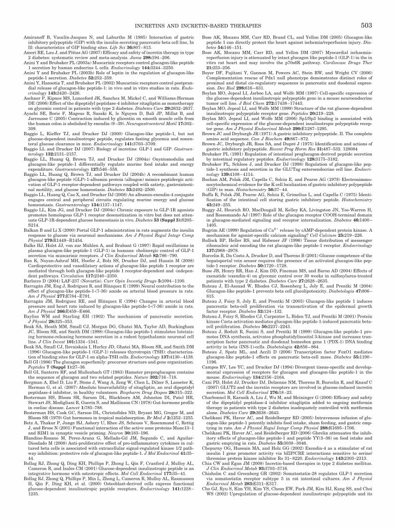
Amiranoff B, Vauclin-Jacques N, and Laburthe M (1985) Interaction of gastricinhibitory polypeptide (GIP) with the insulin-secreting pancreatic beta cell line, Inlll: characteristics of GIP binding sites. Life Sci 36:807–813.
Amori RE, Lau J, and Pittas AG (2007) Efficacy and safety of incretin therapy in type2 diabetes: systematic review and meta-analysis. Jama 298:194–206.
Anini Y and Brubaker PL (2003a) Muscarinic receptors control glucagon-like peptide1 secretion by human endocrine L cells. Endocrinology 144:3244–3250.
Anini Y and Brubaker PL (2003b) Role of leptin in the regulation of glucagon-likepeptide-1 secretion. Diabetes 52:252–259.
Anini Y, Hansotia T, and Brubaker PL (2002) Muscarinic receptors control postpran-dial release of glucagon-like peptide-1: in vivo and in vitro studies in rats. Endo-crinology 143:2420–2426.
Aschner P, Kipnes MS, Lunceford JK, Sanchez M, Mickel C, and Williams-HermanDE (2006) Effect of the dipeptidyl peptidase-4 inhibitor sitagliptin as monotherapyon glycemic control in patients with type 2 diabetes. Diabetes Care 29:2632–2637.
Ayachi SE, Borie F, Magous R, Sasaki K, le Nguyen D, Bali JP, Millat B, andJarrousse C (2005) Contraction induced by glicentin on smooth muscle cells fromthe human colon is abolished by exendin (9–39). Neurogastroenterol Motil 17:302–309.
Baggio L, Kieffer TJ, and Drucker DJ (2000) Glucagon-like peptide-1, but notglucose-dependent insulinotropic peptide, regulates fasting glycemia and nonen-teral glucose clearance in mice. Endocrinology 141:3703–3709.
Baggio LL and Drucker DJ (2007) Biology of incretins: GLP-1 and GIP. Gastroen-terology 132:2131–2157.
Baggio LL, Huang Q, Brown TJ, and Drucker DJ (2004a) Oxyntomodulin andglucagon-like peptide-1 differentially regulate murine food intake and energyexpenditure. Gastroenterology 127:546–558.
Baggio LL, Huang Q, Brown TJ, and Drucker DJ (2004b) A recombinant humanglucagon-like peptide (GLP)-1-albumin protein (albugon) mimics peptidergic acti-vation of GLP-1 receptor-dependent pathways coupled with satiety, gastrointesti-nal motility, and glucose homeostasis. Diabetes 53:2492–2500.
Baggio LL, Huang Q, Cao X, and Drucker DJ (2008) An albumin-exendin-4 conjugateengages central and peripheral circuits regulating murine energy and glucosehomeostasis. Gastroenterology 134:1137–1147.
Baggio LL, Kim JG, and Drucker DJ (2004c) Chronic exposure to GLP-1R agonistspromotes homologous GLP-1 receptor desensitization in vitro but does not atten-uate GLP-1R-dependent glucose homeostasis in vivo. Diabetes 53 (Suppl 3):S205–S214.
Balkan B and Li X (2000) Portal GLP-1 administration in rats augments the insulinresponse to glucose via neuronal mechanisms. Am J Physiol Regul Integr CompPhysiol 279:R1449–R1454.
Balks HJ, Holst JJ, von zur Muhlen A, and Brabant G (1997) Rapid oscillations inplasma glucagon-like peptide-1 (GLP-1) in humans: cholinergic control of GLP-1secretion via muscarinic receptors. J Clin Endocrinol Metab 82:786–790.
Ban K, Noyan-Ashraf MH, Hoefer J, Bolz SS, Drucker DJ, and Husain M (2008)Cardioprotective and vasodilatory actions of glucagon-like peptide 1 receptor aremediated through both glucagon-like peptide 1 receptor-dependent and -indepen-dent pathways. Circulation 117:2340–2350.
Barlocco D (2004) LAF-237 (Novartis). Curr Opin Investig Drugs 5:1094–1100.Barragan JM, Eng J, Rodríguez R, and Blazquez E (1999) Neural contribution to the
effect of glucagon-like peptide-1-(7-36) amide on arterial blood pressure in rats.Am J Physiol 277:E784–E791.
Barragan JM, Rodríguez RE, and Blazquez E (1994) Changes in arterial bloodpressure and heart rate induced by glucagon-like peptide-1-(7-36) amide in rats.Am J Physiol 266:E459–E466.
Bayliss WM and Starling EH (1902) The mechanism of pancreatic secretion.J Physiol 28:325–353.
Beak SA, Heath MM, Small CJ, Morgan DG, Ghatei MA, Taylor AD, BuckinghamJC, Bloom SR, and Smith DM (1998) Glucagon-like peptide-1 stimulates luteiniz-ing hormone-releasing hormone secretion in a rodent hypothalamic neuronal cellline. J Clin Invest 101:1334–1341.
Beak SA, Small CJ, Ilovaiskaia I, Hurley JD, Ghatei MA, Bloom SR, and Smith DM(1996) Glucagon-like peptide-1 (GLP-1) releases thyrotropin (TSH): characteriza-tion of binding sites for GLP-1 on alpha-TSH cells. Endocrinology 137:4130–4138.
Bell GI (1986) The glucagon superfamily: precursor structure and gene organization.Peptides 7 (Suppl 1):27–36.
Bell GI, Santerre RF, and Mullenbach GT (1983) Hamster preproglucagon containsthe sequence of glucagon and two related peptides. Nature 302:716–718.
Bergman A, Ebel D, Liu F, Stone J, Wang A, Zeng W, Chen L, Dilzer S, Lasseter K,Herman G, et al. (2007) Absolute bioavailability of sitagliptin, an oral dipeptidylpeptidase-4 inhibitor, in healthy volunteers. Biopharm Drug Dispos 28:315–322.
Besterman HS, Bloom SR, Sarson DL, Blackburn AM, Johnston DI, Patel HR,Stewart JS, Modigliani R, Guerin S, and Mallinson CN (1978) Gut-hormone profilein coeliac disease. Lancet 1:785–788.
Besterman HS, Cook GC, Sarson DL, Christofides ND, Bryant MG, Gregor M, andBloom SR (1979) Gut hormones in tropical malabsorption. Br Med J 2:1252–1255.
Betz A, Thakur P, Junge HJ, Ashery U, Rhee JS, Scheuss V, Rosenmund C, RettigJ, and Brose N (2001) Functional interaction of the active zone proteins Munc13–1and RIM1 in synaptic vesicle priming. Neuron 30:183–196.
Blandino-Rosano M, Perez-Arana G, Mellado-Gil JM, Segundo C, and Aguilar-Diosdado M (2008) Anti-proliferative effect of pro-inflammatory cytokines in cul-tured beta cells is associated with extracellular signal-regulated kinase 1/2 path-way inhibition: protective role of glucagon-like peptide-1. J Mol Endocrinol 41:35–44.
Bollag RJ, Zhong Q, Ding KH, Phillips P, Zhong L, Qin F, Cranford J, Mulloy AL,Cameron R, and Isales CM (2001) Glucose-dependent insulinotropic peptide is anintegrative hormone with osteotropic effects. Mol Cell Endocrinol 177:35–41.
Bollag RJ, Zhong Q, Phillips P, Min L, Zhong L, Cameron R, Mulloy AL, RasmussenH, Qin F, Ding KH, et al. (2000) Osteoblast-derived cells express functionalglucose-dependent insulinotropic peptide receptors. Endocrinology 141:1228–1235.
Bose AK, Mocanu MM, Carr RD, Brand CL, and Yellon DM (2005) Glucagon-likepeptide 1 can directly protect the heart against ischemia/reperfusion injury. Dia-betes 54:146–151.
Bose AK, Mocanu MM, Carr RD, and Yellon DM (2007) Myocardial ischaemia-reperfusion injury is attenuated by intact glucagon like peptide-1 (GLP-1) in the invitro rat heart and may involve the p70s6K pathway. Cardiovasc Drugs Ther21:253–256.
Boyer DF, Fujitani Y, Gannon M, Powers AC, Stein RW, and Wright CV (2006)Complementation rescue of Pdx1 null phenotype demonstrates distinct roles ofproximal and distal cis-regulatory sequences in pancreatic and duodenal expres-sion. Dev Biol 298:616–631.
Boylan MO, Jepeal LI, Jarboe LA, and Wolfe MM (1997) Cell-specific expression ofthe glucose-dependent insulinotropic polypeptide gene in a mouse neuroendocrinetumor cell line. J Biol Chem 272:17438–17443.
Boylan MO, Jepeal LI, and Wolfe MM (1999) Structure of the rat glucose-dependentinsulinotropic polypeptide receptor gene. Peptides 20:219–228.
Boylan MO, Jepeal LI, and Wolfe MM (2006) Sp1/Sp3 binding is associated withcell-specific expression of the glucose-dependent insulinotropic polypeptide recep-tor gene. Am J Physiol Endocrinol Metab 290:E1287–1295.
Brown JC and Dryburgh JR (1971) A gastric inhibitory polypeptide. II. The completeamino acid sequence. Can J Biochem 49:867–872.
Brown JC, Dryburgh JR, Ross SA, and Dupre J (1975) Identification and actions ofgastric inhibitory polypeptide. Recent Prog Horm Res 31:487–532. 128084
Brubaker PL (1991) Regulation of intestinal proglucagon-derived peptide secretionby intestinal regulatory peptides. Endocrinology 128:3175–3182.
Brubaker PL, Schloos J, and Drucker DJ (1998) Regulation of glucagon-like pep-tide-1 synthesis and secretion in the GLUTag enteroendocrine cell line. Endocri-nology 139:4108–4114.
Buchan AM, Polak JM, Capella C, Solcia E, and Pearse AG (1978) Electronimmu-nocytochemical evidence for the K cell localization of gastric inhibitory polypeptide(GIP) in man. Histochemistry 56:37–44.
Buffa R, Polak JM, Pearse AG, Solcia E, Grimelius L, and Capella C (1975) Identi-fication of the intestinal cell storing gastric inhibitory peptide. Histochemistry43:249–255.
Buggy JJ, Heurich RO, MacDougall M, Kelley KA, Livingston JN, Yoo-Warren H,and Rossomando AJ (1997) Role of the glucagon receptor COOH-terminal domainin glucagon-mediated signaling and receptor internalization. Diabetes 46:1400–1405.
Bugrim AE (1999) Regulation of Ca2� release by cAMP-dependent protein kinase. Amechanism for agonist-specific calcium signaling? Cell Calcium 25:219–226.
Bullock BP, Heller RS, and Habener JF (1996) Tissue distribution of messengerribonucleic acid encoding the rat glucagon-like peptide-1 receptor. Endocrinology137:2968–2978.
Burcelin R, Da Costa A, Drucker D, and Thorens B (2001) Glucose competence of thehepatoportal vein sensor requires the presence of an activated glucagon-like pep-tide-1 receptor. Diabetes 50:1720–1728.
Buse JB, Henry RR, Han J, Kim DD, Fineman MS, and Baron AD (2004) Effects ofexenatide (exendin-4) on glycemic control over 30 weeks in sulfonylurea-treatedpatients with type 2 diabetes. Diabetes Care 27:2628–2635.
Buteau J, El-Assaad W, Rhodes CJ, Rosenberg L, Joly E, and Prentki M (2004)Glucagon-like peptide-1 prevents beta cell glucolipotoxicity. Diabetologia 47:806–815.
Buteau J, Foisy S, Joly E, and Prentki M (2003) Glucagon-like peptide 1 inducespancreatic beta-cell proliferation via transactivation of the epidermal growthfactor receptor. Diabetes 52:124–132.
Buteau J, Foisy S, Rhodes CJ, Carpenter L, Biden TJ, and Prentki M (2001) Proteinkinase Czeta activation mediates glucagon-like peptide-1-induced pancreatic beta-cell proliferation. Diabetes 50:2237–2243.
Buteau J, Roduit R, Susini S, and Prentki M (1999) Glucagon-like peptide-1 pro-motes DNA synthesis, activates phosphatidylinositol 3-kinase and increases tran-scription factor pancreatic and duodenal homeobox gene 1 (PDX-1) DNA bindingactivity in beta (INS-1)-cells. Diabetologia 42:856–864.
Buteau J, Spatz ML, and Accili D (2006) Transcription factor FoxO1 mediatesglucagon-like peptide-1 effects on pancreatic beta-cell mass. Diabetes 55:1190–1196.
Campos RV, Lee YC, and Drucker DJ (1994) Divergent tissue-specific and develop-mental expression of receptors for glucagon and glucagon-like peptide-1 in themouse. Endocrinology 134:2156–2164.
Cani PD, Holst JJ, Drucker DJ, Delzenne NM, Thorens B, Burcelin R, and Knauf C(2007) GLUT2 and the incretin receptors are involved in glucose-induced incretinsecretion. Mol Cell Endocrinol 276:18–23.
Charbonnel B, Karasik A, Liu J, Wu M, and Meininger G (2006) Efficacy and safetyof the dipeptidyl peptidase-4 inhibitor sitagliptin added to ongoing metformintherapy in patients with type 2 diabetes inadequately controlled with metforminalone. Diabetes Care 29:2638–2643.
Chelikani PK, Haver AC, and Reidelberger RD (2005) Intravenous infusion of glu-cagon-like peptide-1 potently inhibits food intake, sham feeding, and gastric emp-tying in rats. Am J Physiol Regul Integr Comp Physiol 288:R1695–1706.
Chelikani PK, Haver AC, and Reidelberger RD (2006) Ghrelin attenuates the inhib-itory effects of glucagon-like peptide-1 and peptide YY(3–36) on food intake andgastric emptying in rats. Diabetes 55:3038–3046.
Chepurny OG, Hussain MA, and Holz GG (2002) Exendin-4 as a stimulator of ratinsulin I gene promoter activity via bZIP/CRE interactions sensitive to serine/threonine protein kinase inhibitor Ro 31–8220. Endocrinology 143:2303–2313.
Chia CW and Egan JM (2008) Incretin-based therapies in type 2 diabetes mellitus.J Clin Endocrinol Metab 93:3703–3716.
Chisholm C and Greenberg GR (2002) Somatostatin-28 regulates GLP-1 secretionvia somatostatin receptor subtype 5 in rat intestinal cultures. Am J PhysiolEndocrinol Metab 283:E311–E317.
Cho GJ, Ryu S, Kim YH, Kim YS, Cheon EW, Park JM, Kim HJ, Kang SS, and ChoiWS (2002) Upregulation of glucose-dependent insulinotropic polypeptide and its
INCRETINS AND INCRETIN-BASED THERAPIES 503
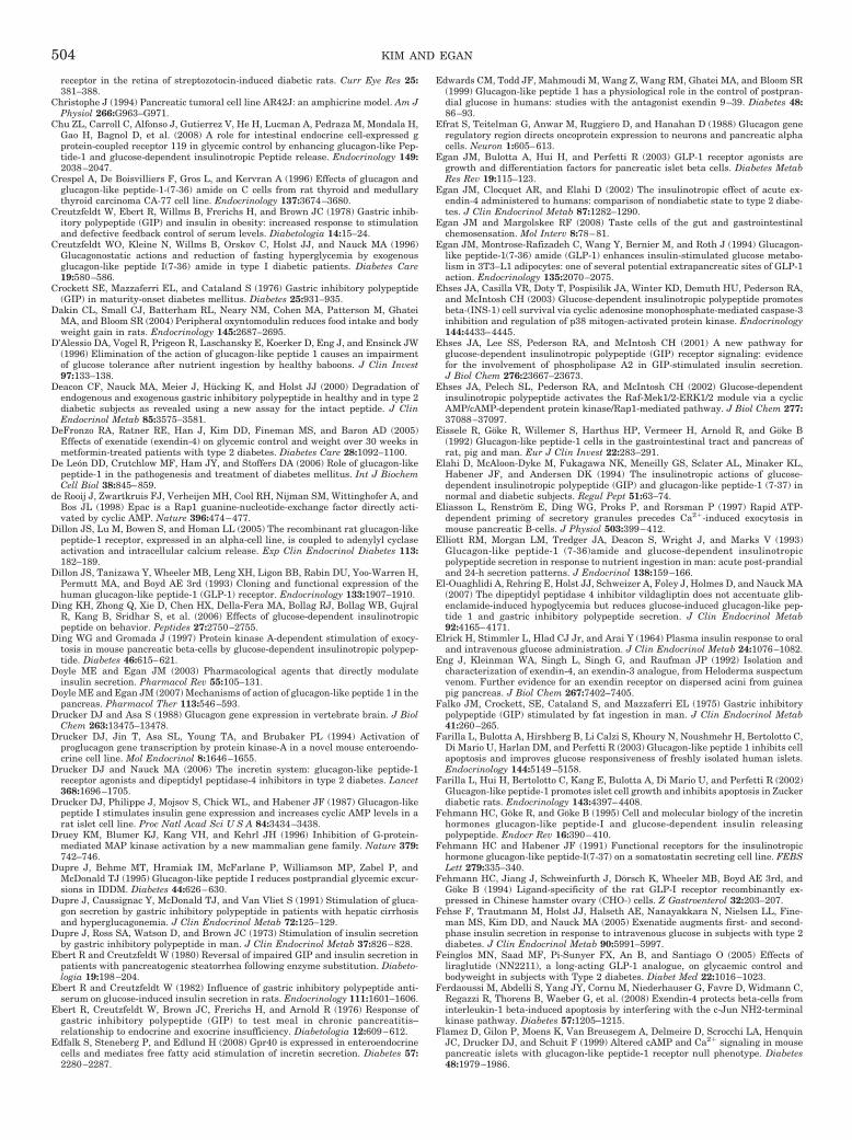
receptor in the retina of streptozotocin-induced diabetic rats. Curr Eye Res 25:381–388.
Christophe J (1994) Pancreatic tumoral cell line AR42J: an amphicrine model. Am JPhysiol 266:G963–G971.
Chu ZL, Carroll C, Alfonso J, Gutierrez V, He H, Lucman A, Pedraza M, Mondala H,Gao H, Bagnol D, et al. (2008) A role for intestinal endocrine cell-expressed gprotein-coupled receptor 119 in glycemic control by enhancing glucagon-like Pep-tide-1 and glucose-dependent insulinotropic Peptide release. Endocrinology 149:2038–2047.
Crespel A, De Boisvilliers F, Gros L, and Kervran A (1996) Effects of glucagon andglucagon-like peptide-1-(7-36) amide on C cells from rat thyroid and medullarythyroid carcinoma CA-77 cell line. Endocrinology 137:3674–3680.
Creutzfeldt W, Ebert R, Willms B, Frerichs H, and Brown JC (1978) Gastric inhib-itory polypeptide (GIP) and insulin in obesity: increased response to stimulationand defective feedback control of serum levels. Diabetologia 14:15–24.
Creutzfeldt WO, Kleine N, Willms B, Orskov C, Holst JJ, and Nauck MA (1996)Glucagonostatic actions and reduction of fasting hyperglycemia by exogenousglucagon-like peptide I(7-36) amide in type I diabetic patients. Diabetes Care19:580–586.
Crockett SE, Mazzaferri EL, and Cataland S (1976) Gastric inhibitory polypeptide(GIP) in maturity-onset diabetes mellitus. Diabetes 25:931–935.
Dakin CL, Small CJ, Batterham RL, Neary NM, Cohen MA, Patterson M, GhateiMA, and Bloom SR (2004) Peripheral oxyntomodulin reduces food intake and bodyweight gain in rats. Endocrinology 145:2687–2695.
D’Alessio DA, Vogel R, Prigeon R, Laschansky E, Koerker D, Eng J, and Ensinck JW(1996) Elimination of the action of glucagon-like peptide 1 causes an impairmentof glucose tolerance after nutrient ingestion by healthy baboons. J Clin Invest97:133–138.
Deacon CF, Nauck MA, Meier J, Hucking K, and Holst JJ (2000) Degradation ofendogenous and exogenous gastric inhibitory polypeptide in healthy and in type 2diabetic subjects as revealed using a new assay for the intact peptide. J ClinEndocrinol Metab 85:3575–3581.
DeFronzo RA, Ratner RE, Han J, Kim DD, Fineman MS, and Baron AD (2005)Effects of exenatide (exendin-4) on glycemic control and weight over 30 weeks inmetformin-treated patients with type 2 diabetes. Diabetes Care 28:1092–1100.
De Leon DD, Crutchlow MF, Ham JY, and Stoffers DA (2006) Role of glucagon-likepeptide-1 in the pathogenesis and treatment of diabetes mellitus. Int J BiochemCell Biol 38:845–859.
de Rooij J, Zwartkruis FJ, Verheijen MH, Cool RH, Nijman SM, Wittinghofer A, andBos JL (1998) Epac is a Rap1 guanine-nucleotide-exchange factor directly acti-vated by cyclic AMP. Nature 396:474–477.
Dillon JS, Lu M, Bowen S, and Homan LL (2005) The recombinant rat glucagon-likepeptide-1 receptor, expressed in an alpha-cell line, is coupled to adenylyl cyclaseactivation and intracellular calcium release. Exp Clin Endocrinol Diabetes 113:182–189.
Dillon JS, Tanizawa Y, Wheeler MB, Leng XH, Ligon BB, Rabin DU, Yoo-Warren H,Permutt MA, and Boyd AE 3rd (1993) Cloning and functional expression of thehuman glucagon-like peptide-1 (GLP-1) receptor. Endocrinology 133:1907–1910.
Ding KH, Zhong Q, Xie D, Chen HX, Della-Fera MA, Bollag RJ, Bollag WB, GujralR, Kang B, Sridhar S, et al. (2006) Effects of glucose-dependent insulinotropicpeptide on behavior. Peptides 27:2750–2755.
Ding WG and Gromada J (1997) Protein kinase A-dependent stimulation of exocy-tosis in mouse pancreatic beta-cells by glucose-dependent insulinotropic polypep-tide. Diabetes 46:615–621.
Doyle ME and Egan JM (2003) Pharmacological agents that directly modulateinsulin secretion. Pharmacol Rev 55:105–131.
Doyle ME and Egan JM (2007) Mechanisms of action of glucagon-like peptide 1 in thepancreas. Pharmacol Ther 113:546–593.
Drucker DJ and Asa S (1988) Glucagon gene expression in vertebrate brain. J BiolChem 263:13475–13478.
Drucker DJ, Jin T, Asa SL, Young TA, and Brubaker PL (1994) Activation ofproglucagon gene transcription by protein kinase-A in a novel mouse enteroendo-crine cell line. Mol Endocrinol 8:1646–1655.
Drucker DJ and Nauck MA (2006) The incretin system: glucagon-like peptide-1receptor agonists and dipeptidyl peptidase-4 inhibitors in type 2 diabetes. Lancet368:1696–1705.
Drucker DJ, Philippe J, Mojsov S, Chick WL, and Habener JF (1987) Glucagon-likepeptide I stimulates insulin gene expression and increases cyclic AMP levels in arat islet cell line. Proc Natl Acad Sci U S A 84:3434–3438.
Druey KM, Blumer KJ, Kang VH, and Kehrl JH (1996) Inhibition of G-protein-mediated MAP kinase activation by a new mammalian gene family. Nature 379:742–746.
Dupre J, Behme MT, Hramiak IM, McFarlane P, Williamson MP, Zabel P, andMcDonald TJ (1995) Glucagon-like peptide I reduces postprandial glycemic excur-sions in IDDM. Diabetes 44:626–630.
Dupre J, Caussignac Y, McDonald TJ, and Van Vliet S (1991) Stimulation of gluca-gon secretion by gastric inhibitory polypeptide in patients with hepatic cirrhosisand hyperglucagonemia. J Clin Endocrinol Metab 72:125–129.
Dupre J, Ross SA, Watson D, and Brown JC (1973) Stimulation of insulin secretionby gastric inhibitory polypeptide in man. J Clin Endocrinol Metab 37:826–828.
Ebert R and Creutzfeldt W (1980) Reversal of impaired GIP and insulin secretion inpatients with pancreatogenic steatorrhea following enzyme substitution. Diabeto-logia 19:198–204.
Ebert R and Creutzfeldt W (1982) Influence of gastric inhibitory polypeptide anti-serum on glucose-induced insulin secretion in rats. Endocrinology 111:1601–1606.
Ebert R, Creutzfeldt W, Brown JC, Frerichs H, and Arnold R (1976) Response ofgastric inhibitory polypeptide (GIP) to test meal in chronic pancreatitis–relationship to endocrine and exocrine insufficiency. Diabetologia 12:609–612.
Edfalk S, Steneberg P, and Edlund H (2008) Gpr40 is expressed in enteroendocrinecells and mediates free fatty acid stimulation of incretin secretion. Diabetes 57:2280–2287.
Edwards CM, Todd JF, Mahmoudi M, Wang Z, Wang RM, Ghatei MA, and Bloom SR(1999) Glucagon-like peptide 1 has a physiological role in the control of postpran-dial glucose in humans: studies with the antagonist exendin 9–39. Diabetes 48:86–93.
Efrat S, Teitelman G, Anwar M, Ruggiero D, and Hanahan D (1988) Glucagon generegulatory region directs oncoprotein expression to neurons and pancreatic alphacells. Neuron 1:605–613.
Egan JM, Bulotta A, Hui H, and Perfetti R (2003) GLP-1 receptor agonists aregrowth and differentiation factors for pancreatic islet beta cells. Diabetes MetabRes Rev 19:115–123.
Egan JM, Clocquet AR, and Elahi D (2002) The insulinotropic effect of acute ex-endin-4 administered to humans: comparison of nondiabetic state to type 2 diabe-tes. J Clin Endocrinol Metab 87:1282–1290.
Egan JM and Margolskee RF (2008) Taste cells of the gut and gastrointestinalchemosensation. Mol Interv 8:78–81.
Egan JM, Montrose-Rafizadeh C, Wang Y, Bernier M, and Roth J (1994) Glucagon-like peptide-1(7-36) amide (GLP-1) enhances insulin-stimulated glucose metabo-lism in 3T3–L1 adipocytes: one of several potential extrapancreatic sites of GLP-1action. Endocrinology 135:2070–2075.
Ehses JA, Casilla VR, Doty T, Pospisilik JA, Winter KD, Demuth HU, Pederson RA,and McIntosh CH (2003) Glucose-dependent insulinotropic polypeptide promotesbeta-(INS-1) cell survival via cyclic adenosine monophosphate-mediated caspase-3inhibition and regulation of p38 mitogen-activated protein kinase. Endocrinology144:4433–4445.
Ehses JA, Lee SS, Pederson RA, and McIntosh CH (2001) A new pathway forglucose-dependent insulinotropic polypeptide (GIP) receptor signaling: evidencefor the involvement of phospholipase A2 in GIP-stimulated insulin secretion.J Biol Chem 276:23667–23673.
Ehses JA, Pelech SL, Pederson RA, and McIntosh CH (2002) Glucose-dependentinsulinotropic polypeptide activates the Raf-Mek1/2-ERK1/2 module via a cyclicAMP/cAMP-dependent protein kinase/Rap1-mediated pathway. J Biol Chem 277:37088–37097.
Eissele R, Goke R, Willemer S, Harthus HP, Vermeer H, Arnold R, and Goke B(1992) Glucagon-like peptide-1 cells in the gastrointestinal tract and pancreas ofrat, pig and man. Eur J Clin Invest 22:283–291.
Elahi D, McAloon-Dyke M, Fukagawa NK, Meneilly GS, Sclater AL, Minaker KL,Habener JF, and Andersen DK (1994) The insulinotropic actions of glucose-dependent insulinotropic polypeptide (GIP) and glucagon-like peptide-1 (7-37) innormal and diabetic subjects. Regul Pept 51:63–74.
Eliasson L, Renstrom E, Ding WG, Proks P, and Rorsman P (1997) Rapid ATP-dependent priming of secretory granules precedes Ca2�-induced exocytosis inmouse pancreatic B-cells. J Physiol 503:399–412.
Elliott RM, Morgan LM, Tredger JA, Deacon S, Wright J, and Marks V (1993)Glucagon-like peptide-1 (7-36)amide and glucose-dependent insulinotropicpolypeptide secretion in response to nutrient ingestion in man: acute post-prandialand 24-h secretion patterns. J Endocrinol 138:159–166.
El-Ouaghlidi A, Rehring E, Holst JJ, Schweizer A, Foley J, Holmes D, and Nauck MA(2007) The dipeptidyl peptidase 4 inhibitor vildagliptin does not accentuate glib-enclamide-induced hypoglycemia but reduces glucose-induced glucagon-like pep-tide 1 and gastric inhibitory polypeptide secretion. J Clin Endocrinol Metab92:4165–4171.
Elrick H, Stimmler L, Hlad CJ Jr, and Arai Y (1964) Plasma insulin response to oraland intravenous glucose administration. J Clin Endocrinol Metab 24:1076–1082.
Eng J, Kleinman WA, Singh L, Singh G, and Raufman JP (1992) Isolation andcharacterization of exendin-4, an exendin-3 analogue, from Heloderma suspectumvenom. Further evidence for an exendin receptor on dispersed acini from guineapig pancreas. J Biol Chem 267:7402–7405.
Falko JM, Crockett, SE, Cataland S, and Mazzaferri EL (1975) Gastric inhibitorypolypeptide (GIP) stimulated by fat ingestion in man. J Clin Endocrinol Metab41:260–265.
Farilla L, Bulotta A, Hirshberg B, Li Calzi S, Khoury N, Noushmehr H, Bertolotto C,Di Mario U, Harlan DM, and Perfetti R (2003) Glucagon-like peptide 1 inhibits cellapoptosis and improves glucose responsiveness of freshly isolated human islets.Endocrinology 144:5149–5158.
Farilla L, Hui H, Bertolotto C, Kang E, Bulotta A, Di Mario U, and Perfetti R (2002)Glucagon-like peptide-1 promotes islet cell growth and inhibits apoptosis in Zuckerdiabetic rats. Endocrinology 143:4397–4408.
Fehmann HC, Goke R, and Goke B (1995) Cell and molecular biology of the incretinhormones glucagon-like peptide-I and glucose-dependent insulin releasingpolypeptide. Endocr Rev 16:390–410.
Fehmann HC and Habener JF (1991) Functional receptors for the insulinotropichormone glucagon-like peptide-I(7-37) on a somatostatin secreting cell line. FEBSLett 279:335–340.
Fehmann HC, Jiang J, Schweinfurth J, Dorsch K, Wheeler MB, Boyd AE 3rd, andGoke B (1994) Ligand-specificity of the rat GLP-I receptor recombinantly ex-pressed in Chinese hamster ovary (CHO-) cells. Z Gastroenterol 32:203–207.
Fehse F, Trautmann M, Holst JJ, Halseth AE, Nanayakkara N, Nielsen LL, Fine-man MS, Kim DD, and Nauck MA (2005) Exenatide augments first- and second-phase insulin secretion in response to intravenous glucose in subjects with type 2diabetes. J Clin Endocrinol Metab 90:5991–5997.
Feinglos MN, Saad MF, Pi-Sunyer FX, An B, and Santiago O (2005) Effects ofliraglutide (NN2211), a long-acting GLP-1 analogue, on glycaemic control andbodyweight in subjects with Type 2 diabetes. Diabet Med 22:1016–1023.
Ferdaoussi M, Abdelli S, Yang JY, Cornu M, Niederhauser G, Favre D, Widmann C,Regazzi R, Thorens B, Waeber G, et al. (2008) Exendin-4 protects beta-cells frominterleukin-1 beta-induced apoptosis by interfering with the c-Jun NH2-terminalkinase pathway. Diabetes 57:1205–1215.
Flamez D, Gilon P, Moens K, Van Breusegem A, Delmeire D, Scrocchi LA, HenquinJC, Drucker DJ, and Schuit F (1999) Altered cAMP and Ca2� signaling in mousepancreatic islets with glucagon-like peptide-1 receptor null phenotype. Diabetes48:1979–1986.
504 KIM AND EGAN
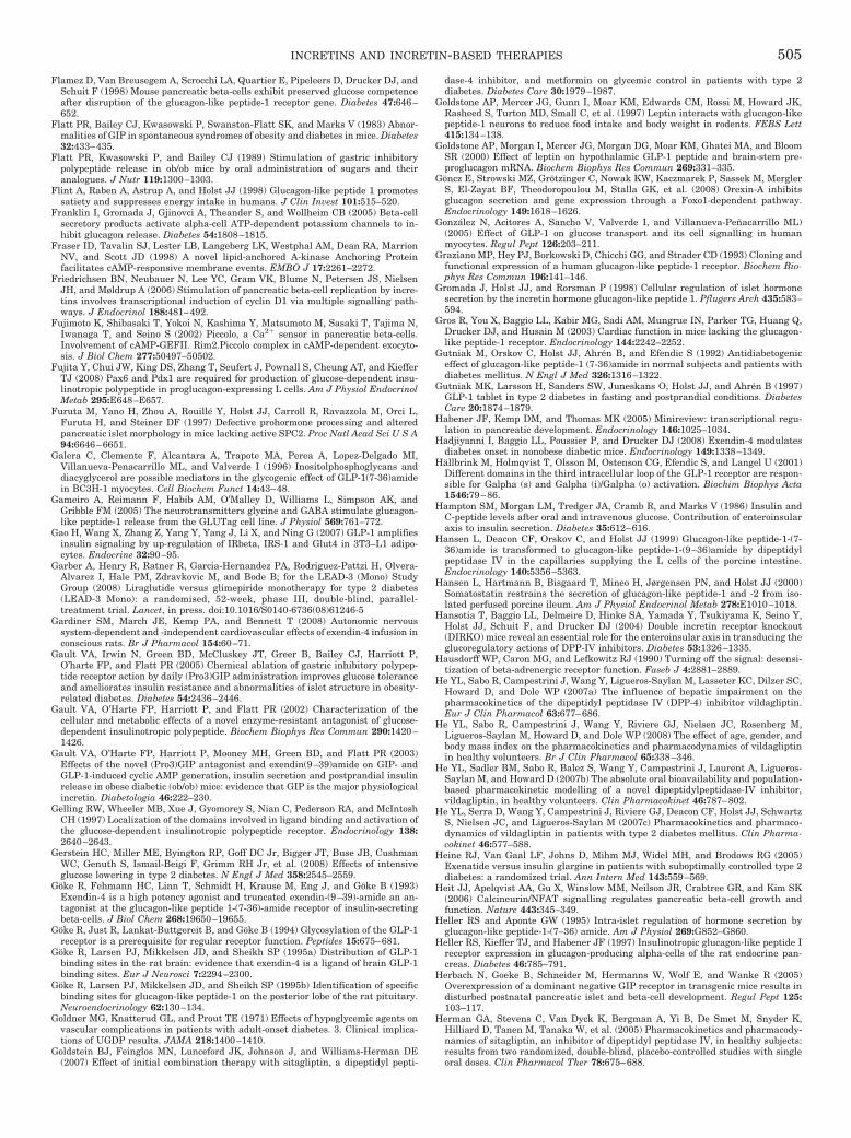
Flamez D, Van Breusegem A, Scrocchi LA, Quartier E, Pipeleers D, Drucker DJ, andSchuit F (1998) Mouse pancreatic beta-cells exhibit preserved glucose competenceafter disruption of the glucagon-like peptide-1 receptor gene. Diabetes 47:646–652.
Flatt PR, Bailey CJ, Kwasowski P, Swanston-Flatt SK, and Marks V (1983) Abnor-malities of GIP in spontaneous syndromes of obesity and diabetes in mice. Diabetes32:433–435.
Flatt PR, Kwasowski P, and Bailey CJ (1989) Stimulation of gastric inhibitorypolypeptide release in ob/ob mice by oral administration of sugars and theiranalogues. J Nutr 119:1300–1303.
Flint A, Raben A, Astrup A, and Holst JJ (1998) Glucagon-like peptide 1 promotessatiety and suppresses energy intake in humans. J Clin Invest 101:515–520.
Franklin I, Gromada J, Gjinovci A, Theander S, and Wollheim CB (2005) Beta-cellsecretory products activate alpha-cell ATP-dependent potassium channels to in-hibit glucagon release. Diabetes 54:1808–1815.
Fraser ID, Tavalin SJ, Lester LB, Langeberg LK, Westphal AM, Dean RA, MarrionNV, and Scott JD (1998) A novel lipid-anchored A-kinase Anchoring Proteinfacilitates cAMP-responsive membrane events. EMBO J 17:2261–2272.
Friedrichsen BN, Neubauer N, Lee YC, Gram VK, Blume N, Petersen JS, NielsenJH, and Møldrup A (2006) Stimulation of pancreatic beta-cell replication by incre-tins involves transcriptional induction of cyclin D1 via multiple signalling path-ways. J Endocrinol 188:481–492.
Fujimoto K, Shibasaki T, Yokoi N, Kashima Y, Matsumoto M, Sasaki T, Tajima N,Iwanaga T, and Seino S (2002) Piccolo, a Ca2� sensor in pancreatic beta-cells.Involvement of cAMP-GEFII. Rim2.Piccolo complex in cAMP-dependent exocyto-sis. J Biol Chem 277:50497–50502.
Fujita Y, Chui JW, King DS, Zhang T, Seufert J, Pownall S, Cheung AT, and KiefferTJ (2008) Pax6 and Pdx1 are required for production of glucose-dependent insu-linotropic polypeptide in proglucagon-expressing L cells. Am J Physiol EndocrinolMetab 295:E648–E657.
Furuta M, Yano H, Zhou A, Rouille Y, Holst JJ, Carroll R, Ravazzola M, Orci L,Furuta H, and Steiner DF (1997) Defective prohormone processing and alteredpancreatic islet morphology in mice lacking active SPC2. Proc Natl Acad Sci U S A94:6646–6651.
Galera C, Clemente F, Alcantara A, Trapote MA, Perea A, Lopez-Delgado MI,Villanueva-Penacarrillo ML, and Valverde I (1996) Inositolphosphoglycans anddiacyglycerol are possible mediators in the glycogenic effect of GLP-1(7-36)amidein BC3H-1 myocytes. Cell Biochem Funct 14:43–48.
Gameiro A, Reimann F, Habib AM, O’Malley D, Williams L, Simpson AK, andGribble FM (2005) The neurotransmitters glycine and GABA stimulate glucagon-like peptide-1 release from the GLUTag cell line. J Physiol 569:761–772.
Gao H, Wang X, Zhang Z, Yang Y, Yang J, Li X, and Ning G (2007) GLP-1 amplifiesinsulin signaling by up-regulation of IRbeta, IRS-1 and Glut4 in 3T3–L1 adipo-cytes. Endocrine 32:90–95.
Garber A, Henry R, Ratner R, Garcia-Hernandez PA, Rodriguez-Pattzi H, Olvera-Alvarez I, Hale PM, Zdravkovic M, and Bode B; for the LEAD-3 (Mono) StudyGroup (2008) Liraglutide versus glimepiride monotherapy for type 2 diabetes(LEAD-3 Mono): a randomised, 52-week, phase III, double-blind, parallel-treatment trial. Lancet, in press. doi:10.1016/S0140-6736(08)61246-5
Gardiner SM, March JE, Kemp PA, and Bennett T (2008) Autonomic nervoussystem-dependent and -independent cardiovascular effects of exendin-4 infusion inconscious rats. Br J Pharmacol 154:60–71.
Gault VA, Irwin N, Green BD, McCluskey JT, Greer B, Bailey CJ, Harriott P,O’harte FP, and Flatt PR (2005) Chemical ablation of gastric inhibitory polypep-tide receptor action by daily (Pro3)GIP administration improves glucose toleranceand ameliorates insulin resistance and abnormalities of islet structure in obesity-related diabetes. Diabetes 54:2436–2446.
Gault VA, O’Harte FP, Harriott P, and Flatt PR (2002) Characterization of thecellular and metabolic effects of a novel enzyme-resistant antagonist of glucose-dependent insulinotropic polypeptide. Biochem Biophys Res Commun 290:1420–1426.
Gault VA, O’Harte FP, Harriott P, Mooney MH, Green BD, and Flatt PR (2003)Effects of the novel (Pro3)GIP antagonist and exendin(9–39)amide on GIP- andGLP-1-induced cyclic AMP generation, insulin secretion and postprandial insulinrelease in obese diabetic (ob/ob) mice: evidence that GIP is the major physiologicalincretin. Diabetologia 46:222–230.
Gelling RW, Wheeler MB, Xue J, Gyomorey S, Nian C, Pederson RA, and McIntoshCH (1997) Localization of the domains involved in ligand binding and activation ofthe glucose-dependent insulinotropic polypeptide receptor. Endocrinology 138:2640–2643.
Gerstein HC, Miller ME, Byington RP, Goff DC Jr, Bigger JT, Buse JB, CushmanWC, Genuth S, Ismail-Beigi F, Grimm RH Jr, et al. (2008) Effects of intensiveglucose lowering in type 2 diabetes. N Engl J Med 358:2545–2559.
Goke R, Fehmann HC, Linn T, Schmidt H, Krause M, Eng J, and Goke B (1993)Exendin-4 is a high potency agonist and truncated exendin-(9–39)-amide an an-tagonist at the glucagon-like peptide 1-(7-36)-amide receptor of insulin-secretingbeta-cells. J Biol Chem 268:19650–19655.
Goke R, Just R, Lankat-Buttgereit B, and Goke B (1994) Glycosylation of the GLP-1receptor is a prerequisite for regular receptor function. Peptides 15:675–681.
Goke R, Larsen PJ, Mikkelsen JD, and Sheikh SP (1995a) Distribution of GLP-1binding sites in the rat brain: evidence that exendin-4 is a ligand of brain GLP-1binding sites. Eur J Neurosci 7:2294–2300.
Goke R, Larsen PJ, Mikkelsen JD, and Sheikh SP (1995b) Identification of specificbinding sites for glucagon-like peptide-1 on the posterior lobe of the rat pituitary.Neuroendocrinology 62:130–134.
Goldner MG, Knatterud GL, and Prout TE (1971) Effects of hypoglycemic agents onvascular complications in patients with adult-onset diabetes. 3. Clinical implica-tions of UGDP results. JAMA 218:1400–1410.
Goldstein BJ, Feinglos MN, Lunceford JK, Johnson J, and Williams-Herman DE(2007) Effect of initial combination therapy with sitagliptin, a dipeptidyl pepti-
dase-4 inhibitor, and metformin on glycemic control in patients with type 2diabetes. Diabetes Care 30:1979–1987.
Goldstone AP, Mercer JG, Gunn I, Moar KM, Edwards CM, Rossi M, Howard JK,Rasheed S, Turton MD, Small C, et al. (1997) Leptin interacts with glucagon-likepeptide-1 neurons to reduce food intake and body weight in rodents. FEBS Lett415:134–138.
Goldstone AP, Morgan I, Mercer JG, Morgan DG, Moar KM, Ghatei MA, and BloomSR (2000) Effect of leptin on hypothalamic GLP-1 peptide and brain-stem pre-proglucagon mRNA. Biochem Biophys Res Commun 269:331–335.
Goncz E, Strowski MZ, Grotzinger C, Nowak KW, Kaczmarek P, Sassek M, MerglerS, El-Zayat BF, Theodoropoulou M, Stalla GK, et al. (2008) Orexin-A inhibitsglucagon secretion and gene expression through a Foxo1-dependent pathway.Endocrinology 149:1618–1626.
Gonzalez N, Acitores A, Sancho V, Valverde I, and Villanueva-Penacarrillo ML)(2005) Effect of GLP-1 on glucose transport and its cell signalling in humanmyocytes. Regul Pept 126:203–211.
Graziano MP, Hey PJ, Borkowski D, Chicchi GG, and Strader CD (1993) Cloning andfunctional expression of a human glucagon-like peptide-1 receptor. Biochem Bio-phys Res Commun 196:141–146.
Gromada J, Holst JJ, and Rorsman P (1998) Cellular regulation of islet hormonesecretion by the incretin hormone glucagon-like peptide 1. Pflugers Arch 435:583–594.
Gros R, You X, Baggio LL, Kabir MG, Sadi AM, Mungrue IN, Parker TG, Huang Q,Drucker DJ, and Husain M (2003) Cardiac function in mice lacking the glucagon-like peptide-1 receptor. Endocrinology 144:2242–2252.
Gutniak M, Orskov C, Holst JJ, Ahren B, and Efendic S (1992) Antidiabetogeniceffect of glucagon-like peptide-1 (7-36)amide in normal subjects and patients withdiabetes mellitus. N Engl J Med 326:1316–1322.
Gutniak MK, Larsson H, Sanders SW, Juneskans O, Holst JJ, and Ahren B (1997)GLP-1 tablet in type 2 diabetes in fasting and postprandial conditions. DiabetesCare 20:1874–1879.
Habener JF, Kemp DM, and Thomas MK (2005) Minireview: transcriptional regu-lation in pancreatic development. Endocrinology 146:1025–1034.
Hadjiyanni I, Baggio LL, Poussier P, and Drucker DJ (2008) Exendin-4 modulatesdiabetes onset in nonobese diabetic mice. Endocrinology 149:1338–1349.
Hallbrink M, Holmqvist T, Olsson M, Ostenson CG, Efendic S, and Langel U (2001)Different domains in the third intracellular loop of the GLP-1 receptor are respon-sible for Galpha (s) and Galpha (i)/Galpha (o) activation. Biochim Biophys Acta1546:79–86.
Hampton SM, Morgan LM, Tredger JA, Cramb R, and Marks V (1986) Insulin andC-peptide levels after oral and intravenous glucose. Contribution of enteroinsularaxis to insulin secretion. Diabetes 35:612–616.
Hansen L, Deacon CF, Orskov C, and Holst JJ (1999) Glucagon-like peptide-1-(7-36)amide is transformed to glucagon-like peptide-1-(9–36)amide by dipeptidylpeptidase IV in the capillaries supplying the L cells of the porcine intestine.Endocrinology 140:5356–5363.
Hansen L, Hartmann B, Bisgaard T, Mineo H, Jørgensen PN, and Holst JJ (2000)Somatostatin restrains the secretion of glucagon-like peptide-1 and -2 from iso-lated perfused porcine ileum. Am J Physiol Endocrinol Metab 278:E1010–1018.
Hansotia T, Baggio LL, Delmeire D, Hinke SA, Yamada Y, Tsukiyama K, Seino Y,Holst JJ, Schuit F, and Drucker DJ (2004) Double incretin receptor knockout(DIRKO) mice reveal an essential role for the enteroinsular axis in transducing theglucoregulatory actions of DPP-IV inhibitors. Diabetes 53:1326–1335.
Hausdorff WP, Caron MG, and Lefkowitz RJ (1990) Turning off the signal: desensi-tization of beta-adrenergic receptor function. Faseb J 4:2881–2889.
He YL, Sabo R, Campestrini J, Wang Y, Ligueros-Saylan M, Lasseter KC, Dilzer SC,Howard D, and Dole WP (2007a) The influence of hepatic impairment on thepharmacokinetics of the dipeptidyl peptidase IV (DPP-4) inhibitor vildagliptin.Eur J Clin Pharmacol 63:677–686.
He YL, Sabo R, Campestrini J, Wang Y, Riviere GJ, Nielsen JC, Rosenberg M,Ligueros-Saylan M, Howard D, and Dole WP (2008) The effect of age, gender, andbody mass index on the pharmacokinetics and pharmacodynamics of vildagliptinin healthy volunteers. Br J Clin Pharmacol 65:338–346.
He YL, Sadler BM, Sabo R, Balez S, Wang Y, Campestrini J, Laurent A, Ligueros-Saylan M, and Howard D (2007b) The absolute oral bioavailability and population-based pharmacokinetic modelling of a novel dipeptidylpeptidase-IV inhibitor,vildagliptin, in healthy volunteers. Clin Pharmacokinet 46:787–802.
He YL, Serra D, Wang Y, Campestrini J, Riviere GJ, Deacon CF, Holst JJ, SchwartzS, Nielsen JC, and Ligueros-Saylan M (2007c) Pharmacokinetics and pharmaco-dynamics of vildagliptin in patients with type 2 diabetes mellitus. Clin Pharma-cokinet 46:577–588.
Heine RJ, Van Gaal LF, Johns D, Mihm MJ, Widel MH, and Brodows RG (2005)Exenatide versus insulin glargine in patients with suboptimally controlled type 2diabetes: a randomized trial. Ann Intern Med 143:559–569.
Heit JJ, Apelqvist AA, Gu X, Winslow MM, Neilson JR, Crabtree GR, and Kim SK(2006) Calcineurin/NFAT signalling regulates pancreatic beta-cell growth andfunction. Nature 443:345–349.
Heller RS and Aponte GW (1995) Intra-islet regulation of hormone secretion byglucagon-like peptide-1-(7–36) amide. Am J Physiol 269:G852–G860.
Heller RS, Kieffer TJ, and Habener JF (1997) Insulinotropic glucagon-like peptide Ireceptor expression in glucagon-producing alpha-cells of the rat endocrine pan-creas. Diabetes 46:785–791.
Herbach N, Goeke B, Schneider M, Hermanns W, Wolf E, and Wanke R (2005)Overexpression of a dominant negative GIP receptor in transgenic mice results indisturbed postnatal pancreatic islet and beta-cell development. Regul Pept 125:103–117.
Herman GA, Stevens C, Van Dyck K, Bergman A, Yi B, De Smet M, Snyder K,Hilliard D, Tanen M, Tanaka W, et al. (2005) Pharmacokinetics and pharmacody-namics of sitagliptin, an inhibitor of dipeptidyl peptidase IV, in healthy subjects:results from two randomized, double-blind, placebo-controlled studies with singleoral doses. Clin Pharmacol Ther 78:675–688.
INCRETINS AND INCRETIN-BASED THERAPIES 505
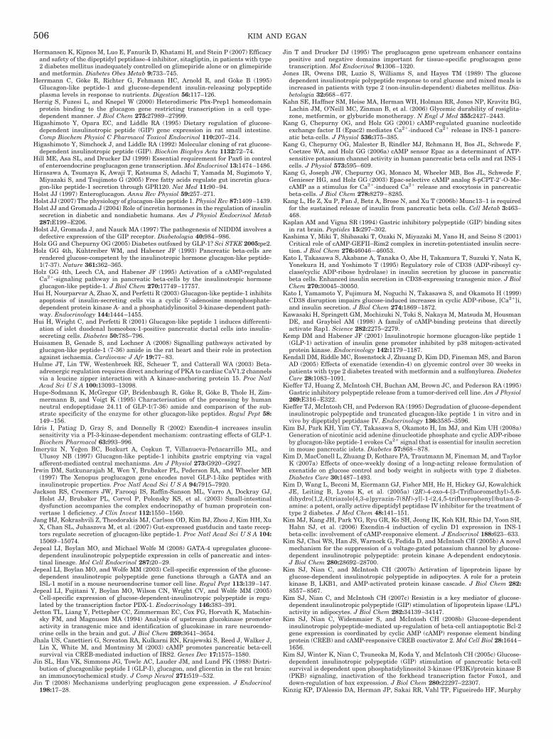
Hermansen K, Kipnes M, Luo E, Fanurik D, Khatami H, and Stein P (2007) Efficacyand safety of the dipeptidyl peptidase-4 inhibitor, sitagliptin, in patients with type2 diabetes mellitus inadequately controlled on glimepiride alone or on glimepirideand metformin. Diabetes Obes Metab 9:733–745.
Herrmann C, Goke R, Richter G, Fehmann HC, Arnold R, and Goke B (1995)Glucagon-like peptide-1 and glucose-dependent insulin-releasing polypeptideplasma levels in response to nutrients. Digestion 56:117–126.
Herzig S, Fuzesi L, and Knepel W (2000) Heterodimeric Pbx-Prep1 homeodomainprotein binding to the glucagon gene restricting transcription in a cell type-dependent manner. J Biol Chem 275:27989–27999.
Higashimoto Y, Opara EC, and Liddle RA (1995) Dietary regulation of glucose-dependent insulinotropic peptide (GIP) gene expression in rat small intestine.Comp Biochem Physiol C Pharmacol Toxicol Endocrinol 110:207–214.
Higashimoto Y, Simchock J, and Liddle RA (1992) Molecular cloning of rat glucose-dependent insulinotropic peptide (GIP). Biochim Biophys Acta 1132:72–74.
Hill ME, Asa SL, and Drucker DJ (1999) Essential requirement for Pax6 in controlof enteroendocrine proglucagon gene transcription. Mol Endocrinol 13:1474–1486.
Hirasawa A, Tsumaya K, Awaji T, Katsuma S, Adachi T, Yamada M, Sugimoto Y,Miyazaki S, and Tsujimoto G (2005) Free fatty acids regulate gut incretin gluca-gon-like peptide-1 secretion through GPR120. Nat Med 11:90–94.
Holst JJ (1997) Enteroglucagon. Annu Rev Physiol 59:257–271.Holst JJ (2007) The physiology of glucagon-like peptide 1. Physiol Rev 87:1409–1439.Holst JJ and Gromada J (2004) Role of incretin hormones in the regulation of insulin
secretion in diabetic and nondiabetic humans. Am J Physiol Endocrinol Metab287:E199–E206.
Holst JJ, Gromada J, and Nauck MA (1997) The pathogenesis of NIDDM involves adefective expression of the GIP receptor. Diabetologia 40:984–986.
Holz GG and Chepurny OG (2005) Diabetes outfoxed by GLP-1? Sci STKE 2005:pe2.Holz GG 4th, Kuhtreiber WM, and Habener JF (1993) Pancreatic beta-cells are
rendered glucose-competent by the insulinotropic hormone glucagon-like peptide-1(7-37). Nature 361:362–365.
Holz GG 4th, Leech CA, and Habener JF (1995) Activation of a cAMP-regulatedCa2�-signaling pathway in pancreatic beta-cells by the insulinotropic hormoneglucagon-like peptide-1. J Biol Chem 270:17749–17757.
Hui H, Nourparvar A, Zhao X, and Perfetti R (2003) Glucagon-like peptide-1 inhibitsapoptosis of insulin-secreting cells via a cyclic 5�-adenosine monophosphate-dependent protein kinase A- and a phosphatidylinositol 3-kinase-dependent path-way. Endocrinology 144:1444–1455.
Hui H, Wright C, and Perfetti R (2001) Glucagon-like peptide 1 induces differenti-ation of islet duodenal homeobox-1-positive pancreatic ductal cells into insulin-secreting cells. Diabetes 50:785–796.
Huisamen B, Genade S, and Lochner A (2008) Signalling pathways activated byglucagon-like peptide-1 (7-36) amide in the rat heart and their role in protectionagainst ischaemia. Cardiovasc J Afr 19:77–83.
Hulme JT, Lin TW, Westenbroek RE, Scheuer T, and Catterall WA (2003) Beta-adrenergic regulation requires direct anchoring of PKA to cardiac CaV1.2 channelsvia a leucine zipper interaction with A kinase-anchoring protein 15. Proc NatlAcad Sci U S A 100:13093–13098.
Hupe-Sodmann K, McGregor GP, Bridenbaugh R, Goke R, Goke B, Thole H, Zim-mermann B, and Voigt K (1995) Characterisation of the processing by humanneutral endopeptidase 24.11 of GLP-1(7-36) amide and comparison of the sub-strate specificity of the enzyme for other glucagon-like peptides. Regul Pept 58:149–156.
Idris I, Patiag D, Gray S, and Donnelly R (2002) Exendin-4 increases insulinsensitivity via a PI-3-kinase-dependent mechanism: contrasting effects of GLP-1.Biochem Pharmacol 63:993–996.
Imeryuz N, Yegen BC, Bozkurt A, Coskun T, Villanueva-Penacarrillo ML, andUlusoy NB (1997) Glucagon-like peptide-1 inhibits gastric emptying via vagalafferent-mediated central mechanisms. Am J Physiol 273:G920–G927.
Irwin DM, Satkunarajah M, Wen Y, Brubaker PL, Pederson RA, and Wheeler MB(1997) The Xenopus proglucagon gene encodes novel GLP-1-like peptides withinsulinotropic properties. Proc Natl Acad Sci U S A 94:7915–7920.
Jackson RS, Creemers JW, Farooqi IS, Raffin-Sanson ML, Varro A, Dockray GJ,Holst JJ, Brubaker PL, Corvol P, Polonsky KS, et al. (2003) Small-intestinaldysfunction accompanies the complex endocrinopathy of human proprotein con-vertase 1 deficiency. J Clin Invest 112:1550–1560.
Jang HJ, Kokrashvili Z, Theodorakis MJ, Carlson OD, Kim BJ, Zhou J, Kim HH, XuX, Chan SL, Juhaszova M, et al. (2007) Gut-expressed gustducin and taste recep-tors regulate secretion of glucagon-like peptide-1. Proc Natl Acad Sci U S A 104:15069–15074.
Jepeal LI, Boylan MO, and Michael Wolfe M (2008) GATA-4 upregulates glucose-dependent insulinotropic polypeptide expression in cells of pancreatic and intes-tinal lineage. Mol Cell Endocrinol 287:20–29.
Jepeal LI, Boylan MO, and Wolfe MM (2003) Cell-specific expression of the glucose-dependent insulinotropic polypeptide gene functions through a GATA and anISL-1 motif in a mouse neuroendocrine tumor cell line. Regul Pept 113:139–147.
Jepeal LI, Fujitani Y, Boylan MO, Wilson CN, Wright CV, and Wolfe MM (2005)Cell-specific expression of glucose-dependent-insulinotropic polypeptide is regu-lated by the transcription factor PDX-1. Endocrinology 146:383–391.
Jetton TL, Liang Y, Pettepher CC, Zimmerman EC, Cox FG, Horvath K, Matschin-sky FM, and Magnuson MA (1994) Analysis of upstream glucokinase promoteractivity in transgenic mice and identification of glucokinase in rare neuroendo-crine cells in the brain and gut. J Biol Chem 269:3641–3654.
Jhala US, Canettieri G, Screaton RA, Kulkarni RN, Krajewski S, Reed J, Walker J,Lin X, White M, and Montminy M (2003) cAMP promotes pancreatic beta-cellsurvival via CREB-mediated induction of IRS2. Genes Dev 17:1575–1580.
Jin SL, Han VK, Simmons JG, Towle AC, Lauder JM, and Lund PK (1988) Distri-bution of glucagonlike peptide I (GLP-I), glucagon, and glicentin in the rat brain:an immunocytochemical study. J Comp Neurol 271:519–532.
Jin T (2008) Mechanisms underlying proglucagon gene expression. J Endocrinol198:17–28.
Jin T and Drucker DJ (1995) The proglucagon gene upstream enhancer containspositive and negative domains important for tissue-specific proglucagon genetranscription. Mol Endocrinol 9:1306–1320.
Jones IR, Owens DR, Luzio S, Williams S, and Hayes TM (1989) The glucosedependent insulinotropic polypeptide response to oral glucose and mixed meals isincreased in patients with type 2 (non-insulin-dependent) diabetes mellitus. Dia-betologia 32:668–677.
Kahn SE, Haffner SM, Heise MA, Herman WH, Holman RR, Jones NP, Kravitz BG,Lachin JM, O’Neill MC, Zinman B, et al. (2006) Glycemic durability of rosiglita-zone, metformin, or glyburide monotherapy. N Engl J Med 355:2427–2443.
Kang G, Chepurny OG, and Holz GG (2001) cAMP-regulated guanine nucleotideexchange factor II (Epac2) mediates Ca2�-induced Ca2� release in INS-1 pancre-atic beta-cells. J Physiol 536:375–385.
Kang G, Chepurny OG, Malester B, Rindler MJ, Rehmann H, Bos JL, Schwede F,Coetzee WA, and Holz GG (2006a) cAMP sensor Epac as a determinant of ATP-sensitive potassium channel activity in human pancreatic beta cells and rat INS-1cells. J Physiol 573:595–609.
Kang G, Joseph JW, Chepurny OG, Monaco M, Wheeler MB, Bos JL, Schwede F,Genieser HG, and Holz GG (2003) Epac-selective cAMP analog 8-pCPT-2�-O-Me-cAMP as a stimulus for Ca2�-induced Ca2� release and exocytosis in pancreaticbeta-cells. J Biol Chem 278:8279–8285.
Kang L, He Z, Xu P, Fan J, Betz A, Brose N, and Xu T (2006b) Munc13–1 is requiredfor the sustained release of insulin from pancreatic beta cells. Cell Metab 3:463–468.
Kaplan AM and Vigna SR (1994) Gastric inhibitory polypeptide (GIP) binding sitesin rat brain. Peptides 15:297–302.
Kashima Y, Miki T, Shibasaki T, Ozaki N, Miyazaki M, Yano H, and Seino S (2001)Critical role of cAMP-GEFII–Rim2 complex in incretin-potentiated insulin secre-tion. J Biol Chem 276:46046–46053.
Kato I, Takasawa S, Akabane A, Tanaka O, Abe H, Takamura T, Suzuki Y, Nata K,Yonekura H, and Yoshimoto T (1995) Regulatory role of CD38 (ADP-ribosyl cy-clase/cyclic ADP-ribose hydrolase) in insulin secretion by glucose in pancreaticbeta cells. Enhanced insulin secretion in CD38-expressing transgenic mice. J BiolChem 270:30045–30050.
Kato I, Yamamoto Y, Fujimura M, Noguchi N, Takasawa S, and Okamoto H (1999)CD38 disruption impairs glucose-induced increases in cyclic ADP-ribose, [Ca2�]i,and insulin secretion. J Biol Chem 274:1869–1872.
Kawasaki H, Springett GM, Mochizuki N, Toki S, Nakaya M, Matsuda M, HousmanDE, and Graybiel AM (1998) A family of cAMP-binding proteins that directlyactivate Rap1. Science 282:2275–2279.
Kemp DM and Habener JF (2001) Insulinotropic hormone glucagon-like peptide 1(GLP-1) activation of insulin gene promoter inhibited by p38 mitogen-activatedprotein kinase. Endocrinology 142:1179–1187.
Kendall DM, Riddle MC, Rosenstock J, Zhuang D, Kim DD, Fineman MS, and BaronAD (2005) Effects of exenatide (exendin-4) on glycemic control over 30 weeks inpatients with type 2 diabetes treated with metformin and a sulfonylurea. DiabetesCare 28:1083–1091.
Kieffer TJ, Huang Z, McIntosh CH, Buchan AM, Brown JC, and Pederson RA (1995)Gastric inhibitory polypeptide release from a tumor-derived cell line. Am J Physiol269:E316–E322.
Kieffer TJ, McIntosh CH, and Pederson RA (1995) Degradation of glucose-dependentinsulinotropic polypeptide and truncated glucagon-like peptide 1 in vitro and invivo by dipeptidyl peptidase IV. Endocrinology 136:3585–3596.
Kim BJ, Park KH, Yim CY, Takasawa S, Okamoto H, Im MJ, and Kim UH (2008a)Generation of nicotinic acid adenine dinucleotide phosphate and cyclic ADP-riboseby glucagon-like peptide-1 evokes Ca2� signal that is essential for insulin secretionin mouse pancreatic islets. Diabetes 57:868–878.
Kim D, MacConell L, Zhuang D, Kothare PA, Trautmann M, Fineman M, and TaylorK (2007a) Effects of once-weekly dosing of a long-acting release formulation ofexenatide on glucose control and body weight in subjects with type 2 diabetes.Diabetes Care 30:1487–1493.
Kim D, Wang L, Beconi M, Eiermann GJ, Fisher MH, He H, Hickey GJ, KowalchickJE, Leiting B, Lyons K, et al. (2005a) (2R)-4-oxo-4-[3-(Trifluoromethyl)-5,6-dihydro[1,2,4]triazolo[4,3-a]pyrazin-7(8H)-yl]-1-(2,4,5-trifluorophenyl)butan-2-amine: a potent, orally active dipeptidyl peptidase IV inhibitor for the treatment oftype 2 diabetes. J Med Chem 48:141–151.
Kim MJ, Kang JH, Park YG, Ryu GR, Ko SH, Jeong IK, Koh KH, Rhie DJ, Yoon SH,Hahn SJ, et al. (2006) Exendin-4 induction of cyclin D1 expression in INS-1beta-cells: involvement of cAMP-responsive element. J Endocrinol 188:623–633.
Kim SJ, Choi WS, Han JS, Warnock G, Fedida D, and McIntosh CH (2005b) A novelmechanism for the suppression of a voltage-gated potassium channel by glucose-dependent insulinotropic polypeptide: protein kinase A-dependent endocytosis.J Biol Chem 280:28692–28700.
Kim SJ, Nian C, and McIntosh CH (2007b) Activation of lipoprotein lipase byglucose-dependent insulinotropic polypeptide in adipocytes. A role for a proteinkinase B, LKB1, and AMP-activated protein kinase cascade. J Biol Chem 282:8557–8567.
Kim SJ, Nian C, and McIntosh CH (2007c) Resistin is a key mediator of glucose-dependent insulinotropic polypeptide (GIP) stimulation of lipoprotein lipase (LPL)activity in adipocytes. J Biol Chem 282:34139–34147.
Kim SJ, Nian C, Widenmaier S, and McIntosh CH (2008b) Glucose-dependentinsulinotropic polypeptide-mediated up-regulation of beta-cell antiapoptotic Bcl-2gene expression is coordinated by cyclic AMP (cAMP) response element bindingprotein (CREB) and cAMP-responsive CREB coactivator 2. Mol Cell Biol 28:1644–1656.
Kim SJ, Winter K, Nian C, Tsuneoka M, Koda Y, and McIntosh CH (2005c) Glucose-dependent insulinotropic polypeptide (GIP) stimulation of pancreatic beta-cellsurvival is dependent upon phosphatidylinositol 3-kinase (PI3K)/protein kinase B(PKB) signaling, inactivation of the forkhead transcription factor Foxo1, anddown-regulation of bax expression. J Biol Chem 280:22297–22307.
Kinzig KP, D’Alessio DA, Herman JP, Sakai RR, Vahl TP, Figueiredo HF, Murphy
506 KIM AND EGAN
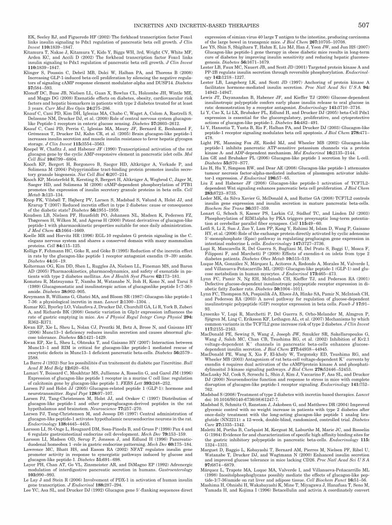
EK, Seeley RJ, and Figueredo HF (2002) The forkhead transcription factor Foxo1links insulin signaling to Pdx1 regulation of pancreatic beta cell growth. J ClinInvest 110:1839–1847.
Kitamura T, Nakae J, Kitamura Y, Kido Y, Biggs WH, 3rd, Wright CV, White MF,Arden KC, and Accili D (2002) The forkhead transcription factor Foxo1 linksinsulin signaling to Pdx1 regulation of pancreatic beta cell growth. J Clin Invest110:1839–1847.
Klinger S, Poussin C, Debril MB, Dolci W, Halban PA, and Thorens B (2008)Increasing GLP-1-induced beta-cell proliferation by silencing the negative regula-tors of signaling cAMP response element modulator-alpha and DUSP14. Diabetes57:584–593.
Klonoff DC, Buse JB, Nielsen LL, Guan X, Bowlus CL, Holcombe JH, Wintle ME,and Maggs DG (2008) Exenatide effects on diabetes, obesity, cardiovascular riskfactors and hepatic biomarkers in patients with type 2 diabetes treated for at least3 years. Curr Med Res Opin 24:275–286.
Knauf C, Cani PD, Kim DH, Iglesias MA, Chabo C, Waget A, Colom A, Rastrelli S,Delzenne NM, Drucker DJ, et al. (2008) Role of central nervous system glucagon-like Peptide-1 receptors in enteric glucose sensing. Diabetes 57:2603–2612.
Knauf C, Cani PD, Perrin C, Iglesias MA, Maury JF, Bernard E, Benhamed F,Gremeaux T, Drucker DJ, Kahn CR, et al. (2005) Brain glucagon-like peptide-1increases insulin secretion and muscle insulin resistance to favor hepatic glycogenstorage. J Clin Invest 115:3554–3563.
Knepel W, Chafitz J, and Habener JF (1990) Transcriptional activation of the ratglucagon gene by the cyclic AMP-responsive element in pancreatic islet cells. MolCell Biol 10:6799–6804.
Knoch KP, Bergert H, Borgonovo B, Saeger HD, Altkruger A, Verkade P, andSolimena M (2004) Polypyrimidine tract-binding protein promotes insulin secre-tory granule biogenesis. Nat Cell Biol 6:207–214.
Knoch KP, Meisterfeld R, Kersting S, Bergert H, Altkruger A, Wegbrod C, Jager M,Saeger HD, and Solimena M (2006) cAMP-dependent phosphorylation of PTB1promotes the expression of insulin secretory granule proteins in beta cells. CellMetab 3:123–134.
Knop FK, Vilsbøll T, Højberg PV, Larsen S, Madsbad S, Vølund A, Holst JJ, andKrarup T (2007) Reduced incretin effect in type 2 diabetes: cause or consequenceof the diabetic state? Diabetes 56:1951–1959.
Knudsen LB, Nielsen PF, Huusfeldt PO, Johansen NL, Madsen K, Pedersen FZ,Thøgersen H, Wilken M, and Agersø H (2000) Potent derivatives of glucagon-likepeptide-1 with pharmacokinetic properties suitable for once daily administration.J Med Chem 43:1664–1669.
Koelle MR and Horvitz HR (1996) EGL-10 regulates G protein signaling in the C.elegans nervous system and shares a conserved domain with many mammalianproteins. Cell 84:115–125.
Kolligs F, Fehmann HC, Goke R, and Goke B (1995) Reduction of the incretin effectin rats by the glucagon-like peptide 1 receptor antagonist exendin (9–39) amide.Diabetes 44:16–19.
Kolterman OG, Kim DD, Shen L, Ruggles JA, Nielsen LL, Fineman MS, and BaronAD (2005) Pharmacokinetics, pharmacodynamics, and safety of exenatide in pa-tients with type 2 diabetes mellitus. Am J Health Syst Pharm 62:173–181.
Komatsu R, Matsuyama T, Namba M, Watanabe N, Itoh H, Kono N, and Tarui S(1989) Glucagonostatic and insulinotropic action of glucagonlike peptide I-(7-36)-amide. Diabetes 38:902–905.
Kreymann B, Williams G, Ghatei MA, and Bloom SR (1987) Glucagon-like peptide-17-36: a physiological incretin in man. Lancet 2:1300–1304. .
Kumar KG, Byerley LO, Volaufova J, Drucker DJ, Churchill GA, Li R, York B, ZuberiA, and Richards BK (2008) Genetic variation in Glp1r expression influences therate of gastric emptying in mice. Am J Physiol Regul Integr Comp Physiol 294:R362–R371.
Kwan EP, Xie L, Sheu L, Nolan CJ, Prentki M, Betz A, Brose N, and Gaisano HY(2006) Munc13–1 deficiency reduces insulin secretion and causes abnormal glu-cose tolerance. Diabetes 55:1421–1429.
Kwan EP, Xie L, Sheu L, Ohtsuka T, and Gaisano HY (2007) Interaction betweenMunc13–1 and RIM is critical for glucagon-like peptide-1 mediated rescue ofexocytotic defects in Munc13–1 deficient pancreatic beta-cells. Diabetes 56:2579–2588.
La Barre J (1932) Sur les possibilites d’un traitement du diabete par l’incretine. BullAcad R Med Belg 12:620–634.
Lamari Y, Boissard C, Moukhtar MS, Jullienne A, Rosselin G, and Garel JM (1996)Expression of glucagon-like peptide 1 receptor in a murine C cell line: regulationof calcitonin gene by glucagon-like peptide 1. FEBS Lett 393:248–252.
Larsen PJ and Holst JJ (2005) Glucagon-related peptide 1 (GLP-1): hormone andneurotransmitter. Regul Pept 128:97–107.
Larsen PJ, Tang-Christensen M, Holst JJ, and Orskov C (1997) Distribution ofglucagon-like peptide-1 and other preproglucagon-derived peptides in the rathypothalamus and brainstem. Neuroscience 77:257–270.
Larsen PJ, Tang-Christensen M, and Jessop DS (1997) Central administration ofglucagon-like peptide-1 activates hypothalamic neuroendocrine neurons in the rat.Endocrinology 138:4445–4455.
Larsson LI, St-Onge L, Hougaard DM, Sosa-Pineda B, and Gruss P (1998) Pax 4 and6 regulate gastrointestinal endocrine cell development. Mech Dev 79:153–159.
Larsson LI, Madsen OD, Serup P, Jonsson J, and Edlund H (1996) Pancreatic-duodenal homeobox 1 -role in gastric endocrine patterning. Mech Dev 60:175–184.
Lawrence MC, Bhatt HS, and Easom RA (2002) NFAT regulates insulin genepromoter activity in response to synergistic pathways induced by glucose andglucagon-like peptide-1. Diabetes 51:691–698.
Layer PH, Chan AT, Go VL, Zinsmeister AR, and DiMagno EP (1992) Adrenergicmodulation of interdigestive pancreatic secretion in humans. Gastroenterology103:990–993.
Le Lay J and Stein R (2006) Involvement of PDX-1 in activation of human insulingene transcription. J Endocrinol 188:287–294.
Lee YC, Asa SL, and Drucker DJ (1992) Glucagon gene 5�-flanking sequences direct
expression of simian virus 40 large T antigen to the intestine, producing carcinomaof the large bowel in transgenic mice. J Biol Chem 267:10705–10708.
Lee YS, Shin S, Shigihara T, Hahm E, Liu MJ, Han J, Yoon JW, and Jun HS (2007)Glucagon-like peptide-1 gene therapy in obese diabetic mice results in long-termcure of diabetes by improving insulin sensitivity and reducing hepatic gluconeo-genesis. Diabetes 56:1671–1679.
Lester LB, Faux MC, Nauert JB, and Scott JD (2001) Targeted protein kinase A andPP-2B regulate insulin secretion through reversible phosphorylation. Endocrinol-ogy 142:1218–1227.
Lester LB, Langeberg LK, and Scott JD (1997) Anchoring of protein kinase Afacilitates hormone-mediated insulin secretion. Proc Natl Acad Sci U S A 94:14942–14947.
Lewis JT, Dayanandan B, Habener JF, and Kieffer TJ (2000) Glucose-dependentinsulinotropic polypeptide confers early phase insulin release to oral glucose inrats: demonstration by a receptor antagonist. Endocrinology 141:3710–3716.
Li Y, Cao X, Li LX, Brubaker PL, Edlund H, and Drucker DJ (2005) beta-Cell Pdx1expression is essential for the glucoregulatory, proliferative, and cytoprotectiveactions of glucagon-like peptide-1. Diabetes 54:482–491.
Li Y, Hansotia T, Yusta B, Ris F, Halban PA, and Drucker DJ (2003) Glucagon-likepeptide-1 receptor signaling modulates beta cell apoptosis. J Biol Chem 278:471–478.
Light PE, Manning Fox JE, Riedel MJ, and Wheeler MB (2002) Glucagon-likepeptide-1 inhibits pancreatic ATP-sensitive potassium channels via a proteinkinase A- and ADP-dependent mechanism. Mol Endocrinol 16:2135–2144.
Lim GE and Brubaker PL (2006) Glucagon-like peptide 1 secretion by the L-cell.Diabetes 55:S70–S77.
Liu H, Hu Y, Simpson RW, and Dear AE (2008) Glucagon-like peptide-1 attenuatestumour necrosis factor-alpha-mediated induction of plasmogen activator inhibi-tor-1 expression. J Endocrinol 196:57–65.
Liu Z and Habener JF (2008) Glucagon-like peptide-1 activation of TCF7L2-dependent Wnt signaling enhances pancreatic beta cell proliferation. J Biol Chem283:8723–8735.
Loder MK, da Silva Xavier G, McDonald A, and Rutter GA (2008) TCF7L2 controlsinsulin gene expression and insulin secretion in mature pancreatic beta-cells.Biochem Soc Trans 36:357–359.
Lonart G, Schoch S, Kaeser PS, Larkin CJ, Sudhof TC, and Linden DJ (2003)Phosphorylation of RIM1alpha by PKA triggers presynaptic long-term potentia-tion at cerebellar parallel fiber synapses. Cell 115:49–60.
Lotfi S, Li Z, Sun J, Zuo Y, Lam PP, Kang Y, Rahimi M, Islam D, Wang P, GaisanoHY, et al. (2006) Role of the exchange protein directly activated by cyclic adenosine5�-monophosphate (Epac) pathway in regulating proglucagon gene expression inintestinal endocrine L cells. Endocrinology 147:3727–3736.
Lupi R, Mancarella R, Del Guerra S, Bugliani M, Del Prato S, Boggi U, Mosca F,Filipponi F, and Marchetti P (2008) Effects of exendin-4 on islets from type 2diabetes patients. Diabetes Obes Metab 10:515–519.
Luque MA, Gonzalez N, Marquez L, Acitores A, Redondo A, Morales M, Valverde I,and Villanueva-Penacarrillo ML (2002) Glucagon-like peptide-1 (GLP-1) and glu-cose metabolism in human myocytes. J Endocrinol 173:465–473.
Lynn FC, Pamir N, Ng EH, McIntosh CH, Kieffer TJ, and Pederson RA (2001)Defective glucose-dependent insulinotropic polypeptide receptor expression in di-abetic fatty Zucker rats. Diabetes 50:1004–1011.
Lynn FC, Thompson SA, Pospisilik JA, Ehses JA, Hinke SA, Pamir N, McIntosh CH,and Pederson RA (2003) A novel pathway for regulation of glucose-dependentinsulinotropic polypeptide (GIP) receptor expression in beta cells. Faseb J 17:91–93.
Lyssenko V, Lupi R, Marchetti P, Del Guerra S, Orho-Melander M, Almgren P,Sjogren M, Ling C, Eriksson KF, Lethagen AL, et al. (2007) Mechanisms by whichcommon variants in the TCF7L2 gene increase risk of type 2 diabetes. J Clin Invest117:2155–2163.
MacDonald PE, Sewing S, Wang J, Joseph JW, Smukler SR, Sakellaropoulos G,Wang J, Saleh MC, Chan CB, Tsushima RG, et al. (2002) Inhibition of Kv2.1voltage-dependent K� channels in pancreatic beta-cells enhances glucose-dependent insulin secretion. J Biol Chem 277:44938–44945.
MacDonald PE, Wang X, Xia F, El-kholy W, Targonsky ED, Tsushima RG, andWheeler MB (2003) Antagonism of rat beta-cell voltage-dependent K� currents byexendin 4 requires dual activation of the cAMP/protein kinase A and phosphati-dylinositol 3-kinase signaling pathways. J Biol Chem 278:52446–52453.
MacLusky NJ, Cook S, Scrocchi L, Shin J, Kim J, Vaccarino F, Asa SL, and DruckerDJ (2000) Neuroendocrine function and response to stress in mice with completedisruption of glucagon-like peptide-1 receptor signaling. Endocrinology 141:752–762.
Madsbad S (2008) Treatment of type 2 diabetes with incretin-based therapies. Lancetdoi: 10.1016/S0140-6736(08)61247-7.
Madsbad S, Schmitz O, Ranstam J, Jakobsen G, and Matthews DR (2004) Improvedglycemic control with no weight increase in patients with type 2 diabetes afteronce-daily treatment with the long-acting glucagon-like peptide 1 analog lira-glutide (NN2211): a 12-week, double-blind, randomized, controlled trial. DiabetesCare 27:1335–1342.
Maletti M, Portha B, Carlquist M, Kergoat M, Laburthe M, Marie JC, and RosselinG (1984) Evidence for and characterization of specific high affinity binding sites forthe gastric inhibitory polypeptide in pancreatic beta-cells. Endocrinology 115:1324–1331.
Marguet D, Baggio L, Kobayashi T, Bernard AM, Pierres M, Nielsen PF, Ribel U,Watanabe T, Drucker DJ, and Wagtmann N (2000) Enhanced insulin secretionand improved glucose tolerance in mice lacking CD26. Proc Natl Acad Sci U S A97:6874–6879.
Marquez L, Trapote MA, Luque MA, Valverde I, and Villanueva-Penacarrillo ML(1998) Inositolphosphoglycans possibly mediate the effects of glucagon-like pep-tide-1(7-36)amide on rat liver and adipose tissue. Cell Biochem Funct 16:51–56.
Mashima H, Ohnishi H, Wakabayashi K, Mine T, Miyagawa J, Hanafusa T, Seno M,Yamada H, and Kojima I (1996) Betacellulin and activin A coordinately convert
INCRETINS AND INCRETIN-BASED THERAPIES 507
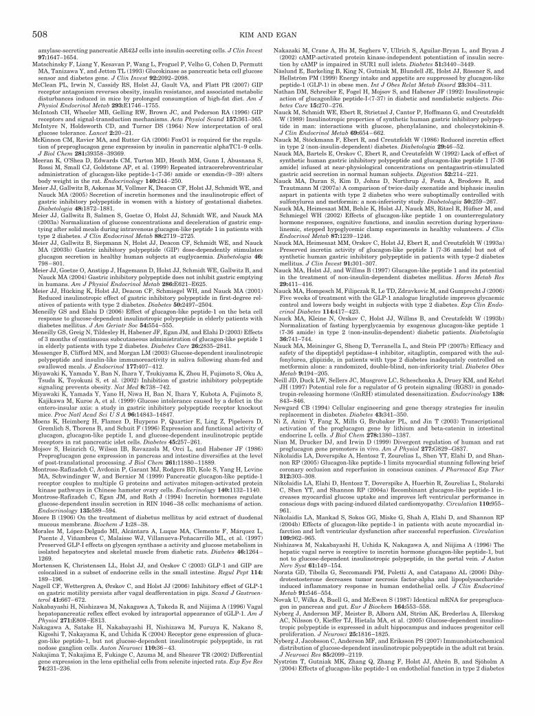
amylase-secreting pancreatic AR42J cells into insulin-secreting cells. J Clin Invest97:1647–1654.
Matschinsky F, Liang Y, Kesavan P, Wang L, Froguel P, Velho G, Cohen D, PermuttMA, Tanizawa Y, and Jetton TL (1993) Glucokinase as pancreatic beta cell glucosesensor and diabetes gene. J Clin Invest 92:2092–2098.
McClean PL, Irwin N, Cassidy RS, Holst JJ, Gault VA, and Flatt PR (2007) GIPreceptor antagonism reverses obesity, insulin resistance, and associated metabolicdisturbances induced in mice by prolonged consumption of high-fat diet. Am JPhysiol Endocrinol Metab 293:E1746–1755.
McIntosh CH, Wheeler MB, Gelling RW, Brown JC, and Pederson RA (1996) GIPreceptors and signal-transduction mechanisms. Acta Physiol Scand 157:361–365.
McIntyre N, Holdsworth CD, and Turner DS (1964) New interpretation of oralglucose tolerance. Lancet 2:20–21.
McKinnon CM, Ravier MA, and Rutter GA (2006) FoxO1 is required for the regula-tion of preproglucagon gene expression by insulin in pancreatic alphaTC1–9 cells.J Biol Chem 281:39358–39369.
Meeran K, O’Shea D, Edwards CM, Turton MD, Heath MM, Gunn I, Abusnana S,Rossi M, Small CJ, Goldstone AP, et al. (1999) Repeated intracerebroventricularadministration of glucagon-like peptide-1-(7-36) amide or exendin-(9–39) altersbody weight in the rat. Endocrinology 140:244–250.
Meier JJ, Gallwitz B, Askenas M, Vollmer K, Deacon CF, Holst JJ, Schmidt WE, andNauck MA (2005) Secretion of incretin hormones and the insulinotropic effect ofgastric inhibitory polypeptide in women with a history of gestational diabetes.Diabetologia 48:1872–1881.
Meier JJ, Gallwitz B, Salmen S, Goetze O, Holst JJ, Schmidt WE, and Nauck MA(2003a) Normalization of glucose concentrations and deceleration of gastric emp-tying after solid meals during intravenous glucagon-like peptide 1 in patients withtype 2 diabetes. J Clin Endocrinol Metab 88:2719–2725.
Meier JJ, Gallwitz B, Siepmann N, Holst JJ, Deacon CF, Schmidt WE, and NauckMA (2003b) Gastric inhibitory polypeptide (GIP) dose-dependently stimulatesglucagon secretion in healthy human subjects at euglycaemia. Diabetologia 46:798–801.
Meier JJ, Goetze O, Anstipp J, Hagemann D, Holst JJ, Schmidt WE, Gallwitz B, andNauck MA (2004) Gastric inhibitory polypeptide does not inhibit gastric emptyingin humans. Am J Physiol Endocrinol Metab 286:E621–E625.
Meier JJ, Hucking K, Holst JJ, Deacon CF, Schmiegel WH, and Nauck MA (2001)Reduced insulinotropic effect of gastric inhibitory polypeptide in first-degree rel-atives of patients with type 2 diabetes. Diabetes 50:2497–2504.
Meneilly GS and Elahi D (2006) Effect of glucagon-like peptide-1 on the beta cellresponse to glucose-dependent insulinotropic polypeptide in elderly patients withdiabetes mellitus. J Am Geriatr Soc 54:554–555.
Meneilly GS, Greig N, Tildesley H, Habener JF, Egan JM, and Elahi D (2003) Effectsof 3 months of continuous subcutaneous administration of glucagon-like peptide 1in elderly patients with type 2 diabetes. Diabetes Care 26:2835–2841.
Messenger B, Clifford MN, and Morgan LM (2003) Glucose-dependent insulinotropicpolypeptide and insulin-like immunoreactivity in saliva following sham-fed andswallowed meals. J Endocrinol 177:407–412.
Miyawaki K, Yamada Y, Ban N, Ihara Y, Tsukiyama K, Zhou H, Fujimoto S, Oku A,Tsuda K, Toyokuni S, et al. (2002) Inhibition of gastric inhibitory polypeptidesignaling prevents obesity. Nat Med 8:738–742.
Miyawaki K, Yamada Y, Yano H, Niwa H, Ban N, Ihara Y, Kubota A, Fujimoto S,Kajikawa M, Kuroe A, et al. (1999) Glucose intolerance caused by a defect in theentero-insular axis: a study in gastric inhibitory polypeptide receptor knockoutmice. Proc Natl Acad Sci U S A 96:14843–14847.
Moens K, Heimberg H, Flamez D, Huypens P, Quartier E, Ling Z, Pipeleers D,Gremlich S, Thorens B, and Schuit F (1996) Expression and functional activity ofglucagon, glucagon-like peptide I, and glucose-dependent insulinotropic peptidereceptors in rat pancreatic islet cells. Diabetes 45:257–261.
Mojsov S, Heinrich G, Wilson IB, Ravazzola M, Orci L, and Habener JF (1986)Preproglucagon gene expression in pancreas and intestine diversifies at the levelof post-translational processing. J Biol Chem 261:11880–11889.
Montrose-Rafizadeh C, Avdonin P, Garant MJ, Rodgers BD, Kole S, Yang H, LevineMA, Schwindinger W, and Bernier M (1999) Pancreatic glucagon-like peptide-1receptor couples to multiple G proteins and activates mitogen-activated proteinkinase pathways in Chinese hamster ovary cells. Endocrinology 140:1132–1140.
Montrose-Rafizadeh C, Egan JM, and Roth J (1994) Incretin hormones regulateglucose-dependent insulin secretion in RIN 1046–38 cells: mechanisms of action.Endocrinology 135:589–594.
Moore B (1906) On the treatment of diabetus mellitus by acid extract of duodenalmucous membrane. Biochem J 1:28–38.
Morales M, Lopez-Delgado MI, Alcantara A, Luque MA, Clemente F, Marquez L,Puente J, Vinambres C, Malaisse WJ, Villanueva-Penacarrillo ML, et al. (1997)Preserved GLP-I effects on glycogen synthase a activity and glucose metabolism inisolated hepatocytes and skeletal muscle from diabetic rats. Diabetes 46:1264–1269.
Mortensen K, Christensen LL, Holst JJ, and Orskov C (2003) GLP-1 and GIP arecolocalized in a subset of endocrine cells in the small intestine. Regul Pept 114:189–196.
Nagell CF, Wettergren A, Ørskov C, and Holst JJ (2006) Inhibitory effect of GLP-1on gastric motility persists after vagal deafferentation in pigs. Scand J Gastroen-terol 41:667–672.
Nakabayashi H, Nishizawa M, Nakagawa A, Takeda R, and Niijima A (1996) Vagalhepatopancreatic reflex effect evoked by intraportal appearance of tGLP-1. Am JPhysiol 271:E808–E813.
Nakagawa A, Satake H, Nakabayashi H, Nishizawa M, Furuya K, Nakano S,Kigoshi T, Nakayama K, and Uchida K (2004) Receptor gene expression of gluca-gon-like peptide-1, but not glucose-dependent insulinotropic polypeptide, in ratnodose ganglion cells. Auton Neurosci 110:36–43.
Nakajima T, Nakajima E, Fukiage C, Azuma M, and Shearer TR (2002) Differentialgene expression in the lens epithelial cells from selenite injected rats. Exp Eye Res74:231–236.
Nakazaki M, Crane A, Hu M, Seghers V, Ullrich S, Aguilar-Bryan L, and Bryan J(2002) cAMP-activated protein kinase-independent potentiation of insulin secre-tion by cAMP is impaired in SUR1 null islets. Diabetes 51:3440–3449.
Naslund E, Barkeling B, King N, Gutniak M, Blundell JE, Holst JJ, Rossner S, andHellstrom PM (1999) Energy intake and appetite are suppressed by glucagon-likepeptide-1 (GLP-1) in obese men. Int J Obes Relat Metab Disord 23:304–311.
Nathan DM, Schreiber E, Fogel H, Mojsov S, and Habener JF (1992) Insulinotropicaction of glucagonlike peptide-I-(7-37) in diabetic and nondiabetic subjects. Dia-betes Care 15:270–276.
Nauck M, Schmidt WE, Ebert R, Strietzel J, Cantor P, Hoffmann G, and CreutzfeldtW (1989) Insulinotropic properties of synthetic human gastric inhibitory polypep-tide in man: interactions with glucose, phenylalanine, and cholecystokinin-8.J Clin Endocrinol Metab 69:654–662.
Nauck M, Stockmann F, Ebert R, and Creutzfeldt W (1986) Reduced incretin effectin type 2 (non-insulin-dependent) diabetes. Diabetologia 29:46–52.
Nauck MA, Bartels E, Orskov C, Ebert R, and Creutzfeldt W (1992) Lack of effect ofsynthetic human gastric inhibitory polypeptide and glucagon-like peptide 1 [7-36amide] infused at near-physiological concentrations on pentagastrin-stimulatedgastric acid secretion in normal human subjects. Digestion 52:214–221.
Nauck MA, Duran S, Kim D, Johns D, Northrup J, Festa A, Brodows R, andTrautmann M (2007a) A comparison of twice-daily exenatide and biphasic insulinaspart in patients with type 2 diabetes who were suboptimally controlled withsulfonylurea and metformin: a non-inferiority study. Diabetologia 50:259–267.
Nauck MA, Heimesaat MM, Behle K, Holst JJ, Nauck MS, Ritzel R, Hufner M, andSchmiegel WH (2002) Effects of glucagon-like peptide 1 on counterregulatoryhormone responses, cognitive functions, and insulin secretion during hyperinsu-linemic, stepped hypoglycemic clamp experiments in healthy volunteers. J ClinEndocrinol Metab 87:1239–1246.
Nauck MA, Heimesaat MM, Orskov C, Holst JJ, Ebert R, and Creutzfeldt W (1993a)Preserved incretin activity of glucagon-like peptide 1 [7-36 amide] but not ofsynthetic human gastric inhibitory polypeptide in patients with type-2 diabetesmellitus. J Clin Invest 91:301–307.
Nauck MA, Holst JJ, and Willms B (1997) Glucagon-like peptide 1 and its potentialin the treatment of non-insulin-dependent diabetes mellitus. Horm Metab Res29:411–416.
Nauck MA, Hompesch M, Filipczak R, Le TD, Zdravkovic M, and Gumprecht J (2006)Five weeks of treatment with the GLP-1 analogue liraglutide improves glycaemiccontrol and lowers body weight in subjects with type 2 diabetes. Exp Clin Endo-crinol Diabetes 114:417–423.
Nauck MA, Kleine N, Orskov C, Holst JJ, Willms B, and Creutzfeldt W (1993b)Normalization of fasting hyperglycaemia by exogenous glucagon-like peptide 1(7-36 amide) in type 2 (non-insulin-dependent) diabetic patients. Diabetologia36:741–744.
Nauck MA, Meininger G, Sheng D, Terranella L, and Stein PP (2007b) Efficacy andsafety of the dipeptidyl peptidase-4 inhibitor, sitagliptin, compared with the sul-fonylurea, glipizide, in patients with type 2 diabetes inadequately controlled onmetformin alone: a randomized, double-blind, non-inferiority trial. Diabetes ObesMetab 9:194–205.
Neill JD, Duck LW, Sellers JC, Musgrove LC, Scheschonka A, Druey KM, and KehrlJH (1997) Potential role for a regulator of G protein signaling (RGS3) in gonado-tropin-releasing hormone (GnRH) stimulated desensitization. Endocrinology 138:843–846.
Newgard CB (1994) Cellular engineering and gene therapy strategies for insulinreplacement in diabetes. Diabetes 43:341–350.
Ni Z, Anini Y, Fang X, Mills G, Brubaker PL, and Jin T (2003) Transcriptionalactivation of the proglucagon gene by lithium and beta-catenin in intestinalendocrine L cells. J Biol Chem 278:1380–1387.
Nian M, Drucker DJ, and Irwin D (1999) Divergent regulation of human and ratproglucagon gene promoters in vivo. Am J Physiol 277:G829–G837.
Nikolaidis LA, Doverspike A, Hentosz T, Zourelias L, Shen YT, Elahi D, and Shan-non RP (2005) Glucagon-like peptide-1 limits myocardial stunning following briefcoronary occlusion and reperfusion in conscious canines. J Pharmacol Exp Ther312:303–308.
Nikolaidis LA, Elahi D, Hentosz T, Doverspike A, Huerbin R, Zourelias L, StolarskiC, Shen YT, and Shannon RP (2004a) Recombinant glucagon-like peptide-1 in-creases myocardial glucose uptake and improves left ventricular performance inconscious dogs with pacing-induced dilated cardiomyopathy. Circulation 110:955–961.
Nikolaidis LA, Mankad S, Sokos GG, Miske G, Shah A, Elahi D, and Shannon RP(2004b) Effects of glucagon-like peptide-1 in patients with acute myocardial in-farction and left ventricular dysfunction after successful reperfusion. Circulation109:962–965.
Nishizawa M, Nakabayashi H, Uchida K, Nakagawa A, and Niijima A (1996) Thehepatic vagal nerve is receptive to incretin hormone glucagon-like peptide-1, butnot to glucose-dependent insulinotropic polypeptide, in the portal vein. J AutonNerv Syst 61:149–154.
Norata GD, Tibolla G, Seccomandi PM, Poletti A, and Catapano AL (2006) Dihy-drotestosterone decreases tumor necrosis factor-alpha and lipopolysaccharide-induced inflammatory response in human endothelial cells. J Clin EndocrinolMetab 91:546–554.
Novak U, Wilks A, Buell G, and McEwen S (1987) Identical mRNA for preprogluca-gon in pancreas and gut. Eur J Biochem 164:553–558.
Nyberg J, Anderson MF, Meister B, Alborn AM, Strom AK, Brederlau A, IllerskogAC, Nilsson O, Kieffer TJ, Hietala MA, et al. (2005) Glucose-dependent insulino-tropic polypeptide is expressed in adult hippocampus and induces progenitor cellproliferation. J Neurosci 25:1816–1825.
Nyberg J, Jacobsson C, Anderson MF, and Eriksson PS (2007) Immunohistochemicaldistribution of glucose-dependent insulinotropic polypeptide in the adult rat brain.J Neurosci Res 85:2099–2119.
Nystrom T, Gutniak MK, Zhang Q, Zhang F, Holst JJ, Ahren B, and Sjoholm A(2004) Effects of glucagon-like peptide-1 on endothelial function in type 2 diabetes
508 KIM AND EGAN
patients with stable coronary artery disease. Am J Physiol Endocrinol Metab287:E1209–1215.
Offield MF, Jetton TL, Labosky PA, Ray M, Stein RW, Magnuson MA, Hogan BL,and Wright CV (1996) PDX-1 is required for pancreatic outgrowth and differenti-ation of the rostral duodenum. Development 122:983–995.
Ohlsson H, Karlsson K, and Edlund T (1993) IPF1, a homeodomain-containingtransactivator of the insulin gene. EMBO J 12:4251–4259.
Orskov C, Holst JJ, Knuhtsen S, Baldissera FG, Poulsen SS, and Nielsen OV (1986)Glucagon-like peptides GLP-1 and GLP-2, predicted products of the glucagon gene,are secreted separately from pig small intestine but not pancreas. Endocrinology119:1467–1475.
Orskov C and Poulsen SS (1991) Glucagonlike peptide-I-(7-36)-amide receptors onlyin islets of Langerhans. Autoradiographic survey of extracerebral tissues in rats.Diabetes 40:1292–1296.
Orskov C, Poulsen SS, Møller M, and Holst JJ (1996) Glucagon-like peptide Ireceptors in the subfornical organ and the area postrema are accessible to circu-lating glucagon-like peptide I. Diabetes 45:832–835.
Otonkoski T and Hayek A (1995) Constitution of a biphasic insulin response toglucose in human fetal pancreatic beta-cells with glucagon-like peptide 1. J ClinEndocrinol Metab 80:3779–3783.
Ozaki N, Shibasaki T, Kashima Y, Miki T, Takahashi K, Ueno H, Sunaga Y, Yano H,Matsuura Y, Iwanaga T, et al. (2000) cAMP-GEFII is a direct target of cAMP inregulated exocytosis. Nat Cell Biol 2:805–811.
Pamir N, Lynn FC, Buchan AM, Ehses J, Hinke SA, Pospisilik JA, Miyawaki K,Yamada Y, Seino Y, McIntosh CH, et al. (2003) Glucose-dependent insulinotropicpolypeptide receptor null mice exhibit compensatory changes in the enteroinsularaxis. Am J Physiol Endocrinol Metab 284:E931–E939.
Park S, Dong X, Fisher TL, Dunn S, Omer AK, Weir G, and White MF (2006)Exendin-4 uses Irs2 signaling to mediate pancreatic beta cell growth and function.J Biol Chem 281:1159–1168.
Parkes D, Jodka C, Smith P, Nayak S, Rinehart L, Gingerich R, Chen K, and YoungA (2001) Pharmacokinetic actions of exendin-4 in the rat: comparison with gluca-gon-like peptide-1. Drug Dev Res 53:260–267.
Pederson RA and Brown JC (1972) Inhibition of histamine-, pentagastrin-, andinsulin-stimulated canine gastric secretion by pure “gastric inhibitory polypep-tide”. Gastroenterology 62:393–400.
Pederson RA and Brown JC (1978) Interaction of gastric inhibitory polypeptide,glucose, and arginine on insulin and glucagon secretion from the perfused ratpancreas. Endocrinology 103:610–615.
Pederson RA, Satkunarajah M, McIntosh CH, Scrocchi LA, Flamez D, Schuit F,Drucker DJ, and Wheeler MB (1998) Enhanced glucose-dependent insulinotropicpolypeptide secretion and insulinotropic action in glucagon-like peptide 1 receptor�/� mice. Diabetes 47:1046–1052.
Pederson RA, Schubert HE, and Brown JC (1975) Gastric inhibitory polypeptide. Itsphysiologic release and insulinotropic action in the dog. Diabetes 24:1050–1056.
Perfetti R, Zhou J, Doyle ME, and Egan JM (2000) Glucagon-like peptide-1 inducescell proliferation and pancreatic-duodenum homeobox-1 expression and increasesendocrine cell mass in the pancreas of old, glucose-intolerant rats. Endocrinology141:4600–4605.
Perley MJ and Kipnis DM (1967) Plasma insulin responses to oral and intravenousglucose: studies in normal and diabetic sujbjects. J Clin Invest 46:1954–1962.
Persson K, Gingerich RL, Nayak S, Wada K, Wada E, and Ahren B (2000) ReducedGLP-1 and insulin responses and glucose intolerance after gastric glucose in GRPreceptor-deleted mice. Am J Physiol Endocrinol Metab 279:E956–E962.
Philippe J (1989) Glucagon gene transcription is negatively regulated by insulin ina hamster islet cell line. J Clin Invest 84:672–677.
Philippe J (1991) Insulin regulation of the glucagon gene is mediated by an insulin-responsive DNA element. Proc Natl Acad Sci U S A 88:7224–7227.
Philippe J, Drucker DJ, Knepel W, Jepeal L, Misulovin Z, and Habener JF (1988)Alpha-cell-specific expression of the glucagon gene is conferred to the glucagonpromoter element by the interactions of DNA-binding proteins. Mol Cell Biol8:4877–4888.
Pipeleers DG, Schuit FC, Van Schravendijk CF, and Van de Winkel M (1985)Interplay of nutrients and hormones in the regulation of glucagon release. Endo-crinology 117:817–823.
Piteau S, Olver A, Kim SJ, Winter K, Pospisilik JA, Lynn F, Manhart S, Demuth HU,Speck M, Pederson RA, et al. (2007) Reversal of islet GIP receptor down-regulationand resistance to GIP by reducing hyperglycemia in the Zucker rat. BiochemBiophys Res Commun 362:1007–1012.
Plamboeck A, Holst JJ, Carr RD, and Deacon CF (2005) Neutral endopeptidase 24.11and dipeptidyl peptidase IV are both mediators of the degradation of glucagon-likepeptide 1 in the anaesthetised pig. Diabetologia 48:1882–1890.
Preitner F, Ibberson M, Franklin I, Binnert C, Pende M, Gjinovci A, Hansotia T,Drucker DJ, Wollheim C, Burcelin R, et al. (2004) Gluco-incretins control insulinsecretion at multiple levels as revealed in mice lacking GLP-1 and GIP receptors.J Clin Invest 113:635–645.
Premont RT, Inglese J, and Lefkowitz RJ (1995) Protein kinases that phosphorylateactivated G protein-coupled receptors. Faseb J 9:175–182.
Prigeon RL, Quddusi S, Paty B, and D’Alessio DA (2003) Suppression of glucoseproduction by GLP-1 independent of islet hormones: a novel extrapancreatic effect.Am J Physiol Endocrinol Metab 285:E701–E707.
Qin X, Shen H, Liu M, Yang Q, Zheng S, Sabo M, D’Alessio DA, and Tso P (2005)GLP-1 reduces intestinal lymph flow, triglyceride absorption, and apolipoproteinproduction in rats. Am J Physiol Gastrointest Liver Physiol 288:G943–G949.
Rachman J, Barrow BA, Levy JC, and Turner RC (1997) Near-normalisation ofdiurnal glucose concentrations by continuous administration of glucagon-like pep-tide-1 (GLP-1) in subjects with NIDDM. Diabetologia 40:205–211.
Ranta F, Avram D, Berchtold S, Dufer M, Drews G, Lang F, and Ullrich S (2006)Dexamethasone induces cell death in insulin-secreting cells, an effect reversed byexendin-4. Diabetes 55:1380–1390.
Raz I, Hanefeld M, Xu L, Caria C, Williams-Herman D, and Khatami H (2006)
Efficacy and safety of the dipeptidyl peptidase-4 inhibitor sitagliptin as mono-therapy in patients with type 2 diabetes mellitus. Diabetologia 49:2564–2571.
Redondo A, Trigo MV, Acitores A, Valverde I, and Villanueva-Penacarrillo ML (2003)Cell signalling of the GLP-1 action in rat liver. Mol Cell Endocrinol 204:43–50.
Reneke JE, Blumer KJ, Courchesne WE, and Thorner J (1988) The carboxy-terminalsegment of the yeast alpha-factor receptor is a regulatory domain. Cell 55:221–234.
Richter G, Feddersen O, Wagner U, Barth P, Goke R, and Goke B (1993) GLP-1stimulates secretion of macromolecules from airways and relaxes pulmonary ar-tery. Am J Physiol 265:L374–L381.
Rinaman L (1999) Interoceptive stress activates glucagon-like peptide-1 neuronsthat project to the hypothalamus. Am J Physiol 277:R582–R590.
Rizos CV, Liberopoulos EN, Mikhailidis DP, and Elisaf MS (2008) Pleiotropic effectsof thiazolidinediones. Expert Opin Pharmacother 9:1087–1108.
Roberge JN and Brubaker PL (1991) Secretion of proglucagon-derived peptides inresponse to intestinal luminal nutrients. Endocrinology 128:3169–3174.
Roberge JN, Gronau KA, and Brubaker PL (1996) Gastrin-releasing peptide is anovel mediator of proximal nutrient-induced proglucagon-derived peptide secre-tion from the distal gut. Endocrinology 137:2383–2388.
Rocca AS and Brubaker PL (1999) Role of the vagus nerve in mediating proximalnutrient-induced glucagon-like peptide-1 secretion. Endocrinology 140:1687–1694.
Rosenstock J, Brazg R, Andryuk PJ, Lu K, and Stein P (2006) Efficacy and safety ofthe dipeptidyl peptidase-4 inhibitor sitagliptin added to ongoing pioglitazone ther-apy in patients with type 2 diabetes: a 24-week, multicenter, randomized, double-blind, placebo-controlled, parallel-group study. Clin Ther 28:1556–1568.
Ross SA, Brown JC, and Dupre J (1977) Hypersecretion of gastric inhibitory polypep-tide following oral glucose in diabetes mellitus. Diabetes 26:525–529.
Ross SA and Dupre J (1978) Effects of ingestion of triglyceride or galactose onsecretion of gastric inhibitory polypeptide and on responses to intravenous glucosein normal and diabetic subjects. Diabetes 27:327–333.
Rothenberg ME, Eilertson CD, Klein K, Zhou Y, Lindberg I, McDonald JK, MackinRB, and Noe BD (1995) Processing of mouse proglucagon by recombinant prohor-mone convertase 1 and immunopurified prohormone convertase 2 in vitro. J BiolChem 270:10136–10146.
Rouille Y, Martin S, and Steiner DF (1995) Differential processing of proglucagon bythe subtilisin-like prohormone convertases PC2 and PC3 to generate either glu-cagon or glucagon-like peptide. J Biol Chem 270:26488–26496.
Rouille Y, Westermark G, Martin SK, and Steiner DF (1994) Proglucagon is pro-cessed to glucagon by prohormone convertase PC2 in alpha TC1–6 cells. Proc NatlAcad Sci U S A 91:3242–3246.
Ruiz-Grande C, Alarcon C, Alcantara A, Castilla C, Lopez Novoa JM, Villanueva-Penacarrillo ML, and Valverde I (1993) Renal catabolism of truncated glucagon-like peptide 1. Horm Metab Res 25:612–616.
Ryan AS, Egan JM, Habener JF, and Elahi D (1998) Insulinotropic hormone gluca-gon-like peptide-1-(7-37) appears not to augment insulin-mediated glucose uptakein young men during euglycemia. J Clin Endocrinol Metab 83:2399–2404.
Sagar SM, Sharp FR, and Curran T (1988) Expression of c-fos protein in brain:metabolic mapping at the cellular level. Science 240:1328–1331.
Salehi A, Meidute Abaraviciene S, Jimenez-Feltstrom J, Ostenson CG, Efendic S,and Lundquist I (2008) Excessive islet NO generation in type 2 diabetic GK ratscoincides with abnormal hormone secretion and is counteracted by GLP-1. PLoSONE 3:e2165.
Sancho V, Nuche B, Arnes L, Cancelas J, Gonzalez N, Díaz-Miguel M, Martín-DuceA, Valverde I, and Villanueva-Penacarrillo ML (2007) The action of GLP-1 andexendins upon glucose transport in normal human adipocytes, and on kinaseactivity as compared with morbidly obese patients. Int J Mol Med 19:961–966.
Sancho V, Trigo MV, Gonzalez N, Valverde I, Malaisse WJ, and Villanueva-Penacarrillo ML (2005) Effects of glucagon-like peptide-1 and exendins on kinaseactivity, glucose transport and lipid metabolism in adipocytes from normal andtype-2 diabetic rats. J Mol Endocrinol 35:27–38.
Sander M, Neubuser A, Kalamaras J, Ee HC, Martin GR, and German MS (1997)Genetic analysis reveals that PAX6 is required for normal transcription of pan-creatic hormone genes and islet development. Genes Dev 11:1662–1673.
Sandhu H, Wiesenthal SR, MacDonald PE, McCall RH, Tchipashvili V, Rashid S,Satkunarajah M, Irwin DM, Shi ZQ, Brubaker PL, et al. (1999) Glucagon-likepeptide 1 increases insulin sensitivity in depancreatized dogs. Diabetes 48:1045–1053.
Sandoval DA, Bagnol D, Woods SC, D’Alessio DA, and Seeley RJ (2008) ArcuateGLP-1 receptors regulate glucose homeostasis but not food intake. Diabetes 57:2046–2054.
Sarkar S, Fekete C, Legradi G, and Lechan RM (2003) Glucagon like peptide-1 (7-36)amide (GLP-1) nerve terminals densely innervate corticotropin-releasing hormoneneurons in the hypothalamic paraventricular nucleus. Brain Res 985:163–168.
Schauder P, Brown JC, Frerichs H, and Creutzfeldt W (1975) Gastric inhibitorypolypeptide: effect on glucose-induced insulin release from isolated rat pancreaticislets in vitro. Diabetologia 11:483–484.
Schinner S, Barthel A, Dellas C, Grzeskowiak R, Sharma SK, Oetjen E, Blume R,and Knepel W (2005) Protein kinase B activity is sufficient to mimic the effect ofinsulin on glucagon gene transcription. J Biol Chem 280:7369–7376.
Schirra J, Katschinski M, Weidmann C, Schafer T, Wank U, Arnold R, and Goke B(1996) Gastric emptying and release of incretin hormones after glucose ingestionin humans. J Clin Invest 97:92–103.
Schirra J, Nicolaus M, Roggel R, Katschinski M, Storr M, Woerle HJ, and Goke B(2006) Endogenous glucagon-like peptide 1 controls endocrine pancreatic secretionand antro-pyloro-duodenal motility in humans. Gut 55:243–251.
Schmid R, Schusdziarra V, Aulehner R, Weigert N, and Classen M (1990) Compar-ison of GLP-1 (7-36amide) and GIP on release of somatostatin-like immunoreac-tivity and insulin from the isolated rat pancreas. Z Gastroenterol 28:280–284.
Schmidt WE, Siegel EG, and Creutzfeldt W (1985) Glucagon-like peptide-1 but notglucagon-like peptide-2 stimulates insulin release from isolated rat pancreaticislets. Diabetologia 28:704–707.
INCRETINS AND INCRETIN-BASED THERAPIES 509

Schwartz AV, Sellmeyer DE, Vittinghoff E, Palermo L, Lecka-Czernik B, FeingoldKR, Strotmeyer ES, Resnick HE, Carbone L, Beamer BA, et al. (2006) Thiazo-lidinedione use and bone loss in older diabetic adults. J Clin Endocrinol Metab91:3349–3354.
Scopsi L, Gullo M, Rilke F, Martin S, and Steiner DF (1995) Proprotein convertases(PC1/PC3 and PC2) in normal and neoplastic human tissues: their use as markersof neuroendocrine differentiation. J Clin Endocrinol Metab 80:294–301.
Scrocchi LA, Brown TJ, MaClusky N, Brubaker PL, Auerbach AB, Joyner AL, andDrucker DJ (1996) Glucose intolerance but normal satiety in mice with a nullmutation in the glucagon-like peptide 1 receptor gene. Nat Med 2:1254–1258.
Scrocchi LA, Marshall BA, Cook SM, Brubaker PL, and Drucker DJ (1998) Identi-fication of glucagon-like peptide 1 (GLP-1) actions essential for glucose homeosta-sis in mice with disruption of GLP-1 receptor signaling. Diabetes 47:632–639.
Shibasaki T, Sunaga Y, and Seino S (2004) Integration of ATP, cAMP, and Ca2�
signals in insulin granule exocytosis. Diabetes 53 (Suppl 3):S59–S62.Shin YK, Martin B, Golden E, Dotson CD, Maudsley S, Kim W, Jang HJ, Mattson
MP, Drucker DJ, Egan JM, et al. (2008) Modulation of taste sensitivity by GLP-1signaling. J Neurochem 106:455–463.
Shiota C, Larsson O, Shelton KD, Shiota M, Efanov AM, Hoy M, Lindner J, Koop-tiwut S, Juntti-Berggren L, Gromada J, et al. (2002) Sulfonylurea receptor type 1knock-out mice have intact feeding-stimulated insulin secretion despite markedimpairment in their response to glucose. J Biol Chem 277:37176–37183.
Shughrue PJ, Lane MV, and Merchenthaler I (1996) Glucagon-like peptide-1 recep-tor (GLP1-R) mRNA in the rat hypothalamus. Endocrinology 137:5159–5162.
Simonsen L, Holst JJ, and Deacon CF (2006) Exendin-4, but not glucagon-likepeptide-1, is cleared exclusively by glomerular filtration in anaesthetised pigs.Diabetologia 49:706–712.
Skoglund G, Hussain MA, and Holz GG (2000) Glucagon-like peptide 1 stimulatesinsulin gene promoter activity by protein kinase A-independent activation of therat insulin I gene cAMP response element. Diabetes 49:1156–1164.
Sokos GG, Nikolaidis LA, Mankad S, Elahi D, and Shannon RP (2006) Glucagon-likepeptide-1 infusion improves left ventricular ejection fraction and functional statusin patients with chronic heart failure. J Card Fail 12:694–699.
Someya Y, Inagaki N, Maekawa T, Seino Y, and Ishii S (1993) Two 3�,5�-cyclic-adenosine monophosphate response elements in the promoter region of the humangastric inhibitory polypeptide gene. FEBS Lett 317:67–73.
Sondhi S, Castellano JM, Chong VZ, Rogoza RM, Skoblenick KJ, Dyck BA, GabrieleJ, Thomas N, Ki K, Pristupa ZB, et al. (2006) cDNA array reveals increasedexpression of glucose-dependent insulinotropic polypeptide following chronic clo-zapine treatment: role in atypical antipsychotic drug-induced adverse metaboliceffects. Pharmacogenomics J 6:131–140.
Song DH, Getty-Kaushik L, Tseng E, Simon J, Corkey BE, and Wolfe MM (2007)Glucose-dependent insulinotropic polypeptide enhances adipocyte developmentand glucose uptake in part through Akt activation. Gastroenterology 133:1796–1805.
Sonne DP, Engstrøm T, and Treiman M (2008) Protective effects of GLP-1 analoguesexendin-4 and GLP-1(9–36) amide against ischemia-reperfusion injury in ratheart. Regul Pept 146:243–249.
Sonoda N, Imamura T, Yoshizaki T, Babendure JL, Lu JC, and Olefsky JM (2008)Beta-Arrestin-1 mediates glucagon-like peptide-1 signaling to insulin secretion incultured pancreatic beta cells. Proc Natl Acad Sci U S A 105:6614–6619.
Starling EH (1905) The Cronian Lectures on the chemical correlation of the functionsof the body. Lancet 2:339–341, 423–425, 501–503, 579–583.
Stein PP, Williams-Herman D, Khatami H, Meininger G, Round E, Sheng D,Sanchez M, Lunceford J, Kaufman KD, and Amatruda JM (2007) Sitagliptin, aselective DPP-4 Inhibitor, is well tolerated in patients with type 2 diabetes: pooledanalysis of 5141 patients in clinical trials for up to 2 years (Abstract). Diabetes56:A142.
Stoffel M, Espinosa R 3rd, Le Beau MM, and Bell GI (1993) Human glucagon-likepeptide-1 receptor gene. Localization to chromosome band 6p21 by fluorescence insitu hybridization and linkage of a highly polymorphic simple tandem repeat DNApolymorphism to other markers on chromosome 6. Diabetes 42:1215–1218.
Stoffers DA, Desai BM, DeLeon DD, and Simmons RA (2003) Neonatal exendin-4prevents the development of diabetes in the intrauterine growth retarded rat.Diabetes 52:734–740.
Stoffers DA, Kieffer TJ, Hussain MA, Drucker DJ, Bonner-Weir S, Habener JF, andEgan JM (2000) Insulinotropic glucagon-like peptide 1 agonists stimulate expres-sion of homeodomain protein IDX-1 and increase islet size in mouse pancreas.Diabetes 49:741–748.
Sunkara G, Sabo R, Wang Y, He YL, Campestrini J, Rosenberg M, Howard D, andDole WP (2007) Dose proportionality and the effect of food on vildagliptin, a noveldipeptidyl peptidase IV inhibitor, in healthy volunteers. J Clin Pharmacol 47:1152–1158.
Syme CA, Zhang L, and Bisello A (2006) Caveolin-1 regulates cellular trafficking andfunction of the glucagon-like Peptide 1 receptor. Mol Endocrinol 20:3400–3411.
Takeda J, Seino Y, Tanaka K, Fukumoto H, Kayano T, Takahashi H, Mitani T,Kurono M, Suzuki T, and Tobe T (1987) Sequence of an intestinal cDNA encodinghuman gastric inhibitory polypeptide precursor. Proc Natl Acad Sci U S A 84:7005–7008.
Talsania T, Anini Y, Siu S, Drucker DJ, and Brubaker PL (2005) Peripheral ex-endin-4 and peptide YY(3–36) synergistically reduce food intake through differentmechanisms in mice. Endocrinology 146:3748–3756.
Tang-Christensen M, Larsen PJ, Goke R, Fink-Jensen A, Jessop DS, Møller M, andSheikh SP (1996) Central administration of GLP-1-(7-36) amide inhibits food andwater intake in rats. Am J Physiol 271:R848–R856.
Tei E, Mehta S, Tulachan SS, Yew H, Hembree M, Preuett B, Snyder CL, YamatakaA, Miyano T, and Gittes GK (2005) Synergistic endocrine induction by GLP-1 andTGF-beta in the developing pancreas. Pancreas 31:138–141.
Tews D, Werner U, and Eckel J (2008) Enhanced protection against cytokine- andfatty acid-induced apoptosis in pancreatic beta cells by combined treatment with
glucagon-like peptide-1 receptor agonists and insulin analogues. Horm Metab Res40:172–180.
Theodorakis MJ, Carlson O, Michopoulos S, Doyle ME, Juhaszova M, Petraki K, andEgan JM (2006) Human duodenal enteroendocrine cells: source of both incretinpeptides, GLP-1 and GIP. Am J Physiol Endocrinol Metab 290:E550–E559.
Theodorakis MJ, Carlson O, Muller DC, and Egan JM (2004) Elevated plasmaglucose-dependent insulinotropic polypeptide associates with hyperinsulinemia inimpaired glucose tolerance. Diabetes Care 27:1692–1698.
Thor P, Laskiewicz J, Konturek JW, Konturek SJ, and Creutzfeldt W (1987) Role ofGIP and insulin in glucose-induced changes in intestinal motility patterns. Am JPhysiol 252:G8–12.
Thorens B (1992) Expression cloning of the pancreatic beta cell receptor for thegluco-incretin hormone glucagon-like peptide 1. Proc Natl Acad Sci U S A 89:8641–8645.
Thorens B, Porret A, Buhler L, Deng SP, Morel P, and Widmann C (1993) Cloningand functional expression of the human islet GLP-1 receptor. Demonstration thatexendin-4 is an agonist and exendin-(9–39) an antagonist of the receptor. Diabetes42:1678–1682.
Thrainsdottir I, Malmberg K, Olsson A, Gutniak M, and Ryden L (2004) Initialexperience with GLP-1 treatment on metabolic control and myocardial function inpatients with type 2 diabetes mellitus and heart failure. Diab Vasc Dis Res1:40–43.
Tibaduiza EC, Chen C, and Beinborn M (2001) A small molecule ligand of theglucagon-like peptide 1 receptor targets its amino-terminal hormone binding do-main. J Biol Chem 276:37787–37793.
Tillmar L, Carlsson C, and Welsh N (2002) Control of insulin mRNA stability in ratpancreatic islets. Regulatory role of a 3�-untranslated region pyrimidine-rich se-quence. J Biol Chem 277:1099–1106.
Todd JF and Bloom SR (2007) Incretins and other peptides in the treatment ofdiabetes. Diabet Med 24:223–232.
Toft-Nielsen MB, Damholt MB, Madsbad S, Hilsted LM, Hughes TE, Michelsen BK,and Holst JJ (2001) Determinants of the impaired secretion of glucagon-likepeptide-1 in type 2 diabetic patients. J Clin Endocrinol Metab 86:3717–3723.
Tornehave D, Kristensen P, Rømer J, Knudsen LB, and Heller RS (2008) Expressionof the GLP-1 receptor in mouse, rat, and human pancreas. J Histochem Cytochem56:841–851.
Tourrel C, Bailbe D, Meile MJ, Kergoat M, and Portha B (2001) Glucagon-likepeptide-1 and exendin-4 stimulate beta-cell neogenesis in streptozotocin-treatednewborn rats resulting in persistently improved glucose homeostasis at adult age.Diabetes 50:1562–1570.
Trapote MA, Clemente F, Galera C, Morales M, Alcantara AI, Lopez-Delgado MI,Villanueva-Penacarrillo ML, and Valverde I (1996) Inositolphosphoglycans arepossible mediators of the glucagon-like peptide 1 (7-36)amide action in the liver. JEndocrinol Invest 19:114–118.
Trinh DK, Zhang K, Hossain M, Brubaker PL, and Drucker DJ (2003) Pax-6 acti-vates endogenous proglucagon gene expression in the rodent gastrointestinalepithelium. Diabetes 52:425–433.
Trumper A, Trumper K, and Horsch D (2002) Mechanisms of mitogenic and anti-apoptotic signaling by glucose-dependent insulinotropic polypeptide in beta(INS-1)-cells. J Endocrinol 174:233–246.
Trumper A, Trumper K, Trusheim H, Arnold R, Goke B, and Horsch D (2001)Glucose-dependent insulinotropic polypeptide is a growth factor for beta (INS-1)cells by pleiotropic signaling. Mol Endocrinol 15:1559–1570.
Tseng CC, Boylan MO, Jarboe LA, Usdin TB, and Wolfe MM (1996a) Chronicdesensitization of the glucose-dependent insulinotropic polypeptide receptor indiabetic rats. Am J Physiol 270:E661–E666.
Tseng CC, Jarboe LA, Landau SB, Williams EK, and Wolfe MM (1993) Glucose-dependent insulinotropic peptide: structure of the precursor and tissue-specificexpression in rat. Proc Natl Acad Sci U S A 90:1992–1996.
Tseng CC, Jarboe LA, and Wolfe MM (1994) Regulation of glucose-dependent insu-linotropic peptide gene expression by a glucose meal. Am J Physiol 266:G887–G891.
Tseng CC, Kieffer TJ, Jarboe LA, Usdin TB, and Wolfe MM (1996b) Postprandialstimulation of insulin release by glucose-dependent insulinotropic polypeptide(GIP). Effect of a specific glucose-dependent insulinotropic polypeptide receptorantagonist in the rat. J Clin Invest 98:2440–2445.
Tseng CC and Zhang XY (1998) Role of regulator of G protein signaling in desensi-tization of the glucose-dependent insulinotropic peptide receptor. Endocrinology139:4470–4475.
Tsuboi T, da Silva Xavier G, Holz GG, Jouaville LS, Thomas AP, and Rutter GA(2003) Glucagon-like peptide-1 mobilizes intracellular Ca2� and stimulates mito-chondrial ATP synthesis in pancreatic MIN6 beta-cells. Biochem J 369:287–299.
Tsukiyama K, Yamada Y, Yamada C, Harada N, Kawasaki Y, Ogura M, Bessho K,Li M, Amizuka N, Sato M, et al. (2006) Gastric inhibitory polypeptide as anendogenous factor promoting new bone formation after food ingestion. Mol Endo-crinol 20:1644–1651.
Tsunekawa S, Yamamoto N, Tsukamoto K, Itoh Y, Kaneko Y, Kimura T, Ariyoshi Y,Miura Y, Oiso Y, and Niki I (2007) Protection of pancreatic beta-cells by exendin-4may involve the reduction of endoplasmic reticulum stress; in vivo and in vitrostudies. J Endocrinol 193:65–74.
Turk J, Gross RW, and Ramanadham S (1993) Amplification of insulin secretion bylipid messengers. Diabetes 42:367–374.
Turk J, Wolf BA, and McDaniel ML (1987) The role of phospholipid-derived media-tors including arachidonic acid, its metabolites, and inositoltrisphosphate and ofintracellular Ca2� in glucose-induced insulin secretion by pancreatic islets. ProgLipid Res 26:125–181.
Turner AJ, Isaac RE, and Coates D (2001) The neprilysin (NEP) family of zincmetalloendopeptidases: genomics and function. Bioessays 23:261–269.
Turton MD, O’Shea D, Gunn I, Beak SA, Edwards CM, Meeran K, Choi SJ, TaylorGM, Heath MM, Lambert PD, et al. (1996) A role for glucagon-like peptide-1 in thecentral regulation of feeding. Nature 379:69–72.
510 KIM AND EGAN
Ugleholdt R, Poulsen ML, Holst PJ, Irminger JC, Orskov C, Pedersen J, RosenkildeMM, Zhu X, Steiner DF, and Holst JJ (2006) Prohormone convertase 1/3 isessential for processing of the glucose-dependent insulinotropic polypeptide pre-cursor. J Biol Chem 281:11050–11057.
Ugleholdt R, Zhu X, Deacon CF, Ørskov C, Steiner DF, and Holst JJ (2004) Impairedintestinal proglucagon processing in mice lacking prohormone convertase 1. En-docrinology 145:1349–1355. .
Usdin TB, Mezey E, Button DC, Brownstein MJ, and Bonner TI (1993) Gastricinhibitory polypeptide receptor, a member of the secretin-vasoactive intestinalpeptide receptor family, is widely distributed in peripheral organs and the brain.Endocrinology 133:2861–2870.
Vaag AA, Holst JJ, Vølund A, and Beck-Nielsen HB (1996) Gut incretin hormones inidentical twins discordant for non-insulin-dependent diabetes mellitus (NIDDM)–evidence for decreased glucagon-like peptide 1 secretion during oral glucose inges-tion in NIDDM twins. Eur J Endocrinol 135:425–432.
Vazquez P, Roncero I, Blazquez E, and Alvarez E (2005a) The cytoplasmic domainclose to the transmembrane region of the glucagon-like peptide-1 receptor containssequence elements that regulate agonist-dependent internalisation. J Endocrinol186:221–231.
Vazquez P, Roncero I, Blazquez E, and Alvarez E (2005b) Substitution of the cysteine438 residue in the cytoplasmic tail of the glucagon-like peptide-1 receptor alterssignal transduction activity. J Endocrinol 185:35–44.
Verdich C, Flint A, Gutzwiller JP, Naslund E, Beglinger C, Hellstrom PM, Long SJ,Morgan LM, Holst JJ, and Astrup A (2001) A meta-analysis of the effect ofglucagon-like peptide-1 (7-36) amide on ad libitum energy intake in humans.J Clin Endocrinol Metab 86:4382–4389.
Villanueva-Penacarrillo ML, Puente J, Redondo A, Clemente F, and Valverde I(2001) Effect of GLP-1 treatment on GLUT2 and GLUT4 expression in type 1 andtype 2 rat diabetic models. Endocrine 15:241–248.
Villhauer EB, Brinkman JA, Naderi GB, Burkey BF, Dunning BE, Prasad K,Mangold BL, Russell ME, and Hughes TE (2003) 1-[[(3-hydroxy-1-adamantyl)ami-no]acetyl]-2-cyano-(S)-pyrrolidine: a potent, selective, and orally bioavailabledipeptidyl peptidase IV inhibitor with antihyperglycemic properties. J Med Chem46:2774–2789.
Vilsbøll T, Agersø H, Krarup T, and Holst JJ (2003a) Similar elimination rates ofglucagon-like peptide-1 in obese type 2 diabetic patients and healthy subjects.J Clin Endocrinol Metab 88:220–224.
Vilsbøll T, Agersø H, Lauritsen T, Deacon CF, Aaboe K, Madsbad S, Krarup T, andHolst JJ (2006) The elimination rates of intact GIP as well as its primary metab-olite, GIP 3-42, are similar in type 2 diabetic patients and healthy subjects. RegulPept 137:168–172.
Vilsbøll T, Krarup T, Deacon CF, Madsbad S, and Holst JJ (2001) Reduced post-prandial concentrations of intact biologically active glucagon-like peptide 1 in type2 diabetic patients. Diabetes 50:609–613.
Vilsbøll T, Krarup T, Madsbad S, and Holst JJ (2002) Defective amplification of thelate phase insulin response to glucose by GIP in obese Type II diabetic patients.Diabetologia 45:1111–1119.
Vilsbøll T, Krarup T, Madsbad S, and Holst JJ (2003b) Both GLP-1 and GIP areinsulinotropic at basal and postprandial glucose levels and contribute nearlyequally to the incretin effect of a meal in healthy subjects. Regul Pept 114:115–121.
Vilsbøll T, Krarup T, Sonne J, Madsbad S, Volund A, Juul AG, and Holst JJ (2003c)Incretin secretion in relation to meal size and body weight in healthy subjects andpeople with type 1 and type 2 diabetes mellitus. J Clin Endocrinol Metab 88:2706–2713.
Vilsbøll T, Zdravkovic M, Le-Thi T, Krarup T, Schmitz O, Courreges JP, VerhoevenR, Buganova I, and Madsbad S (2007) Liraglutide, a long-acting human glucagon-like peptide-1 analog, given as monotherapy significantly improves glycemic con-trol and lowers body weight without risk of hypoglycemia in patients with type 2diabetes. Diabetes Care 30:1608–1610.
Vollmer K, Holst JJ, Baller B, Ellrichmann M, Nauck MA, Schmidt WE, and MeierJJ (2008) Predictors of incretin concentrations in subjects with normal, impaired,and diabetic glucose tolerance. Diabetes 57:678–687.
von Eggelkraut-Gottanka R and Beck-Sickinger AG (2004) Biosynthesis of peptidehormones derived from precursor sequences. Curr Med Chem 11:2651–2665.
Wang Q and Brubaker PL (2002) Glucagon-like peptide-1 treatment delays the onsetof diabetes in 8 week-old db/db mice. Diabetologia 45:1263–1273.
Wang X, Cahill CM, Pineyro MA, Zhou J, Doyle ME, and Egan JM (1999) Glucagon-like peptide-1 regulates the beta cell transcription factor, PDX-1, in insulinomacells. Endocrinology 140:4904–4907.
Wang X, Zhou J, Doyle ME, and Egan JM (2001) Glucagon-like peptide-1 causespancreatic duodenal homeobox-1 protein translocation from the cytoplasm to thenucleus of pancreatic beta-cells by a cyclic adenosine monophosphate/proteinkinase A-dependent mechanism. Endocrinology 142:1820–1827.
Wang Y, Kole HK, Montrose-Rafizadeh C, Perfetti R, Bernier M, and Egan JM(1997a) Regulation of glucose transporters and hexose uptake in 3T3–L1 adipo-cytes: glucagon-like peptide-1 and insulin interactions. J Mol Endocrinol 19:241–248.
Wang Y, Montrose-Rafizadeh C, Adams L, Raygada M, Nadiv O, and Egan JM (1996)GIP regulates glucose transporters, hexokinases, and glucose-induced insulinsecretion in RIN 1046–38 cells. Mol Cell Endocrinol 116:81–87.
Wang Y, Okamoto M, Schmitz F, Hofmann K, and Sudhof TC (1997b) Rim is aputative Rab3 effector in regulating synaptic-vesicle fusion. Nature 388:593–598
Wang Z, Wang RM, Owji AA, Smith DM, Ghatei MA, and Bloom SR (1995) Glucagon-like peptide-1 is a physiological incretin in rat. J Clin Invest 95:417–421.
Wei Y and Mojsov S (1995) Tissue-specific expression of the human receptor forglucagon-like peptide-I: brain, heart and pancreatic forms have the same deducedamino acid sequences. FEBS Lett 358:219–224.
Wettergren A, Maina P, Boesby S, and Holst JJ (1997a) Glucagon-like peptide-1 7-36amide and peptide YY have additive inhibitory effect on gastric acid secretion inman. Scand J Gastroenterol 32:552–555.
Wettergren A, Petersen H, Orskov C, Christiansen J, Sheikh SP, and Holst JJ (1994)
Glucagon-like peptide-1 7-36 amide and peptide YY from the L-cell of the ilealmucosa are potent inhibitors of vagally induced gastric acid secretion in man.Scand J Gastroenterol 29:501–505.
Wettergren A, Schjoldager B, Mortensen PE, Myhre J, Christiansen J, and Holst JJ(1993) Truncated GLP-1 (proglucagon 78–107-amide) inhibits gastric and pancre-atic functions in man. Dig Dis Sci 38:665–673.
Wettergren A, Wøjdemann M, Meisner S, Stadil F, and Holst JJ (1997b) Theinhibitory effect of glucagon-like peptide-1 (GLP-1) 7-36 amide on gastric acidsecretion in humans depends on an intact vagal innervation. Gut 40:597–601.
Wheeler MB, Gelling RW, Hinke SA, Tu B, Pederson RA, Lynn F, Ehses J, andMcIntosh CH (1999) Characterization of the carboxyl-terminal domain of the ratglucose-dependent insulinotropic polypeptide (GIP) receptor. A role for serines 426and 427 in regulating the rate of internalization. J Biol Chem 274:24593–24601.
Wheeler MB, Gelling RW, McIntosh CH, Georgiou J, Brown JC, and Pederson RA(1995) Functional expression of the rat pancreatic islet glucose-dependent insuli-notropic polypeptide receptor: ligand binding and intracellular signaling proper-ties. Endocrinology 136:4629–4639.
Wheeler MB, Lu M, Dillon JS, Leng XH, Chen C, and Boyd AE 3rd (1993) Functionalexpression of the rat glucagon-like peptide-I receptor, evidence for coupling to bothadenylyl cyclase and phospholipase-C. Endocrinology 133:57–62.
White JW and Saunders GF (1986) Structure of the human glucagon gene. NucleicAcids Res 14:4719–4730.
Wideman RD, Covey SD, Webb GC, Drucker DJ, and Kieffer TJ (2007) A switch fromprohormone convertase (PC)-2 to PC1/3 expression in transplanted alpha-cells isaccompanied by differential processing of proglucagon and improved glucose ho-meostasis in mice. Diabetes 56:2744–2752.
Wideman RD, Yu IL, Webber TD, Verchere CB, Johnson JD, Cheung AT, and KiefferTJ (2006) Improving function and survival of pancreatic islets by endogenousproduction of glucagon-like peptide 1 (GLP-1). Proc Natl Acad Sci U S A 103:13468–13473.
Widmann C, Dolci W, and Thorens B (1995) Agonist-induced internalization andrecycling of the glucagon-like peptide-1 receptor in transfected fibroblasts and ininsulinomas. Biochem J 310:203–214.
Widmann C, Dolci W, and Thorens B (1996a) Desensitization and phosphorylation ofthe glucagon-like peptide-1 (GLP-1) receptor by GLP-1 and 4-phorbol 12-myristate13-acetate. Mol Endocrinol 10:62–75.
Widmann C, Dolci W, and Thorens B (1996b) Heterologous desensitization of theglucagon-like peptide-1 receptor by phorbol esters requires phosphorylation of thecytoplasmic tail at four different sites. J Biol Chem 271:19957–19963.
Widmann C, Dolci W, and Thorens B (1997) Internalization and homologous desen-sitization of the GLP-1 receptor depend on phosphorylation of the receptor car-boxyl tail at the same three sites. Mol Endocrinol 11:1094–1102.
Willms B, Werner J, Holst JJ, Orskov C, Creutzfeldt W, and Nauck MA (1996)Gastric emptying, glucose responses, and insulin secretion after a liquid test meal:effects of exogenous glucagon-like peptide-1 (GLP-1)-(7-36) amide in type 2 (non-insulin-dependent) diabetic patients. J Clin Endocrinol Metab 81:327–332.
Winzell MS and Ahren B (2008) Durable islet effects on insulin secretion and proteinkinase A expression following exendin-4 treatment of high-fat diet-fed mice. J MolEndocrinol 40:93–100.
Wishart JM, Horowitz M, Morris HA, Jones KL, and Nauck MA (1998) Relationbetween gastric emptying of glucose and plasma concentrations of glucagon-likepeptide-1. Peptides 19:1049–1053.
Wolfe MM, Hocking MP, Maico DG, and McGuigan JE (1983) Effects of antibodies togastric inhibitory peptide on gastric acid secretion and gastrin release in the dog.Gastroenterology 84:941–948.
Woods SC, West DB, Stein LJ, McKay LD, Lotter EC, Porte SG, Kenney NJ, andPorte D Jr (1981) Peptides and the control of meal size. Diabetologia 20 (Suppl):305–313.
Xie D, Cheng H, Hamrick M, Zhong Q, Ding KH, Correa D, Williams S, Mulloy A,Bollag W, Bollag RJ, et al. (2005) Glucose-dependent insulinotropic polypeptidereceptor knockout mice have altered bone turnover. Bone 37:759–769.
Xie D, Zhong Q, Ding KH, Cheng H, Williams S, Correa D, Bollag WB, Bollag RJ,Insogna K, Troiano N, et al. (2007a) Glucose-dependent insulinotropic peptide-overexpressing transgenic mice have increased bone mass. Bone 40:1352–1360.
Xie T, Chen M, Zhang QH, Ma Z, and Weinstein LS (2007b) Beta cell-specificdeficiency of the stimulatory G protein alpha-subunit Gsalpha leads to reducedbeta cell mass and insulin-deficient diabetes. Proc Natl Acad Sci U S A 104:19601–19606.
Xu G, Kaneto H, Laybutt DR, Duvivier-Kali VF, Trivedi N, Suzuma K, King GL,Weir GC, and Bonner-Weir S (2007) Downregulation of GLP-1 and GIP receptorexpression by hyperglycemia: possible contribution to impaired incretin effects indiabetes. Diabetes 56:1551–1558.
Yamada C, Yamada Y, Tsukiyama K, Yamada K, Udagawa N, Takahashi N, TanakaK, Drucker DJ, Seino Y, and Inagaki N (2008) The murine glucagon-like peptide-1receptor is essential for control of bone resorption. Endocrinology 149:574–579.
Yamada Y, Hayami T, Nakamura K, Kaisaki PJ, Someya Y, Wang CZ, Seino S, andSeino Y (1995) Human gastric inhibitory polypeptide receptor: cloning of the gene(GIPR) and cDNA. Genomics 29:773–776.
Yamamoto H, Kishi T, Lee CE, Choi BJ, Fang H, Hollenberg AN, Drucker DJ, andElmquist JK (2003) Glucagon-like peptide-1-responsive catecholamine neurons inthe area postrema link peripheral glucagon-like peptide-1 with central autonomiccontrol sites. J Neurosci 23:2939–2946.
Yamamoto H, Lee CE, Marcus JN, Williams TD, Overton JM, Lopez ME, HollenbergAN, Baggio L, Saper CB, Drucker DJ, et al. (2002) Glucagon-like peptide-1 recep-tor stimulation increases blood pressure and heart rate and activates autonomicregulatory neurons. J Clin Invest 110:43–52.
Yang H, Egan JM, Wang Y, Moyes CD, Roth J, Montrose MH, and Montrose-Rafizadeh C (1998) GLP-1 action in L6 myotubes is via a receptor different fromthe pancreatic GLP-1 receptor. Am J Physiol 275:C675–C683.
Yasuda K, Inagaki N, Yamada Y, Kubota A, Seino S, and Seino Y (1994) Hamstergastric inhibitory polypeptide receptor expressed in pancreatic islets and clonal
INCRETINS AND INCRETIN-BASED THERAPIES 511
insulin-secreting cells: its structure and functional properties. Biochem BiophysRes Commun 205:1556–1562.
Yew KH, Hembree M, Prasadan K, Preuett B, McFall C, Benjes C, Crowley A, SharpS, Tulachan S, Mehta S, et al. (2005) Cross-talk between bone morphogeneticprotein and transforming growth factor-beta signaling is essential for exendin-4-induced insulin-positive differentiation of AR42J cells. J Biol Chem 280:32209–32217.
Yew KH, Prasadan KL, Preuett BL, Hembree MJ, McFall CR, Benjes CL, CrowleyAR, Sharp SL, Li Z, Tulachan SS, et al. (2004) Interplay of glucagon-like peptide-1and transforming growth factor-beta signaling in insulin-positive differentiation ofAR42J cells. Diabetes 53:2824–2835.
Yi F, Brubaker PL, and Jin T (2005) TCF-4 mediates cell type-specific regulation ofproglucagon gene expression by beta-catenin and glycogen synthase kinase-3beta.J Biol Chem 280:1457–1464.
Yi F, Sun J, Lim GE, Fantus IG, Brubaker PL, and Jin T (2008) Cross talk betweenthe insulin and Wnt signaling pathways: evidence from intestinal endocrine Lcells. Endocrinology 149:2341–2351.
Yip RG, Boylan MO, Kieffer TJ, and Wolfe MM (1998) Functional GIP receptors arepresent on adipocytes. Endocrinology 139:4004–4007.
Yip RG and Wolfe MM (2000) GIP biology and fat metabolism. Life Sci 66:91–103.Yusta B, Baggio LL, Estall JL, Koehler JA, Holland DP, Li H, Pipeleers D, Ling Z,
and Drucker DJ (2006) GLP-1 receptor activation improves beta cell function andsurvival following induction of endoplasmic reticulum stress. Cell Metab 4:391–406.
Zaidi M, Inzerillo AM, Moonga BS, Bevis PJ, and Huang CL (2002) Forty years ofcalcitonin–where are we now? A tribute to the work of Iain Macintyre, FRS. Bone30:655–663.
Zander M, Madsbad S, Madsen JL, and Holst JJ (2002) Effect of 6-week course of
glucagon-like peptide 1 on glycaemic control, insulin sensitivity, and beta-cellfunction in type 2 diabetes: a parallel-group study. Lancet 359:824–830.
Zhao T, Parikh P, Bhashyam S, Bolukoglu H, Poornima I, Shen YT, and Shannon RP(2006) Direct effects of glucagon-like peptide-1 on myocardial contractility andglucose uptake in normal and postischemic isolated rat hearts. J Pharmacol ExpTher 317:1106–1113.
Zhong Q, Itokawa T, Sridhar S, Ding KH, Xie D, Kang B, Bollag WB, Bollag RJ,Hamrick M, Insogna K, et al. (2007) Effects of glucose-dependent insulinotropicpeptide on osteoclast function. Am J Physiol Endocrinol Metab 292:E543–E548.
Zhou J, Livak MF, Bernier M, Muller DC, Carlson OD, Elahi D, Maudsley S, andEgan JM (2007) Ubiquitination is involved in glucose-mediated downregulation ofGIP receptors in islets. Am J Physiol Endocrinol Metab 293:E538–E547.
Zhou J, Montrose-Rafizadeh C, Janczewski AM, Pineyro MA, Sollott SJ, Wang Y,and Egan JM (1999a) Glucagon-like peptide-1 does not mediate amylase releasefrom AR42J cells. J Cell Physiol 181:470–478.
Zhou J, Pineyro MA, Wang X, Doyle ME, and Egan JM (2002) Exendin-4 differen-tiation of a human pancreatic duct cell line into endocrine cells: involvement ofPDX-1 and HNF3beta transcription factors. J Cell Physiol 192:304–314.
Zhou J, Wang X, Pineyro MA, and Egan JM (1999b) Glucagon-like peptide 1 andexendin-4 convert pancreatic AR42J cells into glucagon- and insulin-producingcells. Diabetes 48:2358–2366.
Zhu X, Zhou A, Dey A, Norrbom C, Carroll R, Zhang C, Laurent V, Lindberg I,Ugleholdt R, Holst JJ, et al. (2002) Disruption of PC1/3 expression in mice causesdwarfism and multiple neuroendocrine peptide processing defects. Proc Natl AcadSci U S A 99:10293–10298.
Zinman B, Hoogwerf BJ, Duran García S, Milton DR, Giaconia JM, Kim DD,Trautmann ME, and Brodows RG (2007) The effect of adding exenatide to athiazolidinedione in suboptimally controlled type 2 diabetes: a randomized trial.Ann Intern Med 146:477–485.
512 KIM AND EGAN