in hsod1g93a mice: glutamate-mediated excitotoxicity...
TRANSCRIPT

JPET 174466
1
Exposure to an Environmental Neurotoxicant Hastens the Onset of ALS-Like Phenotype
in hSOD1G93A Mice: Glutamate-Mediated Excitotoxicity
Frank O. Johnson, Yukun Yuan, Ravindra K. Hajela, Alisha Chitrakar, Dawn M.
Parsell, and William D. Atchison
Center for Integrative Toxicology (F.O.J., Y.Y., W.D.A.), Department of
Pharmacology/Toxicology (F.O.J., Y.Y., R.K.H., D.M.P., A.C., W.D.A.) and
Neuroscience Program (Y.Y., R.K.H., W.D.A.), Michigan State University, East Lansing,
MI 48824
JPET Fast Forward. Published on May 17, 2011 as DOI:10.1124/jpet.110.174466
Copyright 2011 by the American Society for Pharmacology and Experimental Therapeutics.

JPET 174466
2
Running Title: MeHg Facilitates Excitotoxicity in hSOD1G93A Mice
Address all correspondence to: Dr. William D. Atchison, Michigan State University Department of Pharmacology and Toxicology B331 Life Sciences Building, East Lansing, MI 48824-1317 Phone: (517) 353-4947 Email: [email protected].
Number of Text Pages: 23
Number of Tables: 1
Number of Figures: 7
Number of References: 60
Total Words in Abstract: 252
Total Words in Introduction: 825
Total Words in Discussion: 1890
Nonstandard Abbreviations: ALS, amyotrophic lateral sclerosis; AMPA, α-amino-
3hydroxyl-5-methyl-4-isoxazole-propionate; CNQX, 6-cyano-7-nitroquinoxaline-2, 3-
dione; EAAT, excitatory amino acid transporter; FALS, familial ALS; KA, kainic acid;
MeHg, methylmercury; NAS, 1-naphthyl acetyl spermine trihydrochloride; NMDA, N-
methyl-Daspartate; NXII, nucleus hypoglossal neurons; SALS, sporadic ALS; SOD1,
Cu2+/Zn2+superoxide dismutase 1; TPEN, N,N,N',N'-tetrakis (pyridylmethyl)
ethylenediamine
Recommended Section Assignment: Toxicology or Neuropharmacology

JPET 174466
3
ABSTRACT
Mice expressing the human SOD1 gene mutation (hSOD1G93A, G93A) were exposed to
methylmercury (MeHg) at concentrations that did not cause overt motor dysfunction. We
hypothesized that low concentrations of MeHg could hasten development of the
Amyotrophic Lateral Sclerosis- (ALS) like phenotype in G93A mice. MeHg (1 or 3
ppm/day in drinking water) concentration-dependently accelerated the onset of rotarod
failure in G93A, but not wt mice. At the time of rotarod failure, MeHg increased Fluo-4
fluorescence ([Ca2+]i), in soma of brainstem-hypoglossal nucleus. These motor neurons
control intrinsic and some extrinsic tongue function, and exhibit vulnerability in bulbar-
onset ALS. The AMPA/KA receptor antagonist CNQX reduced [Ca2+]i in all G93A mice,
irrespective of MeHg treatment. N-acetyl spermine, which antagonizes Ca2+-permeable
AMPA receptors, further reduced [Ca2+]i more effectively in MeHg-treated than
untreated G93A mice, suggesting that MeHg-treated mice have a greater Ca2+-
permeable AMPA receptor contribution. The non-Ca2+ divalent cation chelator
N,N,N',N'-tetrakis (pyridylmethyl) ethylenediamine reduced Fluo-4 fluorescence in all
G93A mice; FluoZin-(Zn2+ indicator) fluorescence was increased in all MeHg-treated
mice. Thus in G93A mice Zn2+ apparently contributed measurably to the MeHg-induced
effect. This is the initial demonstration of accelerated onset of ALS-like phenotype in a
genetically-susceptible organism by exposure to low concentrations of an environmental
neurotoxicant. Increased [Ca2+]i induced by the G93A-MeHg interaction was apparently
associated with Ca2+-permeable AMPA receptors and may contribute to the hastened
development of ALS-like phenotypes by subjecting motor neurons to excessive
elevation of [Ca2+]i, leading to excitotoxic cell death.
JPET 174466
4
INTRODUCTION
Gene-environmental interactions refer to phenotypic effects of environmental
exposures on certain individuals due to genetic or epigenetic predisposition. These
interactions putatively initiate or unmask signs of a disease and/or hasten its
progression in susceptible individuals (Migliore and Coppedè, 2009). Such interactions
have long been postulated to contribute to the etiology of neurodegenerative diseases
such as Alzheimer’s, Parkinson’s Disease and possibly Amyotrophic Lateral Sclerosis
(ALS) (Prasad et al., 1999; Mitchell, 2000; Swash, 2000; Migliore and Coppedè,
2009).This is based on the fact that demonstrable genetic links to these diseases have
either not been identified or comprise only a small fraction of the reported cases.
However, identifying contributory environmental triggers has been difficult, in part due to
the long lag before display of clinical signs and the fact that exposure to an
environmental “stressor” may not have been overt. No specific environmental exposure
has yet been linked unequivocally to a given neurodegenerative disease. Thus, this
hypothesis remains controversial, and for the most part untested.
ALS is a progressive, degenerative and fatal neurological disorder characterized
by decreased skeletal muscle function as a result of loss of upper and/or lower motor
neurons (Rowland and Shneider, 2001). Two general forms of ALS are widely
recognized: familial (FALS) and sporadic (SALS). They present indistinguishable clinical
signs and symptoms, which suggests that similar pathogenic pathways are involved.
FALS accounts for 5-10% of all cases of ALS, whereas SALS makes up over 90% of
the cases. However, lack of a clear identifiable genetic link to the vast majority of ALS
cases makes the potential contribution of environmental exposure appear especially
JPET 174466
5
relevant. Several gene mutations have been associated with FALS, including the
superoxide radical scavenging enzyme Cu/Zn-superoxide dismutase -1 (SOD1) and
recently TDP-43 (TAR DNA Binding Protein-43) and FUS (Fused in Sarcoma) or TLS
(Translocated in Liposarcoma) (Kabashi et al., 2008; Sreedharan et al., 2008). These
latter play a role in nucleic acid synthesis. Gene mutations including those in SOD1
have also been reported in a small percentage of SALS cases (Gruzman et al., 2007).
Recent evidence suggests that such a gene-environment interaction may
contribute to the etiology of ALS. The 2006 report of the Institute of Medicine of the
National Academy of Sciences (IOM, 2006) outlined potential risk factors for ALS,
including head trauma, certain occupations and perhaps military service, with
environmental exposures that facilitate excitotoxicity. Studies of Persian Gulf War
veterans reported an increased incidence of ALS among returning veterans, with a
much earlier age of onset, suggesting that exposure to some environmental factor
triggered the disease or hastened its onset (Karsarkis et al., 1999; Haley, 2003; Horner
et al., 2003).
Certain pesticides and heavy metals have been postulated most frequently as
environmental risk factors for developing ALS (Mitchell, 2000). One such metal is
mercury. Several isolated observations have been noted that are consistent with, but
nonetheless circumstantial with respect to, effects of mercurials on motor neurons.
Following ingestion, organic mercury compounds have been reported to concentrate in
the cerebral cortex, brainstem (Møller-Madsen, 1991) and spinal cord (Arvidson 1992) -
areas of the brain that are known to degenerate during ALS. Both Hg2+ and
methylmercury (MeHg), the principal form of environmental mercury, produce ALS-like
syndromes including disturbances of sensory/motor function and extremity weakness in

JPET 174466
6
animals and human poisoning (Barber, 1978). Nonetheless, no cause and effect
relationship has ever been established between ALS and exposure to any specific
environmental toxicant.
The objective of the present study was to test the hypothesis of a gene-
environment interaction in development of ALS-like phenotype using a genetically-
susceptible animal. The model organism chosen was a well-described transgenic
mouse that develops an ALSlike phenotype (Gurney et al., 1994; Brown, 1995). It over-
expresses a mutated form of the human SOD1 gene in which a glycine for alanine
substitution occurs (hSOD1 G93A - G93A). It is a commonly used and widely accepted
animal model for study of both forms of ALS (see Benmohamed et al.,2010; Synofzik et
al., 2010).
The model neurotoxicant chosen was MeHg. It was selected based on several
criteria. The first was that it has been related at least circumstantially to ALS-like signs
(Barber, 1978). The second was that it shared a common mode of action-
glutamateinduced, Ca2+-dependent neurotoxicity (See review by Allen et al., 2002;
Limke et al., 2004; Grosskreutz et al., 2010)-with development of ALS. The third was
that the primary target of the neurotoxicant not be on motor neurons, so that any effect
caused was clearly due to an interaction as opposed to a primary effect of the
compound. The rationale was that if a common toxic pathway contributed to expression
of disease phenotype, it might be sufficient to provide a multi-hit form of damage (see
review by Le et al., 2009) to the motor system, thereby hastening the onset of ALS
phenotype, even if the target brain regions differed.
Glutamate-induced toxicity has been associated with cerebellar neurotoxicity to
MeHg (Yuan and Atchison, 2007) and is commonly associated with the etiology of ALS.

JPET 174466
7
Motor neuron dysfunction is not a primary effect commonly associated with exposure to
MeHg. Neurotoxicity, instead, typically expresses in cerebellum and visual cortex (Bakir
et al., 1973). Thus, this lack of normally expressed motor dysfunction should make
MeHg a valuable agent with which to test for a gene-environment interaction in that
development of ALS phenotype would not be a normal manifestation of MeHg
neurotoxicity.
We report the role of increases in [Ca2+] and [Zn2+], coupled with greater
sensitivity of Ca2+-permeable AMPA receptors to MeHg toxicity leading to hastened
development of hind limb paralytic phenotype in G93A mice.

JPET 174466
8
MATERIALS AND METHODS Chemicals and solutions. Fluo-4 NW was purchased from Invitrogen, Molecular
Probes (Carlsbad, CA). α-Amino-3-hydroxyl-5-methyl-4-isoxazole-propionate (AMPA),
kainic acid (KA), N-methyl-D-aspartate (NMDA), 1-naphthylacetyl spermine
trihydrochloride (NAS), 6cyano-7-nitroquinoxaline-2,3-dione (CNQX), and 5-, N,N,N',N'-
tetrakis (pyridylmethyl) ethylenediamine (TPEN) were all purchased from Sigma-Aldrich
(St. Louis, MO). Methylmercuric chloride (MeHg) was purchased from ICN Biomedicals
Inc. (Aurora, OH). Fluo-4 NW stocks (1X) were prepared by combining 5 ml of artificial
cerebrospinal fluid (ACSF) and 100 µl of 250 μM probenecid (5 μM final concentration)
to each vial of Fluo-4. Vials were then sonicated, solution aliquoted and stored in a foil-
wrapped (eppendorf) container at -20°C. Stock solutions of chemicals were prepared as
follows: NMDA and AMPA (10 mM) were dissolved in distilled water, filtered, aliquoted
and kept in the freezer at -20°C. KA (10 mM) stock solution was prepared by adding 1-2
drops of 1N NaOH to the appropriate volume of distilled water prior to storage at -20°C.
The corresponding chemicals were then included in the ACSF perfusate and aerated
with 95% O2/ 5% CO2 at room temperature of 23-25°C, before (pre-drug) and during the
experiments. MeHg stock solution, 500 ml of 20 mg/L concentration, was prepared by
dissolving it in MilliQ® filtered water and stored at 4°C.
Oxygenated slicing solution contained (in mM): 125, NaCl; 2.5, KCl; 4, MgCl2; 1.25;
KH2PO4; 26, NaHCO3; 1, CaCl2 and 25, D-glucose (pH 7.35 - 7.4 when saturated with
95% O2/5% CO2 at room temperature of 22 - 25oC). ACSF, in which all experiments
were conducted, was identical in composition to the slicing solution, except that MgCl2
was reduced to 1 mM and CaCl2 was increased to 2 mM (Yuan and Atchison, 2007).

JPET 174466
9
Some experiments, utilized 10 mM and 40 mM KCl to depolarize the plasma
membrane. The composition of these solutions was otherwise identical to normal ACSF
except that equimolar substitutions of K+ for Na+ were made to maintain osmolarity.
Breeding and PCR-Genotyping. All animal use and breeding protocols were approved
by the Institutional Animal Care and Use Committee (IACUC) of Michigan State
University and were carried out in accordance with the National Institute of Health (NIH)
guidelines for the care and use of laboratory animals. Transgenic male mice
hemizygous for the mutated human SOD1 gene, (stock #002726 [B6SJL-TgN
(SOD1G93A) 1Gur/J], Jackson Laboratory, Bar Harbor, ME) were used for breeding stock
(Gurney et al., 1994). Tail snip DNA and PCR were used to confirm the presence of the
SOD1G93A gene. Upon arrival of breeders, one heterozygote male was cohabitated with
two B6SJLF1/J females (stock # 100012 Jackson Laboratory) in a 1:2 mating scheme.
This design provided both sufficiently large litter numbers and a genetic pairing
favorable for maintaining stable copy numbers of the hSOD1 mutation. Mice were
housed and maintained in an AAALAC -accredited, climate-controlled facility. They were
supplied free choice with Harlan Teklad #8640 feed and MilliQ® -filtered water. Pups
were genotyped before weaning as previously described (Alexander et al., 2004).
Chronic MeHg Exposure. The exposure paradigm and time course of experimentation
is depicted schematically in Figure 1. After weaning (PND 21) and genotyping, cohorts
of male G93A and wt mice were randomly assigned to groups treated with 0, 1, or 3
ppm of MeHg. These concentrations were selected based on reported effects of low
dose, lifetime exposure of MeHg on motor function and their ability to accumulate in

JPET 174466
10
brain regions similar to those affected in ALS (Stern et al., 2001; Weiss et al., 2005).
The MeHg dosing solution was diluted on a weekly basis by adding the appropriate
quantity of stock concentration to 5 nM Na2CO3 buffering solution to produce 1-ppm and
3-ppm. Mice received MeHg freechoice via drinking water starting on PND29. This time
point was selected to avoid any neurotoxic effects of MeHg on postnatal brain
development which could have affected learning of the task independent of its effects
on motor neuron degeneration. The 0 ppm control groups received the 5 nM-Na2CO3
drinking solution. Animal weights were recorded thrice weekly.
Total Tissue Mercury. Trunk blood (100 µl) and brainstem samples (0.1 g) were taken
from G93A and 3-ppm wt mice at the onset of hind limb paralysis, typically at 3-4 mos of
age. Untreated wt mice were culled at 3-4 mos. Samples were collected from 10 mice of
each genotype and treatment group and stored at -20◦C until analyzed. Total Hg
concentration was determined using cold-vapor atomic absorption spectrometry by the
Diagnostic Center for Population and Animal Health, Michigan State University.
Rotarod Test. The time course of development of motor dysfunction associated with
the ALS-like phenotype was tracked using a variable speed rotarod (diameter 3.175 cm,
IITC, Life Science, Woodland Hills, CA). This is a simple test that depends on both CNS
and peripheral neuromuscular function. While it is not specific for motor neuron function,
it is easy to use, reliable, and a commonly accepted test of motor function. The inability
of each mouse to maintain coordination and balance for 10 s at increasing speed (10-13
rpm in 10 s) heralds the irreversible and debilitating process of paralysis in this ALS
mouse model. Beginning on PND50 (see Fig. 1), each mouse was trained on the

JPET 174466
11
rotarod with three trials per day for 130 s each. Trials were spaced 10 min apart and
training lasted for three consecutive days. The experimental phase was started on
PND57 and continued for three consecutive days per week using similar parameters as
the habituation phase described above. Rotarod performance was started on PND 50,
21 days following first exposure to MeHg as a precaution against potential
developmental effects of MeHg which could affect task acquisition in this test (cf-
Newland and Rasmussen, 2000; Newland et al., 2004; Bellum et al., 2007). This still
permitted detection of hind limb dysfunction in the MeHg-treated G93A group well
before the expected time of paralysis without MeHg, usually 120 days. “Failure” was
scored if a mouse could not remain on the rotarod for at least 10 s for 2 of 3 consecutive
trials on two consecutive days. Once a mouse failed the rotarod test, they were
maintained with or without MeHg, dependent on the treatment group, until dysfunction
was clearly evident in at least one hind limb. This interval was typically short- 2-3 days,
reflecting the rapid progression of the hind limb dysfunction once it commenced.
Preparation of Adult Mouse Brainstem Slices. Brainstem slices were collected for a
given mouse 2-3 days after the onset of rotarod failure (typically 3 - 4 mos old). They
were used to test specifically for lower motor neuron function using nucleus
hypoglossus (NXII) and Fluo-4 and/or FluoZin fluorescence confocal microscopy
techniques and tissue Hg measurement as described below. NXII motor neurons control
intrinsic and some extrinsic tongue function and are typically among the most affected
muscles in both bulbar and spinal onset forms of ALS (DePaul et al., 1988). For all
experiments, the mice were decapitated under deep CO2 anesthesia; their brains were
isolated quickly and immersed immediately in ice-cold slicing solution. Coronal

JPET 174466
12
brainstem slices of 200 μm thickness were then collected and incubated in ACSF for a
minimum of two hrs before being used for confocal microscopy studies.
Laser Scanning Confocal Microscopy. For divalent cation fluorescence
measurement, slices were incubated at room temperature for 1-2 hr in ACSF containing
Fluo-4 NW with or without 5 μM N,N,N',N'-tetrakis (pyridylmethyl) ethylenediamine
(TPEN) (see below results). Hypoglossal cells (NXII) were visualized under a 10x
objective fitted to a Leica DM LFSA (Leica Optics, Bannockburn, IL). NXII identification
was based on its anatomical proximity to the central canal and vagus nerve. Fluo-4 and
FluoZin fluorescence were measured in NXII motor neuronal soma using laser scanning
confocal microscopy with a 40x water immersion objective (NA 0.75). Slices loaded with
1x Fluo-4 (no wash) and/or 5 μM FluoZin were excited by 488 nm argon laser light
attenuated to 10%; emitted fluorescence was collected at 515 or 560 nm, respectively.
Images (512 x 512 pixels, xyz scan mode) for FluoZin and/or Fluo-4 mediated
fluorescence were collected before (predrug) and during perfusion with modified ACSF-
containing 10 or 40 mM KCl. In addition, Fluo-4 mediated fluorescence was modulated
with bath application of specific glutamate receptor agonists or antagonists. All
experiments were carried out at room temperature of 23-25°C and data analysis was
done offline using Leica software.
Statistical Analysis. Body weight and rotarod data were analyzed using two-way
analysis of variance (ANOVA) with repeated measures. Total Hg concentration of blood
and brainstem from an individual mouse was analyzed using one-way ANOVA.
Normalized Fluo4 and/or FluoZin fluorescence (F/F0) data were calculated after
subtracting background fluorescence and by dividing the Ca2+ and/or Zn2+-mediated

JPET 174466
13
fluorescence stimulated by various pharmacological agents by the mean pre-drug Ca2+
fluorescence (ACSF only). The data were then analyzed using two-way ANOVA with
repeated measures. If not indicated otherwise, values are expressed as mean ±
standard error of the mean (SEM) and represent a minimum of 5-8 separate slices
(1/mouse) for in vitro studies and 14 mice per treatment group for rotarod analyses.
Significant mean differences were calculated using Dunnett’s posthoc multiple
comparison test. Statistical significance was set at p<0.05.
JPET 174466
14
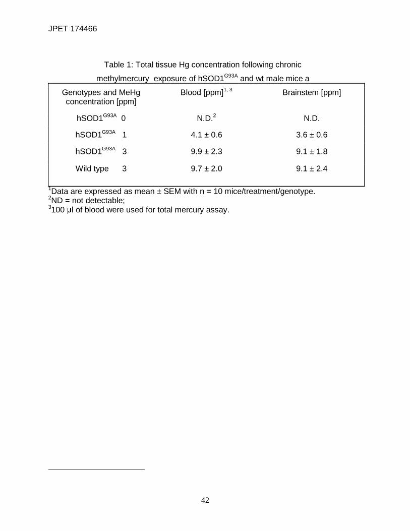
RESULTS Total Tissue Mercury Concentration is Not Affected by Genotypes. MeHg
accumulation in brainstem and blood following drinking water administration was
measured (Table 1) to determine a) if MeHg treatment resulted in concentration-
dependent increases in [Hg] as previously reported (Stern and Korn, 2001), and b)
whether there was a differential accumulation of Hg by wt and G93A mice. Both G93A
and wt mice exposed chronically to 1 or 3 ppm MeHg, accumulated approximately
equivalent total Hg in blood and brainstem (p>0.05). Further there was no genotypic
difference in Hg accumulation in blood or brainstem. Hg accumulation was, however,
exposure concentration-dependent, (p<0.05).
Effects of MeHg Exposure on Body Weight. Overexpression of mutant human
Cu2+/Zn2+SOD1G93A (G93A) genes in males typically generates smaller animals than wt
littermates (Fig. 2A). Thus we wanted to test whether MeHg would further reduce body
weight, perhaps contributing to a weakened state of the G93A mice or alternatively,
reflecting effects on feeding behavior due to dysfunction of hypoglossal nerve
innervated muscles. Mean body weight difference between G93A and treated wt at the
start of rotarod training was 9% and at the symptomatic stages was 11%. The rate of
incremental weight gain in treated wt was significantly (p<0.05) faster compared to
treated or untreated G93A mice, with MeHg-treated wt mice attaining significantly
greater body weight throughout the treatment period. For the G93A group, mice
exposed to MeHg at either concentration, gained weight at least equal to or greater than
untreated G93A mice. Body weights of untreated G93A and 3 ppm MeHg G93A were

JPET 174466
15
similar at the start of exposure, however, on week 13 there was a significant (p<0.05)
increase in weight gain in treated G93A mice that continued until week 15 (end stage of
the disease). When compared to untreated G93A, mice exposed to 1 ppm MeHg exhibit
a significantly (p<0.05) increased body weight starting already on week 9 until week 16
when they became paralyzed (end stage of the disease). G93A mice did gain weight,
albeit at a slower rate, until the very end, at which point a larger increase was noted.
This may reflect weights of animals that took a longer time to develop paresis. Over the
entire experimental period, the body weight of untreated G93A mice did not increase
appreciably. Shown in Panel B are comparative data for wt mice either unexposed or
exposed to 3 ppm MeHg. There was little difference in either the rate of body weight
gain, or the final weights achieved by wt mice in the presence of MeHg. Overall, MeHg
exposure was not associated with a generalized metabolic decline, as reflected by body
weight loss.
MeHg Exposure Hastened the Time to Onset of Rotarod Failure in G93A Mice. A
rotarod test was used to track motor coordination and balance from pre-symptomatic
(PND50) to symptomatic stages of ALS-like symptoms in both genotypes and treatment
groups (Fig. 3). Normally, G93A mice developed paralytic hind limb phenotypes, as
evidenced by failure to perform on the rotarod, which progressed to death in ~120 days,
whereas their wt littermates exhibited no motor deficits and survived (Alexander et al.,
2004). Exposure of wt mice to 3 ppm MeHg caused no decrement in rotarod
performance over the duration of the observation period. They survived more than 120
days, at which point the experiment was terminated. In contrast, both untreated and
MeHg-treated G93A mice exhibited time-dependent failure of motor coordination.

JPET 174466
16
However, MeHg treatment significantly (p<0.05) hastened the time to onset of rotarod
failure and hind limb paralysis in G93A mice. Exposure to either 1 or 3 ppm MeHg
caused 50% of G93A mice to fail the test (median time) by 91 and 84 days,
respectively, compared to 104 days in untreated G93A mice (Fig. 3A). This reduction
was concentration-dependent (p < 0.05). At the point in the disease process at which
50% of males exposed to 3 ppm MeHg failed the rotarod test, only 21% of mice in the 1
ppm group failed, and no failures were recorded in either the untreated G93A control
group or wt mice exposed to 3 ppm MeHg. The mean age to onset of ALS-like
phenotype was also significantly (p<0.05) shortened at 1 or 3 ppm MeHg, compared to
untreated G93A mice. Once failure in the rotarod test occurred, the time of progression
(dragging of hind limb) to hind limb paralysis was significantly (p<0.05) shortened in
G93A mice exposed to 3 but not 1 ppm MeHg when compared to untreated G93A
controls.
Elevation of Intracellular Divalent Cations in G93A Mice at Time of Rotarod
Failure. KCl-induced depolarization increased Fluo-4 fluorescence in both genotypes,
(F/F0 >1), with differential responses to MeHg treatment (Fig. 4A). At 10 mM, [KCl]
significantly (p<0.05) augmented Fluo-4-fluorescence only in the 3 ppm MeHg-treated
G93A mice, as compared to untreated G93A or wt (untreated and treated) slices.
However, 40 mM [KCl] significantly (p<0.05) increased [Ca2+]i in all treatment groups of
the G93A mice, compared to wt. MeHg had no further effect to increase [Ca2+]i above
that in untreated G93A mice.
Fluo-4 binds not only Ca2+ but also other divalent cations such as Zn2+ which
could artificially elevate the apparent increase in [Ca2+]i fluorescence. MeHg releases a
JPET 174466
17
non-Ca2+ divalent cation (Denny et al., 1993; Hare et al., 1993). In synaptosomes
(Denny and Atchison, 1994) this was identified as Zn2+. Additionally, in the G93A mice,
impaired metal binding to SOD1 could result in an increase in intracellular Zn2+
(Beckman et al., 2001). As such, it was important to test for the possibility that more
than one Fluo-4-chelatable cation contributed to the [KCl] depolarization-induced
increase in Fluo-4 fluorescence. TPEN is a cell-permeant divalent cation chelator, which
binds Zn2+ with high, and Ca2+ and Mg2+ with low affinity (Shmist et al., 2005). It does
not bind MeHg (Hare et al., 1993). Incubation of brainstem slices for 2 hrs with Fluo-4 in
the presence of 5 μM TPEN significantly (p<0.05) decreased [KCl]-induced Fluo-4
fluorescence compared to untreated slices (Fig. 4B). However, even in the presence of
TPEN, Fluo-4 fluorescence was increased in a [KCl]-dependent manner. Consequently,
we added TPEN to the Fluo-4 dye mix in all subsequent experiments.
Increased NXII Fluo-4 Fluorescence is Mediated Primarily by AMPA Receptors. To
identify pharmacologically which types of glutamate receptors are responsible for the
increased [Ca2+]i in hypoglossal motor neurons, we compared effects of different
glutamate receptor agonists on Fluo-4 fluorescence with or without MeHg treatment.
Brainstem slices were stimulated sequentially with puff-application of NMDA, AMPA, or
KA, each at 50 μM, for 120 s (Fig. 5). Neither NMDA nor KA significantly (p>0.05)
increased mean normalized fluorescence in any genotype. Conversely, AMPA
significantly (p<0.05) increased Fluo-4 fluorescence in both MeHg-treated and untreated
G93A mice compared to wt. However, no further increase was seen upon exposure to
MeHg as compared to untreated G93A mice (p>0.05).
JPET 174466
18
AMPA receptor activation could result in increased Fluo-4 fluorescence if there
was an increase in expression or activity of Ca2+-permeable AMPA receptors
(Kawahara et al., 2004; Tortarolo et al., 2006; Kwak et al., 2010). If such an effect
occurred following MeHg exposure, it may not have been possible to detect an increase
in MeHg-treated G93A hypoglossal motor neurons over that of untreated control using
AMPA receptor agonists if the receptors had desensitized, thereby obscuring a potential
effect. Consequently, treatment with an AMPA receptor antagonist was used in an
attempt to attenuate the postsynaptic response. CNQX (20 μM) reduced mean
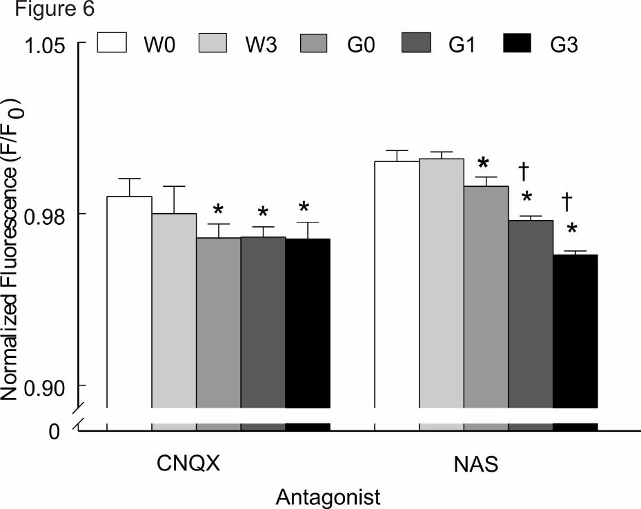
normalized Fluo-4 fluorescence in both genotypes (F/F0 <1) (Fig. 6). However, the
effects were significantly (p<0.05) larger in the G93A group. MeHg-treatment did not
further reduce [Ca2+]i compared to untreated G93A slices (p>0.05).
AMPA receptors lacking GluR2 subunits comprise a subset of CNQX-sensitive
receptors that are present in spinal motor neurons in humans (Kawahara et al., 2004)
and G93A mice (Tateno et al., 2004; Rembach et al., 2004; Tortarolo et al., 2006).
These receptors have a high Ca2+ conductance. We examined if Ca2+-permeable AMPA
receptors could contribute to increase Ca2+ in NXII neurons. NAS (50 μM) is a specific
antagonist of Ca2+-permeable AMPA receptors. It had little effect in wt as evidenced by
normalized fluorescence >1 (Fig. 6). In comparison, it significantly (p<0.05) attenuated
Fluo-4 fluorescence in G93A slices. Importantly, when compared to untreated G93A
slices, normalized Fluo-4 fluorescence was further significantly (p<0.05) attenuated in
MeHg treated G93A slices. The effect appeared to be MeHg-concentration dependent.
Thus, NAS-sensitive, and presumably Ca2+-permeable AMPA receptors, contribute
significantly to MeHg-induced increase in Fluo-4 fluorescence in the G93A mice.
JPET 174466
19
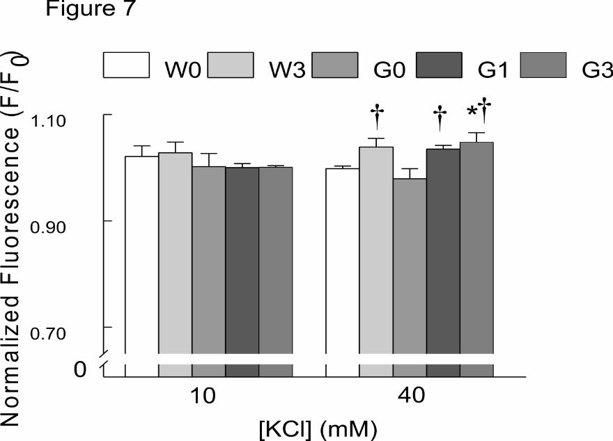
MeHg Elevation of Intracellular FluoZin Fluorescence in G93A and Wild-type Mice
at the Time of Rotarod Failure. To test directly for a contribution of Zn2+ to the
TPENsensitive component of Fluo-4 fluorescence, we used the Zn2+-sensitive
fluorophore FluoZin. Given the presence of Zn2+ in glutamatergic vesicles, KCl-induced
depolarization could increase [Zn2+] secondary to release of glutamate. KCl-induced
depolarization increased FluoZin fluorescence in both genotypes, (F/F0 >1), with
differential responses to MeHg treatment (Fig. 7). At 10 mM, [KCl] did not significantly
increase FluoZin fluorescence in any genotype or at either MeHg concentration.
However, at 40 mM, [KCl] application significantly (p<0.05) augmented FluoZin
fluorescence in all MeHg-treated groups irrespective of genotypes. Further, 40 mM [KCl]
significantly (p<0.05) increased [Zn2+]i in G93A mice exposed to 3 ppm MeHg,
compared to untreated wt.
JPET 174466
20
DISCUSSION
The objective of the present study was to test the concept that exposure to a
known environmental neurotoxicant, at levels which were not themselves overtly
neurotoxic, would accelerate the onset of the ALS phenotype in a genetically-
susceptible mouse. Studies focused on brainstem hypoglossal neuronal soma (NXII),
motor neurons which control functions of the tongue including chewing, swallowing, and
speaking. In humans, degeneration of NXII motor neurons produces signs and
symptoms collectively referred to as bulbar onset ALS (neck and head). This form of
ALS is associated with increased morbidity and mortality (Smittkamp et al., 2008). We
hypothesized that the common mode of excitotoxic action of MeHg would synergize with
that associated with ALS etiology to hasten development of motor dysfunction.
Results of the present study are consistent with the following conclusions. First,
chronic exposure to MeHg caused a concentration-dependent shift in rotarod
performance to shorten the time to onset of failure, an effect indicative of impairment of
motor function, in the genetically-susceptible G93A mice. Second, the SOD1-G93A
genotype was associated with elevation in motor neurons of both Ca2+ and another non-
Ca2+ divalent cation- presumably [Zn2+], which is itself neurotoxic. Third, Ca2+ and Zn2+-
permeable AMPA receptors contributed to enhanced divalent cation levels in G93A
motor neurons and MeHg treatment exacerbated this effect.
This is the first unequivocal demonstration of a gene-environment interaction to
facilitate development of the ALS phenotype in the G93A mouse. Chronic MeHg
exposure in male mice expressing an ALS phenotype (G93A) markedly accelerates the
time to onset of hind limb dysfunction. This effect was concentration-dependent. It is

JPET 174466
21
G93A-specific and associated with MeHg exposure. It was not seen in wt mice exposed
to the highest MeHg concentration (3 ppm) for 120 days. Thus MeHg/G93A interaction
was required. Rotarod failure was associated with MeHg accumulation, but MeHg levels
in wt and G93A mice were equivalent, so the enhancement of motor dysfunction was
not due simply to increased accumulation of MeHg in the G93A mice. The
concentrations of MeHg used were without effects on generalized function because
body weight gain was not reduced by MeHg treatment in either wt nor G93A mice. Once
rotarod failure ensued, the interval between onset of hind limb dysfunction and paresis
was shortened in animals treated with the highest concentration of MeHg. This suggests
that even after motor neuron damage begins, MeHg continued to enhance the
processes associated with cell death.
ALS pathophysiology coalesces around several mechanisms, including
glutamatemediated excitotoxicity (Cluskey and Ramsden, 2001). Included in this is
enhanced release of glutamate (Milanese et al., 2010). This causes repetitive firing and
dysregulated Ca2+ entry leading to cell death, and is a well-known model of brain
neurotoxicity (Sinor et al., 2000). MeHg-induced increase in presynaptic [Ca2+]i is an
extensively described mechanism contributing to its neurotoxicity (Limke et al., 2004),
as is elevated [Ca2+]i in spinal cord nerve terminals of the SOD1 mutants (Milanese et
al., 2010). Thus a prospective interaction between the MeHg-induced increase in [Ca2+]i,
and enhanced glutamate-mediated excitotoxicity in G93A group represented a logical
point of intersection for a gene/environment interaction. We determined if hastened
onset of ALS-like phenotype in G93A mice is related to the effects of MeHg to increase
[Ca2+]i and the potential contribution of Ca2+-permeable AMPA receptors to this effect.
Absolute measurements of [Ca2+]i were not possible because of differences among
JPET 174466
22
preparations in dye loading efficiency. Consequently, measurements of Fluo-4
fluorescence had to be made in relative terms. After rotarod failure occurred, [Ca2+]i in
brainstem-hypoglossal neuronal soma increased and AMPA receptor antagonists
blunted this effect. At a low level of depolarization (10 mM KCl), only the highest MeHg
concentration increased Fluo-4 fluorescence. With stronger depolarization (40 mM KCl),
a greater level of Fluo-4 fluorescence was seen in all G93A mice irrespective of MeHg
treatment. Thus, the increase at low levels of depolarization could reflect a direct effect
of MeHg with relatively low contribution of extracellular Ca2+, whereas at higher levels of
depolarization, effects of the G93A mutation became more evident. A potentially greater
effect of MeHg to increase [Ca2+]i could have been blunted by the well described ability
of MeHg to block voltage-gated Ca2+ channels (Shafer and Atchison, 1991; Hajela et al.,
2003). The ability of AMPA receptor agonists to replicate the increase in Fluo-4
fluorescence in the G93A group demonstrated that this effect likely resulted from
enhanced release of glutamate in the 40 mM KCl group. The ability of AMPA receptor
antagonists to counteract the increase in Fluo-4 fluorescence implies that, even in the
absence of depolarization, some level of glutamate release is occurring spontaneously
in G93A mice.
Increased NXII somal [Ca2+] in the MeHg-treated G93A mice could result from
pre- or postsynaptic effects, or a combination of the two. Numerous studies have
demonstrated that acute exposure to MeHg increases [Ca2+]i (Hare et al., 1993; Limke
et al., 2003), an effect that should elevate the spontaneous release of neurotransmitters
including glutamate (Yuan and Atchison, 2007). MeHg reportedly increases extracellular
glutamate levels in rat primary motor cortex (Juárez et al., 2002), a result consistent
with increased [Ca2+]i in presynaptic glutamatergic terminals. We have also
JPET 174466
23
demonstrated that MeHg increased glutamatergic synaptic neurotransmission in brain
slices in vitro (Yuan and Atchison, 2007). Thus a reasonable expectation is that MeHg
treatment could enhance glutamate release secondary to elevation of presynaptic
[Ca2+]i.
A TPEN-sensitive pool contributed to the Fluo-4 signal of divalent cations in
MeHgtreated animals. We suggest that it is Zn2+ based on results of previous studies
(Denny and Atchison, 1994) and our results with FluoZin. Elevation of [Zn2+]i could
provide a synergistic effect of MeHg on motor neuron function and degeneration of
these neurons in ALS. At higher concentrations, Zn2+ is itself cytotoxic. It can enter
neurons through Ca2+-permeable AMPA receptors, and it is contained in high
concentrations in glutamatergic vesicles. Thus an increased spontaneous release of
glutamate could reasonably be expected to increase synaptic levels of Zn2+. MeHg itself
liberates a non-Ca2+ divalent cation (Denny and Atchison, 1994), potentially contributing
further to a releasable Zn2+ pool. The extent to which a putative effect of Zn2+
contributes to the MeHg/G93A interaction merits further analyses.
Postsynaptic effects of MeHg on glutamate receptors could be an alternate or
additional site of action of MeHg. An increased function of Ca2+/Zn2+-permeant AMPA
receptors could hasten excitotoxicity in vulnerable motor neurons. The net effect would
be to increase postsynaptic [Ca2+]i, secondary to glutamate receptor activation. The
response of AMPA receptors to MeHg has not been reported. Consequently we tested
whether glutamate agonists could facilitate increases in [Ca2+]i in MeHg-treated mice. A
stimulatory effect of MeHg could result from a) preferential effects to impede expression
or function of Ca2+-impermeable AMPA receptors; b) enhanced function of Ca2+-
permeable AMPA receptors; or c) an indirect effect to depolarize the membrane

JPET 174466
24
sufficiently to activate NMDA receptors. NXII neurons contain unique combinations of
AMPA/KA or NMDA receptors (Essin, 2002). At symptomatic stages of ALS, untreated
G93A mice exhibited increased sensitivity to AMPA but not NMDA or KA; MeHg did not
change the sensitivity of NMDA receptors. Thus an indirect effect involving NMDA
receptor activation can be ruled out. The stimulatory effect of AMPA on Fluo-4
fluorescence was not enhanced further by MeHg, suggesting a lack of a direct effect on
the receptor above that seen with the G93A mutation. However, a lack of effect of MeHg
to increase Fluo-4 fluorescence further could result from the methodological approach.
Methodologically, if the increased [Ca2+]i elicited by the G93A mutation was of
sufficiently high magnitude, the ability of Fluo-4 to report further changes in
fluorescence could have been impeded due to dye saturation. Alternatively, a potential
increase in glutamate release, or prolonged contact with the AMPA receptor (see below)
could have desensitized the receptor, making it incapable of responding to further
activation by agonist.
Taking the converse approach we used antagonists to attempt to quell an
increase in Fluo-4 fluorescence in the G93A group, as well as a potential enhancement
by MeHg should it occur. CNQX reduced Fluo-4 fluorescence and effectively attenuated
[Ca2+]i in G93A and wt slices. CNQX produced a more prominent reduction of [Ca2+]i in
the G93A group compared to wt. This implies that AMPA receptors contributed
significantly to increased [Ca2+]i. This effect was not enhanced by MeHg. Thus
AMPA/KA receptors are apparently active and contribute to increased [Ca2+]i. We then
used a specific receptor blocker to test if increased [Ca2+]i was primarily mediated in the
G93A group by the Ca2+permeable AMPA receptors. NAS, a polyamine toxin derived
from Joro spider venom, binds specifically to GluR2R-lacking AMPA receptors inside
JPET 174466
25
the receptor pore, so that Ca2+ influx is prevented (Blaschke et al., 1993). Ca2+ was not
precluded from entering NXII neurons from wt slices, as NAS had no effect on Fluo-4
fluorescence. Conversely, NAS significantly reduced [Ca2+]i in the G93A group,
indicating that a subset of CNQX-sensitive, Ca2+permeable receptors was responsible
for increased [Ca2+]i. MeHg enhanced this effect above that of untreated G93A mice in
that group, but the MeHg-treated wt group did not display NAS sensitivity. As such,
MeHg appears to increase [Ca2+]i of NXII motor neurons by potentiating [Ca2+]i influx
through Ca2+-permeable AMPA receptors. These, in turn, were only found in the G93A
group. This once again points to a gene-environmental interaction between the
susceptible G93A genotype and MeHg exposure.
In addition to its direct effects on neurons, MeHg could influence motor neuron
Ca2+ regulation indirectly, particularly by effects on astrocyte function. MeHg
accumulates in cortical astrocytes, where it inhibits glutamate uptake through the
excitatory amino acid transporter 1 (EAAT-I, also known as GLAST) (Mutkus et al.,
2005). The effects of MeHg on brainstem astrocytes have not been examined, but in
both patients with ALS, and the mouse model, levels of EAAT-2 are reduced (Barbeito
et al., 2004; Boillée et al., 2006). Astrocytes play a critical role in maintaining glutamate
homeostasis (see Allen et al., 2002; Aschner et al., 1993; reviewed in Aschner et al.,
2000). Thus, a potential increase in the residence time of glutamate in the synaptic cleft
due to MeHg-induced disruption of EAAT-2 could theoretically exacerbate glutamate-
mediated excitotoxicity of sensitive motor neurons.
In conclusion, our results clearly demonstrate a gene-environment interaction in
which MeHg interacts in an organism with a specific gene mutation to hasten the time of
onset of development of ALS-like phenotypes. This apparent gene-environment
JPET 174466
26
interaction provides evidence of a potential contribution of MeHg to the development of
ALS in genetically-susceptible organisms. MeHg can exhibit a panoply of effects
contributing to the acceleration of the ALS phenotype. However in the absence of the
synergistic effect of the G93A mutation, the actions of MeHg are not sufficient in and of
themselves to induce motorneuron dysfunction over a period of 120 days. Our results
support a role for Ca2+permeable AMPA receptors in the MeHg effect. Motor neurons in
the G93A mice have an increased abundance of GluR3 mRNA and protein expression
coupled with a decrease in protein expression of GluR2 subunits (Tortarolo et al., 2006).
This could, in theory, lead to a greater proportion of Ca2+-permeable AMPA receptors
which could increase glutamateinduced motor neuron excitotoxicity due to increased
[Ca2+]i . Additionally, Yin et al. (2007) demonstrated that intrathecal infusion of NAS
reduced motor neuron loss in G93A rats. This effect is presumably due to action on
Ca2+-permeable AMPA receptors. A combination of MeHg-induced enhanced response
of Ca2+-permeable AMPA receptors coupled with an increase in glutamate release, or
decrease in astrocytic EAAT-2 function could predispose motor neurons to excitotoxic
damage, thereby unmasking or hastening the onset of ALS in an individual with a
known (or, conceivably, an unknown) predilection to the disease. The results of this
study also point to the utility of using environmental agents to examine the pathogenesis
of other neurodegenerative conditions.
JPET 174466
27
ACKNOWLEDGMENTS: The authors acknowledge the excellent word processing
assistance of Elizabeth Anne Hill and Julie Van Raemdonck and graphical assistance of
Sarah Metzger. Preliminary reports of portions of this project were presented at the
Annual Meetings of the Society of Toxicology, 2010 in Salt Lake City, UT, the 25th
International Neurotoxicology Conference, October 12-16, 2008, Rochester, NY and the
Society of Neuroscience, November 19, 2008 in Washington DC.
AUTHORS CONTRIBUTIONS
Participated in research design: Atchison, Hajela, Johnson, Parsell, Yuan
Conducted experiments: Chitrakar, Hajela, Johnson, Yuan
Performed data analyses: Atchison, Chitrakar, Hajela, Johnson, Yuan
Wrote or contributed to the writing of the manuscript: Atchison, Hajela, Johnson, Yuan
Other: Atchison acquired funding for research and oversaw the entire project; Parsell
was responsible for setting up and maintaining the transgenic mouse colonies.
JPET 174466
28
REFERENCES
Alexander GM, Erwin KL, Byers N, Deitch JS, Augelli BJ, Blankenhorn EP, and Heiman-
Patterson TD (2004) Effect of transgene copy number on survival in the SOD1G93A
SOD1 transgenic mouse model of ALS. Brain Res Mol Brain Res 130: 7-15.
Allen JW, Shanker G, Tan KH, and Aschner M (2002) The consequences of
methylmercury exposure on interactive functions between astrocytes and neurons.
Neurotoxicology. 23:755-9.
Arvidson B (1992) Inorganic mercury is transported from muscular nerve terminals to
spinal and brainstem motoneurons. Muscle Nerve 15: 1089-94.
Aschner M, Du YL, Gannon M, and Kimelberg HK (1993) Methylmercury-induced
alterations in excitatory amino acid transport in rat primary astrocyte cultures. Brain Res
602: 181-6.
Aschner M, Yao CP, Allen JW, and Tan KH (2000) Methylmercury alters glutamate
transport in astrocytes. Neurochem Int 37: 199-206.
Bakir F, Damluji SF, Amin-Zaki L, Murtadha M, Khalidi A, Al-Rawi NY, Tikriti S, Dahahir
HI, Clarkson TW, Smith JC, and Doherty RA (1973) Methylmercury poisoning in Iraq.
Science 181:230-41.
JPET 174466
29
Barbeito LH, Pehar M, Cassina P, Vargas MR, Peluffo H, Viera L, Estevez AG, and
Beckman JS (2004) A role for astrocytes in motor neuron loss in amyotrophic lateral
sclerosis. Brain Res Brain Res Rev 47:263-74.
Barber TE (1978) Inorganic mercury intoxication reminiscent of amyotrophic lateral
sclerosis. J Occup Med 20: 667-9.
Beckman JS, Estévez AG, Crow JP, and Barbeito L (2001) Superoxide dismutase and
the death of motoneurons in ALS. Trends Neurosci 24: S15-20.
Bellum S, Thuett KA, Grajeda R, and Abbott LC (2007) Coordination deficits induced in
young adult mice treated with methylmercury. Int J Toxicol 26:115-21
Benmohamed R, Arvanites AC, Kim J, Ferrante RJ, Silverman RB, Morimoto RI, and
Kirsch DR (2011) Identification of compounds protective against G93A-SOD1 toxicity for
the treatment of amyotrophic lateral sclerosis. Amyotroph Lateral Scler 12:87-96.
Blaschke M, Keller BU, Rivosecchi R, Hollmann M, Heinemann S, and Konnerth A
(1993) A single amino acid determines the subunit-specific spider toxin block of alpha-
amino-3 hydroxy-5-methylisoxazole-4-propionate/kainate receptor channels. Proc Natl
Acad Sci USA 90:6528-32.
Boillée S, Vande Velde C, and Cleveland DW (2006) ALS: A disease of motor neurons
and their nonneuronal neighbors. Neuron 52:39-59.
JPET 174466
30
Brown RH Jr. (1995) Amyotrophic Lateral Sclerosis: Recent insights from genetics and
transgenic mice Cell 80:687-92
Cluskey S and Ramsden DB (2001) Mechanisms of neurodegeneration in amyotrophic
lateral sclerosis. Mol Pathol 54: 386-92.
Denny MF, Hare MF, and Atchison WD (1993) Methylmercury alters intrasynaptosomal
concentrations of endogenous polyvalent cations. Toxicol Appl Pharmacol 122: 222-32.
Denny MF and Atchison WD (1994) Methylmercury-induced elevations in intra-
synaptosomal zinc concentrations: an 19F-NMR study. J Neurochem 63: 383-6.
DePaul R, Abbs JH, Caligiuri M, Gracco VL, and Brooks BR (1988) Hypoglossal,
trigeminal, and facial motoneuron involvement in amyotrophic lateral sclerosis.
Neurology 38:281-3.
Essin K, Nistri A, and Magazanik L (2002) Evaluation of GluR2 subunit involvement in
AMPA receptor function of neonatal rat hypoglossal motoneurons. Eur J Neurosci 15:
1899906.
Grosskreutz J, Van Den Bosch L, and Keller BU (2010) Calcium dysregulation in
amyotrophic lateral sclerosis. Cell Calcium 47:165-74.

JPET 174466
31
Gruzman A, Wood WL, Alpert E, Prasad MD, Miller RG, Rothstein JD, Bowser R,
Hamilton R, Wood TD, Cleveland DW, Lingappa VR, and Liu J (2007) Common
molecular signature in SOD1 for both sporadic and familial amyotrophic lateral
sclerosis. Proc Natl Acad Sci USA 104:12524-9.
Gurney ME, Pu H, Chiu AY, Dal Canto MC, Polchow CY, Alexander DD, Caliendo J,
Hentati A, Kwon YW, Deng HX, Chen W, Zhai P, Sufit RL, and Siddique T (1994)
Motorneuron degeneration in mice that express a human Cu, Zn superoxide dismutase.
Science 264: 1772-5.
Haley RW (2003) Excess incidence of ALS in young Gulf War veterans. Neurology 61:
750-6.
Hajela RK, Peng SQ, and Atchison WD (2003) Comparative effects of methylmercury
and Hg2+ on human neuronal N- and R-type high-voltage activated calcium channels
transiently expressed in human embryonic kidney 293 cells. J Pharmacol Exp Ther
306:1129-36.
Hare MF, McGinnis KM, and Atchison WD (1993) Methylmercury increases intracellular
concentrations of Ca++ and heavy metals in NG108-15 cells. J Pharmacol Exp Ther 266:
1626-35.
Horner RD, Kamins KG, Feussner JR, Grambow SC, Hoff-Lindquist J, Harati Y,
Mitsumoto H, Pascuzzi R, Spencer PS, Tim R, Howard D, Smith TC, Ryan MA,

JPET 174466
32
Coffman CJ, and Kasarskis EJ (2003) Occurrence of amyotrophic lateral sclerosis
among Gulf War veterans. Neurology 61: 742-9.
Institute of Medicine of the National Academies (2006) Amyotrophic Lateral Sclerosis in
Veterans: Review of the Scientific Literature, Board on Population Health and Public
Health Practice, Released 11/ 6/2006. http://books.nap.edu/openbook.php? record_
id=1175.
Juárez BI, Martínez ML, Montante M, Dufour L, García E, and Jiménez-Capdeville ME
(2002) Methylmercury increases glutamate extracellular levels in frontal cortex of awake
rats. Neurotoxicol Teratol 24: 767-71.
Kabashi E, Valdmanis PN, and Dion P (2008) TARDBP mutations in individuals with
sporadic and familial amyotrophic lateral sclerosis. Nat Genet 40:572–4.
Kamel F, Umbach DM, Stallone L, Richards M, Hu H, and Sandler DP (2008)
Association of lead exposure with survival in amyotrophic lateral sclerosis. Environ
Health Perspect 116: 943-7.
Karsarkis EJ, Scarlata D, Hill R, Fuller C, Stambler N, and Cedarbaum JM (1999) A
retrospective study of percutaneous endoscopic gastrostomy in ALS patients during the
BDNF and CNTF trials. J Neurol Sci 169: 118-25.

JPET 174466
33
Kawahara Y, Ito K, Sun H, Aizawa H, Kanazawa I, and Kwak S (2004) Glutamate
receptors: RNA editing and death of motor neurons. Nature 427: 801.
Kwak S, Hideyama T, Yamashita T, and Aizawa H (2010) AMPA receptor-mediated
neuronal death in sporadic ALS. Neuropathology 30: 182-8.
Le W, Chen S, and Jankovic J (2009) Etiopathogenesis of Parkinson Disease: a new
beginning? Neuroscientist 15: 28-35.
Limke TL, Otero-Montañez JK, and Atchison WD (2003) Evidence for interactions
between intracellular calcium stores during methylmercury-induced intracellular calcium
dysregulation in rat cerebellar granule neurons. J Pharmacol Exp Ther 304: 949-58.
Limke TL, Heidemann SR, and Atchison WD (2004) Disruption of intraneuronal divalent
cation regulation by methylmercury: are specific targets involved in altered neuronal
development and cytotoxicity in methylmercury poisoning. Neurotoxicology 25: 741-60.
Migliore L and Coppedè F (2009) Genetics, environmental factors and the emerging
role of epigenetics in neurodegenerative diseases. Mutat Res 667: 82-97.
Milanese M, Zappettini S, Onofri F, Musazzi L, Tardito D, Bonifacino T, Messa M,
Racagni G, Usai C, Benfenati F, Popoli M, and Bonanno G. (2011) Abnormal exocytotic
release of glutamate in a mouse model of amyotrophic lateral sclerosis. J Neurochem
116:1028-42.

JPET 174466
34
Mitchell JD (2000) Amyotrophic lateral sclerosis: toxins and environment. Amyotroph
Lateral Scler Other Motor Neuron Disord. 1: 235-50.
Møller-Madsen B (1991) Localization of mercury in CNS of the rat III. Oral
administration of methylmercuric chloride (CH3HgCl). Fundam Appl Toxicol 16: 172-87.
Mutkus L, Aschner JL, Syversen T, and Aschner M (2005) Methylmercury alters the in
vitro uptake of glutamate in GLAST- and GLT-1-transfected mutant CHO-K1 cells. Biol
Trace Elem 107:231-45.
Newland MC and Rasmussen EB (2000) Aging unmasks adverse effects of gestational
exposure to methylmercury in rats. Neurotoxicol Teratol 22:819-28.
Newland MC, Reile PA, and Langston JL (2004) Gestational exposure to methylmercury
retards choice in transition in aging rats. Neurotoxicol Teratol 26:179-94.
Prasad KN, Cole WC, and Kumar B (1999) Multiple antioxidants in the prevention and
treatment of Parkinson's disease. J Am Coll Nutr 18: 413-23.
Rembach A, Turner BJ, Bruce S, Cheah IK, Scott RL, Lopes EC, Zagami CJ, Beart PM,
Cheung NS, Langford SJ, and Cheema SS (2004) Antisense peptide nucleic acid
targeting GluR3 delays disease onset and progression in the SOD1 G93A mouse model
of familial ALS. J Neurosci Res 77:573-82.
JPET 174466
35
Rowland LP and Shneider NA (2001) Amyotrophic lateral sclerosis. N Engl J Med 11:
11312.
Shafer TJ, and Atchison WD (1991) Methylmercury blocks N- and L-type Ca++ channels
in nerve growth factor-differentiated pheochromocytoma (PC12) cells. J Pharmacol Exp
Ther 258:149-57.
Sinor JD, Du S, Venneti S, Blitzblau RC, Leszkiewicz DN, Rosenberg PA, and
Aizenman E (2000) NMDA and glutamate evoke excitotoxicity at distinct cellular
locations in rat cortical neurons in vitro. J Neurosci 20: 8831-7.
Shmist YA, Kamburg R, Ophir G, Kozak A, Shneyvays V, Appelbaum YJ, and
Shainberg A (2005) N,N,N',N'-tetrakis(2-pyridylmethyl)-ethylenediamine improves
myocardial protection against ischemia by modulation of intracellular Ca2+ homeostasis.
J Pharmacol Exp Ther 313: 1046-57.
Smittkamp SE, Brown JW, and Stanford JA (2008) Time-course and characterization of
orolingual motor deficits in B6SJL-Tg(SOD1-SOD1G93A)1Gur/J mice. Neuroscience 151:
613-21.
Sreedharan J, Blair IP, Tripathi VB, Hu X, Vance C, Rogelj B, Ackerley S, Durnall JC,
Williams KL, Buratti E, Baralle F, de Belleroche J, Mitchell JD, Leigh PN, Al-Chalabi A,
Miller CC, Nicholson G, and Shaw CE (2008) TDP-43 mutations in familial and sporadic
amyotrophic lateral sclerosis. Science 319:1668-72.

JPET 174466
36
Stern AH and Korn LR (2001) How useful is linear regression analysis in detecting the
existence of dose-response relationships in large-scale epidemiologic studies when only
a fraction of the population is sensitive? The case of methylmercury. Regul Toxicol
Pharmacol 33: 29-36.
Stern S, Cox C, Cernichiari E, Balys M, and Weiss B (2001) Perinatal and lifetime
exposure to methylmercury in the mouse: blood and brain concentrations of mercury to
26 months of age. Neurotoxicology 22:467-77.
Swash M (2000) Nature and nurture in ALS. Amyotroph Lateral Scler 1: 223.
Synofzik M, Fernández-Santiago R, Maetzler W, Schöls L, and Andersen PM (2010)
The human G93A SOD1 phenotype closely resembles sporadic amyotrophic lateral
sclerosis. J Neurol Neurosurg Psych 7:764-7.
Tateno M, Sadakata H, Tanaka M, Itohara S, Shin RM, Miura M, Masuda M, Aosaki T,
Urushitani M, Misawa H, and Takahashi R (2004) Calcium-permeable AMPA receptors
promote misfolding of mutant SOD1 protein and development of amyotrophic lateral
sclerosis in a transgenic mouse model. Hum Mol Genet 13: 2183-96
.
Tortarolo M, Grignaschi G, Calvaresi N, Zennaro E, Spaltro G, Colovic M, Fracasso C,
Guiso G, Elger B, Schneider H, Seilheimer B, Caccia S, and Bendotti C (2006)
Glutamate AMPA receptors change in motor neurons of SOD1 transgenic mice and
JPET 174466
37
their inhibition by a noncompetitive antagonist ameliorates the progression of
amytrophic lateral sclerosis-like disease. J Neurosci Res 83: 134-46.
Weiss B, Stern S, Cox C, and Balys M (2005) Perinatal and lifetime exposure to
methylmercury in the mouse: behavioral effects. Neurotoxicology 26: 675-90.
Yin HZ, Tang DT, Weiss JH (2007) Intrathecal infusion of a Ca(2+)-permeable AMPA
channel blocker slows loss of both motor neurons and of the astrocyte glutamate
transporter, GLT-1 in a mutant SOD1 rat model of ALS. Exp Neurol. 207:177-85.
Yuan Y, and Atchison WD (2007) Methylmercury-induced increase of intracellular Ca2+
increases spontaneous synaptic current frequency in rat cerebellar slices. Mol
Pharmacol 71: 1109-21.
JPET 174466
38
FOOTNOTES This study was supported by the following grants from the National
Institute of Environmental Health Sciences: [5T32 ES007255], [R21ES014357], and
[R01ES03299] including an ARRA supplement to this grant. Alisha Chitrakar was
supported in part by a Summer Undergraduate Research Fellowship from the American
Society of Pharmacology and Experimental Therapeutics and was the recipient of a
Pfizer Undergraduate Research Award from the Society of Toxicology (2010) for her
role on the project.
JPET 174466
39
FIGURE LEGENDS Figure 1. Time course of experimental design for MeHg exposure, rotarod training
and development of ALS phenotype. After weaning (PND 21) and genotyping,
cohorts of male G93A and wt mice were randomly assigned to groups treated with 0, 1,
or 3 ppm of MeHg. Mice received MeHg free-choice via drinking water starting on
PND29 and continuing until sacrifice.
Figure 2. Comparison of mean body weight during chronic exposure of
SOD1G93A and wt male mice to MeHg. (A) Time course of changes in body weight in
untreated G93A male mice (■ ) or those exposed to 1 (�) or 3 ppm (▼) MeHg and wt
mice exposed to 3 ppm MeHg (●). (B) Wt males were exposed to 0 ppm (�) or 3 ppm
(●) MeHg. All values represent the mean ± SEM, n = 14 mice/treatment/genotype. The
asterisk (*) indicates a value significantly different from wt (p<0.05).
Figure 3. Chronic MeHg exposure hastened disease onset and progression in
SOD1G93A mice. (A) Time course of onset of rotarod failure in, 0 ( ■) , 1 (�) or 3 ppm
(▼) MeHg-treated G93A and 3 ppm MeHg-treated wt ( ●). 50% Cumulative failure of the
population occurred at 84 and 91 days (arrows) compared to 104 days for the control
group; no failure occurred in wt mice. (B) Mean time of onset and progression of
paralytic ALS-like phenotypes in G93A mice. The asterisk (*) depicts a significant
(p<0.05) difference between untreated and treated G93A mice: A concentration-
dependence occurred to time of onset but not time to progression. All values represent
JPET 174466
40
mean ± SEM, n = 14 mice/treatment/genotype; Key: W3= wt 3 ppm; G0= G93A 0 ppm;
G1= G93A 1 ppm; G3= G93A 3 ppm.
Figure 4. Chronic MeHg exposure, increased [Ca2+]i Fluo-4 fluorescence in G93A
slices following incubation with TPEN and puff application of [KCl]. (A) Membrane
depolarization with [KCl] (40 mM) increased Fluo-4 Ca2+ fluorescence in NXII nuclei of
G93A mice. (B) N,N,N',N'-Tetrakis (pyridylmethyl) ethylenediamine (TPEN) significantly
reduced Fluo-4 fluorescence. All values represent mean ± SEM, n = 8
mice/treatment/genotype; (p<0.05). The asterisk (*) indicates a significant difference
between wt and G93A mice (p<0.05); the cross (†) indicates a significant difference
between MeHg-treated and untreated G93A mice following puff application of [KCl]
(p<0.05); ªp<0.05 differences between TPEN-treated and untreated slices; the plus (+)
indicates a significant difference between [KCl] at 10 mM and 40 mM in TPEN-treated
and untreated slices. Key: W0= wt 0 ppm; W3= wt 3 ppm; G0= G93A 0 ppm; G1= G93A
1 ppm; G3= G93A 3 ppm.
Figure 5. Pharmacological identification of glutamate receptors regulating [Ca2+]I
in XII nuclei in Wt and G93A mice. Application of AMPA increased Ca2+ entry through
AMPA/KA receptors. All values represent mean ± SEM, n = 8 mice/treatment/genotype.
*p<0.05 difference between genotypes; ‡p<0.05 differences between treated wt and
G93A mice slices. Key: W0= wt 0 ppm; W3= wt 3 ppm; G0= G93A 0 ppm; G1= G93A 1
ppm; G3= G93A 3 ppm.
Figure 6. Pharmacological identification of AMPA receptor subunits regulating
[Ca2+]I in XII nuclei of Wt and G93A male mice. Application of glutamate receptor

JPET 174466
41
antagonists decreased Ca2+ entry via AMPA-type receptors including those presumably
lacking the GluR2 subunit (NAS-sensitive). All values represent the mean ± SEM, n = 8
mice/ treatment/genotype; The asterisk (*) depicts a significant difference (p<0.05)
between genotypes. The cross (†) depicts a significant difference (p<0.05) between
MeHg-treated and untreated G93A mice; Dunnett’s posthoc tests. Key: W0= wt 0 ppm;
W3= wt 3 ppm; G0= G93A 0 ppm; G1= G93A 1 ppm; G3= G93A 3 ppm.
Figure 7. Chronic MeHg exposure, increased FluoZin fluorescence in G93A slices
following puff application of [KCl]. (A) Membrane depolarization with [KCl] (40 mM)
increased FluoZin (Zn2+) fluorescence in NXII nuclei of treated mice irrespective of
genotypes. All values represent the mean ± SEM, n = 8 mice/treatment/genotype;
(p<0.05). The asterisk (*) indicates a significant difference between untreated wt mice
(p<0.05); the cross (†) indicates a significant difference between MeHg-treated and
untreated G93A mice following puff application of [KCl] (p<0.05). Key: W0= wt 0 ppm;
W3= wt 3 ppm; G0= G93A 0 ppm; G1= G93A 1 ppm; G3= G93A 3 ppm.
JPET 174466
42
Table 1: Total tissue Hg concentration following chronic
methylmercury exposure of hSOD1G93A and wt male mice a
Genotypes and MeHg concentration [ppm]
Blood [ppm]1, 3 Brainstem [ppm]
hSOD1G93A 0 N.D.2 N.D.
hSOD1G93A 1 4.1 ± 0.6 3.6 ± 0.6
hSOD1G93A 3 9.9 ± 2.3 9.1 ± 1.8
Wild type 3 9.7 ± 2.0 9.1 ± 2.4
1Data are expressed as mean ± SEM with n = 10 mice/treatment/genotype. 2ND = not detectable; 3100 μl of blood were used for total mercury assay.