in saccharomyces cerevisiae - universiteit utrecht · pdf filegrowth rate-regulated expression...
TRANSCRIPT

Growth rate-regulated expression of the hexose transporter HXT5
in Saccharomyces cerevisiae
Groeisnelheid gereguleerde expressie van de hexose transporter HXT5
in Saccharomyces cerevisiae
(met een samenvatting in het Nederlands)
Proefschrift
ter verkrijging van de graad van doctor aan de Universiteit Utrecht
op gezag van de Rector Magnificus, Prof. Dr. W.H. Gispen, ingevolge het besluit van het College voor Promoties
in het openbaar te verdedigen op maandag 10 november 2003 des middags te 2.30 uur
door
René Verwaal
Geboren op 18 oktober 1975 te Gouda

Promotoren:
Prof. Dr. J. Boonstra (Verbonden aan de leerstoelgroep Moleculaire Celbiologie, Faculteit Biologie, Universiteit Utrecht) Prof. Dr. Ir. C.T. Verrips (Verbonden aan de leerstoelgroep Moleculaire Celbiologie, Faculteit Biologie, Universiteit Utrecht) Prof. Dr. A.J. Verkleij (Verbonden aan de leerstoelgroep Moleculaire Celbiologie, Faculteit Biologie, Universiteit Utrecht) The research described in this thesis was performed at the department of Molecular Cell Biology and the Institute of Biomembranes, Faculty of Biology, Utrecht University, Utrecht, The Netherlands, and financially supported by Unilever Research Vlaardingen, The Netherlands.

Beoordelingscommissie: Prof. Dr. G.F.B.P. van Meer (Verbonden aan de vakgroep Membraan Enzymologie, Faculteit Scheikunde, Universiteit Utrecht) Dr. H.H.W. Silljé (Verbonden aan het Max Planck Institute of Biochemistry, Department of Cell Biology, Martinsried, Germany) Prof. Dr. J.C.M. Smeekens (Verbonden aan de vakgroep Moleculaire Plantenfysiologie, Faculteit Biologie, Universiteit Utrecht) Prof. Dr. H.A.B. Wösten (Verbonden aan de vakgroep Moleculaire Microbiologie, Faculteit Biologie, Universiteit Utrecht) Paranimfen: Ronald Verwaal
Henri Versteeg Afbeelding omslag: Denise van Suylekom, Lan Nguyen Do Ngoc en Bruno
Humbel (Electron microscopic micrograph of a cryosectioned stationary Saccharomyces cerevisiae cell expressing HA-tagged Hxt5p, immunogold-labelled to locate Hxt5-HA)
Ontwerp omslag: Pieter van Dorp van Vliet
(Beeldverwerking en Vormgeving, Faculteit Biologie, Universiteit Utrecht)
Reproductie: Ridderprint offsetdrukkerij b.v. Ridderkerk ISBN: 90-393-3523-0


Table of contents Chapter 1 General introduction 7 Chapter 2 HXT5 expression is determined by growth rates 29
in Saccharomyces cerevisiae Chapter 3 HXT5 expression is under control of STRE 47
and HAP elements in the HXT5 promoter Chapter 4 The role of Hxt5p in trehalose accumulation 67
in Saccharomyces cerevisiae Chapter 5 Identification of genes that are expressed similar 87
to HXT5 upon an increase in G1 phase duration Chapter 6 General discussion 109 Summary 123 Samenvatting 127 Dankwoord 133 List of publications 137 Curriculum vitae 139


General introduction
Cha
pter
1

Chapter 1
8
General introduction Introduction
Yeasts are unicellular fungi that are able to produce ethanol and carbon dioxide from sugars, an ability which yeast has developed in its natural habitat, the grape juice. Already at the time of the ancient Egyptians at 3000 BC, this feature was used for preparation of food products like beer, bread and wine (1). It took until the 19th century when Louis Pasteur discovered that the yeast Saccharomyces cerevisiae was responsible for production of alcohol and CO2, a process that was called fermentation (2,3). Since then, yeast has been thoroughly studied concerning its physiology and application in biochemistry and biotechnology. Molecular genetic tools provided a means to use yeast in several biotechnological applications, such as manufacturing baker’s yeast, production of heterologous proteins, or production of metabolites that are used for various purposes (4). One of the metabolites with special interest is trehalose, which is not constitutively accumulated but only formed within cells during certain growth conditions to which cells may be exposed in nature. Trehalose is a non-reducing disaccharide that accumulates in bacteria, eukaryotic microorganisms like yeast, plants, insects and invertebrates, but so far not in mammals (5). Trehalose accumulation has been thoroughly studied in Saccharomyces cerevisiae, where it was initially thought to serve as a reserve carbohydrate (6). Nowadays, trehalose is known as a metabolite that stabilizes proteins and biological membranes under a variety of conditions, including increased temperature, hydrostatic pressure, desiccation, nutrient starvation, osmotic or oxidative stress, and exposure to toxic chemicals (5,7,8,9,10,11,12,13,14,15,16). Because trehalose has several applicable protective properties, it has become an important target for biotechnology. Trehalose may have several applications in food preparation such as dried and frozen food products, it may be used as a sweetener, in vaccine protection in hot climates, in cosmetic products as a liposome stabilizer, and may even stabilize organs that are used for transplantation (17). High trehalose contents protect cells from autolysis and increase the leavening capacity in frozen dough, making it an interesting target for baker’s yeast (18,19). Saccharomyces cerevisiae is able to consume various carbon sources, of which the fermentable monosaccharide glucose is most preferred. Yeast cells are able to grow on a wide variety of different glucose concentrations and show a great ability to adapt to changes in the environmental sugar concentration (20). Glucose, but also other carbon

General introduction
9
sources, may serve as precursors for trehalose. When glucose is used for trehalose production, it has to be transported into the cell and converted into glucose intermediates that serve as precursors for trehalose. Sugar metabolism
Sugar specific carriers transport sugars across the plasma membrane, before they are metabolized. On overview transport of different sugars and their subsequent metabolism is depicted in Figure 1.
Sucrose
Glucose + Fructose
Mannose
Glucose + Fructose + GalactoseMaltose
Raffinose
Hexose transportersMaltose
transportersGalactosetransporter
xxxxxxxxxxxxxxxxxxxxxxxxxxxxxxxxxxxxxxxxxxxxxxxxxxxxxxxxxxxxxxxxxxxxxxxxxxxxxxxxxx
xxxxxxxxxxxxxxxxxxxxxxxxxxxxxxxxxxxxxxxxxxxxxxxxxxxxxxxxxxxxxxxxxxxxxxxxxxxxxxxxxxxxxxxxxxxxxxxxxxxx
xxxxxxxxxxxxxxxxxxxxxxxxxxxxxxxxxxxxxxxxxxxxxxxxxxxxxxxx
Glucose
Glucose-6-phosphate
Fructose-6-phosphate
Pyruvate
Maltose 2 glucose
xxxxxxxxxxxxxxxxxxxxxxxx
Galactose
Fructose
Mannose
Glucose-1-phosphate
UDP-glucose
TrehaloseGLYCOLYSIS
TREHALOSE
ACCUMULATION
Acetyl-CoA
RESPIRATION
Acetate
AcetaldehydeEthanol
FERMENTATION
TCA cycle and
oxidative phosphorylation
Lactate
Glycerol
Structural
carbohydrates
Figure 1: Overview of carbon metabolism in Saccharomyces cerevisiae. See text for details.

Chapter 1
10
The monosaccharides glucose, fructose and mannose, are transported into cells by hexose transporter (Hxt) proteins (21,22,23,24). Upon entry, glucose is phosphorylated into glucose-6-phosphate, fructose and mannose enter glycolysis at the level of fructose-6-phosphate, and are further metabolized in glycolysis. Galactose enters glycolysis after transport by Gal2p (25,26) and conversion into glucose-1-phophate and subsequently into glucose-6-phosphate. Maltose is transported into cells by the family of maltose transporter proteins, which include Mph2p, Mph3p, Agt1p, Mal31p and Mal61p (27,28,29,30).
Glucose-6-phosphate is the precursor for UDP-glucose, and glucose-6-phosphate and UDP-glucose are used to produce trehalose. UDP-glucose is not exclusively used as precursor for trehalose, but also as precursor for structural carbohydrates, which are used as components of the cell wall. Di- and trisaccharides like sucrose, raffinose and maltose are first cleaved by glycosidases in the periplasmic space or in the cytoplasm before they are further metabolized in glycolysis. The end-point of glycolysis is pyruvate, and it depends on the growth condition whether pyruvate is used for respiration or fermentation. When cells grow in an anaerobic environment, sugars are exclusively fermented into CO2 and ethanol. However, also when cells grow aerobically but only in the presence of a high glucose concentration, cells are able to use glucose for fermentation. This phenomenon is called the Crabtree effect (31,32). In an aerobic environment, only at low growth rates in the presence of low carbon source concentrations, sugars are completely used by respiration. During respiratory growth, pyruvate is converted into acetyl-CoA, which subsequently enters the tricarboxylic acid (TCA) cycle for conversion into CO2, which yields NADH and FADH. In the mitochondrial respiratory chain, NADH and FADH are reoxidized to yield ATP via oxidative phosphorylation. Yeast cells are also able to grow on non-fermentable carbon sources like glycerol, ethanol, acetate and lactate, and these carbon sources are exclusively metabolized by respiration to generate ATP. Because glucose is the most preferred carbon source, the most widely studied carbon source and because it is involved in a variety of regulatory processes within Saccharomyces cerevisiae, next the first step of glucose metabolism, being glucose transport, is described in more detail.

General introduction
11
Glucose transport by hexose transporters In Saccharomyces cerevisiae, a multigene family consisting of 20 genes, which
include HXT1-HXT17, GAL2, SNF3 and RGT2, encodes Hxt proteins (21,22,23,24). All HXT family members have been identified by characterization of mutant yeast strains and, after completion of the yeast-sequencing project, by sequence similarities to the known hexose transporters in yeast and various organisms (33,34). Hxt proteins have 12 putative transmembrane segments, whose amino- and carboxy-terminal regions are localized in the cytoplasm (Table 1, 23). Hxt proteins are highly homologous concerning their amino acid sequence, although the amino- and carboxy-terminal regions considerably differ in amino acid composition. However, Hxt5p is considerably larger compared to the major Hxt proteins and Hxt8-17p, because it contains a larger amino-terminal region (Table 1). Table 1: Total number of amino acids and the number of amino acids in the potential cytoplasmic N- and C-terminal domains of the hexose transporter protein family members. Data was obtained from the Swiss-Prot database (http://www.expasy.ch/sprot).
Individual expression of HXT1-4 and HXT6-7 in the MC996 background strain, which was deleted for HXT1-7 (hxt null mutant), resulted in regaining the ability to grow on glucose. Therefore, HXT1-4 and HXT6-7 were initially thought to encode the major Hxt proteins (35). Overexpression of HXT5 or HXT8-17 (except HXT12, which
Protein Total no. amino acids No. amino acids in cytoplasmic N-terminus
No. amino acids in cytoplasmic C-terminus
Hxt1p 570 60 58 Hxt2p 541 49 36 Hxt3p 567 57 58 Hxt4p 576 66 58 Hxt5p 592 82 58 Hxt6p 570 60 58 Hxt7p 570 60 58 Hxt8p 569 61 55 Hxt9p 567 56 57 Hxt10p 546 44 50 Hxt11p 567 56 57 Hxt12p 457 2 57 Hxt13p 564 52 59 Hxt14p 540 56 19 Hxt15p 567 55 59 Hxt16p 567 55 59 Hxt17p 564 52 59

Chapter 1
12
probably encodes a pseudogene) individually in the CEN.PK background strain deleted for hxt1-17 and gal2 restored growth on glucose, indicating that Hxt5p, Hxt8-11p and Hxt13-17p are also able to transport glucose (36). Hxt8-11p and Hxt13-17p do not seem to play an important role in glucose uptake, because these genes are expressed to very low levels under physiological growth condition (24,37). On the contrary, it is described later in this thesis that HXT5 is expressed during specific growth conditions. Hxt5p may indeed contribute to glucose (and fructose) transport, because transport capacity for these hexoses was measured (38,39). SNF3 and RGT2 encode plasma membrane proteins that are involved in sensing the amount of glucose to induce expression of specific HXT genes (40,41), and the product of GAL2 is involved in transport of galactose (25,26).
The affinity for glucose of the major Hxt protein was determined by individual expression of these transporters in the hxt null strain (42). It appeared that HXT1 and HXT3 encode transporters with a low affinity for glucose (Km= 50-100 mM) and are probably required during growth at high glucose concentrations. HXT2 encodes transporter with bi-phasic uptake kinetics (Km= 1.5mM and 60 mM). HXT4 encodes a Hxt protein with moderate affinity for glucose (Km= 9 mM) and HXT6 and HXT7 encode proteins with high affinity for glucose (Km= 1-2 mM). Thus, Hxt2p, Hxt4p, Hxt6p and Hxt7p are probably required for glucose transport when low glucose levels are present. The major Hxt proteins also have affinity for fructose and mannose (42). Hxt5p has moderate affinity for glucose (Km= 10 mM), moderate to low affinity for fructose (Km= 40 mM) and low affinity for mannose (Km >100 mM) (38). It should however be noted that most of the data on the kinetics of glucose transport was obtained from individual expression of HXT genes in the hxt null mutant, so that the results may not reflect the in vivo functions of the Hxt proteins. A single Hxt protein might behave differently in terms of affinity, which may be modulated by means of interaction between different Hxt proteins. Furthermore, the missing HXT genes may be important for regulation of expression of other HXT genes (24,43). Transcriptional regulation of the major hexose transporter genes
Expression of the major HXT genes is mainly regulated by the extracellular glucose concentration (24). In general, expression of HXT1 is induced only by high concentrations of glucose (44,45). HXT2 and HXT4 expression is only induced by low glucose concentrations (44,46). Expression of HXT3 is thought to be induced

General introduction
13
independent of the sugar concentration (44,47), although another study indicated that HXT3 was not expressed at low glucose concentrations (37). Expression of HXT6 and HXT7, encoding highly homologous proteins that only differ in two amino acids, is repressed by high levels of glucose, but expression of HXT6 is only modestly induced by low glucose (48). HXT7 expression is undetectable at high concentrations of glucose, HXT7 becomes expressed when the glucose concentration decreases, and expression is absent upon glucose depletion (49).
In yeast, glucose regulates expression of genes by two different pathways, which are the glucose repression and the glucose induction pathway. The major component of the glucose repression pathway is the Mig1p transcriptional repressor complex, which recruits the general co-repressors Ssn6p and Tup1p to the promoters of specific genes when the extracellular glucose concentration is high (50). The serine/threonine kinase Snf1p plays an important role in the localization of Mig1p. At low glucose concentrations, Snf1p phosphorylates Mig1p, which is subsequently transported out of the nucleus, resulting in expression of glucose-repressed genes (51,52,53).
The signal for glucose induction of HXT expression is generated by the glucose sensors Snf3p and Rgt2p that both contain an exceptionally long, cytoplasmically localized carboxy-terminal domain. Snf3p is required for induction of certain HXT genes at low glucose concentrations, whereas Rgt2p exhibits this function at high glucose concentrations for other HXT genes (40,41). Expression of HXT genes is determined by a signal that is generated by Snf3p and Rgt2p that finally act on the transcription factor Rgt1p, which acts as a repressor of HXT transcription (24,54). Transcriptional repression activity of Rgt1p is dependent on the co-repressors Ssn6p and Tup7p (44). Another component that is required for expression of HXT genes is the ubiquitin ligase Grr1p, which was suggested to inactivate the repressing function of Rgt1p by ubiquitination in the presence of glucose. How ubiquitination of Rgt1p, or possibly a regulator of Rgt1p, alters the function of Rgt1p remains unclear (44,55). The proteins Std1p and Mth1p physically interact with the C-terminal domain of Snf3p and Rgt2p and are presumably involved in transmission of the glucose signal (56,57,58). In the absence of glucose, Rgt1p functions as a transcriptional repressor of HXT1, HXT2, HXT3 and HXT4 (24,44). Besides ubiquitination, phosphorylation of Rgt1p also provides a manner of regulation of the function of Rgt1p. At high glucose concentrations, Rgt1p becomes hyper-phosphorylated, dissociates from HXT promoters

Chapter 1
14
and is converted into a transcriptional activator of HXT expression (54,59). At low levels of glucose, Rgt1p has a neutral role, neither repressing nor activating transcription. Expression of HXT2, HXT4 and HXT6 is subjected to glucose repression at high glucose concentrations and expression is therefore fully activated at low glucose concentrations when Rgt1p is inactive (44). Expression of HXT7 is probably regulated similarly like expression of HXT2, HXT4 and HXT6 (49). The affinity for Rgt1p for HXT promoters depends on the amount of glucose available, with the highest affinity in cells grown in the absence of glucose, modest affinity in cells grown at low levels of glucose, and very low affinity in cells grown at high levels of glucose (59). Recently, it was shown that protein kinase Pkc1p, which is known to be involved in control of cell wall integrity, also plays a role in regulation of HXT1, HXT2 and HXT4 expression, whose expression was strongly decreased in cells deleted for pkc1 (60). The authors suggested that Rtg1p might be a target of Pkc1p. Expression of HXT5 is regulated in a completely different manner compared to expression of the major hexose transporters, as described later in this thesis.
Thus, Saccharomyces cerevisiae possesses a sophisticated means of regulation of HXT gene expression, which enables cells to grow on a wide variety of glucose concentrations. In the laboratory these conditions can be mimicked in a variety of experimental set-ups by growing yeast cells on solid media like agar or liquid media like growth in batch cultures. Batch growth on glucose
When yeast cells are inoculated into a complete fermentable medium containing glucose as carbon source, and subsequently incubated under optimal physiological growth conditions, the cells will consume glucose and other nutrients present in the medium. During aerobic batch growth on glucose, distinct growth phases are observed. When glucose-deprived cells are inoculated on a medium containing a high amount of glucose, cells enter the lag phase, which represents a short period during which cells do not divide, although the length of the lag phase depends on the composition of the growth medium and the “history” of the culture that was used for inoculation (4). During this phase, the cells adapt their metabolism to take maximum advantage of the new enriched environment. Expression of genes encoding ribosomal proteins, proteins involved in proteins synthesis and glycolytic proteins are induced to establish subsequent growth at a higher growth rate (61,62). Expression of HXT1 and

General introduction
15
HXT3 is also induced during this phase, due to the high concentration of glucose in the medium (62). Expression of genes encoding proteins involved in respiratory metabolism or proteins required for the utilization of alternative carbon sources like galactose, sucrose or maltose, are repressed by general glucose repression (63,64).
During the next phase, when the cells have adapted to their new environment, cells grow logarithmically and this phase is therefore called the exponential phase of cell growth. Glucose is consumed completely by fermentation due to the Crabtree effect, and cells produce large amounts of ethanol, which accumulates in the growth medium (31,32). Expression of genes encoding proteins involved in glycolysis is highly induced, and the enzymatic activity of these proteins is increased (65,66).
When glucose starts to become exhausted, the cells transiently arrest growth and switch to a respiratory mode of energy production. This transition point is called the diauxic shift. Major changes are observed with respect to metabolite patterns and enzymatic activities (67), protein expression patterns as determined by two-dimensional gel electrophoresis (68,69,70,71), and gene expression using DNA microarrays (65,72). Due to exhaustion of glucose, many glucose-repressed genes become de-repressed in this phase of batch growth. However, other signal transduction pathways besides the general glucose-repression pathway are regulating expression of certain genes at this stage, which will become clear later in this thesis. Examples of genes whose expression is repressed, include those encoding proteins involved in glycolysis, transcription and translation. Genes encoding proteins involved in the TCA or glyoxylate cycle, and genes encoding proteins required for production of reserve carbohydrates, were induced upon entry in the diauxic shift phase (65). Notably, expression of some of these genes, including those involved in metabolism of the reserve carbohydrates trehalose and glycogen, was induced when (some) glucose and other essential nutrients were still available in the growth medium.
In the subsequent post-diauxic growth phase, when glucose is completely exhausted, cells use the accumulated ethanol as their new carbon source. Cells grow with a substantial decrease in the growth rate as compared to the exponential phase during growth on glucose. Gluconeogenesis provides metabolites for biosynthesis, energy is obtained by respiration, and protein synthesis is strongly decreased (66,71,73).
When all ethanol is finally exhausted, growth is completely impaired and the stationary phase is reached, which may take several days after inoculation on fresh medium (70,71,74). Some genes are specifically expressed in the stationary phase, for

Chapter 1
16
example PMA1 and PMA2 (75), and members of the SNZ family (76,77), although the true function of these gene products in the stationary phase remains to be elucidated. After prolonged periods in the stationary phase, cells may autolyse and die.
In conclusion, during batch growth the fate of glucose during growth depends on the specific phase of growth. In general, when the growth rate is high during the exponential phase of cell growth, glucose is fully fermented. On the other hand, just prior to depletion of the available glucose in the medium, upon a decrease in the growth rate of cells, glucose can be converted into reserve carbohydrates, which is discussed later. Other methods of cultivation
Most studies in yeast are performed by growing cells in batch cultures in shake flasks, merely because it is easy to set-up. However, batch cultures have some disadvantages: Many parameters, like the glucose and ethanol concentration, pH and oxygen availability are constantly changing and have a major impact on for example the growth rate of yeast cells. These problems are overcome by using continuous cultures, where cells grow for a prolonged period at exponential growth rates, without a lag or stationary phase. Parameters like pH, temperature, oxygen availability, CO2 and ethanol production are carefully controlled and monitored. The chemostat consists of a culture into which fresh medium is added at a constant rate, and the culture volume is kept constant by continuous removal of culture. The change in biomass (x) is described by
the difference between the rate of growth (µ) and the rate of removal of cells; dx / dt =
µx - Dx. The dilution rate D (flow rate per unit volume per hour) is describes as F / V,
where F is the medium flow rate and V is the culture volume. In steady state, when dx /
dt = 0, the growth rate is equal to the dilution rate (µ = D). The dilution rate controls the rate of cell growth via the concentration of the growth-limiting nutrient in the medium, which may be for example a carbon or nitrogen source. Furthermore, cells can be grown at specific dilution or growth rates, by increasing or decreasing the amount of culture medium that is pumped into the continuous culture system (78,79,80). Another method to cultivate yeast cells is by growing cells in fed-batch cultures, which also specifically allows controlling the growth rate of yeast cells by limitation for one specific nutrient (79). Furthermore, fed-batch cultures may be used to study events associated with an increased G1 phase duration, for example gene expression (81) or accumulation of

General introduction
17
reserve carbohydrates (81,82), when a low amount of galactose is continuously added to synchronized cells. Reserve carbohydrates in Saccharomyces cerevisiae Saccharomyces cerevisiae accumulates two different kinds of glucose stores, being trehalose and glycogen. An early study on accumulation of trehalose and glycogen showed that yeast cells accumulate these reserve carbohydrates when nutrients become limited (6). Several carbon sources, like glucose, galactose or ethanol may serve as precursors for trehalose and glycogen (see Figure 2). Both trehalose and glycogen have functions as reserve carbohydrates in yeast, and may be used as nutrient source during scarcity of nutrients. A method to demonstrate this is by performing viability assays; Upon a sudden removal of the carbon source, cells with high levels of accumulated trehalose and glycogen survived much longer compared to cells unable to produce these carbohydrates (83). Trehalose also has a specific function as a stress protectant (5,84). Various conditions that are harmful for cells result in accumulation of trehalose, including increased temperature, hydrostatic pressure, desiccation, nutrient starvation, osmotic or oxidative stress, and exposure to toxic chemicals (5,7,8,9,10,11,12,13,14,15,16). Trehalose can protect native proteins from denaturation and prevents aggregation of denatured proteins (14,85,86). Trehalose is also capable of protecting DNA and lipids (16). Furthermore, trehalose may protect membranes from desiccation to maintain membrane integrity by substituting water molecules and binding to the polar headgroups of phospholipids (87,88). Although the nutrient availability is an important determinant for reserve carbohydrate accumulation, recent observations indicate that the accumulation of trehalose and glycogen is related to and may even be regulated by the duration of the G1 phase of the cell cycle, or in other words the growth rate of yeast cells (81,82). A tight correlation between G1 phase elongation and accumulation of trehalose and glycogen was observed during growth in fed-batch cultures; An increase in the G1 phase duration was accompanied with higher trehalose and glycogen accumulation when the galactose consumption rate was lower than 20 fmol cell-1.hr-1 in haploid cells (82), and below 31 fmol cell-1.hr-1 in diploid cells (81). Lowering the galactose consumption rate resulted in a greater increase in G1 phase duration and even higher levels of trehalose and glycogen accumulation. In haploid and diploid cells, trehalose was not accumulated above galactose consumption rates of 20 and 31 fmol cell-1.hr-1 respectively, although still

Chapter 1
18
some glycogen accumulated (81,82). In conclusion, cells grown in fed-batch cultures accumulate trehalose and glycogen concomitant with an increase in the duration of the G1 phase. Furthermore, the specific relation between growth rate and reserve carbohydrate accumulation was also observed by using nitrogen-limited continuous cultures using glucose as carbon source. Trehalose accumulation was only observed at
D ≤0.10 h-1, whereas glycogen accumulation gradually increased at decreasing growth
rates and was also accumulated when D was ≥0.10 h-1 (82).
Growth rate-regulated accumulation of reserve carbohydrates is also observed when yeast cells are grown by other cultivation methods like batch cultures in shake flasks. During exponential growth on fermentable carbon sources, like glucose, galactose or fructose, cells exhibit high growth rates and have low trehalose and glycogen levels. Several studies indicate that the trehalose accumulation pattern differs from that of glycogen (6,89,90). When glucose is still abundant in the medium, glycogen is accumulated, whereas trehalose accumulated only upon entry in the diauxic shift, when glucose was depleted from the medium (89,90), although another study showed that trehalose is also accumulated prior to glucose depletion (6). Thus, upon depletion of glucose, the growth rate of the cells decreases, resulting in elongation of the G1 phase duration and accumulation of trehalose and glycogen. Other conditions, like growth at increased temperature, hydrostatic pressure, desiccation, osmotic or oxidative stress, and exposure to toxic chemicals are also associated with decreased growth rates of cells, which may be a general mechanism to regulate reserve carbohydrate accumulation. The proteins involved in accumulation of reserve carbohydrates are discussed below. Proteins involved in trehalose and glycogen metabolism Several carbon sources may be used as precursors for trehalose and glycogen (Figure 2). Upon transport, glucose is phosphorylated into glucose-6-phosphate by hexokinase (91) and converted into glucose-1-phosphate by phosphoglucomutase, encoded by PGM1 or PGM2 (92). Subsequently, glucose-1-phosphate is converted into UDP-glucose by UDP-pyrophosphorylase (Ugp1p, 93). Galactose may serve as precursor for trehalose and glycogen as depicted in Figure 2 (94). Non-fermentable carbon sources like ethanol or glycerol, which can be ultimately converted into glucose-6-phosphate by gluconeogenesis, are also used as precursors for trehalose. During conditions that result in accumulation of reserve carbohydrates, the flux towards the

General introduction
19
precursors of trehalose and glycogen may be increased by induction of PGM2 and UGP1 expression, as well as regulation of the enzymatic activities of the enzymes (92,93,95). Figure 2: Overview of trehalose and glycogen metabolism in Saccharomyces cerevisiae. Many carbon sources can serve as precursors for trehalose and glycogen. The proteins and metabolites that are involved in trehalose and glycogen metabolism are indicated. Tps1p, Tps2p, Tps3p and Tsl1p are present in the trehalose synthase complex. For details see text.
As indicated in Figure 2, trehalose is synthesized by the conversion of glucose-6-phosphate and UDP-glucose into trehalose-6-phosphate, which is catalyzed by trehalose-6-phosphate synthase, encoded by TPS1 (96,97). Subsequently, trehalose-6-phosphate phosphatase (Tps2p) mediates the conversion of trehalose-6-phospate into trehalose and free phosphate (98,99). Tps1p and Tps2p are part of a large protein complex called the trehalose synthase complex, which also contains the products of the TSL1 and TPS3 genes. Tsl1p and Tps3p are regulatory components that probably function to stabilize the complex (12,96,100). Trehalose can be degraded by the action of two types of trehalases. The first type, encoded by the NTH1 gene (101,102), is a cytoplasmic protein (103), has its maximal activity at pH 6.8 – 7.0, and is therefore called neutral trehalase (104). NTH2 encodes a protein with 77% similarity to Nth1p, although the cellular role of Nth2p remains unclear (105). The second type of trehalase
Glucose
Glucose-6-phosphate
Hxk1p Hxk2p Glk1p
Glucose-1-phosphate
UDP-glucose
Ugp1p
Trehalose-6-phosphate
Trehalose
Tps2p
Glycogen
Glucose
Glc3p
Gsy2p
Gsy1p
Gdb1p Nth1p Nth2p Ath1p
Gph1p
Tsl1p
Tps3p
Tps1p
UTP
Pgm2p
Pgm1p
Galactose
Galactose-1-phosphate
Gal1p
Gal7p
Non-fermentablecarbon sources
Glg2p
Glg1p

Chapter 1
20
is found in vacuoles (103) and is encoded by ATH1 (106). The enzyme is optimally active at pH 4.5 – 5.0 and therefore designated as acidic trehalase (104). Synthesis of glycogen involves several modification steps including initiation by auto-glucosylation of the glycogenin proteins Glg1p and Glg2p (107), elongation by Gsy1p and Gsy2p (108) and ramification by the transglucosidase Glc3p (109). Glycogen degradation is mediated by the proteins encoded by GPH1 and GDB1 (110,111). The role of Tps1p in yeast Yeast cells deleted for tps1 display a variety of phenotypes, of which the inability to grow on rapidly fermentable carbon sources like glucose is the most profound (112). Upon addition of glucose, tps1 deletion mutants hyperaccumulate glycolytic intermediates, and have reduced intracellular levels of inorganic phosphate and ATP (113,114). Reduction of ATP levels suggests an imbalance between the upper part of glycolysis, in which ATP is consumed, and the lower part of glycolysis, where ATP is generated. Mutations that decrease sugar transport reduce the glucose-growth defect, indicating an excessive sugar influx into glycolysis in tps1 deletion mutants (115,116). Deletion of hxk2 in the tps1 deletion mutant, thereby lowering the glucose phosphorylation rate, results in regaining the ability to grow on glucose (115,117). These results imply that the system responsible for trehalose production is also important for the control of glucose influx in glycolysis (112,118). It was proposed that Tps1p acts as a direct regulator of glucose transport and phosphorylation, in conjunction with a sugar carrier and a sugar kinase to form the “general glucose-sensing complex” (96,100,112,119). The observation that Tps1p may exists both as a free form and as a component of the trehalose synthase complex supports the suggestion that Tps1p might exhibit additional regulatory functions (100). Furthermore, these observations also suggest that a link exists between glucose transport and accumulation of trehalose. Glucose, which can be transported into cells by hexose transporters, is used as a precursor for trehalose, which is accumulated during conditions when glucose becomes limited, or during conditions when trehalose is required to protect cells against harmful conditions. This glucose may be gained by lowering the amount of glucose destined for glycolysis, or by increasing the uptake of glucose, for example by increasing the amount of active glucose transporters. A system that couples glucose transport directly to accumulation of trehalose during conditions of slow growth is beneficial for the yeast cell.

General introduction
21
Outline of this thesis In general, trehalose accumulates in Saccharomyces cerevisiae upon conditions that result in a decrease in growth rates of the cells. Glucose, which is the most preferred monosaccharides for yeast, may serve as a precursor for trehalose. During growth on glucose, which is transported into cells by specific hexose transporters, trehalose is accumulated when the growth rate of cells decreases as a consequence of glucose exhaustion or conditions that are disadvantageous for growth. Earlier studies suggested that Tps1p, one of components of the protein complex involved in trehalose production, might control glucose transport. Thus, trehalose accumulation and glucose transport seem to be connected. Therefore, the aim of this study was to identify a hexose transporter that is involved in trehalose accumulation or regulation of trehalose accumulation during conditions of slow growth. In Chapter 2, Hxt5p was identified as the hexose transporter whose expression is determined by the growth rate of cells. HXT5 expression was induced upon conditions resulting in a decrease in the growth rate of cells, as determined by a variety of experimental set-ups and growth on different carbon sources. In contrast to the major HXT genes, expression of HXT5 is not regulated by the extracellular glucose concentration. In Chapter 3, the putative regulatory elements in the promoter of HXT5 that contribute to growth rate-regulated HXT5 expression were identified. These results indicate that HXT5 expression is indeed regulated in a completely different manner as compared to the major HXT genes. In Chapter 4, the role of Hxt5p in trehalose accumulation was investigated. HXT5 expression correlates with the pattern of trehalose accumulation during a variety of conditions, including growth in batch, fed-batch and continuous cultures. Furthermore, expression of HXT5 and TPS1 is regulated by a common signal transduction pathway, being the cAMP/PKA pathway. However, upon deletion of hxt5, trehalose accumulation was decreased to only 80% of wildtype levels, indicating that Hxt5p may contribute to, but is not exclusively involved in trehalose accumulation. In Chapter 5, DNA microarray experiments were performed to determine genome-wide expression of cells that grow with an increased G1 phase duration. Using this method, genes that were expressed similar to HXT5 were identified. With this information, we elucidated whether Hxt5p is involved in other processes besides glucose transport that specifically occur when the growth rate of cells is decreased. In Chapter 6, the results of this thesis are discussed in a broader perspective.

Chapter 1
22
References 1 Jackson, M. (1977). The world guide to beer. Ballantine Books, New York. 2 Barnett, J. A. (2000). A history of research on yeasts 2: Louis Pasteur and his contemporaries,
1850-1880. Yeast 16, 755-71. 3 Barnett, J. A. (2003). Beginnings of microbiology and biochemistry: the contribution of yeast
research. Microbiology 149, 557-67. 4 Walker, G. M. (1998). Yeast physiology and biotechnology. John Wiley & Sonds Ltd., West
Sussex. 5 Elbein, A. D., Pan, Y. T., Pastuszak, I. and Carroll, D. (2003). New insights on trehalose: a
multifunctional molecule. Glycobiology 13, 17-27. 6 Lillie, S. H. and Pringle, J. R. (1980). Reserve carbohydrate metabolism in Saccharomyces
cerevisiae: responses to nutrient limitation. J Bacteriol 143, 1384-94. 7 Crowe, J. H., Whittam, M. A., Chapman, D. and Crowe, L. M. (1984). Interactions of phospholipid
monolayers with carbohydrates. Biochim Biophys Acta 769, 151-9. 8 Attfield, P. V. (1987). Trehalose accumulates in Saccharomyces cerevisiae during exposure to
agents that induce heat shock response. FEBS Lett 225, 259-63. 9 Wiemken, A. (1990). Trehalose in yeast, stress protectant rather than reserve carbohydrate. Antonie
Van Leeuwenhoek 58, 209-17. 10 De Virgilio, C., Hottiger, T., Dominguez, J., Boller, T. and Wiemken, A. (1994). The role of
trehalose synthesis for the acquisition of thermotolerance in yeast. I. Genetic evidence that trehalose is a thermoprotectant. Eur J Biochem 219, 179-86.
11 Hottiger, T., De Virgilio, C., Hall, M. N., Boller, T. and Wiemken, A. (1994). The role of trehalose synthesis for the acquisition of thermotolerance in yeast. II. Physiological concentrations of trehalose increase the thermal stability of proteins in vitro. Eur J Biochem 219, 187-93.
12 Reinders, A., Burckert, N., Hohmann, S., Thevelein, J. M., Boller, T., Wiemken, A. and De Virgilio, C. (1997). Structural analysis of the subunits of the trehalose-6-phosphate synthase/phosphatase complex in Saccharomyces cerevisiae and their function during heat shock. Mol Microbiol 24, 687-95.
13 Hounsa, C. G., Brandt, E. V., Thevelein, J., Hohmann, S. and Prior, B. A. (1998). Role of trehalose in survival of Saccharomyces cerevisiae under osmotic stress. Microbiology 144 ( Pt 3), 671-80.
14 Singer, M. A. and Lindquist, S. (1998). Thermotolerance in Saccharomyces cerevisiae: the Yin and Yang of trehalose. Trends Biotechnol 16, 460-8.
15 Iwahashi, H., Nwaka, S. and Obuchi, K. (2000). Evidence for contribution of neutral trehalase in barotolerance of Saccharomyces cerevisiae. Appl Environ Microbiol 66, 5182-5.
16 Benaroudj, N., Lee, D. H. and Goldberg, A. L. (2001). Trehalose accumulation during cellular stress protects cells and cellular proteins from damage by oxygen radicals. J Biol Chem 276, 24261-7.
17 Schiraldi, C., Di Lernia, I. and De Rosa, M. (2002). Trehalose production: exploiting novel approaches. Trends Biotechnol 20, 420-5.
18 Attfield, P. V. (1997). Stress tolerance: the key to effective strains of industrial baker's yeast. Nat Biotechnol 15, 1351-7.

General introduction
23
19 Randez-Gil, F., Sanz, P. and Prieto, J. A. (1999). Engineering baker's yeast: room for improvement. Trends Biotechnol 17, 237-44.
20 Barnett, J. A. (1997). Sugar utilization by Saccharomyces cerevisiae in Zimmermann, F. K. and Entian, K. D. (Eds), Yeast Sugar Metabolism: Biochemistry, genetics, biotechnology, and applications, Technomic Publishing Company Inc., Basel.
21 Bisson, L. F., Coons, D. M., Kruckeberg, A. L. and Lewis, D. A. (1993). Yeast sugar transporters. Crit. Rev. Biochem. Mol. Biol. 28, 259-308.
22 Boles, E. and Hollenberg, C. P. (1997). The molecular genetics of hexose transport in yeasts. FEMS Microbiol. Rev. 21, 85-111.
23 Kruckeberg, A. L. (1996). The hexose transporter family of Saccharomyces cerevisiae. Arch. Microbiol. 166, 283-92.
24 Ozcan, S. and Johnston, M. (1999). Function and regulation of yeast hexose transporters. Microbiol. Mol. Biol. Rev. 63, 554-69.
25 Tschopp, J. F., Emr, S. D., Field, C. and Schekman, R. (1986). GAL2 codes for a membrane-bound subunit of the galactose permease in Saccharomyces cerevisiae. J Bacteriol 166, 313-8.
26 Szkutnicka, K., Tschopp, J. F., Andrews, L. and Cirillo, V. P. (1989). Sequence and structure of the yeast galactose transporter. J Bacteriol 171, 4486-93.
27 Han, E. K., Cotty, F., Sottas, C., Jiang, H. and Michels, C. A. (1995). Characterization of AGT1 encoding a general alpha-glucoside transporter from Saccharomyces. Mol Microbiol 17, 1093-107.
28 Brondijk, T. H., van der Rest, M. E., Pluim, D., de Vries, Y., Stingl, K., Poolman, B. and Konings, W. N. (1998). Catabolite inactivation of wild-type and mutant maltose transport proteins in Saccharomyces cerevisiae. J Biol Chem 273, 15352-7.
29 Day, R. E., Higgins, V. J., Rogers, P. J. and Dawes, I. W. (2002). Characterization of the putative maltose transporters encoded by YDL247w and YJR160c. Yeast 19, 1015-27.
30 Wang, X., Bali, M., Medintz, I. and Michels, C. A. (2002). Intracellular maltose is sufficient to induce MAL gene expression in Saccharomyces cerevisiae. Eukaryot Cell 1, 696-703.
31 Crabtree, H. G. (1929). Observations on the carbohydrate metabolism of tumors. Biochem J 23, 536-45.
32 De Deken, R. H. (1966). The Crabtree effect: a regulatory system in yeast. J Gen Microbiol 44, 149-56.
33 Paulsen, I. T., Sliwinski, M. K., Nelissen, B., Goffeau, A. and Saier, M. H., Jr. (1998). Unified inventory of established and putative transporters encoded within the complete genome of Saccharomyces cerevisiae. FEBS Lett 430, 116-25.
34 Nelissen, B., De Wachter, R. and Goffeau, A. (1997). Classification of all putative permeases and other membrane plurispanners of the major facilitator superfamily encoded by the complete genome of Saccharomyces cerevisiae. FEMS Microbiol Rev 21, 113-34.
35 Reifenberger, E., Freidel, K. and Ciriacy, M. (1995). Identification of novel HXT genes in Saccharomyces cerevisiae reveals the impact of individual hexose transporters on glycolytic flux. Mol. Microbiol. 16, 157-67.
36 Wieczorke, R., Krampe, S., Weierstall, T., Freidel, K., Hollenberg, C. P. and Boles, E. (1999). Concurrent knock-out of at least 20 transporter genes is required to block uptake of hexoses in Saccharomyces cerevisiae. FEBS Lett. 464, 123-8.

Chapter 1
24
37 Diderich, J. A., Schepper, M., van Hoek, P., Luttik, M. A., van Dijken, J. P., Pronk, J. T., Klaassen, P., Boelens, H. F., de Mattos, M. J., van Dam, K. and Kruckeberg, A. L. (1999). Glucose uptake kinetics and transcription of HXT genes in chemostat cultures of Saccharomyces cerevisiae. J. Biol. Chem. 274, 15350-9.
38 Diderich, J. A., Schuurmans, J. M., Van Gaalen, M. C., Kruckeberg, A. L. and Van Dam, K. (2001). Functional analysis of the hexose transporter homologue HXT5 in Saccharomyces cerevisiae. Yeast 18, 1515-24.
39 Buziol, S., Becker, B., Baumeister, A., Jung, S., Mauch, K., Reuss, M. and Boles, E. (2002). Determination of the in vivo kinetics of the starvation-induced Hxt5 glucose transporter of Saccharomyces cerevisiae. FEMS Yeast Research 2, 283-91.
40 Ozcan, S., Dover, J., Rosenwald, A. G., Wolfl, S. and Johnston, M. (1996). Two glucose transporters in Saccharomyces cerevisiae are glucose sensors that generate a signal for induction of gene expression. Proc. Natl. Acad. Sci. USA 93, 12428-32.
41 Ozcan, S., Dover, J. and Johnston, M. (1998). Glucose sensing and signaling by two glucose receptors in the yeast Saccharomyces cerevisiae. EMBO J. 17, 2566-73.
42 Reifenberger, E., Boles, E. and Ciriacy, M. (1997). Kinetic characterization of individual hexose transporters of Saccharomyces cerevisiae and their relation to the triggering mechanisms of glucose repression. Eur J Biochem 245, 324-33.
43 Meijer, M. M. (1998). The role of the external glucose concentration in glucose repression in Saccharomyces cerevisiae, PhD-thesis, Utrecht University, Utrecht, The Netherlands.
44 Ozcan, S. and Johnston, M. (1995). Three different regulatory mechanisms enable yeast hexose transporter (HXT) genes to be induced by different levels of glucose. Mol Cell Biol 15, 1564-72.
45 Ozcan, S., Leong, T. and Johnston, M. (1996). Rgt1p of Saccharomyces cerevisiae, a key regulator of glucose-induced genes, is both an activator and a repressor of transcription. Mol Cell Biol 16, 6419-26.
46 Ozcan, S. and Johnston, M. (1996). Two different repressors collaborate to restrict expression of the yeast glucose transporter genes HXT2 and HXT4 to low levels of glucose. Mol Cell Biol 16, 5536-45.
47 Ko, C. H., Liang, H. and Gaber, R. F. (1993). Roles of multiple glucose transporters in Saccharomyces cerevisiae. Mol Cell Biol 13, 638-48.
48 Liang, H. and Gaber, R. F. (1996). A novel signal transduction pathway in Saccharomyces cerevisiae defined by Snf3-regulated expression of HXT6. Mol Biol Cell 7, 1953-66.
49 Ye, L., Berden, J. A., van Dam, K. and Kruckeberg, A. L. (2001). Expression and activity of the Hxt7 high-affinity hexose transporter of Saccharomyces cerevisiae. Yeast 18, 1257-67.
50 Treitel, M. A. and Carlson, M. (1995). Repression by Ssn6-Tup1 is directed by Mig1, a repressor/activator protein. Proc Natl Acad Sci U S A 92, 3132-6.
51 DeVit, M. J., Waddle, J. A. and Johnston, M. (1997). Regulated nuclear translocation of the Mig1 glucose repressor. Mol Biol Cell 8, 1603-18.
52 Ostling, J. and Ronne, H. (1998). Negative control of the Mig1p repressor by Snf1p-dependent phosphorylation in the absence of glucose. Eur J Biochem 252, 162-8.
53 Treitel, M. A., Kuchin, S. and Carlson, M. (1998). Snf1 protein kinase regulates phosphorylation of the Mig1 repressor in Saccharomyces cerevisiae. Mol Cell Biol 18, 6273-80.

General introduction
25
54 Mosley, A. L., Lakshmanan, J., Aryal, B. K. and Ozcan, S. (2003). Glucose-mediated phosphorylation converts the transcription factor Rgt1 from a repressor to an activator. J Biol Chem 278, 10322-7.
55 Li, F. N. and Johnston, M. (1997). Grr1 of Saccharomyces cerevisiae is connected to the ubiquitin proteolysis machinery through Skp1: coupling glucose sensing to gene expression and the cell cycle. Embo J 16, 5629-38.
56 Schmidt, M. C., McCartney, R. R., Zhang, X., Tillman, T. S., Solimeo, H., Wolfl, S., Almonte, C. and Watkins, S. C. (1999). Std1 and Mth1 proteins interact with the glucose sensors to control glucose-regulated gene expression in Saccharomyces cerevisiae. Mol Cell Biol 19, 4561-71.
57 Lafuente, M. J., Gancedo, C., Jauniaux, J. C. and Gancedo, J. M. (2000). Mth1 receives the signal given by the glucose sensors Snf3 and Rgt2 in Saccharomyces cerevisiae. Mol Microbiol 35, 161-72.
58 Schulte, F., Wieczorke, R., Hollenberg, C. P. and Boles, E. (2000). The HTR1 gene is a dominant negative mutant allele of MTH1 and blocks Snf3- and Rgt2-dependent glucose signaling in yeast. J Bacteriol 182, 540-2.
59 Kim, J. H., Polish, J. and Johnston, M. (2003). Specificity and regulation of DNA binding by the yeast glucose transporter gene repressor Rtg1. Mol Cell Biol 23, 5208-16.
60 Brandao, R. L., Etchebehere, L., Queiroz, C. C., Tropia, M. J., Ernandes, J. R., Goncalves, T., Loureiro-Dias, M. C., Winderickx, J., Thevelein, J. M., Leiper, F. C., Carling, D. and Castro, I. M. (2002). Evidence for involvement of Saccharomyces cerevisiae protein kinase C in glucose induction of HXT genes and derepression of SUC2. FEM Yeast Res 2, 93-102.
61 Brejning, J. and Jespersen, L. (2002). Protein expression during lag phase and growth initiation in Saccharomyces cerevisiae. Int J Food Microbiol 75, 27-38.
62 Brejning, J., Jespersen, L. and Arneborg, N. (2003). Genome-wide transcriptional changes during the lag phase of Saccharomyces cerevisiae. Arch Microbiol 179, 278-94.
63 Gancedo, J. M. (1998). Yeast carbon catabolite repression. Microbiol Mol Biol Rev 62, 334-61. 64 Schuller, H. J. (2003). Transcriptional control of nonfermentative metabolism in the yeast
Saccharomyces cerevisiae. Curr Genet 43, 139-60. 65 DeRisi, J. L., Iyer, V. R. and Brown, P. O. (1997). Exploring the metabolic and genetic control of
gene expression on a genomic scale. Science 278, 680-6. 66 Goncalves, P. and Planta, R. J. (1998). Starting up yeast glycolysis. Trends Microbiol 6, 314-9. 67 Francois, J., Eraso, P. and Gancedo, C. (1987). Changes in the concentration of cAMP, fructose
2,6-bisphosphate and related metabolites and enzymes in Saccharomyces cerevisiae during growth on glucose. Eur J Biochem 164, 369-73.
68 Boucherie, H. (1985). Protein synthesis during transition and stationary phases under glucose limitation in Saccharomyces cerevisiae. J Bacteriol 161, 385-92.
69 Boy-Marcotte, E., Tadi, D., Perrot, M., Boucherie, H. and Jacquet, M. (1996). High cAMP levels antagonize the reprogramming of gene expression that occurs at the diauxic shift in Saccharomyces cerevisiae. Microbiology 142 ( Pt 3), 459-67.
70 Werner-Washburne, M., Braun, E., Johnston, G. C. and Singer, R. A. (1993). Stationary phase in the yeast Saccharomyces cerevisiae. Microbiol Rev 57, 383-401.

Chapter 1
26
71 Werner-Washburne, M., Braun, E. L., Crawford, M. E. and Peck, V. M. (1996). Stationary phase in Saccharomyces cerevisiae. Mol Microbiol 19, 1159-66.
72 Gasch, A. P., Spellman, P. T., Kao, C. M., Carmel-Harel, O., Eisen, M. B., Storz, G., Botstein, D. and Brown, P. O. (2000). Genomic expression programs in the response of yeast cells to environmental changes. Mol. Biol. Cell. 11, 4241-57.
73 Fuge, E. K., Braun, E. L. and Werner-Washburne, M. (1994). Protein synthesis in long-term stationary-phase cultures of Saccharomyces cerevisiae. J Bacteriol 176, 5802-13.
74 Herman, P. K. (2002). Stationary phase in yeast. Curr Opin Microbiol 5, 602-7. 75 Fernandes, A. R. and Sa-Correia, I. (2003). Transcription patterns of PMA1 and PMA2 genes and
activity of plasma membrane H+-ATPase in Saccharomyces cerevisiae during diauxic growth and stationary phase. Yeast 20, 207-19.
76 Braun, E. L., Fuge, E. K., Padilla, P. A. and Werner-Washburne, M. (1996). A stationary-phase gene in Saccharomyces cerevisiae is a member of a novel, highly conserved gene family. J Bacteriol 178, 6865-72.
77 Padilla, P. A., Fuge, E. K., Crawford, M. E., Errett, A. and Werner-Washburne, M. (1998). The highly conserved, coregulated SNO and SNZ gene families in Saccharomyces cerevisiae respond to nutrient limitation. J Bacteriol 180, 5718-26.
78 Tempest, D. W. (1969). The continuous cultivation of microorganisms: 1. Theory of the chemostat. Academic Press, London & New York.
79 Fiechter, A. (1992). Advances in Biochemical Engineering-Biotechnology: Bioseparation. Springer Verlag, New York.
80 Sierkstra, L. N., ter Schure, E. G., Verbakel, J. M. and Verrips, C. T. (1994). A nitrogen-limited, glucose-repressed, continuous culture of Saccharomyces cerevisiae. Microbiology 140, 593-9.
81 Silljé, H. H., ter Schure, E. G., Rommens, A. J., Huls, P. G., Woldringh, C. L., Verkleij, A. J., Boonstra, J. and Verrips, C. T. (1997). Effects of different carbon fluxes on G1 phase duration, cyclin expression, and reserve carbohydrate metabolism in Saccharomyces cerevisiae. J. Bacteriol. 179, 6560-5.
82 Paalman, J. W., Verwaal, R., Slofstra, S. H., Verkleij, A. J., Boonstra, J. and Verrips, C. T. (2003). Trehalose and glycogen accumulation is related to the duration of the G1 phase of Saccharomyces cerevisiae. FEM Yeast Res 3, 261-8.
83 Silljé, H. H., Paalman, J. W., ter Schure, E. G., Olsthoorn, S. Q., Verkleij, A. J., Boonstra, J. and Verrips, C. T. (1999). Function of trehalose and glycogen in cell cycle progression and cell viability in Saccharomyces cerevisiae. J Bacteriol 181, 396-400.
84 Francois, J. and Parrou, J. L. (2001). Reserve carbohydrates metabolism in the yeast Saccharomyces cerevisiae. FEMS Microbiol Rev 25, 125-45.
85 Winkler, K., Kienle, I., Burgert, M., Wagner, J. C. and Holzer, H. (1991). Metabolic regulation of the trehalose content of vegetative yeast. FEBS Lett 291, 269-72.
86 Arguelles, J. C. (1994). Heat-shock response in a yeast tps1 mutant deficient in trehalose synthesis. FEBS Lett 350, 266-70.
87 Crowe, J. H., Hoekstra, F. A. and Crowe, L. M. (1992). Anhydrobiosis. Annu Rev Physiol 54, 579-99.

General introduction
27
88 Sano, F., Asakawa, N., Inoue, Y. and Sakurai, M. (1999). A dual role for intracellular trehalose in the resistance of yeast cells to water stress. Cryobiology 39, 80-7.
89 Francois, J., Neves, M. J. and Hers, H. G. (1991). The control of trehalose biosynthesis in Saccharomyces cerevisiae: evidence for a catabolite inactivation and repression of trehalose-6-phosphate synthase and trehalose-6-phosphate phosphatase. Yeast 7, 575-87.
90 Parrou, J. L., Enjalbert, B., Plourde, L., Bauche, A., Gonzalez, B. and Francois, J. (1999). Dynamic responses of reserve carbohydrate metabolism under carbon and nitrogen limitations in Saccharomyces cerevisiae. Yeast 15, 191-203.
91 Gancedo, J. M., Clifton, D. and Fraenkel, D. G. (1977). Yeast hexokinase mutants. J Biol Chem 252, 4443-4.
92 Boles, E., Liebetrau, W., Hofmann, M. and Zimmermann, F. K. (1994). A family of hexosephosphate mutases in Saccharomyces cerevisiae. Eur J Biochem 220, 83-96.
93 Daran, J. M., Dallies, N., Thines-Sempoux, D., Paquet, V. and Francois, J. (1995). Genetic and biochemical characterization of the UGP1 gene encoding the UDP-glucose pyrophosphorylase from Saccharomyces cerevisiae. Eur J Biochem 233, 520-30.
94 Bhat, P. J. and Murthy, T. V. (2001). Transcriptional control of the GAL/MEL regulon of yeast Saccharomyces cerevisiae: mechanism of galactose-mediated signal transduction. Mol Microbiol 40, 1059-66.
95 Daran, J. M., Bell, W. and Francois, J. (1997). Physiological and morphological effects of genetic alterations leading to a reduced synthesis of UDP-glucose in Saccharomyces cerevisiae. FEMS Microbiol Lett 153, 89-96.
96 Bell, W., Klaassen, P., Ohnacker, M., Boller, T., Herweijer, M., Schoppink, P., Van der Zee, P. and Wiemken, A. (1992). Characterization of the 56-kDa subunit of yeast trehalose-6-phosphate synthase and cloning of its gene reveal its identity with the product of CIF1, a regulator of carbon catabolite inactivation. Eur J Biochem 209, 951-9.
97 Vuorio, O. E., Kalkkinen, N. and Londesborough, J. (1993). Cloning of two related genes encoding the 56-kDa and 123-kDa subunits of trehalose synthase from the yeast Saccharomyces cerevisiae. Eur J Biochem 216, 849-61.
98 Vandercammen, A., Francois, J. and Hers, H. G. (1989). Characterization of trehalose-6-phosphate synthase and trehalose-6- phosphate phosphatase of Saccharomyces cerevisiae. Eur J Biochem 182, 613-20.
99 Londesborough, J. and Vuorio, O. (1991). Trehalose-6-phosphate synthase/phosphatase complex from bakers' yeast: purification of a proteolytically activated form. J Gen Microbiol 137, 323-30.
100 Bell, W., Sun, W., Hohmann, S., Wera, S., Reinders, A., De Virgilio, C., Wiemken, A. and Thevelein, J. M. (1998). Composition and functional analysis of the Saccharomyces cerevisiae trehalose synthase complex. J Biol Chem 273, 33311-9.
101 Kopp, M., Muller, H. and Holzer, H. (1993). Molecular analysis of the neutral trehalase gene from Saccharomyces cerevisiae. J Biol Chem 268, 4766-74.
102 Kopp, M., Nwaka, S. and Holzer, H. (1994). Corrected sequence of the yeast neutral trehalase-encoding gene (NTH1): biological implications. Gene 150, 403-4.
103 Keller, F., Schellenberg, M. and Wiemken, A. (1982). Localization of trehalase in vacuoles and of trehalose in the cytosol of yeast (Saccharomyces cerevisiae). Arch Microbiol 131, 298-301.

Chapter 1
28
104 Londesborough, J. and Varimo, K. (1984). Characterization of two trehalases in baker's yeast. Biochem J 219, 511-8.
105 Wolfe, K. H. and Lohan, A. J. (1994). Sequence around the centromere of Saccharomyces cerevisiae chromosome II: similarity of CEN2 to CEN4. Yeast 10, S41-6.
106 Destruelle, M., Holzer, H. and Klionsky, D. J. (1995). Isolation and characterization of a novel yeast gene, ATH1, that is required for vacuolar acid trehalase activity. Yeast 11, 1015-25.
107 Cheng, C., Mu, J., Farkas, I., Huang, D., Goebl, M. G. and Roach, P. J. (1995). Requirement of the self-glucosylating initiator proteins Glg1p and Glg2p for glycogen accumulation in Saccharomyces cerevisiae. Mol Cell Biol 15, 6632-40.
108 Farkas, I., Hardy, T. A., Goebl, M. G. and Roach, P. J. (1991). Two glycogen synthase isoforms in Saccharomyces cerevisiae are coded by distinct genes that are differentially controlled. J. Biol. Chem. 266, 15602-7.
109 Rowen, D. W., Meinke, M. and LaPorte, D. C. (1992). GLC3 and GHA1 of Saccharomyces cerevisiae are allelic and encode the glycogen branching enzyme. Mol Cell Biol 12, 22-9.
110 Hwang, P. K., Tugendreich, S. and Fletterick, R. J. (1989). Molecular analysis of GPH1, the gene encoding glycogen phosphorylase in Saccharomyces cerevisiae. Mol Cell Biol 9, 1659-66.
111 Teste, M. A., Enjalbert, B., Parrou, J. L. and Francois, J. M. (2000). The Saccharomyces cerevisiae YPR184w gene encodes the glycogen debranching enzyme. FEMS Microbiol Lett 193, 105-10.
112 Thevelein, J. M. and Hohmann, S. (1995). Trehalose synthase: guard to the gate of glycolysis in yeast? Trends Biochem. Sci. 20, 3-10.
113 Gonzalez, M. I., Stucka, R., Blazquez, M. A., Feldmann, H. and Gancedo, C. (1992). Molecular cloning of CIF1, a yeast gene necessary for growth on glucose. Yeast 8, 183-92.
114 Van Aelst, L., Hohmann, S., Bulaya, B., de Koning, W., Sierkstra, L., Neves, M. J., Luyten, K., Alijo, R., Ramos, J. and Coccetti, P. (1993). Molecular cloning of a gene involved in glucose sensing in the yeast Saccharomyces cerevisiae. Mol Microbiol 8, 927-43.
115 Blazquez, M. A. and Gancedo, C. (1994). Identification of extragenic suppressors of the cif1 mutation in Saccharomyces cerevisiae. Curr Genet 25, 89-94.
116 Neves, M. J., Hohmann, S., Bell, W., Dumortier, F., Luyten, K., Ramos, J., Cobbaert, P., de Koning, W., Kaneva, Z. and Thevelein, J. M. (1995). Control of glucose influx into glycolysis and pleiotropic effects studied in different isogenic sets of Saccharomyces cerevisiae mutants in trehalose biosynthesis. Curr Genet 27, 110-22.
117 Hohmann, S., Neves, M. J., de Koning, W., Alijo, R., Ramos, J. and Thevelein, J. M. (1993). The growth and signalling defects of the ggs1 (fdp1/byp1) deletion mutant on glucose are suppressed by a deletion of the gene encoding hexokinase PII. Curr Genet 23, 281-9.
118 Bonini, B. M., Van Vaeck, C., Larsson, C., Gustafsson, L., Ma, P., Winderickx, J., Van Dijck, P. and Thevelein, J. M. (2000). Expression of escherichia coli otsA in a Saccharomyces cerevisiae tps1 mutant restores trehalose 6-phosphate levels and partly restores growth and fermentation with glucose and control of glucose influx into glycolysis. Biochem J 350 Pt 1, 261-8.
119 Thevelein, J. M. (1992). The RAS-adenylate cyclase pathway and cell cycle control in Saccharomyces cerevisiae. Antonie Van Leeuwenhoek 62, 109-30.

HXT5 expression is determined by growth rates
in Saccharomyces cerevisiae
René Verwaal, Johannes W.G. Paalman, Astrid Hogenkamp, Arie J. Verkleij, C. Theo Verrips and Johannes Boonstra
Department of Molecular Cell Biology and the Institute of Biomembranes, Utrecht University, Padualaan 8, 3584 CH Utrecht, The Netherlands
Published in Yeast (2002): 19, 1029-1038
Cha
pter
2

Chapter 2
30
Abstract In the yeast Saccharomyces cerevisiae hexose transporter (Hxt) proteins
transport glucose across the plasma membrane. The Hxt proteins are encoded by a multigene family with 20 members, of which Hxt1-4p and Hxt6-7p are the major hexose transporters. The remaining Hxt proteins have other or unknown functions. In this study, expression of HXT5 under different experimental set-ups is determined. In glucose-grown batch cultures, HXT5 is expressed prior to glucose depletion. Independent of the carbon source used in batch cultures, HXT5 is expressed after 24 hours of growth and during growth on ethanol or glycerol, which indicates that growth on glucose is not necessary for expression of HXT5. Increasing the temperature or osmolarity of the growth medium also induces expression of HXT5. In fed-batch cultures, expression of HXT5 is only observed at low glucose consumption rates, independent of the extracellular glucose concentration. The only common parameter in these experiments is that an increase of HXT5 expression is accompanied by a decrease of the growth rate of cells. To determine whether HXT5 expression is determined by the growth rate, cells were grown in a nitrogen-limited continuous culture, which enables modulation of only the growth rate of cells. Indeed, HXT5 is expressed only at low dilution rates. Therefore, our results indicate that expression of HXT5 is regulated by growth rates of cells, rather than by extracellular glucose concentrations, as is the case for the major HXTs. A possible function for Hxt5p and factors responsible for increased expression of HXT5 upon low growth rates will be discussed.
Introduction
The initial step in glucose metabolism is uptake of glucose, which is carried out by specific hexose transporter (Hxt) proteins that are localized in the plasma membrane (1,2,3,4). Uptake of glucose is mediated by facilitated diffusion. A multigene family of 20 genes, which encode different putative HXTs, namely HXT1-HXT17, GAL2, SNF3 and RGT2, has been identified. HXT1-4 and HXT6-7 encode the major hexose transporters, because expression of each of these genes in the MC996 background strain deleted for HXT1-7 allows growth on glucose (5). Overexpression of HXT8-17 (except HXT12) individually in an hxt1-17 gal2 deletion strain in the CEN.PK

Growth rate-regulated HXT5 expression
31
background restored growth on glucose, indicating that Hxt8-11p and Hxt13-17p are also able to transport glucose (6), but expression is low under normal growth conditions (4). Recently, it was shown that Hxt5p also has glucose transport capacity (7). Expression of the major HXTs is mainly regulated by extracellular glucose concentrations (4). SNF3 and RGT2 encode plasma membrane proteins that are involved in sensing the amount of glucose to induce expression of specific HXTs (8,9).
HXTs encode highly homologous proteins with 12 putative transmembrane segments, with amino- and carboxy-terminal domains localized in the cytoplasm (2). The intracellular amino-terminal domains of Hxt proteins show little homology amongst each other in contrast to the remaining domains. The HXT5 gene encodes a protein of 592 amino acids, which is approximately 20 amino acids larger compared to the major hexose transporters. The intracellular amino-terminal domains of Hxt1p, Hxt6p and Hxt7p contain 59 amino acids, those of Hxt2p, Hxt3p and Hxt4p 50, 56 and 65 amino acids respectively, but the amino-terminal intracellular domain of Hxt5p is 82 amino acids. Because Hxt5p has a longer amino-terminal domain, it is tempting to speculate that Hxt5p may have a specific function in addition to glucose transport in yeast. Furthermore, HXT5 has a different expression pattern compared to the major hexose transporters, as determined by DNA micro array experiments. In glucose-grown batch culture experiments, expression of HXT5 was highly induced upon glucose depletion (10), which was confirmed at the protein level by monitoring expression of Hxt5-GFP (7). Other studies indicated that expression of HXT5 is induced by increasing the osmolarity of the growth medium after addition of NaCl or sorbitol (11,12,13,14), or by increasing the temperature (11). Increased expression of HXT5 upon glucose depletion, temperature and osmotic up-shift suggests a specific role for Hxt5p in adaptation of cells to these new conditions. To test whether Hxt5p was essential for growth, HXT5 was deleted, which did not result in a clear phenotype. Inoculation of stationary phase cells, which normally would have expressed HXT5, into fresh medium containing high levels of glucose, resulted in slightly slower growth of the HXT5 deletion strain compared to wild type cells (7).
In this study we determined the expression of the hexose transporter HXT5 to obtain insight in the regulation of expression and to obtain clues about a possible function for Hxt5p in addition to glucose transport. HXT5 expression was determined under different conditions, including batch growth, fed-batch growth and well-defined growth conditions in continuous cultures. It was shown that expression of HXT5 mRNA

Chapter 2
32
and Hxt5p is not regulated by extracellular glucose concentrations, as is the case for major Hxt proteins, but merely by the growth rates of cells. Based on the unique expression profile of HXT5 and the presence of an extended amino-terminal domain, a possible function for Hxt5p will be discussed.
Materials and Methods Strains, media and growth conditions
The Saccharomyces cerevisiae strains used in this study include CEN.PK 113-7D (MATa, SUC2, MAL2-8c) and was kindly provided by P. Kötter (Institut für Mikrobiologie, Johann Wolfgang Goethe-Universität, Frankfurt am Main, Germany), KY98 (MATa, SUC2, MAL2-8c, HXT5::GFP) by A. Kruckeberg (E. C. Slater Institute, University of Amsterdam, Amsterdam, The Netherlands) and strain JBY20 (MATa, SUC2, MAL2-8c, ura3, HXT5::HA) by J. Becker (Institut für Mikrobiologie, Heinrich-Heine-Universität, Düsseldorf, Germany). During batch culture experiments yeast cells were grown on 0.67% (w/v) yeast nitrogen base without amino acids (Difco) and 2% (w/v) of the carbon source as indicated in the text. Cells were grown at 30ºC at 180 rpm in a shaking incubator (New Brunswick Scientific). For temperature up-shift experiments cells were grown to an OD600 of 1.2 (± 0.1). The culture was subdivided into 15 ml falcon tubes and incubated in a water bath of 42ºC, while shaking the tubes every 10 minutes. For the osmotic up-shift experiments the cells were also grown to an OD600 of 1.2 (± 0.1) and concentrated 5 M NaCl was added to a final concentration of 0.7 M. Samples were directly frozen in liquid nitrogen and stored at –80ºC. Fed-batch growth conditions
Experiments with synchronous cultures in fed-batch experiments were performed as described earlier, with some modifications (15). Cells were grown in YNB medium without amino acids with glucose as carbon source at a cell density of 1.2x10⁷ cells.ml-1, and an initial extracellular glucose concentration of 0.05 mM. Glucose dissolved in YNB medium was continuously added at rates of 10 fmol and 50 fmol cell-1.h-1 respectively. The number of cells and the external glucose concentration was monitored throughout the experiment.

Growth rate-regulated HXT5 expression
33
Cell synchronization Centrifugal elutriation was performed as described previously, with some
modifications (16). Cells were grown until the exponential phase in YNB medium containing 1% galactose at 30ºC. 2x10¹⁰ cells were loaded in a 40 ml chamber of a Beckman J-6MI centrifuge with a JE-5.0 rotor at 30ºC and 2000 rpm. Cells were grown in the elutriation chamber on YNB 1% galactose medium. Newborn daughter cells were collected on ice, centrifuged and kept overnight on ice in YNB 1% galactose medium. The cell size was monitored with a Coulter Multizer II, and the flow rate of the elutriation was adapted to maintain a constant cell size. Continuous culturing
Cells were grown in a 2 L BiofloIII chemostat (New Brunswick Scientific) connected to a computer controller unit with Advanced Fermentation Software (New Brunswick Scientific). Cells were inoculated in the EGLI culture medium as described previously (17) and continuous feed was connected after overnight batch growth. During the different dilution rates, the NH4
+ concentration was adapted to 1.5 g.l-1 to maintain nitrogen limitation, the glucose concentration in the feed was 200 mM. Steady state samples were taken as described (17). Glucose concentration
Extracellular glucose concentrations were determined as described (18). Samples were mixed with hexokinase/glucose-6-phosphate dehydrogenase (Boehringer Mannheim), NADP+ (Roche) in a 100 mM imidazole, 10 mM MgCl2 buffer pH 7.0 and the conversion of NADP+ into NADPH was determined using a spectrophotometer (Pharmacia Biotech). Northern blot analysis
To isolate total RNA, yeast cells were broken with 0.45 mm glass beads in a Bead Beater (Biospec Products Inc.) in phenol and RNA extraction buffer (1 mM EDTA, 100 mM LiCl, 100 mM Tris-HCl pH 7.5, 0.5% (w/v) lithium dodecylsulfide). A phenol/chloroform extraction was performed, and total RNA was precipitated by adding 3 M NaAc pH 5.6. Samples were washed with ethanol, air-dried and suspended in
DMPC-treated water. Equal amounts of total RNA (10 µg) were loaded on a 1% denaturing formamide/formaldehyde gel and RNA was separated by electrophoresis.

Chapter 2
34
RNA was transferred to Hybond-N membrane (Amersham Pharmacia Biotech) and cross-linked using UV light in a UV stratalinker (Stratagene). 15 pmol of HXT5-specific oligonucleotides (5’-TCCCAAGGGGCCTTGATGAGCGTT-3’) was labelled with T4
polynucleotide kinase (USB) and 50 µCi γ-32P-ATP (Amersham Pharmacia Biotech), and purified using the QIAquick nucleotide removal kit (Qiagen). The blots were washed once in 2x SSC at room temperature, incubated for prehybridisation in hybridisation mixture (1 mM EDTA, 7% SDS, 0.5 M NaPO4 pH 7.5) for at least one hour at 45ºC in a micro-4 hybridization oven (Biozym). Labelled oligonucleotides were added and hybridised overnight at 45ºC. After hybridisation the blots were washed for two minutes in 2x SSC at room temperature, twice for 20 minutes in 2x SSC, 0.1% SDS, 0.1% NaPPi at 45ºC, and once for 20 minutes with 0.5x SSC, 0.1% SDS, 0.1% NaPPi at 45ºC. After a final wash for 10 minutes in 2x SSC at room temperature, the membrane was wrapped in Saran (Dow Chemicals), and autoradiograms were developed using hyperfilm MP (Amersham). To control whether equal amounts of RNA was loaded, the gels were checked for ethidium bromide staining by UV light and the membranes were probed with an oligonucleotide against ACT1 (5’-TGTCTTGGTCTACCGACGATAGATGGGAAG-3’). Western blot analysis
JBY20 cells were collected in Falcon tubes, frozen immediately in liquid nitrogen and stored at –80ºC. The cells were thawed on ice, collected by centrifugation, washed in ice-cold water, and resuspended in PBS containing Complete protease inhibitors (Boehringer). Equal amounts of cells were lysed by shaking vigorously with 0.45 mm glass beads in a Bead Beater. Equal amounts of protein were loaded on a 10% SDS-PAGE gel, and transferred to PVDF membrane (Roche) after electrophoresis. The membranes were blocked in 5% Protifar (Nutricia) in TBST buffer (50 mM Tris-HCl pH 7.4, 10 mM NaCl and 0.1% Tween20) for 1 hour at room temperature. The membranes were incubated with 12CA5 antibody (Roche) with TBST/0.5% Protifar for one hour at room temperature. The primary antibody was detected using peroxide-conjugated rabbit anti-mouse (Jackson Immunoresearch). Proteins were visualized by Enhanced Chemoluminescence (Renaissance).

Growth rate-regulated HXT5 expression
35
Results Expression of HXT5 mRNA and Hxt5 protein in glucose-grown batch cultures
To determine whether HXT5 expression was related to the extracellular glucose concentration, overnight-grown JBY20 cells were inoculated into fresh medium containing 2% glucose and grown in batch cultures. In JBY20 cells, HXT5 mRNA expression was induced 9 hours after inoculation (Figure 1a). Hxt5-HA protein was expressed after 9 hours after inoculation (Figure 1b). Yeast cells had different growth rates during growth on glucose in batch cultures (Figure 1c).
HXT5
1 2 3 4 5 6 7 8 9 10 11 12 13 14 15 16 17 18 Time (hr)
a.
Hxt5-HA
Hxt5-HA
1 2 3 4 5 6 7 8 9 Time (hr)
10 11 12 13 14 15 16 17 18 Time (hr)
b.
c.
rRNA
Time (hr)
log
OD
60
0
Extra
cellu
lar g
luco
se
(mM
)
0.1
1
10
0 1 2 3 4 5 6 7 8 9 10 11 12 13 14 15 16 17 180
10
20
30
40
50
60
70
80
90
100
110
JBY20 cells exhibited fast growth during the exponential growth phase
(µ=0.36 h-1) and slow growth upon entry in the stationary phase approximately 9 hours
after inoculation (µ=0.028 h-1). No HXT5 mRNA or Hxt5-HA was expressed during the
Figure 1: Expression of HXT5during growth on glucose inbatch cultures. Strain JBY20was grown on YNB 2% glucosein batch cultures. At theindicated time points afterinoculation cells were harvested,RNA and proteins were extractedand the extracellular glucoseconcentration was measured. (a)Northern blot analysis of HXT5mRNA. (b) Western blot analysisof Hxt5-HA. (c) Optical densityat 600 nm ( ) and extracellularglucose concentrations ( ).Results shown are representativeof at least three differentexperiments.

Chapter 2
36
exponential growth phase. Expression of Hxt5-HA was observed in slowly growing cells until 2 hours after inoculation. This was probably a remainder of the Hxt5-HA that was expressed in the overnight-grown cells used for inoculation, and probably not the result of de novo synthesis, because HXT5 mRNA was not expressed at these time points. Furthermore, JBY20 cells grown exponentially overnight did not show expression of Hxt5-HA at the same optical densities (data not shown).
To establish whether the increased expression was related to the extracellular glucose concentration, extracellular glucose levels in the medium were measured. Extracellular glucose levels decreased, starting from 105 mM at inoculation and decreasing to 0.55 mM after 15 hours of growth, and to 0.03 mM after 18 hours of growth (Figure 1c). HXT5 expression was already initiated 9 hours after inoculation when still 48 mM glucose was present. However, when glucose was depleted (<1 mM) 15 hours after inoculation, HXT5 expression was maximally induced. However, Hxt5-HA expression remained constant from approximately 12 hours after inoculation. Therefore, regulation of HXT5 expression by glucose alone seems unlikely, but a parameter that correlated with increased expression of HXT5 after 9 hours of growth was a decrease in growth rate of the cells. Expression of HXT5 on different carbon sources
To investigate whether HXT5 expression was dependent on growth on glucose, CEN.PK 113-7D cells were grown in batch cultures on YNB media containing different carbon sources. On easily fermentable carbon sources, including glucose, galactose, fructose, mannose, sucrose and maltose, cells exhibited high growth rates during the exponential phase of cell growth. Cells growing on non-easily fermentable carbon sources, i.e. ethanol and glycerol, exhibited low growth rates during the exponential phase of cell growth (Table 1).
Carbon source µ (h-1) exponential growth
Glucose 0.35 Galactose 0.19 Fructose 0.34 Mannose 0.30 Ethanol 0.10 Sucrose 0.33 Maltose 0.26 Glycerol 0.01
Table 1: Growth rates of CEN.PK 113-7D cells growing exponen-tially on YNB medium with 2% of different carbon sources in batch cultures.

Growth rate-regulated HXT5 expression
37
Independently of the carbon source that was used, HXT5 was expressed after 24 hours of growth when cells entered the stationary phase and exhibited low growth rates (Figure 2). Cells growing exponentially 6 hours after inoculation on medium containing the carbon sources glucose, galactose, fructose, mannose, sucrose and maltose did not express HXT5. Expression of HXT5 was observed during the exponential phase of cell growth, when cells were grown on ethanol or glycerol (Figure 2). These results indicate that glucose is not a determining factor in the regulation of HXT5 expression, but a decrease in growth due to the carbon source was accompanied by induction of HXT5 expression.
Figure 2: Expression of HXT5 mRNA during the exponential and stationary phase of growth on different carbon sources. Strain CEN.PK 113-7D was grown on YNB medium containing 2% of each different carbon source indicated. During the exponential and stationary phase of batch growth cells were harvested and mRNA was extracted. Subsequently, expression of HXT5 mRNA was determined by Northern blot analysis. Results shown are representative of at least three different experiments.
Expression of HXT5 during environmental changes leading to low growth rates
Changing growth conditions by increasing the temperature or osmolarity creates conditions that influence the growth rate of cells. When the temperature of the medium of cells growing exponentially on YNB 2% glucose was changed from 30ºC to
42ºC over a 90-minute-period, growth rates decreased from µ=0.36 h-1 to µ=0.14 h-1. Cells growing exponentially at 30ºC did not express HXT5. Increasing the temperature to 42ºC induced expression of HXT5 (Figure 3a). To determine whether expression of
HXT5
ACT1
gluc
ose
galactos
e
fructos
e
man
nose
etha
nol
sucr
ose
malto
se
glyc
erol
gluc
ose
galactos
e
fructos
e
man
nose
etha
nol
sucr
ose
malto
se
glyc
erol
exponential stationary

Chapter 2
38
Hxt5 protein followed the expression pattern of HXT5 mRNA, expression of Hxt5-HA during temperature up-shift was determined. Hxt5-HA was expressed 20 minutes after temperature up-shift and was present throughout the treatment (Figure 3b). The extracellular glucose concentration remained higher than 60 mM, indicating that increased expression of HXT5 was not a result of glucose depletion.
Figure 3: Expression of HXT5 upon temperature up-shift to 42ºC. CEN.PK 113-7D cells and JBY20 cells were grown at 30ºC on YNB 2% glucose in batch cultures until the exponential phase. Then, the temperature of the medium was increased to 42ºC and at the indicated time points culture samples were harvested. mRNA was extracted from CEN.PK 113-7D cells and proteins were extracted from JBY20 cells. (a) Northern blot analysis of HXT5 mRNA. (b) Western blot analysis of Hxt5-HA. Experiments were performed at least three time and similar results were obtained: representative blots are shown.
Stress and hence a lower growth rate was also introduced by adding NaCl to a final concentration of 0.7 M to cells growing exponentially on YNB 2% glucose
(µ=0.35 h-1 for the non-treated and µ=0.25 h-1 for the treated cells). This switch in
external conditions was accompanied by a transient expression of HXT5 (Figure 4a). This transient induction is not due to glucose limitation, as the glucose concentration remained higher than 60 mM throughout the experiment. During the switch of external conditions the pattern on the Hxt5-HA protein level was different from that of HXT5 mRNA, as the protein was present throughout the experiment, and HXT5 mRNA was
a.
b.
HXT5
0 10 20 30 45 60 75 90 Time (min)
rRNA
0 10 20 30 45 60 75 90 Time (min)
Hxt5-HA

Growth rate-regulated HXT5 expression
39
expressed transiently (Figure 4b). Again, the results of these experiments indicate that a decrease in the growth rate of cells correlates with increased expression of HXT5.
Figure 4: Expression of HXT5 after increasing the osmolarity to 0.7 M NaCl. CEN.PK 113-7D cells and JBY20 cells were grown on YNB 2% glucose in batch cultures until the exponential phase. The osmolarity of the medium was increased to 0.7 M NaCl by adding a concentrated solution of NaCl and at the indicated time points culture samples were harvested. mRNA was extracted from CEN.PK 113-7D cells and proteins were extracted from JBY20 cells. (a) Northern blot analysis of HXT5 mRNA. (b) Western blot analysis of Hxt5-HA. Experiments were performed at least three time and similar results were obtained: representative blots are shown.
Expression of HXT5 during low growth rates in fed-batch cultures To study the effect of G1 phase elongation, and hence low growth rates, on
expression of HXT5, Hxt5-GFP tagged cells that were synchronized early in the G1 phase, were grown in fed-batch cultures. Low growth rates were induced by growing the cells on 10 fmol glucose cell-1.h-1, while 50 fmol glucose cell-1.h-1 was used to induce high growth rates (15). At a consumption rate of 10 fmol glucose cell-1.h-1, HXT5 mRNA was detected. No HXT5 mRNA was detected at a consumption rate of 50 fmol glucose cell-1.h-1 (Figure 5a). Fluorescence studies confirmed that at the consumption rate of 10 fmol glucose cell-1.h-1, Hxt5-GFP was incorporated in the plasma membrane within two hours, whereas at the consumption rate of 50 fmol glucose cell-1.h-1 even after eight hours of growth no fluorescence was observed (data not shown).
0 10 20 30 40 50 60 75 90
HXT5
ACT1
Time (min)
0 10 20 30 40 50 60 75 90
Hxt5-HA
Time (min)
a.
b.

Chapter 2
40
Even under these conditions the external glucose concentration did not have a regulatory function in HXT5 expression. During the experiment extracellular glucose levels of the culture growing on 10 fmol glucose cell-1.h-1 remained at a concentration of approximately 0.1 mM (Figure 5b). The fast growing cells with a consumption rate of 50 fmol glucose cell-1.h-1 initially have higher extracellular glucose levels, varying between 0.2 and 0.4 mM. After 5 hours of growth, the extracellular glucose concentration diminishes to between 0.1 and 0.15 mM. These concentrations were also found in the culture growing at a consumption rate of 10 fmol glucose cell-1.h-1 (Figure 5b). The cells growing at higher growth rates did not express HXT5 and Hxt5-GFP after 5 hours, even when the extracellular glucose concentration is comparable to concentrations of the slowly growing culture. During slow growth in fed-batch cultures the parameter that correlated with increased expression of HXT5 was again a decrease of the growth rate, and the growth rate per se might therefore regulate the expression of HXT5.
HXT5
ACT1
0 1 2 3 4 5 6 7 8 0 1 2 3 4 5 6 7 8 Time (hr)
10 fmol glucose/cell.hr 50 fmol glucose/cell.hr
a.
b.
0
0.05
0.1
0.15
0.2
0.25
0.3
0.35
0.4
0 1 2 3 4 5 6 7 8
Time (hr)
Extr
ace
llu
lar
glu
co
se
(m
M)
Figure 5. Expression of HXT5in synchronized cells in fed-batch cultures. KY98 cells weresynchronized by elutriation andgrown at either 10 or 50 fmolglucose cell-1.h-1 in fed-batchcultures. At the indicated timepoints samples were taken andexamined for HXT5 expressionand residual glucoseconcentration. (a) Northern blotanalysis of HXT5 mRNA. (b)Extracellular glucose concentra-tions of cells growing on either10 ( ) or 50 fmol ( ) glucosecell-1.h-1 in fed-batch cultures.Results shown are representativeof at least three differentexperiments.

Growth rate-regulated HXT5 expression
41
Expression of HXT5 in a nitrogen-limited continuous culture To determine whether the growth rate was the only parameter that determines
expression of HXT5, CEN.PK 113-7D cells were grown in a nitrogen-limited continuous culture. This experimental set-up allowed modulation of only the growth rate of yeast, by changing the dilution rate of the culture (17). Other parameters like temperature, agitation, pH and oxygen availability were constant. Also the concentration of intracellular metabolites including glucose-6-phosphate, fructose-6-phosphate, glucose-1-phosphate and ATP were constant at different dilution rates (data not shown). The concentration of the limiting compound, in this case nitrogen, was also constant, namely virtually 0 mM at all dilution rates (19,20). HXT5 was only expressed at dilution rates lower than 0.10 h-1, whereas no HXT5 expression was observed at dilution rates higher than 0.13 h-1 (Figure 6). At the dilution rate of 0.10 h-1 HXT5 was expressed to a lower extent compared expression at dilution rates of 0.068 h-1 and 0.071 h-1. The dilution rate is the only parameter that is changed in the continuous culture experiments. Low dilution rates, and hence low growth rates, result in increased expression of HXT5. Therefore, these results clearly indicate that the growth rate determines expression of HXT5 in Saccharomyces cerevisiae.
Figure 6: Expression of HXT5 in a nitrogen-limited continuous culture. CEN.PK 113-7D cells were cultivated in a nitrogen-limited continuous culture. At the different dilution rates indicated, cells were harvested and mRNA was extracted. Expression of HXT5 mRNA was determined by Northern blot analysis.
0.06
8
0.07
1
0.10
0.10
0.13
0.13
0.13
3
0.15
4
0.17
8
HXT5
ACT1
0.18
2
0.19
6D (h )-1

Chapter 2
42
Discussion
To obtain clues about the function of Hxt5, and to find mechanisms that are involved in expression of HXT5, its expression was determined in different experimental set-ups. Batch culture experiments revealed that HXT5 is expressed both at the mRNA and protein level when still ample glucose is available in the medium, and remained present after glucose depletion. These results are largely in agreement with earlier observations (10), however HXT5 is also expressed prior to glucose depletion in our experiments. This suggests that Hxt5p might contribute to glucose transport, which is supported by the observation that Hxt5p is indeed able to transport glucose across the plasma membrane (7). Independent of the carbon source in which cells are inoculated, HXT5 is expressed after 24 hours of growth, and cells grown in ethanol or glycerol already expressed HXT5 in the exponential phase of batch growth. These results are in agreement with earlier observations, where expression of Hxt5-GFP in cells growing on different carbon sources in batch cultures was studied (7). Increasing the temperature or osmolarity of the growth medium of exponentially growing cells resulted in increased expression of HXT5 at both the mRNA and the protein level. These results confirm the results of various DNA micro-array experiments, which show increased expression of HXT5 after increasing the temperature (11) or osmolarity (11,12,13,14). In fed-batch cultures HXT5 was expressed when cells were grown at 10 fmol glucose cell-1.h-1, whereas no HXT5 expression was observed at 50 fmol glucose cell-1.h-1. Taken together, our results indicate that during all experiments one parameter that results in induction of HXT5 expression is in common, being a decrease in the growth rate. These results were confirmed by continuous culture experiments, which were used for modulation of only the growth rate of cells under well-defined growth conditions. HXT5 is expressed only at dilution rates lower than 0.10 h-1, and expression of HXT5 is increased even more when the growth rate is further diminished to dilution rates of 0.068 h-1. The concentration of the growth limiting substrate, in this case nitrogen, is extremely low at virtually all growth rates and is the only substrate that determines the growth rate (19,20).
Our results suggest that expression of HXT5 is not regulated by glucose and not subjected to glucose repression. Furthermore, in a hexokinase II deletion mutant, a protein known to be involved in the regulation of glucose repression, HXT5 expression

Growth rate-regulated HXT5 expression
43
is not derepressed at high extracellular glucose concentrations in batch cultures (21). This result indicates that HXT5 expression is not regulated by glucose repression. Interestingly, expression of HXT7 is derepressed at high extracellular glucose concentrations, showing that expression of certain HXTs can be repressed by glucose (21). Also Snf3 and Rgt2 are not involved in regulation of HXT5 expression, as cells deleted for snf3 or rgt2 still expressed HXT5 in glucose-grown batch cultures (data not shown). The Snf3/Rgt2 pathway does regulate expression of the major HXTs (4). Because glucose repression and the Snf3/Rgt2 pathway are not involved in regulation of HXT5 expression, another mechanism probably regulates expression of HXT5. Indeed, our results indicate that expression of HXT5 is induced upon a decrease in growth rates of cells.
To obtain further insight how HXT5 expression is regulated, the promoter region of the HXT5 gene was analysed to reveal elements that might be involved in regulation of expression (22). The HXT5 promoter contains two stress responsive elements (STREs; -472 bp and -304 bp relative to the translation initiation site respectively), two HAP2/3/4/5 binding sites (-854 bp and -785 bp respectively) and one PDS element (-544 bp) (23,24,25). Surprisingly, the promoter of HXT5 appears to be homologous with the promoter of GSY2, encoding glycogen synthase 2, which is involved in glycogen synthesis (26). The promoter of GSY2 also contains two STREs, two putative HAP2/3/4/5 complex binding sites and one PDS element. Furthermore, expression of GSY2 exhibits a similar expression pattern as HXT5 (27), indicating that HXT5 and GSY2 expression could be regulated in a similar matter. The involvement of low growth rates in HXT5 expression does not exclude the involvement of these elements in the promoter of HXT5 in determining expression of HXT5 during low growth rates. Furthermore, the transcriptional elements may even be activated under conditions that cause low growth rates to induce expression of HXT5.
Cell cycle duration is a well-organized process of which environmental conditions are the main regulators. It was shown that cell cycle duration could be greatly elongated by growing cells in fed-batch cultures on low amounts of carbon source. Concomitantly, trehalose and glycogen were accumulated in these cells (15). Similar results were obtained from cells that were grown in continuous cultures, where also an increase in cell cycle duration is accompanied with elevated trehalose and glycogen levels (28). Surprisingly, we observed that HXT5 was expressed whenever trehalose was accumulated during growth in batch cultures, fed-batch cultures and

Chapter 2
44
continuous cultures (see Chapter 4). Furthermore, genome-wide analysis of stressful conditions including temperature up-shift, adding chemical compounds that are hazardous for cells and increased osmolarity, revealed that expression of HXT5 is induced concomitantly with genes involved in reserve carbohydrate metabolism (11). None of the other hexose transporter has this specific expression pattern. Furthermore, Hxt5p is structurally different compared to the major Hxt proteins, because it contains a larger intracellular amino-terminal domain. Taken together, these observations suggest a specific role for Hxt5p in the accumulation or metabolism of reserve carbohydrates. The precursor for trehalose is glucose and Hxt5p may specifically regulate uptake of glucose that is designated for production of trehalose during conditions that induce low growth rates. Furthermore, it was postulated that Tps1p, a protein involved in trehalose synthesis, might function as a direct regulator of glucose transport, probably by interacting with a hexose transporter (29). In our opinion, Hxt5p seems a good candidate to interact with Tps1p, thereby regulating accumulation of trehalose.
In conclusion, our results indicate that expression of HXT5 is determined by growth rates of cells, and not dependent on the extracellular glucose concentration. The promoter of HXT5 contains putative regulatory elements, which may contribute to expression of HXT5 during low growth rates. The extended amino-terminal domain of Hxt5p and the unique expression pattern of HXT5 during various kinds of conditions leading to low growth rates that are concomitant to accumulation of trehalose, suggest a role for Hxt5p in accumulation of this reserve carbohydrate besides glucose transport.
Acknowledgements
We thank Jessica Becker for providing strain JBY20 and Arle Kruckeberg for providing strain KY98 and oligonucleotides for analysing HXT5 expression. We are grateful to Sjoukje Slofstra for technical assistance.

Growth rate-regulated HXT5 expression
45
References 1 Bisson, L. F., Coons, D. M., Kruckeberg, A. L. and Lewis, D. A. (1993). Yeast sugar transporters.
Crit. Rev. Biochem. Mol. Biol. 28, 259-308. 2 Kruckeberg, A. L. (1996). The hexose transporter family of Saccharomyces cerevisiae. Arch.
Microbiol. 166, 283-92. 3 Boles, E. and Hollenberg, C. P. (1997). The molecular genetics of hexose transport in yeasts.
FEMS Microbiol. Rev. 21, 85-111. 4 Ozcan, S. and Johnston, M. (1999). Function and regulation of yeast hexose transporters.
Microbiol. Mol. Biol. Rev. 63, 554-69. 5 Reifenberger, E., Freidel, K. and Ciriacy, M. (1995). Identification of novel HXT genes in
Saccharomyces cerevisiae reveals the impact of individual hexose transporters on glycolytic flux. Mol. Microbiol. 16, 157-67.
6 Wieczorke, R., Krampe, S., Weierstall, T., Freidel, K., Hollenberg, C. P. and Boles, E. (1999). Concurrent knock-out of at least 20 transporter genes is required to block uptake of hexoses in Saccharomyces cerevisiae. FEBS Lett. 464, 123-8.
7 Diderich, J. A., Schuurmans, J. M., Van Gaalen, M. C., Kruckeberg, A. L. and Van Dam, K. (2001). Functional analysis of the hexose transporter homologue HXT5 in Saccharomyces cerevisiae. Yeast 18, 1515-24.
8 Ozcan, S., Dover, J., Rosenwald, A. G., Wolfl, S. and Johnston, M. (1996). Two glucose transporters in Saccharomyces cerevisiae are glucose sensors that generate a signal for induction of gene expression. Proc. Natl. Acad. Sci. USA 93, 12428-32.
9 Ozcan, S., Dover, J. and Johnston, M. (1998). Glucose sensing and signaling by two glucose receptors in the yeast Saccharomyces cerevisiae. EMBO J. 17, 2566-73.
10 DeRisi, J. L., Iyer, V. R. and Brown, P. O. (1997). Exploring the metabolic and genetic control of gene expression on a genomic scale. Science 278, 680-6.
11 Gasch, A. P., Spellman, P. T., Kao, C. M., Carmel-Harel, O., Eisen, M. B., Storz, G., Botstein, D. and Brown, P. O. (2000). Genomic expression programs in the response of yeast cells to environmental changes. Mol. Biol. Cell. 11, 4241-57.
12 Posas, F., Chambers, J. R., Heyman, J. A., Hoeffler, J. P., de Nadal, E. and Arino, J. (2000). The transcriptional response of yeast to saline stress. J. Biol. Chem. 275, 17249-55.
13 Rep, M., Krantz, M., Thevelein, J. M. and Hohmann, S. (2000). The transcriptional response of Saccharomyces cerevisiae to osmotic shock. Hot1p and Msn2p/Msn4p are required for the induction of subsets of high osmolarity glycerol pathway-dependent genes. J. Biol. Chem. 275, 8290-300.
14 Yale, J. and Bohnert, H. J. (2001). Transcript expression in Saccharomyces cerevisiae at high salinity. J. Biol. Chem. 276, 15996-6007.
15 Silljé, H. H., ter Schure, E. G., Rommens, A. J., Huls, P. G., Woldringh, C. L., Verkleij, A. J., Boonstra, J. and Verrips, C. T. (1997). Effects of different carbon fluxes on G1 phase duration, cyclin expression, and reserve carbohydrate metabolism in Saccharomyces cerevisiae. J. Bacteriol. 179, 6560-5.

Chapter 2
46
16 Woldringh, C. L., Huls, P. G. and Vischer, N. O. (1993). Volume growth of daughter and parent cells during the cell cycle of Saccharomyces cerevisiae a/alpha as determined by image cytometry. J. Bacteriol. 175, 3174-81.
17 Meijer, M. M., Boonstra, J., Verkleij, A. J. and Verrips, C. T. (1998). Glucose repression in Saccharomyces cerevisiae is related to the glucose concentration rather than the glucose flux. J. Biol. Chem. 273, 24102-7.
18 Bergmeyer, H. U. (1974). Mehods of Enzymatic Analysis. Verlag Chemie Weinheim, Weinheim, Germany.
19 Herbert, D., Elsworth, R. and Telling, R. C. (1956). The continunuous culture of bacteria; a theoretical and experimental study. J Gen Microbiol 14, 601-622.
20 Tempest, D. W. (1969). The continuous cultivation of microorganisms: 1. Theory of the chemostat. Academic Press, London & New York.
21 Petit, T., Diderich, J. A., Kruckeberg, A. L., Gancedo, C. and Van Dam, K. (2000). Hexokinase regulates kinetics of glucose transport and expression of genes encoding hexose transporters in Saccharomyces cerevisiae. J Bacteriol 182, 6815-8.
22 van Helden, J., Andre, B. and Collado-Vides, J. (2000). A web site for the computational analysis of yeast regulatory sequences. Yeast 16, 177-87.
23 Olesen, J., Hahn, S. and Guarente, L. (1987). Yeast Hap2 and Hap3 activators both bind to the CYC1 upstream activation site, UAS2, in an interdependent manner. Cell 51, 953-61.
24 Boorstein, W. R. and Craig, E. A. (1990). Regulation of a yeast HSP70 gene by a cAMP responsive transcriptional control element. EMBO J. 9, 2543-53.
25 Estruch, F. (2000). Stress-controlled transcription factors, stress-induced genes and stress tolerance in budding yeast. FEMS Microbiol Rev 24, 469-86.
26 Farkas, I., Hardy, T. A., Goebl, M. G. and Roach, P. J. (1991). Two glycogen synthase isoforms in Saccharomyces cerevisiae are coded by distinct genes that are differentially controlled. J. Biol. Chem. 266, 15602-7.
27 Parrou, J. L., Enjalbert, B. and Francois, J. (1999). STRE- and cAMP-independent transcriptional induction of Saccharomyces cerevisiae GSY2 encoding glycogen synthase during diauxic growth on glucose. Yeast 15, 1471-84.
28 Paalman, J. W., Verwaal, R., Slofstra, S. H., Verkleij, A. J., Boonstra, J. and Verrips, C. T. (2003). Trehalose and glycogen accumulation is related to the duration of the G1 phase of Saccharomyces cerevisiae. FEM Yeast Res 3, 261-8.
29 Thevelein, J. M. and Hohmann, S. (1995). Trehalose synthase: guard to the gate of glycolysis in yeast? Trends Biochem. Sci. 20, 3-10.

HXT5 expression is under control of STRE and HAP elements
in the HXT5 promoter
René Verwaal, Megumi Arako, Rick Kapur, Arie J. Verkleij, C. Theo Verrips and Johannes Boonstra
Department of Molecular Cell Biology and the Institute of Biomembranes, Utrecht University, Padualaan 8, 3584 CH Utrecht, The Netherlands
Submitted for publication
Cha
pter
3

Chapter 3
48
Abstract
Hexose transporter (Hxt) proteins transport glucose across the plasma membrane in the yeast Saccharomyces cerevisiae. Recently, we have shown that expression of HXT5 is regulated by the growth rate of the cells. Because gene expression is regulated by binding of specific transcription factors to regulatory elements in the promoter of genes, the presence of putative regulatory elements in the promoter of HXT5 was determined by computer-assisted analysis. This revealed the presence of two putative stress responsive elements (STREs), one putative post-diauxic shift (PDS) element and two putative Hap2/3/4/5p (HAP) complex binding elements. The involvement of these elements was studied by using mutations in a HXT5-promoter LacZ fusion construct. Growth during various conditions that result in low growth rates of yeast cells revealed that the STRE most proximal to the translation initiation site seemed to be involved in particular in regulation of HXT5 expression during growth at decreased growth rates, whereas the HAP elements seemed to be required during growth on non-fermentable carbon sources. The PDS element and to a lesser extent the other STRE showed particular involvement in regulation of HXT5 expression during growth on ethanol. A possible mechanism by which expression of HXT5 could be regulated by the transcriptional regulatory elements in the promoter is discussed.
Introduction Hexose transport (Hxt) proteins, encoded by the HXT gene family consisting of 20 members, transport glucose across the plasma membrane of the yeast Saccharomyces cerevisiae (1,2,3,4). Expression of the major hexose transporter (HXT) genes, which include HXT1,2,3,4,6 and HXT7, is regulated by the extracellular glucose concentration (4). In contrast, expression of HXT5, which encodes a Hxt protein that is also able to transport glucose (5), is determined by the growth rate of cells and not by the extracellular glucose concentration. In batch cultures, HXT5 is expressed upon a decrease in the growth rate of cells when glucose is still available in the medium, and expression was maximally induced upon glucose depletion (6). During batch growth on glucose, cells grow exponentially by fermentation of the available glucose, and ethanol is produced (7).

STRE- and HAP-dependent regulation of HXT5 expression
49
When glucose becomes exhausted, cells use the accumulated ethanol as their new carbon source and continue to growth with decrease growth rates in this so-called diauxic shift phase. Several studies revealed major changes in the pattern of gene expression, with the common feature that genes encoding components of the glycolytic pathway and cell growth machinery are repressed, and glucose-repressed genes are de-repressed (8,9,10,11). However, a subset of genes that is characteristic for this phase was already induced when glucose and all other nutrients were still available in the medium, and expression was induced far in advance of other so-called diauxic and stationary phase events (12,13,14). Examples include genes involved in the synthesis of reserve carbohydrates (for review see 15) and the general stress-response gene CTT1 (16). Expression of the hexose transporter HXT5 was also induced when still ample glucose was present in the medium (6). Therefore, expression of HXT5 is as well an example of a gene whose expression is induced prior to the diauxic shift.
Expression of genes is regulated by the binding of transcription factors to specific binding sites in the promoter region. Therefore, expression of HXT5 is possibly regulated by transcription factors that induce expression upon a decrease in the growth rate of cells. In the present study we identified five putative regulatory elements in the promoter of the HXT5 gene by computer-assisted analysis. These include two stress responsive elements (STREs, 17,18,19,20), one PDS element (21) and two Hap2/3/4/5p (HAP) complex binding sites (22). These putative regulatory elements were mutated by site-directed mutagenesis to determine whether these elements were involved in the growth-rate regulated expression of HXT5. We provide evidence that expression of HXT5 upon a decrease in growth rates was fully dependent on the presence of one STRE. In addition, the two HAP elements seemed to be involved in regulation of HXT5 expression during growth on non-fermentable carbon sources. The PDS and the remaining STRE element are important during growth on ethanol. Possible mechanisms that regulate HXT5 expression will be discussed in the scope of these results.
Materials and Methods Plasmids and site-directed mutagenesis
Plasmid BM3555 contained 1342 bp upstream of the translation initiation site of the HXT5 gene (kindly donated by M. Johnston, Washington University, St. Louis,

Chapter 3
50
MO, USA). This plasmid was used as template to mutate the putative regulatory elements by site-directed mutagenesis via inverse PCR (23). Primers used to introduce mutation in the STRE1, STRE2, PDS, HAP1 and HAP2 elements are indicated in Table 1.
Table 1: List of primers (forwards and reverse) used to introduce mutations in the putative regulatory elements in the promoter of HXT5 on plasmid BM3555. The introduced mutations are underlined.
Primer name
Primer Sequence (5’-3’)
STRE1 for GGGCATGGGTTAATTAGTTTAGATCTCACGGAGTAAACAAGAAAGG STRE1 rev CCTTTCTTGTTTACTCCGTGAGATCTAAACTAATTAACCCATGCCC STRE2 for CCCCGTGGCTGTGCACATAGATCTACAGTAAGTAATTCAC STRE2 rev GTGAATTACTTACTGTAGATCTATGTGCACAGCCACGGGG PDS for CTGTTACTTCCCGGATTAAGATCTGCATTCTAACTCTGTGCGC PDS rev GCGCACAGAGTTAGAATGCAGATCTTAATCCGGGAAGTAACAG HAP1 for CCGATACGGGAATATCTCGAGCAATACTTATCACTTCGG HAP1 rev CCGAAGTGATAAGTATTGCTCGAGATATTCCCGTATCGG HAP2 for GCAATATTTACCTTATTTTCTCGAGTACTTTTCTATACGC HAP2 rev GCGTATAGAAAAGTACTCGAGAAAATAAGGTAAATATGGC
Inverse PCR was performed using Pfu Turbo DNA polymerase (Statagene, La Jolla, CA, USA) and the following parameters: initial denaturation for 30 s at 95ºC, then 18 cycles of subsequent denaturation for 30 s at 95ºC, annealing for 1 min at 55ºC and elongation for 18 min 36 s at 68ºC. The amplified product was digested with DpnI and transformed into E.coli cells (DH10α). The BamHI/EcoRI promoter fragments were subcloned into YIp357R (24) and introduction of the desired mutation was confirmed by sequencing (BaseClear, Leiden, The Netherlands). Using the procedure described above, mutations of both STRE and both HAP elements were obtained. The plasmids were restricted with StuI for targeting as a single-copy integration into the ura3 locus (25) during subsequent transformation into strain CEN.PK 115-7D by electroporation (26). Integration was confirmed by Southern blotting. Stains, media and growth conditions
During batch culture experiments, yeast cells were grown on 0.67% (w/v) yeast nitrogen base without amino acids (Difco, Becton, Dickinson and Company, Sparks, USA) and 2% (w/v) of the carbon source as indicated in the text. The strains used during this study are indicated in Table 2.

STRE- and HAP-dependent regulation of HXT5 expression
51
Table 2: List of strains used during this study.
Yeast strain Genotype Source CEN.PK 115-7D MATa, SUC2, MAL2-8c, ura3-52 P. Kötter RKY01 MATa, URA3::HXT5pr-LacZ This study RKY02 MATa, URA3::∆STRE1-HXT5pr-LacZ This study RKY03 MATa, URA3::∆STRE2-HXT5pr-LacZ This study RKY04 MATa, URA3::∆STRE1/2-HXT5pr-LacZ This study RKY05 MATa, URA3::∆PDS-HXT5pr-LacZ This study RKY06 MATa, URA3::∆HAP1-HXT5pr-LacZ This study RKY07 MATa, URA3::∆HAP2-HXT5pr-LacZ This study RKY08 MATa, URA3::∆HAP1/2-HXT5pr-LacZ This study
Cells were grown at 30ºC at 180 rpm in a shaking incubator (New Brunswick Scientific, Nijmegen, The Netherlands). For batch growth experiments, the strains were inoculated on YNB 2% glucose medium and grown overnight, until the OD600 of the cells was 0.50 (± 0.1). For temperature up-shift experiments cells were grown overnight to an OD600 of 1.0 (± 0.1) on YNB 2% glucose medium. The culture was subdivided into 15 ml falcon tubes and incubated in a water bath of 42ºC, while shaking the tubes every 10 minutes. For the osmotic up-shift experiments the cells were grown overnight to an OD600 of 1.0 (± 0.1) on YNB 2% glucose medium and 5 M NaCl was added to a final concentration of 0.5 M. Samples were treated as described below. Continuous culturing
Continuous culturing was performed as described earlier (6). Cells were grown at dilution rates of 0.07 and 0.17 h-1. β-galactosidase assays
Samples were directly centrifuged, washed with PBS, resuspended in 0.1 M Tris/Triton X-100 buffer pH 7.0 and stored at -80ºC to permeabilize the cells. Upon thawing at 4ºC, Z-buffer (60 mM Na2HPO4, 40 mM NaH2PO4, 10 mM KCl, 0.25 mM
DTT, pH 7.0) with o-nitrophenyl β-galactoside (ONPG, 4 mg/ml in 0.1 M potassium
phosphate buffer, pH 7.0) was added to initiate the reaction, which was terminated by addition of 1.0 M Na2CO3. The conversion of ONPG into galactose and o-nitrophenyl, which is a yellow product, was measured on a spectofotometer at OD420 (Pharmacia Biotech, Cambridge, England). Miller Units are defined as OD420 x 1,000/OD600 x t (min) x volume (ml) (27).

Chapter 3
52
Results Identification of putative regulatory elements in the promoter of HXT5
Recently, we showed that expression of HXT5 mRNA and protein is induced upon a decrease in the growth rate of cells, independent of the extracellular glucose concentration (6). Because expression of genes is regulated by binding of specific transcription factors to regulatory elements in their promoter, the promoter of HXT5 was analysed by computer-assisted analysis to determine whether putative regulatory elements were present (28). Five putative regulatory elements were identified, that may be involved in transcriptional activation of HXT5 expression in response to a decrease in the growth rate of cells (Figure 1).
Figure 1: Putative regulatory elements in the promoter of HXT5 and the result of site-directed mutagenesis. The coordinates indicated are relative to the translation initiation site. Mutagenesis results in (partial) replacement of the consensus sequence by unrelated sequences.
Two sequences had striking resemblance to STREs (stress responsive elements), which have the core consensus sequence AGGGG (18). The two STREs were located at the positions –304 and –472 bp relative to the translation initiation site. Furthermore, one sequence matched with the conserved sequence of the PDS (post-diauxic shift) element (core consensus T(T/A)AGGGAT) at –544 bp (21). Finally, two Hap2/3/4/5p (HAP) complex binding sites at the positions –785 and –854 bp matched the core consensus ACCAATNA (27,29), although the putative HAP site most distal to the translation initiation site at –854 bp was present in the reversed orientation (TNATTGGT). In order to determine whether these elements play a role in regulation of
-1000 -800 -600 -400 -200 -1
ATG ...
Consensus:
Sequence in
HXT5 promoter:
Result of
mutagenesis:
TNATTGGT ACCAATNA T(T/A)AGGGAT AGGGG AGGGG
TTATTGGT ACCAATCA TAAGGGAT AGGGG AGGGG-854 -847 -785 -778 -544 -537 -472 -468 -304 -300
GATCTGATCTTAAGATCTCTCGAGCATTCTCGAG * ** * ** * * *** * *
HAP2 HAP1 PDS STRE2 STRE1
* * * * * * * *

STRE- and HAP-dependent regulation of HXT5 expression
53
HXT5 expression, we used site-directed mutagenesis to inactivate the putative STRE, HAP and PDS elements in a HXT5-promoter LacZ fusion construct, as described in materials and methods. The putative elements were mutated individually or as combinations where either both STREs or both HAP binding sites were mutated resulting in wildtype, STRE1, STRE2, STRE1/2, PDS, HAP1, HAP2 and HAP1/2 HXT5-promoter LacZ fusion constructs (Figure 1). We chose specifically for site-directed mutagenesis instead of deletion of the putative regulatory elements to avoid a possible alteration of the promoter structure. The LacZ expression constructs were transformed into yeast, resulting in strains carrying single-copy integrations of the wildtype and mutated HXT5-promoter LacZ construct, as indicated in Table 2. Expression of HXT5-promoter LacZ fusion constructs during batch growth on glucose
To establish the effect of mutations of the putative regulatory elements in the promoter of HXT5, cells containing single-copy integrations of wildtype and mutated HXT5-promoter LacZ constructs (strains RKY01-RKY08) were grown in batch cultures containing YNB 2% glucose. During the first 6 hours after inoculation, when cells grow at a maximal growth rate (data not shown), virtually no β-galactosidase activity was measured in all strains. After approximately 6 hours of growth β-galactosidase activity increased in all transformed strains, except those containing a mutation in the STRE1 element in the HXT5-promoter LacZ construct, indicating an important role for STRE1 in regulation of HXT5 expression (Figure 2). At this time point, the growth rate of all transformed strains started to decrease, whereas glucose was still present in the medium (data not shown). Thus, the STRE1 element may be important for regulation of HXT5 expression when the growth rate of cells decrease. Mutations of the STRE2, PDS, HAP1 and HAP2 elements did not affect expression of the HXT5-promoter LacZ fusion construct during the remaining growth on glucose until 24 hours after inoculation (Figure 2), suggesting that these elements do not contribute to HXT5 expression under these conditions. Mutation of both HAP elements resulted in a similar induction of HXT5-LacZ expression compared to the wildtype HXT5-LacZ construct until 9 hours after inoculation. After this time-point, however, glucose was depleted from the medium (data not shown) and β-galactosidase activity of the HXT5-promoter ∆HAP1/2-LacZ fusion construct remained constant during subsequent growth (Figure 2). The latter results suggest that the HAP elements are also involved in regulation of HXT5

Chapter 3
54
expression during batch growth on glucose, especially when glucose is depleted from the medium.
-10
10
30
50
70
90
110
130
150
0 3 6 9 12 15 18 21 24
Time (hr)
β-g
ala
cto
sid
se a
cti
vit
y
(Mil
ler
Un
its)
Figure 2: Effect of mutations in the putative regulatory elements in the promoter of HXT5 on the expression of a HXT5-promoter LacZ construct during batch growth on glucose. Strain CEN.PK 115-7D was transformed with YIp357R derivative plasmids to obtain single copy integrations of the wildtype or mutated promoter region of HXT5 fused to LacZ. The strains RKY01-RKY08 were inoculated on YNB containing 2% glucose and grown for 24 hours in shake flasks. Samples for β-galactosidase determination were taken every 3 hours. Symbols indicate integrated HXT5-promoter LacZ constructs containing either wildtype ( ) or mutations in the putative STRE1 ( ), STRE2 ( ), STRE1/2( ), PDS ( ), HAP1 ( ), HAP2 ( ) and HAP1/2 ( ) elements.
Expression of HXT5-promoter LacZ fusion constructs during growth at increased temperature or osmolarity
It was shown that expression of HXT5 was induced upon a decrease in the growth rate of cells by increasing the temperature or the osmolarity of the growth medium (6). Earlier studies indicated that STREs were absolutely required for induction of expression upon an increase in the temperature or osmolarity for many genes, for example for GSY2 (30) and GPH1 (31). In order to determine whether the STREs were also required for regulation of HXT5 expression during increased temperature or

STRE- and HAP-dependent regulation of HXT5 expression
55
osmolarity, RKY01-RKY04 cells were grown at 30ºC to the exponential phase of cells growth. Subsequently, the cells were shifted to 37ºC or to medium containing 0.5M NaCl for 90 minutes, and samples were taken at the indicated time-points. During growth at 37ºC (Figure 3a) and 0.5M NaCl (Figure 3b), β-galactosidase activity of the wildtype and mutated STRE2 HXT5-promoter LacZ constructs increased, whereas no expression of the mutated STRE1 and STRE1/2 LacZ constructs was observed. These results indicated that the STRE1 element is important in regulation of HXT5 expression when cells the growth rate is decreased at conditions of increased temperature or osmolarity. Figure 3: Expression pattern of HXT5-promoter LacZ fusion constructs containing wildtype or mutated STRE1, STRE2 or STRE1/2 upon increase of temperature or osmolarity of the growth medium. The yeast strains RKY01-RKY04 were grown at 30ºC on YNB 2% glucose in batch cultures until the exponential phase. Then, the temperature of the medium was increased to 37ºC or osmolarity was increased to 0.5M NaCl by adding concentrated NaCl. At the indicated time points culture samples were taken for β-galactosidase of the culture grown at increased temperature (a) or osmolarity (b). Symbols indicate integrated HXT5-promoter LacZ constructs containing either wildtype ( ) or mutations in STRE1 ( ), STRE2 ( ), STRE1/2( ).
Time (min)
-5
0
5
10
15
20
25
30
0 15 30 45 60 75 90
β-g
ala
cto
sid
se a
cti
vit
y
(Mil
ler
Un
its)
a.
-10
0
10
20
30
40
50
60
0 15 30 45 60 75 90
Time (min)
β-g
ala
cto
sid
se a
cti
vit
y
(Mil
ler
Un
its)
b.

Chapter 3
56
Expression of HXT5-promoter LacZ fusion constructs during batch growth on ethanol or glycerol
Growth on the non-fermentable carbon sources ethanol and glycerol results in a substantial decrease in the growth rate of cells, and thereby in induction of HXT5 expression (6). To determine whether any of the putative regulatory elements identified in the promoter of HXT5 are involved in the regulation of HXT5 expression during growth on ethanol or glycerol, the RKY01-RKY08 strains were grown for 24 hours on YNB medium containing these non-fermentable carbon sources. All strains had reached similar optical densities and therefore had similar growth rates after 24 hours of growth (data not shown). β-galactosidase activity was always highest in strains containing the wildtype HXT5-promoter LacZ constructs in cells grown for 24 hours on YNB 2% ethanol or glycerol medium. Therefore, β-galactosidase activity of the mutated HXT5-promoter LacZ constructs is indicated as the percentage of expression of the wildtype construct (Figure 4). During growth on ethanol, strains containing the mutated HAP1 or HAP2 HXT5-promoter LacZ constructs had a 2-fold decrease in β-galactosidase activity compared to the wildtype HXT5-promoter LacZ construct, whereas β-galactosidase activity was even decreased 3-fold when STRE2 was mutated. β-galactosidase activity was again completely inhibited in strains containing mutated STRE1 and STRE1/2 HXT5-promoter LacZ constructs. Strikingly, mutation of the PDS element and both of the HAP elements together resulted 5-fold decrease in β-galactosidase activity (Figure 4a). These results suggest that the PDS and the HAP elements play an additional role next to STRE1 in the regulation of HXT5 expression during growth on ethanol. Growing the transformed strains for 24 hours on medium containing YNB 2% glycerol revealed that β-galactosidase activity was similar for the wildtype, the mutated STRE2 and HAP2 constructs (Figure 4b), indicating no involvement of these elements during growth on glycerol. β-galactosidase activity was diminished to about two-fold in the mutated HAP1 and PDS HXT5-promoter LacZ strains. Expression was again completely inhibited in the strains containing mutations in the STRE1 element, and a 4-fold decrease in β-galactosidase activity compared to the wildtype construct was observed when both HAP elements were mutated (Figure 4b). Furthermore, the loss of one HAP element can be complemented by the remaining HAP element, as the decrease in β-galactosidase activity was only severe when both HAP elements were mutated. Therefore, these results indicate that during batch growth on the non-fermentable carbon sources ethanol and glycerol the STRE1 element again plays a crucial role in the

STRE- and HAP-dependent regulation of HXT5 expression
57
regulation of HXT5 expression, whereas the HAP elements again seem to serve an additional role. Furthermore, the experiments revealed a role for the PDS element and possibly STRE2 specifically during batch growth on ethanol.
HAP1
HAP2
HAP1/
2
b.
0
20
40
60
80
100
120
Wild
type
STR
E1
STR
E2
STR
E1/
2PDS
%exp
ressio
n r
ela
tive t
o w
ild
typ
e
a.
0
20
40
60
80
100
120
Wild
type
STR
E1
STR
E2
STR
E1/
2PDS
HAP1
HAP2
HAP1/
2
%exp
ressio
n r
ela
tive t
o w
ild
typ
e
Expression of HXT5-promoter LacZ in a nitrogen-limited continuous culture
Our earlier studies indicated that expression of HXT5 was specifically induced at dilution rates of 0.10 h-1 and lower during growth in nitrogen-limited continuous cultures (6). β-galactosidase activity of the wildtype and mutated HXT5-promoter LacZ constructs was determined during a fixed growth rate of 0.07 and 0.17 h-1 in a nitrogen-limited continuous culture. Similar to expression of HXT5 mRNA, the wildtype HXT5-promoter LacZ construct was highly expressed at a dilution rate of 0.07 (Figure 5a), and severely decreased at a dilution rate of 0.17 h-1 (Figure 5b). β-galactosidase activity was about 2-fold decreased in the strains transformed with the mutated PDS and STRE2
Figure 4: Effect of mutations inthe putative regulatory elementson the expression HXT5 after 24hr of batch growth on YNBmedium containing non-fermentable carbon sources.RKY01-RKY08 strains weregrown overnight on YNB 2%glucose, washed and inoculated atan OD600 of 0.25 on YNBmedium containing either 2%ethanol or 2% glycerol. After 24hr of growth, samples were takenfor β-galactosidase assay forgrowth on ethanol (a) or glycerol(b). The experiments wereperformed four times and themean β-galactosidase activity,indicated as percentage expressionof wildtype, of the results isindicated. Error bars representstandard deviation (n=4).

Chapter 3
58
construct at a dilution rate of 0.07 h-1, indicating that the PDS and STRE2 element could play a role in regulation of HXT5 expression during low growth rates. Mutation of the HAP1 element resulted in a slight decrease in β-galactosidase activity compared to the wildtype promoter, whereas β-galactosidase activity was more decreased in the strain containing a mutated HAP2 HXT5-promoter LacZ construct, when grown at a dilution rate of 0.07 h-1 (Figure 5a). Mutation of both HAP elements resulted in a 3-fold decrease in β-galactosidase activity compared to the wildtype promoter at a dilution rate of 0.07 h-1 (Figure 5a), which was also observed during batch growth experiments on glucose, ethanol and glycerol. β-galactosidase activity was inhibited in all transformed strains when the growth rate was increased to 0.17 h-1 (Figure 5b).
a.
0
50
100
150
200
250
Wild
type
STRE1
STRE2
STRE1/
2PD
S
HAP1
HAP2
HAP1/
2
b-g
ala
cto
sid
as
e a
cti
vit
y
(Mille
r U
nit
s)
b.
0
50
100
150
200
250
Wild
type
STRE1
STRE2
STRE1/
2PD
S
HAP1
HAP2
HAP1/
2
b-g
ala
cto
sid
as
e a
cti
vit
y
(Mille
r U
nit
s)
Once again, expression was virtually absent at a dilution rate of 0.07 h-1 in the strains transformed containing a mutation in STRE1, indicating that the STRE1 element in the HXT5 promoter is extremely important in the regulation of HXT5 expression during low growth rates. Finally, these results indicate that the regulation of HXT5
Figure 5: Contribution of the putative regulatory elements on the expression HXT5 as measured by β–galactosidase activity of a HXT5-promoter LacZ construct during low or high growth rates in a nitrogen-limited continuous culture. Strains RKY01-RKY08 were grown specifically at low growth rates of D=0.07 h-1 or high growth rates of D=0.17 h-1. Results shown are the mean of six β-galactosidase assays of two independent steady-states at the two different dilution rates. Error bars represent the standard deviation (n=4).

STRE- and HAP-dependent regulation of HXT5 expression
59
expression by the growth rate of cells is in particular determined by the STRE1 element in the promoter of HXT5, and to some extent by the HAP elements.
Discussion
In this study we have investigated whether putative regulatory elements in the promoter of HXT5 are involved in the regulation of HXT5 expression, whose expression is determined by the growth rate of cells (6). Computer-assisted analysis revealed the presence of two STREs, one PDS element and two Hap2/3/4/5p complex binding sites. These putative regulatory elements were mutated by site-directed mutagenesis, fused to LacZ, and transformed into yeast to determine the contribution of these elements in the growth rate-regulated expression of HXT5. The results from the batch culture experiments suggest that the STRE1 element is involved in induction of HXT5
expression upon a decrease in the growth rate of cells, because β-galactosidase activity
was not induced upon a decrease in the growth rate in the strain containing the ∆STRE1-HXT5-promoter LacZ fusion construct. Furthermore, during other conditions used in
this study to decrease the growth rates of cells and expression of HXT5 (6), β-
galactosidase activity was completely absent when the STRE located most proximal to the translation initiation site (STRE1) was mutated in the HXT5-promoter LacZ fusion construct. These results indicate that the STRE1 element plays an important role in growth rate-regulated HXT5 expression. In addition, the HAP elements also play an
important role in regulation of HXT5 expression. In batch cultures, β-galactosidase
activity in the HAP1/2 mutant was not further increased upon glucose depletion. In the next period of growth, cells may utilize ethanol that was accumulated as a result of fermentation of the available glucose (data not shown). It was shown previously that HXT5 mRNA expression was fully induced when glucose was depleted from the medium (6). The results of the present study suggest that this full induction is mediated by the HAP elements present in the HXT5 promoter. Furthermore, the HAP elements are also involved in regulation of HXT5 expression during batch growth for 24 hours on the non-fermentable carbon sources ethanol and glycerol and during low growth rates in continuous cultures, when ethanol was produced (data not shown). Apparently, the HAP elements are involved in regulation of HXT5 expression during growth on non-fermentable carbon sources. Mutation of the HAP1 and HAP2 elements individually

Chapter 3
60
also influenced expression β-galactosidase activity to some extent, but never showed the
substantial decrease in β-galactosidase activity when both HAP elements were mutated.
These results suggest that, when one HAP element is mutated, the remaining one can
take over the other’s function. β-galactosidase activity of the mutated STRE2 and PDS element was decreased approximately 2-fold in the continuous culture experiments and more substantial after growth for 24 hours on ethanol, especially for the PDS mutation.
This increase in β-galactosidase activity was not observed during batch growth on
glucose and during growth on glycerol. Thus, the PDS and STRE2 element could be specifically involved in regulation of HXT5 expression when ethanol is present in the medium. In conclusion, expression of HXT5 is mediated by the STRE1 and both HAP elements in the promoter of HXT5. STRE1 seems to be involved in the induction of HXT5 expression during conditions that decrease the growth rate of cells. The HAP elements seem required for maximal induction of HXT5 expression when glucose is depleted from the medium, or during growth on non-fermentable carbon sources, as well as the PDS element during growth on ethanol. Because the regulatory elements involved in HXT5 expression are now identified, a prediction can be made about the transcription factors that bind to the regulatory elements in the HXT5 promoter. STREs are bound and activated by the transcription factors Msn2p and Msn4p (32,33), which may bind to STRE1 in the promoter of HXT5 and could therefore be the most important transcription factors involved in regulation of HXT5 expression. The Hap2/3/4/5p complex binds to HAP elements (22) and could therefore bind to the HAP elements in the promoter of HXT5 during growth on non-fermentable carbon sources. Activity of the transcription factor Gis1p that binds to PDS elements (34,35), might be specifically regulated by low growth rates during growth on ethanol.
A major question that remains, concerns the regulation of HXT5 expression by the growth rate. The observation that the STRE1 element is extremely important in the regulation of HXT5 expression suggests that pathways upstream of Msn2/4p could be involved. In this respect, the availability of cyclic AMP (cAMP) may play a prominent role. When extracellular glucose concentrations decrease below a certain level, resulting in decreased growth rates of cells, the level of cAMP decreases (36). This decrease results in down-regulation of the cAMP-regulated cAMP protein kinase A (PKA) signalling pathway. This pathway is involved in many cellular processes, including

STRE- and HAP-dependent regulation of HXT5 expression
61
nutrient sensing, carbohydrate storage and stress responses (18,37,38). PKA activity may also control the growth rate, mediated by regulation of translation of the G1 cyclin CLN3, as cAMP increases Cln3 protein levels (39). The PKA pathway has a negative effect on the induction of a large number of genes at the diauxic transition of growth, for instance CTT1, DDR1, HSP12, TPS2 and GSY2 (30,40,41). Most of the genes that are induced during the diauxic or just prior to the diauxic shift posses one or more STREs in their promoter region (17,18,19,20). The activity of the PKA pathway controls the localization of Msn2/4p. Low cAMP levels and hence low activity of the PKA pathway results in nuclear localization of Msn2/4p, which could subsequently lead to activation of STRE-driven gene expression (42). Therefore, our results suggest that the PKA pathway could regulate expression of HXT5 by acting on STRE1 via Msn2/4p. Furthermore, the PKA pathway also regulates expression of genes containing PDS elements, for example SSA3 (34). Activity of Gis1p, the transcription factor thought to mediate regulation of PDS expression, is under regulation of Rim15p, whose activity is negatively regulated by the PKA pathway (34,43). These observations might explain the
decrease in β-galactosidase activity upon mutation of the PDS element in the HXT5-
promoter LacZ construct during growth on ethanol. Finally, the HAP elements are also important factors involved in regulation of HXT5 expression. Activity of the Hap2/3/4/5p complex, which probably binds to the HAP elements in the HXT5 promoter, is dependent on the Hap4p subunit, whose transcription is repressed by glucose (11,44,45). Thus, the HAP elements may also contribute to HXT5 expression specifically when glucose is absent, i.e. upon depletion of glucose during batch growth, and during growth on non-fermentable carbon sources. Indeed, HXT5 was induced to maximal extent when glucose was depleted from the medium in batch growth cultures, although expression was already induced earlier upon a decrease in growth rate of cells when glucose was still present (6). This growth rate-regulated induction might therefore be under control of a transcription factor that binds to STRE1, whereas the full activation of HXT5 expression might be under the control of the Hap2/3/4/5p binding complex that binds to the HAP elements in the HXT5 promoter.
In conclusion, our data show that mutation of one of the STREs present in the promoter of HXT5 completely abolishes expression of a HXT5-promoter LacZ construct during various conditions that result in a decrease in the growth rate of cells. These conditions might be associated with decreased activity of the PKA pathway. In that way, expression of HXT5 could be under control of the PKA pathway, which represses

Chapter 3
62
expression of genes containing STREs in their promoter during optimal growth conditions. Therefore, we suggest that the PKA pathway may be involved in the regulation the growth rate, and in regulation of expression of genes, including HXT5, that are induced upon a decrease in the growth rate. Furthermore, the Hap2/3/4/5p complex is involved in regulation of HXT5 expression during conditions of glucose depletion, and growth on non-fermentable carbon sources, whereas the PDS element could have a regulatory role in HXT5 expression during growth on ethanol.

STRE- and HAP-dependent regulation of HXT5 expression
63
References 1 Bisson, L. F., Coons, D. M., Kruckeberg, A. L. and Lewis, D. A. (1993). Yeast sugar transporters.
Crit. Rev. Biochem. Mol. Biol. 28, 259-308. 2 Kruckeberg, A. L. (1996). The hexose transporter family of Saccharomyces cerevisiae. Arch.
Microbiol. 166, 283-92. 3 Boles, E. and Hollenberg, C. P. (1997). The molecular genetics of hexose transport in yeasts.
FEMS Microbiol. Rev. 21, 85-111. 4 Ozcan, S. and Johnston, M. (1999). Function and regulation of yeast hexose transporters.
Microbiol. Mol. Biol. Rev. 63, 554-69. 5 Diderich, J. A., Schuurmans, J. M., Van Gaalen, M. C., Kruckeberg, A. L. and Van Dam, K.
(2001). Functional analysis of the hexose transporter homologue HXT5 in Saccharomyces cerevisiae. Yeast 18, 1515-24.
6 Verwaal, R., Paalman, J. W. G., Hogenkamp, A., Verkleij, A. J., Verrips, C. T. and Boonstra, J. (2002). HXT5 expression is determined by growth rates in Saccharomyces cerevisiae. Yeast 19, 1029-38.
7 De Deken, R. H. (1966). The Crabtree effect: a regulatory system in yeast. J Gen Microbiol 44, 149-56.
8 Boucherie, H. (1985). Protein synthesis during transition and stationary phases under glucose limitation in Saccharomyces cerevisiae. J Bacteriol 161, 385-92.
9 Boy-Marcotte, E., Garreau, H. and Jacquet, M. (1987). Cyclic AMP controls the switch between division cycle and resting state programs in response to ammonium availability in Saccharomyces cerevisiae. Yeast 3, 85-93.
10 Bataillé, N., Regnacq, M. and Boucherie, H. (1991). Induction of a heat-shock-type response in Saccharomyces cerevisiae following glucose limitation. Yeast 7, 367-78.
11 DeRisi, J. L., Iyer, V. R. and Brown, P. O. (1997). Exploring the metabolic and genetic control of gene expression on a genomic scale. Science 278, 680-6.
12 Werner-Washburne, M., Braun, E., Johnston, G. C. and Singer, R. A. (1993). Stationary phase in the yeast Saccharomyces cerevisiae. Microbiol Rev 57, 383-401.
13 Werner-Washburne, M., Braun, E. L., Crawford, M. E. and Peck, V. M. (1996). Stationary phase in Saccharomyces cerevisiae. Mol Microbiol 19, 1159-66.
14 Herman, P. K. (2002). Stationary phase in yeast. Curr Opin Microbiol 5, 602-7. 15 Francois, J. and Parrou, J. L. (2001). Reserve carbohydrates metabolism in the yeast
Saccharomyces cerevisiae. FEMS Microbiol Rev 25, 125-45. 16 Marchler, G., Schuller, C., Adam, G. and Ruis, H. (1993). A Saccharomyces cerevisiae UAS
element controlled by protein kinase A activates transcription in response to a variety of stress conditions. Embo J 12, 1997-2003.
17 Mager, W. H. and De Kruijff, A. J. (1995). Stress-induced transcriptional activation. Microbiol Rev 59, 506-31.
18 Ruis, H. and Schuller, C. (1995). Stress signaling in yeast. Bioessays 17, 959-65. 19 Siderius, M. and Mager, W. H. (1997). General stress response: in search of a common
denominator in Hohmann, S. and Mager, W. H. (Eds), Yeast Stress Responses, pp. 213-230.

Chapter 3
64
20 Estruch, F. (2000). Stress-controlled transcription factors, stress-induced genes and stress tolerance in budding yeast. FEMS Microbiol Rev 24, 469-86.
21 Boorstein, W. R. and Craig, E. A. (1990). Regulation of a yeast HSP70 gene by a cAMP responsive transcriptional control element. EMBO J. 9, 2543-53.
22 Olesen, J., Hahn, S. and Guarente, L. (1987). Yeast Hap2 and Hap3 activators both bind to the CYC1 upstream activation site, UAS2, in an interdependent manner. Cell 51, 953-61.
23 Weiner, M. P., Costa, G. L., Schoettlin, W., Cline, J., Mathur, E. and Bauer, J. C. (1994). Site-directed mutagenesis of double-stranded DNA by the polymerase chain reaction. Gene 151, 119-23.
24 Myers, A. M., Tzagoloff, A., Kinney, D. M. and Lusty, C. J. (1986). Yeast shuttle and integrative vectors with multiple cloning sites suitable for construction of lacZ fusions. Gene 45, 299-310.
25 Orr-Weaver, T. L., Szostak, J. W. and Rothstein, R. J. (1983). Genetic applications of yeast transformation with linear and gapped plasmids. Methods Enzymol 101, 228-45.
26 Delorme, E. (1989). Transformation of Saccharomyces cerevisiae by electroporation. Appl Environ Microbiol 55, 2242-6.
27 Dang, V. D., Bohn, C., Bolotin-Fukuhara, M. and Daignan-Fornier, B. (1996). The CCAAT box-binding factor stimulates ammonium assimilation in Saccharomyces cerevisiae, defining a new cross-pathway regulation between nitrogen and carbon metabolisms. J Bacteriol 178, 1842-9.
28 van Helden, J., Andre, B. and Collado-Vides, J. (2000). A web site for the computational analysis of yeast regulatory sequences. Yeast 16, 177-87.
29 Flattery-O'Brien, J. A., Grant, C. M. and Dawes, I. W. (1997). Stationary-phase regulation of the Saccharomyces cerevisiae SOD2 gene is dependent on additive effects of HAP2/3/4/5- and STRE-binding elements. Mol Microbiol 23, 303-12.
30 Parrou, J. L., Enjalbert, B. and Francois, J. (1999). STRE- and cAMP-independent transcriptional induction of Saccharomyces cerevisiae GSY2 encoding glycogen synthase during diauxic growth on glucose. Yeast 15, 1471-84.
31 Sunnarborg, S. W., Miller, S. P., Unnikrishnan, I. and LaPorte, D. C. (2001). Expression of the yeast glycogen phosphorylase gene is regulated by stress-response elements and by the HOG MAP kinase pathway. Yeast 18, 1505-14.
32 Schmitt, A. P. and McEntee, K. (1996). Msn2p, a zinc finger DNA-binding protein, is the transcriptional activator of the multistress response in Saccharomyces cerevisiae. Proc. Natl. Acad. Sci. USA 93, 5777-82.
33 Martinez-Pastor, M. T., Marchler, G., Schuller, C., Marchler-Bauer, A., Ruis, H. and Estruch, F. (1996). The Saccharomyces cerevisiae zinc finger proteins Msn2p and Msn4p are required for transcriptional induction through the stress response element (STRE). Embo J 15, 2227-35.
34 Pedruzzi, I., Burckert, N., Egger, P. and De Virgilio, C. (2000). Saccharomyces cerevisiae Ras/cAMP pathway controls post-diauxic shift element-dependent transcription through the zinc finger protein Gis1. Embo J 19, 2569-79.
35 Oshiro, J., Han, G. S., Iwanyshyn, W. M., Conover, K. and Carman, G. M. (2003). Regulation of the yeast DPP1-encoded diacylglycerol pyrophosphate phosphatase by transcription factor Gis1p. J Biol Chem.

STRE- and HAP-dependent regulation of HXT5 expression
65
36 Russell, M., Bradshaw-Rouse, J., Markwardt, D. and Heideman, W. (1993). Changes in gene expression in the Ras/adenylate cyclase system of Saccharomyces cerevisiae: correlation with cAMP levels and growth arrest. Mol Biol Cell 4, 757-65.
37 Thevelein, J. M. (1994). Signal transduction in yeast. Yeast 10, 1753-90. 38 Thevelein, J. M. and de Winde, J. H. (1999). Novel sensing mechanisms and targets for the cAMP-
protein kinase A pathway in the yeast Saccharomyces cerevisiae. Mol Microbiol 33, 904-18. 39 Hall, D. D., Markwardt, D. D., Parviz, F. and Heideman, W. (1998). Regulation of the Cln3-Cdc28
kinase by cAMP in Saccharomyces cerevisiae. Embo J 17, 4370-8. 40 Boy-Marcotte, E., Tadi, D., Perrot, M., Boucherie, H. and Jacquet, M. (1996). High cAMP levels
antagonize the reprogramming of gene expression that occurs at the diauxic shift in Saccharomyces cerevisiae. Microbiology 142 ( Pt 3), 459-67.
41 Boy-Marcotte, E., Perrot, M., Bussereau, F., Boucherie, H. and Jacquet, M. (1998). Msn2p and Msn4p control a large number of genes induced at the diauxic transition which are repressed by cyclic AMP in Saccharomyces cerevisiae. J Bacteriol 180, 1044-52.
42 Gorner, W., Durchschlag, E., Martinez-Pastor, M. T., Estruch, F., Ammerer, G., Hamilton, B., Ruis, H. and Schuller, C. (1998). Nuclear localization of the C2H2 zinc finger protein Msn2p is regulated by stress and protein kinase A activity. Genes Dev 12, 586-97.
43 Reinders, A., Burckert, N., Hohmann, S., Thevelein, J. M., Boller, T., Wiemken, A. and De Virgilio, C. (1997). Structural analysis of the subunits of the trehalose-6-phosphate synthase/phosphatase complex in Saccharomyces cerevisiae and their function during heat shock. Mol Microbiol 24, 687-95.
44 Forsburg, S. L. and Guarente, L. (1989). Identification and characterization of HAP4: a third component of the CCAAT-bound HAP2/HAP3 heteromer. Genes Dev 3, 1166-78.
45 Brons, J. F., De Jong, M., Valens, M., Grivell, L. A., Bolotin-Fukuhara, M. and Blom, J. (2002). Dissection of the promoter of the HAP4 gene in S. cerevisiae unveils a complex regulatory framework of transcriptional regulation. Yeast 19, 923-32.

66

The role of Hxt5p in trehalose accumulation
in Saccharomyces cerevisiae
René Verwaal, Johannes W.G. Paalman, Arie J. Verkleij, C. Theo Verrips and Johannes Boonstra
Department of Molecular Cell Biology and the Institute of Biomembranes, Utrecht University, Padualaan 8, 3584 CH Utrecht, The Netherlands
Manuscript in preparation
Cha
pter
4

Chapter 4
68
Abstract
The reserve carbohydrate trehalose accumulates under conditions when cells grow with a low growth rate, which also results in expression of the hexose transporter HXT5. In the present study, the role of Hxt5p in trehalose accumulation was determined. During various growth conditions, including batch growth, fed-batch growth and low growth rates in a nitrogen-limited continuous culture, HXT5 was expressed concomitantly with trehalose accumulation. An important protein involved in trehalose accumulation is Tps1p, which is believed to interact with hexose transporters to control glucose influx. During batch growth on glucose, HXT5 was the only hexose transporter that remained expressed upon glucose depletion when trehalose accumulation still continued and TPS1 remained expressed. By using a ras2 mutant, it was demonstrated that expression of HXT5 and TPS1 is regulated by a common regulatory pathway, being the cAMP/PKA pathway. These observations suggest that Hxt5p plays a role in trehalose accumulation. However, trehalose accumulation was affected, but not completely abolished, in a strain deleted for hxt5. Furthermore, no direct physical interaction between Hxt5p and Tps1p was determined. These finding suggest an indirect interaction, which was supported by the observation that Hxt5p was phosphorylated under conditions when trehalose accumulates. The possible involvement of the protein kinase Kns1p in this process is discussed.
Introduction
In Saccharomyces cerevisiae, hexose transporter (Hxt) proteins mediate transport of glucose across the plasma membrane (1, 2, 3, 4). The Hxt proteins are encoded by a multigene family consisting of 20 members. It was shown that HXT1-4 and HXT6-7 encode the major Hxt proteins (5), whose expression is regulated by extracellular glucose concentrations (4). Hxt5p, which was thought not to contribute to glucose transport, was however found to be able to transport glucose and enabled cells to grow, albeit slowly, when expressed solely in cells deleted for the major HXT genes (6, 7). Moreover, expression of HXT5 was not determined by the extracellular glucose concentration, but by the growth rate of cells (8). Hxt5p is also structurally different as

Role of Hxt5p in trehalose accumulation
69
compared to the major hexose transporters, as it contains a longer cytoplasmic localized N-terminal domain (Swiss-Prot, http://www.expasy.org/sprot/).
When glucose is inside the cell, it will be immediately phosphorylated by hexokinase into glucose-6-phosphate (G-6-P). Subsequently, G-6-P enters glycolysis during conditions when growth is favourable (9), or is used for formation of the reserve carbohydrates trehalose and glycogen when growth conditions are unfavourable or when glucose becomes scarce (10, 11, 12). Trehalose production is mediated by a protein complex consisting of Tps1p, Tps2p, Tps3p and Tsl1p (13, 14). The TPS1 gene encodes trehalose-6-phosphate synthase, which converts UDP-glucose and glucose-6-phosphate into trehalose-6-phosphate (13,15). Trehalose-6-phosphate phosphatase (Tps2p) mediates the conversion of trehalose-6-phospate into trehalose and free phosphate (16,17). Tsl1p and Tps3p are regulatory components that probably function to stabilize the complex (13,14,18). Tps1p can be present in the cell either in complex with Tps2p and Tps3p/Tsl1p (14,18) or as a monomeric form (13,14). Strains deleted for tps1 are unable to grow on the fermentable carbon sources glucose and fructose (19), and do not accumulate trehalose (20). It is considered that Tps1p itself, or its product trehalose-6-phosphate, might restrict glucose influx into glycolysis (21). For instance, it was proposed that Tps1p could have an inhibitory effect on sugar influx during growth on glucose by interacting with hexose transporters and/or with hexokinase (13,14,21).
Since the precursor of trehalose is glucose, which is transported by hexose transporters, and the suggestion that glucose transports may interact with Tps1p, we suggested that the hexose transporters are specifically involved in the accumulation of trehalose. Our results show that during various growth conditions when trehalose accumulated, concomitantly the hexose transporter HXT5 is expressed, and that expression of HXT5 and TPS1 is regulated by the cAMP/PKA pathway, which suggest that Hxt5p is involved in trehalose accumulation. However, trehalose accumulation was affected, but not completely abolished, in cells deleted for hxt5 compared to wildtype cells after 24 hours of growth. In order to reveal the mechanism behind the involvement of Hxt5p in trehalose accumulation, we sought to determine whether Hxt5p and Tps1p interacted physically, which was, despite various efforts, not established. However, the observation that Hxt5p is phosphorylated in vivo suggests the involvement of a kinase and provides an indication about how Hxt5p may play a role in trehalose accumulation.

Chapter 4
70
Materials and methods Strains, media and growth conditions
The Saccharomyces cerevisiae strains used in this study are indicated in Table 1. Cells were grown at 30ºC at 180 rpm in a shaking incubator (New Brunswick Scientific) in 0.67% (w/v) yeast nitrogen base without amino acids (Difco) and 2% (w/v) of the carbon source as indicated in the text. Strains KT1112 and JF415 were grown in 1% (w/v) yeast extract, 2% (w/v) bacto-peptone and 1% (w/v) glucose medium in batch cultures.
Table 1: List of strains used. Yeast strain Genotype Source CEN.PK 113-7D MATa SUC2 MAL2-8c P. Kötter (Frankfurt,
Germany) MSY1 MATa SUC2 MAL2-8c leu2-3,112
∆hxt5::LEU2 J. Diderich (Amsterdam, the Netherlands)
KY98 MATa SUC2 MAL2-8c HXT5::GFP J. Diderich (Amsterdam, the Netherlands)
JBY20 MATa, SUC2, MAL2-8c, ura3, HXT5::HA J. Becker (Düsseldorf, Germany)
KT1112 MATa leu2 ura3 his3 K. Tatchell (Shreveport, USA)
JF415 MATa leu2 ura3 ras2::HIS3 Jean François (Toulouse, France)
Elutriation and subsequent fed-batch experiments using glucose as carbon source were performed as described (8,22). Continuous culture experiments were performed as described previously (8,23). Northern blot analysis
Total RNA was isolated, separated on agarose gels and transferred to Hybond-N membrane (Amersham Pharmacia Biotech) as described earlier (8). Oligonucleotides directed against HXT1-7 (24), ACT1 (5’-TGTCTTGGTCTACCGACGATAGATGGG-AAG-3’) and TPS1 (5’-AATCTCTAGCCCAGGCCATCCGAACCACTT-3’) were labelled as described (8). A probe against U2 was obtained by PCR, using plasmid pRS313U2 as template and primers 5’-GTTTTGCAAGGAAAGG-3’ and 5’-
CGAGAGAAACATCAAGCG-3’. The probe was labelled with [α-32P]dCTP using

Role of Hxt5p in trehalose accumulation
71
prime-a-gene labelling system according to manufacturer’s protocol (Promega, Madison,Wisconsin, USA). Hybridization and washing was performed as described earlier (8). Determination of trehalose levels
The amount of accumulated trehalose was determined enzymatically as described previously (22). Yeast 2-hybrid DNA binding (pACT2, Clontech Laboratories, USA) and transactivating vector (pGBT8, Clontech Laboratories, USA) expressing either the N-terminal, cytoplasmic domain of Hxt5p (amino acids 1-82) or full-length Tps1p were constructed as follows: The N-terminal half of HXT5 was amplified by PCR using the primers 5’-GGGGGGGATCCTGATGTCGGAACTTGAAAACGCT-3’ and 5’-CCCCGAGCTC-ATCCGACTTCGATTTCTTCTC-3’, full-length TPS1 was amplified using the primers 5’-GGGGGGATCCTGATGACTACGGATAACGCTAAG -3’ and 5’-CCCCGAGCT-CTCAAGTTTTTGGTGGCAGAGGA-3’. The products were BamHI/SacI ligated into either pACT2 or pGBT8. The plasmids were transformed into strain AH109 (Clontech Laboratories, USA), plated on selective agar plates containing 2% glucose and grown at 30ºC. Co-immunoprecipitation of Hxt5p and Tps1p KY98 and JBY20 cells were grown overnight on YNB 2% glucose medium until glucose depletion. Cells were washed twice with water and subsequently broken with 0.45 mm glass beads in a Bead Beater (Biospec Products Inc.) in Tris buffer (0.05 M Tris.HCl, 0.15 M NaCl, 0.01 M EDTA, pH 7.5) containing Complete protease inhibitors (Boehringer), the protease inhibitor benzamidine (1 mM) and the phosphatase inhibitor NaF (1 mM). After disruption, the cell debris was transferred into a 1.5 ml reaction tube and the same volume of Tris buffer containing 1% Trition X-100, protease and phosphatase inhibitors was added. 20 µl CL-4B Protein A sepharose (Pharmacia, 0.07 µg in 500 µl Tris buffer containing 0.5% Triton X-100), precoupled to monoclonal anti-GFP or anti-HA antibody (Roche, Indianapolis, USA) or polyclonal Tps1p antibody (Kindly donated by J. Thevelein, Leuven, Belgium) was added to the cell lysate and incubated overnight at 4ºC in a rotary incubator. After incubation, the sepharose beads were washed 4 times with Tris buffer containing 0.5% Trition X-100,

Chapter 4
72
protease and phosphatase inhibitors, and 20 µl 1.2x sample buffer was added. Proteins were separated using electrophoresis and Western blot was performed to determine co-immunoprecipitation of Hxt5p and Tps1p using either an antibody directed against Tps1p when Hxt5p-HA or Hxt5p-GFP was immunoprecipitated or anti-GFP or anti-HA when Tps1p was immunoprecipitated. Conditions for Western blotting were described previously (8). Immunoprecipitation of phosphorylated Hxt5p KY98 cells were grown overnight on YNB 2% glucose medium in shake flasks until the exponential phase (OD600 0.80) and beyond glucose depletion (OD600 4.80). The absence or presence of Hxt5p-GFP was confirmed by fluorescence microscopy. Cells with an equivalent of 50 OD600 units were centrifuged and washed twice with sterile H2O. Cells were resuspended in a 15 ml falcon tube in YNB medium without phosphate and 500 µCi [32P] carrier-free orthophosphate in dilute HCl solution (Amersham Biosciences). 2% glucose (w/v) was added to the exponentially growing cells, and no glucose was added to the glucose-depleted cells. The exponential cells were incubated for 1 hour, and the glucose-depleted cells for 4 hours in a water bath at 30ºC. After incubation, cells were disrupted in 8 cycles of vortexing with glass beads in Tris buffer (0.05 M Tris.HCl, 0.15 M NaCl, 0.01 M EDTA, pH 7.5) containing Complete protease inhibitors (Boehringer), the protease inhibitor benzamidine and the phosphatase inhibitor NaF, and further treated as described above under co-immunoprecipitation. After overnight rotation, the beads were washed for 10 times with Tris buffer containing 0.5% Trition X-100, protease and phosphatase inhibitors. After boiling the samples, a quarter of the sample was used to check for correct immunoprecipitation by Western blotting using anti-GFP antibody (Roche, Indianapolis, USA). The remaining sample was used for electrophoresis. The gel was dried and autoradiography was performed.

Role of Hxt5p in trehalose accumulation
73
Results
HXT expression patterns and trehalose accumulation correlate during various growth conditions
Expression of the major hexose transporters was analyzed to establish whether any of the hexose transporters was specifically expressed during trehalose accumulation. During batch growth on glucose, HXT1, HXT3 and HXT4 were expressed until 13 hours after inoculation, although HXT3 and HXT4 expression was more induced 9 hours after inoculation as compared to the first 8 hours of growth (Figure 1a). HXT2 mRNA was present only at 1 hour after inoculation. HXT7 expression was induced 9 hours after inoculation when the growth rate of the cells decreased, but expression was repressed 15 hours after inoculation upon glucose depletion (data not shown). HXT5 expression was also induced upon a decrease in the growth rate of cells 9 hours after inoculation, but was maximally induced 15 hours after inoculation, when HXT7 expression was repressed (Figure 1a). Expression of the major hexose transporter HXT6 was not observed. TPS1 expression was induced 5 hours after inoculation and expression was maximally induced between 9 and 14 hours after inoculation. TPS1 remained expressed until 18 hours after inoculation, when none of the major hexose transporters, except HXT5, were expressed (Figure 1a).
It is well known that during batch growth, yeast cells accumulate the reserve carbohydrate trehalose (10,11). Trehalose accumulation during batch growth on glucose was measured in the same samples used to determine HXT1-7 expression. It was assumed that if one of these transporters was involved in trehalose accumulation, both accumulation and expression of the hexose transporter should occur at the same time. During batch growth on glucose, HXT5 expression specifically correlated with the accumulation pattern of trehalose (Figure 1b). HXT1, HXT3, HXT4 and HXT7 expression correlated only partially with trehalose accumulation; HXT1, HXT3 and HXT4 expression was repressed 13 hours after inoculation when trehalose accumulation still continued, whereas HXT7 expression was absent 15 hours after inoculation. After this time point, TPS1 was still expressed, trehalose was still accumulated, and HXT5 was the only hexose transporter that was expressed (Figure 1b).

Chapter 4
74
Figure 1: Expression of HXT1,2,3,4,5,7 and TPS1 during bath growth on glucose and accumulation of trehalose. Wildtype strain CEN.PK 113-7D was grown on YNB 2% glucose in batch cultures. At the indicated time points after inoculation cells were harvested for RNA isolation and trehalose determination. (a) Northern blot analysis of HXT1,2,3,4,5,7 and TPS1 mRNA. U2 represents the loading control. (b) Optical density of the cells at 600 nm ( ) and the amount of accumulated trehalose ( ).
HXT5
1 2 3 4 5 6 7 8 9 10 11 12 13 14 15 16 17 18 Time (hr)
a.
U2
Time (hr)
HXT1
HXT3
HXT7
b.
HXT2
HXT4
TPS1
log
OD
60
0
Tre
ha
lose
accu
mu
latio
n
(µM
glu
co
se
eq
/OD
un
its)
60
0
0.1
1
10
1 2 3 4 5 6 7 8 9 10 11 12 13 14 15 16 17 18
0
1
2
3
4
5
6

Role of Hxt5p in trehalose accumulation
75
In order to determine whether other growth conditions resulted in similar correlation between HXT5 expression and trehalose accumulation, cells were grown in fed-batch cultures, using glucose as carbon source. HXT5 expression was only observed when cells were grown at glucose consumption rates of 10 fmol cell-1.h-1, whereas no HXT5 expression was observed during growth at 50 fmol cell-1.h-1 (Figure 2a). Trehalose was only accumulated when cells were grown at glucose consumption rates of 10 fmol cell-1. h-1, which again indicated a correlation between HXT5 expression and trehalose accumulation (Figure 2b).
HXT5
ACT1
0 1 2 3 4 5 6 7 8 0 1 2 3 4 5 6 7 8 Time (hr)
10 fmol glucose/cell.hr 50 fmol glucose/cell.hr
a.
b.
Time (hr)
1 2 3 4 5 6 7 80-2
18
2
6
10
14
T
reh
alo
se
accu
mu
latio
n
(µM
glu
cose e
q/O
D units)
60
0
Finally, to show the relation between growth rates, HXT5 expression and trehalose accumulation, cells were grown at different dilution rates in a nitrogen-limited continuous culture. HXT5 expression and trehalose accumulation only occurred at dilution rates lower than 0.10 h-1, whereas no HXT5 expression or trehalose accumulation was observed when the dilution rate 0.13 h-1 or higher (Figure 3). The correlation between HXT5 expression, TPS1 expression and trehalose accumulation suggests that Hxt5p plays a role in accumulation of trehalose.
Figure 2: HXT5 ex-pression and trehaloseaccumulation duringgrowth in fed-batchcultures. Wildtype strainCEN.PK 113-7D wasgrown in fed-batch cultureswith glucose consumptionrates of either 10 fmolcell-1.h-1 or 50 fmolcell-1.h-1. (a) Northern blotanalysis of HXT5expression, including ACT1as loading control. (b)Trehalose accumulationduring glucose consump-tion rates of 10 fmolcell-1.h-1 ( ) or 50 fmolcell-1.h-1 ( ).

Chapter 4
76
0.06
8
0.10
0
0.13
0
0.15
4
0.17
8
0.19
6
HXT5
ACT1
D (h )-1
0
5
10
15
20
0.05 0.1 0.15 0.2
Dilution rate (h-1
)
Tre
ha
lose
(µ
g g
luco
se
[m
g D
W]-
1)
a.
b.
Expression of HXT5 and TPS1 is regulated by a common regulatory pathway If Hxt5p is involved in trehalose accumulation, it is expected that expression of HXT5 and TPS1 is regulated in a similar manner. It is known that the activity of the cAMP/PKA pathway is involved in regulation of trehalose accumulation. Inactivation of this pathway, for example by deletion of the ras2 gene, results in hyperaccumulation of trehalose, even during conditions when normally no trehalose is accumulated (25,26). In order to establish whether HXT5 and TPS1 expression was influenced by the activity of the cAMP/PKA pathway, cells deleted for ras2 and the corresponding wildtype strain were grown in batch cultures containing YP 1% glucose. In the wildtype strain KT1112, HXT5 expression was induced after 7 hours of growth upon a decrease in the growth rate (Figure 4a/b), whereas increased expression of TPS1 was observed earlier (Figure 4b). However, in the corresponding strain deleted for ras2, expression of both HXT5 and TPS1 was induced after 2 hours of growth, which was not observed in the wildtype strain (Figure 4b). After 4 hours, expression of HXT5 and TPS1 decreased in the ras2 deletion mutant. These results suggest that a common regulatory pathway, being the cAMP/PKA pathway, regulates expression of HXT5 and TPS1.
Figure 3: Expression of HXT5 andtrehalose accumulation during growthin a nitrogen-limited continuousculture. Wildtype strain CEN.PK 113-7D was grown in a nitrogen-limitedcontinuous culture at various dilutionrates. At the indicated dilution rates,cells were harvested, RNA wasextracted and the amount of trehaloseaccumulation was determined. (a)Northern blot analysis of HXT5expression. ACT1 is included as loadingcontrol. (b) Trehalose accumulationpattern ( ).

Role of Hxt5p in trehalose accumulation
77
1 2 3 4 5 6 7 1 2 3 4 5 6 7
KT1112 (wildtype) JF415 (ras2 deletion)
Time (hr)
HXT5
ACT1
TPS1
a.
b.
OD
60
0
Time (hr)
1 2 3 4 5 6 70
0.5
1
1.5
2
2.5
3
3.5
4
0
However, we have shown previously that expression of HXT5 was determined by the growth rate of yeast cells (8). Thus the induction of HXT5 in the ras2 deletion mutant might not be the result of decreased activity of the cAMP/PKA pathway, but merely the result of the decrease in growth rate. Therefore, the wildtype strain and the strain deleted for ras2 were grown in a nitrogen-limited continuous culture, which allows to determine whether decreased activity of the cAMP/PKA pathway and not a decrease in the growth rate is responsible for the induction of HXT5 expression. HXT5 expression in strain KT1112 was similar as observed for strain CEN.PK113-7D (Figure 3a), as HXT5 was only expressed at low dilution rates of 0.07 h-1, and not at dilution rates of 0.17 and 0.27 h-1. In the ras2 mutant, HXT5 expression was higher at D=0.07 h-1 compared to wildtype levels. Furthermore, HXT5 was still expressed at D=0.17 and 0.27 h-1 compared to HXT5 expression in the wildtype strain, although expression was lower compared to D=0.07 h-1 in the ras2 mutant (Figure 5). On the other hand, TPS1 was expressed to some extent in the wildtype strain at D=0.07, 0.17 and 0.27 h-1, but in the ras2 deletion mutant TPS1 expression was much more increased compared to the wildtype strain at these dilution rates (Figure 5). These results indicate that expression of HXT5 and TPS1 is regulated in a similar manner, being the cAMP/PKA pathway.
Figure 4: Expression ofHXT5 and TPS1 in wildtypeand ras2 deletion strainsduring batch growth on YP1% glucose. Wildtype strainKT1112 and strain JF415deleted for ras2 were grown inbatch cultures containing YP1% glucose. Samples weretaken every hour to determine(a) OD600 of the KT1112wildtype cells ( ) and JF415deleted for ras2 ( ) and (b)expression of HXT5 and TPS1as determined by Northernblot analysis. ACT1 expressionwas used as loading control.

Chapter 4
78
Figure 5: Expression of HXT5 and TPS1 in wildtype and ras2 deletion strains during growth in a nitrogen-limited continuous culture. Wildtype strain KT1112 and strain JF415 deleted for ras2 were grown in a nitrogen-limited continuous culture at dilution rates of 0.07, 0.17 and 0.27 h-1. At each steady state, samples were taken in duplo to determine HXT5 and TPS1 expression, indicated as (1) and (2). ACT1 expression was determined as loading control. Trehalose accumulation is affected in a hxt5 deletion mutant
In order to determine the involvement of Hxt5p in trehalose accumulation, a strain deleted for the hxt5 gene was used to determine whether accumulation of trehalose was affected. Overnight-grown CEN.PK113-7D (Wildtype) and hxt5 deletion (MSY1) strains were inoculated on YNB 2% glucose medium and grown in batch cultures. After 24 hours trehalose accumulation was measured, which revealed that the amount of trehalose accumulated in a strain deleted for hxt5 was 80% of the amount of trehalose accumulated in the wildtype strain (Figure 6).
0
2
4
6
8
10
12
14
Wildtype MSY1
T
reh
alo
se
accu
mu
latio
n
(µM
glu
co
se
eq
/OD
un
its)
60
0
Figure 6: Accumulation of trehalose in a hxt5deletion strain. Strain MSY1, which is deleted forhxt5, and its isogenic wildtype strain CEN.PK113-7D were inoculated at equal optical densitieson YNB medium containing 2% glucose. After 24hours, cells were harvested and trehaloseaccumulation was measured. (S.E.M., n=6, isindicated).
TPS1
ACT1
HXT5
KT1112 (wildtype) JF415 (ras2 deletion)
0.07
(1)
0.07
(2)
0.17
(1)
0.17
(2)
0.27
(1)
0.27
(2)
0.07
(1)
0.07
(2)
0.17
(1)
0.17
(2)
0.27
(1)
0.27
(2)

Role of Hxt5p in trehalose accumulation
79
The observation that accumulation of trehalose is affected in the hxt5 deletion strains suggests an involvement of Hxt5p in trehalose accumulation. However, trehalose is still accumulated in the hxt5 deletion strain, which demonstrates that trehalose accumulation is not exclusively dependent on Hxt5p in the mutant strain. On the other hand, other hexose transporters may take over the specific function of Hxt5p, when hxt5 is deleted. No physical interaction between Hxt5p and Tps1p
It was suggested that Tps1p could be specifically involved in regulation of glucose influx into cells, probably by acting directly on the hexose transporters (21,27). Because our results indicate a correlation between HXT5 expression and trehalose accumulation, similar regulation of HXT5 and TPS1 expression, and because trehalose accumulation is affected in a hxt5 deletion strain, we hypothesized that Tps1p might specifically interact with Hxt5p, by binding to the extended cytoplasmic N-terminal domain of Hxt5p. Despite various efforts to demonstrate an interaction between Hxt5p and Tps1p using co-immunoprecipitation experiments and yeast 2-hybrid studies, no physical interaction between Hxt5p and Tps1p was found (data not shown). It is therefore likely to suggest that, if Hxt5p is involved in trehalose accumulation by interacting with Tps1p, this interaction is established not by direct but possibly by indirect interaction. Hxt5p is phosphorylated during a condition that results in trehalose accumulation
It was reported recently that Hxt5p is part of a protein complex that associates with the serine/threonine protein phosphatase Sit4p (28,29), which suggests that Sit4p may dephosphorylate Hxt5p, and that Hxt5p may be phosphorylated during certain conditions. Therefore, cells containing GFP-tagged Hxt5p were grown into the exponential phase and overnight until glucose depletion, and labelled [32P] orthophosphate was added to determine phosphorylation of immunoprecipitated Hxt5p-GFP. During exponential growth, a condition known not to express HXT5 mRNA and protein (8), no Hxt5p-GFP was immunoprecipitated and no phosphorylated Hxt5p-GFP was detected (Figure 7). After overnight growth until depletion of glucose, a condition that results in expression of Hxt5p (8), Hxt5p-GFP was immunoprecipitated and phosphorylated (Figure 7). This result shows that Hxt5p can be phosphorylated in vivo.

Chapter 4
80
Although the exact role of Hxt5p phosphorylation remains unknown, it might be important for a regulatory role of Hxt5p in trehalose accumulation. Figure 7: In vivo phosphorylation of Hxt5p. Strain KY98 was grown into the exponential phase or overnight until glucose depletion. [32P] orthophosphate was added to the cells as described in materials and methods. After incubation, Hxt5p-GFP was immunoprecipitated and detected by Western blot (lane 1 and 2). Phosphorylated Hxt5-GFP was determined using autoradiography (lane 3 and 4). Immunoprecipitates of exponentially growing cells are indicated in lane 1 and 3, those of the glucose-depleted cells in lane 2 and 4. The protein size is indicated on the left (kDa). The arrow indicates the position of Hxt5p-GFP, which runs at the appropriate height (approximately 97 kDa).
Discussion
During batch growth on glucose, yeast cells accumulate the reserve carbohydrate trehalose when the amount of glucose in the medium becomes scarce (10,11), a condition that results in decreased growth rates of cells. On the other hand, trehalose is also accumulated during various other growth rate-decreasing conditions, for example growth at higher temperatures (30) or growth on ethanol (31). The relationship between the growth rate and trehalose accumulation was demonstrated recently (32). Several observations in this study suggest that the hexose transporter Hxt5p plays a role in trehalose accumulation in Saccharomyces cerevisiae. Initially, a correlation was observed between trehalose accumulation and expression of the hexose
45
97
66
115
1 2 3 4

Role of Hxt5p in trehalose accumulation
81
transporter HXT5 during various growth conditions that all result in a decreased growth rate of cells. These conditions include growth on glucose in batch cultures, in fed-batch cultures and growth in nitrogen-limited continuous cultures. Other conditions, for example growth at elevated temperatures or growth on non-fermentable carbon sources, also resulted in expression of HXT5 and accumulation of trehalose (data not shown). During batch growth on glucose, HXT5 expression was induced when expression of TPS1, encoding a key protein involved in trehalose accumulation, was up-regulated. From this moment, trehalose was accumulated in the cells. Furthermore, when glucose was depleted from the growth medium, none of the major hexose transporters was expressed, whereas HXT5 and TPS1 were still expressed and trehalose accumulation still continued. This observation provides an indication that Hxt5p might play a role in trehalose accumulation.
We also provided evidence that expression of HXT5 and TPS1 is regulated by a common regulatory signal transduction pathway, being the cAMP/PKA pathway. It was shown that expression of HXT5 was regulated by a Stress Responsive Element (STRE) in the HXT5 promoter (Chapter 3). The promoter of TPS1 contains 6 STREs, which may be important for regulation of TPS1 expression (33). Furthermore, the activity of the cAMP/PKA pathway controls expression of STRE-controlled genes (34,35), which may control expression of both HXT5 and TPS1. Indeed, increased expression of HXT5 and TPS1 was observed in mutant yeast strain with constitutive low activity of the cAMP/PKA pathway. During growth on glucose in batch cultures, however, increased expression of HXT5 and TPS1 was transient in the ras2 deletion mutant, but were expressed in a similar pattern, suggesting co-regulated expression. During growth of the ras2 deletion mutant in a nitrogen-limited continuous culture, HXT5 remained expressed at D=0.17 and 0.27 h-1, although expression was lower as compared to growth at D=0.07 h-1. An explanation for this observation might be that full expression is dependent on activation of STREs but also HAP elements in the HXT5 promoter (Chapter 3). Probably, at high dilution rates the HAP2/3/4/5 complex is not involved in regulation of expression and HXT5 expression is not fully activated. Indeed, the level of TPS1 expression was similar during growth at D=0.07, 0.17 and 0.27 h-1, probably because the HAP2/3/4/5 complex is not involved in regulation of TPS1 expression, because HAP elements are not present in the TPS1 promoter. However, the observation that expression of HXT5 and TPS1 is regulated in a similar manner, strongly suggest

Chapter 4
82
that their functions may be connected and that Hxt5p is involved in trehalose accumulation by cooperating with Tps1p.
The ultimate experiment to show that Hxt5p is indeed involved in trehalose accumulation is by determining the amount of trehalose accumulation in a strain deleted for hxt5. However, trehalose accumulation was only 20% decreased in a hxt5 deletion strain compared to wildtype cells during 24 hours of batch growth on glucose. If only Hxt5p was involved in trehalose accumulation, it was expected that trehalose accumulation was completely abolished in the hxt5 deletion strain. Thus, other factors or other hexose transporters probably also play a role in trehalose accumulation. Indeed, involvement of other hexose transporters in trehalose accumulation may be partially explained by the results of the batch growth experiments using glucose as carbon source. These experiments revealed that from that time-point after inoculation when trehalose accumulated, expression of HXT5 was induced, but HXT1, HXT3, HXT4 and HXT7 were also expressed (Figure 1a). Because the available glucose, which is likely to be transported into the cells by these transporters, may also serve as precursor for trehalose, these other transporters may also contribute to trehalose accumulation. However, upon depletion of glucose, trehalose accumulation still continued and HXT5 was the sole hexose transporter that was expressed. From this moment, cells consume accumulated ethanol that could serve as a precursor for trehalose (31) and Hxt5p might serve a regulatory role in trehalose accumulation from this moment, because Hxt5p does not seem to serve a direct role in ethanol uptake or metabolism.
It was proposed previously that Tps1p might regulate glucose influx into cells, probably by interaction with a hexose transporter (21,27). Because trehalose accumulation correlated with expression of HXT5, and HXT5 expression was co-regulated with expression of TPS1, we suggested that Tps1p might specifically interact with Hxt5p to regulate glucose uptake and subsequently trehalose accumulation. Because Hxt5p contains a longer cytoplamic N-terminal domain compared to the major Hxt proteins, we speculated that this domain could form an interacting domain for Tps1p. However, direct interaction between the cytoplamic N-terminal domain of Hxt5p and Tps1p was not measured, as determined by yeast-2-hybrid studies. Also, no direct interaction was measured either between full-length Hxt5p and Tps1p as determined by co-immunoprecipitation. These results suggest that a possible role of Hxt5p in trehalose accumulation is probably not mediated through direct interaction between Hxt5p and Tps1p. However, large scale yeast 2-hybrid studies showed that Hxt5p interacts with the

Role of Hxt5p in trehalose accumulation
83
product of YLL020C (36,37). YLL020C is part of the ORF encoded by YLL019C (38), which encodes the serine/threonine kinase Kns1p belonging to the LAMMER kinase family (39). Surprisingly, studies conducted to identify protein complexes by mass spectrometry revealed an interaction between Tps1p and Kns1p (29). Also, the expression profiles of HXT5 and KNS1 are similar as determined by DNA microarray studies during a variety of conditions that result in low growth rates of cells (40). Furthermore, it was observed in our study that Hxt5p-GFP is phosphorylated in vivo upon glucose depletion, a condition known to result in trehalose accumulation and TPS1 expression. Thus, Kns1p might be the protein kinase responsible for Hxt5p-GFP phosphorylation. Kns1p might also phosphorylate Tps1p and in that way Kns1p could function as a docking protein to bring Tps1p and Hxt5p in each other’s proximity. The observation that the same kinase may phosphorylate both Tps1p and Hxt5p, implies a possible importance of both proteins within the same process, being accumulation of trehalose.
Acknowledgements We thank Peter Kötter, Jasper Diderich and Jessica Becker for providing yeast
strains and Laura Newcomb for providing plasmid pRS313U2. We are grateful to Rick Kapur, Megumi Arako and Sjoukje Slofstra for technical assistance.

Chapter 4
84
References 1 Bisson, L. F., Coons, D. M., Kruckeberg, A. L. and Lewis, D. A. (1993). Yeast sugar transporters.
Crit. Rev. Biochem. Mol. Biol. 28, 259-308. 2 Kruckeberg, A. L. (1996). The hexose transporter family of Saccharomyces cerevisiae. Arch.
Microbiol. 166, 283-92. 3 Boles, E. and Hollenberg, C. P. (1997). The molecular genetics of hexose transport in yeasts.
FEMS Microbiol. Rev. 21, 85-111. 4 Ozcan, S. and Johnston, M. (1999). Function and regulation of yeast hexose transporters.
Microbiol. Mol. Biol. Rev. 63, 554-69. 5 Reifenberger, E., Freidel, K. and Ciriacy, M. (1995). Identification of novel HXT genes in
Saccharomyces cerevisiae reveals the impact of individual hexose transporters on glycolytic flux. Mol. Microbiol. 16, 157-67.
6 Diderich, J. A., Schuurmans, J. M., Van Gaalen, M. C., Kruckeberg, A. L. and Van Dam, K. (2001). Functional analysis of the hexose transporter homologue HXT5 in Saccharomyces cerevisiae. Yeast 18, 1515-24.
7 Buziol, S., Becker, B., Baumeister, A., Jung, S., Mauch, K., Reuss, M. and Boles, E. (2002). Determination of the in vivo kinetics of the starvation-induced Hxt5 glucose transporter of Saccharomyces cerevisiae. FEMS Yeast Research 2, 283-91.
8 Verwaal, R., Paalman, J. W. G., Hogenkamp, A., Verkleij, A. J., Verrips, C. T. and Boonstra, J. (2002). HXT5 expression is determined by growth rates in Saccharomyces cerevisiae. Yeast 19, 1029-38.
9 Entian, K. D. (1997). Sugar phosphorylation in yeast. Technomic, Lancaster. 10 Lillie, S. H. and Pringle, J. R. (1980). Reserve carbohydrate metabolism in Saccharomyces
cerevisiae: responses to nutrient limitation. J Bacteriol 143, 1384-94. 11 Parrou, J. L., Enjalbert, B., Plourde, L., Bauche, A., Gonzalez, B. and Francois, J. (1999). Dynamic
responses of reserve carbohydrate metabolism under carbon and nitrogen limitations in Saccharomyces cerevisiae. Yeast 15, 191-203.
12 Francois, J. and Parrou, J. L. (2001). Reserve carbohydrates metabolism in the yeast Saccharomyces cerevisiae. FEMS Microbiol Rev 25, 125-45.
13 Bell, W., Klaassen, P., Ohnacker, M., Boller, T., Herweijer, M., Schoppink, P., Van der Zee, P. and Wiemken, A. (1992). Characterization of the 56-kDa subunit of yeast trehalose-6-phosphate synthase and cloning of its gene reveal its identity with the product of CIF1, a regulator of carbon catabolite inactivation. Eur J Biochem 209, 951-9.
14 Bell, W., Sun, W., Hohmann, S., Wera, S., Reinders, A., De Virgilio, C., Wiemken, A. and Thevelein, J. M. (1998). Composition and functional analysis of the Saccharomyces cerevisiae trehalose synthase complex. J Biol Chem 273, 33311-9.
15 Vuorio, O. E., Kalkkinen, N. and Londesborough, J. (1993). Cloning of two related genes encoding the 56-kDa and 123-kDa subunits of trehalose synthase from the yeast Saccharomyces cerevisiae. Eur J Biochem 216, 849-61.

Role of Hxt5p in trehalose accumulation
85
16 Vandercammen, A., Francois, J. and Hers, H. G. (1989). Characterization of trehalose-6-phosphate synthase and trehalose-6- phosphate phosphatase of Saccharomyces cerevisiae. Eur J Biochem 182, 613-20.
17 Londesborough, J. and Vuorio, O. (1991). Trehalose-6-phosphate synthase/phosphatase complex from bakers' yeast: purification of a proteolytically activated form. J Gen Microbiol 137, 323-30.
18 Reinders, A., Burckert, N., Hohmann, S., Thevelein, J. M., Boller, T., Wiemken, A. and De Virgilio, C. (1997). Structural analysis of the subunits of the trehalose-6-phosphate synthase/phosphatase complex in Saccharomyces cerevisiae and their function during heat shock. Mol Microbiol 24, 687-95.
19 Gonzalez, M. I., Stucka, R., Blazquez, M. A., Feldmann, H. and Gancedo, C. (1992). Molecular cloning of CIF1, a yeast gene necessary for growth on glucose. Yeast 8, 183-92.
20 Van Aelst, L., Hohmann, S., Bulaya, B., de Koning, W., Sierkstra, L., Neves, M. J., Luyten, K., Alijo, R., Ramos, J. and Coccetti, P. (1993). Molecular cloning of a gene involved in glucose sensing in the yeast Saccharomyces cerevisiae. Mol Microbiol 8, 927-43.
21 Thevelein, J. M. and Hohmann, S. (1995). Trehalose synthase: guard to the gate of glycolysis in yeast? Trends Biochem. Sci. 20, 3-10.
22 Silljé, H. H., ter Schure, E. G., Rommens, A. J., Huls, P. G., Woldringh, C. L., Verkleij, A. J., Boonstra, J. and Verrips, C. T. (1997). Effects of different carbon fluxes on G1 phase duration, cyclin expression, and reserve carbohydrate metabolism in Saccharomyces cerevisiae. J. Bacteriol. 179, 6560-5.
23 Meijer, M. M., Boonstra, J., Verkleij, A. J. and Verrips, C. T. (1998). Glucose repression in Saccharomyces cerevisiae is related to the glucose concentration rather than the glucose flux. J. Biol. Chem. 273, 24102-7.
24 Diderich, J. A., Schepper, M., van Hoek, P., Luttik, M. A., van Dijken, J. P., Pronk, J. T., Klaassen, P., Boelens, H. F., de Mattos, M. J., van Dam, K. and Kruckeberg, A. L. (1999). Glucose uptake kinetics and transcription of HXT genes in chemostat cultures of Saccharomyces cerevisiae. J. Biol. Chem. 274, 15350-9.
25 Verma, R., Iida, H. and Pardee, A. B. (1988). Modulation of expression of the stress-inducible p118 of Saccharomyces cerevisiae by cAMP. II. A study of p118 expression in mutants of the cAMP cascade. J Biol Chem 263, 8576-82.
26 Cannon, J. F., Pringle, J. R., Fiechter, A. and Khalil, M. (1994). Characterization of glycogen-deficient glc mutants of Saccharomyces cerevisiae. Genetics 136, 485-503.
27 Thevelein, J. M. (1992). The RAS-adenylate cyclase pathway and cell cycle control in Saccharomyces cerevisiae. Antonie Van Leeuwenhoek 62, 109-30.
28 Sutton, A., Immanuel, D. and Arndt, K. T. (1991). The Sit4 protein phosphatase functions in late G1 for progression into S phase. Mol Cell Biol 11, 2133-48.
29 Ho, Y., Gruhler, A., Heilbut, A., Bader, G. D., Moore, L., Adams, S. L., Millar, A., Taylor, P., Bennett, K., Boutilier, K., Yang, L., Wolting, C., Donaldson, I., Schandorff, S., Shewnarane, J., Vo, M., Taggart, J., Goudreault, M., Muskat, B., Alfarano, C., Dewar, D., Lin, Z., Michalickova, K., Willems, A. R., Sassi, H., Nielsen, P. A., Rasmussen, K. J., Andersen, J. R., Johansen, L. E., Hansen, L. H., Jespersen, H., Podtelejnikov, A., Nielsen, E., Crawford, J., Poulsen, V., Sorensen, B. D., Matthiesen, J., Hendrickson, R. C., Gleeson, F., Pawson, T., Moran, M. F., Durocher, D.,

Chapter 4
86
Mann, M., Hogue, C. W., Figeys, D. and Tyers, M. (2002). Systematic identification of protein complexes in Saccharomyces cerevisiae by mass spectrometry. Nature 415, 180-3.
30 Parrou, J. L., Teste, M. A. and Francois, J. (1997). Effects of various types of stress on the metabolism of reserve carbohydrates in Saccharomyces cerevisiae: genetic evidence for a stress-induced recycling of glycogen and trehalose. Microbiology 143, 1891-900.
31 Mansure, J. J., Panek, A. D., Crowe, L. M. and Crowe, J. H. (1994). Trehalose inhibits ethanol effects on intact yeast cells and liposomes. Biochim Biophys Acta 1191, 309-16.
32 Paalman, J. W., Verwaal, R., Slofstra, S. H., Verkleij, A. J., Boonstra, J. and Verrips, C. T. (2003). Trehalose and glycogen accumulation is related to the duration of the G1 phase of Saccharomyces cerevisiae. FEM Yeast Res 3, 261-8.
33 Winderickx, J., de Winde, J. H., Crauwels, M., Hino, A., Hohmann, S., Van Dijck, P. and Thevelein, J. M. (1996). Regulation of genes encoding subunits of the trehalose synthase complex in Saccharomyces cerevisiae: novel variations of STRE-mediated transcription control? Mol. Gen. Genet. 252, 470-82.
34 Moskvina, E., Schuller, C., Maurer, C. T., Mager, W. H. and Ruis, H. (1998). A search in the genome of Saccharomyces cerevisiae for genes regulated via stress response elements. Yeast 14, 1041-50.
35 Gorner, W., Durchschlag, E., Martinez-Pastor, M. T., Estruch, F., Ammerer, G., Hamilton, B., Ruis, H. and Schuller, C. (1998). Nuclear localization of the C2H2 zinc finger protein Msn2p is regulated by stress and protein kinase A activity. Genes Dev 12, 586-97.
36 Ito, T., Tashiro, K., Muta, S., Ozawa, R., Chiba, T., Nishizawa, M., Yamamoto, K., Kuhara, S. and Sakaki, Y. (2000). Toward a protein-protein interaction map of the budding yeast: A comprehensive system to examine two-hybrid interactions in all possible combinations between the yeast proteins. Proc Natl Acad Sci U S A 97, 1143-7.
37 Ito, T., Chiba, T., Ozawa, R., Yoshida, M., Hattori, M. and Sakaki, Y. (2001). A comprehensive two-hybrid analysis to explore the yeast protein interactome. Proc Natl Acad Sci U S A 98, 4569-74.
38 Cherry, J. M., Adler, C., Ball, C., Chervitz, S. A., Dwight, S. S., Hester, E. T., Jia, Y., Juvik, G., Roe, T., Schroeder, M., Weng, S. and Botstein, D. (1998). SGD: Saccharomyces Genome Database. Nucleic Acids Res 26, 73-9.
39 Lee, K., Du, C., Horn, M. and Rabinow, L. (1996). Activity and autophosphorylation of LAMMER protein kinases. J Biol Chem 271, 27299-303.
40 Gasch, A. P., Spellman, P. T., Kao, C. M., Carmel-Harel, O., Eisen, M. B., Storz, G., Botstein, D. and Brown, P. O. (2000). Genomic expression programs in the response of yeast cells to environmental changes. Mol. Biol. Cell. 11, 4241-57.

Identification of genes that are expressed similar to HXT5 upon an
increase in G1 phase duration
René Verwaal, Victor J. Winter, Sung Schoondermark-Stolk, Arie J. Verkleij, C. Theo Verrips and Johannes Boonstra
Department of Molecular Cell Biology and the Institute of Biomembranes, Utrecht University, Padualaan 8, 3584 CH Utrecht, The Netherlands
Manuscript in preparation
Cha
pter
5

Chapter 5
88
Abstract
It is known that a decrease in the growth rate of cells results in expression of the hexose transporter HXT5. Here, we describe the analysis of a genome-wide expression array of Saccharomyces cerevisiae growing at a low growth rate of 19 fmol galactose cell-1.h-1 in fed-batch cultures to identify genes with similar transcription profiles as HXT5. This approach may provide indications about additional functions of Hxt5p besides glucose transport. Genes encoding proteins involved in trehalose accumulation were expressed similar to HXT5. This observation strengthens our hypothesis that Hxt5p plays a role in trehalose accumulation. Also genes encoding proteins involved in respiration, protein folding, stress responses, and genes encoding proteins involved in metabolism of carbon that were not present in the growth medium, were similarly expressed. The role of Hxt5p in any of these processes remains unclear. Results from this and other genome-wide analysis studies suggest that the genes that were expressed similar like HXT5 are part of a group of genes whose expression is merely induced as a result of a decrease in the growth rate of cells. The promoter regions of many of these genes were enriched in so-called stress responsive elements (STREs), suggesting these elements are important in mediating induction of genes in response to decreased growth rates. Because it is known that the protein kinase A (PKA) pathway regulates expression of STRE-controlled genes, this pathway might be specifically involved in growth rate-regulated gene expression.
Introduction
During optimal growth conditions when essential nutrients are not limiting, cells grow with maximal growth rates and produce as much offspring as possible. When a nutrient source becomes limited, or when cells are exposed to stress conditions, cells change their cellular components and metabolism to adapt to these circumstances, and the growth rate of cells decreases. Conditions that result in decreased growth rates induce accumulation of the reserve carbohydrates trehalose and glycogen (1,2,3,4).
Accumulation of trehalose in particular might not be the result of a change in the environmental condition as such, but seems regulated by a general mechanism. It was observed for haploid cells that trehalose is only accumulated at low growth rates

Genes with an expression pattern similar to HXT5
89
(5). This was amongst others demonstrated by using continuous cultures, where trehalose was only accumulated when the growth rate was lower than 0.1 h-1. Furthermore, in fed-batch cultures trehalose accumulation was only observed when the G1 phase duration was longer than 160 minutes, and accumulation of trehalose increased with increasing G1 phase duration (5). Similar regulation of trehalose accumulation was observed in diploid cells (2). A decrease in the growth rate of cells is the result of an increase in the G1 phase duration of the cell cycle, because the duration of the S, G2 and M phase remains nearly constant (6). Therefore, it was concluded that the G1 phase duration, and hence the growth rate of cells, determines whether trehalose is accumulated in Saccharomyces cerevisiae (5) On the other hand, regulation by the growth rate is not limited to accumulation of trehalose, but may also be a regulatory mechanism for expression of specific genes. We have shown previously that expression of the hexose transporter HXT5 is regulated by the growth rate (7), and not by the extracellular glucose concentration, which regulates expression of other genes belonging to the hexose transporter family (8). Several observations suggest that product of the HXT5 gene is possibly involved in accumulation of trehalose; the expression pattern of HXT5 specifically correlates with trehalose accumulation during various growth conditions, and trehalose accumulation is affected in a hxt5 deletion mutant (Chapter 4). It was suggested that trehalose synthase, encoded by TPS1, which is a protein involved in trehalose accumulation, interacts with a hexose transporter and a sugar kinase to control glucose influx, thereby forming the so-called “general glucose-sensing complex” (9,10). We reasoned that Hxt5p could be the specific hexose transporter in this complex and could interact with Tps1p. However, physical interaction between Hxt5p and Tps1p was not observed using co-immunoprecipitation and yeast two-hybrid studies. By identifying genes with expression patterns similar to HXT5, other members of the putative complex may be identified, because the proteins encoded by genes with similar expression patterns may be involved in similar process. A method to determine expression of virtually all genes present in the yeast genome is DNA microarray analysis (11,12). The general procedure to perform genome-wide analysis is to compare two conditions to identify genes whose expression is specifically induced or repressed at a certain condition, for example growth at higher osmolarity versus growth at normal osmolarity (13,14,15). However, another selection providing much more insight in the cellular behavior during growth at a low growth rate is by using the HXT5 expression

Chapter 5
90
pattern as selection criterion for all expression data. This approach revealed some of the typical events that occur during growth at decreased growth rates, and provides information about a possible function of Hxt5p besides glucose transport. In the present study we show that 72 genes were similarly expressed as HXT5, which included genes encoding proteins involved in trehalose accumulation, including TPS1, TPS2 and TSL1, suggesting again an involvement of Hxt5p in trehalose accumulation. Also, the hexokinase encoded by GLK1 exhibited a similar pattern, which may be the specific sugar kinase component of the general glucose-sensing complex. On the other hand, also other genes exhibited expression pattern similar to expression of HXT5. These mainly include genes encoding proteins involved in the tricarboxylic acid (TCA) cycle, genes that respond to stress, genes encoding proteins involved in protein folding and genes encoding proteins that are required for growth on carbon sources, which were actually not present in the growth medium used to elongate the G1 phase. The actual role of these genes with respect to Hxt5p or low growth rates of cells is discussed.
It is known that genes with similar expression patterns often have common regulatory elements in their promoter. It was observed that genes with expression patterns similar to HXT5 were enriched in stress responsive elements (STREs) in their promoter region, suggesting that expression of genes that are expressed like HXT5 is regulated in a similar manner during conditions that result in decreased growth rates of cells.
Materials and methods Elutriation of cells
Wildtype strain CEN.PK 113-7D was used during this study. Cells were pre-grown in batch cultures on YNB medium (Difco) containing 1% galactose until the exponential phase of cell growth (OD 1.0). Synchronized cells were obtained as described earlier (7). G1 phase elongation
To elongate the G1 phase duration, synchronized cells were grown in fed-batch cultures as described earlier (7), with some modifications. Cells were grown in YNB medium without amino acids with galactose as carbon source at a cell density of 1.2x10⁷ cells.ml-1, and an initial extracellular galactose concentration of 0.15 mM.

Genes with an expression pattern similar to HXT5
91
Galactose dissolved in YNB medium was continuously added at rates of 19 fmol cell-1.h-1. Progression through the cell cycle was monitored by determining the percentage of budded cells. The end of the G1 phase was indicated as the point, where 50% of the cells were budded. Samples were taken at 30, 90, 150 and 210 minutes after inoculation, immediately frozen in liquid nitrogen and stored at -80°C. mRNA extraction and Northern blot analysis
The samples were defrosted on ice, and total RNA was isolated as using phenol/chloroform extraction as described earlier (7). Northern blot analysis was performed as described using HXT5- and ACT1-specific oligonucleotides (7). 33P CTP-labelled cDNA synthesis
cDNA was produced and radioactively labeled using total mRNA as template; 4 µg RNA (measured at 260 nm) was mixed with 2 µl OligodT (1µg/µl) to a final volume of 10 µl. The following components were added: 6 µl first strand buffer (Life Technologies, Breda, The Netherlands), 1 µl 0.1 M dithiothreitol, 1.5 µl of a mixed solution containing 100 mM of dATP, dGTP and dTTP, 300 units Superscript II reverse transcriptase (Life Technologies) and 100 µCi [33P]CTP (Amersham Biosciences, Roosendaal, The Netherlands). The mixture was kept at 37°C for 90 min and next 70 µl STE (0.1 M NaCl, 10 mM Tris.HCl, 1 mM EDTA, pH 8.0) was added. The newly synthesized cDNA was purified by passage through a Sephadex G-50 column (Amersham Biosciences), washed with 350 µl STE and finally eluted with 500 µl of STE. Prior to hybridization, the labeled cDNA was denatured by heating to 100°C for 3 min. Filter hybridization
Yeast GeneFilter microarrays (ResGen, Invitrogen, Breda, The Netherlands) were washed for 5 min with boiling 0.5% SDS. The membranes were prehybridized for 4 h with 5 ml MicroHyb solution (ResGen) and 5 µl Oligo-dA (ResGen) at 42°C in a roller incubator (Thermo Hybaid, Landgraaf, The Netherlands). The labeled cDNA probes were added to the filters and incubated overnight at 42°C. The next day, the filters were rinsed twice with 2x SSC (diluted from 20x SSC which contains 3 M NaCl, and 0.3 M Na-citrate, pH 7.0) and 1% SDS for 20 minutes at 50°C. Subsequently, the filters were washed with 0.5x SSC, 1% SDS for 15 minutes at room temperature. The filters were then transferred to a humid 3-layer Whatman filter, wrapped in Saran foil

Chapter 5
92
and placed against a Phosphor screen (Molecular Dynamics, Sunnyvale, CA, USA). After 144 hours of exposure the screen was scanned by a phosphorimager SI (Molecular Dynamics) coupled to a computer at 50 µm resolution using Image-Quant 5.1 (Molecular Dynamics). The samples were labeled and hybridized in duplo to different microarray filters. Spot validation and data analysis
The intensities of the spots were monitored using ImaGene® 4.2 software (BioDiscovery, Marina del Rey, CA, USA). The signal mean value was subtracted by the background mean, and the resulting value was normalized against the total signal of an individual membrane on which the spot was located. Spots were flagged either automatically by the software package if the spot was negative, or of low or poor quality. Spots were flagged by hand if the normalized spot value was below 0.1.
On average of two independent microarray experiments, HXT5 expression was induced 5.28-fold from 30 to 90 minutes after inoculation, decreased 1.84-fold from 90 to 150 minutes and was modestly induced with 1.28-fold from 150 to 210 minutes after inoculation. When these exact parameters were used to determine which genes were expressed like HXT5, no genes exhibited the same expression pattern. Therefore, the criteria for genes with expression patterns similar to HXT5 were set less stringent. Genes were regarded as coinduced with HXT5 when expression was, on average of two microarray hybridizations or in one independent microarray hybridization, 2-fold induced between 30 and 90 minutes, and 1.5-fold reduced between 90 and 150 minutes. Furthermore, expression should be at least 2-fold decreased or absent in synchronous cells grown at high growth rates on galactose (16). The data was analyzed using Microsoft Excel 2000. Computer-assisted analysis of promoter regions
The promoter regions of induced genes were analyzed using RSA tools (17). To discover unknown patterns in the promoters of the induced genes, oligonucleotide analysis was used to reveal the presence of certain motifs. In order to search for the presence of known putative regulatory elements in the promoters of induced genes, upstream sequences were retrieved without prevention of overlap with upstream ORFs. In the promoter regions of the majority of genes demonstrated previously to be STRE controlled, the majority of STREs were present between -600 to -100 from the start

Genes with an expression pattern similar to HXT5
93
codon ATG (18). Therefore, in our queries only the sequences -600 to 100 upstream of the translation initiation site were included to determine presence of STREs, whereas the sequences -1000 to -1 upstream were used to determine the presence of HAP elements. As query patterns, CCCCT was used to determine the presence of STRE elements, and ACCAAYNA for HAP elements.
Results and discussion
The G1 phase of the yeast cell cycle, and hence the growth rate of cells, can be regulated by growing cells in fed-batch cultures by controlled addition of low amounts of galactose (2,5). Synchronized cells were grown at 19 fmol galactose cell-1.h-1 to increase the G1 phase duration to 210 minutes (Figure 1).
Figure 1: Elongation of G1 phase duration. Synchronized cells were grown in fed-batch cultures upon addition of 19 fmol galactose cell-1.h-1 ( ) to elongate the G1 phase duration to approximately 210 minutes. The G1 phase duration was determined as the time point from inoculation where 50% of the cells were budded. Arrows indicate the time points at which samples for DNA microarray analysis were taken. The time points where CLN1 and CLN2 expression were maximally induced during growth at 1% galactose (*) and growth at 19 fmol galactose cell-1.h-1 (**) is indicated.
This is an increase in G1 phase duration of approximately 90 minutes as compared to cells grown in the presence of 1% galactose, which have a G1 phase duration of 120 minutes (5). The G1 phase duration was determined as the time point from inoculation
0
10
20
30
40
50
60
70
80
90
100
0 30 60 90 120 150 180 210 240 270 300 330 360 390
Time (min)
Bu
d%
***

Chapter 5
94
where 50% of the cells were budded. Samples were taken early in the G1 phase (30 and 90 minutes after inoculation), in mid-G1 (150 minutes after inoculation) and at the end of the G1 phase (210 minutes after inoculation).
It is known that HXT5 is expressed when the growth rate of cells decreases (7). To confirm HXT5 expression in our samples, and thereby also justifying that the growth rate of the cells was indeed decreased, expression of HXT5 was initially determined by Northern blot analysis. This revealed that HXT5 was indeed expressed, however not at constant levels (Figure 2a). At 30 minutes, expression was low, expression was induced at 90 minutes, and expression decreased and remained constant at 150 and 210 minutes after inoculation (Figure 2a).
30'
90'
150'
210'
HXT5
ACT1
a. b.
Time (min)
0 30 60 90 120 150 180 210
Time (min)
No
rma
lize
d e
xp
ressio
n v
alu
e
0
0.1
0.2
0.3
0.4
0.5
0.6
0.7
0.8
0.9
Figure 2: Expression of HXT5 during growth at low growth rates as determined by Northern blot and DNA microarray analysis. Synchronized cells were grown in fed-batch cultures upon addition of 19 fmol galactose cell-1.h-1 to elongate the G1 phase duration. (a) Cells were harvested at the indicated time points, mRNA was isolated and subjected to Northern blot analysis. Oligonucleotides that specifically detect HXT5 or ACT1 mRNA were used for hybridization. (b) Expression pattern of HXT5 in duplicate DNA microarray experiments.
Subsequently, DNA microarray analysis was performed to identify genes that exhibited an expression profile similar to HXT5, which provides insight in the cellular behavior during growth at low growth rates, and may also provide information about additional functions of Hxt5p besides glucose transport. Next, the proper criteria were selected to identify genes that were expressed similar to HXT5. Genes whose expression

Genes with an expression pattern similar to HXT5
95
was 2-fold induced between 30 and 90 minutes after inoculation, and 1.5-fold decreased between 90 and 150 minutes after inoculation were initially identified. Furthermore, expression should also be induced 2-fold or more as compared to genome-wide expression data from synchronized cells grown at 1% galactose, which results in high growth rates of cells. This data set was available from another study and will be published separately (16). From a total of 6144 open reading frames (ORFs) analyzed, 72 genes showed an expression pattern that was similar to the expression pattern of HXT5. Increasing the 2-fold induction between 30 and 90 minutes after inoculation or the 1.5-fold reduction in expression between 90 and 150 minutes after inoculation always resulted in a similar set of genes (data not shown). This justified that the selection criteria applied were appropriate.
Expression of HXT5 as determined by Northern blot and DNA microarray analysis was similar, and showed that the results of the DNA microarray experiments were indeed a reflection of expression at the mRNA level (Figure 2b). This was also concluded from the expression data of the cyclins CLN1 and CLN2. It was shown previously that expression of CLN1 and CLN2 occurred just prior to bud emergence, and CLN1 and CLN2 expression was delayed in cells grown at low growth rates compared to cells grown at a higher growth rate (2). In our study, expression of CLN1 and CLN2 was induced at 150 minutes after inoculation of the cells (Figure 1). Synchronized cells grown at high growth rates already expressed CLN1 and CLN2 to maximal levels after 90 minutes (Figure 1), indicating that the growth rates of the cells used in this study were indeed decreased.
In order to categorize the identified genes, over-represented gene groups encoding proteins involved in common biological processes were found by using the SGD Gene Ontology Term Finder and applying a cut-off of p<0.01. This analysis revealed that most of the co-induced genes have functions in carbohydrate metabolism, protein folding, response to stress, energy pathways, coenzyme metabolism, lipid metabolism, and a large group of proteins with unknown biological functions (Table 1). The functions of these proteins will be described below.

Chapter 5
96
Table 1: Gene groups with common biological processes found to be over-represented (p<0.01) according to the SGD Gene Ontology Term Finder (http://db.yeastgenome.org/cgi-bin/SGD/GO/goTermFinder) among the transcripts that were expressed similar to HXT5. Some genes may be present in several groups. Only over-represented main- and subgroups are shown. Common biological process
Co-regulated genes
Carbohydrate metabolismTricarboxylic acid cycle
ATH1 CYB2 FUM1 GLK1 GRE3 GUT2 JEN1 KGD2
LSC1 LSC2 MDH1 PGM2 SDH2 TKL2 TPS1 TPS2
TSL1
Response to stress AHA1 ATH1 DDR2 GRE3 GRX1 GTT1 HSP104
HSP26 HSP42 HSP78 TPS1 TPS2 TSL1 YGP1
YJL144W
Protein folding AHA1 HSP104 HSP26 HSP78 MPD2 SBA1 SSE2
Energy pathwaysEnergy derivation by oxidationof organic compounds
ADH3 ATH1 FUM1 ISF1 KGD2 LSC1 LSC2 MDH1
SDH2 TKL2 TSL1
Coenzyme metabolism COQ4 GUT2 LSC1 LSC2 PNC1 TKL2
Lipid metabolismFatty acid oxidation
FAA1 POX1 TES1
Biological process unknown 24 ORFs (not shown)
The identified genes may be roughly divided into four categories: I) Genes encoding proteins that are involved in trehalose metabolism. II) Genes that encode proteins that are known to be required for growth at low growth rates. III) Genes that encode proteins involved in protein folding, which is part of a larger group of genes known to respond to stress. IV) Genes encoding proteins that are involved in metabolism of carbon sources, which were not present in the growth medium.
From the first group, four genes with expression patterns similar like HXT5 encode proteins involved in trehalose metabolism, a process in which Hxt5p may be involved (Chapter 4). These include TPS1, encoding trehalose phosphate synthase (19,20), TPS2 encodes trehalose-6-phosphate phosphatase (21,22), TSL1 encodes the regulatory subunit of the trehalose phosphate synthase complex (23) and ATH1 encodes acidic trehalase, which is involved in trehalose degradation (24). Indeed, it was observed previously that an increase in G1 phase duration results in accumulation of trehalose during growth at 19 fmol galactose cell-1.h-1 (5) and similar amounts of trehalose were measured in the cells used for the DNA microarray experiments (data not shown). Expression of PGM2, encoding phosphoglucomutase was also induced. This protein converts glucose-6-phosphate to glucose-1-phosphate, which is an intermediate

Genes with an expression pattern similar to HXT5
97
required for trehalose production (25). It is described that Tps1p might regulate sugar influx in conjunction with a hexose transporter and a sugar kinase, to form the “general glucose-sensing complex” (9,10). Glucokinase, encoded by GLK1 (26), was also expressed similar to HXT5. Therefore, Glk1p and Hxt5p seem logical candidates to form the components of this complex during conditions of slow growth. Our results indicate that expression of HXT5 and TPS1 is similar during low growth rates on galactose, a carbon source that is not transported by Hxt5p (27). Expression of HXT5, TPS1 and GLK1 was also induced after 30 minutes of ethanol stress (28), a condition that also results in accumulation of trehalose (29). Hxt5p and Glk1p do not seem have to clear functions in the metabolism of ethanol. Thus, the putative complex consisting of Hxt5p, Tps1p and Glk1p might have a regulatory role in accumulation of trehalose, independent of the carbon source present in the growth medium.
It is known that cells grow completely respiratory during sugar limitation in fed-batch cultures and low growth rates in continuous cultures (30,31,32). During G1 phase elongation in this study cells also exhibited respiratory growth, because genes involved in respiratory metabolism were expressed similar to expression of HXT5, and fall within the second group of genes. It is not clear whether Hxt5p is actually involved in respiratory processes. From this group, 6 genes encode mitochondrial proteins that function in the tricarboxylic acid (TCA) cycle. The TCA cycle is involved formation of NADH that is subsequently reoxidized to NAD+ via mitochondrial respiration to yield ATP (33,34). These genes include FUM1 encoding fumarate hydratase (35), KGD2 encoding 2-oxoglutarate dehydrogenase (36), LSC1 and LSC2 encoding the alpha and beta subunit of succinyl-CoA synthetase respectively (37) MDH1, encoding malate dehydrogenase (38), and SDH2 encoding succinate dehydrogenase (39). Genes encoding proteins involved in coenzyme metabolism, which may be closely linked to respiratory growth, were also expressed similar like HXT5. Maintenance of intracellular redox balance by controlling the NADH/NAD+ ratio is extremely important during growth, and the proteins encoded by the following genes may be involved in maintaining this balance. These genes include GUT2, encoding mitochondrial glycerol-3-phosphate dehydrogenase (40). COQ4 encodes a protein involved in biosynthesis of coenzyme Q (ubiquinone), which is part of the mitochondrial electron transport chain (41,42). PNC1 encodes a protein with protein involved in NAD+ metabolism (43,44). TKL2 encodes an enzyme with transketolase activity, which is needed for biosynthesis

Chapter 5
98
of aromatic amino acids (45). It is known that amino acid synthesis results in production of NADH (46). The third group consists of genes that are known to be induced by various so-called “stress” conditions. Surprisingly, a large number of these genes are involved in folding of proteins. These include the heat shock genes HSP26 and HSP42. These genes encode two proteins with high similarity that may exhibit chaperone activity (47,48). HSP78 encodes a mitochondrial heatshock protein (49) and HSP104 encodes a protein that is required for disassembly of aggregates of denatured proteins (50,51,52). The protein encoded by SSE2 is a HSP70 family member and is thought to exhibit chaperone activity (53). Three other genes with chaperone-like functions include AHA1 encoding a co-chaperone that activates Hsp82p ATPase activity (54,55), and SBA1 that encodes a putative co-chaperone of Hsp82 (56). MPD2 encodes a potential protein disulfide isomerase, which also functions in protein folding (57,58). The actual function of the several heatshock genes that are induced upon an increase in the G1 phase duration remains quite puzzling. During other conditions that result in a decrease in growth rate, for example heat shock or ethanol stress, expression of HSP genes was also induced and under those conditions their physiological role seems more evident (28,59). Hsp proteins have functions in preventing protein denaturation and aggregation of unfolded proteins, processes that occur during heat or ethanol stress (60). However, denaturation of proteins is not known as a response to nutrient limitation, which was used here to elongate the G1 phase duration. Hsp78, which is present in the mitochondrion, might be specifically required for proper folding of proteins required for respiration. However, the actual physiological role of all the other chaperones during growth at a low growth rate remains unclear, as well as a possible role of Hxt5p in these processes.
Other genes that were recognized as to respond to stress were DDR2, encoding a protein with unknown functions, whose expression is induced upon a variety of stress conditions, including DNA damage (61). GRX1 encodes a glutaredoxin, which displays glutathione peroxidase activity and is involved in resistance to hydroperoxides (62). GTT1 encodes a protein involved in glutathione metabolism (63). The product of YGP1 is a secreted glycoprotein with unknown functions, whose expression is amongst others induced upon nutrient limitation (64) and YJL144w encodes a protein of unknown function, which is expressed under conditions of hyperosmotic stress (65). Again, the growth conditions that were used to elongate the G1 phase duration are not known to cause stress conditions like DNA damage, oxidative or osmotic stress, which are known

Genes with an expression pattern similar to HXT5
99
to induce expression of these genes. It is not likely that Hxt5p plays a role in any of these processes. Apparently, another mechanism than the actual stress itself might be responsible for the induction of these genes. The GRE3 gene, which responds to osmotic stress (66), encodes an aldolase reductase with NADPH specificity (67), which catalyzes reduction of xylose to xylitol (68,69). Because wildtype yeast strains are unable to grow on xylose, the true function of Gre3p in xylose metabolism remains unclear. Recently, it was shown that Hxt5p was able to transport xylose (70) and HXT5 expression was induced in a recombinant yeast strain with a higher ability to utilize xylose (71). Therefore, the products of HXT5 and GRE3 may have connecting functions specifically during decreased growth rates. Expression of genes involved in trehalose metabolism (TPS1, TPS2, TSL1, ATH1, PGM2) were also shown to respond to stress conditions. Indeed, trehalose accumulation has an important physiological function during growth at low growth rates, by serving as a reserve carbohydrate (2) or as a protectant of proteins and membranes during stress conditions (72,73). The fourth category includes genes encoding proteins involved in metabolism of carbon sources that were not present in the growth medium. Three genes of this group function in lipid metabolism; FAA1, which encodes long-chain fatty acid CoA ligase, may be involved in the import process of exogenous fatty acids (74,75). Furthermore, Faa1p is required for transcriptional regulation of POX1 (75), encoding acyl-CoA oxidase, which functions in fatty acid beta-oxidation in the peroxisome (76,77,78). Another peroxisomal protein belonging to this group is TES1, encoding acyl-CoA thioesterase, which is probably involved in fatty acid oxidation (79). JEN1, encoding a lactate transporter (80) and CYB2, which encodes a protein involved in the conversion of lactate into pyruvate (81) were expressed similar to HXT5, although lactate was not present in the growth medium. The function of these genes during low growth in the absence of their substrates is unknown. Maybe yeast cells induce expression of these genes during growth at low growth rates to be prepared to changing conditions, in case these carbon sources are available in a new environment. It is noteworthy to mention that Hxt5p was suggested to function as a reserve transporter, which might be involved in the initial uptake of glucose when glucose becomes available again. This was based on the observation that glucose-depleted cells deleted for hxt5 have a slightly increased lag phase upon inoculation on a fresh glucose medium, compared to wildtype cells grown in the same way (27). In this respect, induction of the genes encoding chaperones, which was observed in our study, may

Chapter 5
100
have a similar function as well, by rapidly enabling folding of newly synthesized proteins when growth conditions suddenly improve. Whether this is really the case remains to be elucidated.
Results of other studies may provide indications about why the genes found to be expressed similar to HXT5 upon an increase in the G1 phase duration were specifically induced. Previously, the existence of the family of environmental stress response (ESR) genes was described, which corresponds to a cluster of genes that have similar expression patterns under a variety of stress conditions, for example heat shock, osmotic shock, growth into the stationary phase and nitrogen depletion (59). One of the ESR genes is HXT5. Among the 300 genes up-regulated in the ESR, 13 genes were expressed like HXT5 during an increase of G1 phase duration in our study. These genes include ATH1, GLK1, GRX1, GTT1, HSP104, HSP26, HSP42, HSP78, PGM2, SSE2, TPS1, TPS2 and TSL1. Expression of HXT5 was also induced in other genome-wide expression studies, including addition of salt (13,14,15), cell damaging agents (82,83) or high amounts of ethanol (28). Surprisingly, many of the genes that were expressed like HXT5 in our study were also induced together with HXT5 in those studies (Table 2). It is known that all conditions that result in expression of those genes and HXT5 have a common feature, being a reduction in the growth rate of cells. We have shown earlier that expression of HXT5 is determined by the growth rate of cells (7). Thus, the results of these studies and our present study indicate that growth rate-regulated expression of genes is not limited to expression of HXT5 alone. Expression of several genes was induced upon a decrease in the growth rate of cells in this study, and they do not seem to have a clear function during the conditions that were used in this study. Most strikingly, several HSP genes encoding proteins involved in protein folding were induced, although it is unlikely that a decrease in the growth rates of cells in our experimental set-up results in protein denaturation or protein aggregation. Thus, the genes that are expressed similar to HXT5 may be part of a common set of genes whose expression is merely induced upon a decrease in growth rate of cells, independent of the specific condition that results in the decrease in the growth rate of cells. Apparently, not all genes that are induced, exhibit a clear physiological or metabolic function during every slow growth-inducing condition, but are merely expressed as a consequence of the decrease of the growth rate of cells.

Genes with an expression pattern similar to HXT5
101
Table 2: Expression of HXT5 co-regulated genes in other studies. Most of the genes that were expressed similar to HXT5 upon an increase in G1 duration are also expressed like HXT5 during a variety of other conditions, as determined by DNA microarray analysis. (1, ref 13), (2, ref 14), (3, ref 15), (4, ref 59), (5, ref 82), (6, ref 83), (7, ref 28).
Genes with similar expression patterns often have common regulatory elements in their promoter region. These elements are bound by similar transcription factors, which was observed earlier for ribosomal proteins (84) and stress-induced genes (85). The genes that were expressed similar to HXT5 might therefore contain similar regulatory elements in their promoters. Indeed, 36% of the genes that were expressed similar to HXT5 contain two or more STREs in their promoter region, in comparison to
0.4M NaCl (1) 0.7M NaCl (2) 1M NaCl (3) ESR (4) Sulfometuron Cell damaging Ethanol methyl (5) conditions (6) stress (7)
ADH3AHA1ATH1 xCOQ4 xCYB2 x xDDR2 x x xFAA1 x x xFUM1GLK1 x x x x x xGRE3 x x x xGRX1 x x xGTT1 x xGUT2HSP104 x x x x x xHSP26 x x x x xHSP42 x x x x x xHSP78 x x x x xHXT5 x x x x x x xISF1 xJEN1 xKGD2 xLSC1 xLSC2 x xMDH1 x xMPD2PGM2 x x x x xPNC1 x xPOX1SBA1SDH2 x xSSE2 x x x xTES1TKL2TPS1 x x x x xTPS2 x x x x xTSL1 x x x x xYGP1 x x x xYJL144W x

Chapter 5
102
only 11% of all ORFs, and 74% of the genes in this study contained one or more STRE in their promoter region versus 36% of all ORFs (Table 3, ref 17). This suggests that a significant part of the transcriptional response to a decrease in the growth rate was mediated by STRE-regulated expression.
Table 3: % of genes that were expressed similar to HXT5 that contained STREs in their promoter region, as determined by RSAT (17). 72 genes exhibited an expression pattern similar to HXT5. The consensus sequence for STREs is CCCCT.
No. STREs: 1 2 3 4 5 6 Total no.
genes No. genes (% of total no. genes) 27 (38%) 9 (12%) 10 (14%) 3 (4%) 2 (3%) 2 (3%) 53 (74%)
The involvement of STREs in growth rate-regulated expression was shown for HXT5, because mutation of one of the two STREs present in the HXT5 promoter completely abolished HXT5 expression (Chapter 3). Furthermore, expression of a subset of these genes were also found to be dependent on functional STREs, including GLK1, HSP104, HSP42, HSP78 (18), DDR2 (86), HSP26 (87) and TPS1, TPS2 and TSL1 (88). It is known that the activity of the PKA pathway plays an important role in regulation of expression of STRE-controlled genes (89). Many of the conditions that induce HXT5 expression, and at the same time expression of other STRE-controlled genes, all have in common that the growth rate of cells is decreased. Therefore, the growth rate may in some way regulate the activity of the PKA pathway, and by that expression of STRE-regulated genes. It seems likely that the growth rate and the activity of the PKA pathway are connected, because HXT5 was expressed in a mutant strain with constitutive low activity of the PKA pathway during growth at high growth rates in a continuous culture (Chapter 4). However, not all genes that were expressed like HXT5 contained STRE elements in their promoter region, indicating that expression of growth-rate regulated genes is not entirely dependent on the presence of STREs in the promoter. Those genes with expression patterns similar to HXT5 that were involved in respiration were enriched in HAP2/3/4/5 complex binding sites. It is known that expression of many respiratory genes is transcriptionally regulated by the carbon source and that the HAP2/3/4/5 complex binding site regulates expression of these genes (90,91). Thus, other regulatory mechanism may also be involved in growth-rate

Genes with an expression pattern similar to HXT5
103
regulated expression of genes, although expression of the majority of genes seems to be regulated by STRE-mediated expression.
Conclusion
In this study, genes were identified that exhibited expression patterns similar to HXT5 during an increase in the G1 phase duration by using DNA microarray analysis. Of those genes, only a small number may actually give indications about additional functions of Hxt5p, which were genes encoding proteins involved in trehalose metabolism, as we previously suggested that Hxt5p is involved in this process. Other co-induced genes provided no clear indications for additional functions of Hxt5p besides glucose transport. These include genes encoding proteins involved in respiration, genes encoding chaperones, stress-induced genes, and genes involved in carbon metabolism. Genes that were expressed similar to HXT5 in this study were also co-induced during various other slow growth-inducing conditions. Cells may be prepared to rapidly and specifically respond to harmful conditions by inducing a common set of genes encoding proteins with protective properties. The trigger for this induction might be a decrease in the growth rate of cells. Because a high percentage of these genes contain STREs in their promoter region, this element may be important for growth rate-regulated expression of genes, which is likely to be coordinated by the PKA pathway.

Chapter 5
104
References 1 Lillie, S. H. and Pringle, J. R. (1980). Reserve carbohydrate metabolism in Saccharomyces
cerevisiae: responses to nutrient limitation. J Bacteriol 143, 1384-94. 2 Silljé, H. H., ter Schure, E. G., Rommens, A. J., Huls, P. G., Woldringh, C. L., Verkleij, A. J.,
Boonstra, J. and Verrips, C. T. (1997). Effects of different carbon fluxes on G1 phase duration, cyclin expression, and reserve carbohydrate metabolism in Saccharomyces cerevisiae. J. Bacteriol. 179, 6560-5.
3 Parrou, J. L., Teste, M. A. and Francois, J. (1997). Effects of various types of stress on the metabolism of reserve carbohydrates in Saccharomyces cerevisiae: genetic evidence for a stress-induced recycling of glycogen and trehalose. Microbiology 143, 1891-900.
4 Parrou, J. L., Enjalbert, B., Plourde, L., Bauche, A., Gonzalez, B. and Francois, J. (1999). Dynamic responses of reserve carbohydrate metabolism under carbon and nitrogen limitations in Saccharomyces cerevisiae. Yeast 15, 191-203.
5 Paalman, J. W., Verwaal, R., Slofstra, S. H., Verkleij, A. J., Boonstra, J. and Verrips, C. T. (2003). Trehalose and glycogen accumulation is related to the duration of the G1 phase of Saccharomyces cerevisiae. FEM Yeast Res 3, 261-8.
6 Carter, B. L. and Jagadish, M. N. (1978). Control of cell division in the yeast Saccharomyces cerevisiae cultured at different growth rates. Exp Cell Res 112, 373-83.
7 Verwaal, R., Paalman, J. W. G., Hogenkamp, A., Verkleij, A. J., Verrips, C. T. and Boonstra, J. (2002). HXT5 expression is determined by growth rates in Saccharomyces cerevisiae. Yeast 19, 1029-38.
8 Ozcan, S. and Johnston, M. (1999). Function and regulation of yeast hexose transporters. Microbiol. Mol. Biol. Rev. 63, 554-69.
9 Thevelein, J. M. (1992). The RAS-adenylate cyclase pathway and cell cycle control in Saccharomyces cerevisiae. Antonie Van Leeuwenhoek 62, 109-30.
10 Thevelein, J. M. and Hohmann, S. (1995). Trehalose synthase: guard to the gate of glycolysis in yeast? Trends Biochem. Sci. 20, 3-10.
11 Schena, M., Shalon, D., Davis, R. W. and Brown, P. O. (1995). Quantitative monitoring of gene expression patterns with a complementary DNA microarray. Science 270, 467-70.
12 DeRisi, J. L., Iyer, V. R. and Brown, P. O. (1997). Exploring the metabolic and genetic control of gene expression on a genomic scale. Science 278, 680-6.
13 Posas, F., Chambers, J. R., Heyman, J. A., Hoeffler, J. P., de Nadal, E. and Arino, J. (2000). The transcriptional response of yeast to saline stress. J. Biol. Chem. 275, 17249-55.
14 Rep, M., Krantz, M., Thevelein, J. M. and Hohmann, S. (2000). The transcriptional response of Saccharomyces cerevisiae to osmotic shock. Hot1p and Msn2p/Msn4p are required for the induction of subsets of high osmolarity glycerol pathway-dependent genes. J. Biol. Chem. 275, 8290-300.
15 Yale, J. and Bohnert, H. J. (2001). Transcript expression in Saccharomyces cerevisiae at high salinity. J. Biol. Chem. 276, 15996-6007.
16 Winter, V. J. (2004). Oxygen stress defenses during ageing and the cell cycle in Saccharomyces cerevisiae, PhD-thesis, Utrecht University, Utrecht, The Netherlands.
17 van Helden, J., Andre, B. and Collado-Vides, J. (2000). A web site for the computational analysis of yeast regulatory sequences. Yeast 16, 177-87.
18 Moskvina, E., Schuller, C., Maurer, C. T., Mager, W. H. and Ruis, H. (1998). A search in the genome of Saccharomyces cerevisiae for genes regulated via stress response elements. Yeast 14, 1041-50.
19 Bell, W., Klaassen, P., Ohnacker, M., Boller, T., Herweijer, M., Schoppink, P., Van der Zee, P. and Wiemken, A. (1992). Characterization of the 56-kDa subunit of yeast trehalose-6-phosphate

Genes with an expression pattern similar to HXT5
105
synthase and cloning of its gene reveal its identity with the product of CIF1, a regulator of carbon catabolite inactivation. Eur J Biochem 209, 951-9.
20 Vuorio, O. E., Kalkkinen, N. and Londesborough, J. (1993). Cloning of two related genes encoding the 56-kDa and 123-kDa subunits of trehalose synthase from the yeast Saccharomyces cerevisiae. Eur J Biochem 216, 849-61.
21 Vandercammen, A., Francois, J. and Hers, H. G. (1989). Characterization of trehalose-6-phosphate synthase and trehalose-6- phosphate phosphatase of Saccharomyces cerevisiae. Eur J Biochem 182, 613-20.
22 Londesborough, J. and Vuorio, O. (1991). Trehalose-6-phosphate synthase/phosphatase complex from bakers' yeast: purification of a proteolytically activated form. J Gen Microbiol 137, 323-30.
23 Reinders, A., Burckert, N., Hohmann, S., Thevelein, J. M., Boller, T., Wiemken, A. and De Virgilio, C. (1997). Structural analysis of the subunits of the trehalose-6-phosphate synthase/phosphatase complex in Saccharomyces cerevisiae and their function during heat shock. Mol Microbiol 24, 687-95.
24 Destruelle, M., Holzer, H. and Klionsky, D. J. (1995). Isolation and characterization of a novel yeast gene, ATH1, that is required for vacuolar acid trehalase activity. Yeast 11, 1015-25.
25 Boles, E., Liebetrau, W., Hofmann, M. and Zimmermann, F. K. (1994). A family of hexosephosphate mutases in Saccharomyces cerevisiae. Eur J Biochem 220, 83-96.
26 Albig, W. and Entian, K. D. (1988). Structure of yeast glucokinase, a strongly diverged specific aldo-hexose-phosphorylating isoenzyme. Gene 73, 141-52.
27 Diderich, J. A., Schuurmans, J. M., Van Gaalen, M. C., Kruckeberg, A. L. and Van Dam, K. (2001). Functional analysis of the hexose transporter homologue HXT5 in Saccharomyces cerevisiae. Yeast 18, 1515-24.
28 Alexandre, H., Ansanay-Galeote, V., Dequin, S. and Blondin, B. (2001). Global gene expression during short-term ethanol stress in Saccharomyces cerevisiae. FEBS Lett 498, 98-103.
29 Mansure, J. J., Panek, A. D., Crowe, L. M. and Crowe, J. H. (1994). Trehalose inhibits ethanol effects on intact yeast cells and liposomes. Biochim Biophys Acta 1191, 309-16.
30 Beudekker, R. F., Van Dam, H. W., Van der Plaat, J. B. and Vellenga, K. (1990). Developments in baker's yeast production in erachtert, H. a. D. M., R. (Ed), Yeast Biotechnology and Biocatalysis, Dekker, pp. 103-46.
31 Hensing, M. C., Rouwenhorst, R. J., Heijnen, J. J., van Dijken, J. P. and Pronk, J. T. (1995). Physiological and technological aspects of large-scale heterologous-protein production with yeasts. Antonie Van Leeuwenhoek 67, 261-79.
32 van Dijken, J. P., Weusthuis, R. A. and Pronk, J. T. (1993). Kinetics of growth and sugar consumption in yeasts. Antonie Van Leeuwenhoek 63, 343-52.
33 de Vries, S. and Marres, C. A. (1987). The mitochondrial respiratory chain of yeast. Structure and biosynthesis and the role in cellular metabolism. Biochim Biophys Acta 895, 205-39.
34 de Winde, J. H. and Grivell, L. A. (1993). Global regulation of mitochondrial biogenesis in Saccharomyces cerevisiae. Prog Nucleic Acid Res Mol Biol 46, 51-91.
35 Stein, I., Peleg, Y., Even-Ram, S. and Pines, O. (1994). The single translation product of the FUM1 gene (fumarase) is processed in mitochondria before being distributed between the cytosol and mitochondria in Saccharomyces cerevisiae. Mol Cell Biol 14, 4770-8.
36 Repetto, B. and Tzagoloff, A. (1990). Structure and regulation of KGD2, the structural gene for yeast dihydrolipoyl transsuccinylase. Mol Cell Biol 10, 4221-32.
37 Przybyla-Zawislak, B., Dennis, R. A., Zakharkin, S. O. and McCammon, M. T. (1998). Genes of succinyl-CoA ligase from Saccharomyces cerevisiae. Eur J Biochem 258, 736-43.
38 McAlister-Henn, L. and Thompson, L. M. (1987). Isolation and expression of the gene encoding yeast mitochondrial malate dehydrogenase. J Bacteriol 169, 5157-66.

Chapter 5
106
39 Robinson, K. M. and Lemire, B. D. (1996). Covalent attachment of FAD to the yeast succinate dehydrogenase flavoprotein requires import into mitochondria, presequence removal, and folding. J Biol Chem 271, 4055-60.
40 Larsson, C., Pahlman, I. L., Ansell, R., Rigoulet, M., Adler, L. and Gustafsson, L. (1998). The importance of the glycerol 3-phosphate shuttle during aerobic growth of Saccharomyces cerevisiae. Yeast 14, 347-57.
41 Poon, W. W., Do, T. Q., Marbois, B. N. and Clarke, C. F. (1997). Sensitivity to treatment with polyunsaturated fatty acids is a general characteristic of the ubiquinone-deficient yeast coq mutants. Mol Aspects Med 18 Suppl, S121-7.
42 Belogrudov, G. I., Lee, P. T., Jonassen, T., Hsu, A. Y., Gin, P. and Clarke, C. F. (2001). Yeast COQ4 encodes a mitochondrial protein required for coenzyme Q synthesis. Arch Biochem Biophys 392, 48-58.
43 Ghislain, M., Talla, E. and Francois, J. M. (2002). Identification and functional analysis of the Saccharomyces cerevisiae nicotinamidase gene, PNC1. Yeast 19, 215-24.
44 Sandmeier, J. J., Celic, I., Boeke, J. D. and Smith, J. S. (2002). Telomeric and rDNA silencing in Saccharomyces cerevisiae are dependent on a nuclear NAD(+) salvage pathway. Genetics 160, 877-89.
45 Schaaff-Gerstenschlager, I., Mannhaupt, G., Vetter, I., Zimmermann, F. K. and Feldmann, H. (1993). TKL2, a second transketolase gene of Saccharomyces cerevisiae. Cloning, sequence and deletion analysis of the gene. Eur J Biochem 217, 487-92.
46 Albers, E., Lidén, G., Larsson, C. and Gustafsson, L. (1998). Anaerobic redox balance and nitrogen metabolism in Saccharomyces cerevisiae. Recent Res. Dev. Microbiol. 2, 253–79.
47 Gu, J., Emerman, M. and Sandmeyer, S. (1997). Small heat shock protein suppression of Vpr-induced cytoskeletal defects in budding yeast. Mol Cell Biol 17, 4033-42.
48 Haslbeck, M., Walke, S., Stromer, T., Ehrnsperger, M., White, H. E., Chen, S., Saibil, H. R. and Buchner, J. (1999). Hsp26: a temperature-regulated chaperone. Embo J 18, 6744-51.
49 Schmitt, M., Neupert, W. and Langer, T. (1996). The molecular chaperone Hsp78 confers compartment-specific thermotolerance to mitochondria. J Cell Biol 134, 1375-86.
50 Parsell, D. A., Kowal, A. S., Singer, M. A. and Lindquist, S. (1994). Protein disaggregation mediated by heat-shock protein Hsp104. Nature 372, 475-8.
51 Lindquist, S., Patino, M. M., Chernoff, Y. O., Kowal, A. S., Singer, M. A., Liebman, S. W., Lee, K. H. and Blake, T. (1995). The role of Hsp104 in stress tolerance and [PSI+] propagation in Saccharomyces cerevisiae. Cold Spring Harb Symp Quant Biol 60, 451-60.
52 Glover, J. R. and Lindquist, S. (1998). Hsp104, Hsp70, and Hsp40: a novel chaperone system that rescues previously aggregated proteins. Cell 94, 73-82.
53 Mukai, H., Kuno, T., Tanaka, H., Hirata, D., Miyakawa, T. and Tanaka, C. (1993). Isolation and characterization of SSE1 and SSE2, new members of the yeast HSP70 multigene family. Gene 132, 57-66.
54 Panaretou, B., Siligardi, G., Meyer, P., Maloney, A., Sullivan, J. K., Singh, S., Millson, S. H., Clarke, P. A., Naaby-Hansen, S., Stein, R., Cramer, R., Mollapour, M., Workman, P., Piper, P. W., Pearl, L. H. and Prodromou, C. (2002). Activation of the ATPase activity of hsp90 by the stress-regulated cochaperone aha1. Mol Cell 10, 1307-18.
55 Lotz, G. P., Lin, H., Harst, A. and Obermann, W. M. (2003). Aha1 binds to the middle domain of Hsp90, contributes to client protein activation, and stimulates the ATPase activity of the molecular chaperone. J Biol Chem 278, 17228-35.
56 Fang, Y., Fliss, A. E., Rao, J. and Caplan, A. J. (1998). SBA1 encodes a yeast hsp90 cochaperone that is homologous to vertebrate p23 proteins. Mol Cell Biol 18, 3727-34.
57 Tachikawa, H., Takeuchi, Y., Funahashi, W., Miura, T., Gao, X. D., Fujimoto, D., Mizunaga, T. and Onodera, K. (1995). Isolation and characterization of a yeast gene, MPD1, the overexpression

Genes with an expression pattern similar to HXT5
107
of which suppresses inviability caused by protein disulfide isomerase depletion. FEBS Lett 369, 212-6.
58 Norgaard, P., Westphal, V., Tachibana, C., Alsoe, L., Holst, B. and Winther, J. R. (2001). Functional differences in yeast protein disulfide isomerases. J Cell Biol 152, 553-62.
59 Gasch, A. P., Spellman, P. T., Kao, C. M., Carmel-Harel, O., Eisen, M. B., Storz, G., Botstein, D. and Brown, P. O. (2000). Genomic expression programs in the response of yeast cells to environmental changes. Mol. Biol. Cell. 11, 4241-57.
60 Piper, P. W. (1995). The heat shock and ethanol stress responses of yeast exhibit extensive similarity and functional overlap. FEMS Microbiol Lett 134, 121-7.
61 Kobayashi, N., McClanahan, T. K., Simon, J. R., Treger, J. M. and McEntee, K. (1996). Structure and functional analysis of the multistress response gene DDR2 from Saccharomyces cerevisiae. Biochem Biophys Res Commun 229, 540-7.
62 Collinson, E. J., Wheeler, G. L., Garrido, E. O., Avery, A. M., Avery, S. V. and Grant, C. M. (2002). The yeast glutaredoxins are active as glutathione peroxidases. J Biol Chem 277, 16712-7.
63 Choi, J. H., Lou, W. and Vancura, A. (1998). A novel membrane-bound glutathione S-transferase functions in the stationary phase of the yeast Saccharomyces cerevisiae. J Biol Chem 273, 29915-22.
64 Destruelle, M., Holzer, H. and Klionsky, D. J. (1994). Identification and characterization of a novel yeast gene: the YGP1 gene product is a highly glycosylated secreted protein that is synthesized in response to nutrient limitation. Mol Cell Biol 14, 2740-54.
65 Garay-Arroyo, A., Colmenero-Flores, J. M., Garciarrubio, A. and Covarrubias, A. A. (2000). Highly hydrophilic proteins in prokaryotes and eukaryotes are common during conditions of water deficit. J Biol Chem 275, 5668-74.
66 Garay-Arroyo, A. and Covarrubias, A. A. (1999). Three genes whose expression is induced by stress in Saccharomyces cerevisiae. Yeast 15, 879-92.
67 Lee, H. (1998). The structure and function of yeast xylose (aldose) reductases. Yeast 14, 977-84. 68 Richard, P., Toivari, M. H. and Penttila, M. (1999). Evidence that the gene YLR070c of
Saccharomyces cerevisiae encodes a xylitol dehydrogenase. FEBS Lett 457, 135-8. 69 Traff, K. L., Otero Cordero, R. R., van Zyl, W. H. and Hahn-Hagerdal, B. (2001). Deletion of the
GRE3 aldose reductase gene and its influence on xylose metabolism in recombinant strains of Saccharomyces cerevisiae expressing the xylA and XKS1 genes. Appl Environ Microbiol 67, 5668-74.
70 Hamacher, T., Becker, J., Gardonyi, M., Hahn-Hagerdal, B. and Boles, E. (2002). Characterization of the xylose-transporting properties of yeast hexose transporters and their influence on xylose utilization. Microbiology 148, 2783-8.
71 Wahlbom, C. F., Cordero Otero, R. R., van Zyl, W. H., Hahn-Hagerdal, B. and Jonsson, L. J. (2003). Molecular analysis of a Saccharomyces cerevisiae mutant with improved ability to utilize xylose shows enhanced expression of proteins involved in transport, initial xylose metabolism, and the pentose phosphate pathway. Appl Environ Microbiol 69, 740-6.
72 Singer, M. A. and Lindquist, S. (1998). Thermotolerance in Saccharomyces cerevisiae: the Yin and Yang of trehalose. Trends Biotechnol 16, 460-8.
73 Francois, J. and Parrou, J. L. (2001). Reserve carbohydrates metabolism in the yeast Saccharomyces cerevisiae. FEMS Microbiol Rev 25, 125-45.
74 Johnson, D. R., Knoll, L. J., Levin, D. E. and Gordon, J. I. (1994). Saccharomyces cerevisiae contains four fatty acid activation (FAA) genes: an assessment of their role in regulating protein N-myristoylation and cellular lipid metabolism. J Cell Biol 127, 751-62.
75 Faergeman, N. J., Black, P. N., Zhao, X. D., Knudsen, J. and DiRusso, C. C. (2001). The Acyl-CoA synthetases encoded within FAA1 and FAA4 in Saccharomyces cerevisiae function as

Chapter 5
108
components of the fatty acid transport system linking import, activation, and intracellular utilization. J Biol Chem 276, 37051-9.
76 Dmochowska, A., Dignard, D., Maleszka, R. and Thomas, D. Y. (1990). Structure and transcriptional control of the Saccharomyces cerevisiae POX1 gene encoding acyl-coenzyme A oxidase. Gene 88, 247-52.
77 Schafer, H., Nau, K., Sickmann, A., Erdmann, R. and Meyer, H. E. (2001). Identification of peroxisomal membrane proteins of Saccharomyces cerevisiae by mass spectrometry. Electrophoresis 22, 2955-68.
78 Klein, A. T., van den Berg, M., Bottger, G., Tabak, H. F. and Distel, B. (2002). Saccharomyces cerevisiae acyl-CoA oxidase follows a novel, non-PTS1, import pathway into peroxisomes that is dependent on Pex5p. J Biol Chem 277, 25011-9.
79 Jones, J. M., Nau, K., Geraghty, M. T., Erdmann, R. and Gould, S. J. (1999). Identification of peroxisomal acyl-CoA thioesterases in yeast and humans. J Biol Chem 274, 9216-23.
80 Casal, M., Paiva, S., Andrade, R. P., Gancedo, C. and Leao, C. (1999). The lactate-proton symport of Saccharomyces cerevisiae is encoded by JEN1. J Bacteriol 181, 2620-3.
81 Davis, E. S., Becker, A., Heitman, J., Hall, M. N. and Brennan, M. B. (1992). A yeast cyclophilin gene essential for lactate metabolism at high temperature. Proc Natl Acad Sci U S A 89, 11169-73.
82 Jia, M. H., Larossa, R. A., Lee, J. M., Rafalski, A., Derose, E., Gonye, G. and Xue, Z. (2000). Global expression profiling of yeast treated with an inhibitor of amino acid biosynthesis, sulfometuron methyl. Physiol Genomics 3, 83-92.
83 Jelinsky, S. A., Estep, P., Church, G. M. and Samson, L. D. (2000). Regulatory networks revealed by transcriptional profiling of damaged Saccharomyces cerevisiae cells: Rpn4 links base excision repair with proteasomes. Mol Cell Biol 20, 8157-67.
84 Mager, W. H. and Planta, R. J. (1991). Coordinate expression of ribosomal protein genes in yeast as a function of cellular growth rate. Mol Cell Biochem 104, 181-7.
85 Boy-Marcotte, E., Perrot, M., Bussereau, F., Boucherie, H. and Jacquet, M. (1998). Msn2p and Msn4p control a large number of genes induced at the diauxic transition which are repressed by cyclic AMP in Saccharomyces cerevisiae. J Bacteriol 180, 1044-52.
86 Kobayashi, N. and McEntee, K. (1993). Identification of cis and trans components of a novel heat shock stress regulatory pathway in Saccharomyces cerevisiae. Mol Cell Biol 13, 248-56.
87 Martinez-Pastor, M. T., Marchler, G., Schuller, C., Marchler-Bauer, A., Ruis, H. and Estruch, F. (1996). The Saccharomyces cerevisiae zinc finger proteins Msn2p and Msn4p are required for transcriptional induction through the stress response element (STRE). Embo J 15, 2227-35.
88 Winderickx, J., de Winde, J. H., Crauwels, M., Hino, A., Hohmann, S., Van Dijck, P. and Thevelein, J. M. (1996). Regulation of genes encoding subunits of the trehalose synthase complex in Saccharomyces cerevisiae: novel variations of STRE-mediated transcription control? Mol. Gen. Genet. 252, 470-82.
89 Gorner, W., Durchschlag, E., Martinez-Pastor, M. T., Estruch, F., Ammerer, G., Hamilton, B., Ruis, H. and Schuller, C. (1998). Nuclear localization of the C2H2 zinc finger protein Msn2p is regulated by stress and protein kinase A activity. Genes Dev 12, 586-97.
90 Gancedo, J. M. (1998). Yeast carbon catabolite repression. Microbiol Mol Biol Rev 62, 334-61. 91 Schuller, H. J. (2003). Transcriptional control of nonfermentative metabolism in the yeast
Saccharomyces cerevisiae. Curr Genet 43, 139-60.

General discussion
Cha
pter
6

Chapter 6
110
General discussion
In this thesis regulation of expression, and the function of the hexose transporter Hxt5p was examined to reveal the involvement of a specific hexose transporter in accumulation of the reserve carbohydrate trehalose during conditions of slow growth in Saccharomyces cerevisiae. In Chapter 2 it appeared that the growth rate of cells determines expression of the hexose transporter HXT5. This was concluded from the observations that HXT5 expression initiated during conditions that result in a decease in the growth rate of cells, and from the observation that HXT5 was only expressed at low dilution rates in continuous cultures. However, the growth rate by itself is probably not involved in regulation of HXT5 expression, because growth rate is in fact a quite arbitrary term. Basically, yeast cells can grow in two ways, by proliferation or cell growth. Proliferation is cell division, which leads to an increase in cell number, whereas growth is macromolecular synthesis, which leads to an increase in cell mass or size. In this thesis, we have always regarded the increase in cell number as the parameter for growth rate. There is also a connection between the increase in cell size and cell number, because in general cells divide when cells reach a critical cell size per cell.
What determines the growth rate of cells? In principle, the growth rate is determined by the concentration of the growth limiting nutrient, which is described by the Monod equation: µ = µmax.S / (Ks+S) (1). In this equation, µ is the specific growth rate of cells, µmax is the maximum growth rate for growth on the limiting nutrient source, S is the concentration of the limiting nutrient and Ks is the Monod coefficient, which corresponds to the nutrient concentration at which µ is half of its maximum. During batch growth on glucose, the growth rate µ is virtually equal to maximal growth rate µmax when the concentration of glucose is high or not limiting, whereas the growth rate µ decreases when glucose becomes limiting and all other essential nutrients remain present. In addition, the growth rate will be influenced independent of the availability of nutrients by certain environmental conditions that are harmful for cells, like growth at increased temperature or osmolarity (2). By examining the influence of nutrients on the growth rate, it appeared that the cell cycle is largely determined by the duration of the G1 phase. The G1 phase of the cell cycle can be greatly elongated, whereas the other phases, S, G2, M, remain fairly constant (3). There is a connection between the growth rate, or the G1 phase duration, and the carbon consumption rate of cells, which was
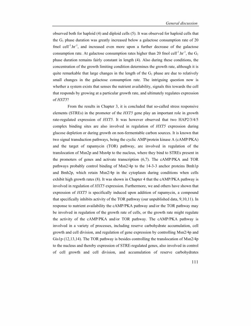
General discussion
111
observed both for haploid (4) and diploid cells (5). It was observed for haploid cells that the G1 phase duration was greatly increased below a galactose consumption rate of 20 fmol cell-1.hr-1, and increased even more upon a further decrease of the galactose consumption rate. At galactose consumption rates higher than 20 fmol cell-1.hr-1, the G1 phase duration remains fairly constant in length (4). Also during these conditions, the concentration of the growth limiting condition determines the growth rate, although it is quite remarkable that large changes in the length of the G1 phase are due to relatively small changes in the galactose consumption rate. The intriguing question now is whether a system exists that senses the nutrient availability, signals this towards the cell that responds by growing at a particular growth rate, and ultimately regulates expression of HXT5?
From the results in Chapter 3, it is concluded that so-called stress responsive elements (STREs) in the promoter of the HXT5 gene play an important role in growth rate-regulated expression of HXT5. It was however observed that two HAP2/3/4/5 complex binding sites are also involved in regulation of HXT5 expression during glucose depletion or during growth on non-fermentable carbon sources. It is known that two signal transduction pathways, being the cyclic AMP/protein kinase A (cAMP/PKA) and the target of rapamycin (TOR) pathway, are involved in regulation of the translocation of Msn2p and Msn4p to the nucleus, where they bind to STREs present in the promoters of genes and activate transcription (6,7). The cAMP/PKA and TOR pathways probably control binding of Msn2/4p to the 14-3-3 anchor proteins Bmh1p and Bmh2p, which retain Msn2/4p in the cytoplasm during conditions when cells exhibit high growth rates (8). It was shown in Chapter 4 that the cAMP/PKA pathway is involved in regulation of HXT5 expression. Furthermore, we and others have shown that expression of HXT5 is specifically induced upon addition of rapamycin, a compound that specifically inhibits activity of the TOR pathway (our unpublished data, 9,10,11). In response to nutrient availability the cAMP/PKA pathway and/or the TOR pathway may be involved in regulation of the growth rate of cells, or the growth rate might regulate the activity of the cAMP/PKA and/or TOR pathway. The cAMP/PKA pathway is involved in a variety of processes, including reserve carbohydrate accumulation, cell growth and cell division, and regulation of gene expression by controlling Msn2/4p and Gis1p (12,13,14). The TOR pathway is besides controlling the translocation of Msn2/4p to the nucleus and thereby expression of STRE-regulated genes, also involved in control of cell growth and cell division, and accumulation of reserve carbohydrates

Chapter 6
112
(8,15,16,17). The cAMP/PKA pathway and the TOR pathway probably act independently, because glycogen accumulation still occurred in a hyperactive cAMP/PKA mutant upon addition of rapamycin (15). An overview of the components that are involved in the regulation of the cAMP/PKA pathway and downstream components of this pathway is depicted in Figure 1, and are shortly described below (for review see 12,18,19,20).
Figure 1: Overview of the PKA pathway and additional pathways that are involved in controlling the localization of Msn2/4p. For details see text.
High glucose
Gpr1pHxtp's
Glucosephosphorylation
Gpa2p
Cyr1pRas1,2p
Cdc25p
Sdc25p
Hsp70
Stress
Ira1,2p
?
CAP
Adenylatecyclase
cAMP
ATP
Tpk1,2,3pBcy1p
Protein kinase A
Pde1,2p
AMP
xxxxxxxxxxxxxxxxxxxxxxxxxxx xxxxxxxxxxxxxxxxxxxxxxxxxxx
xxxxxxxxxx
xxxxxxxxxxxxxxxxxxxxxxxxxxxxxxxxxxxxxxxxxxxxxxxx ?
Sch9p
?
?xxxxxxxxxxxxxxxxxxxxxxxxxxxxxxxxxxxx
Tor1,2p
Glucose and
all essential nutrients
Nutrients
(nitrogen)
cAMP pathway FGM pathway TOR pathway
Msn2/4p
Expression of
STRE-regulated
genes
Carbohydrate
accumulation
Intracellular
acidification
Tpk1,2,3pBcy1p
cAMPCell growth and
cell proliferation
xxxxxxxxxxxxxxxxxxxxxxxxxxxxxxxxxx
Bmh1,2p
Msn2/4pExpression of
PDS-regulated
genes
Gis1p
Translocation
to nucleus
?
?
Rapamycin
Cytoplasm

General discussion
113
The activity of the PKA pathway is regulated by transient activation, or by sustained activation. The first components of the cAMP/PKA pathway that were cloned were the RAS1 and the RAS2 genes. Ras2p is active when GTP is bound, and becomes inactive when GTP is hydrolysed to GDP. The exchange of GDP for GTP is mediated by the Cdc25p and Sdc25p, and Ira1p and Ira2p stimulate exchange of GTP for GDP. A downstream target of Ras2p is adenylate cyclase, encoded by CYR1. CAP/Srv2p associates with Cyr1p, and is required for activation of adenylate cyclase by Ras. Adenylate cyclase mediates the conversion of ATP into cAMP. The target of cAMP is the cAMP-dependent protein kinase A, known as PKA. It consists of a regulatory subunit, which is encoded by BCY1, and three catalytic subunits, encoded by TPK1, TPK2 and TPK3. In non-growing cells, or cells that grow with a low growth rate, PKA is an inactive tetramer consisting of two regulatory subunits and two catalytic subunits. In response to extracellular signals that result in increased cAMP levels, cAMP binds to the regulatory subunits. Subsequently, a conformational change in the regulatory subunits results in a decreased affinity for the catalytic subunits, which are released and present in a free and active conformation. Hydrolysis of cAMP by Pde1p and Pde2p restores PKA to the resting, inactive state. Three mechanisms are probably involved in transient up-regulation of the activity of the cAMP/PKA pathway. Firstly, intracellular acidification seems to serve upstream of the cAMP/PKA activity during conditions of nutrient depletion (21). It is believed that intracellular acidification blocks activity of Ira1p and Ira2p, which results in a long lasting increase in cAMP levels and increased activity of the cAMP/PKA pathway. It was suggested that intracellular acidification might be involved controlling the maintenance of a proper intracellular pH and ATP levels during conditions of carbon starvation (22). Secondly, it was suggested that Cdc25p is activated by the HSP70 heat shock protein Ssa1p, which binds directly to Cdc25p, and thereby controls activity of the cAMP/PKA pathway (23). The authors proposed that after stress conditions, transient accumulation of unfolded proteins occurs, which reduces the availability of HSP70 for Cdc25p and reduces the activity of the cAMP/PKA pathway. Thirdly, a system exists that specifically activates the cAMP/PKA pathway in response to addition of glucose to glucose-deprived cells, which results in a transient increase in cAMP levels. This system involves the proteins Gpr1p and Gpa2p. For proper activation of adenylate cyclase by this pathway, glucose transport and glucose phosphorylation are also required (24). There seems to exist a system that is responsible for maintaining a sustained high activity of the cAMP/PKA

Chapter 6
114
pathway during growth on glucose or other rapidly fermented sugars. This pathway, called the fermentable growth-medium-induced (FGM) pathway, has been proposed on basis of physiological data that showed that only a complete growth medium containing glucose or a related fermentable carbon source is able to maintain the glucose effect of targets of the cAMP/PKA pathway (25). For example, starvation for nitrogen in cells growing on glucose results in a slow increase in the cAMP level rather than a decrease, although phenomena associated with decreased activity of the cAMP/PKA pathway were observed (26). Down-regulation of targets of the cAMP/PKA pathway is apparently also regulated in a cAMP independent manner, suggesting the involvement of an additional mechanism that regulates cAMP/PKA activity, being the FGM pathway. It was shown that the protein kinase Sch9p, which has high homology to the catalytic subunits of PKA (27), is involved in activation of the FGM pathway (28). The FGM pathway may control the activity PKA directly, or the downstream targets of the cAMP/PKA pathway. It remains however unclear how glucose and other rapidly fermentable sugars are sensed and activate the FGM pathway. Thus, it seems that transient activation of PKA is mediated by cAMP, whereas the FGM pathway mediates sustained activation of PKA.
In yeast, a molecular machinery consisting of cyclins and cyclin-dependent kinases (CDKs) regulates progression through the cell cycle, and is therefore an important regulator of the growth rate of cells. In some way the availability of nutrients should be connected to the cell cycle machinery in order to regulate the length of the G1 phase, which is determined by the time when cells enter the passage from the G1 to the S phase. Cdc28p, a CDK, is activated throughout the cell cycle by binding of different cyclins, and controls the major events of the yeast cell division cycle (29). Transition from the G1 to the S phase is mediated by a peak in expression of CLN1 and CLN2 (30,31,32). The timing of this event is regulated by Cln3p, whose abundance is regulated by transcription and translation (33,34,35). Translation of Cln3p is regulated by activity of the cAMP/PKA and TOR pathway, and in addition CLN3 mRNA may be kept in an untranslated state, which is mediated by Whi3p (15,36,37,38).
Thus, the nutrient availability is involved in controlling the growth rate of cells, which may be governed by the cAMP/PKA, the TOR and the FGM pathway. At the same time, these pathways are involved in expression of STRE-regulated genes. It is therefore an interesting question whether the growth rate really regulates expression of HXT5 and STRE-regulated genes. If this is the case, it would mean that certain

General discussion
115
components of the cell cycle machinery, e.g. cyclins, would also function upstream of Msn2/4p. In other words, the cAMP/PKA pathway might regulate expression of HXT5 and the growth rate separately. By experiments described in Chapter 4, the growth rate and the activity of the cAMP/PKA pathway was uncoupled by using continuous cultures. A ras2 deletion mutant, which always exhibits low PKA activity, and has decreased growth rates in batch cultures as compared to wildtype cells, was grown at low and high dilution rates in nitrogen-limited continuous cultures. In this mutant, HXT5 remained expressed even during growth at high dilution rates, which was not observed in wildtype cells. If a high growth rate per se results in repression of HXT5 expression, this would also have occurred in the ras2 deletion mutant, unless Ras2p or a pathway controlled by Ras2p is involved in HXT5 expression. This suggests that the cAMP/PKA pathway, and not the growth rate which is determined by the activity of the cAMP/PKA pathway, is important for regulation of HXT5 expression. However, if Ras2p or the cAMP/PKA pathway was directly involved in regulation of the growth rate of cells, ras2 deleted cells would probably be unable to grow at high dilution rates, and cells should be washed out during growth in continuous cultures. Because the latter does not occur, additional mechanisms besides the cAMP/PKA pathway should exist to regulate the growth rate of cells, which subsequently determines expression of HXT5. Additionally, the TOR pathway may regulate the growth rate of cells, as well as the FGM pathway. However, other components may be involved in controlling the growth rate as well.
This is best discussed when we know some additional regulatory features of the cAMP/PKA pathway, concerning the dynamics of reserve carbohydrate accumulation as observed for synchronized cells. This provides a link between accumulation of reserve carbohydrates and regulation of the growth rate of cells. Similar like the relation between the G1 phase duration and the carbon availability, the accumulation of reserve carbohydrates and in particular trehalose also correlated with the availability of the carbon source. A tight correlation was observed between the duration of the G1 phase and the level of reserve carbohydrate accumulation; upon a longer duration of the G1 phase, the maximal level of accumulation increased (4,5). Moreover, when the G1 phase duration was specifically elongated, the accumulation pattern of trehalose and glycogen proved to be quite dynamic. During conditions when the G1 phase is elongated, trehalose levels gradually increased during progression through the G1 phase. However, at a sudden point towards the end of the G1 phase, the

Chapter 6
116
levels of accumulated reserve carbohydrates suddenly dropped (5). This decrease was suggested to be required for a proper progression to the S phase, where the liberated glucose would ultimately result in increased sugar flux and increased CLN1 and CLN2 expression, making progression to the S phase possible. This also prompted the hypothesis that accumulation of reserve carbohydrates by themselves caused the increase in G1 phase duration. Indeed, cells unable to synthesize trehalose and glycogen have a short G1 phase duration of 120 minutes when grown at galactose consumption rates of 20 fmol cell-1.hr-1 and lower, whereas wildtype cells increased their G1 phase duration to maximally 520 minutes during the same conditions (39). Thus, accumulation of reserve carbohydrates is also involved in regulation of the growth rate of cells. In wildtype cells, trehalose and glycogen probably decrease the amount of carbon source that may be used for G1 phase progression. In cells unable to synthesize reserve carbohydrates, signalling through the cAMP/PKA pathway may be increased because of higher carbon availability, resulting in higher translation of Cln3p and faster progression through the G1 phase.
A recent study revealed the mechanism responsible for the characteristic behaviour of reserve carbohydrates in the G1 phase at low growth rates (40). In this study, cells were grown in fed-batch cultures under glucose-limiting conditions, resulting in extracellular glucose concentrations that were always lower than 0.17 mM, thereby not activating the Gpr1-Gpa2 sensing system or the FGM pathway, which both exhibit maximum activation at glucose concentrations of about 20 mM (24,40). It was observed that at the end of the G1 phase, just prior to the S phase, intracellular cAMP levels increase, and decrease again subsequently in the G2 and M phase (40). This increase in cAMP was suggested to transiently activate the cAMP/PKA pathway. A decrease in the oxygen tension was observed, which is known to be associated with a rise in glycolytic flux and occurrence of overflow of metabolism. This increase in glycolytic flux is probably due to breakdown of trehalose and glycogen, because increased PKA activity results in increased trehalase activity and increased breakdown of glycogen by inhibiting its accumulation and stimulating its degradation (41). Thus, a transient increase in cAMP might be important for progression through cell cycle during growth at low growth rates, which suggests that cells may not progress through the cell cycle if there is no transient increase in cAMP. Indeed, a transient increase in intracellular cAMP levels was also observed at high extracellular glucose concentrations, but now in the M phase of the cell cycle (40). A transient increase in the

General discussion
117
cAMP levels during the cell cycle might be required for proper progression through the cell cycle, and the nutrient availability, inherent to the growth rate of cells, might determine in which phase of the cell cycle this occurs. It remains however unknown how this sudden increase in cAMP was generated, but the possibility that components upstream of Cyr1p are involved were excluded (40). Maybe, this transient cAMP increase is also involved in regulation of expression of HXT5. It is known that sudden addition of a high amount of glucose results in a transient increase of cAMP, which is caused by the activation of the Gpr1p-Gpa2p system (24), and will probably also occur in synchronized cells early in the G1 phase. During subsequent growth at high growth rates, intracellular cAMP levels are higher compared to growth at low growth rates (40), and the transient increase in cAMP was observed again in the M phase, as if the cells might build in a control to govern the growth rate. Thus, the increase in intracellular cAMP might be important for governing the growth rate and subsequent expression of genes. During growth at low growth rates at low nutrient availability, something else might happen; the initial peak in cAMP is not observed, thereby rendering the cAMP/PKA pathway inactive, and resulting in expression of HXT5. Throughout the G1 phase, the levels of cAMP remains low, thereby still inducing HXT5 expression. However, at the end of the G1 phase, the increase in cAMP might transiently decrease HXT5 expression, although this has not been demonstrated experimentally.
If accumulation of reserve carbohydrates itself has a major impact on the growth rate of cells, other factors that are important for the regulation of reserve carbohydrate accumulation may be important for regulation of the growth rate as well. In this respect, as we suggested that Hxt5p is involved in trehalose accumulation, Hxt5p itself may play a role in regulation of the growth rate by controlling to some degree trehalose accumulation. The suggestion that Kns1p might phosphorylate Hxt5p (Chapter 4) and Tps1p indicates that this kinase may be important in this process. Remarkably, expression of KNS1 was similar to expression of HXT5 during a variety of conditions that result in low growth rates (42). Phosphorylation of Hxt5p and Tps1p by Kns1p might render Hxt5p and Tps1p more active, or might induce a conformational change so that Hxt5p and Tps1p may interact when both proteins are phosphorylated. Subsequently, the glucose flux may be driven towards accumulation of trehalose. At the G1/S transition, trehalose is no longer accumulated, maybe because the activity of Kns1p is cell cycle-regulated and decreases at the G1/S transition. It will be interesting to determine whether the activity of Kns1p is under control of the PKA pathway.

Chapter 6
118
In order to change the sugar flux from glycolysis towards reserve carbohydrate accumulation during low growth rates, other changes in the control of metabolism have to occur as well. The flux has to be changed towards glucose-1-phosphate and UDP-glucose, by stimulating expression of PGM1, PGM2 and UGP1, or by rendering these enzymes more active. Recently, the PAS kinases Psk1p and Psk2p were identified to have a role in the regulation of changing the flux towards and from glycolysis and reserve carbohydrate accumulation (43). Psk1p and Psk2p were originally identified to be required for growth on galactose at 39ºC (Galts). Interestingly, cells grown on galactose exhibit lower maximal growth rates as compared to growth on glucose, and during growth at 39ºC the growth rate is decreased compared to growth at 30ºC. Thus, the Psk proteins may be involved in regulation the growth rate of cells, or regulation of certain processes that are involved in growth rate regulation. A suppression screen revealed that overexpression of PGM1 or PGM2 rescued the Galts growth defect, thus Psk1p and Psk2p are somehow involved in the control of the interconversion of glucose-6-phosphate to glucose-1-phosphate. Furthermore, it was shown that Psk2p was able to phosphorylate Ugp1p, thereby probably rendering it inactive. This was concluded because trehalose and glycogen accumulation was increased in exponentially growing cells deleted for psk1 and psk2. Thus, low growth rates might result in inactivation of Psk1p and Psk2p, dephosphorylation and activation of Ugp1p and pushing the flux towards UDP-glucose and reserve carbohydrate accumulation. During high growth rates, Psk1p and Psk2p are probably active, and direct the sugar flux towards glycolysis. The signal that regulates the activity of PAS kinases is however unknown. It would be interesting to determine whether the activity of PAS kinase in cell cycle regulated during low growth rates, thereby modulating the carbon flux during the G1 phase.
During conditions of slow growth expression of a number of genes is specifically induced, and activity of certain proteins may be modulated to drive the carbon flux away from glycolysis towards other metabolic routes, like accumulation of reserve carbohydrates. These changes are well coordinated by the cAMP/PKA, TOR and the FGM pathway, and additionally by proteins like Psk1/2p, and may also be controlled by Hxt5p.

General discussion
119
References 1 Monod, J. (1942). Recherches sur la Croissance des Cultures Bactériennes. Hermann, Paris,
France. 2 Pirth, S. J. (1975). Priciples of microbe and cell cultivation. Blackwell Scientific Publications,
London. 3 Carter, B. L. and Jagadish, M. N. (1978). Control of cell division in the yeast Saccharomyces
cerevisiae cultured at different growth rates. Exp Cell Res 112, 373-83. 4 Paalman, J. W., Verwaal, R., Slofstra, S. H., Verkleij, A. J., Boonstra, J. and Verrips, C. T. (2003).
Trehalose and glycogen accumulation is related to the duration of the G1 phase of Saccharomyces cerevisiae. FEM Yeast Res 3, 261-8.
5 Silljé, H. H., ter Schure, E. G., Rommens, A. J., Huls, P. G., Woldringh, C. L., Verkleij, A. J., Boonstra, J. and Verrips, C. T. (1997). Effects of different carbon fluxes on G1 phase duration, cyclin expression, and reserve carbohydrate metabolism in Saccharomyces cerevisiae. J. Bacteriol. 179, 6560-5.
6 Martinez-Pastor, M. T., Marchler, G., Schuller, C., Marchler-Bauer, A., Ruis, H. and Estruch, F. (1996). The Saccharomyces cerevisiae zinc finger proteins Msn2p and Msn4p are required for transcriptional induction through the stress response element (STRE). Embo J 15, 2227-35.
7 Schmitt, A. P. and McEntee, K. (1996). Msn2p, a zinc finger DNA-binding protein, is the transcriptional activator of the multistress response in Saccharomyces cerevisiae. Proc. Natl. Acad. Sci. USA 93, 5777-82.
8 Beck, T. and Hall, M. N. (1999). The TOR signalling pathway controls nuclear localization of nutrient-regulated transcription factors. Nature 402, 689-92.
9 Hardwick, J. S., Kuruvilla, F. G., Tong, J. K., Shamji, A. F. and Schreiber, S. L. (1999). Rapamycin-modulated transcription defines the subset of nutrient-sensitive signaling pathways directly controlled by the Tor proteins. Proc Natl Acad Sci U S A 96, 14866-70.
10 Shamji, A. F., Kuruvilla, F. G. and Schreiber, S. L. (2000). Partitioning the transcriptional program induced by rapamycin among the effectors of the Tor proteins. Curr Biol 10, 1574-81.
11 Crespo, J. L. and Hall, M. N. (2002). Elucidating TOR signaling and rapamycin action: lessons from Saccharomyces cerevisiae. Microbiol Mol Biol Rev 66, 579-91.
12 Thevelein, J. M., Cauwenberg, L., Colombo, S., De Winde, J. H., Donation, M., Dumortier, F., Kraakman, L., Lemaire, K., Ma, P., Nauwelaers, D., Rolland, F., Teunissen, A., Van Dijck, P., Versele, M., Wera, S. and Winderickx, J. (2000). Nutrient-induced signal transduction through the protein kinase A pathway and its role in the control of metabolism, stress resistance, and growth in yeast. Enzyme Microb Technol 26, 819-25.
13 Gorner, W., Durchschlag, E., Martinez-Pastor, M. T., Estruch, F., Ammerer, G., Hamilton, B., Ruis, H. and Schuller, C. (1998). Nuclear localization of the C2H2 zinc finger protein Msn2p is regulated by stress and protein kinase A activity. Genes Dev 12, 586-97.
14 Pedruzzi, I., Burckert, N., Egger, P. and De Virgilio, C. (2000). Saccharomyces cerevisiae Ras/cAMP pathway controls post-diauxic shift element-dependent transcription through the zinc finger protein Gis1. Embo J 19, 2569-79.
15 Barbet, N. C., Schneider, U., Helliwell, S. B., Stansfield, I., Tuite, M. F. and Hall, M. N. (1996). TOR controls translation initiation and early G1 progression in yeast. Mol. Biol. Cell. 7, 25-42.
16 Thomas, G. and Hall, M. N. (1997). TOR signalling and control of cell growth. Curr Opin Cell Biol 9, 782-7.
17 Schmelzle, T. and Hall, M. N. (2000). TOR, a central controller of cell growth. Cell 103, 253-62. 18 Thevelein, J. M. and de Winde, J. H. (1999). Novel sensing mechanisms and targets for the cAMP-
protein kinase A pathway in the yeast Saccharomyces cerevisiae. Mol Microbiol 33, 904-18.

Chapter 6
120
19 Estruch, F. (2000). Stress-controlled transcription factors, stress-induced genes and stress tolerance in budding yeast. FEMS Microbiol Rev 24, 469-86.
20 Gagiano, M., Bauer, F. F. and Pretorius, I. S. (2002). The sensing of nutritional status and the relationship to filamentous growth in Saccharomyces cerevisiae. FEM Yeast Res 2, 433-70.
21 Thevelein, J. M. (1991). Fermentable sugars and intracellular acidification as specific activators of the RAS-adenylate cyclase signalling pathway in yeast: the relationship to nutrient-induced cell cycle control. Mol Microbiol 5, 1301-7.
22 Colombo, S., Ma, P., Cauwenberg, L., Winderickx, J., Crauwels, M., Teunissen, A., Nauwelaers, D., de Winde, J. H., Gorwa, M. F., Colavizza, D. and Thevelein, J. M. (1998). Involvement of distinct G-proteins, Gpa2 and Ras, in glucose- and intracellular acidification-induced cAMP signalling in the yeast Saccharomyces cerevisiae. Embo J 17, 3326-41.
23 Geymonat, M., Wang, L., Garreau, H. and Jacquet, M. (1998). Ssa1p chaperone interacts with the guanine nucleotide exchange factor of ras Cdc25p and controls the cAMP pathway in Saccharomyces cerevisiae. Mol Microbiol 30, 855-64.
24 Rolland, F., De Winde, J. H., Lemaire, K., Boles, E., Thevelein, J. M. and Winderickx, J. (2000). Glucose-induced cAMP signalling in yeast requires both a G-protein coupled receptor system for extracellular glucose detection and a separable hexose kinase-dependent sensing process. Mol Microbiol 38, 348-58.
25 Thevelein, J. M. (1994). Signal transduction in yeast. Yeast 10, 1753-90. 26 Ma, P., Goncalves, T., Maretzek, A., Dias, M. C. and Thevelein, J. M. (1997). The lag phase rather
than the exponential-growth phase on glucose is associated with a higher cAMP level in wild-type and cAPK-attenuated strains of the yeast Saccharomyces cerevisiae. Microbiology 143 ( Pt 11), 3451-9.
27 Toda, T., Cameron, S., Sass, P. and Wigler, M. (1988). SCH9, a gene of Saccharomyces cerevisiae that encodes a protein distinct from, but functionally and structurally related to, cAMP-dependent protein kinase catalytic subunits. Genes Dev 2, 517-27.
28 Crauwels, M., Donaton, M. C., Pernambuco, M. B., Winderickx, J., de Winde, J. H. and Thevelein, J. M. (1997). The Sch9 protein kinase in the yeast Saccharomyces cerevisiae controls cAPK activity and is required for nitrogen activation of the fermentable-growth-medium-induced (FGM) pathway. Microbiology 143 ( Pt 8), 2627-37.
29 Mendenhall, M. D. and Hodge, A. E. (1998). Regulation of Cdc28 cyclin-dependent protein kinase activity during the cell cycle of the yeast Saccharomyces cerevisiae. Microbiol Mol Biol Rev 62, 1191-243.
30 Hadwiger, J. A., Wittenberg, C., Richardson, H. E., de Barros Lopes, M. and Reed, S. I. (1989). A family of cyclin homologs that control the G1 phase in yeast. Proc Natl Acad Sci U S A 86, 6255-9.
31 Richardson, H. E., Wittenberg, C., Cross, F. and Reed, S. I. (1989). An essential G1 function for cyclin-like proteins in yeast. Cell 59, 1127-33.
32 Wittenberg, C., Sugimoto, K. and Reed, S. I. (1990). G1-specific cyclins of S. cerevisiae: cell cycle periodicity, regulation by mating pheromone, and association with the p34CDC28 protein kinase. Cell 62, 225-37.
33 Dirick, L., Bohm, T. and Nasmyth, K. (1995). Roles and regulation of Cln-Cdc28 kinases at the start of the cell cycle of Saccharomyces cerevisiae. Embo J 14, 4803-13.
34 Stuart, D. and Wittenberg, C. (1995). CLN3, not positive feedback, determines the timing of CLN2 transcription in cycling cells. Genes Dev 9, 2780-94.
35 Tyers, M., Tokiwa, G. and Futcher, B. (1993). Comparison of the Saccharomyces cerevisiae G1 cyclins: Cln3 may be an upstream activator of Cln1, Cln2 and other cyclins. Embo J 12, 1955-68.
36 Hall, D. D., Markwardt, D. D., Parviz, F. and Heideman, W. (1998). Regulation of the Cln3-Cdc28 kinase by cAMP in Saccharomyces cerevisiae. Embo J 17, 4370-8.

General discussion
121
37 Polymenis, M. and Schmidt, E. V. (1997). Coupling of cell division to cell growth by translational control of the G1 cyclin CLN3 in yeast. Genes Dev 11, 2522-31.
38 Gari, E., Volpe, T., Wang, H., Gallego, C., Futcher, B. and Aldea, M. (2001). Whi3 binds the mRNA of the G1 cyclin CLN3 to modulate cell fate in budding yeast. Genes Dev 15, 2803-8.
39 Paalman, J. W. G. (2001). The role of nutrients in G1 phase progression of Saccharomyces cerevisiae, PhD-thesis, Universiteit Utrecht, Utrecht, The Netherlands.
40 Muller, D., Exler, S., Aguilera-Vazquez, L., Guerrero-Martin, E. and Reuss, M. (2003). Cyclic AMP mediates the cell cycle dynamics of energy metabolism in Saccharomyces cerevisiae. Yeast 20, 351-67.
41 Francois, J. and Parrou, J. L. (2001). Reserve carbohydrates metabolism in the yeast Saccharomyces cerevisiae. FEMS Microbiol Rev 25, 125-45.
42 Gasch, A. P., Spellman, P. T., Kao, C. M., Carmel-Harel, O., Eisen, M. B., Storz, G., Botstein, D. and Brown, P. O. (2000). Genomic expression programs in the response of yeast cells to environmental changes. Mol. Biol. Cell. 11, 4241-57.
43 Rutter, J., Probst, B. L. and McKnight, S. L. (2002). Coordinate regulation of sugar flux and translation by PAS kinase. Cell 111, 17-28.

122

Summary
123
Summary
Glucose, which is transported into yeast cells by hexose transporter (Hxt) proteins, is the most preferred carbon source for the yeast Saccharomyces cerevisiae. A multigene family consisting of 20 members encodes the Hxt proteins. HXT1-4 and HXT6-7 encode the major Hxt proteins, the remaining Hxt proteins have other or unknown functions. Hxt proteins localize to the plasma membrane, have 12 membrane-spanning domains, and their amino- and carboxy-terminal domains are present in the cytoplasm. Expression of the major HXT genes is regulated by the extracellular glucose concentration. Some of the major HXT genes are expressed when the glucose concentration is high, whereas others are expressed when the concentration is low.
Comparing the number of amino acids of all Hxt proteins revealed that the Hxt5 protein contains a longer amino-terminal domain as compared to the major Hxt proteins, which suggests that Hxt5 has additional functions besides glucose transport. In Chapter 2 we determined the expression pattern of HXT5 to obtain insight in regulation of HXT5 expression. It appeared that expression of HXT5 is not regulated by the extracellular glucose concentration, but by the growth rate of yeast cells. This was concluded by growing cells in batch cultures on glucose, ethanol and glycerol, growth at a higher temperature or osmolarity, growth in fed-batch cultures and continuous cultures. The results indicated that low growth rates of cells result in induction of HXT5 expression.
Gene expression is regulated by binding of transcription factors to regulatory elements in the promoter of genes. Therefore, the growth rate should somehow be involved in binding of specific transcription factors to the promoter of the HXT5 gene. In Chapter 3, the putative regulatory elements in the promoter of HXT5 that contribute to growth rate-regulated HXT5 expression were identified. Computer-assisted analysis revealed the presence of two putative stress responsive elements (STREs), one putative post-diauxic shift (PDS) element and two putative Hap2/3/4/5 (HAP) complex binding elements. The involvement of these elements was studied by mutation of these putative regulatory elements in a HXT5-promoter LacZ fusion construct. It appeared that one of the STREs is extremely important for growth-rate regulated HXT5 expression, whereas in addition the HAP elements are important for regulation of HXT5 expression during growth on ethanol and glycerol.

124
Glucose is not only used for metabolism, but it may also serve as a precursor for the reserve carbohydrates trehalose and glycogen. Earlier studies in our laboratory have shown that trehalose in particular accumulates when the growth rate of cells is decreased. It was suggested in literature that Tps1, one of components of the protein complex involved in trehalose production, might control glucose transport by interacting with a hexose transporter. Because Hxt5 contains a longer amino-terminal domain that may serve as an interaction domain for Tps1, and expression of HXT5 and accumulation of trehalose are both regulated by the growth rate of cells, the role of Hxt5 in trehalose accumulation was investigated in Chapter 4. It appeared that HXT5 was indeed expressed when trehalose accumulated during low growth rates in glucose-grown batch cultures, fed-batch cultures and continuous cultures. Furthermore, expression of both HXT5 and TPS1 was found to be regulated by the cAMP/PKA signal transduction pathway. In addition we observed that Hxt5 was phosphorylated upon glucose depletion, a condition that results in trehalose accumulation. Other studies indicated that the kinase that is likely to phosphorylate Hxt5 binds physically to Tps1. These observations strongly suggested that Hxt5 plays a role in trehalose accumulation. However, trehalose accumulation was affected, but not completely abolished, in a strain deleted for hxt5. Furthermore, no direct physical interaction between Hxt5 and Tps1 was determined as determined by yeast 2-hybrid studies and co-immunoprecipitation. Therefore, the exact role of Hxt5 in trehalose accumulation remains to be further explored.
To establish whether Hxt5 is involved in other processes, genes that were expressed similar to HXT5 during growth at a low growth rate in fed-batch cultures were identified using DNA microarray analysis. This approach was chosen, because genes that are similarly expressed may be involved in similar processes. From a total of 6144 genes, 72 genes exhibited an expression pattern similar to HXT5. These included genes encoding proteins involved in trehalose accumulation, which strengthens our hypothesis that Hxt5 plays a role in trehalose accumulation. Also genes encoding proteins involved in respiration, protein folding, stress responses, and genes encoding proteins involved in metabolism of carbon that were not present in the growth medium, were similarly expressed. How Hxt5 is involved in any of these processes remains to be determined. It appeared that the genes that were identified in our study were also similarly expressed in a variety of other studies that all had one common feature, being a reduction in the growth rate of cells. We suggested that cells might be pre-
Summary
125
programmed to rapidly and specifically respond to harmful conditions by inducing a common set of genes encoding proteins with protective properties. Expression of these genes is triggered by a reduction in the growth rate of cells. A high percentage of these genes contain STREs in their promoter region. This regulatory element may therefore be involved in growth rate-regulated expression of genes. Because it is known that the cAMP/PKA pathway is involved in STRE-mediated gene expression, this pathway seems important for regulation of growth rate-regulated gene expression in Saccharomyces cerevisiae. In Chapter 6, the results of this thesis are discussed in a broader perspective, and we explained which factors and processes might contribute to regulation of the growth rate and growth rate-regulated expression of genes in Saccharomyces cerevisiae.

126

Samenvatting
127
Samenvatting Gisten zijn ééncellige organismen. Er zijn veel verschillende soorten gisten, waarvan Saccharomyces cerevisiae, oftewel bakkersgist, de bekendste is. Gisten worden al sinds de tijd van de oude Egyptenaren gebruikt voor de bereiding van bier, brood en wijn, omdat ze in staat zijn ethanol (alcohol) en koolzuur te produceren. Om te kunnen groeien en overleven hebben gisten voedingsbronnen als stikstof en koolstof nodig. Voorbeelden van koolstofbronnen zijn ethanol en glycerol en de suikers glucose en galactose. Gisten hebben een voorkeur voor bepaalde koolstofbronnen, omdat deze makkelijk te gebruiken zijn voor productie van energie, een proces dat metabolisme heet. Van alle koolstofbronnen wordt glucose het makkelijkst gemetaboliseerd en gisten hebben daardoor een voorkeur om te groeien op glucose. Op het moment dat gisten in aanraking komen met glucose, zal de samenstelling van de gistcel zó veranderen, dat glucose zo snel en efficiënt mogelijk gemetaboliseerd kan worden. Om dit te bewerkstelligen zijn eiwitten nodig. Om te begrijpen hoe die eiwitten gemaakt worden, volgt nu een stukje “basiskennis” (Figuur 1). Gistcellen bevatten, net als bijvoorbeeld menselijke cellen, DNA. Op het DNA, dat erg groot is, zijn kleine afzonderlijke stukjes aanwezig, genaamd genen die de informatie bevatten om eiwitten te maken. In Saccharomyces cerevisiae zijn 6144 genen aanwezig en van die genen kunnen in principe uiteindelijk evenzoveel eiwitten worden gemaakt, die het “werk” in de cel doen. Van een gen wordt eerst het zogenaamde boodschapper RNA of messenger RNA (mRNA) gemaakt. Dit mRNA dient vervolgens als boodschapper voor eiwitsynthese, wat gebeurt met behulp van speciale eiwitten die ribosomen heten. De aanmaak (expressie) van mRNA wordt gereguleerd door een klein stukje DNA, de zogenaamde promoter, dat voor een gen ligt. Aan kleine gebiedjes in de promoter, de regulatoire elementen, kunnen transcriptie factoren binden, wat eiwitten zijn die reguleren of een gen tot expressie wordt gebracht (Figuur 1).

128
Figuur 1: Van DNA tot eiwit. In de promoter van genen zijn regulatoire elementen aanwezig, waaraan transcriptie factoren kunnen binden. Als dit gebeurt, kan het gen worden vertaald in boodschapper RNA (mRNA). Het mRNA wordt gelezen door ribosomen die zorgen voor aanmaak van het eiwit.
Glucose, de meest geprefereerde koolstofbron, kan niet zomaar door het omhulsel (membraan) van een gistcel heenkomen. Het wordt specifiek de cel in getransporteerd door eiwitten die hexose transporters (Hxt’s) heten. Deze eiwitten zijn aanwezig in het membraan van de gistcel. Zie hiervoor de kaft van mijn proefschrift: De paarse stipjes aan de rand van de grote ronde cel geven de locatie van het Hxt5 eiwit in de membraan van de cel aan. Er zijn 17 verschillende HXT genen die 17 verschillende Hxt eiwitten kunnen maken in Saccharomyces cerevisiae. Niet alle Hxt’s even belangrijk. Alleen Hxt1,2,3,4,5,6 en Hxt7, de zogenaamde “hoofd” Hxt’s, dragen daadwerkelijk bij tot glucose transport, HXT8-17 komen bijna niet tot expressie. Expressie van de “hoofd” hexose transporters wordt bepaald door de hoeveelheid glucose die aanwezig is in de omgeving van de gistcel. Ze worden niet allemaal tegelijk tot expressie gebracht. Sommige van de “hoofd” HXT genen komen tot expressie als er veel glucose, en andere als er weinig glucose aanwezig is. In Hoofdstuk 2 is aangetoond dat expressie van HXT5 niet door de glucose concentratie, maar op een andere manier gereguleerd wordt, namelijk door de snelheid
DNA
mRNA
Eiwit
Promoter Gen
Regulatoireelementen
Transcriptie factor
mRNA
EiwitRibosoom

Samenvatting
129
waarmee gistcellen groeien, kortweg de groeisnelheid. Deze term is gedefinieerd als de tijdsduur om van één cel twee cellen te maken, wat in bakkersgist gebeurt door middel van celdeling. HXT5 komt niet niet tot expressie wanneer de groeisnelheid hoog is, maar juist wel bij lage groeisnelheden. Dit is uitgezocht met behulp van verschillende opgroeimethoden. Zo komt HXT5 tijdens groei op glucose in batch culturen, waarbij de hoeveelheid glucose na verloop van tijd wordt opgebruikt, niet tot expressie wanneer cellen groeien met een maximale groeisnelheid. HXT5 komt pas tot expressie nadat de groeisnelheid na verloop van tijd afneemt. Ook komt HXT5 tot expressie tijdens groei op ethanol of glycerol, koolstofbronnen waarop de groeisnelheid laag is. Een hoge temperatuur of aanwezigheid van een hoge concentratie zout leidt ook tot een verlaging van de groeisnelheid en expressie van HXT5. De concentratie glucose in het groeimedium is dan nog steeds hoog. In fed-batch culturen, waarbij de groeisnelheid door middel van zeer gedoseerde toevoeging van de hoeveelheid suiker gereguleerd kan worden, is HXT5 expressie alleen aangetoond bij lage groeisnelheden, onafhankelijk van de concentratie glucose in het groeimedium. In continu culturen, waarmee de groeisnelheid heel specifiek gereguleerd kan worden, komt HXT5 alleen tot expressie bij lage groeisnelheden en is expressie weer onafhankelijk van de glucose concentratie. De conclusie van dit onderzoek was dat expressie van HXT5 gereguleerd wordt door de groeisnelheid. Uiteindelijk moet de groeisnelheid op de één of andere manier aangrijpen op transcriptie factoren, omdat deze eiwitten expressie van genen reguleren. Daarom is in Hoofdstuk 3 uitgezocht welke regulatoire elementen in de promoter van het HXT5 gen betrokken zijn bij HXT5 expressie. Door middels van een computeranalyse werden 5 mogelijke regulatoire elementen in de promoter van het HXT5 gen geïdentificeerd, te weten 2 stress responsive elements (STREs), 1 post-diauxic shift (PDS) element en 2 Hap2/3/4/5 transcriptie factor binding (HAP) elements. Het is bekend dat de transcriptie factoren Msn2 en Msn4 binden aan STREs, Gis1 bindt aan PDS en het HAP2/3/4/5 complex bindt aan HAP elementen. Deze transcriptie factoren kunnen zo expressie van genen reguleren, die deze regulatoire elementen in de promoter bevatten. Door nu de mogelijke elementen zó te veranderen (=muteren), dat de transcriptie factoren niet meer kunnen binden, kon de rol van de afzonderlijke elementen in de regulatie van HXT5 expressie worden bestudeerd. Het bleek dat één van de STREs uitermate belangrijk is voor groeisnelheid gereguleerde HXT5 expressie. Op het moment dat dit element gemuteerd was, kwam HXT5 niet meer tot expressie tijdens groeicondities die

130
resulteerden in een langzame groeisnelheid. Verder bleek dat de HAP elementen van belang zijn bij regulatie van HXT5 expressie tijdens groei op ethanol en glycerol. Omdat één van de STREs zo belangrijk is voor regulatie van HXT5 expressie, en omdat het uit de literatuur bekend is dat de transcriptie factoren Msn2 en Msn4 onder controle staan van de protein kinase A (PKA) signaal transductie route, suggereerden wij dat deze route een belangrijke rol zou kunnen spelen bij groeisnelheid gereguleerde HXT5 expressie. Glucose kan door cellen worden gemetaboliseerd, maar daarnaast onder bepaalde omstandigheden ook worden opgeslagen in de gistcel als de reservestoffen trehalose en glycogeen. Eerder onderzoek in ons laboratorium heeft uitgewezen dat de opbouw van met name trehalose, net als expressie van HXT5, ook door groeisnelheid gereguleerd wordt. Verder is in de literatuur gesuggereerd dat één van de eiwitten die betrokken is bij trehalose opbouw, genaamd Tps1, de opname van glucose zou kunnen reguleren door te binden aan hexose transporters. Het Hxt5 eiwit is wat betreft zijn structuur ook nog eens verschillend van de andere Hxt eiwitten, omdat het een langere, unieke "staart" bezit die zich in de binnenkant van de cel bevindt. Ons idee was dat Tps1 hier specifiek aan zou kunnen binden en vervolgens de naar binnen getransporteerde glucose direct in de richting van trehalose opbouw zou dirigeren. In Hoofdstuk 4 is de mogelijke rol van Hxt5 in trehalose opbouw dan ook onderzocht. In glucose gegroeide batch culturen kwam het expressie patroon van HXT5 geheel overeen met het opbouw patroon van trehalose, terwijl het expressie patroon van de andere "hoofd" HXT genen niet of maar gedeeltelijk overeen kwam. In fed-batch culturen kwam HXT5 alleen tot expressie én werd trehalose opgebouwd bij een lage groeisnelheid, terwijl dit voor beide processen niet het geval was bij een hoge groeisnelheid. Ook in continu culturen was er alleen HXT5 expressie en trehalose opbouw bij lage groeisnelheden. Verder is aangetoond dat expressie van zowel HXT5 als TPS1 gereguleerd wordt door de PKA signaal transductie route. Al deze bevindingen suggereerden in sterke mate dat Hxt5 betrokken is bij de opbouw van trehalose in gistcellen. De eerder gesuggereerde binding tussen Hxt5 en Tps1 kon niet worden aangetoond, dus de manier waarop Hxt5 betrokken zou kunnen zijn bij trehalose opbouw is (nog) onbekend. Verder wezen de resultaten erop dat Hxt5 niet exclusief betrokken is bij trehalose opbouw in gist; in een giststam die geen Hxt5 meer kan aanmaken werd nog steeds 80% van de hoeveelheid trehalose opgebouwd ten opzichte van een stam die Hxt5 nog wel bevat.

Samenvatting
131
Om erachter te komen of Hxt5 eventueel betrokken is bij andere processen in gist, is in Hoofdstuk 5 onderzocht welke genen hetzelfde expressie patroon als HXT5 hebben tijdens langzame groei in een fed-batch cultuur. Genen die op hetzelfde moment tot expressie komen, zouden immers iets met elkaar te maken kunnen hebben. Met behulp van de DNA microarray techniek kan het expressie patroon van alle 6144 genen in gist tegelijk worden bepaald. Hiermee werd aangetoond dat 72 genen een expressie patroon hadden dat gelijk was aan het expressie patroon van HXT5 tijdens langzame groei. Deze genen werden vervolgens onderverdeeld in groepen met gemeenschappelijke functies. 1). Genen die betrokken zijn bij trehalose opbouw, wat dus weer een betrokkenheid van Hxt5 bij trehalose opbouw suggereert. 2). Genen die betrokken zijn bij respiratie, een proces dat zorgt voor energie productie tijdens langzame groeisnelheden. 3). Genen die betrokken zijn bij de vouwing van eiwitten. Deze genen behoren tot een grotere groep genen, waarvan bekend is dat ze tot expressie komen tijdens “stress”condities. 4). Genen die betokken zijn bij metabolisme van koolstofbronnen die verrassenderwijs niet aanwezig waren in het groeimedium. Ook was er een aantal genen waarvan de functie (tot nu toe) nog niet geïdentificeerd is. Hoe en of Hxt5 betrokken is bij deze processen, is onbekend. Het was echter opvallend dat veel van de genen die hetzelfde expressiepatroon als HXT5 hadden in onze studie, ook tegelijk met HXT5 tot expressie kwamen na toevoeging van verschillende hoeveelheden zout, verhoging van de temperatuur of het toevoegen van chemicaliën die schadelijk zijn voor de cel. Dit was uitgezocht in andere studies waarin op dezelfde manier het totale expressie plaatje van genen was onderzocht. Tussen al die studies en de onze is één overeenkomst, namelijk dat de condities alle leiden tot een verlaging van de groeisnelheid van cellen. Dus HXT5 zou samen met de in onze studie geïdentificeerde genen tot een gemeenschappelijke groep van genen kunnen behoren die tot expressie wordt gebracht als de groeisnelheid van cellen wordt verlaagd, ongeacht de conditie die resulteert in een verlaging van de groeisnelheid. Het was verder opvallend dat een hoog percentage van de genen met hetzelfde expressie patroon als HXT5 één of meerdere STREs bevatte. In Hoofdstuk 3 is aangetoond dat dit element enorm belangrijk is voor groeisnelheid gereguleerde expressie van HXT5. Omdat bekend is dat de PKA signaal transductie route een belangrijke rol speelt bij expressie van STRE-gereguleerde genen, en deze route van belang is voor regulatie van HXT5 expressie, zou de PKA signaal transductie route in het algemeen een belangrijke rol kunnen spelen in groeisnelheid gereguleerde genexpressie in Saccharomyces cerevisiae.

132
In Hoofdstuk 6 zijn de bevindingen uit het proefschrift in een breder perspectief bediscussieerd. Tevens is geprobeerd om te verklaren hoe groeisnelheid exact gereguleerd kan worden en hoe groeisnelheid kan aangrijpen op expressie van genen in Saccharomyces cerevisiae.

133
Dankwoord
In dit stukje wil ik graag iets zeggen tegen iedereen die op de één of andere manier heeft bijgedragen aan mijn proefschrift.
In de eerste plaats dank ik mijn promotoren Johannes, Theo en Arie. Ik heb de samenwerking met jullie als erg prettig ervaren en wil jullie bedanken voor het vertrouwen dat jullie mij hebben gegeven. Johannes, ik vond het fijn dat jouw deur altijd openstond. Hoe verder ik op weg was, des te meer kwam ik je kamer in om te discussiëren over resultaten en andere interessante zaken. In de toekomst zal ik proberen niet alles drie keer te herhalen en zal ik mij echt aan deadlines proberen te houden, wellicht scheelt dat een hoop stress. Theo, ook jij bedankt voor je uitstekende begeleiding en de vele stimulerende discussies die we gevoerd hebben. Verder vond ik het erg leuk dat we tegelijk op de congressen in Praag en Madison waren en denk ik met plezier terug aan de gesprekken die we daar gevoerd hebben. Arie, in mijn ogen ben jij degene geweest die de grote lijn van mijn project altijd in de gaten gehouden heeft. Je wist altijd precies waar ik mee bezig was en had daardoor vaak verrassende en goede voorstellen. Ik zal proberen in de toekomst nog een hoop mooie doelpunten te maken!
Aan mijn (ex-)collega's in het "gistlab": Cees, toen ik als AIO begon was jij er ook nog even. Jij hebt mijn interesse voor gistwerk aangewakkerd. Bedankt voor alles! Hans, dank voor het leren elutriëren en alle andere dingen die je me bijgebracht hebt. En natuurlijk voor het minipreppen en sequencen! Yvonne, er is nu een grotere kans dat we elkaar tegen zullen komen boven op de berg. Dan praten we bij! Victor, nog even en dan heb jij het ook gehaald. Succes en op jouw promotiedag ben ik graag van de partij! Sung, onze gezamelijke congresbezoeken en alles daaromheen (met als hoogtepunt de foute disco...) waren echt super! Hou me op de hoogte van de kleine en succes met het afronden van jouw promotie. Denise, je hebt nog even te gaan, maar ik weet zeker dat het je allemaal gaat lukken! Hierbij wil ik ook "mijn" studenten bedanken voor het werk dat ze gedaan hebben. Astrid, je krijgt nog steeds een reprint van me, maar ik denk dat ik die je samen met mijn proefschrift kan geven op een maandagavond in Mick O'Connels. Succes met jouw AIO-schap! Rick, jij hebt de basis voor Hoofdstuk 3 gelegd. Bedankt voor al je inzet. Ik weet het, je hebt nog steeds iets van me tegoed… Megumi, je kwam bij ons vanuit Japan, sprak wat Engels en geen Nederlands. Nu kan ik je zelfs bedanken in mijn eigen taal! Jij hebt een heleboel werk gedaan voor Hoofdstuk 3 en nog veel meer andere experimenten. Veel succes in de toekomst en ik hoop je nog

134
een keer te zien. Verder wil ik Sjoukje bedanken voor de fermentaties en Raoul voor de gezellige Radio 538 dance party op het lab in de zomer van 2001. En Lan, bedankt voor het mooie plaatje dat nu op de kaft van mijn proefschrift staat.
Voor alle andere MCB-AIO's: Angélique, David, Edward, Ilse, Irina, Maarten, Peter en Rinze, bedankt voor de leuke tijd en succes met het afronden van jullie promotie. Vergeet tezijnertijd niet om mij een boekje te sturen! Verder dank ik Anje, Antje, Adri, Bart, Bram, Bruno, Chris, Cor, Elsa, Fons, Fred, Frits, Hans, Heather, Henk, Jan, Jappie, Jarno, Jord, Jose, Koert, Louise, Mirjam, Nuria, Paul, Pim, Quint, Ramon, Rob, Theo, Thomas, Ulrike, Wally, Willie, alle MCB-AIO’s die gepromoveerd zijn tijdens mijn AIO-schap, alle studenten die in de tijd dat ik AIO was stage liepen bij de vakgroep en iedereen die ik per ongeluk vergeten ben voor van alles en nog wat. Ik denk met plezier terug aan de borrels, Nieuwjaars vieringen, promotie feestjes, labuitjes, het jaarlijke voetbalpartijtje tegen de studenten en jullie meeleven als Feyenoord weer eens verloren had… Ook dank ik Cor en Ton voor het rondbrengen van de spullen en Ton, ik zal de gesprekken over onze club missen.
Uiteraard hebben mijn vrienden ook een grote bijdrage aan mijn proefschrift geleverd, ookal wisten jullie lang niet allemaal waar ik nou precies mee bezig was. Martijn en Hendrik, avondjes stappen met jullie zijn altijd leuk en vochtig. Verder waren de novemberse tripjes naar de eilanden van de zon onvergetelijk. Kasper en Wouter, jullie ook bedankt voor de gezellige en ontspannende avondjes. Ik hoop dat we elkaar in het vervolg weer wat vaker zullen zien, al wordt een afspraak met Kasper het komende jaar wat lastig in verband met zijn wereldreis. Veel plezier! Sander en Henri, de stapsessies, avondjes kolonisten, squash-partijen (Henri) en natuurlijk de pubquiz samen met Erwin zijn/waren altijd gezellig. Sander, succes met de laatste loodjes als AIO en ik hoop dat je een mooie post-doc plek zult vinden. Wanneer gaan we vieren dat jouw Genes and Development verhaal geaccepteerd is? Henri, ik weet zeker dat jouw optreden als paranimf je net zo goed zal afgaan als je eigen verdediging afgelopen mei. Bedankt dat jij nu mijn paranimf wilt zijn. Pieter, Claudia en Geert-Jan, bedankt voor die leuke tijd op de Cambridgelaan en beneden in de bar. Occasionally, I ran into Mika there, besides meeting him at work. Thanks for all the fun, advice and the nice holiday in Finland. Michiel, laten we binnenkort echt weer eens een squash-poging wagen. Jij ook succes met het afronden van je promotie. Verder dank ik de jongens uit vak QQ voor de ontspanning tijdens de verrichtingen van onze club Feyenoord. Iris en Martin, bedankt voor de leuke en ontspannende vakantie in Side.

135
Ook wil ik mijn familie bedanken voor hun steun en interesse. Ik vind het erg fijn dat jullie bij mijn verdediging en de daarop volgende feestvieringen aanwezig kunnen zijn. Luuk, Lia en Wendy, dank voor jullie gastvrijheid en warmte (en natuurlijk het krantje en de altijd lekkere soep in het weekend). Ronald, broeder, ik vind het een grote eer dat jij ook mijn paranimf wilt zijn. Lees mijn proefschrift maar goed door, als ik het niet meer weet zul jij toch echt moeten antwoorden... Lieve pa en ma, bedankt dat jullie mij altijd hebben gesteund, mijn eigen keuzes hebben laten maken en mij de kans hebben gegeven te laten studeren. Bianca, liefie, bedankt voor al je steun, geduld, begrip en zorg, vooral in de afgelopen maanden.

136

137
List of publications C.M.J. Sagt, B. Kleizen, R. Verwaal, M.D. de Jong, W.H. Muller, A. Smits, C. Visser, J. Boonstra, A.J. Verkleij, C.T. Verrips. (2000). Introduction of an N-glycosylation site increases secretion of heterologous proteins in yeasts. Appl. Environ. Microbiol. 66, 4940-4944. R. Verwaal, J.W.G. Paalman, A. Hogenkamp, A.J. Verkleij, C.T. Verrips, J. Boonstra. (2002). HXT5 expression is determined by growth rates in Saccharomyces cerevisiae. Yeast 19, 1029-1038. J.W.G. Paalman, R. Verwaal, S.H. Slofstra, A.J. Verkleij, J. Boonstra, C.T. Verrips. (2003). Trehalose and glycogen accumulation is related to the duration of the G1 phase of Saccharomyces cerevisiae. FEMS Yeast Res. 3, 261-268. R. Verwaal, M. Arako, R. Kapur, A.J. Verkleij, C.T. Verrips, J. Boonstra. HXT5 expression is under control of STRE and HAP elements in the HXT5 promoter. Submitted for publication. R. Verwaal, J.W.G. Paalman, A.J. Verkleij, C.T. Verrips, J. Boonstra. The role of Hxt5p in trehalose accumulation in Saccharomyces cerevisiae. Manuscript in preparation. R. Verwaal, V.J. Winter, S. Schoondermark-Stolk, A.J. Verkleij, C.T. Verrips, J. Boonstra. Identification of genes that are expressed similar to HXT5 upon an increase in G1 phase duration. Manuscript in preparation.

138

139
Curriculum vitae
De schrijver van dit proefschrift werd geboren op 18 oktober 1975 in Gouda. In 1994 behaalde hij zijn VWO diploma aan het Coornhert Gymnasium in Gouda. In datzelfde jaar startte hij met zijn studie Biologie aan de Universiteit Utrecht, met als afstudeerrichting Fundamentele Biomedische Wetenschappen. Tijdens zijn studie werd een eerste stage gelopen bij de vakgroep Moleculaire Celbiologie aan de Universiteit Utrecht (Dr. C.M.J. Sagt en Prof. Dr. Ir. C.T. Verrips). Een tweede stage werd gelopen bij de sectie Cellulaire Biochemie van het Nederlands Kanker Instituut in Amsterdam (Dr. B.N.G. Giepmans en Prof. Dr. W.H. Moolenaar). In juni 1999 werd zijn studie voltooid, waarna bij de vakgroep Moleculaire Celbiologie aan de Universiteit Utrecht begonnen werd aan het promotie onderzoek dat in dit proefschrift beschreven staat (Prof. Dr. J. Boonstra, Prof. Dr. Ir. C.T. Verrips en Prof. Dr. A.J. Verkleij). Vanaf september 2003 is de schrijver werkzaam als post-doctoraal onderzoeker bij de sectie Fungal Genomics van het laboratorium voor Microbiologie aan de Wageningen Universiteit (Prof. Dr. Ir. A.J.J. van Ooyen).

140