in situ activation of penile progenitor cells with …...acid in phosphate buffer 0.1 mol/l (ph ¼...
TRANSCRIPT

In Situ Activation of Penile Progenitor Cells With Low-IntensityExtracorporeal Shockwave Therapy
Guiting Lin, MD, PhD,1 Amanda B. Reed-Maldonado, MD,1 Bohan Wang, MD,1,2 Yung-chin Lee, MD,1,3
Jun Zhou, MD,1,4 Zhihua Lu, MD,1,5 Guifang Wang, MD,1 Lia Banie, BS,1 and Tom F. Lue, MD1
ABSTRACT
Received Oc1Knuppe MoMedicine, U2DepartmenZhejiang, C
3DepartmenDepartmensiung Med
J Sex Med
Background: We previously reported that progenitor cells, or stem cells, exist within penile tissue. Wehypothesized that acoustic wave stimulation by low-intensity extracorporeal shockwave therapy (Li-ESWT)would activate local stem or progenitor cells within the penis, producing regenerative effects.
Aims: To study the feasibility of in situ penile progenitor cell activation by Li-ESWT.
Methods: We performed a cohort analysis of young and middle-age male Sprague-Dawley rats treated with5-ethynyl-20-deoxyuridine (EdU) pulse followed by Li-ESWT. In addition, Li-ESWT was applied to culturedSchwann cells and endothelial cells to study the molecular mechanism involved in cell proliferation. Thirtyminutes before Li-ESWT, each rat received an intraperitoneal injection of EdU. Li-ESWT was applied to thepenis at very low (0.02 mJ/mm2 at 3 Hz for 300 pulses) or low (0.057 mJ/mm2 at 3 Hz for 500 pulses) energylevels. The endothelial and Schwann cells were treated with very low energy (0.02 mJ/mm2 at 3 Hz for 300pulses) in vitro.
Outcomes: At 48 hours or 1 week after Li-ESWT, penile tissues were harvested for histologic study to assessEdUþ and Ki-67þ cells, and cell proliferation, Ki-67 expression, Erk1/2 phosphorylation, translocation, andangiogenesis were examined in cultured Schwann and endothelial cells after Li-ESWT.
Results: Li-ESWT significantly increased EdUþ cells within penile erectile tissues (P < .01) at 48 hours and 1week. There were more cells activated in young animals than in middle-age animals, and the effect depended ondosage. Most activated cells were localized within subtunical spaces. In vitro studies indicated that Li-ESWTstimulated cell proliferation through increased phosphorylation of Erk1/2.
Clinical Translation: The present results provide a possible explanation for the clinical benefits seen withLi-ESWT.
Strengths and Limitations: The main limitation of the present project was the short period of study and theanimal model used. Li-ESWT could be less effective in improving erectile function in old animals because of thedecreased number and quality of penile stem or progenitor cells associated with aging.
Conclusion: Li-ESWT activation of local penile progenitor cells might be one of the mechanisms that contributeto the beneficial effects of shockwave treatment for erectile dysfunction, which represents a non-invasive alter-native to exogenous stem cell therapy. Lin G, Reed-Maldonado HB, Wang B, et al. In Situ Activation ofPenile Progenitor Cells With Low-Intensity Extracorporeal Shockwave Therapy. J Sex Med2017;XX:XXXeXXX.
Copyright � 2017, International Society for Sexual Medicine. Published by Elsevier Inc. All rights reserved.
Key Words: Penile Progenitor Cells; Stem Cells; Low-Intensity Extracorporeal Shockwave Therapy;Endogenous Stem Cells; Erectile Dysfunction
tober 24, 2016. Accepted February 3, 2017.
lecular Urology Laboratory, Department of Urology, School ofniversity of CaliforniaeSan Francisco, San Francisco, CA, USA;
t of Urology, The Second Hospital, Zhejiang University,hina;
t of Urology, Kaohsiung Medical University Hospital, andt of Urology, Faculty of Medicine, College of Medicine, Kaoh-ical University, Kaohsiung, Taiwan;
4Department of Urology, The Third XiangYa Hospital, Central SouthUniversity, Changsha, China;
5Department of Urology, The First Hospital of Jilin University, Changchun,China
Copyright ª 2017, International Society for Sexual Medicine. Published byElsevier Inc. All rights reserved.http://dx.doi.org/10.1016/j.jsxm.2017.02.004
2017;-:1e9 1
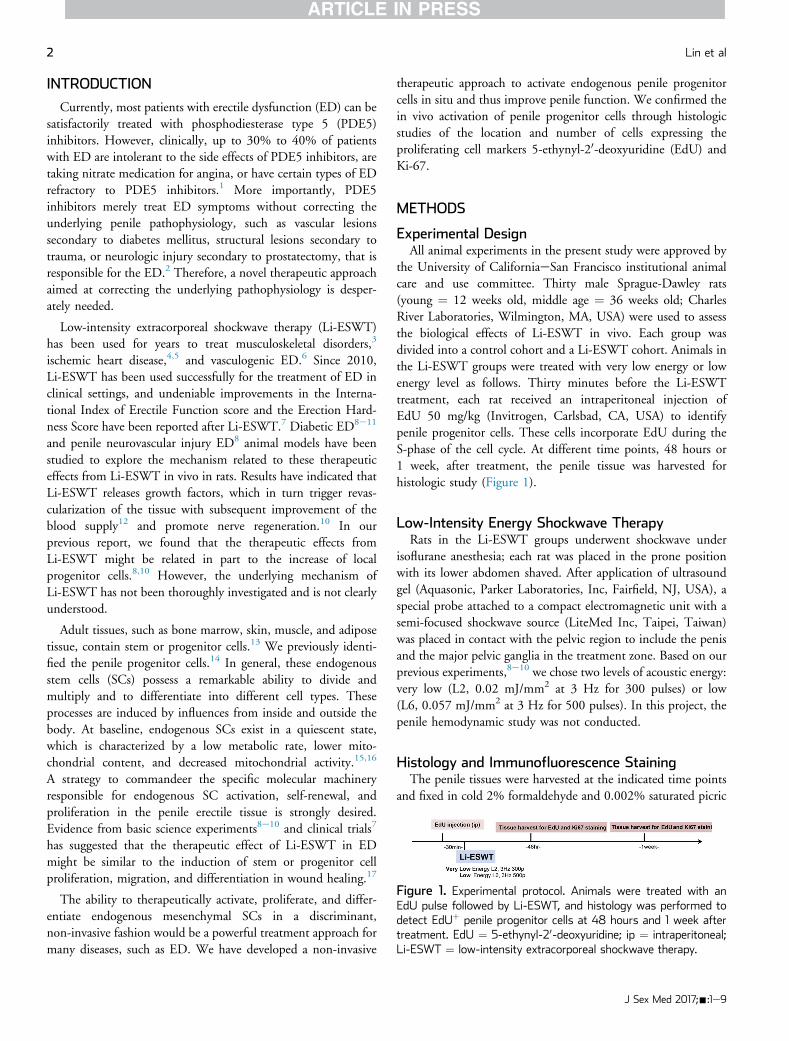
Figure 1. Experimental protocol. Animals were treated with anEdU pulse followed by Li-ESWT, and histology was performed todetect EdUþ penile progenitor cells at 48 hours and 1 week aftertreatment. EdU ¼ 5-ethynyl-20-deoxyuridine; ip ¼ intraperitoneal;Li-ESWT ¼ low-intensity extracorporeal shockwave therapy.
2 Lin et al
INTRODUCTION
Currently, most patients with erectile dysfunction (ED) can besatisfactorily treated with phosphodiesterase type 5 (PDE5)inhibitors. However, clinically, up to 30% to 40% of patientswith ED are intolerant to the side effects of PDE5 inhibitors, aretaking nitrate medication for angina, or have certain types of EDrefractory to PDE5 inhibitors.1 More importantly, PDE5inhibitors merely treat ED symptoms without correcting theunderlying penile pathophysiology, such as vascular lesionssecondary to diabetes mellitus, structural lesions secondary totrauma, or neurologic injury secondary to prostatectomy, that isresponsible for the ED.2 Therefore, a novel therapeutic approachaimed at correcting the underlying pathophysiology is desper-ately needed.
Low-intensity extracorporeal shockwave therapy (Li-ESWT)has been used for years to treat musculoskeletal disorders,3
ischemic heart disease,4,5 and vasculogenic ED.6 Since 2010,Li-ESWT has been used successfully for the treatment of ED inclinical settings, and undeniable improvements in the Interna-tional Index of Erectile Function score and the Erection Hard-ness Score have been reported after Li-ESWT.7 Diabetic ED8e11
and penile neurovascular injury ED8 animal models have beenstudied to explore the mechanism related to these therapeuticeffects from Li-ESWT in vivo in rats. Results have indicated thatLi-ESWT releases growth factors, which in turn trigger revas-cularization of the tissue with subsequent improvement of theblood supply12 and promote nerve regeneration.10 In ourprevious report, we found that the therapeutic effects fromLi-ESWT might be related in part to the increase of localprogenitor cells.8,10 However, the underlying mechanism ofLi-ESWT has not been thoroughly investigated and is not clearlyunderstood.
Adult tissues, such as bone marrow, skin, muscle, and adiposetissue, contain stem or progenitor cells.13 We previously identi-fied the penile progenitor cells.14 In general, these endogenousstem cells (SCs) possess a remarkable ability to divide andmultiply and to differentiate into different cell types. Theseprocesses are induced by influences from inside and outside thebody. At baseline, endogenous SCs exist in a quiescent state,which is characterized by a low metabolic rate, lower mito-chondrial content, and decreased mitochondrial activity.15,16
A strategy to commandeer the specific molecular machineryresponsible for endogenous SC activation, self-renewal, andproliferation in the penile erectile tissue is strongly desired.Evidence from basic science experiments8e10 and clinical trials7
has suggested that the therapeutic effect of Li-ESWT in EDmight be similar to the induction of stem or progenitor cellproliferation, migration, and differentiation in wound healing.17
The ability to therapeutically activate, proliferate, and differ-entiate endogenous mesenchymal SCs in a discriminant,non-invasive fashion would be a powerful treatment approach formany diseases, such as ED. We have developed a non-invasive
therapeutic approach to activate endogenous penile progenitorcells in situ and thus improve penile function. We confirmed thein vivo activation of penile progenitor cells through histologicstudies of the location and number of cells expressing theproliferating cell markers 5-ethynyl-20-deoxyuridine (EdU) andKi-67.
METHODS
Experimental DesignAll animal experiments in the present study were approved by
the University of CaliforniaeSan Francisco institutional animalcare and use committee. Thirty male Sprague-Dawley rats(young ¼ 12 weeks old, middle age ¼ 36 weeks old; CharlesRiver Laboratories, Wilmington, MA, USA) were used to assessthe biological effects of Li-ESWT in vivo. Each group wasdivided into a control cohort and a Li-ESWT cohort. Animals inthe Li-ESWT groups were treated with very low energy or lowenergy level as follows. Thirty minutes before the Li-ESWTtreatment, each rat received an intraperitoneal injection ofEdU 50 mg/kg (Invitrogen, Carlsbad, CA, USA) to identifypenile progenitor cells. These cells incorporate EdU during theS-phase of the cell cycle. At different time points, 48 hours or1 week, after treatment, the penile tissue was harvested forhistologic study (Figure 1).
Low-Intensity Energy Shockwave TherapyRats in the Li-ESWT groups underwent shockwave under
isoflurane anesthesia; each rat was placed in the prone positionwith its lower abdomen shaved. After application of ultrasoundgel (Aquasonic, Parker Laboratories, Inc, Fairfield, NJ, USA), aspecial probe attached to a compact electromagnetic unit with asemi-focused shockwave source (LiteMed Inc, Taipei, Taiwan)was placed in contact with the pelvic region to include the penisand the major pelvic ganglia in the treatment zone. Based on ourprevious experiments,8e10 we chose two levels of acoustic energy:very low (L2, 0.02 mJ/mm2 at 3 Hz for 300 pulses) or low(L6, 0.057 mJ/mm2 at 3 Hz for 500 pulses). In this project, thepenile hemodynamic study was not conducted.
Histology and Immunofluorescence StainingThe penile tissues were harvested at the indicated time points
and fixed in cold 2% formaldehyde and 0.002% saturated picric
J Sex Med 2017;-:1e9
In Situ Activation of Penile Progenitor Cells With Li-ESWT 3
acid in phosphate buffer 0.1 mol/L (pH ¼ 8.0) for 4 hoursfollowed by overnight immersion in buffer containing 30%sucrose. The specimens were embedded in OCT compound(Sakura Finetic USA, Torrance, CA, USA) and stored at �70�Cuntil use. Fixed frozen tissue specimens were cut at 10 mm,mounted onto SuperFrost-Plus charged slides (Fisher Scientific,Pittsburgh, PA, USA), and air dried for 5 minutes. The tissuesection was subjected to EdU staining with or without immu-nostaining for Ki-67. For immunostaining, the slides were placedin 0.3% H2O2 and methanol for 10 minutes, washed twice inphosphate buffered saline (PBS) for 5 minutes, and incubatedwith 3% horse serum in PBS and 0.3% Triton X-100 for 30minutes at room temperature. After draining this solutionfrom the tissue section, the slides were incubated at roomtemperature with antieKi-67 antibody (1:500; Abcam Inc,Cambridge, MA, USA) for 1.5 hours. Control tissue sectionswere similarly prepared except no primary antibody was added.After rinses with PBS, the sections were incubated with fluo-rescein isothiocyanateeconjugated secondary antibody (JacksonImmunoResearch Laboratories, West Grove, PA, USA). Afterrinses with PBS, the slides were incubated with freshly madeClick-iT reaction cocktail (Thermo Fisher Scientific Inc,Waltham, MA, USA) for 30 minutes at room temperaturewithout light followed by staining with 40,6-diamidino-2-phenylindole 1 mg/mL (for nuclear staining; Sigma-Aldrich,St Louis, MO, USA). Immunofluorescence staining with Ki-67and pErk1/2 was conducted as previously reported.14
Primary Schwann Cell Isolation and CultureRat Schwann cells were harvested as previously described.8,18
Briefly, sciatic nerves were harvested from Sprague-Dawley ratsand enzymatically dissociated by incubation at 37�C sequentiallywith 1% collagenase and 0.125% trypsin for 30 and 10 minutes,respectively. The mixture was triturated, centrifuged, andresuspended in 10% fetal bovine serum in Dulbecco’s ModifiedEagle Medium. The cell pellets were plated on dishes pre-coatedwith poly-L-lysine for incubation in the same medium. On thefollowing day, cytosine arabinoside 10 mmol/L was added andallowed to incubate for an additional 48 hours to removefibroblasts. The cell culture was maintained in Dulbecco’sModified Eagle Medium supplemented with 10% fetal bovineserum, forskolin 2 mmol/L (Sigma, St Louis, MO, USA), andheregulin 2 ng/mL (Sigma) to stimulate Schwann cell prolifera-tion. For further purification, the cell culture was gently trypsi-nized, pelleted, and incubated with anti-Thy1 antibody (AbDSerotec, Raleigh, NC, USA) on ice for 2 hours, followed byincubation in complement (Jackson ImmunoResearch Labora-tories) for an additional 2 hours.
Effect of Low-Intensity Shockwave on ActivatingSchwann Cells In VitroSchwann cells were resuspended in fresh, pre-warmed (37�C)
complete medium. The Schwann cells were counted and plated
J Sex Med 2017;-:1e9
on 96-well plates pre-coated with 0.01% poly-L-lysine. The cellswere treated with or without Li-ESWT at 0.02 mJ/mm2 at 3 Hzfor 300 pulses. Growth curves were generated using the MTTassay to measure changes in cell number at 24, 48, 72, and 96hours after Li-ESWT. The phosphorylation level of Erk1/2 inthe cells was checked with western blot, and the location ofactivated Erk1/2 was checked with immunofluorescence as pre-viously reported.19 In addition, expression of Ki-67 in those cellswas checked 48 hours after Li-ESWT.
Effect of Low-Intensity Shockwave on ActivatingEndothelial Cells In Vitro
Human umbilical vein endothelial cells (HUVECs) were usedand maintained in endothelial cell basal medium supplementedwith Bullet Kit (EBM-2, Lonza Inc, Walkersville, MD, USA) inculture flasks coated with 0.1% gelatin and maintained at 37�Cwith humidified 5% carbon dioxide. HUVECs cultured frompassages 4 to 8 were used for this experiment. HUVECs weretreated with or without Li-ESWT at the energy level of 0.02 mJ/mm2 at 3 Hz for 300 pulses. For the tube formation assay, a totalof 30,000 HUVECs with different treatments were seeded inMatrigel in serum-free medium in 24-well plates in triplicate andincubated at 37�C for 6 hours. Tubules were visualized by lightmicroscopy at low magnification (40�). Photomicrographs fromeach well were captured, and total tubule length and number oftubules were analyzed using ImageJ 2.02 (National Institutes ofHealth, Bethesda, MD, USA).
Image and Statistical AnalysesFor image analysis, five randomly selected fields per slide for
each treatment group were photographed and recorded using aRetiga Q Image digital still camera and ACT-1 software (NikonInstruments Inc, Melville, NY, USA). The images were quanti-fied using Image-Pro Plus (Media Cybernetics, Silver Spring,MD, USA). Total EdUþ cells in each section were counteddouble-blindly by different investigators. Data were analyzedusing Prism 5 (GraphPad Software, San Diego, CA, USA) andexpressed as mean ± standard error of mean. Multiple groupswere compared using t-test and one-way analysis of variancefollowed by the Tukey-Kramer test for post hoc comparisons.Statistical significance was set at a P value less than .05.
RESULTS
Li-ESWTActivates Penile Progenitor Cells in Youngand Middle-Age Rats
Previously reported results have shown that Li-ESWT allevi-ates ED in neurovascular and diabetic ED animal models10,11;however, the underlying mechanisms are not well elucidated.Therefore, to clarify the possible mechanisms of these functionalimprovements and histologic changes, we examined penile cellproliferation after Li-ESWT. For this purpose, EdU pulsing wasused in the present project.
4 Lin et al
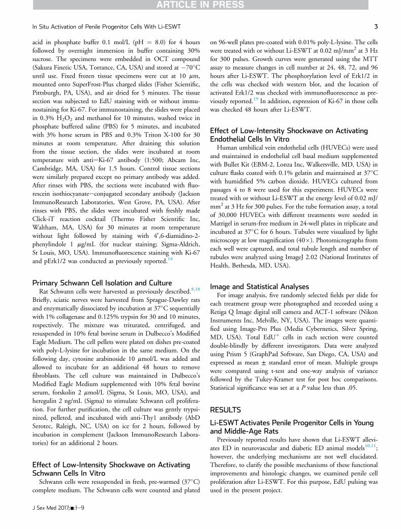
In the young rats, Li-ESWT significantly increased EdUþ cellswithin penile erectile tissues (P < .01) at 48 hours and 1 week.This finding strongly suggests that penile progenitor cells can be“activated” by appropriate levels of Li-ESWT. Low-energyLi-ESWT activated more penile progenitor cells than very low-energy Li-ESWT in the young rats (Figure 2). The differencebetween the middle-age rats and young rats also was significant:Li-ESWT activated fewer penile cells in middle-age animalscompared with young animals. The middle-age rats had one fifththe cellular response that the young rats had at the verylow-energy setting, and the use of low-energy Li-ESWT doubledthis activation. Interestingly, in the low-energy group of middle-age rats, EdUþ cells activated by Li-ESWT decreased significantlyby 1 week after Li-ESWT (P < .01; P < .05; Figure 2), whichsuggests that some activated EdUþ cells proliferate and becometerminally differentiated cells and thus lose the EdU marker.
Location of Penile Progenitor Cells Activated byLi-ESWT
We previously reported that penile progenitor cells exist in thesubtunical and para-sinusoidal regions within the penis.14 In thepresent project, the location of the EdUþ cells after Li-ESWTwas extensively studied in all tissue samples. As observed inFigure 2, most EdUþ cells were localized in the subtunicalregion, and, interestingly, some cells were clustered togetherwithin the tunica (Figure 3).
Approximately 70% to 80% of EdUþ cells localized in thesubtunical space, 10% to 19% localized in the para-sinusoid area,
Figure 2. Representative images show actiation and proliferation of pa rat’s penis. Panel B shows EdUþ penile progenitor cells in young (left)(bottom) groups. Most EdUþ cells (arrows) are localized in the subtuncells between young and middle-age rats 48 hours after Li-ESWT. Panafter Li-ESWT (*P < .01; #P < .05). EdU ¼ 5-ethynyl-20-deoxyuridine
1.9% to 5.3% localized in the penile nerve, and approximately3.8% to 6.7% localized in penile blood vessels. Interestingly,very low-energy Li-ESWT activated more progenitor cells in thepenile nerves and blood vessels compared with low-energyLi-ESWT (P < .01). There was no significant difference be-tween very low- and low-energy treatments in the subtunical andpara-sinusoidal space distribution (P > .05). In addition, therewere some EdUþ cells within the penile dorsal nerve, whichimplies that Schwann cells were activated by Li-ESWT, similar toour previous report.8 EdUþ cells also were observed in smallvessels, including capillaries, arterioles, and venules, whichimplies that the endothelium also was activated by Li-ESWT(Figure 3).
Cellular Markers of Penile Progenitor CellsActivated by Li-ESWTTo define those EdUþ cells activated by Li-ESWT, we stained
for antigen Ki-67, a nuclear marker of cell proliferation. Inter-estingly, cells expressing Ki-67 do not colocalize with EdUþ cells(Figure 4), which suggests that the two markers might identifyproliferating cells in different stages of the cell cycle. Only cells atthe S-phase of the cell cycle incorporate EdU and thus stainstrongly.
Low-Intensity Shockwave Activates Schwann CellsIn VitroAs we previously noted, Schwann cells were activated by
Li-ESWT in vivo. To confirm this effect, we isolated the primary
enile progenitor cells by Li-ESWT. Panel A shows a cross-section ofand middle-age (right) rats of the sham control (top) and Li-ESWTical region. Panel C shows a comparison of EdUþ penile progenitorel D shows more EdUþ penile progenitor cells in young rats 1 week; Li-ESWT ¼ low-intensity extracorporeal shockwave therapy.
J Sex Med 2017;-:1e9

Figure 3. Location of EdUþ penile progenitor cells (red areas) after low-intensity extracorporeal shockwave therapy. Panel A shows thepenile subtunical region (70%e80% localized cells). Panel B shows the para-sinusoid region (10%e19% localized cells). Panel C shows thepenile dorsal nerve (1.9%e5.3% localized cells). Panel D shows the penile small blood vessel (3.8%e6.7% localized cells). Upper panels aremagnified �200, and lower panels are magnified �400. EdU ¼ 5-ethynyl-20-deoxyuridine; L ¼ lumen; S ¼ sinusoid (*P < .01).
In Situ Activation of Penile Progenitor Cells With Li-ESWT 5
Schwann cells and treated them with Li-ESWT. Li-ESWTpromoted Schwann cell proliferation significantly at 48 hoursafter treatment (P < .05; Figure 5A), and this effect lasted for 96hours. Those activated Schwann cells expressed high levels ofKi-67 (P < .05; Figure 5B, C). To explore the underlyingmechanism of this activation, the phosphorylation level of theErk1/2 pathway was checked in those cells. This demonstratedthat low-energy shockwave increased Erk1/2 phosphorylationsignificantly (P < .05; Figure 5D) and that activated Erk1/2translocated into the cell nucleus (Figure 5E).
Li-ESWT Activates Endothelium and PromotesAngiogenesisIt has been reported that Li-ESWT promotes angiogenesis
mainly through enhanced vascular endothelial growth factorexpression. In the present experiment, we assessed the effect ofLi-ESWT on new blood vessel formation in vitro. HUVECsformed a robust tube network within 6 hours after seeding afterLi-ESWT. The tube length and branch points increasedapproximately 42% and 43%, respectively, compared with thatof the control groups (P < .05; Figure 6).
J Sex Med 2017;-:1e9
DISCUSSION
Since the first reported use of SCs for ED therapy in 2004,20
SC-based therapies have been extensively studied in the man-agement of ED with the goal of complete replacement of lost ordamaged cells.21 In recent years, a spate of reports related to theprogress of SC-based ED therapy has been published.22 Differenttherapeutic forms of SCs have been developed, includingmultiple sources of SCs, gene-transfected SCs, SC lysate, andSCs seeded on tissue matrices.23 However, in recent years,tremendous limitations in the use of exogenous SCs for EDtherapy have become obvious. These include the need for inva-sive tissue harvest, complex isolation techniques, issues related toincorporation of exogenous proteins during cell culture, andconcerns about finding the few SCs remaining in the penis aftertransplantation. More importantly, migration of implanted SCsto existing malignant tumors, enhancing tumor growth, also hasbeen reported in animal experiments.24 We previously reportedthat there are endogenous stem or progenitor cells in penileerectile tissue.14 Therefore, local activation of penile endogenousSCs for ED would be an ideal approach for ED to avoid many ofthe aforementioned limitations to the use of exogenous SCs.

Figure 4. Expression of Ki-67 (green areas) and EdUþ (red areas) in penile progenitor cells after low-intensity extracorporeal shockwavetherapy. EdU ¼ 5-ethynyl-20-deoxyuridine; S ¼ sinusoid.
6 Lin et al
Of course, a well-designed comparison study will be needed toconfirm this conjecture.
Signals that play critical roles in SC activation include solubleSC niche signals (growth factors and cytokines), whereas the fateof SCs is influenced by coexisting adhesive, mechanical, andtopologic cues.25 For decades, scientists have attempted to usechemistry to steer the fate of SCs, but with limited success.26,27
Recent demonstrations of the effects of low-energy shockwave onSCs in culture have suggested the possibility of using mechano-biological methods to drive the growth and fate SCs in vivo,28
thus avoiding the requirement for SC harvest, culture, prepara-tion, and transplantation.29
Figure 5. Low-intensity extracorporeal shockwave therapy stimulatesErk pathway and increasing Ki-67 expression. Panel A shows the prolifeof Ki-67 in Schwann cells after low-intensity extracorporeal shockwavecells by low-intensity extracorporeal shockwave therapy (n ¼ 3 each; *Plow-intensity extracorporeal shockwave therapy. b-ACT ¼ b-actin; ctrl ¼
It has been well demonstrated that Li-ESWT subjectively andobjectively improves erectile function. However, the mechanismsunderlying these beneficial effects have yet to be fully elucidated.In the penis there are many kinds of cells, including terminallydifferentiated cells and stem and progenitor cells, as we reportedin 2015.14 Most penile smooth muscle cells and fibroblasts areterminally differentiated and cannot be activated to proliferate.In contrast, penile progenitor cells, including subtunical penileprogenitor cells, para-sinusoidal penile progenitor cells, Schwanncell progenitor cells, and endothelial progenitor cells, can beactivated to re-enter the cell cycle and to proliferate and differ-entiate into mature penile cells. In our present study, Li-ESWT
proliferation of primary rat Schwann cells in vitro by activating theration assay of Schwann cells in vitro. Panel B shows the expressiontherapy. Panel D shows activation of the Erk1/2 pathway in Schwann< .05). Panel E shows translocation of pErk1/2 to the nucleus aftercontrol; DAPI ¼ 40,6-diamidino-2-phenylindole; SW ¼ shockwave.
J Sex Med 2017;-:1e9

Figure 6. Low-intensity extracorporeal shockwave therapy promotes tube formation of human umbilical vein endothelial cells in cellculture. ctrl ¼ control; DAPI ¼ 40,6-diamidino-2-phenylindole; SW ¼ shockwave.
In Situ Activation of Penile Progenitor Cells With Li-ESWT 7
activated these cells and induced them to re-enter the cell cycle.Cells incorporate EdU during the S-phase of the cell cycle andtherefore could be identified with EdU staining.
It has been reported that Li-ESWT influences cell prolifera-tion by altering major extracellular factors and signaling pathwaysinvolved in cell proliferation. It has been hypothesized thatextracellular adenosine triphosphate (ATP), released in an energylevel-dependent and pulse number-dependent manner, is thetrigger of the biological effects of shockwave treatment. Biolog-ically, endogenous SCs activating out of quiescence to generateproliferating progeny require ATP to provide energy. The level ofATP in quiescent endogenous SCs might be insufficient for SCactivation. Several studies have demonstrated that Li-ESWTenhances cellular ATP significantly and that the production ofATP is related to the activation of the Erk1/2 and p38 mitogen-activated protein kinase pathway.17
In the 1980s, ESWT was described as “mechanotherapy,”with original applications for urological lithotripsy. Morerecently, it has been successfully applied for regenerative medi-cine. The molecular mechanisms of Li-ESWT are related todifferent pathways of biological reactions through a “mechano-transduction” process. From extensive basic science research, ithas been demonstrated that Li-ESWT does not evoke amechanical disruption of tissues and cells, but rather inducesbiological effects that activate a series of cellular events respon-sible for the therapeutic effects of Li-ESWT.30,31 Activation ofSCs by Li-ESWT is a focused treatment and therefore would
J Sex Med 2017;-:1e9
cause a minimum of off-site effects. As a non-invasive treatmentapproach, Li-ESWT is characterized by the absence of major sideeffects, repeatability, good tolerability, and excellent complianceby patients.
Many cells, including SCs, bone marrow stromal cells, oste-oblasts, endothelial cells, and Schwann cells, are potential targetsfor mechano-transduction using Li-ESWT.32e35 In the penis,approximately 70% to 80% of the Li-ESWTeactivated cellswere localized within the subtunical space, which was the samelocation noted in our previous report.14 Although EdU wasinjected into newborn rats and immunochemical detectionwas performed when the rats grew to adulthood, the previousreport identified the subtunical region as the “niche” of theEdU-retaining SCs. This further confirms that theLi-ESWTeactivated EdUþ cells in the present study were SCsactivated in situ and not cells that migrated from other locations.Moreover, approximately 1.5% to 6.7% of the activated cellswere located within the penile nerve and penile vessels. Thisseems to confirm that Li-ESWT has the ability to activatemultiple stem or progenitor cells, resulting in regeneration ofblood vessels, nerves, and musclesobserved in previous clin-ical7,36e38 and animal8e11 experiments. To further study thepotential of activation of endothelial and Schwann cells, the twocells were treated with low-energy shockwave in vitro. The resultsclearly indicate that Li-ESWT can activate endothelial andSchwann cells, and this response was related to activation of theErk1/2 cellular signaling pathway.

8 Lin et al
As expected, the level of activation of penile progenitor cellsvaries with the amount of energy applied. Low-energy Li-ESWTactivated more penile progenitor cells compared with very low-energy Li-ESWT. There were fewer penile cells activated byLi-ESWT in the middle-age animals compared with younganimals. To maximize outcomes, further experiments are neededto identify the best treatment protocols for young and olderanimals and humans.
The major limitation of this study is the short timeline usedand the animal models used. Li-ESWT might be less effective inimproving erectile function in old animals because of thedecreased number and quality of penile stem or progenitor cellsassociated with aging. With a longer period of study in youngand old rats, we hope to better define the true benefits andlimitations of this therapy.
CONCLUSION
Li-ESWT activation of local penile progenitor cells in situmight be one of the mechanisms that contribute to the beneficialeffects of Li-ESWT for ED, which represents a non-invasivealternative to exogenous SC therapy.
ACKNOWLEDGMENT
The manuscript was awarded the Zorgniotti-Newman Prize atthe International Society for Sexual Medicine Meeting in Beijing,China, 2016.
Corresponding Author: Tom F. Lue, MD, Knuppe MolecularUrology Laboratory, Department of Urology, School of Medi-cine, University of CaliforniaeSan Francisco, San Francisco, CA94143, USA. Tel: 415-353 7339; Fax: 415-476 3803; E-mail:[email protected]
Conflicts of Interest: Tom F. Lue is a consultant to Acoustic WaveCell Therapy, Inc. All other authors have no conflict of interest.
Funding: This work was supported by the US Army, Navy, AirForce, Veterans’ Administration, and Health Affairs and theNational Institutes of Health to support the AFIRM II effort(award W81XWH-13-2-0052). The US Army Medical ResearchAcquisition Activity (820 Chandler Street, Fort Detrick MD,21702-5014) is the awarding and administering acquisitionoffice. Opinions, interpretations, conclusions, and recommen-dations are those of the authors and are not necessarily endorsedby the Department of Defense.
STATEMENT OF AUTHORSHIP
Category 1
(a) Conception and Design
Guiting Lin; Amanda B. Reed-Maldonado; Tom F. Lue(b) Acquisition of Data
Guiting Lin; Bohan Wang; Yung-chin Lee; Jun Zhou; Zhihua Lu;Guifang Wang; Lia Banie(c) Analysis and Interpretation of Data
Guiting Lin; Amanda B. Reed-Maldonado; Bohan Wang;Tom F. Lue
Category 2
(a) Drafting the Article
Guiting Lin; Amanda B. Reed-Maldonado; Tom F. Lue(b) Revising It for Intellectual Content
Amanda B. Reed-Maldonado; Tom F. LueCategory 3
(a) Final Approval of the Completed Article
Tom F. LueREFERENCES1. Dorsey P, Keel C, Klavens M, et al. Phosphodiesterase type 5
(PDE5) inhibitors for the treatment of erectile dysfunction.Expert Opin Pharmacother 2010;11:1109-1122.
2. Hatzichristou D, d’Anzeo G, Porst H, et al. Tadalafil 5 mg oncedaily for the treatment of erectile dysfunction during a6-month observational study (EDATE): impact of patientcharacteristics and comorbidities. BMC Urol 2015;15:111.
3. Al-Abbad H, Simon JV. The effectiveness of extracorporealshock wave therapy on chronic Achilles tendinopathy: asystematic review. Foot Ankle Int 2013;34:33-41.
4. Abe Y, Ito K, Hao K, et al. Extracorporeal low-energy shock-wave therapy exerts anti-inflammatory effects in a rat modelof acute myocardial infarction. Circ J 2014;78:2915-2925.
5. Ito K, Fukumoto Y, Shimokawa H. Extracorporeal shock wavetherapy for ischemic cardiovascular disorders. Am JCardiovasc Drugs 2011;11:295-302.
6. Gruenwald I, Appel B, Vardi Y. Low-intensity extracorporealshock wave therapy—a novel effective treatment for erectiledysfunction in severe ED patients who respond poorly to PDE5inhibitor therapy. J Sex Med 2012;9:259-264.
7. Lu Z, Lin G, Reed-Maldonado A, et al. Low-intensity extra-corporeal shock wave treatment improves erectile function: asystematic review and meta-analysis. Eur Urol 2017;71:223-233.
8. Li H, Matheu MP, Sun F, et al. Low-energy shock wave therapyameliorates erectile dysfunction in a pelvic neurovascular in-juries rat model. J Sex Med 2016;13:22-32.
9. Liu J, Zhou F, Li GY, et al. Evaluation of the effect of differentdoses of low energy shock wave therapy on the erectilefunction of streptozotocin (STZ)-induced diabetic rats.Int J Mol Sci 2013;14:10661-10673.
10. Qiu X, Lin G, Xin Z, et al. Effects of low-energy shockwavetherapy on the erectile function and tissue of a diabetic ratmodel. J Sex Med 2013;10:738-746.
11. Assaly-Kaddoum R, Giuliano F, Laurin M, et al. Low intensityextracorporeal shock wave therapy improves erectile functionin a model of type II diabetes independently of NO/cGMPpathway. J Urol 2016;196:950-956.
12. Rassweiler JJ, Knoll T, Kohrmann KU, et al. Shock wavetechnology and application: an update. Eur Urol 2011;59:784-796.
J Sex Med 2017;-:1e9

In Situ Activation of Penile Progenitor Cells With Li-ESWT 9
13. Lin G, Garcia M, Ning H, et al. Defining stem and progenitorcells within adipose tissue. Stem Cells Dev 2008;17:1053-1063.
14. Lin G, Alwaal A, Zhang X, et al. Presence of stem/progenitorcells in the rat penis. Stem Cells Dev 2015;24:264-270.
15. Lunt SY, Vander Heiden MG. Aerobic glycolysis: meeting themetabolic requirements of cell proliferation. Annu Rev CellDev Biol 2011;27:441-464.
16. Hsu P, Qu CK. Metabolic plasticity and hematopoietic stem cellbiology. Curr Opin Hematol 2013;20:289-294.
17. Weihs AM, Fuchs C, Teuschl AH, et al. Shock wave treatmentenhances cell proliferation and improves wound healing byATP release-coupled extracellular signal-regulated kinase(ERK) activation. J Biol Chem 2014;289:27090-27104.
18. Shen YA, Chen Y, Dao DQ, et al. Phosphorylation of LKB1/Par-4 establishes Schwann cell polarity to initiate andcontrol myelin extent. Nat Commun 2014;5:4991.
19. Lin G, Shindel AW, Banie L, et al. Molecular mechanismsrelated to parturition-induced stress urinary incontinence. EurUrol 2009;55:1213-1222.
20. Bochinski D, Lin GT, Nunes L, et al. The effect of neuralembryonic stem cell therapy in a rat model of cavernosal nerveinjury. BJU Int 2004;94:904-909.
21. Strong TD, Gebska MA, Burnett AL, et al. Endothelium-specific gene and stem cell-based therapy for erectiledysfunction. Asian J Androl 2008;10:14-22.
22. Zhang H, Albersen M, Jin X, Lin G. Stem cells: novel players inthe treatment of erectile dysfunction. Asian J Androl 2012;14:145-155.
23. Albersen M, Fandel TM, Lin G, et al. Injections of adiposetissue-derived stem cells and stem cell lysate improve recov-ery of erectile function in a rat model of cavernous nerve injury.J Sex Med 2011;7:3331-3340.
24. Lin G, Yang R, Banie L, et al. Effects of transplantation ofadipose tissue-derived stem cells on prostate tumor. Prostate2010;70:1066-1073.
25. Sun Y, Chen CS, Fu J. Forcing stem cells to behave: abiophysical perspective of the cellular microenvironment.Annu Rev Biophys 2012;41:519-542.
26. Ma Q, Jones D, Borghesani PR, et al. Impaired B-lympho-poiesis, myelopoiesis, and derailed cerebellar neuron migrationin CXCR4- and SDF-1edeficient mice. Proc Natl Acad Sci U S A1998;95:9448-9453.
J Sex Med 2017;-:1e9
27. Chavakis E, Hain A, Vinci M, et al. High-mobility group box 1activates integrin-dependent homing of endothelial progenitorcells. Circ Res 2007;100:204-212.
28. Zhao Y, Wang J, Wang M, et al. Activation of bone marrow-derived mesenchymal stromal cells—a new mechanism ofdefocused low-energy shock wave in regenerative medicine.Cytotherapy 2013;15:1449-1457.
29. Ventura C. Tuning stem cell fate with physical energies.Cytotherapy 2013;15:1441-1443.
30. d’Agostino MC, Craig K, Tibalt E, et al. Shock wave asbiological therapeutic tool: from mechanical stimulation torecovery and healing, through mechanotransduction. Int JSurg 2015;24:147-153.
31. Sukubo NG,Tibalt E, Respizzi S, et al. Effect of shock waves onmacrophages: a possible role in tissue regeneration andremodeling. Int J Surg 2015;24:124-130.
32. Notarnicola A, Moretti B. The biological effects of extracor-poreal shock wave therapy (ESWT) on tendon tissue. MusclesLigaments Tendons J 2012;2:33-37.
33. Romeo P, Lavanga V, Pagani D, et al. Extracorporeal shockwave therapy in musculoskeletal disorders: a review. MedPrinc Pract 2014;23:7-13.
34. Tara S, Miyamoto M, Takagi G, et al. Low-energy extracorpo-real shock wave therapy improves microcirculation blood flowof ischemic limbs in patients with peripheral arterial disease:pilot study. J Nippon Med School 2014;81:19-27.
35. Tepekoylu C,Wang FS, Kozaryn R, et al. Shock wave treatmentinduces angiogenesis and mobilizes endogenous CD31/CD34-positive endothelial cells in a hindlimb ischemia model:implications for angiogenesis and vasculogenesis. J ThoracCardiovasc Surg 2013;146:971-978.
36. Olsen AB, Persiani M, Boie S, et al. Can low-intensity extra-corporeal shockwave therapy improve erectile dysfunction?A prospective, randomized, double-blind, placebo-controlledstudy. Scand J Urol 2015;49:329-333.
37. Chung E, Cartmill R. Evaluation of clinical efficacy, safety andpatient satisfaction rate after low-intensity extracorporealshockwave therapy for the treatment of male erectiledysfunction: an Australian first open-label single-arm pro-spective clinical trial. BJU Int 2015;115(Suppl 5):46-49.
38. Frey A, Sonksen J, Fode M. Low-intensity extracorporealshockwave therapy in the treatment of postprostatectomyerectile dysfunction: a pilot study. Scand J Urol 2016;50:123-127.