in situ cell death detection kit, fluorescein -...
TRANSCRIPT
1684795a.fm Page 1 Tuesday, November 26, 2002 10:32 AM
For research purposes only. Not for use for in vitro diagnosticprocedures for clinical diagnosis.
In Situ Cell Death Detection Kit, FluoresceinKit for detection and quantification of apoptosis (programmed cell death) at single cell level, based on labeling of DNA strand breaks (TUNEL technology): Analysis by fluorescence microscopy or flow cytometry
Cat. No. 1 684 795 Store at �15 to �25°C1 Kit (50 tests)
Instruction ManualVersion 3, November 2002
1684795a.fm Page 2 Tuesday, November 26, 2002 10:32 AM
Roche Applied Science2
1. Preface
1.1 Table of contents
1. Preface ......................................................................................................................21.1 Table of contents ............................................................................................................................ 21.2 Kit contents ....................................................................................................................................... 3
2. Introduction ..............................................................................................................52.1 Product overview ............................................................................................................................ 52.2 Background information .............................................................................................................. 8
3. Procedures and required materials ................................................................. 103.1 Flow chart ........................................................................................................................................103.2 Preparation of sample material ...............................................................................................113.2.1 Cell suspension .............................................................................................................................113.2.2 Adherent cells, cell smears, and cytospin preparations ................................................123.2.3 Tissue sections ..............................................................................................................................133.2.3.1 Treatment of paraffin-embedded tissue ...............................................................................133.2.3.2 Treatment of cryopreserved tissue ........................................................................................153.3 Labeling protocol ..........................................................................................................................163.3.1 Before you begin ..........................................................................................................................163.3.2 Labeling protocol for cell suspensions ................................................................................173.3.3 Labeling protocol for adherent cells, cell smears,
cytospin preparations, and tissues..........................................................................................183.3.4 Labeling protocol for difficult tissue .....................................................................................19
4. Typical results ....................................................................................................... 20
5. Appendix ................................................................................................................ 215.1 Trouble-shooting ..........................................................................................................................215.2 References ......................................................................................................................................245.3 Related products ..........................................................................................................................25
1684795a.fm Page 3 Tuesday, November 26, 2002 10:32 AM
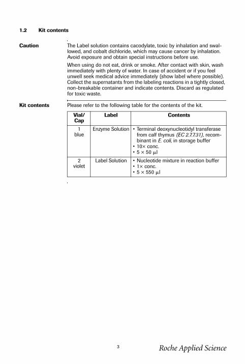
1.2 Kit contents
Caution The Label solution contains cacodylate, toxic by inhalation and swal-lowed, and cobalt dichloride, which may cause cancer by inhalation. Avoid exposure and obtain special instructions before use.When using do not eat, drink or smoke. After contact with skin, wash immediately with plenty of water. In case of accident or if you feel unwell seek medical advice immediately (show label where possible).Collect the supernatants from the labeling reactions in a tightly closed, non-breakable container and indicate contents. Discard as regulated for toxic waste.
Kit contents Please refer to the following table for the contents of the kit.
Vial/Cap
Label Contents
1blue
Enzyme Solution • Terminal deoxynucleotidyl transferase from calf thymus (EC 2.7.7.31), recom-binant in E. coli, in storage buffer
• 10× conc.• 5 × 50 �l
2violet
Label Solution • Nucleotide mixture in reaction buffer• 1× conc.• 5 × 550 �l
Roche Applied Science3
1.2 Kit contents, continued
1684795a.fm Page 4 Tuesday, November 26, 2002 10:32 AM
Roche Applied Science4
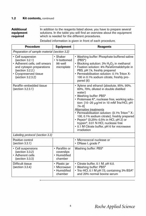
Additional equipment required
In addition to the reagents listed above, you have to prepare several solutions. In the table you will find an overview about the equipment which is needed for the different procedures.Detailed information is given in front of each procedure.
Procedure Equipment Reagents
Preparation of sample material (section 3.2)
• Cell suspension (section 3.2.1)
• Adherent cells, cell smears and cytospin preparations (section 3.2.2.)
• Cryopreserved tissue (section 3.2.3.2)
• Shaker• V-bottomed
96-well microplate
• Washing buffer: Phosphate buffered saline (PBS*)
• Blocking solution: 3% H2O2 in methanol• Fixation solution: 4% Paraformaldehyde in
PBS, pH 7.4, freshly prepared• Permeabilisation solution: 0.1% Triton X-
100 in 0.1% sodium citrate, freshly pre-pared (6)
Paraffin-embedded tissue (section 3.2.3.1)
• Xylene and ethanol (absolute, 95%, 90%, 80%, 70%, diluted in double distilled water)
• Washing buffer: PBS*• Proteinase K*, nuclease free, working solu-
tion: [10–20 �g/ml in 10 mM Tris/HCl, pH 7.4–8]
Alternative treatments• Permeabilisation solution: (0.1% Triton1) X–
100, 0.1% sodium citrate), freshly prepared• Pepsin* (0.25%–0.5% in HCl, pH 2) or
trypsin*, 0.01 N HCl, nuclease free• 0.1 M Citrate buffer, pH 6 for microwave
irradiation
Labeling protocol (section 3.3)
Positive control(section 3.3.1)
• Micrococcal nuclease or • DNase I, grade I*
• Cell suspensions (section 3.3.2)
• Adherent cells (section 3.3.3)
• Parafilm or coverslips
• Humidified chamber
Washing buffer: PBS*
Difficult tissue(section 3.3.4)
• Plastic jar• Microwave• Humidified
chamber
• Citrate buffer, 0.1 M, pH 6.0.• Washing buffer: PBS*• Tris-HCl, 0.1 M pH 7.5, containing 3% BSA*
and 20% normal bovine serum
1684795a.fm Page 5 Tuesday, November 26, 2002 10:32 AM
Roche Applied Science5
2. Introduction
2.1 Product overview
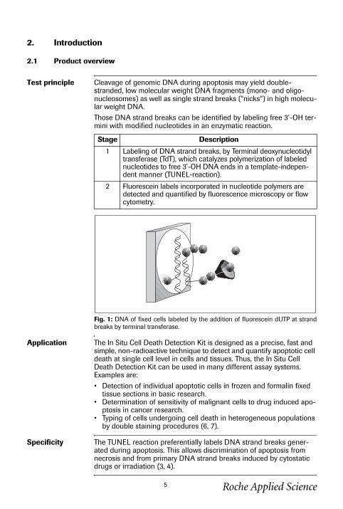
Test principle Cleavage of genomic DNA during apoptosis may yield double-stranded, low molecular weight DNA fragments (mono- and oligo-nucleosomes) as well as single strand breaks (“nicks“) in high molecu-lar weight DNA.Those DNA strand breaks can be identified by labeling free 3’-OH ter-mini with modified nucleotides in an enzymatic reaction.
Fig. 1: DNA of fixed cells labeled by the addition of fluorescein dUTP at strandbreaks by terminal transferase.
Application The In Situ Cell Death Detection Kit is designed as a precise, fast and simple, non-radioactive technique to detect and quantify apoptotic cell death at single cell level in cells and tissues. Thus, the In Situ Cell Death Detection Kit can be used in many different assay systems. Examples are:• Detection of individual apoptotic cells in frozen and formalin fixed
tissue sections in basic research.• Determination of sensitivity of malignant cells to drug induced apo-
ptosis in cancer research.• Typing of cells undergoing cell death in heterogeneous populations
by double staining procedures (6, 7).
Specificity The TUNEL reaction preferentially labels DNA strand breaks gener-ated during apoptosis. This allows discrimination of apoptosis from necrosis and from primary DNA strand breaks induced by cytostatic drugs or irradiation (3, 4).
Stage Description
1 Labeling of DNA strand breaks, by Terminal deoxynucleotidyl transferase (TdT), which catalyzes polymerization of labeled nucleotides to free 3’-OH DNA ends in a template-indepen-dent manner (TUNEL-reaction).
2 Fluorescein labels incorporated in nucleotide polymers are detected and quantified by fluorescence microscopy or flow cytometry.
2.1 Product overview, continued
1684795a.fm Page 6 Tuesday, November 26, 2002 10:32 AM
Test interference False negative results: DNA cleavage can be absent or incomplete in some forms of apoptotic cell death (37). Sterical hindrance such as extracellular matrix components can prevent access of TdT to DNA strand breaks. In either case false negative results may be obtained.False positive results: Extensive DNA fragmentation may occur in cer-tain forms of necrosis (38).DNA strand breaks may also be prominent in cell populations with high proliferative or metabolic activity. In either case false positive results may be obtained.To confirm apoptotic mode of cell death, the morphology of respective cells should be examined very carefully. Morphological changes dur-ing apoptosis have a characteristic pattern. Therefore evaluation of cell morphology is an important parameter in situations where there is any ambiguity regarding interpretation of results.
Sample material • Cell suspensions from• permanent cell lines• lymphocytes and leukemic cells from peripheral blood (4),• thymocytes (1, 6),• bone marrow cells• fine needle biopsies (5)
• Cytospins and cell smear preparations• Adherent cells cultured on chamber slides (31)• Frozen or formalin-fixed, paraffin-embedded tissue sections (1, 25,
26, 29, 30, 32–34, 36, 39)
continued on next page
Roche Applied Science6
2.1 Product overview, continued
1684795a.fm Page 7 Tuesday, November 26, 2002 10:32 AM
Assay time 1-2 hours, excluding culture, fixation and permeabilisation of cells and preparation of tissue sections.
Number of tests The kit is designed for 50 tests.
Kit storage/ stability
The unopened kit is stable at �15 to �25°C through the expiration date printed on the label.Note: The TUNEL reaction mixture should be prepared immediately before use and should not be stored. Keep TUNEL reaction mixture on ice until use.
Advantage Please refer to the following table.
Benefit Feature
Sensitive Detection of apoptotic cell death at single cell level via fluorescence microscope and at cell populations via FACS analysis at very early stages (1, 2, 6).
Specific Preferential labeling of apoptosis versus necrosis (3, 4).
Fast Short assay time (1-2 h).
Convenient • No secondary detection system required.• One incubation and one washing step
only.• Reagents are provided in stable, opti-
mized form.• No dilution steps required.
Flexible • Suitable for fixed cells and tissue. This allows accumulation, storage and trans-port of samples (2, 5).
• Double staining enables identification of type and differentiation state of cells undergoing apoptosis (6).
Function-tested Every lot is function-tested on apoptotic cells in comparison to a master lot.
Roche Applied Science7
1684795a.fm Page 8 Tuesday, November 26, 2002 10:32 AM
2.2 Background information
Cell death Two distinct modes of cell death, apoptosis and necrosis, can be distin-guished based on differences in morphological, biochemical and molecular changes of dying cells.Programmed cell death or apoptosis is the most common form of eukaryotic cell death. It is a physiological suicide mechanism that pre-serves homeostasis, in which cell death naturally occurs during normal tissue turnover (8, 9). In general, cells undergoing apoptosis display a characteristic pattern of structural changes in nucleus and cytoplasm, including rapid blebbing of plasma membrane and nuclear disintegra-tion. The nuclear collapse is associated with extensive damage to chromatin and DNA-cleavage into oligonucleosomal length DNA frag-ments after activation of a calcium-dependent endogenous endonu-clease (10, 11). However, very rare exceptions have been described where morphological features of apoptosis are not accompanied with oligonucleosomal DNA cleavage (37).
Apoptosis Apoptosis is essential in many physiological processes, including maturation and effector mechanisms of the immune system (12, 13), embryonic development of tissue, organs and limbs (14), development of the nervous system (15, 16) and hormone-dependent tissue remodeling (17). Inappropriate regulation of apoptosis may play an important role in many pathological conditions like ischemia, stroke, heart disease, cancer, AIDS, autoimmunity, hepatotoxicity and degen-erative diseases of the central nervous system (18–20).In oncology, extensive interest in apoptosis comes from the observa-tion, that this mode of cell death is triggered by a variety of antitumor drugs, radiation and hyperthermia, and that the intrinsic propensity of tumor cells to respond by apoptosis is modulated by expression of several oncogenes and may be a prognostic marker for cancer treat-ment (21).
Roche Applied Science8
2.2 Background information, continued
1684795a.fm Page 9 Tuesday, November 26, 2002 10:32 AM
Identification of apoptosis
Several methods have been described to identify apoptotic cells (22– 24). Endonucleolysis is considered as the key biochemical event of apoptosis, resulting in cleavage of nuclear DNA into oligonucleosome-sized fragments. Therefore, this process is commonly used for detec-tion of apoptosis by the typical “DNA ladder” on agarose gels during electrophoresis. This method, however, can not provide information regarding apoptosis in individual cells nor relate cellular apoptosis to histological localization or cell differentiation. This can be done by enzymatic in situ labeling of apoptosis induced DNA strand breaks.DNA polymerase as well as terminal deoxynucleotidyl transferase (TdT) (1-6, 25-36) have been used for the incorporation of labeled nucleotides to DNA strand breaks in situ. The tailing reaction using TdT, which was also described as ISEL (in situ end labeling) (5, 35) or TUNEL (TdT-mediated dUTP nick end labeling) (1, 6, 31, 33) technique, has several advantages in comparison to the in situ nick translation (ISNT) using DNA polymerase:• Label intensity of apoptotic cells is higher with TUNEL compared to
ISNT, resulting in an increased sensitivity (2, 4).• Kinetics of nucleotide incorporation is very rapid with TUNEL com-
pared to the ISNT (2, 4).• TUNEL preferentially labels apoptosis in comparison to necrosis,
thereby discriminating apoptosis from necrosis and from primary DNA strand breaks induced by antitumor drugs or radiation (3, 4).
Roche Applied Science9
1684795a.fm Page 10 Tuesday, November 26, 2002 10:32 AM
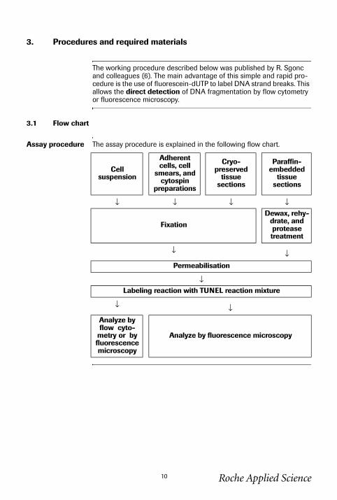
3. Procedures and required materials
The working procedure described below was published by R. Sgonc and colleagues (6). The main advantage of this simple and rapid pro-cedure is the use of fluorescein-dUTP to label DNA strand breaks. This allows the direct detection of DNA fragmentation by flow cytometry or fluorescence microscopy.
3.1 Flow chart
Assay procedure The assay procedure is explained in the following flow chart.
Cell suspension
Adherent cells, cell
smears, and cytospin
preparations
Cryo-preserved
tissue sections
Paraffin-embedded
tissue sections
↓ ↓ ↓ ↓
Fixation
Dewax, rehy-drate, and protease treatment
↓ ↓Permeabilisation
↓Labeling reaction with TUNEL reaction mixture
↓ ↓Analyze by flow cyto-
metry or by fluorescence microscopy
Analyze by fluorescence microscopy
Roche Applied Science10
1684795a.fm Page 11 Tuesday, November 26, 2002 10:32 AM
3.2 Preparation of sample material
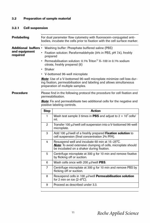
3.2.1 Cell suspension
Prelabeling For dual parameter flow cytometry with fluorescein-conjugated anti-bodies, incubate the cells prior to fixation with the cell surface marker.
Additional buffers and equipment required
• Washing buffer: Phosphate buffered saline (PBS)• Fixation solution: Paraformaldehyde (4% in PBS, pH 7.4), freshly
prepared• Permeabilisation solution: 0.1% Triton1) X–100 in 0.1% sodium
citrate, freshly prepared (6)• Shaker• V-bottomed 96-well microplateNote: Use of a V-bottomed 96-well microplate minimize cell loss dur-ing fixation, permeabilisation and labeling and allows simultaneous preparation of multiple samples.
Procedure Please find in the following protocol the procedure for cell fixation and permeabilisation.Note: Fix and permeabilisate two additional cells for the negative and positive labeling controls.
Step Action
1 Wash test sample 3 times in PBS and adjust to 2 × 10⁷ cells/ml.
2 Transfer 100 �l/well cell suspension into a V-bottomed 96-well microplate.
3 Add 100 �l/well of a freshly prepared Fixation solution to cell suspension (final concentration 2% PFA).
4 Resuspend well and incubate 60 min at 15–25°C.Note: To avoid extensive clumping of cells, microplate should be incubated on a shaker during fixation.
5 Centrifuge microplate at 300 g for 10 min and remove fixative by flicking off or suction.
6 Wash cells once with 200 �l/well PBS.
7 Centrifuge microplate at 300 g for 10 min and remove PBS by flicking off or suction.
8 Resuspend cells in 100 �l/well Permeabilisation solution for 2 min on ice (2–8°C).
9 Proceed as described under 3.3.
Roche Applied Science11
1684795a.fm Page 12 Tuesday, November 26, 2002 10:32 AM
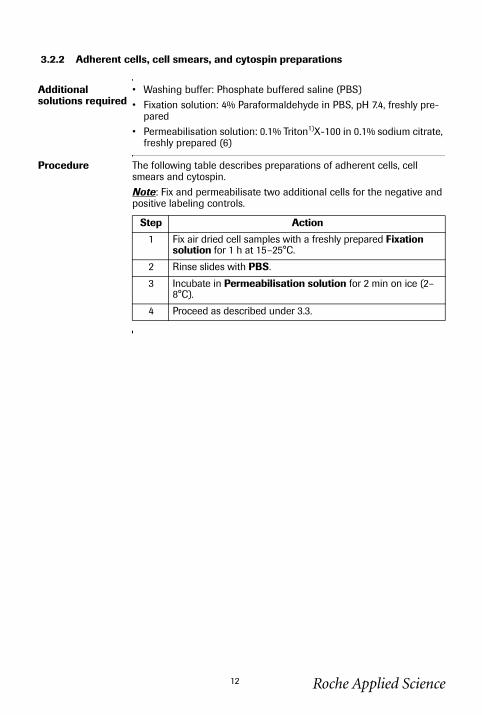
3.2.2 Adherent cells, cell smears, and cytospin preparations
Additional solutions required
• Washing buffer: Phosphate buffered saline (PBS)• Fixation solution: 4% Paraformaldehyde in PBS, pH 7.4, freshly pre-
pared• Permeabilisation solution: 0.1% Triton1)X-100 in 0.1% sodium citrate,
freshly prepared (6)
Procedure The following table describes preparations of adherent cells, cell smears and cytospin.Note: Fix and permeabilisate two additional cells for the negative and positive labeling controls.
Step Action
1 Fix air dried cell samples with a freshly prepared Fixation solution for 1 h at 15–25°C.
2 Rinse slides with PBS.
3 Incubate in Permeabilisation solution for 2 min on ice (2–8°C).
4 Proceed as described under 3.3.
Roche Applied Science12
1684795a.fm Page 13 Tuesday, November 26, 2002 10:32 AM
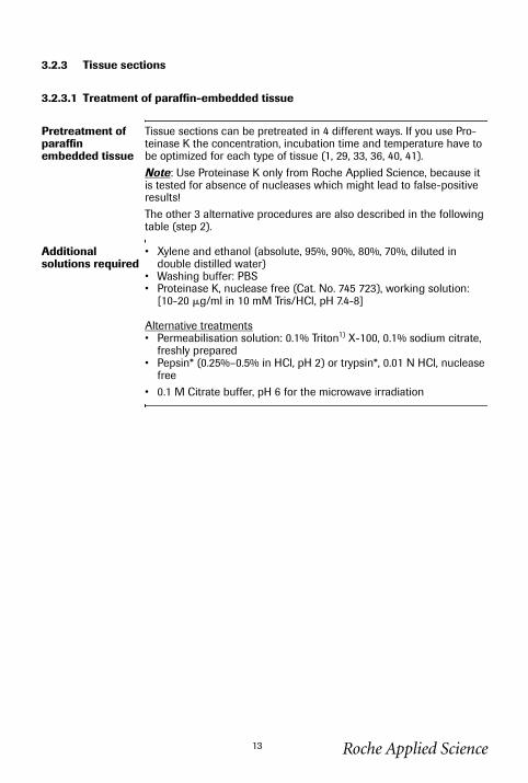
3.2.3 Tissue sections
3.2.3.1 Treatment of paraffin-embedded tissue
Pretreatment of paraffin embedded tissue
Tissue sections can be pretreated in 4 different ways. If you use Pro-teinase K the concentration, incubation time and temperature have to be optimized for each type of tissue (1, 29, 33, 36, 40, 41).Note: Use Proteinase K only from Roche Applied Science, because it is tested for absence of nucleases which might lead to false-positive results!The other 3 alternative procedures are also described in the following table (step 2).
Additional solutions required
• Xylene and ethanol (absolute, 95%, 90%, 80%, 70%, diluted in double distilled water)
• Washing buffer: PBS• Proteinase K, nuclease free (Cat. No. 745 723), working solution:
[10-20 �g/ml in 10 mM Tris/HCl, pH 7.4-8]
Alternative treatments• Permeabilisation solution: 0.1% Triton1) X-100, 0.1% sodium citrate,
freshly prepared• Pepsin* (0.25%–0.5% in HCl, pH 2) or trypsin*, 0.01 N HCl, nuclease
free• 0.1 M Citrate buffer, pH 6 for the microwave irradiation
Roche Applied Science13
3.2.3.1 Treatment of paraffin-embedded tissue, continued
1684795a.fm Page 14 Tuesday, November 26, 2002 10:32 AM
Procedure In the following table the pretreatment of paraffin-embedded tissue with Proteinase K treatment and 3 alternative procedures are described.Note: Add additional tissue sections for the negative and positive labeling controls.
Step Action1 Dewax and rehydrate tissue section according to standard
protocols (e.g. by heating at 60°C followed by washing in xylene and rehydration through a graded series of ethanol and double dist. water) (1, 33, 36).
2 Incubate tissue section for 15-30 min at 21–37°C with Pro-teinase K working solution.
Alternatives: Treatment:
1. Permeabilisa-tion solution
Incubate slides for 8 min.
2. Pepsin* (30, 40) or trypsin*
15-60 min at 37°C.
3. Microwave irradiation
• Place the slide(s) in a plastic jar containing 200 ml 0.1 M Citrate buffer, pH 6.0.
• Apply 350 W microwave irradiation for 5 min.
3 Rinse slide(s) twice with PBS.4 Proceed as described under 3.3.
Roche Applied Science14
1684795a.fm Page 15 Tuesday, November 26, 2002 10:32 AM
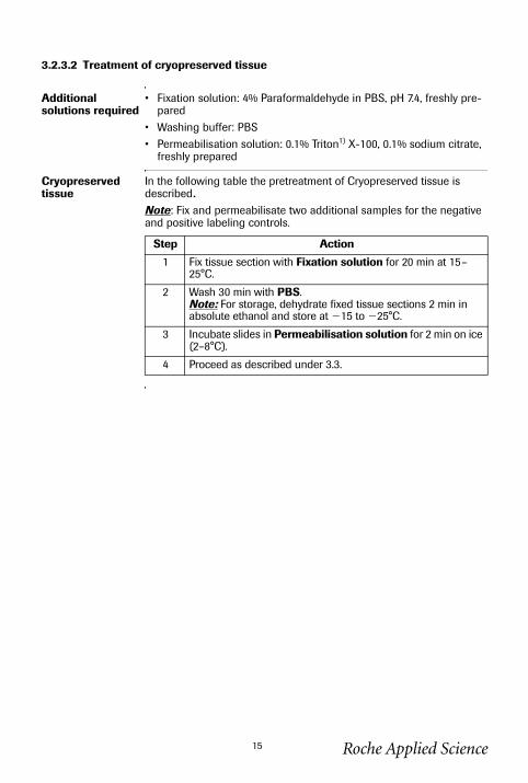
3.2.3.2 Treatment of cryopreserved tissue
Additional solutions required
• Fixation solution: 4% Paraformaldehyde in PBS, pH 7.4, freshly pre-pared
• Washing buffer: PBS• Permeabilisation solution: 0.1% Triton1) X-100, 0.1% sodium citrate,
freshly prepared
Cryopreserved tissue
In the following table the pretreatment of Cryopreserved tissue is described.Note: Fix and permeabilisate two additional samples for the negative and positive labeling controls.
Step Action
1 Fix tissue section with Fixation solution for 20 min at 15–25°C.
2 Wash 30 min with PBS.Note: For storage, dehydrate fixed tissue sections 2 min in absolute ethanol and store at �15 to �25°C.
3 Incubate slides in Permeabilisation solution for 2 min on ice (2–8°C).
4 Proceed as described under 3.3.
Roche Applied Science15
1684795a.fm Page 16 Tuesday, November 26, 2002 10:32 AM
3.3 Labeling protocol
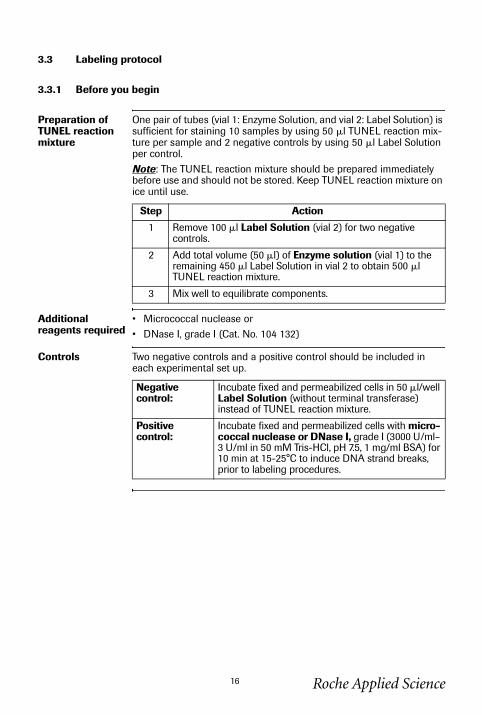
3.3.1 Before you begin
Preparation of TUNEL reaction mixture
One pair of tubes (vial 1: Enzyme Solution, and vial 2: Label Solution) is sufficient for staining 10 samples by using 50 �l TUNEL reaction mix-ture per sample and 2 negative controls by using 50 �l Label Solution per control.Note: The TUNEL reaction mixture should be prepared immediately before use and should not be stored. Keep TUNEL reaction mixture on ice until use.
Additional reagents required
• Micrococcal nuclease or • DNase I, grade I (Cat. No. 104 132)
Controls Two negative controls and a positive control should be included in each experimental set up.
Step Action
1 Remove 100 �l Label Solution (vial 2) for two negative controls.
2 Add total volume (50 �l) of Enzyme solution (vial 1) to the remaining 450 �l Label Solution in vial 2 to obtain 500 �l TUNEL reaction mixture.
3 Mix well to equilibrate components.
Negative control:
Incubate fixed and permeabilized cells in 50 �l/well Label Solution (without terminal transferase) instead of TUNEL reaction mixture.
Positive control:
Incubate fixed and permeabilized cells with micro-coccal nuclease or DNase I, grade I (3000 U/ml– 3 U/ml in 50 mM Tris-HCl, pH 7.5, 1 mg/ml BSA) for 10 min at 15-25°C to induce DNA strand breaks, prior to labeling procedures.
Roche Applied Science16
1684795a.fm Page 17 Tuesday, November 26, 2002 10:32 AM
3.3.2 Labeling protocol for cell suspensions
Additional euipment and solutions required
• Washing buffer: PBS• Humidified chamber
Procedure Please refer to the following table.
Step Action
1 Wash cells twice with PBS (200 �l/well).
2 Resuspend in 50 �l/well TUNEL reaction mixture.Note: For the negative control add 50 �l Label solution.
3 Add lid and incubate for 60 min at 37°C in a humidified atmosphere in the dark.
4 Wash samples twice in PBS.
5 Transfer cells in a tube to a final volume of 250–500 �l in PBS.
6 Samples can directly be analyzed under a fluorescence micro-scope or embedded with antifade prior to analysis. Use an exitation wavelength in the range of 450–500 nm (e.g. 488 nm) and detection in the range of 515–565 nm (green).
Roche Applied Science17
1684795a.fm Page 18 Tuesday, November 26, 2002 10:32 AM
3.3.3 Labeling protocol for adherent cells, cell smears, cytospin preparations and tissues
Additional equipment and solutions required
• Washing buffer: PBS• Parafilm or coverslip• Humidified chamber
Procedure Please refer to the following table.
Step Action
1 Rinse slides twice with PBS.
2 Dry area around sample.
3 Add 50 �l TUNEL reaction mixture on sample.Note: For the negative control add 50 �l Label solution each. To ensure a homogeneous spread of TUNEL reaction mixture across cell monolayer and to avoid evaporative loss, samples should be covered with parafilm or coverslip during incuba-tion.
4 Incubate slide in a humidified atmosphere for 60 min at 37°C in the dark.
5 Rinse slide 3× with PBS.
6 Samples can directly be analyzed by flow cytometry or fluo-rescence microscopy. Use an excitation wavelength in the range of 450–500 nm (e.g. 488 nm) or by detection in the range of 515–565 nm (green).
Roche Applied Science18
1684795a.fm Page 19 Tuesday, November 26, 2002 10:32 AM
3.3.4 Labeling protocol for difficult tissue
Additional equipment and solutions required
• Citrate buffer, 0.1 M, pH 6.0.• Washing buffer: PBS• Tris-HCl, 0.1 M pH 7.5, containing 3% BSA and 20% normal bovine
serum• Humidified chamber• Microwave
Procedure Please refer to the following table.
Step Action
1 Dewax paraformaldehyde- or formalin-fixed tissue sections according to standard procedures.
2 Place the slide(s) in a plastic jar containing 200 ml 0.1 M Cit-rate buffer, pH 6.0.
3 • Apply 750 W (high) microwave irradiation for 1 min.• Cool rapidly by immediately adding 80 ml double dist. water
(20–25°C).• Transfer the slide(s) into PBS (20–25°C).DO NOT perform a Proteinase K treatment!
4 Immerse the slide(s) for 30 min at 15-25°C in Tris-HCl, 0.1 M pH 7.5, containing 3% BSA and 20% normal bovine serum.
5 • Rinse the slide(s) twice with PBS at 15-25°C.• Let excess fluid drain off.
6 Add 50 �l of TUNEL reaction mixture on the section.Note: For the negative control add 50 �l Label solution.
7 Incubate for 60 min at 37°C in a humidified atmosphere in the dark.
8 • Rinse slide(s) three times in PBS for 5 min each.• Evaluate the section under a fluorescence microscope.
Roche Applied Science19
1684795a.fm Page 20 Tuesday, November 26, 2002 10:32 AM
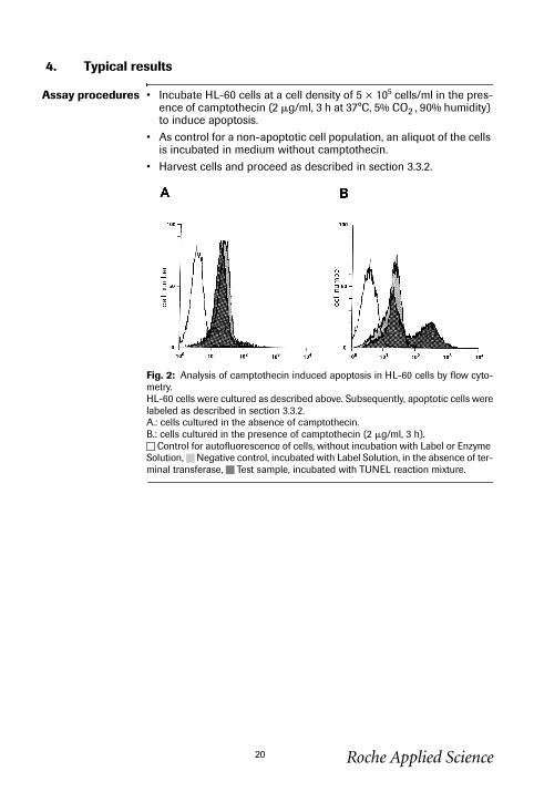
4. Typical results
Assay procedures • Incubate HL-60 cells at a cell density of 5 × 10⁵ cells/ml in the pres-ence of camptothecin (2 �g/ml, 3 h at 37°C, 5% CO2 , 90% humidity) to induce apoptosis.
• As control for a non-apoptotic cell population, an aliquot of the cells is incubated in medium without camptothecin.
• Harvest cells and proceed as described in section 3.3.2.
Fig. 2: Analysis of camptothecin induced apoptosis in HL-60 cells by flow cyto-metry.HL-60 cells were cultured as described above. Subsequently, apoptotic cells werelabeled as described in section 3.3.2. A.: cells cultured in the absence of camptothecin.B.: cells cultured in the presence of camptothecin (2 �g/ml, 3 h).
Control for autofluorescence of cells, without incubation with Label or Enzyme Solution, Negative control, incubated with Label Solution, in the absence of ter-minal transferase, Test sample, incubated with TUNEL reaction mixture.
Roche Applied Science20

1684795a.fm Page 21 Tuesday, November 26, 2002 10:32 AM
Roche Applied Science21
5. Appendix
5.1 Trouble-shooting
This table describes various troubleshooting parameters.
Problem Step/Reagent of Procedure
Possible cause Recommendation
Nonspecificlabeling
Embedding of tissue
UV-irradiation forpolymerization of embedding material (e.g. methacrylate) leads to DNA strand breaks
Try different embedding material or different polymerization reagent.
Fixation Acidic fixatives (e.g. methacarn, Carnoy’s fixative)
• Try 4% buffered paraformalde-hyde.
• Try formalin or glutaraldehyde.
TUNEL reaction
TdT concentration too high
Reduce concentration of TdT by diluting it 1:2 up to 1:3 with TUNEL Dilution Buffer (Cat. No. 1 966 006).
Nucleases, Polymerases
Some tissues (e.g. smooth muscles) show DNA strand breaks very soon after tissue preparation
• Fix tissue immediately after organ preparation.
• Perfuse fixative through liver vein.
Some enzymes are still active
Block with a solution containing ddUTP and dATP.
High back-ground
Measurement of samples
Measuring via micro-plate reader not possi-ble because of too high background
Try to reduce background by the following recommendations.
Sample Mycoplasma contami-nation
Mycoplasma Detection Kit (Cat. No. 1 296 449).
Highly proliferating cells
Double staining, e.g. with Annexin-V-Fluos (Cat. No. 1 828 681).Note: Measuring via microplate reader not possible because of too high background.
Erythrocytes high autofluorescence because of hemoglobin
Use dUTP-rhodamine.
Fixation Formalin fixation leads to a yellowish staining of cells containing mel-anin precursors
Try methanol for fixation but take into account that this might lead to reduced sensitivity.
TUNEL reaction
Concentration of label-ing mix is too high for mamma carcinoma
Reduce concentration of labeling mix to 50% by diluting with TUNEL Dilution Buffer (Cat. No. 1 966 006).
continued on next page
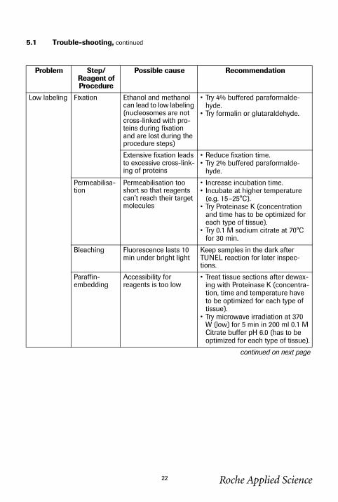
5.1 Trouble-shooting, continued
1684795a.fm Page 22 Tuesday, November 26, 2002 10:32 AM
Low labeling Fixation Ethanol and methanol can lead to low labeling (nucleosomes are not cross-linked with pro-teins during fixation and are lost during the procedure steps)
• Try 4% buffered paraformalde-hyde.
• Try formalin or glutaraldehyde.
Extensive fixation leads to excessive cross-link-ing of proteins
• Reduce fixation time.• Try 2% buffered paraformalde-
hyde.
Permeabilisa-tion
Permeabilisation too short so that reagents can’t reach their target molecules
• Increase incubation time.• Incubate at higher temperature
(e.g. 15–25°C).• Try Proteinase K (concentration
and time has to be optimized for each type of tissue).
• Try 0.1 M sodium citrate at 70°C for 30 min.
Bleaching Fluorescence lasts 10 min under bright light
Keep samples in the dark after TUNEL reaction for later inspec-tions.
Paraffin-embedding
Accessibility for reagents is too low
• Treat tissue sections after dewax-ing with Proteinase K (concentra-tion, time and temperature have to be optimized for each type of tissue).
• Try microwave irradiation at 370 W (low) for 5 min in 200 ml 0.1 M Citrate buffer pH 6.0 (has to be optimized for each type of tissue).
continued on next page
Problem Step/Reagent of Procedure
Possible cause Recommendation
Roche Applied Science22
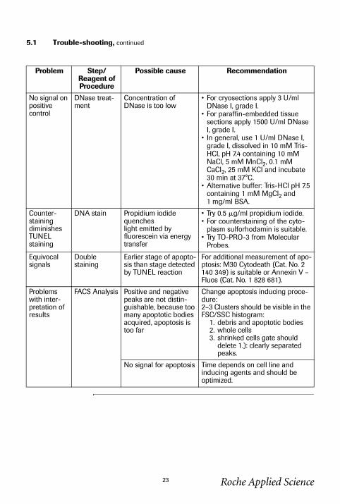
5.1 Trouble-shooting, continued
1684795a.fm Page 23 Tuesday, November 26, 2002 10:32 AM
No signal on positive control
DNase treat-ment
Concentration of DNase is too low
• For cryosections apply 3 U/ml DNase I, grade I.
• For paraffin-embedded tissue sections apply 1500 U/ml DNase I, grade I.
• In general, use 1 U/ml DNase I, grade I, dissolved in 10 mM Tris-HCl, pH 7.4 containing 10 mM NaCl, 5 mM MnCl2, 0.1 mM CaCl2, 25 mM KCl and incubate 30 min at 37°C.
• Alternative buffer: Tris-HCl pH 7.5 containing 1 mM MgCl2 and 1 mg/ml BSA.
Counter-staining diminishes TUNEL staining
DNA stain Propidium iodide quencheslight emitted by fluorescein via energy transfer
• Try 0.5 �g/ml propidium iodide.• For counterstaining of the cyto-
plasm sulforhodamin is suitable.• Try TO-PRO-3 from Molecular
Probes.
Equivocal signals
Double staining
Earlier stage of apopto-sis than stage detected by TUNEL reaction
For additional measurement of apo-ptosis: M30 Cytodeath (Cat. No. 2 140 349) is suitable or Annexin V – Fluos (Cat. No. 1 828 681).
Problems with inter-pretation of results
FACS Analysis Positive and negative peaks are not distin-guishable, because too many apoptotic bodies acquired, apoptosis is too far
Change apoptosis inducing proce-dure:2–3 Clusters should be visible in the FSC/SSC histogram:
1. debris and apoptotic bodies2. whole cells3. shrinked cells gate should
delete 1.): clearly separated peaks.
No signal for apoptosis Time depends on cell line and inducing agents and should be optimized.
Problem Step/Reagent of Procedure
Possible cause Recommendation
Roche Applied Science23
1684795a.fm Page 24 Tuesday, November 26, 2002 10:32 AM
Roche Applied Science24
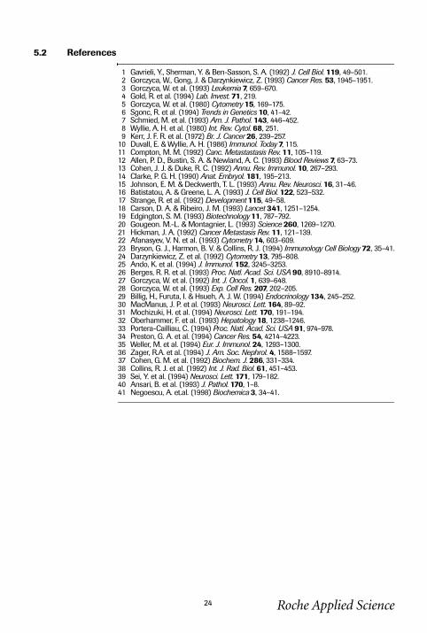
5.2 References
1 Gavrieli, Y., Sherman, Y. & Ben-Sasson, S. A. (1992) J. Cell Biol. 119, 49–501.2 Gorczyca, W., Gong, J. & Darzynkiewicz, Z. (1993) Cancer Res. 53, 1945–1951.3 Gorczyca, W. et al. (1993) Leukemia 7, 659–670.4 Gold, R. et al. (1994) Lab. Invest. 71, 219.5 Gorczyca, W. et al. (1980) Cytometry 15, 169–175.6 Sgonc, R. et al. (1994) Trends in Genetics 10, 41–42.7 Schmied, M. et al. (1993) Am. J. Pathol. 143, 446–452.8 Wyllie, A. H. et al. (1980) Int. Rev. Cytol. 68, 251.9 Kerr, J. F. R. et al. (1972) Br. J. Cancer 26, 239–257.
10 Duvall, E. & Wyllie, A. H. (1986) Immunol. Today 7, 115.11 Compton, M. M. (1992) Canc. Metastastasis Rev. 11, 105–119.12 Allen, P. D., Bustin, S. A. & Newland, A. C. (1993) Blood Reviews 7, 63–73.13 Cohen, J. J. & Duke, R. C. (1992) Annu. Rev. Immunol. 10, 267–293.14 Clarke, P. G. H. (1990) Anat. Embryol. 181, 195–213.15 Johnson, E. M. & Deckwerth, T. L. (1993) Annu. Rev. Neurosci. 16, 31–46.16 Batistatou, A. & Greene, L. A. (1993) J. Cell Biol. 122, 523–532.17 Strange, R. et al. (1992) Development 115, 49–58.18 Carson, D. A. & Ribeiro, J. M. (1993) Lancet 341, 1251–1254.19 Edgington, S. M. (1993) Biotechnology 11, 787–792.20 Gougeon. M.-L. & Montagnier, L. (1993) Science 260, 1269–1270.21 Hickman, J. A. (1992) Cancer Metastasis Rev. 11, 121–139.22 Afanasyev, V. N. et al. (1993) Cytometry 14, 603–609.23 Bryson, G. J., Harmon, B. V. & Collins, R. J. (1994) Immunology Cell Biology 72, 35–41.24 Darzynkiewicz, Z. et al. (1992) Cytometry 13, 795–808.25 Ando, K. et al. (1994) J. Immunol. 152, 3245–3253.26 Berges, R. R. et al. (1993) Proc. Natl. Acad. Sci. USA 90, 8910–8914.27 Gorczyca, W. et al. (1992) Int. J. Oncol. 1, 639–648.28 Gorczyca, W. et al. (1993) Exp. Cell Res. 207, 202–205.29 Billig, H., Furuta, I. & Hsueh, A. J. W. (1994) Endocrinology 134, 245–252.30 MacManus, J. P. et al. (1993) Neurosci. Lett. 164, 89–92.31 Mochizuki, H. et al. (1994) Neurosci. Lett. 170, 191–194.32 Oberhammer, F. et al. (1993) Hepatology 18, 1238–1246.33 Portera-Cailliau, C. (1994) Proc. Natl. Acad. Sci. USA 91, 974–978.34 Preston, G. A. et al. (1994) Cancer Res. 54, 4214–4223.35 Weller, M. et al. (1994) Eur. J. Immunol. 24, 1293–1300.36 Zager, R.A. et al. (1994) J. Am. Soc. Nephrol. 4, 1588–1597.37 Cohen, G. M. et al. (1992) Biochem. J. 286, 331–334.38 Collins, R. J. et al. (1992) Int. J. Rad. Biol. 61, 451–453.39 Sei, Y. et al. (1994) Neurosci. Lett. 171, 179–182.40 Ansari, B. et al. (1993) J. Pathol. 170, 1–8.41 Negoescu, A. et.al. (1998) Biochemica 3, 34–41.
1684795a.fm Page 25 Tuesday, November 26, 2002 10:32 AM
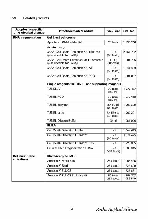
5.3 Related products
Apoptosis-specific physiological change Detection mode/Product Pack size Cat. No.
DNA fragmentation Gel Electrophoresis
Apoptotic DNA-Ladder Kit 20 tests 1 835 246
In situ assay
In Situ Cell Death Detection Kit, TMR red (also useable for FACS)
1 kit (50 tests)
2 156 792
In Situ Cell Death Detection Kit, Fluorescein (also useable for FACS)
1 kit (50 tests)
1 684 795
In Situ Cell Death Detection Kit, AP 1 kit (50 tests)
1 684 809
In Situ Cell Death Detection Kit, POD 1 kit (50 tests)
1 684 817
Single reagents for TUNEL and supporting reagents
TUNEL AP 70 tests (3.5 ml)
1 772 457
TUNEL POD 70 tests (3.5 ml)
1 772 465
TUNEL Enzyme 2× 50 �l (20 tests)
1 767 305
TUNEL Label 3× 550 �l (30 tests)
1 767 291
TUNEL Dilution Buffer 20 ml 1 966 006
ELISA
Cell Death Detection ELISA 1 kit 1 544 675
Cell Death Detection ELISAPLUS 1 kit (96 tests)
1 774 425
Cell Death Detection ELISAPLUS, 10× 1 kit 1 920 685
Cellular DNA Fragmentation ELISA 1 kit (500 tests)
1 585 045
Cell membrane alterations
Microscopy or FACS
Annexin-V-Alexa 568 250 tests 1 985 485
Annexin-V-Biotin 250 tests 1 828 690
Annexin-V-FLUOS 250 tests 1 828 681
Annexin-V-FLUOS Staining Kit 50 tests250 tests
1 858 7771 988 549
Roche Applied Science25
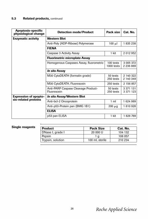
5.3 Related products, continued
1684795a.fm Page 26 Tuesday, November 26, 2002 10:32 AM
26
Single reagents
Enzymatic activity Western Blot
Anti-Poly (ADP-Ribose) Polymerase 100 �l 1 835 238
FIENA
Caspase 3 Activity Assay 1 kit 2 012 952
Fluorimetric microplate Assay
Homogenous Caspases Assay, fluorometric 100 tests1000 tests
3 005 3722 236 869
In situ Assay
M30 CytoDEATH (formalin grade) 50 tests250 tests
2 140 3222 140 349
M30 CytoDEATH, Fluorescein 250 tests 2 156 857
Anti-PARP Caspase Cleavage Product-Fluorescein
50 tests250 tests
3 371 1313 371 123
Expression of apopto-sis-related proteins
In situ Assay/Western Blot
Anti-bcl-2 Oncoprotein 1 ml 1 624 989
Anti-p53-Protein pan (BMG 1B1) 200 �g 1 810 928
ELISA
p53 pan ELISA 1 kit 1 828 789
Apoptosis-specific physiological change Detection mode/Product Pack size Cat. No.
Product Pack Size Cat. No.DNase I, grade I 20 000 U 104 132Pepsin 1 g 108 057Trypsin, solution 100 ml, sterile 210 234
Roche Applied Science
1684795a.fm Page 27 Tuesday, November 26, 2002 10:32 AM
For further information please access our web-site address at:http://biochem.roche.comor the Apoptosis special interest site:http://www.roche-applied-science.com/apoptosis
*available from Roche Applied Science1)Triton is a trademark of Rohm & Haas, Philadelphia, USA.Sold through an arrangement with ENZO DIAGNOSTICS, INC.Purchase of this product does not include any right of license to exploit this product commercially.This product or the use may be covered by one or more ENZO patents, including the following:U.S. Patent Nos. 4,711,955; 5,328,824; 5,449,767; 5,241,060; 4,994,373; and 5,175,269; EP 0 063 897 BI; EP 0 117 440 BI;EP 0 122 614 BI; and EP 0 128 332 BI; and Canadian Patent Nos. 1,219,824; 1,223,831; 1,309,672; 1,254,525; and 1,228,811.
Roche Applied Science27

Roche Diagnostics GmbHRoche Applied ScienceNonnenwald 282372 PenzbergGermany
1102
.116
9727
7�
www.roche-applied-science.com
to order, solve technical queries, find product information, or contact your local sales representative.
www.roche-applied-science/pack-insert/1684795a.pdf
Enzo
1684795a.fm Page 28 Tuesday, November 26, 2002 10:32 AM