in the pathogenesis of amyotrophic lateral do mthfr c677t
TRANSCRIPT

Page 1/27
Do MTHFR C677T Genetic Polymorphism In�uencein the Pathogenesis of Amyotrophic LateralSclerosis?Rômulo Morais Azevedo
Universidade Federal de GoiasKamilla de Faria Santos
Universidade Federal de GoiasRayana Pereira Dantas de Oliveira
Universidade Federal de GoiasJúllia Costa Pereira
Universidade Federal de GoiasDhiogo da Cruz Pereira Bento
Universidade Federal de GoiasAngela Adamski da Silva Reis
Universidade Federal de GoiasRodrigo da Silva Santos ( [email protected] )
Universidade Federal de Goiás https://orcid.org/0000-0002-9480-4362
Research Article
Keywords: Neurodegenerative disease, Genetic polymorphism, Biomarker, MTHFR, Personalized medicine
Posted Date: October 1st, 2021
DOI: https://doi.org/10.21203/rs.3.rs-939291/v1
License: This work is licensed under a Creative Commons Attribution 4.0 International License. Read Full License

Page 2/27
AbstractAmyotrophic Lateral Sclerosis (ALS) is a progressive and lethal neurodegenerative disease without ade�nitive diagnostic test and effective treatment. A plethora of studies suggest that genetic factors playan important role in ALS development, and potentially link folate pathway dysregulation to diseasepathogenesis. This study aims to evaluate folate dysregulation due to MTHFR C677T polymorphism andother factors such as sociodemographic and clinical, to better elucidate the involvement of these factorsin ALS pathogenesis, and to investigate possible biomarkers for use as disease diagnostics orprognostics. This hospital-based case-control study analyzed 101 patients diagnosed with ALS and 119considered healthy, with no suspicion or diagnosis of neurodegenerative disease. Blood samples werecollected, stored, and underwent DNA extraction. Clinical and sociodemographic data from patients werecollected through a questionnaire, as well as consultation of medical records. Genotypic analyses wereperformed using PCR-RFLP, and statistical analysis of clinical and genotypic data was conducted withSPSS software, version 23. The results show a higher presence of the mutant genotype (p = 0.02) in thecase group, and suggest that mutant allele (T) is a risk factor for ALS susceptibility (OR = 1.54; 95% CI = 1.05–2.29; p = 0.03). Mutant genotype (T/T) interacts with both demographics (White p = 0.005 / Brownp = 0,001) and clinical factors (Physical activity p = 0.006) as risk factors for ALS. Also, a signi�cantdifference in alcohol consumption (p = 0.001) between the case and control group was observed.Moreover, a statistical trend towards faster disease progression and death was observed for patients withthe mutant allele (T) (p = 0.06). Thus, the results of this study suggest that folate de�ciency due toMTHFR C677T polymorphism is implicated in ALS through pathogenic mechanisms and interaction withother risk factors, resulting in faster disease progression and early death.
IntroductionAmyotrophic Lateral Sclerosis (ALS) is a lethal and progressive neurodegenerative disease thatselectively affects the upper and lower motor neurons. It is characterized by symptoms such as cramps,fasciculations, muscle weakness and atrophy, dysphagia, dysarthria, and respiratory failure, which is theleading cause of death [1–3]. The disease has an annual incidence of about 1.55–1.96 / 100,000inhabitants and a prevalence of 5.2–6.2 / 100,000 inhabitants, predominantly affecting individuals aged55 to 75 years, Caucasian populations, and males [4, 5].
The causes of disease development are still unde�ned. ALS is thought to be a multifactorial pathology,associated with smoking, alcoholism, prolonged physical activities, and genetic factors [6, 7]. There isample evidence that genetic factors contribute to ALS susceptibility, and several genes have already beenimplicated in the neurodegenerative process, such as SOD1, VAPB, C9orf72, FUS, NEK1, TRPM7, andothers [8, 9]. Despite the continued discovery of an increasing number of genetic biomarkers in ALS, over50% of patients do not have a genetic diagnosis [10].
Without a speci�c diagnostic test for the disease, ALS diagnosis is currently performed through clinicalevaluation aided by neuroimaging and electroneuromyography tests [11]. The average time for the

Page 3/27
diagnosis is estimated to be nine months to one year, from the symptom onset [12, 13]. Riluzole, aglutamate antagonist, is currently the only medicine with proven e�cacy in improving ALS survival, but itonly increases life expectancy by two to three months. It is known that early diagnosis associated withdrug therapy using riluzole may enhance its e�cacy in clinical improvement [14, 15].
Variation in genes involved in the folate pathway has been suggested as a possible factor implicated inALS since this vitamin is essential to DNA synthesis and homocysteine (Hcy) metabolism, thedysregulation folate metabolism unbalances the cellular homeostasis, and could contribute toneurodegenerative processes in motor neurons [16, 17]. The Methylenetetrahydrofolate reductase(MTHFR) gene stands out as a potential target, considering that it is responsible for encoding the MTHFRenzyme, which plays a crucial role in intracellular folate metabolism [18]. MTHFR enzyme regulates theintracellular methylation pathway and in�uences DNA synthesis by converting 5,10-methylenetetrahydrofolate (5,10-MTHF) into 5-methyltetrahydrofolate (5-MTHF), the most abundantcirculating form of folic acid in the body [18–22].
It is known that polymorphisms in the MTHFR gene result in the production of a defective enzyme, whichderegulates the entire intracellular biochemical process of folate. Among the main polymorphismsstudied in the gene, the C677T (rs1801133) mutation codes for a thermolabile enzyme with reducedactivity by up to 65% at 35 ºC, resulting in increased plasma levels of Hcy [23, 24]. The mutation has beenidenti�ed as a risk factor for several diseases, such as cardiovascular diseases [25, 26], cancers [27, 28],diabetes [29], hypertension [30], and congenital malformations [31].
In the nervous system, dysregulation of the folate cycle directly impacts neuronal physiology by causinghyperhomocysteinemia (HHcy). Homocysteine is a neurotoxic factor and is associated with severalneuronal death mechanisms [32, 33]. Thus, increasing numbers of studies have hypothesized the folatemetabolic dysfunctions in ALS pathogenesis, especially after the detection of HHcy in ALS patientsaffected by the disease and the discovery of its effect on the motor neuron [34, 35].
Therefore, this study aims to evaluate the MTHFR C677T polymorphism, socio-demographic and clinicalfactors in ALS patients in the population of central Brazil, to understand how genetic causes ofintracellular folate dysregulation, as well the in�uences of environmental and clinical factor affectdisease susceptibility and progression.
Material And MethodsPopulation study (Patients)
This hospital-based case-control study consists of 101 patients diagnosed with ALS (case group) whounderwent medical follow-up at the referral hospital for neuromuscular diseases in Goiânia, GO, Brazil, Dr.Henrique Santillo Rehabilitation and Readaptation Center (CRER). The control group consists of 119individuals not diagnosed with ALS or other neurodegenerative diseases, collected from the Laboratory ofClinical Analysis and Health Education of the Federal University of Goiás, Brazil (LACES-UFG).
Page 4/27
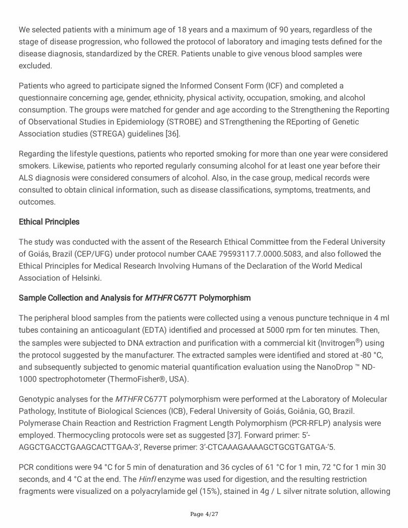
We selected patients with a minimum age of 18 years and a maximum of 90 years, regardless of thestage of disease progression, who followed the protocol of laboratory and imaging tests de�ned for thedisease diagnosis, standardized by the CRER. Patients unable to give venous blood samples wereexcluded.
Patients who agreed to participate signed the Informed Consent Form (ICF) and completed aquestionnaire concerning age, gender, ethnicity, physical activity, occupation, smoking, and alcoholconsumption. The groups were matched for gender and age according to the Strengthening the Reportingof Observational Studies in Epidemiology (STROBE) and STrengthening the REporting of GeneticAssociation studies (STREGA) guidelines [36].
Regarding the lifestyle questions, patients who reported smoking for more than one year were consideredsmokers. Likewise, patients who reported regularly consuming alcohol for at least one year before theirALS diagnosis were considered consumers of alcohol. Also, in the case group, medical records wereconsulted to obtain clinical information, such as disease classi�cations, symptoms, treatments, andoutcomes.
Ethical Principles
The study was conducted with the assent of the Research Ethical Committee from the Federal Universityof Goiás, Brazil (CEP/UFG) under protocol number CAAE 79593117.7.0000.5083, and also followed theEthical Principles for Medical Research Involving Humans of the Declaration of the World MedicalAssociation of Helsinki.
Sample Collection and Analysis for MTHFR C677T Polymorphism
The peripheral blood samples from the patients were collected using a venous puncture technique in 4 mltubes containing an anticoagulant (EDTA) identi�ed and processed at 5000 rpm for ten minutes. Then,the samples were subjected to DNA extraction and puri�cation with a commercial kit (Invitrogen®) usingthe protocol suggested by the manufacturer. The extracted samples were identi�ed and stored at -80 °C,and subsequently subjected to genomic material quanti�cation evaluation using the NanoDrop ™ ND-1000 spectrophotometer (ThermoFisher®, USA).
Genotypic analyses for the MTHFR C677T polymorphism were performed at the Laboratory of MolecularPathology, Institute of Biological Sciences (ICB), Federal University of Goiás, Goiânia, GO, Brazil. Polymerase Chain Reaction and Restriction Fragment Length Polymorphism (PCR-RFLP) analysis wereemployed. Thermocycling protocols were set as suggested [37]. Forward primer: 5’-AGGCTGACCTGAAGCACTTGAA-3’, Reverse primer: 3’-CTCAAAGAAAAGCTGCGTGATGA-‘5.
PCR conditions were 94 °C for 5 min of denaturation and 36 cycles of 61 °C for 1 min, 72 °C for 1 min 30seconds, and 4 °C at the end. The HinfI enzyme was used for digestion, and the resulting restrictionfragments were visualized on a polyacrylamide gel (15%), stained in 4g / L silver nitrate solution, allowing

Page 5/27
for the identi�cation of wildtype genotypes (C/C) 198bp fragment, heterozygotes (C/T) 198bp, 175bp,and 23bp fragments, and mutants (T/T) 175bp and 23bp fragments.
Statistical analysis
Statistical analysis was performed using the SPSS statistical package, version 23. The generalcharacterization of the case group and control group individuals was performed with descriptivestatistics, with absolute (n) and relative (%) frequencies reported, while mean and standard deviationwere applied to continuous variables.
For genotypic analysis, PostHoc Chi-square tests and Pearson’s test (MacDonald and Gardner, 2000) wereperformed. To study survival (time between ALS diagnosis and death) according to genotype, Kaplan-Meier curves were created. For all results, a signi�cance level of 5% was used.
ResultsSociodemographic and clinical data
Sociodemographic data were evaluated for ALS (101 individuals) and control (119 individuals) groups.Mean ages were 57.3 ± 12.8 years for the case group and 58.0 ± 10.3 years for the control group (Table1).
The case group had 44.6% of female patients (45 individuals) and 55.4% of male patients (56individuals). In the control group, 57.1% were female (68 individuals), and 42.9% were male (51individuals). Age (p = 0.64) and gender (p=0.08) showed no statistically signi�cant associations with thedisease.
In the analysis of risk factors, smoking was not different between the case and control groups (p = 0.28).However, alcohol consumption was higher in the case group than in the control group (p = 0.01),therefore, it is a possible risk condition for the disease among the studied groups (Table 1).
Table 1 - Comparison of clinical and sociodemographic variables between case and control group.

Page 6/27
Samples n (%) Total p
Control 119(54.1)
Case 101(45.9)
Age (Mean ±SD)
58.0 ± 10.3 57.3 ± 12.8 57.7 ±11.5
0.64**
Gender
Female 68 (57.1) 45 (44.6) 113(51.4)
0.08*
Male 51 (42.9) 56 (55.4) 107(48.6)
Alcohol Intake
No 84 (70.6) 55 (54.5) 139(63.2)
0.01*†
Yes 35 (29.4) 46 (45.5) 81 (36.8)
Smoking
No 68 (57.1) 65 (64.4) 133(60.5)
0.28*
Yes 51 (42.9) 36 (35.6) 87 (39.5)
*Pearson’s Chi-square test, ** Student t-test, † p < 0,05.
n= absolute frequency, % = relative frequency
SD = standard deviation, ALS- Amyotrophic Lateral Sclerosis,
In the ALS group, 48.5% identi�ed as white and 44.6% identi�ed as brown. When asked about physicalactivity, 46.5% declared that they did not engage in any type of physical activity before the diagnosis ofthe disease, while 53.5% reported physical activity. The reported physical activities included walking,soccer, water aerobics, workout, pilates, and others. Most cases reported work in the following categories:general service category (36.6%), housework (13.9%), administrative services (12.9%), commerce (9.9%),teachers (7.9%), health professional (5.9%), chef cook (1%), student (1%), and others (5%). However, 5.9%chose not to inform.
The patients in the case group were classi�ed according to the El Escorial World Federation of Neurology,as reported in their medical records. 5.9% of patients reported a recurrence of ALS in the family (fALS),and 94.1% were considered sporadic cases (sALS). Moreover, 72.3% �t the cALS category, 23.8% werebALS, and 4% were juvenile ALS. Previous pathologies were cited by 46.5% in the case group, and 40.6%reported a history of another neurological disease in the family.
Page 7/27
Regarding treatment with riluzole, 75.2% of the case group said they followed the treatment, and 24.8%reported not using it. During the research period, 10.9% of the ALS patients in the case group died.Therefore, death was considered as a disease outcome in this study.
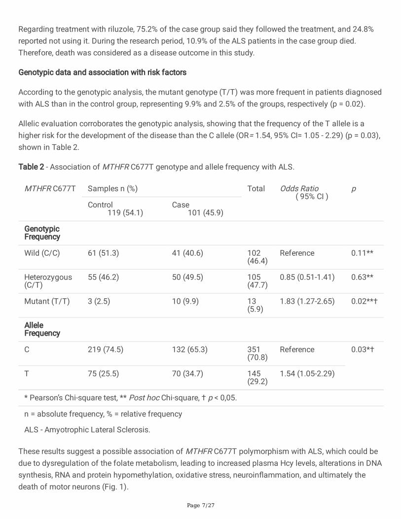
Genotypic data and association with risk factors
According to the genotypic analysis, the mutant genotype (T/T) was more frequent in patients diagnosedwith ALS than in the control group, representing 9.9% and 2.5% of the groups, respectively (p = 0.02).
Allelic evaluation corroborates the genotypic analysis, showing that the frequency of the T allele is ahigher risk for the development of the disease than the C allele (OR= 1.54, 95% CI= 1.05 - 2.29) (p = 0.03),shown in Table 2.
Table 2 - Association of MTHFR C677T genotype and allele frequency with ALS.
MTHFR C677T Samples n (%) Total Odds Ratio ( 95% CI )
p
Control 119 (54.1)
Case 101 (45.9)
GenotypicFrequency
Wild (C/C) 61 (51.3) 41 (40.6) 102(46.4)
Reference 0.11**
Heterozygous(C/T)
55 (46.2) 50 (49.5) 105(47.7)
0.85 (0.51-1.41) 0.63**
Mutant (T/T) 3 (2.5) 10 (9.9) 13(5.9)
1.83 (1.27-2.65) 0.02**†
AlleleFrequency
C 219 (74.5) 132 (65.3) 351(70.8)
Reference 0.03*†
T 75 (25.5) 70 (34.7) 145(29.2)
1.54 (1.05-2.29)
* Pearson’s Chi-square test, ** Post hoc Chi-square, † p < 0,05.
n = absolute frequency, % = relative frequency
ALS - Amyotrophic Lateral Sclerosis.
These results suggest a possible association of MTHFR C677T polymorphism with ALS, which could bedue to dysregulation of the folate metabolism, leading to increased plasma Hcy levels, alterations in DNAsynthesis, RNA and protein hypomethylation, oxidative stress, neuroin�ammation, and ultimately thedeath of motor neurons (Fig. 1).

Page 8/27
When analyzing associations of clinical data with the results of the genotypic analysis of ALS patients,the patients’ gender (p = 0.85), alcohol intake (p = 0.16), smoking (p = 0.42), disease classi�cation (p =0.57), neurological disease in the family (p = 0.99), and previous pathologies (p = 0.90) did not present astatistically signi�cant association with genotype in disease development (Table 3). However, ethnicity,when compared with genotypic pro�le, was signi�cantly associated with (T/T) genotype patients,speci�cally for self-identi�ed white (p = 0.005) and brown (p = 0.001) individuals. Engaging in physicalactivity was also associated with the (T/T) genotype (p = 0.006), shown in Table 3.
Table 3 - Association between MTHFR C677T polymorphism genotype with the demographic and clinicalpro�le of ALS patients.
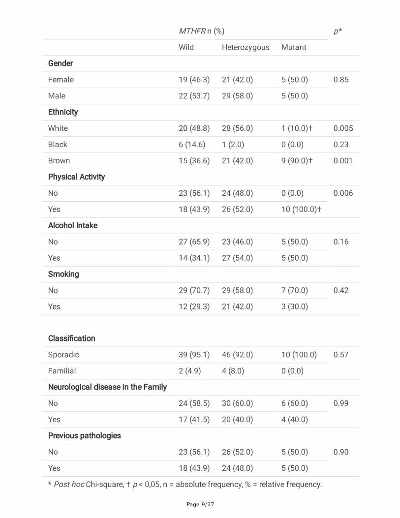
Page 9/27
MTHFR n (%) p*
Wild Heterozygous Mutant
Gender
Female 19 (46.3) 21 (42.0) 5 (50.0) 0.85
Male 22 (53.7) 29 (58.0) 5 (50.0)
Ethnicity
White 20 (48.8) 28 (56.0) 1 (10.0)† 0.005
Black 6 (14.6) 1 (2.0) 0 (0.0) 0.23
Brown 15 (36.6) 21 (42.0) 9 (90.0)† 0.001
Physical Activity
No 23 (56.1) 24 (48.0) 0 (0.0) 0.006
Yes 18 (43.9) 26 (52.0) 10 (100.0)†
Alcohol Intake
No 27 (65.9) 23 (46.0) 5 (50.0) 0.16
Yes 14 (34.1) 27 (54.0) 5 (50.0)
Smoking
No 29 (70.7) 29 (58.0) 7 (70.0) 0.42
Yes 12 (29.3) 21 (42.0) 3 (30.0)
Classi�cation
Sporadic 39 (95.1) 46 (92.0) 10 (100.0) 0.57
Familial 2 (4.9) 4 (8.0) 0 (0.0)
Neurological disease in the Family
No 24 (58.5) 30 (60.0) 6 (60.0) 0.99
Yes 17 (41.5) 20 (40.0) 4 (40.0)
Previous pathologies
No 23 (56.1) 26 (52.0) 5 (50.0) 0.90
Yes 18 (43.9) 24 (48.0) 5 (50.0)
* Post hoc Chi-square, † p < 0,05, n = absolute frequency, % = relative frequency.

Page 10/27
Association of genotypic pro�le and ALS outcome
Survival analysis was performed with the onset of symptoms as the starting point and death as theoutcome according to the MTHFR C667T polymorphism genotypes. This analysis demonstrated astatistical trend towards more signi�cant progression and earlier death of patients with the heterozygousgenotype (C/T) when compared to (C/C) patients (p = 0.06), presented in Fig. 2.
DiscussionALS is a heterogeneous disease with ethnic and sociodemographic differences. In this study, the ALSgroup has an average age of 57.3 ± 12.8 years, younger patients represent 4% of the group, and 55.4% aremales. ALS disease affects mainly men, with a male-to-female ratio between 1 and 2, and the initialsymptoms usually manifest between 55–75 years [2, 7, 38]. Despite the prevalence in the elderly, youngpeople with an average age of 20 years are also affected, representing 1–10% of all cases, classi�ed asjuvenile ALS [39]. In this study, the average age and gender proportions in the case group correspond tothe literature [40]. However, age and gender are not signi�cantly different between groups (Table 1).
The reason for the difference in occurrence between the genders is not yet known. Some studiesassociate a lower prevalence of ALS in women with a hormonal protective effect or lower exposure to riskfactors [41, 42]. Recent studies suggest that estrogen acts as a protective factor in neurodegenerativeprocesses by countering mechanisms of oxidative stress and excitotoxicity [43–45]. Despite the lowerincidence of women among ALS patients in general, they are more associated with a disease-speci�cclassi�cation, the FTD-ALS cases, which has been justi�ed by genetic factors [46]. However, furtherstudies are needed to elucidate gender biases in ALS.
Looking at the sociodemographic make-up of the case group, white people comprised the mostsubstantial proportion, representing 48.5% of the individuals, and 44.6% were brown. These resultscorroborate other studies that report a higher incidence of the disease in white populations, which hasbeen associated with genetic factors [5, 47]. Besides, the white population has more easy access tohealth programs than the black population, traditionally considered a minority in a society that oftenneglects social rights [48].
Among the modi�able variables analyzed, alcohol consumption as a risk factor for ALS is stillcontroversial. Some studies show the consumption of alcohol as a protective factor for the disease [49,50]. However, in the present study, alcohol consumption was more reported in the case group; thus, it is apossible risk condition for the disease (p = 0.01). Such a hypothesis does have a potential mechanisticexplanation. Excessive alcohol consumption increases the sensitivity of glutamate receptors andincreases the concentration of glutamate in the central nervous system, leading to excitotoxicity andneuronal death [51]. Additionally, it is known that alcohol consumption can activate astrocytes throughalterations in proteins expressed in microglia, implying in neuroin�ammatory mechanisms [52].

Page 11/27
Smoking as a risk factor for ALS is also controversial. Some studies relate smoking to an increased riskfor the disease, possibly linked to the toxic effects of cigarette substances [49, 53]. Smoking results inneuronal death since nicotine interacts with acetylcholine receptors in the central nervous system,causing the dysregulation of Ca2+, Na+, and K+ ion concentrations, increased cell permeability, oxidativestress, and neuroin�ammation [54, 55]. Nevertheless, there was no statistically signi�cant difference inidenti�cation as a smoker between the groups in this study (p = 0.28).
Other factors were investigated in the case group, such as physical activity and occupation before thediagnosis of ALS. Some studies indicate that physical activity could contribute to the risk of diseasedevelopment, as it results in increased metabolic rates in the body, which is related to the production ofreactive oxygen species and oxidative stress [56, 57]. In this sense, the hypermetabolic condition wasreported as a characteristic of ALS pathogenesis, and it has been discussed as a determining factor inneurodegenerative processes [58, 59]. Besides, physical activity has been associated with neuronal death,due to its relation to the increase of pro-in�ammatory cytokine expression resulting in neuroin�ammationprocesses. Additionally, it is known that physical injuries during such practices may contribute to thegenesis of the pathological process [57, 60]. In the present study, physical activity was reported by mostALS individuals, representing 53.5% in the case group (Table 3). Similar results were found in studies by[57] and [61].
In the professional occupation analysis of the ALS group, most individuals reported working inprofessions that require physical exertion or repetitive activities, like general services (36.6%), housework(13.9%), administrative services (12.9%), commerce (9.9%), teachers (7.9%), and health professionals(5.9%). In general, activities in which professionals are exposed to chemicals and require signi�cantphysical performance are considered risk factors for ALS [62, 63]. The present study corroborates theliterature regarding occupational exposure, in which professions such as health professionals, teachers,scientists, and athletes are most affected by the disease [64, 65]. However, further research is needed onthe association of occupational factors and clinical factors with disease susceptibility.
ALS patients were also classi�ed into major disease variations according to the recommendations of theEl Escorial World Federation of Neurology and information contained in medical records. We observed ahigher percentage of sALS (94.7%) and fALS (5.9%). Moreover, 72.3% �t into the cALS category, 23.8%were bALS, and 4% were juvenile cases. These data corroborate the prevalence of the classi�cationsdescribed in the literature, where sALS represents ~ 90 to 95% of all ALS cases, and ~ 5 to 10% are fALS.The other classi�cations also follow the same pattern, in which the classical form of the disease is mostprevalent ~ 50 to 75%, the bulbar form affects ~ 20 to 25% of all cases, and juveniles vary from ~ 1 to10% [3, 39].
Also, in the case group, individuals were asked about their past medical history and cases ofneurodegenerative diseases in the family. Research on patients’ previous diseases and the occurrence ofneurodegenerative diseases in the families of ALS patients has grown considerably in recent years due tothe discovery of the common etiopathogenesis among some diseases, which aroused scientists’

Page 12/27
attention to a better understanding of the mechanisms involved in ALS [66, 67]. The present studyshowed that 46.5% of the patients in the case group reported some type of disease before the ALSdiagnosis, and 40.6% reported cases of neurodegenerative diseases in the family. Signi�cant results ofneurological disease history in family and ALS were found in the studies by [66], [68] and [69].
Regarding drug treatment, 75.2% of ALS patients reported taking riluzole, and 24.8% reported not havingfollowed the treatment. Riluzole is the only drug approved for ALS treatment in Brazil. Nevertheless,studies report no signi�cant increase in patient survival attributed to this drug treatment – only about twoto three months [14, 70]. Other medications are currently being studied for ALS treatment, such asEdaravone (Radicut), Masitinib [14], Tirasemtiv [71], and others. However, the progressive character of thedisease and the lack of effective medication contribute to the short life expectancy after the diagnosis,which varies between three and �ve years.
Concerning the genotyping analyses, this study demonstrates a higher prevalence of genotype (T/T) inALS patients, making it a possible risk factor for the disease (OR = 1,83; 95% CI = 1.27–2.65) (p = 0.02).Similarly, the allele frequency analysis showed that the T allele confers risk for disease susceptibility (OR = 1.54; 95% CI = 1.05–2.29) (p = 0.03) (Table 2). The same results were found in studies by [72] and [73].
The hypothesis that MTHFR C677T polymorphism may contribute to ALS pathogenesis is supported byevidence showing that this polymorphism causes an imbalance of the intracellular folate pathway [74],which is associated with several pathogenic mechanisms, such as hypomethylation, increased Hcy levelsin the organism, and altered nucleotide synthesis. These mechanisms have been implicated in centralnervous system malformation, cognitive impairment, depression, epilepsy, Down syndrome, and thedevelopment of neurodegenerative diseases [75, 76].
Intracellular folate dysregulation could be driven by several factors, especially alterations in genes actingon its biochemical pathway [23]. Remarkably, the MTHFR gene stands out due to its coding of theenzyme MTHFR, which plays a crucial role in folate intracellular metabolism by converting 5,10-MTHFinto 5-MTHF, the main circulating form of folic acid and most abundant form in the body [77]. Theenzyme MTHFR directly regulates the intracellular methylation cycle, since the 5-MTHF reduced by theenzyme is an intermediate necessary for the methylation of Hcy to produce methionine. Such a process isessential to cellular homeostasis, and vital to the synthesis of SAM, the main active molecule in folic acidintracellular methylation processes [24]. Also, the enzyme MTHFR indirectly in�uences purine synthesisvia the Methylenetetrahydrofolate dehydrogenase (MTHFD) pathway through the use of the 5,10-MTHFmolecule, which can be used in both pathways [21].
The MTHFR gene is more expressed in the brain, muscle, placenta, and stomach, possibly due to thegreater need for folate homeostasis in these tissues [78]. More than 20 genetic variations in the gene areknown to result in the coding of non-functional enzymes. Among these, the C677T single nucleotidepolymorphism – SNP is the main alteration studied. Cytosine is replaced with thymine at position 677 inexon 4, causing the change in coding for the amino acid alanine to valine. This change results in theproduction of a thermolabile enzyme with activities reduced by 35% in the case of heterozygous (C/T)

Page 13/27
and 65% in homozygous (T/T) at 35 ºC [22, 24]. Various diseases are associated with this production,such as cardiovascular disease [25, 26], diabetes [29], hypertension [30], congenital anomalies [31],cancers [27, 28], and neurodegenerative diseases such as Alzheimer [79, 80] and Parkinson [81, 82].
The C677T polymorphism directly interferes with the available concentration of 5-MTHF, a molecule thatis used in the bioconversion of Hcy to methionine and is essential in the production of SAM [24]. LowSAM synthesis is a limiting factor in the methylation cycle, which is crucial for DNA interactions,chromatin structure, and transcription rates, all of which are involved in epigenetic mechanisms that areessential for neuronal development and differentiation [22, 83].
Epigenetic changes during embryonic development are known to result in life-long functional changes,predisposing individuals to various neurological disorders [22]. Although not well understood, somestudies report that folate cycle dysregulation and epigenetic changes are associated with alterations instem cell differentiation and proliferation rates of neurons, the formation of neuromuscular junctions,neural cell specialization, and decreased cells in cortical regions [74, 75, 84]. Thus, the importance ofepigenetic changes for motor neurons should be considered, as they are highly differentiated with largeprolongations in the central nervous system and responsible for specialized functions in the contractionof effector muscles in the periphery [85].
Furthermore, hypomethylation resulting from the MTHFR C677T polymorphism could be associated withALS pathogenesis since it may affect proteins that compound the neuronal cytoskeleton such as tubulin,β-actin [86], myelin, and cell membrane phospholipids, thus supporting the neurodegenerative processthrough structural changes and demyelination of the nervous system [83, 87]. Moreover, disturbances inthe methylation process are associated with lower synthesis of inhibitory neurotransmitters such asgamma-aminobutyric acid (GABA) and serotonin, which causes neuroplasticity dysregulation andexposes motor neurons to glutamate excitotoxicity in ALS [88–90].
Additionally, HHcy due to the MTHFR C677T polymorphism is known to affect cellular redox states,associated with higher production of reactive oxygen species and oxidative stress [35] (Fig. 1). HHcy alsoaffects ion channels, inducing intracellular calcium in�ux associated with mitochondrial dysregulation,DNA damage, and cell death [91–93]. Studies on ALS have reported HHcy in patients plasma andcerebrospinal �uids [94, 95], discussed as a factor in the predisposition of motor neurons to the processof excitotoxicity and oxidative stress, due to interactions in glutamatergic receptors, which causes highersensitivity in the release process of glutamate neurotransmitters [94, 96].
Moreover, the increased Hcy level in the bloodstream could contribute to muscle denervation andneurodegeneration, given that the concentration of this amino acid is ten times higher in the bloodstreamof ALS patients than in their central nervous system [95]. This suggests that Hcy might act atneuromuscular junctions and skeletal muscles [35].
Of the main process arising from the MTHFR gene polymorphism liked to HHcy, oxidative stress presentsitself as a mechanism implicated in many intracellular alterations in ALS neurodegeneration [97]. In the

Page 14/27
transsulfuration pathway, Hcy is converted to cystathionine and later to cysteine, the precursor moleculein protein and glutathione (GSH) synthesis. Glutathione is an essential antioxidant compound that actsagainst oxidative imbalance and cell damage by capturing reactive oxygen species (ROS) [98, 99].Intracellular folate de�ciency due to MTHFR gene polymorphism compromises glutathione metabolismsince SAM allosterically regulates GSH production [100, 101] (Fig. 1). In brain tissue, the conversion ofcystathionine to cysteine does not occur, and the process of GSH synthesis is dependent on cysteinetransporters [102]. In this way, studies report low levels of glutathione in brain tissue, causing greatersusceptibility of neurons to oxidative stress and development of ALS [103, 104] (Fig. 1). On the otherhand, folate de�ciency and HHcy contribute to oxidative stress, once the Hcy interacts with cysteinetransporters preventing GSH synthesis [105].
As previously mentioned, the MTHFR C677T polymorphism can also affect DNA synthesis via thethymidylate pathway by MTHFD and TMS enzymes [19]. In this pathway, the 5,10-MTHF is reduced andoxidized to 10-Formiltetrahydrofolate, which donates a methyl group for the conversion of deoxyuridine-5-monophosphate (dUMP) to dTMP [106, 107] (Fig. 2). Although not well understood, de�ciency in theMTHFR enzyme is known to result in the accumulation of the 10-Formiltetrahydrofolate molecule due tothe non-conversion of 5,10-MTHFR to 5-MTHF, stimulating dTMP synthesis [108]. Thus, the synthesis ofnew thymidylate is an alternative for intracellular folate reactions in cases of MTHFR enzyme de�ciency,possibly increasing the dTMP concentration in plasma.
The increased production of thymidylate contributes to oxidative stress, as its entire synthesis process isperformed by oxidation-reduction reactions that depend on NADPH [20, 21]. NADPH is a major ROSgenerator that contributes to the development of neurodegenerative diseases, including ALS, due to therelease of free radicals in the brain and the vulnerability of this tissue to oxidizing agents [109]. Increasedplasma thymidine levels have been reported in neurogastrointestinal mitochondrial encephalomyopathydisease (MNGIE), discussed as a cause in mitochondrial DNA imbalance associated with symptoms suchas mental regression, ophthalmoplegia, and fatal gastrointestinal complications [110]. Moreover,increased thymidylate was reported in carcinoma cells as an angiogenic factor associated with oxidativestress, stimulation of pro-in�ammatory interleukin secretion, and vascular endothelial growth factorsecretion (VEGF) [111].
In addition to analyzing the MTHFR C677T polymorphism as a possible risk factor for ALS, this studyalso identi�ed associations between the genotypic pro�le and sociodemographic and clinical factorsreported by the individuals in the case group. Interactions between genotype (T/T) and factors such asethnicity in white (p = 0.005) and brown (p = 0.001) individuals, as well as physical activity (p = 0.006)were observed (Table 3). Other important variables, such as gender, alcohol consumption, smoking, ALSclassi�cation, neurological disease in the family, and previous pathologies showed no interactions withthe genotypic pro�le presented in the case group.
The association of ethnicity with the MTHFR C677T polymorphism is consistent with the higherprevalence of this genetic variable in white people such as Hispanic, Italian, Californian, and others.

Page 15/27
Conversely, the lowest incidence was reported in Black people, including North America, Brazil, and sub-Saharan Africa [24]. Since studies suggest that genetic in�uences can explain the higher frequency inwhite populations [5], it is possible that the variation in the MTHFR gene is a factor, in agreement with theresults presented in our study.
Physical activity is considered a risk factor for ALS due to the increase in energy expenditure associatedwith oxidative stress (discussed earlier in this study). Moreover, the relationship between physical activityand genetic contributions in ALS susceptibility has been discussed [61]. In this context, the resultsobtained in this study show an interaction between physical activity and MTHFR C677T polymorphism indisease susceptibility. This interaction perhaps results from the possibility that both of these risk factorscontribute to oxidative stress.
ALS is a rapidly progressing disease and has a median survival time of three to �ve years. However, thereare discrepant cases in which survival exceeds ten years, as was the case of the famous physicistStephen Hawking [112, 113]. The precise causes of these longer survival cases are still unknown;however, genetic and environmental factors are possible causes [114].
In this study, we performed a comparative analysis between the C677T MTHFR polymorphism and thedeath, which showed a statistical trend towards more signi�cant ALS progression and early death inpatients with the heterozygous (C/T) genotype compared to the wild genotype (C/C) (p = 0.06) (Fig. 2).Until the data collection period, there were no cases of death with the genotype (T/T). A study by Tsang etal. [115] reports that the heterozygous genotype (C/T) exhibited the most signi�cant effect of MTHFRC677T polymorphism on decreasing blood folate, which surprisingly corroborates our data. This resultsuggests a higher pathogenic risk and earlier death in heterozygous (C/T) than mutant homozygous(T/T) genotypes.
ConclusionThere is ample evidence that folate de�ciency and MTHFR C677T polymorphism are associated with thedevelopment of various disorders, including neurodegenerative diseases. This polymorphism is known toresult in hypomethylation, HHcy, aberrant stimulation of nucleotide synthesis, oxidative stress, and celldeath, which may be implicated in the pathogenesis and progression of ALS neurodegeneration.
We have found that the MTHFR C677T polymorphism is possibly implicated in the pathogenesis of ALSand is a risk factor for the development and progression of the disease. Furthermore, we found a riskinteraction of the mutant genotype with sociodemographic and clinical factors, which increasessusceptibility to the disease. Moreover, within the modi�able factors, alcohol consumption was a possiblerisk factor, independently of genotype.
Since ALS is a fast-progressing disease without a de�nitive diagnosis and effective treatment, the resultsof this study are highly relevant in the search for a possible biomarker for its diagnosis and prognosis.Such a biomarker could serve as a complementary tool in early diagnosis and improve the effectiveness

Page 16/27
of riluzole treatment. This study also highlights the possible relationship between folate cycledysregulation to the pathogenesis of the disease; however, further studies are needed to elucidate howthese factors contribute to ALS pathogenesis.
DeclarationsAcknowledgment
The authors would like to thank the reference hospital for neuromuscular disease, Dr. Henrique SantilhoRehabilitation and Readaptation Center (CRER), and the Laboratory of Clinical Analysis and HealthEducation of the Federal University of Goiás (LACES-UFG) for providing samples to realize this study. Wewould like to thank all the participants in this study, especially ALS patients. We mourn all the deaths thatoccurred during the study and sympathize with the painful loss of the families.
Author contributions
Conceptualization: Rômulo Morais Azevedo, Angela Adamski da Silva Reis and Rodrigo da Silva Santos,Methodology and Validation: Rômulo Morais Azevedo, Kamilla de Faria Santos, Rayana Pereira Dantasde Oliveira, Júllia Costa Pereira, Dhiogo da Cruz Pereira Bento, Angela Adamski da Silva Reis and Rodrigoda Silva Santos, Formal analysis: Rômulo Morais Azevedo, Kamilla de Faria Santos, Rayana PereiraDantas de Oliveira and Júllia Costa Pereira, Investigation: Rômulo Morais Azevedo, Kamilla de FariaSantos, Rayana Pereira Dantas de Oliveira, Júllia Costa Pereira, Dhiogo da Cruz Pereira Bento, AngelaAdamski da Silva Reis and Rodrigo da Silva Santos, Resources: Rômulo Morais Azevedo, AngelaAdamski da Silva Reis and Rodrigo da Silva Santos, Data curation: Rômulo Morais Azevedo, Kamilla deFaria Santos, Rayana Pereira Dantas de Oliveira, Júllia Costa Pereira, Angela Adamski da Silva Reis andRodrigo da Silva Santos, Writing: Rômulo Morais Azevedo, Angela Adamski da Silva Reis and Rodrigo daSilva Santos, Writing-review and editing: Rômulo Morais Azevedo, Kamilla de Faria Santos, Júllia CostaPereira, Angela Adamski da Silva Reis and Rodrigo da Silva Santos, Visualization: Rômulo MoraisAzevedo, Kamilla de Faria Santos, Rayana Pereira Dantas de Oliveira, Júllia Costa Pereira, AngelaAdamski da Silva Reis and Rodrigo da Silva Santos, Supervision and Project administration: AngelaAdamski da Silva Reis and Rodrigo da Silva Santos, Funding acquisition: Angela Adamski da Silva Reisand Rodrigo da Silva Santos.
Funding
This work was supported by the Postgraduate Program in Assessment and Health Care from Faculty ofPharmacy, Federal University of Goiás (PPGAAS-FF-UFG), and personal resources from the researchcoordinators (Rodrigo da Silva Santos, Ph.D., and Angela Adamski da Silva Reis, Ph.D.).
Compliance with ethical standards
Con�ict of interest: The authors declare that they have no con�ict of interest or no �nancial con�ict ofinterest to disclose.

Page 17/27
Ethical approval: All procedures performed in studies involving human participants were in accordancewith the ethical standards of the institutional and/or national research committee and with the 1964Helsinki declaration and its later amendments or comparable ethical standards. This study was approvedby the Ethical Committee of Federal University of Goiás (protocol: CAAE 79593117.7.0000.5083).
Informed consent: Patients signed their written informed consent, which was approved by the EthicalCommittee of Federal University of Goiás (protocol: CAAE 79593117.7.0000.5083). The informed consentwas obtained from the pacient(s) and/or legally autorized representative (LAR) signed informed consentregarding publishing their data.
References1. Al-Chalabi, A., Hardiman, O., Kiernan, M.C., Chiò, A., Rix-Brooks, B., van den Berg, L.H., (2016)
Amyotrophic lateral sclerosis: moving towards a new classi�cation system. Lancet Neurol. 15,1182–1194. https://doi.org/10.1016/S1474-4422(16)30199-5
2. Oskarsson, B., Gendron, T.F., Staff, N.P., 2018. Amyotrophic Lateral Sclerosis: An Update for (2018)Mayo Clin. Proc. 93, 1617–1628. https://doi.org/10.1016/j.mayocp.2018.04.007
3. Grollemund, V., Pradat, P.-F., Querin, G., Delbot, F., Le Chat, G., Pradat-Peyre, J.-F., Bede, P., (2019)Machine Learning in Amyotrophic Lateral Sclerosis: Achievements, Pitfalls, and Future Directions.Front. Neurosci. 13, 1–28. https://doi.org/10.3389/fnins.2019.00135
4. Marin, B., Boumédiene, F., Logroscino, G., Couratier, P., Babron, M.-C., Leutenegger, A.L., Copetti, M.,Preux, P.-M., Beghi, E., (2017) Variation in worldwide incidence of amyotrophic lateral sclerosis: ameta-analysis. Int. J. Epidemiol. 46, 57–74. https://doi.org/10.1093/ije/dyw061
5. Wei, Q., Chen, X., Chen, Y., Ou, R., Cao, B., Hou, Y., Zhang, L., Shang, H.F., (2019) Unique characteristicsof the genetics epidemiology of amyotrophic lateral sclerosis in China. Sci. China Life Sci. 62, 517–518. https://doi.org/10.1007/s11427-018-9453-x
�. Hardiman, O., Al-Chalabi, A., Chio, A., Corr, E.M., Logroscino, G., Robberecht, W., Shaw, P.J., Simmons,Z., van den Berg, L.H., (2017) Amyotrophic lateral sclerosis. Nat. Rev. Dis. Prim. 3, 17071.https://doi.org/10.1038/nrdp.2017.71
7. Longinetti, E., Fang, F., (2019) Epidemiology of amyotrophic lateral sclerosis: an update of recentliterature. Curr. Opin. Neurol. 32, 1–6. https://doi.org/10.1097/WCO.0000000000000730
�. Bertazzi, R.N., Martins, F.R., Saade, S.Z.Z., Guedes, V.R., (2017) Esclerose lateralamiotró�ca/Amyotrophic lateral sclerosis. Rev. Patol. do Tocantins 4, 54.https://doi.org/10.20873/uft.2446-6492.2017v4n3p54
9. Chia, R., Chiò, A., Traynor, B.J., (2018) Novel genes associated with amyotrophic lateral sclerosis:diagnostic and clinical implications. Lancet Neurol. 17, 94–102. https://doi.org/10.1016/S1474-4422(17)30401-5
10. Souza, P.V.S. de, Pinto, W.B.V. de R., Chieia, M.A.T., Oliveira, A.S.B., (2015) Clinical and genetic basisof familial amyotrophic lateral sclerosis. Arq. Neuropsiquiatr. 73, 1026–1037.

Page 18/27
https://doi.org/10.1590/0004-282X20150161
11. Van den Bos, M.A.J., Geevasinga, N., Higashihara, M., Menon, P., Vucic, S., (2019) Pathophysiologyand diagnosis of ALS: Insights from advances in neurophysiological techniques. Int. J. Mol. Sci.https://doi.org/10.3390/ijms20112818
12. Lacomis, D., Gooch, C., (2019) Upper motor neuron assessment and early diagnosis in ALS.Neurology 92, 255–256. https://doi.org/10.1212/WNL.0000000000006867
13. Mitchell, J.D., Callagher, P., Gardham, J., Mitchell, C., Dixon, M., Addison-Jones, R., Bennett, W., O’Brien,M.R., (2010) Timelines in the diagnostic evaluation of people with suspected amyotrophic lateralsclerosis (ALS)/motor neuron disease (MND) – a 20-year review: Can we do better? Amyotroph.Lateral Scler. 11, 537–541. https://doi.org/10.3109/17482968.2010.495158
14. Zhou, L., Tian, Z., Yao, M., Chen, X., Song, Y., Ye, J., Yi, N., Cui, X., Wang, Y., (2019) Riluzole promotesneurological function recovery and inhibits damage extension in rats following spinal cord injury: ameta‐analysis and systematic review. J. Neurochem. 150, 6–27. https://doi.org/10.1111/jnc.14686
15. Zoing, M.C., Burke, D., Pamphlett, R., Kiernan, M.C., (2006) Riluzole therapy for motor neuronedisease: An early Australian experience (1996-2002). J. Clin. Neurosci. 13, 78–83.https://doi.org/10.1016/j.jocn.2004.04.011
1�. Liew, S.-C., (2016) Folic acid and diseases - supplement it or not? Rev. Assoc. Med. Bras. 62, 90–100.https://doi.org/10.1590/1806-9282.62.01.90
17. Zur-Wyrozumska, K., Pera, J., Dziubek, A., Sado, M., Golenia, A., Słowik, A., Dziedzic, T., (2017)Association between C677T polymorphism of MTHFR gene and risk of amyotrophic lateral sclerosis:Polish population study and a meta-analysis. Neurol. Neurochir. Pol. 51, 135–139.https://doi.org/10.1016/j.pjnns.2017.01.008
1�. Wan, L., Li, Y., Zhang, Z., Sun, Z., He, Y., Li, R., (2018) Methylenetetrahydrofolate reductase andpsychiatric diseases. Transl. Psychiatry 8, 242. https://doi.org/10.1038/s41398-018-0276-6
19. Stover, P.J., (2009) One-Carbon Metabolism–Genome Interactions in Folate-Associated Pathologies.J. Nutr. 139, 2402–2405. https://doi.org/10.3945/jn.109.113670
20. Neagos, D., Cretu, R., Tutulan-Cunita, A., Stoian, V., Bohiltea, L.C., (2010) Methylenetetrahydrofolatedehydrogenase (MTHFD) enzyme polymorphism as a maternal risk factor for trisomy 21: a clinicalstudy. J. Med. Life 3, 454–457.
21. Field, M.S., Kamynina, E., Watkins, D., Rosenblatt, D.S., Stover, P.J., (2015) Human mutations inmethylenetetrahydrofolate dehydrogenase 1 impair nuclear de novo thymidylate biosynthesis. Proc.Natl. Acad. Sci. 112, 400–405. https://doi.org/10.1073/pnas.1414555112
22. Froese, D.S., Fowler, B., Baumgartner, M.R., (2019) Vitamin B12, folate, and the methionineremethylation cycle-biochemistry, pathways, and regulation. J. Inherit. Metab. Dis. 42, 673–685.https://doi.org/10.1002/jimd.12009
23. Hiraoka, M., Kagawa, Y., (2017) Genetic polymorphisms and folate status. Congenit. Anom. (Kyoto).57, 142–149. https://doi.org/10.1111/cga.12232

Page 19/27
24. Liew, S.-C., Gupta, E. Das., (2015) Methylenetetrahydrofolate reductase (MTHFR) C677Tpolymorphism: Epidemiology, metabolism and the associated diseases. Eur. J. Med. Genet. 58, 1–10.https://doi.org/10.1016/j.ejmg.2014.10.004
25. Husemoen, L.L.N., Skaaby, T., Jørgensen, T., Thuesen, B.H., Fenger, M., Grarup, N., Sandholt, C.H.,Hansen, T., Pedersen, O., Linneberg, A., (2014) MTHFR C677T genotype and cardiovascular risk in ageneral population without mandatory folic acid forti�cation. Eur. J. Nutr. 53, 1549–1559.https://doi.org/10.1007/s00394-014-0659-2
2�. Li, A., Shi, Y., Xu, L., Zhang, Y., Zhao, H., Li, Q., Zhao, X., Cao, X., Zheng, H., He, Y., (2017) A possiblesynergistic effect of MTHFR C677T polymorphism on homocysteine level variations increased riskfor ischemic stroke. Medicine (Baltimore). 96, 1–5. https://doi.org/10.1097/MD.0000000000009300
27. Galbiatti, A.L.S., Ruiz, M.T., Maniglia, J.V., Raposo, L.S., Pavarino-Bertelli, É.C., Goloni-Bertollo, E.M.,(2012) Head and neck cancer: genetic polymorphisms and folate metabolism. Braz. J.Otorhinolaryngol. 78, 132–139. https://doi.org/10.1590/S1808-86942012000100021
2�. Izmirli, M., (2013) A literature review of MTHFR (C677T and A1298C polymorphisms) and cancer risk.Mol. Biol. Rep. 40, 625–637. https://doi.org/10.1007/s11033-012-2101-2
29. Kheradmand, M., Maghbooli, Z., Salemi, S., Sanjari, M., (2017) Associations of MTHFR C677Tpolymorphism with insulin resistance, results of NURSE Study (Nursing Unacquainted Related StressEtiologies). J. Diabetes Metab. Disord. 16, 22. https://doi.org/10.1186/s40200-017-0303-9
30. Qian, X., Lu, Z., Tan, M., Liu, H., Lu, D., (2007) A meta-analysis of association between C677Tpolymorphism in the methylenetetrahydrofolate reductase gene and hypertension. Eur. J. Hum.Genet. 15, 1239–1245. https://doi.org/10.1038/sj.ejhg.5201914
31. Rai, V., (2018) Strong Association of C677T Polymorphism of Methylenetetrahydrofolate ReductaseGene With Nosyndromic Cleft Lip/Palate (nsCL/P). Indian J. Clin. Biochem. 33, 5–15.https://doi.org/10.1007/s12291-017-0673-2
32. Coşar, A., İpçioğlu, O.M., Özcan, Ö., Gültepe, M., (2014) Folate and homocysteine metabolisms andtheir roles in the biochemical basis of neuropsychiatry. Turkish J. Med. Sci. 44, 1–9.https://doi.org/10.3906/sag-1211-39
33. Zheng, Z., Wang, J., Yi, L., Yu, H., Kong, L., Cui, W., Chen, H., Wang, C., (2014) Correlation betweenBehavioural and Psychological Symptoms of Alzheimer Type Dementia and Plasma HomocysteineConcentration. Biomed Res. Int. 2014, 1–6. https://doi.org/10.1155/2014/383494
34. Zoccolella, S., Bendotti, C., Beghi, E., Logroscino, G., (2010) Homocysteine levels and amyotrophiclateral sclerosis: A possible link. Amyotroph. Lateral Scler. 11, 140–147.https://doi.org/10.3109/17482960902919360
35. Bukharaeva, E., Shakirzyanova, A., Khuzakhmetova, V., Sitdikova, G., Giniatullin, R., (2015)Homocysteine aggravates ROS-induced depression of transmitter release from motor nerveterminals: potential mechanism of peripheral impairment in motor neuron diseases associated withhyperhomocysteinemia. Front. Cell. Neurosci. 9, 1–8. https://doi.org/10.3389/fncel.2015.00391
Page 20/27
3�. Little, J., Higgins, J.P.., Ioannidis, J.P.., Moher, D., Gagnon, F., von Elm, E., Khoury, M.J., Cohen, B.,Davey-Smith, G., Grimshaw, J., Scheet, P., Gwinn, M., Williamson, R.E., Zou, G.Y., Hutchings, K.,Johnson, C.Y., Tait, V., Wiens, M., Golding, J., van Duijn, C., McLaughlin, J., Paterson, A., Wells, G.,Fortier, I., Freedman, M., Zecevic, M., King, R., Infante-Rivard, C., Stewart, A., Birkett, N., (2009)STrengthening the REporting of Genetic Association Studies (STREGA) — An Extension of theSTROBE Statement. PLoS Med. 6, 0151–0153. https://doi.org/10.1371/journal.pmed.1000022
37. Keku, T., Millikan, R., Worley, K., Winkel, S., Eaton, A., Biscocho, L., Martin, C., Sandler, R., (2002) 5,10-Methylenetetrahydrofolate reductase codon 677 and 1298 polymorphisms and colon cancer inAfrican Americans and whites. Cancer Epidemiol. Biomarkers Prev. 11, 1611–21.
3�. Ingre, C., Roos, P.M., Piehl, F., Kamel, F., Fang, F., (2015) Risk factors for amyotrophic lateral sclerosis.Clin. Epidemiol. 7, 181–193. https://doi.org/10.2147/CLEP.S37505
39. Grad, L.I., Rouleau, G.A., Ravits, J., Cashman, N.R., (2017) Clinical Spectrum of Amyotrophic LateralSclerosis (ALS). Cold Spring Harb. Perspect. Med. 7, a024117.https://doi.org/10.1101/cshperspect.a024117
40. Ragagnin, A.M.G., Shadfar, S., Vidal, M., Jamali, M.S., Atkin, J.D., (2019) Motor Neuron Susceptibilityin ALS/FTD. Front. Neurosci. 13, 1-37. https://doi.org/10.3389/fnins.2019.00532
41. McCombe, P.A., Henderson, R.D., (2010) Effects of gender in amyotrophic lateral sclerosis. Gend.Med. 7, 557–570. https://doi.org/10.1016/j.genm.2010.11.010
42. Rooney, J.P.K., Visser, A.E., D’Ovidio, F., Vermeulen, R., Beghi, E., Chio, A., Veldink, J.H., Logroscino, G.,van den Berg, L.H., Hardiman, O., (2017) A case-control study of hormonal exposures as etiologicfactors for ALS in women. Neurology 89, 1283–1290.https://doi.org/10.1212/WNL.0000000000004390
43. Slowik, A., Beyer, C., (2015) In�ammasomes are neuroprotective targets for sex steroids. J. SteroidBiochem. Mol. Biol. 153, 135–143. https://doi.org/10.1016/j.jsbmb.2015.02.013
44. Lejri, I., Grimm, A., Eckert, A., (2018) Mitochondria, Estrogen and Female Brain Aging. Front. AgingNeurosci. 10, 1–12. https://doi.org/10.3389/fnagi.2018.00124
45. Slowik, A., Lammerding, L., Hoffmann, S., Beyer, C., (2018) Brain in�ammasomes in stroke anddepressive disorders: Regulation by oestrogen. J. Neuroendocrinol. 30, e12482.https://doi.org/10.1111/jne.12482
4�. Ashley F. Curtis, Mario Masellis, Ging-Yuek Robin Hsiung, Rahim Moineddin, Kathy Zhang, Bonnie Au,Geneva Millett, Ian Mackenzie, Ekaterina Rogaeva, Mary C. Tierney, (2017) Sex differences in theprevalence of genetic mutations in FTD and ALS A meta-analysis. Neurology 89, 1633–1642.
47. Roberts, A.L., Johnson, N.J., Chen, J.T., Cudkowicz, M.E., Weisskopf, M.G., (2016) Race/ethnicity,socioeconomic status, and ALS mortality in the United States. Neurology 87, 2300–2308.https://doi.org/10.1212/WNL.0000000000003298
4�. Rechtman, L., Jordan, H., Wagner, L., Horton, D.K., Kaye, W., (2015) Racial and ethnic differencesamong amyotrophic lateral sclerosis cases in the United States. Amyotroph. Lateral Scler. Front.Degener. 16, 65–71. https://doi.org/10.3109/21678421.2014.971813
Page 21/27
49. Wang, M.-D., Little, J., Gomes, J., Cashman, N.R., Krewski, D., (2017) Identi�cation of risk factorsassociated with onset and progression of amyotrophic lateral sclerosis using systematic review andmeta-analysis. Neurotoxicology 61, 101–130. https://doi.org/10.1016/j.neuro.2016.06.015
50. D’Ovidio, F., Rooney, J.P.K., Visser, A.E., Manera, U., Beghi, E., Logroscino, G., Vermeulen, R.C.H.,Veldink, J.H., van den Berg, L.H., Hardiman, O., Chiò, A., (2019) Association between alcohol exposureand the risk of amyotrophic lateral sclerosis in the Euro-MOTOR study. J. Neurol. Neurosurg.Psychiatry 90, 11–19. https://doi.org/10.1136/jnnp-2018-318559
51. Holmes, A., Spanagel, R., Krystal, J.H., (2013) Glutamatergic targets for new alcohol medications.Psychopharmacology (Berl). 229, 539–554. https://doi.org/10.1007/s00213-013-3226-2
52. Rossetti, I., Zambusi, L., Maccioni, P., Sau, R., Provini, L., Castelli, M.P., Gonciarz, K., Colombo, G.,Morara, S., (2019) Predisposition to Alcohol Drinking and Alcohol Consumption Alter Expression ofCalcitonin Gene-Related Peptide, Neuropeptide Y, and Microglia in Bed Nucleus of Stria Terminalis ina Subnucleus-Speci�c Manner. Front. Cell. Neurosci. 13, 1–14.https://doi.org/10.3389/fncel.2019.00158
53. Alonso, A., Logroscino, G., Jick, S.S., Hernán, M.A., (2010) Association of smoking with amyotrophiclateral sclerosis risk and survival in men and women: a prospective study. BMC Neurol. 10, 6.https://doi.org/10.1186/1471-2377-10-6
54. Alkam, T., Nabeshima, T., (2019) Molecular mechanisms for nicotine intoxication. Neurochem. Int.125, 117–126. https://doi.org/10.1016/j.neuint.2019.02.006
55. Alrouji, M., Manouchehrinia, A., Gran, B., Constantinescu, C.S., (2019) Effects of cigarette smoke onimmunity, neuroin�ammation and multiple sclerosis. J. Neuroimmunol. 329, 24–34.https://doi.org/10.1016/j.jneuroim.2018.10.004
5�. Chen, A., Montes, J., Mitsumoto, H., (2008) The Role of Exercise in Amyotrophic Lateral Sclerosis.Phys. Med. Rehabil. Clin. N. Am. 19, 545–557. https://doi.org/10.1016/j.pmr.2008.02.003
57. Beghi, E., (2013) Are professional soccer players at higher risk for ALS. Amyotroph. Lateral Sclerosis.Front. Degener. 14, 501–506. https://doi.org/10.3109/21678421.2013.809764
5�. Vandoorne, T., De Bock, K., Van Den Bosch, L., (2018) Energy metabolism in ALS: anunderappreciated opportunity?. Acta Neuropathol. 135, 489–509. https://doi.org/10.1007/s00401-018-1835-x
59. Kirk, S.E., Tracey, T.J., Steyn, F.J., Ngo, S.T., (2019) Biomarkers of Metabolism in Amyotrophic LateralSclerosis. Front. Neurol. 10, 191. https://doi.org/10.3389/fneur.2019.00191
�0. Svensson, M., Lexell, J., Deierborg, T., (2015) Effects of Physical Exercise on Neuroin�ammation,Neuroplasticity, Neurodegeneration, and Behavior. Neurorehabil. Neural Repair 29, 577–589.https://doi.org/10.1177/1545968314562108
�1. Huisman, M.H.B., Seelen, M., De Jong, S.W., Dorresteijn, K.R.I.S., Van Doormaal, P.T.C., Van Der Kooi,A.J., De Visser, M., Schelhaas, H.J., Van Den Berg, L.H., Veldink, J.H., (2013) Lifetime physical activityand the risk of amyotrophic lateral sclerosis. J. Neurol. Neurosurg. Psychiatry 84, 976–981.https://doi.org/10.1136/jnnp-2012-304724

Page 22/27
�2. Wang, M.-D., Gomes, J., Cashman, N.R., Little, J., Krewski, D., (2014) A Meta-Analysis of ObservationalStudies of the Association Between Chronic Occupational Exposure to Lead and Amyotrophic LateralSclerosis. J. Occup. Environ. Med. 56, 1235–1242.https://doi.org/10.1097/JOM.0000000000000323
�3. Bozzoni, V., (2016) Amyotrophic lateral sclerosis and environmental factors. Funct. Neurol. 31, 7–19.https://doi.org/10.11138/FNeur/2016.31.1.007
�4. Lacorte, E., Ferrigno, L., Leoncini, E., Corbo, M., Boccia, S., Vanacore, N., (2016) Physical activity, andphysical activity related to sports, leisure and occupational activity as risk factors for ALS: Asystematic review. Neurosci. Biobehav. Rev. 66, 61–79.https://doi.org/10.1016/j.neubiorev.2016.04.007
�5. Peters, T.L., Kamel, F., Lundholm, C., Feychting, M., Weibull, C.E., Sandler, D.P., Wiebert, P., Sparén, P.,Ye, W., Fang, F., (2017) Occupational exposures and the risk of amyotrophic lateral sclerosis. Occup.Environ. Med. 74, 87–92. https://doi.org/10.1136/oemed-2016-103700
��. Fallis, B.A., Hardiman, O., (2009) Aggregation of neurodegenerative disease in ALS kindreds.Amyotroph. Lateral Scler. 10, 95–98. https://doi.org/10.1080/17482960802209664
�7. Longinetti, E., Mariosa, D., Larsson, H., Ye, W., Ingre, C., Almqvist, C., Lichtenstein, P., Piehl, F., Fang, F.,(2017) Neurodegenerative and psychiatric diseases among families with amyotrophic lateralsclerosis. Neurology 89, 578–585. https://doi.org/10.1212/WNL.0000000000004179
��. Byrne, S., Heverin, M., Elamin, M., Bede, P., Lynch, C., Kenna, K., Maclaughlin, R., Walsh, C., Al Chalabi,A., Hardiman, O., (2013) Aggregation of neurologic and neuropsychiatric disease in amyotrophiclateral sclerosis kindreds: A population-based case-control cohort study of familial and sporadicamyotrophic lateral sclerosis. Ann. Neurol. 74, 699–708. https://doi.org/10.1002/ana.23969
�9. Huisman, M.H.B., de Jong, S.W., Verwijs, M.C., Schelhaas, H.J., van der Kooi, A.J., de Visser, M.,Veldink, J.H., van den Berg, L.H., (2011) Family history of neurodegenerative and vascular diseases inALS: A population-based study. Neurology 77, 1363–1369.https://doi.org/10.1212/WNL.0b013e318231530b
70. Jaiswal, M.K., (2019) Riluzole and edaravone: A tale of two amyotrophic lateral sclerosis drugs. Med.Res. Rev. 39, 733–748. https://doi.org/10.1002/med.21528
71. Shefner, J.M., Cudkowicz, M.E., Hardiman, O., Cockroft, B.M., Lee, J.H., Malik, F.I., Meng, L., Rudnicki,S.A., Wolff, A.A., Andrews, J.A., (2019) A phase III trial of tirasemtiv as a potential treatment foramyotrophic lateral sclerosis. Amyotroph. Lateral Scler. Front. Degener. 0, 1–11.https://doi.org/10.1080/21678421.2019.1612922
72. Kühnlein, P., Jung, H., Farkas, M., Keskitalo, S., Ineichen, B., Jelcic, I., Petersen, J., Semmler, A., Weller,M., Ludolph, A.C., Linnebank, M., (2011) The thermolabile variant of 5,10-methylenetetrahydrofolatereductase is a possible risk factor for amyotrophic lateral sclerosis. Amyotroph. Lateral Scler. 12,136–139. https://doi.org/10.3109/17482968.2010.536985
73. Sazci, A., Ozel, M.D., Emel, E., Idrisoglu, H.A., (2012) Gender-Speci�c Association ofMethylenetetrahydrofolate Reductase Gene Polymorphisms with Sporadic Amyotrophic Lateral

Page 23/27
Sclerosis. Genet. Test. Mol. Biomarkers 16, 716–721. https://doi.org/10.1089/gtmb.2011.0313
74. Lintas, C., (2019) Linking genetics to epigenetics: The role of folate and folate-related pathways inneurodevelopmental disorders. Clin. Genet. 95, 241–252. https://doi.org/10.1111/cge.13421
75. Reynolds, E., (2006) Vitamin B12, folic acid, and the nervous system. Lancet Neurol. 5, 949–960.https://doi.org/10.1016/S1474-4422(06)70598-1
7�. Craenen, K., Verslegers, M., Baatout, S., Abderra� Benotmane, M., (2019) An appraisal of folates askey factors in cognition and ageing-related diseases. Crit. Rev. Food Sci. Nutr. 0, 1–18.https://doi.org/10.1080/10408398.2018.1549017
77. Schwahn, B., Rozen, R., (2001) Polymorphisms in the methylenetetrahydrofolate reductase gene:Clinical consequences. Am. J. Pharmacogenomics. 1, 189-201. https://doi.org/10.2165/00129785-200101030-00004
7�. Gaughan, D.J., Barbaux, S., Kluijtmans, L.A.J., Whitehead, A.S., (2000) The human and mousemethylenetetrahydrofolate reductase (MTHFR) genes: genomic organization, mRNA structure andlinkage to the CLCN6 gene. Gene 257, 279–289. https://doi.org/10.1016/S0378-1119(00)00392-9
79. Rai V. (2016) Folate Pathway Gene Methylenetetrahydrofolate Reductase C677T Polymorphism andAlzheimer Disease Risk in Asian Population. Indian J Clin Biochem. 31(3): 245-52.https://doi.org/10.1007/s12291-015-0512-2
�0. Chang, Yu-Tzu, Hsu, S.-W., Tsai, S.-J., Chang, Ya-Ting, Huang, C.-W., Liu, M.-E., Chen, N.-C., Chang, W.-N., Hsu, J.-L., Lee, C.-C., Chang, C.-C., (2017) Genetic effect of MTHFR C677T polymorphism on thestructural covariance network and white-matter integrity in Alzheimer’s disease. Hum. Brain Mapp.38, 3039–3051. https://doi.org/10.1002/hbm.23572
�1. Zhu, Y., Zhu, R.-X., He, Z.-Y., Liu, X., Liu, H.-N., (2015) Association of MTHFR C677T with totalhomocysteine plasma levels and susceptibility to Parkinson’s disease: a meta-analysis. Neurol. Sci.36, 945–951. https://doi.org/10.1007/s10072-014-2052-6
�2. Liu, L., Zhang, L., Guo, L., Yu, Q., Li, H., Teng, J., Xie, A., (2018) MTHFR C677T and A1298Cpolymorphisms may contribute to the risk of Parkinson’s disease: A meta-analysis of 19 studies.Neurosci. Lett. 662, 339–345. https://doi.org/10.1016/j.neulet.2017.10.060
�3. Warzyszynska, J.E., Kim, Y.-I.J., (2014) Folate in Human Health and Disease, in: ELS. John Wiley &Sons, Ltd, Chichester, UK, pp. 193–194. https://doi.org/10.1002/9780470015902.a0002268.pub2
�4. Mattson, M.P., Shea, T.B., (2003) Folate and homocysteine metabolism in neural plasticity andneurodegenerative disorders. Trends Neurosci. 26, 137–146. https://doi.org/10.1016/S0166-2236(03)00032-8
�5. Stifani, N., (2014) Motor neurons and the generation of spinal motor neuron diversity. Front. Cell.Neurosci. https://doi.org/10.3389/fncel.2014.00293
��. Vermillion, K.L., Lidberg, K.A., Gammill, L.S., (2014) Cytoplasmic protein methylation is essential forneural crest migration. J. Cell Biol. 204, 95–109. https://doi.org/10.1083/jcb.201306071
�7. Surtees, R., Leonard, J., Austin, S., (1991) Association of demyelination with de�ciency ofcerebrospinal-�uid S-adenosylmethionine in inborn errors of methyl-transfer pathway. Lancet 338,

Page 24/27
1550–1554. https://doi.org/10.1016/0140-6736(91)92373-A
��. Jadavji, N.M., Wieske, F., Dirnagl, U., Winter, C., (2015) Methylenetetrahydrofolate reductase de�ciencyalters levels of glutamate and γ-aminobutyric acid in brain tissue. Mol. Genet. Metab. Reports 3, 1–4.https://doi.org/10.1016/j.ymgmr.2015.02.001
�9. Bakulin, I.S., Chervyakov, A. V., Suponeva, N.A., Zakharova, M.N., Piradov, M.A., (2016) Motor CortexHyperexcitability, Neuroplasticity, and Degeneration in Amyotrophic Lateral Sclerosis, in: Update onAmyotrophic Lateral Sclerosis. InTech, p. 13. https://doi.org/10.5772/63310
90. Vermeiren, Y., Janssens, J., Van Dam, D., De Deyn, P.P., (2018) Serotonergic Dysfunction inAmyotrophic Lateral Sclerosis and Parkinson’s Disease: Similar Mechanisms, Dissimilar Outcomes.Front. Neurosci. 12, 1–9. https://doi.org/10.3389/fnins.2018.00185
91. Lewinski, F. von, Keller, B.U., (2005) Ca2+, mitochondria and selective motoneuron vulnerability:implications for ALS. Trends Neurosci. 28, 494–500. https://doi.org/10.1016/j.tins.2005.07.001
92. Coppedè, F., Migliore, L., (2015) DNA damage in neurodegenerative diseases. Mutat. Res. Mol. Mech.Mutagen. 776, 84–97. https://doi.org/10.1016/j.mrfmmm.2014.11.010
93. Leal, S.S., Gomes, C.M., (2015) Calcium dysregulation links ALS defective proteins and motor neuronselective vulnerability. Front. Cell. Neurosci. 9, 1–6. https://doi.org/10.3389/fncel.2015.00225
94. Zoccolella, S., Simone, I.L., Lamberti, P., Samarelli, V., Tortelli, R., Serlenga, L., Logroscino, G., (2008)Elevated plasma homocysteine levels in patients with amyotrophic lateral sclerosis. Neurology 70,222–225. https://doi.org/10.1212/01.wnl.0000297193.53986.6f
95. Valentino, F., Bivona, G., Butera, D., Paladino, P., Fazzari, M., Piccoli, T., Ciaccio, M., La Bella, V., (2010)Elevated cerebrospinal �uid and plasma homocysteine levels in ALS. Eur. J. Neurol. 17, 84–89.https://doi.org/10.1111/j.1468-1331.2009.02752.x
9�. Bhatia, P., Singh, N., (2015) Homocysteine excess: delineating the possible mechanism ofneurotoxicity and depression. Fundam. Clin. Pharmacol. 29, 522–528.https://doi.org/10.1111/fcp.12145
97. Bozzo, F., Mirra, A., Carrì, M.T., (2017) Oxidative stress and mitochondrial damage in thepathogenesis of ALS: New perspectives. Neurosci. Lett. 636, 3–8.https://doi.org/10.1016/j.neulet.2016.04.065
9�. Nazki, F.H., Sameer, A.S., Ganaie, B.A., (2014) Folate: Metabolism, genes, polymorphisms and theassociated diseases. Gene 533, 11–20. https://doi.org/10.1016/j.gene.2013.09.063
99. Chen, Y., Han, M., Matsumoto, A., Wang, Y., Thompson, D.C., Vasiliou, V., (2018) Glutathione andTranssulfuration in Alcohol-Associated Tissue Injury and Carcinogenesis, in: Advances inExperimental Medicine and Biology. pp. 37–53. https://doi.org/10.1007/978-3-319-98788-03
100. Shan, X., (2001) Mutations in the regulatory domain of cystathionine beta-synthase can functionallysuppress patient-derived mutations in cis. Hum. Mol. Genet. 10, 635–643.https://doi.org/10.1093/hmg/10.6.635
101. García-Giménez, J.L., Pallardó, F. V., (2014) Maintenance of glutathione levels and its importance inepigenetic regulation. Front. Pharmacol. 5, 1504–1509. https://doi.org/10.3389/fphar.2014.00088

Page 25/27
102. Finkelstein, J.D., (1998) The metabolism of homocysteine: pathways and regulation. Eur. J. Pediatr.157, S40–S44. https://doi.org/10.1007/PL00014300
103. Vargas, M.R., Johnson, D.A., Johnson, J.A., (2011) Decreased glutathione accelerates neurologicalde�cit and mitochondrial pathology in familial ALS-linked hSOD1G93A mice model. Neurobiol. Dis.43, 543–551. https://doi.org/10.1016/j.nbd.2011.04.025
104. Weiduschat, N., Mao, X., Hupf, J., Armstrong, N., Kang, G., Lange, D.J., Mitsumoto, H., Shungu, D.C.,(2014) Motor cortex glutathione de�cit in ALS measured in vivo with the J-editing technique.Neurosci. Lett. 570, 102–107. https://doi.org/10.1016/j.neulet.2014.04.020
105. Veeranki, S., Tyagi, S., (2013) Defective Homocysteine Metabolism: Potential Implications forSkeletal Muscle Malfunction. Int. J. Mol. Sci. 14, 15074–15091.https://doi.org/10.3390/ijms140715074
10�. Skibola, C.F., (2002) Polymorphisms in the thymidylate synthase and serinehydroxymethyltransferase genes and risk of adult acute lymphocytic leukemia. Blood. 99, 3786–3791. https://doi.org/10.1182/blood.V99.10.3786
107. Hagner, N., Joerger, M., (2010) Cancer chemotherapy: targeting folic acid synthesis. Cancer Manag.Res. 2, 293–301.
10�. Quinlivan, E.P., Davis, S.R., Shelnutt, K.P., Henderson, G.N., Ghandour, H., Shane, B., Selhub, J., Bailey,L.B., Stacpoole, P.W., Gregory, J.F., (2005) Methylenetetrahydrofolate Reductase 677C→TPolymorphism and Folate Status Affect One-Carbon Incorporation into Human DNADeoxynucleosides. J. Nutr. 135, 389–396. https://doi.org/10.1093/jn/135.3.389
109. Ma, M.W., Wang, J., Zhang, Q., Wang, R., Dhandapani, K.M., Vadlamudi, R.K., Brann, D.W., (2017)NADPH oxidase in brain injury and neurodegenerative disorders. Mol. Neurodegener. 12, 7.https://doi.org/10.1186/s13024-017-0150-7
110. Yadak, R., Sillevis Smitt, P., van Gisbergen, M.W., van Til, N.P., de Coo, I.F.M., (2017) MitochondrialNeurogastrointestinal Encephalomyopathy Caused by Thymidine Phosphorylase Enzyme De�ciency:From Pathogenesis to Emerging Therapeutic Options. Front. Cell. Neurosci. 11, 1-14.https://doi.org/10.3389/fncel.2017.00031
111. Brown, N.S., Jones, A., Fujiyama, C., Harris, A.L., Bicknell, R., (2000) Thymidine phosphorylaseinduces carcinoma cell oxidative stress and promotes secretion of angiogenic factors. Cancer Res.60, 6298–6302.
112. Preskill, J., (2018) Stephen Hawking (1942–2018). Science. 360, 156–156.https://doi.org/10.1126/science.aat6775
113. Foster, L.A., Salajegheh, M.K., (2019) Motor Neuron Disease: Pathophysiology, Diagnosis, andManagement. Am. J. Med. 132, 32–37. https://doi.org/10.1016/j.amjmed.2018.07.012
114. Brown, R.H., Al-Chalabi, A., (2017) Amyotrophic Lateral Sclerosis. N. Engl. J. Med. 377, 162–172.https://doi.org/10.1056/NEJMra1603471
115. Tsang, B.L., Devine, O.J., Cordero, A.M., Marchetta, C.M., Mulinare, J., Mersereau, P., Guo, J., Qi, Y.P.,Berry, R.J., Rosenthal, J., Crider, K.S., Hamner, H.C., (2015) Assessing the association between the

Page 26/27
methylenetetrahydrofolate reductase (MTHFR) 677C/T polymorphism and blood folateconcentrations: a systematic review and meta-analysis of trials and observational studies. Am. J.Clin. Nutr. 101, 1286–1294. https://doi.org/10.3945/ajcn.114.099994
Figures
Figure 1
Hypothetical Model: MTHFR C677T polymorphism and possible folate cycle implication pathways.

Page 27/27
Figure 2
Kaplan-Meier curve analyzing survival at the onset of symptoms to outcome according to (C/C) and(C/T) MTHFR C667T polymorphism.