in the registration - fsl
TRANSCRIPT

Brain Extraction, Registration & EPI Distortion Correction

What use is Registration?Some common uses of registration:
Combining across individuals in group studies: including fMRI & diffusion
Quantifying structural change
Correcting for motion
time
Overview
• Brain Extraction (BET)
• Registration concepts (FLIRT & FNIRT)
• Practical applications (FLIRT & FNIRT)• Single-stage registration• Multi-stage registrations• EPI distortion correction• Pathological image registration

Overview
• Brain Extraction (BET)
• Registration concepts (FLIRT & FNIRT)
• Practical applications (FLIRT & FNIRT)• Single-stage registration• Multi-stage registrations• EPI distortion correction• Pathological image registration

BET: Brain Extraction ToolBrain / non-brain segmentation
Preparation step for registration and segmentation
Eliminates non-brain tissues with highly variable contrast and geometry (e.g. scalp, marrow, etc.) - works best if some fat sat is used
Robust to bias fields (by using • local intensity changes)
S.M. Smith; Fast robust automated brain extraction; HBM 17(3), 2002.
Works with a wide range of MRI sequences (T1, T2, etc.) and resolutions

Example Results
Brain Surface Model
Extracted Brain Surface(not what we aim for here)

Example Results

Example Results
Original Brain Extracted Brain Mask

Difficulties
Marrow
Blood (sinus)Membranes Marrow

Example ResultsWant to remove the majority of non-brain structures, leaving all the brain intact.Leaving small pieces of non-brain is unimportant for linear registration, but it is important for segmentation.
Overview
• Brain Extraction (BET)
• Registration concepts (FLIRT & FNIRT)
• Practical applications (FLIRT & FNIRT)• Single-stage registration• Multi-stage registrations• EPI distortion correction• Pathological image registration

What is Registration?

What is Registration?
Voxel location = anatomical locationwith accurate intensity values

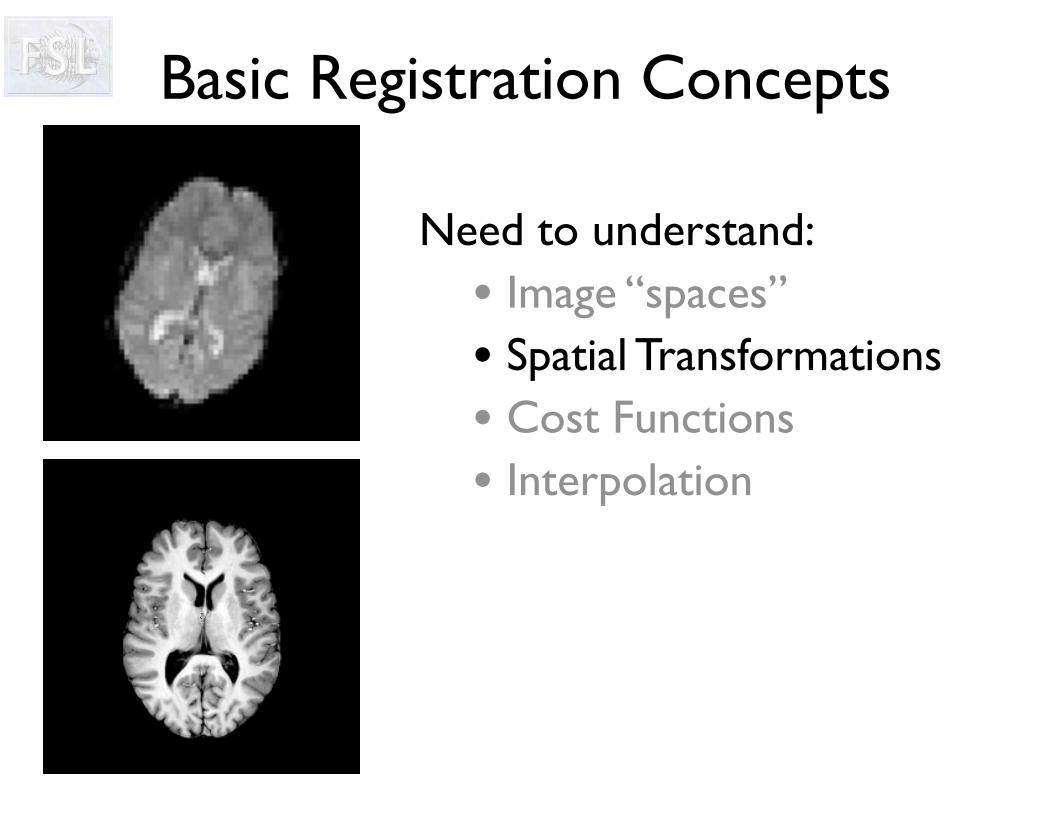
Need to understand:• Image “spaces” • Spatial Transformations• Cost Functions• Interpolation
Basic Registration Concepts

Basic Registration Concepts
Need to understand:• Image “spaces” • Spatial Transformations• Cost Functions• Interpolation

Standard Space• Common reference coordinate
system for reporting/describing
• Register all members of a group to this space for group studies
• Original Talairach & Tournoux coords based on one post-mortem brain
• Now use standard images based on non-linear group average (MNI152)
• MNI is not quite Talairach

Other “Spaces”
FMRI Structural Standard
• All images in the same “space” are aligned
• Different images ⇒ different “spaces” e.g. standard space, structural space, functional space
• Can have different resolution images in the same space e.g. 1mm and 2mm versions of standard space images
• Want to move image-related info between spaces e.g. a mask from standard space to structural space

Other “Spaces”
FMRI Structural Standard
• Need to registration between spaces (via images) and get the transformations before transforming/moving/resampling any image-related info (like masks or atlas ROIs)
• Can have versions of the same “image” (e.g. a mask) in several different spaces
• FSL tools (e.g. FEAT) often move things between spaces
TransformTransform

Other “Spaces”
FMRI Structural Standard
TransformTransform
Inverse Transform Inverse Transform
• Need to registration between spaces (via images) and get the transformations before transforming/moving/resampling any image-related info (like masks or atlas ROIs)
• Can have versions of the same “image” (e.g. a mask) in several different spaces
• FSL tools (e.g. FEAT) often move things between spaces

Image (Voxel) Coordinates
Voxel coordinates in FSL:• Integers between 0 and N-1 inclusive• Refer to the whole voxel• Origin in the lower-left corner: (0,0,0)
Confusingly, there are many types of coordinates
FSLeyes reports theseUsed by FSL commands & same as NIfTI coords
• Axes are not aligned with the anatomy• Cannot distinguish left from right by voxel coordinate values
y
x
z
Ny-1
Nx-1
Nz-1
0

Standard Space CoordinatesStandard Space coordinates in FSL:• Real numbers, in units of mm• Origin (0,0,0) near centre of image (anatomical landmark; e.g. anterior commisure)• Axes aligned with anatomy (left and right specified)
Several standard spaces exist: MNI, Talairach, BrainWeb, etc
y
x
z
0
A
P
RL
S
I
FSLeyes also reports these when possible
Need to understand:• Image “spaces” • Spatial Transformations• Cost Functions• Interpolation
Basic Registration Concepts

Spatial Transformations
• To align images must transform them
Examples:
Rigid Body (6 DOF)Affine (12 DOF)Non-linear (12 - millions DOF)
• Degrees of Freedom (DOF) partially describe transform
• Many types of transformation

Rigid-Body Transformations
• 6 DOF in 3D• Includes:
– 3 Rotations

Rigid-Body Transformations
• 6 DOF in 3D• Includes:
– 3 Rotations
Used for within-subject registrations
– 3 Translations

Affine Transformations
• 12 DOF in 3D• Linear Transf.• Includes:
– 3 Rotations– 3 Translations
– 3 Scalings

Affine Transformations
• 12 DOF in 3D• Linear Transf.• Includes:
– 3 Rotations– 3 Translations
Used for eddy current correction and initialising non-linear registration
– 3 Scalings
– 3 Skews/Shears

Non-Linear Transformations• More than 12 DOF• Can be purely local
• Subject to constraints:
– Basis Functions• e.g. B-Splines
– Regularisation
– Topology-preservation
Used for good quality between-subjectregistrations

Nonlinear Registration
Before Registration
Reference (MNI152)
Linear Registration
Non-Linear Transformations

Rigid body (6 DOF)- within-subject motion
Non-linear (lots of DOF!)- high-quality image (resolution, contrast) & same modality
of reference/template- better with a non-linear template (e.g. MNI152_T1_2mm)
Affine (12 DOF)- needed as a starting point for non-linear- align to affine template, or using lower quality images, or
eddy current correction Global scaling (7 DOF)
- within-subject but with global scaling (equal in x,y,z)- corrects for scanner scaling drift in longitudinal studies
More DOF is NOT always better (e.g. within-subject)
What transform/DOF do I use?

What do the transformations look like?
A =
�
⇧⇧⇤
a11 a12 a13 a14
a21 a22 a23 a24
a31 a32 a33 a34
0 0 0 1
⇥
⌃⌃⌅
An affine transformation is represented by these 12
numbers.This matrix multiplies
coordinate vectors to define the transformed coordinates.
A non-linear transformation can be
represented by a deformation field.

Non-linear deformation
mm
Deformation-field
x-component
y-component
The deformation-field (also called a “displacement-field” or just “warp”) is stored using images with one 3D image per component of the vector
NB: this is not the default output in FSL (the default is a coefficient file)

Non-linear deformationRegularisation, Warp Resolution and DOF
Spacing of points = warp resolution =
regularisation = DOF
• Various ways of controlling warp smoothness
• Less DOF = smoother
• Lower warp resolution = smoother
• Higher regularisation = smoother

Non-linear deformationHigh Regularisation Lower Regularisation
MNI
Input

Non-linear deformationRegularisation, Warp Resolution and DOF
Spacing of points = warp resolution =
regularisation = DOF
• Various ways of controlling warp smoothness
• Less DOF = smoother
• Lower warp resolution = smoother
• Higher regularisation = smoother
• Default warp resolution of 10mm is a good compromise for MNI152
• Between two subjects can use less smooth warps (less regularisation, higher warp resolution, more DOF)

Need to understand:• Image “spaces” • Spatial Transformations• Cost Functions• Interpolation
Basic Registration Concepts

Cost FunctionMeasures “goodness” of alignment
Seek the minimum valueSeveral main varieties
_ =
Similarity function is opposite (maximum sought)

FLIRT: Cost Functions
FMRIB’s Linear Image Registration Tool

FLIRT: Cost FunctionsImportant: Allowable image modalities
Less important: DetailsLeast Squares Same modality
(exact sequence parameters)
Normalised Correlation Same modality (can change brightness & contrast)
Correlation Ratio Any MR modalities
Mutual Information Any modalities (including CT, PET, etc.)
Normalised Mutual Info. Any modalities (including CT, PET, etc.)
BBR Within-subject EPI to structural (see later)

FNIRT: Cost Functions
FMRIB’s Non-linear Image Registration Tool

• Only uses Least Squares as cost functionso images must be of the same modality/sequence
• Also includes an explicit model for bias field (RF inhomog.)• Estimate displacement field and RF bias field together• Options exist to control bias field (turn off/on, smoothness)
Template With RF modellingWithout RF modelling
FNIRT: Cost Functions
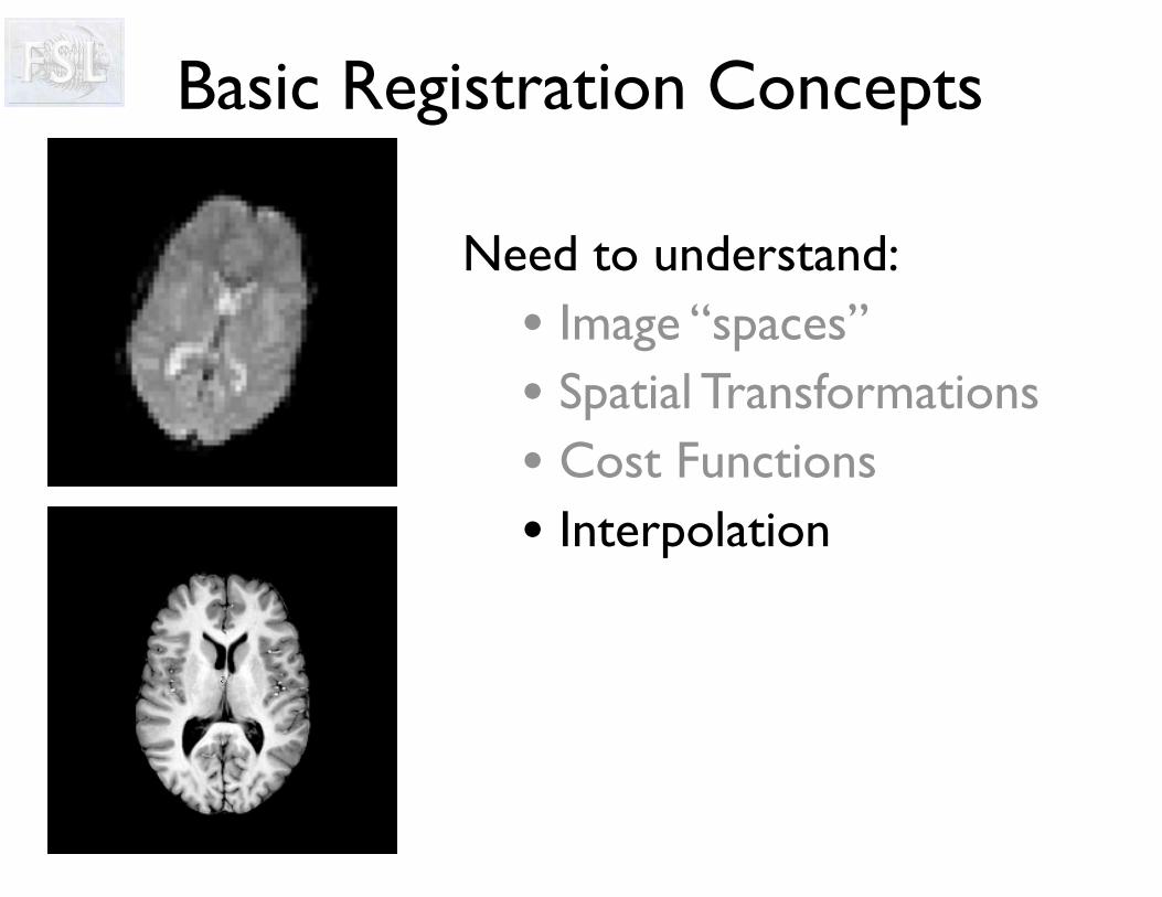
Need to understand:• Image “spaces” • Spatial Transformations• Cost Functions• Interpolation
Basic Registration Concepts

InterpolationFinds intensity values between grid points
Various types include• Nearest Neighbour• Trilinear• Spline• Sinc• k-Space methods
Fast, but blocky - can be used for discrete labels

InterpolationFinds intensity values between grid points
Various types include• Nearest Neighbour• Trilinear• Spline • Sinc• k-Space methods
Fast, with some blurring - most common option

InterpolationFinds intensity values between grid points
Various types include• Nearest Neighbour• Trilinear• Spline• Sinc• k-Space methods
Slower (spline is fairly fast) - creates sharp images but can create values outside the original range

• Affects accuracy of subsequent analysis• Important for quantitative imaging • Can affect size of artefacts
Interpolation
Nearest Neighbour Trilinear Spline

Applying Transformations
transformation
• Step 1: Estimating a transformation• finding the transformation• no resampling

transformation
• Step 2: Resampling• applying a transformation• thus creating a new, modified image
• Step 1: Estimating a transformation• finding the transformation• no resampling
• “Registration” can mean either
• Usually delay resampling as it reduces image quality
• Other terms: coregistration & spatial normalisation
Applying Transformations

• Mask values are normally 0 and 1 (integer format)• Interpolation gives values in between
if rounded to integer mask "shrinks”
• Ensure output datatype = float (applywarp & flirt default)• Re-threshold (binarize) the transformed mask
• "Correct" thresholding depends on the particular case• Threshold near 0.0 to include partial-volume edges• Threshold near 1.0 to exclude partial-volume edges• Threshold at 0.5 to keep the same size (approx)
Transforming Masks

0.1 Threshold0.5 Threshold0.9 Threshold
Transforming Masks

Overview
• Brain Extraction (BET)
• Registration concepts (FLIRT & FNIRT)
• Practical applications (FLIRT & FNIRT)• Single-stage registration• Multi-stage registrations• EPI distortion correction• Pathological image registration

Registration with FSLTwo main tools:
FNIRT & FLIRT(FMRIB’s Non-Linear/Linear Image
Registration Tool)
Both tools used by FMRI and Diffusion tools
(FEAT, MELODIC & FDT)
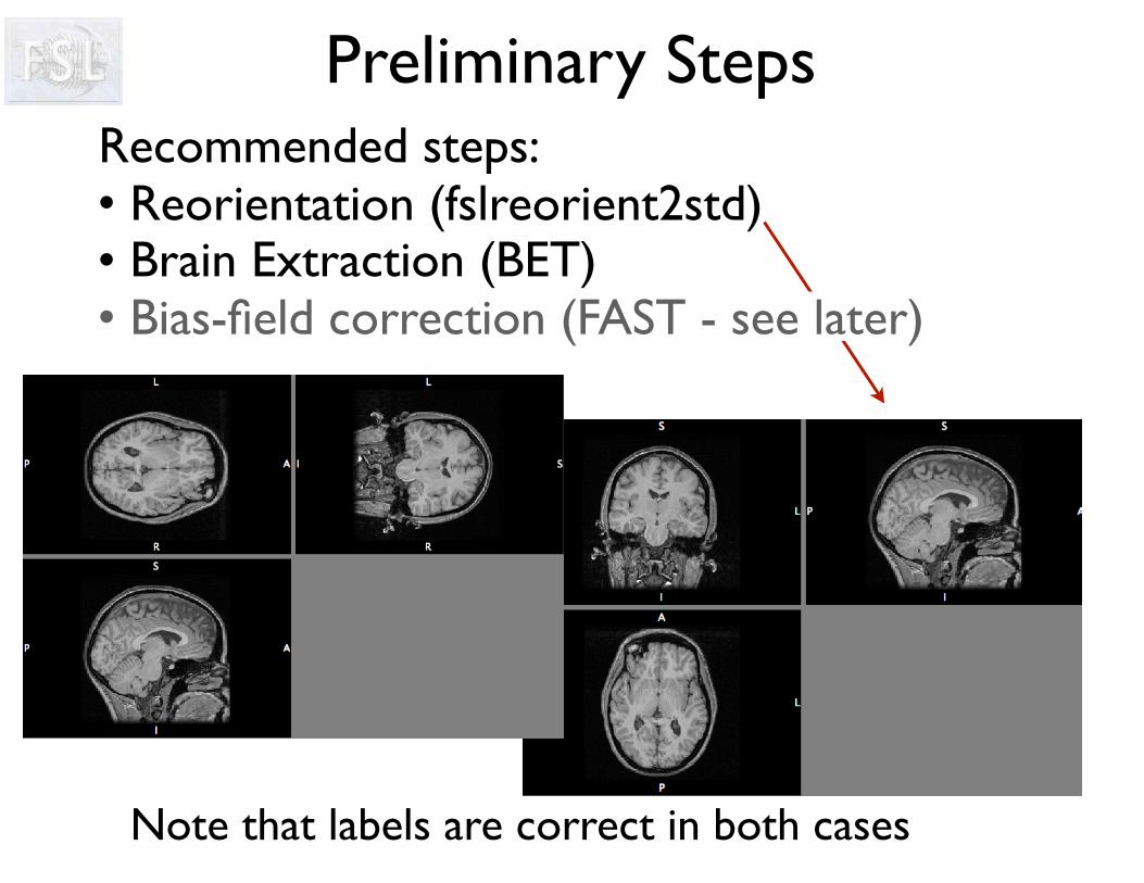
Preliminary StepsRecommended steps:• Reorientation (fslreorient2std)• Brain Extraction (BET)• Bias-field correction (FAST - see later)
Note that labels are correct in both cases
Overview
• Brain Extraction (BET)
• Registration concepts (FLIRT & FNIRT)
• Practical applications (FLIRT & FNIRT)• Single-stage registration• Multi-stage registrations• EPI distortion correction• Pathological image registration

Case Study
Scenario:Have two (or more) different types of images from
the same subjectFor example, T1-weighted and T2-weighted images
Objective:Have images aligned so that, for example, they can be
used for multi-modal segmentation
Solution:FLIRT with 6 DOF (rigid-body)

Single-stage Registration
BET
Input Reference
• Single subject ⇒ 6 DOF = FLIRT
• T2-wt to T1-wt multi-modal cost function
(e.g. default of correlation ratio)
• Run brain extraction on both images
• Choose image with better resolution or contrast as the reference
• Always check your output
⇒

Visual CheckAlways assess registration quality visually!Can use:• FSLeyes (using overlay or flicking between images)• slices for a static view use (as in FEAT)
slices T2_to_T1im T1im
✔
✘
Red edges from second image
Grayscale from first image

Registration in FSLInput Reference
• In FSL the reference image controls the FOV and resolution of the output image
• Transformations are given:
from input space to reference space
• Inverse transformations can easily be calculated to go from reference space to input space when needed
• Can overlay images in FSLeyes with different FOV or resolution: i.e. images can be in different spaces and resolutions
• Images can be resampled into a different space by applying a previously derived transformation

Troubleshooting
• Check that voxel size is correct for both images
• Check if brain extraction is OK: small, isolated errors are fine, but large or consistent shifts are not
• Limit search (if initially fairly well aligned)
• Try different (but still appropriate) cost functions
• Check for artefacts and pathologies (see later - cost function weighting)

Overview
• Brain Extraction (BET)
• Registration concepts (FLIRT & FNIRT)
• Practical applications (FLIRT & FNIRT)• Single-stage registration• Multi-stage registrations• EPI distortion correction• Pathological image registration

Case Study
Scenario:Doing a functional (or diffusion) studyHave EPI and T1-weighted of each subject
Objective:Need to register images to a common (standard)
space to allow the group study to be performed
Solution:2-stage registration with FLIRT & FNIRT (in FEAT)

Two Stage RegistrationRegistering very different images is difficult due to:• Differences in individual anatomies• Different contrasts in various modalities• Distortions which differ between images
6
DOF
FLIRT
DOF
>12
FNIRT**
To register an EPI to a standard space template (e.g. MNI152) use a structural intermediate image
Automatically done by FEAT GUI (some user control)Need to manually run brain extraction (not on EPI usually*)

Registration for FMRI Analysis (FEAT)
FMRI(implicit)
MainStructural
Standard
FLIRT
FNIRT
NB: actually need brain extracted and original images for FNIRT
FLIRT +

MNI Space
Registration Registration

Registration for FMRI Analysis

Registration for FMRI Analysis
Functional image in standard space: fmri in grey + red lines from MNI (standard space template)

Registration for FMRI Analysis
Example func (fmri) in highres (structural) space: top line = fmri in grey + red lines from structural bottom line = structural in grey + red lines from fmriAlso: fsleyes highres example_func2highres (in reg subdirectory of feat directory)


Registration for FMRI Analysis
Highres (structural) in standard space (MNI) top line = structural in grey + red lines from MNI bottom line = MNI in grey + red lines from structuralAlso: fsleyes standard highres2standard


Registration for FMRI Analysis
Example func (fmri) in standard space (MNI) top line = fmri in grey + red lines from MNI bottom line = MNI in grey + red lines from fmriAlso: fsleyes standard example_func2standard

Registration for FMRI Analysis
Core registrations are: example_func 2 highres + highres 2 standard
The example_func2standard is derived from these
Any failures need to be fixed in the core registrations (and then can run updatefeatreg)

Overview
• Brain Extraction (BET)
• Registration concepts (FLIRT & FNIRT)
• Practical applications (FLIRT & FNIRT)• Single-stage registration• Multi-stage registrations• EPI distortion correction• Pathological image registration

Case Study
Scenario:Doing a functional (or diffusion) study
Objective:Want to correct for distortions in EPI
as otherwise the registrations are inaccurate
Solution:Fieldmap-based correction using FUGUE/FEAT

Registration of EPI
Solution: - undo distortion by
“unwarping” - ignore areas of high
signal loss - needs a fieldmap
(special acquisition)
Problem: - EPI images distorted
and suffer signal loss - standard registration
does not work well
EPI T1-weightedanatomical

T1-weighted(aligned)
Signal Loss
Distortion

Courtesy of D. Greve, MGH
EPI is very sensitive to any deviations from a perfectly uniform B0 field
Air-tissue interfaces cause magnetic disturbances
A separate fieldmap image measures the B0 deviations
fieldmap EPI
B0 Field Inhomogeneities
distortionsignal loss

Using FieldmapsFrom the fieldmap image we get:
EPI
B0 Fieldmap
Only takes a few minutes to acquire one fieldmap - and it massively improves registration
Need a new fieldmap for each scanning session as it changes (e.g. it depends on head orientation)
Magnitude of spatial distortions • (phase-encode direction only)Estimate of signal loss
PE

Unwarping with FieldmapsFieldmap Original EPI
Unwarped EPI
Used to improveregistration of EPIand structural scan
It does not restoresignal in the frontal lobe

Unwarping with Fieldmaps
It does use fieldmap image to calculate distortion and “unwarp” EPI
Fieldmap Original EPI
Unwarped EPI
It does deweight areas with substantial signal loss in the registration
Used to improveregistration of EPIand structural scan
It does not do anythingabout motion correction
It does not restoresignal in the frontal lobe

Fieldmap AcquisitionFieldmaps are becoming standard sequences
Only takes a few minutes to acquire - best either immediately before or after EPI scans (but this is not crucial)
• Gradient Echo• Asymmetric Spin Echo• EPI• Blip-reversed b=0 pair (EPI)
✔✔✘✔
Each based on a pair of images with different TE (record these TE values)
Four main types of acquisitions:
Crucially requires the phase information (not only the magnitude, unlike the vast majority of other images)
Distortion & Signal Loss
Magnitude part of fieldmap Phase difference of images

• EPI to structural registration (Greve & Fischl, NeuroImage, 2009)• incorporates fieldmap correction (previously FUGUE)• used in FEAT (B0 unwarping)
Boundary-Based Registration (BBR)
T1w T1w + boundaries EPIEPI +
boundaries
• Uses white-matter boundaries (via T1w segmentation)• Need good structurals (not too much bias field)• Also requires anatomical contrast in the EPI• Driven by intensity difference across boundary (samples)
• More robust to pathologies and artefacts in EPI

Structural Image
Registration with Distortion Correction
Registration without Distortion Correction
Distortion Correction

Distortion Correction within FEAT

Fieldmap in rad/s
Need to prepare the fieldmap image: Fsl_prepare_fieldmap (for Siemens)
B0 Field
(rad/s)
=
Phase difference (rad)
TE difference
(sec)
Distortion Correction within FEAT

Fieldmap in rad/s
Fieldmap Magnitude... needs this ...
... and aggressive BET (leave no non-brain) for best performance
Input file = brain extracted file ... but also needs to find original*
Distortion Correction within FEAT

Fieldmap in rad/s
EPI echo spacing (ms)
Also called dwell time
Normally about 0.5-0.7ms
xxv
v
v
Time between echos in k-space
Fieldmap Magnitude
Divide value by any acceleration factor
Distortion Correction within FEAT

Fieldmap in rad/s
EPI echo spacing (ms)
EPI echo time (ms)
Normally about 30-40msat 3T
Fieldmap Magnitude
Distortion Correction within FEAT

Fieldmap in rad/s
EPI echo spacing (ms)
EPI echo time (ms)
Unwarp (PE) direction- Often A-P but can be anything- Cannot tell if it is + or -- Try both and see what works (see practical)
Fieldmap Magnitude
Distortion Correction within FEAT

Fieldmap in rad/s
EPI echo spacing (ms)
EPI echo time (ms)
Unwarp (PE) direction
Signal loss thresh %Ignore voxels with more than this signal loss in registration
Fieldmap Magnitude
Distortion Correction within FEAT

Fieldmap use in FEAT

This should be fairly uniform everywhere except where the field is not uniform - inferior temporal and frontal lobes
Fieldmap use in FEAT

This should be mostly yellow - red voxels get ignored in the registration (lots of red is bad)
Fieldmap use in FEAT

This shows how much each voxel moves - check that the range is sensible (anything from +/- 3 to +/- 20 is common)
Fieldmap use in FEAT

This shows the white matter edges from the structural on the fieldmap (to check fieldmap to structural registration - not EPI)
Fieldmap use in FEAT

Fieldmap to highres (structural)
Functional (EPI) to highres (structural)
- no correction
Fieldmap use in FEAT
Functional (EPI) to highres (structural)
- with fieldmap correction

Fieldmap use in FEAT
Functional (EPI) to highres (structural)
- no correction
Functional (EPI) to highres (structural)
- with fieldmap correction
Movie of EPI with and without correction

Fieldmap use in FEAT
Functional (EPI) to highres (structural)
- no correction
Functional (EPI) to highres (structural)
- with fieldmap correction
Look for areas where unwarping (correction) changes brain shape
Movie of EPI with and without correction

Fieldmap use in FEAT
Functional (EPI) to highres (structural)
- with fieldmap correction
Functional (EPI) to highres (structural)
- no correction
Look for areas where unwarping (correction) changes brain shape
Movie of EPI with and without correction
See if these areas are better aligned with or without correction but don’t trust borders with signal loss areasNB: Using FSLeyes is often better

BBR and Fieldmaps
Standard FLIRT

BBR FLIRT
BBR and Fieldmaps

BBR FLIRT with Fieldmap
BBR and Fieldmaps

BBR and Fieldmaps
Standard FLIRT

BBR FLIRT
BBR and Fieldmaps

BBR FLIRT with Fieldmap
BBR and Fieldmaps

Overview
• Brain Extraction (BET)
• Registration concepts (FLIRT & FNIRT)
• Practical applications (FLIRT & FNIRT)• Single-stage registration• Multi-stage registrations• EPI distortion correction• Pathological image registration

Case Study
Scenario:Have images containing a known pathology (or
artefact) which looks different in different images For example, some sequences (e.g. FLAIR) highlight
lesions that are hard to see in other sequences
Objective:Align the images based on the healthy tissue, but
“ignoring” the area of the pathology (or artefact)
Solution:Cost-Function Weighting (FLIRT or FNIRT)

Cost Function Weighting
Artefacts and pathologies introduce non-matching
image regions
Cost (similarity) functions assume that all of the
images can be matched
Use a weighting image to down-weight non-
matching regions
black=0; white=1
weighting imageweighting image

Cost Function Weighting• All FLIRT & FNIRT cost functions can be weighted
• Do not assign zero to the background as then the brain/background contrast is lost
- Large values for important areas/regions e.g. ventricular matching
• Voxel weights are relative, reflecting its importance in overall matching
• Weighting for reference image, input image or both
- Zero, or small, values for corrupted areas e.g. gross pathology or artefact

Troubleshooting Registrations
• Check the images: voxel sizes, artefacts, large bias field
• Check the brain extraction: look for large/consistent errors
• For EPI: acquire and use fieldmap to unwarp distortion
• For FMRI or diffusion: use multi-stage registration (e.g. via
GUIs) with a structural image for best results
• If pathologies/artefacts exist: use cost-function deweighting
• If images are nearly aligned: try limiting the search
• For FLIRT: can try different cost functions
• For FNIRT: check initial affine alignment is OK
• For small FOV: acquire whole-brain EPI for multi-stage reg

Advanced Registration
•2D - 3D Registration
•Severe Pathology
•Surface-based Registratione.g. connectivity-driven (FMRI)
•Other Image Modalitiese.g. diffusion imaging data MR Spectroscopic Imaging (MRSI)

That’s all folks

• Small FOV• Advanced non-linear registration (FNIRT)
Advanced Topics

Case Study
Scenario:Functional study using a small FOV (e.g. a few slices)Often done to obtain better resolution scans over ROI
Objective:Get activation results registered well to the full brain
(and standard space)
Solution:Scan one whole-brain EPI and use a 3-stage registration
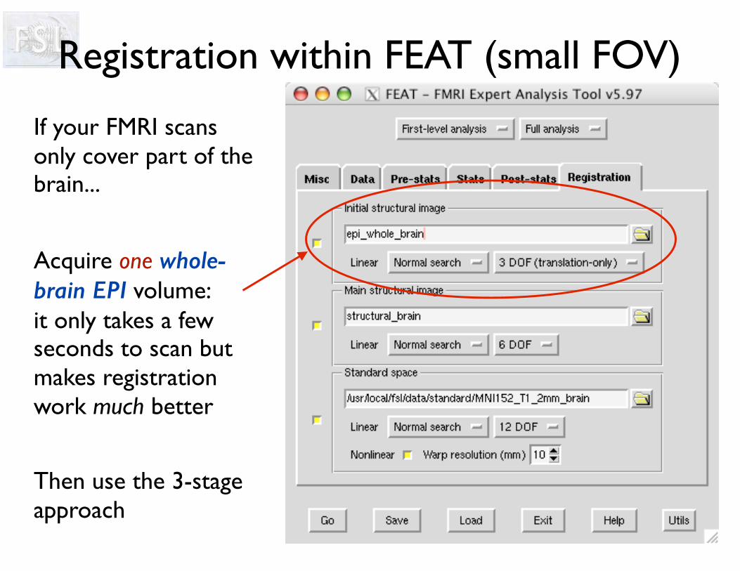
Registration within FEAT (small FOV)
Acquire one whole-brain EPI volume: it only takes a few seconds to scan but makes registration work much better
If your FMRI scans only cover part of the brain...
Then use the 3-stage approach

Partial Brain EPI & UnwarpingIn partial FOV studies, registration is massively improved by multi-stage registration:
3. Structural to Standard
2. Full Brain EPI to Structural• apply unwarping (full brain field map)
1. Partial Brain to Full Brain EPI• Desirable for full brain to contain
exactly the same slices so that registration is simple
Partial Brain FMRI timeseries
Full Brain Single Image
(an extra acquisition - but only takes seconds!)
All taken care of when using the FEAT GUI

Registration within FEAT (small FOV)

Registration within FEAT (small FOV)

Registration within FEAT (small FOV)

Advanced Topics
• Small FOV• Advanced non-linear registration (FNIRT)
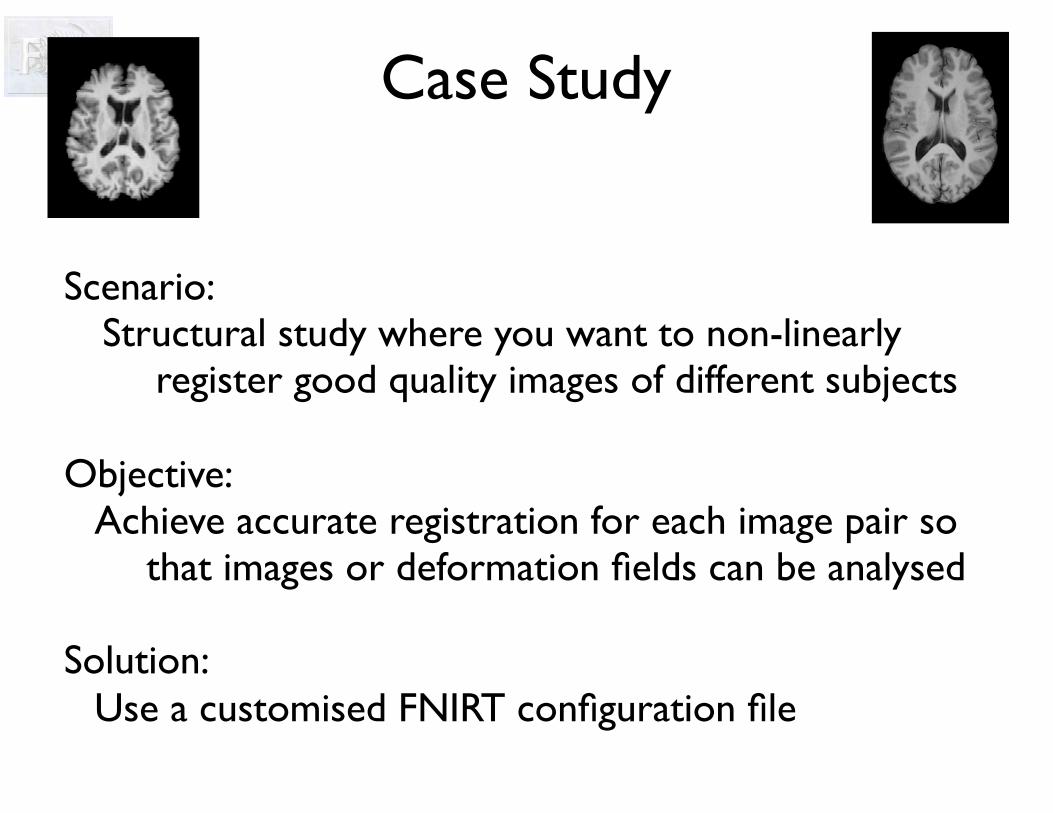
Case Study
Scenario:Structural study where you want to non-linearly
register good quality images of different subjects
Objective:Achieve accurate registration for each image pair so
that images or deformation fields can be analysed
Solution:Use a customised FNIRT configuration file

FNIRT Controls
FNIRT has many different controls available to the user:
• warp resolution (control/knot point spacing)
• cost function (bias correction or not)
• desired outputs (deformation field, Jacobian, etc)
• config file – advanced stuff!• multi-resolution scales• regularization strength• cost-function weighting• number of iterations
...

Multi-resolutionWhy use lower resolutions?• Faster calculations
• 8mm - 80 thousand calcs• 1mm - 40 million calcs
• Insensitive to fine structure
Progressively improve resolution during registration
8mm
1mm

Sub-sampleby 4
Sub-sampling in FNIRT
Ref
InputYielding these
warps
FLIRT matrix
M
Sub-sampleby 2
Sub-sampleby 1
Allows optimization to avoid distracting detail
4
2
1

Regularization
Affine λ=1000 λ=100 λ=10
Deformation-fields
• Need to prevent deformation from changing topology (folding/tearing/etc)
• Control “strength” of regularization vs image similarity (cost) by a constant, λ - often linked with multi-res
• Final results not too sensitive to λ