in-vitro characterization of human brain
TRANSCRIPT
IN-VITRO CHARACTERIZATION OF HUMAN BRAIN
MICROVASCULAR ENDOTHELIAL CELLS INFECTED WITH WEST
NILE VIRUS TO STUDY TIGHT JUNCTION INTEGRITY
A THESIS SUBMITTED TO THE GRADUATE DMSION OF THE
UNIVERSITY OF HAWAI'IIN PARTIAL FULFILLMENT OF THE
REQUIREMENTS FOR THE DEGREE OF
MASTER OF SCIENCE
IN
BIOMEDICAL SCIENCES (TROPICAL MEDICINE)
MAY 2008
By Yeung Y. Lo
Thesis Committee:
VlVek R. Nerurkar, Chairperson Saguna Verma
Frederic Mercier

We certify that we have read this thesis and that, In our opinion, It Is
satisfactory In scope and quality as thesis for the degree of Master of
Science In Biomedical Sciences (Tropical Medicine).
THESIS COMMITTEE:
Chairperson
ii

Dedications
I would like to dedicate this work to my loved ones who have given me their love
and support unconditionally and will motivate me for many more challenges to
come.
Tao Chih Lo
Lai Fong Kong
Sin Sin Lo
Ning Ning Lo
FaiLo
Aja Merkel Razumny
Roxanne Menade/ook
AnahShah
Robert Mourant
Louise Mourant
Michael TutteffOw
iii

Acknowledgments
I thank my committee members and the funding sources for making this project
possible. I would like to give special thanks to Dr. Saguna Verma for her day-to
day guidance, from the beginning to the end of this project, and beyond. I also
appreciate her patience and support throughout my time in the laboratory. My
funding sources included grants from the Centers for Biomedical Research
Excellence (P20RR018727), Research Centers in Minority Institutions Program
(G12RR003061), NCRR, NIH; and Hawaii Community Foundation (20050405).
In addition, I would like to dedicate my sincere appreciation to my fellow students
and co-workers who have helped me in many different ways to make this project
come through. Special thanks go to Ulziijargal Gurjav, Laami Submibcay, James
Kelley, Haiyan Luo, Stephanie Lum, Austin Nakatsuka, Pakieli Kaufusi, Moti
Chapagain, Becky Nakama and Juliene Co.
iv

Abstract
West Nile virus (WNV) is the predominant cause of viral encephalitis in the
United States today. Infection of neurons and induction of inflammatory
response are cardinal features of fatal WN encephalitis. WNV neuroinvasion has
been implicated to occur by hematogenous spread and possibly via the blood
brain barrier (BBB) interface. However, the precise mechanisms of viral entry
into the CNS through the brain vascular endothelium, key component of the BBB,
are unclear. Cellular and molecular modulations of tight junction proteins (T JP),
cell adhesion molecules (CAM) and matrix metalloproteinases (MMP) on brain
vascular endothelial cells in response to WNV infection were highly likely to
influence transmigration of WNV into the CNS. We hypothesized that WNV
could infect and replicate in human brain microvascular endothelial (HBMVE)
cells. Further we hypothesized that infection with WNV would lead to modulation
in the expressions of TJP, CAM and MMP, thereby leading to enhanced
migration of the virus across the BBB. Our results demonstrate that WNV could
efficiently replicate in HBMVE cells. In correspondence to the maximal viral
replication and virion release on day 2 after infection, WNV claudin-1
transcription and translation were significantly up-regulated. In addition,
significant induction of E-selectin and VCAM-1 transcription was detected.
VCAM-1 increase was further confirmed at post-translational level. WNV
selectively modulated the expressions of CAM and TJP, but none of the MMP
examined. These modulations are confirmed to be direct effects of WNV
replication as changes in CAM or T JP expressions were not observed in HBMVE
v
cells infected with UV-inactivated WNV. Our in vitro results suggest an active
role of brain endothelial cells in WNV neuroinvasion.
vi

Table of Contents
Contents Page
Dedications ...................................................................................... iii
Acknowledgements .......................................................................... .iv
Abstract ........................................................................................... v
Table of Contents .............................................................................. vii
List of Tables ...... '" ........................................................................... x
List of Figures ......................................................... '" ....................... xi
List of Abbreviations and Symbols ......................................................... xii
Chapter 1. Introduction
1.1 West Nile Virus and Human Diseases ........................................... 1
1.1.1 West Nile Virus ................................................................ 1
1.1.2 Epidemiology of WN Diseases ............................................ 4
1.1.3 Acquisition and Dissemination of WNV in Humans .................. 6
1.1.4 InfiammatoryWNNDand Pathology ..................................... 7
1.1.5 Immune Response to WNV Infection ................................... 1 0
1.1.6 Diagnosis. Treatment and Prognosis ................................... 14
1.1.7 Vaccines and Disease Prevention ....................................... 15
1.2 Blood-Brain Barrier and Viral Infection .......................................... 17
1.2.1 Introduction to BBB and Transmigration Pathways
Through BBB .................................................................. 17
1.2.2 Dysfunction of BBB in Viral Infections ................................... 20
1.2.2.1 T JP and BBB Permeability ................................ 20
1.2.2.2 MMP and TJP ................................................ 24
1.2.2.3 CAM and Immune Cell Transmigration ................ 25
vii

Chapter 2. Thesis Scope
2.1 Background for Research Question: Whether HBMVE cells play
a role in WNV-CNS entry? ................................................................... 32
2.2 Objectives and Hypothesis ........................................................ 32
2.3 Specific Aims ......................................................................... 33
2.4 Significance ............................................................................ 33
Chapter 3. Methods
3.1 Experimental Design for Specific Aim 1 ........................................ 34
3.1.1 Infection of HBMVE cells in 6-well plates and
on cover slips ................................................................ 34
3.1.2 Collection of culture supernatants, harvesting of cells
and fixation of cells on cover slips ...................................... 34
3.1.3 Detection of WNV on cover slips by immunofluorescence ....... 36
3.1.4 Quantitation of virus titer in supernatants by Plaque assay ...... 36
3.1.5 Quantitation of virus RNA copy in supernatant by qRT -PCR. ... 37
3.1.5.1 Extraction of viral RNA from supernatants ......... 38
3.1.5.2 Synthesis of cDNA from viral RNA
from supernatants ........................................ 38
3.1.6 Quantitation of virus RNA copy in infected HBMVE cells
by qRT-PCR ................................................................. 38
3.1.6.1 Extraction of cellular RNA from celllysates ........ 39
3.1.6.2 Synthesis of cDNA from cellular RNA ............... 39
3.2 Experimental Design for Specific Aim 2 ...................................... .40
3.2.1 Infection of HBMVE cells in 6-well plates, T-25 tissue
culture flasks and on cover slips ........................................ 40
3.2.2 Harvesting of cells and fixation of cells on cover slips ........... .40
3.2.3 Evaluation of transcription fold-change of genes
of interest by qRT-PCR. ................................................. .40
3.2.4 Detection of the expression of proteins of interest of
viii

bywestem blot.. ................................................ : ........... 41
3.2.5 Detection of proteins of interest on cover slips by
immunofluorescence ...................................................... .41
3.3 Materials ............................................................................... 43
3.3.1 HBMVE cells ................................................................ .43
3.3.2 WN virus ...................................................................... 43
3.3.3 Antibodies ..................... '" ...... '" ................................... 44
Chapter 4. Results
4.1 WNV infected and replicated in HBMVE cells ................................ 45
4.2 Replication-competent WNV, but not W-inactivated WNV,
differentially up-regulated T JP expressions in HBMVE cells ............. 54
4.3 Replication-competent WNV, but not W-inactivated WNV,
selectively induced CAM expressions in HBMVE cells .................... 59
4.4 Infection of HBMVE cells with WNV did not result in
MMP or TIMP expressions ........................................................ 64
Chapter 5. Discussion ................................................................... 65
References ..................................................................................... 72
ix

Tables
Table 1
Table 2
List of Tables
Page
Vaccines licensed or in clinical trials .................................... 17
Percent ofWNV-infected cells by IFS staining ....................... 46
x

Figures
Fig. 1
Fig. 2
Fig. 3
Fig. 4
Fig. 5
Fig. 6
Fig. 7
Fig. 8
Fig. 9
Fig. 10
List of Figures
Page
Schematic of WNV genome ............................................... 2
Schematic of WNV intracellular life cycle .............................. 3
Proposed interactions of junctional complex proteins ............. 23
Adhesion receptors involved in the diapedesis process .......... 29
WNV could infect and replicate in HBMVE cells .................... 46
WNV titer recovered from cell culture supematant quantitated by plaque assay ............................................ .48
WNV transcripts recovered from cell culture Supematant quantitated by qRT-PCR. ................................ 50
Intracellular WNV RNA transcripts quantitated by qRT-PCR .................................................................. 52
WNV differentially modulated T JP expressions quantitated by qRT- PCR, we and IFS ............................... 56
WNV selectively induced CAM expression quantitated by qRT-PCR, WB and IFS ................................. 61
xi

(k)Da
(s)PECAM
I-Ig
I-Im
ACBRI
AFP
AP
B(M)VEC
BBB
BSA
C
CAM
CCl-12
CCR5(l132)
CDC
cDNA
CMV
CNS
C02
CSF
D
DC-SIGN
ddH20
DEET
DNA
DNase
E
ECl
ECM
List of Abbreviations and Symbols
kilodalton
soluble platelet endothelial cell adhesion molecule
microgram
micron
Applied Cell Biology Research Institute
acute flaccid paralysis
alkaline phosphatase
brain (micro)vascular endothelial cells
blood-brain barrier
bovine serum albumin
capsid
cell adhesion molecule
Chemokine (C-C motif) ligand 2
chemokine (C-C motif) receptor 5 delta 32
Center for Disease Control and Prevention
complementary deoxyribonucleic acid
cytomegalovirus
central nervous system
carbon dioxide
cerebral-spinal fluid
day
dendritic-cell-specific, ICAM-3-grabbing non-integrin
double-distilled water
N,N'-Diethyl-m-toluamide
deoxyribonucleic acid
deoxyribonuclease
envelope
enhanced chemiluminescence
extracellular matrix
xii

EGF
EHV-1
h
HAD
HBMVE
HBV
HCMV
HIV
HSV
ICAM
IFN
IFS
IgMlG
IL
IP-10
IRF-3
JAM
MDCK
MHC
MIG
min
mL
MMP
MOl
MV
NeuroAIDS
NF-KB
NK
NMDA
NS
NY99
epidermal growth factor
equine herpes virus-1
hour
HIV-associated dementia
human brain microvascular endothelial
hepatitis B virus
human cytomegalovirus
human immunodeficiency virus
herpes simplex virus
intercellular adhesion molecule
interferon
immunofluorescence staining
immunoglobulin M or G
interleukin
Chemokine (C-X-C motif) ligand 10
Interferon regulatory factor 3
junctional adhesion molecule
Madin-Darby canine kidney
major histocompatibility complex
monokine induced by gamma interferon
minute
milliliter
matrix metalloproteinase
multiplicity of infection
measles virus
neuro aquired immunodeficiency syndrome
nuclear factor-kappa B
natural killer
N-methyl D-aspartate
nonstructural
New York 99
xiii

°C PBS PCR
PFA PFU prM
PVDF qRT-PCR
RNA rpm
RT
50 50S-PAGE
slV
TBs(T)
TIMP
TJP
TLR
TMEV
TNF
UV
VCAM
we WNF
WNNO
WNV
WNVE
degree Celsius
phosphate buffer saline
polymerase chain reaction
paraformaldehyde
plaque-forming unit
pre-membrane
polyvinylidene fluoride
quantitative reverse-transcriptase polymerase chain
reaction
ribonucleic acid
revolutions per minute
room temperature
standard deviation
sodium dodecyl sulfate polyacrylamide gel
electrophoresis
simian immunodeficiency virus
tris-bufferred saline (Tween)
tissue inhibitor of metalloproteinase
tight junction protein
toll-like receptor
Theiler's murine enceaphalomyelitis virus
tumor necrosis factor
ultraviolet
vascular cell adhesion molecule
western blot
West Nile fever
West Nile neurological disease
West Nile virus
Wes Nile virus encephalitis
xiv

Chapter 1. Introduction
1.1 West Nile virus and human diseases
1.1.1 West Nile virus
West Nile virus (WNV) is an arthropod-borne virus transmitted by mosquito
vectors first isolated from a febrile woman in the West Nile region of Uganda in
1937 (Smithbum K, 1940). It belongs to the Japanese encephalitis serocomplex
of the genus Flavivirus within the family of Flaviviridae (Brinton, 2002). WN
virions are spherical in shape with icosahedral symmetry. Its envelope measures
50 nm in diameter and it has a smooth spike-less outer surface (Mukhopadhyay
et a/., 2003).
WNV is a single-stranded positive-sense RNA virus of approximately 10.8 kb
genome size consisting of three structural proteins and seven non-structural (NS)
proteins. Structural proteins include (i) capsid (C) protein which is responsible for
viral RNA binding, (ii) membrane (prM) protein that blocks premature fusion and
chaperone envelope (E)-protein folding, and (iii) envelope (E) which mediates
viral attachment, and membrane fusion and viral assembly (Mukhopadhyay et a/.,
2005). The seven NS proteins are NS1, NS2A, NS2B, NS3, NS4A, NS4B and
NS5 (see Fig.1) (Mukhopadhyay et a/., 2003). The viral NS proteins regulate
viral transcription, replication and attenuate host antiviral responses (Samuel &
Diamond, 2006). The WNV genome is contained within a capsid, which is
surrounded by a host-derived lipid membrane embedded with virus-encoded E

and prM glycoproteins (Mukhopadhyay et aI., 2003). The E protein contains
epitopes recognizable by neutralizing antibodies and is responsible for receptor
binding and fusion with target cell membrane (Brinton, 2002; Chu et a/., 2005).
Structura l NCR NCR NCR
5{) )) )) C-prM-E NS1-NS2A-NS2B-NS3-NS4A-NS4B-NS5
POIYP~ ~o- 1 1 and post-translational modification Serine protease RNA polymerase
NTPase RNA helicase
Fig.1 Schematic of WNV genome [ Mukhopadhyay S. et aI., Nat. Rev. Microbial., 2005,3(1 ):13-22]
The linear positive-sense genome serves directly as messenger RNA with a
single open reading frame containing the necessary RNA-dependent RNA
polymerase, encoded by NS proteins, for replication (Brinton, 2002).
Immunoglobulin-like Domain III of WNV envelope protein is responsible for
binding to receptor on the surface of Vero and C6/36 mosquito host cells (Chu et
a/., 2005). While WNV receptor on neurons is unclear, a putative WNV receptor,
integrin av133, has been identified on non-neuronal cells such as Vero and CS-1
melanoma cells (Chu & Ng, 2004b; Lee et a/., 2006). Other molecules which
have been implicated in the attachment of WNV to cells in vitro include DC-SIGN
and DC-SIGN-R (Davis et aI., 2006a).
2
WNV enters host cells via clathrin-mediated endocytic pathway where virus is
transported toward the endoplasmic reticulum (ER) via lysosomal vesicles (Chu
& Ng, 2004a). At low pH, the viral envelope fuses with the lysosomal membrane
and viral RNA is subsequently released into the cytoplasm. Virus replication
occurs in the cytoplasm in close proximity to rough ER. Assembly of virions
takes place within the ER lumen, and the nascent virions are transported within
vesicles to the cell surface and released by exocytosis (Brinton, 2002). See Fig.
2.
.. RIlla CD'" 8IIIf/IJIg Md _-_...... PnIf8III rlllJ&' II'OLI
",,-.. - "...... - .. ,.. .-. II"-........ - .. ~ ...... - .. 1
Fig. 2 Schematic of WNV Intracellular life cycle. WNV attaches to susceptible cells through an as yet uncharacterized cell surface receptor and enters cells via receptor-mediated endocytosis. After acidification of the endosome, the E protein undergoes a conformational change that facilitates membrane fusion and nucleocapsid escape into the cytoplasm. Viral RNA binds to ribosomes in the cytoplasm and Is recruited to the rough endoplasmic reticulum where translation of the polyprotein from a single open reading frame ensues. The polyprotein is cleaved post-translationally by viral and host proteases. Translation, replication, and packaging are coupled processes and nascent viruses accumulate in membrane-derived vesicles prior to secretion by the cellular exocytosis machinery. [Diamond MS et ai, Viral Immuno/., 2003, 16(3):259-78]
3

1.1.2 Epidemiology of WN diseases
Since 1937, WNV has caused epidemics of febrile illnesses and sporadic
encephalitis throughout Africa, Europe, Asia and Australia (Hayes & Gubler,
2006; Murgue et al., 2002). In 1990s, fatal WNV encephalitis started to
frequently manifest in epidemics in Romania, Russia, and Israel. In 1999, WNV
infection was documented the first time in North America in New York City,
causing 59 cases of encephalitis and 7 deaths (Hayes & Gubler, 2006; Murgue et
al., 2002). Nucleotide sequencing of the envelope gene from isolated WNV
strain New York 99 (NY99) was analyzed and it was revealed to be closely
related (>99.8% amino acid homology) to an Israeli goose isolate in 1998
(Lanciotti et al., 1999). After its introduction to the U.S. in 1999, the virus has
spread dramatically westward across the United States, southward into Central
America and the Caribbean, and northward into Canada, resulting in the largest
epidemics of neuroinvasive WNV disease ever reported (Hayes et al., 2005a).
Specifically, the U.S. epidemic in 2003 was the largest WNV encephalitis
outbreak ever reported (Solomon, 2004). WNV is divided into lineages 1 and 2
based on genetic sequencing (Hayes & Gubler, 2006). Lineage 1 includes
pathogenic strains from North America, Europe, Australia, Africa, and Asia,
whereas lineage 2 includes only the less virulent strains from Africa (including the
original Uganda strain) and Madagascar (Lanciotti et al., 2002). Among lineage
1, WNV is categorized into four clades based on unique amino acids
polymorph isms or deletions in their envelope proteins (Brinton, 2002; Kuno et al.,
4

1998): Indian, Kunjin, A, and B (Beasley at al., 2004). Isolates from the US are in
clade B of lineage 1 and are closely related to strains from Israel.
WNV is enzootically maintained between more than 200 species of birds and
many species of mosquitoes (van der Meulen at al., 2005). In the U.S., different
species of mosquitoes play roles in WNV transmission in different parts of the
U.S. depending on the geographical location: Culax tarsalis and quinquafasciatus
in the West, Culax quinquafasciatus, and nigripalpus in the Southeast, and Culax
pipians and restuans in the Northeast. Mosquito acquires WNV from infected
blood through a blood meal, after penetrating the gut. WNV replicates in the
nervous system and salivary gland and persists throughout the life of the
mosquito without producing disease (Girard et al., 2005). Most birds support
high-titer of WNV replication in the blood, some can last for up to 100 days
without developing fatal illnesses, therefore the amplifying hosts (van der Meulen
at al., 2005). In naturally infected crows and blue jays, WNV has been detected
in the brains, livers, lungs, spleens, hearts and kidneys (Samuel & Diamond,
2006). Many other vertebrates are also susceptible to WNV infection but
neurological disease is only common in horses (Kleiboeker at al., 2004). It has
been suggested that the initial WNV introduction into New York city came from
infected migratory birds (Rappole at al., 2000), or imported exotic birds. Humans
can also contract WNV by means other than mosquito bites though at significant
lower probability. The less common routes of human WNV transmission include
transfusion of WNV-infected blood products (Kleinman at al., 2005; Pealer at al.,
5

2003; Stramer et aI., 2005), transplantation of WNV-infected organs (Bragin
Sanchez & Chang, 2005; Cushing et al., 2004; Wadei et al., 2004), vertical
mother-to-child transplacental spread (2002a), breast feeding of milk from WNV
infected mother (2002c) and accidental laboratory exposure (2002b).
Multiple epidemiological data suggests people with compromised immune
systems, including the AIDS and transplant patients (Guamer et a/., 2004), and
older age, being male, having underlying condition of hypertension, or diabetes
mellitus are among the high risk group to develop severe WNV diseases (Jean
CM, ; Murray et al., 2006). In addition, retrospective chart reviews of 172 WNV
cases hospitalized in Houston, Texas, between 2002 and 2004 indicates that
being African-American, being positive for hepatitiS 'C virus and having chronic
renal disease are risk factors for death after age adjustment (Murray et al., 2006).
Further, an underlying genetic risk factor is identified to be homozygosity for
defective CCR5 allele (CCR51132) in a cohort of WNV-infected Caucasian
patients from Arizona and Colorado (Glass at al., 2006). CCR5, critical
chemokine receptor, has been demonstrated to regulate leukocyte trafficking into
the brain for WNV clearance and is crucial for survival in mice infected with WNV
(Glass at al., 2005).
1.1.3 Acquisition and dissemination of WNV in humans
Humans acquire WNV intradermally from the saliva of infected mosquito upon
feeding. WNV is believed to be initially taken up by the skin Langerhans cells
6
(Chambers & Diamond, 2003), which then travel to and replicate in regional
draining lymph nodes where the virus enters the bloodstream for the first time
leading to primary viremia (Johnston et sl., 2000). Secondary viremia follows at
significantly higher titers as a result of WNV dissemination and replication in both
the primary and secondary lymphoid organs (Chambers & Diamond, 2003; Davis
et al., 2006b; Samuel & Diamond, 2006). Virus can be detected in blood as early
as 1 to 2 days after initial inoculation, and viremia typically lasts for a week
(Davis et al., 2006b). Common self-limiting clinical features of WNF include the
abrupt onset of fever, headache, and fatigue, with variable malaise, anorexia,
nausea, myalgia, lymphadenopathy, and a nonpruritic generalized
maculopapular rash (Campbell et al., 2002; Ferguson et al., 2005; Tilley et al.,
2007). Further, through un-identified mechanisms, WNV enters the CNS and
causes an array of West Nile neurological disease (WNND) including meningitiS,
encephalitis, and acute flaccid paralysis/polio-like myelitis (AFP) (Bode et sl.,
2006; Weiss et sl., 2001).
1.1.4 Inflammatory WNND and pathology
Severe outcomes of WN infection are the development of WNND. Postmortem
histologic examinations of WNV encephalitic brains demonstrate perivascular
inflammation, microglial nodules, variable necrosis, and loss of neurons (Guamer
et al., 2004; Kleinschmidt-DeMasters et aI., 2004). Among the CNS, the deep
gray nuclei, brainstem, and spinal cord show most pathology (Guamer et sl.,
2004; Kleinschmidt-DeMasters et sl., 2004). Microglial nodules, loss of anterior
7

hom cells and parivascular infiltration of T- and B-Iymphocytes in the spinal cord
is observed in patients with AFP (Kelley st a/., 2003; Kleinschmidt-DeMasters at
a/., 2004). Immunohistochemistry revealed that WNV mostly being localized
within neurons but also in glia (van Marie st a/., 2007) primarily in the brain stem
and anterior horns. WNV antigen staining is generally focal and sparse.
However, virus can be detected throughout the eNS and in other systemic
organs in immunosuppressed patients (Armah st a/., 2007; Guamer st a/., 2004).
Experimental eNS pathology studies of WNV infection in animal models also
report similar findings as in patients with WNVE, such as infection and injury of
brain stem, hippocampal, and spinal cord neurons but rarely in non-neuronal
tissues (Samuel & Diamond, 2006). Sequential histopathological events
unraveled in a mouse model with WNVE demonstrates that peripheral infection
and replication in the skin, spleen and kidney preceded infection of the eNS
(Garcia-Tapia et a/., 2007).
Acute or chronic neuroinflammation can result in various degree of BBB
breakdown and allow enhanced infiltration of circulating immune cells into the
eNS (Aktas st a/., 2007). Activation of glial cells is increasingly being recognized
to be important in the disease process (Aktas st a/., 2007). Microglia, the resident
immune cells of the brain, as well as astrocytes are capable of releasing a
diverse array of soluble factors, such as proinflammatory cytokines and free
radicals when activated by pathogens such as HIV-1, TNF-a and IL-1(3 are
secreted by HIV-1-infected and activated macrophages as well as microglia and
8

play critical role in HIV-associated dementia, which is characterized by leukocyte
infiltration, microglia activation, aberrant chemokine expression, blood-brain
barrier (BBB) disruption, and ultimately damage and death of neurons (Eugenin
at a/., 2006b). Further, excitatory neurotransmitter such as glutamate can act as
a neurotoxin if it is produced in excess or is not re-uptaken properly by astrocytes
(Tilleux & Hermans, 2007). As seen in NeuroAIDS, TNF-a and IL-113
synergistically over stimulate glutamate receptor N-methyl-D-aspartate (NMDA)
and leads to neuronal cell death by increased calcium influx (Brabers & Nottet,
2006).
In WN meningoencephalitis, infiltration of lymphocytes, perivascular
accumulation of macrophages and microglial nodules (Hayes & Gubler, 2006;
Hayes st a/., 2005b) are grossly observed in biopsies of diseased individuals.
Molecular pro inflammatory parameters such as cytokines and chemokines, MCP-
5 (or CCl12), IP-10 (or CXCl10), and MIG (or CXCl9), IFN-y and TNF-a
(Garcia-Tapia st a/., 2007), are found to be specifically triggered in experimental
WNV CNS infections It is recently shown that the expression of the WNV-NY99
capsid protein by implantation into the striatum of rats results in
neuroinflammation, together with induction of CXCl10 and diminished
expression of the protective astrocyte-specific endoplasmic reticulum stress
sensor" gene (OASIS), which is consistent with that observed in neurons and
astrocytes transfected with the same WNV capsid (van MarIe st a/., 2007). On
similar note, in vitro experimental infection of different human brain cell
9

populations confirmed that both neurons, and to a lesser extent glial cells are
susceptible to WNV infection and support replication at various degrees
(Cheeran at a/., 2005). Despite the borderline low level of WNV infection and
replication in astrocytes and microglia, robust immune mediators are released
including proinflammatory cytokines (ll-6, TNF-a), and chemokines (CXCl10,
CCL2, CCl5) which playa critical role in the recruitment of virus-specific T cells
into the CNS (Glass at a/., 2005; Klein at a/., 2005). In addition to
proinflammatory cytokines, the levels of a critical mediator of the inflammatory
cascade, macrophage migration inhibitory factor (MIF) is found to be increased in
the plasma and CSF of WNV-infected patients (Aljona at al., 2007a). Increased
MIF expression facilitates WNV neuroinvasion by compromising BBB integrity
and is associated with increased mortality in a mouse model (Aljona at al.,
2007a). Elevated levels of MIF has also been suggested to play roles in viral
pathogenesis, including dengue hemorrhagic fever (Chan at aI" 2006) and HBV
in patients (Zhang at al., 2002) and HCMV (Bacher at a/., 2002) and influenza
virus in vitro (Arndt at al., 2002).
1.1.5 Immune response to WNV Infection
Innate and adaptive immunity are both crucial in controlling WNV infection
(Diamond at a/., 2003a). Antiviral type I interferon (IFN-a and (3) production
mediated by RIG-I and MDA5 is essential in suppressing viral titers in the brain
and peripheral organs and is directly associated with reduced lethality in mice
(Fredericksen at al., 2008). In vitro treatment of cells, including primary neurons,
10

with IFN-a~ before or after WNV infection reduces viral titer and inhibits
cytopathology (Samuel & Diamond, 2005). Activation of the complement system
is required for the induction of protective antibodies against WNV, specifically
C1q, C3, C4, factor B, factor D and complement receptors 1 and 2 are critical in
controlling WNV infection in mice (Samuel & Diamond, 2005). Moreover,
altemative complement pathway is important for CD8+ T cell recruitment
whereas classical and lectin pathways are important in modulating both Band T
cells responses to WNV infection (Mehlhop at al., 2005). Toll-like receptor 3
(TLR-3), another pattem recognition receptor that recognizes intracellular dsRNA
to mediate type I interferon production via NF-K8 (Alexopoulou at al., 2001), has
been shown to trigger TNF-a signaling pathway, which leads to augmentation of
WNV entry into the brain to cause lethal encephalitis in mouse models (Wang at
al., 2004). Both humoral (Chung at al., 2007; Diamond at al., 2003b; Engle &
Diamond, 2003; Mehlhop & Diamond, 2008; Nybakken at al., 2005) and cell
mediated immunity (Brien at al., 2007; Klein at al., 2005; Shrestha & Diamond,
2004; Sitati at al., 2007; Wang at al., 2003a; Wang at aI., 2003b) are important in
WNV clearance. Early induction of neutralizing IgM is particularly crucial in
limiting viremia and WNV dissemination into tha brain which protects against
lethality. Anti-WNV IgM induction later also plays role in anti-WNV IgG response
(Diamond at al., 2003b). Passive transfer of WNV antibody prior to viral
challenge has been shown to confer protection in immunocompetent mice,
however humoral immunity is insufficient in rescuing RAG1 knockout mice which
lack function T and B cells from WNV infection (Diamond et al., 2003a; Engle &
11

Diamond, 2003). One mechanism of clearance of WNV-infected cells is by Fc
gamma receptor 1- and/or IV-mediated phagocytosis through monoclonal IgG
antibodies recognition of cell surface-associated NS1 (Chung at a/., 2007). Both
CD 4+ (Sitati & Diamond, 2006), CD 8+ cytotoxic T cells (Brien at a/., 2007;
Klein at a/., 2005; Shrestha & Diamond, 2004; Shrestha at a/., 2006; Sitati at a/.,
2007; Wang at a/., 2003b) and IFN-y-producing gamma delta (y6) T cells (Wang
at aI., 2003a) play roles in WNV infection control. In wild-type mice, yi5-T cells
expanded significantly during WNV infection and produced IFN-y. Adoptive
transfer of IFN-y-producing yi5-T cells to knockout mice increase survival of
infected mice (Wang at aI., 2003a). While IgM appears to be crucial factor to
restrict viremia, animals lacking CD8+ T cells or MHC class la have functional
humoral response compared to animal which have CD8+ T cell but bear higher
viral loads in the CNS have higher mortality rates (Shrestha & Diamond, 2004;
Wang at a/., 2003b). Adoptive transfer of naive, unprimed splenic CD8+ T cells to
RAG-KO animals (which lack mature T and B cells) appear to be sufficient in
providing protection against lethal dose WNV infection (Engle & Diamond, 2003).
On the other hand, CD8+ cytotoxic T cells directed against specific WNV antigen
peptides have also been shown to carry out clearance (Brien at a/., 2007; Purtha
at a/., 2007). A study demonstrated that one way CD8+ T cells cleared WNV
from infected neurons is mediated by perf orin pathways (Shrestha at a/., 2006).
Migration of activated CD8+ T cells into the brain is important for viral clearance
as a mouse model that lack CD8+ T cells or major histocompatibility complex
12

class la (MHC-I) antigens, antigen peptide presenting complex to CD8+ T cells,
shows up to 1,000-fold higher titers of WNV in the CNS and increased mortality
after WNV inoculation (Shrestha & Diamond, 2004). CD4+ T cells also
participate in the WNV immunity in promoting the maturation of antibody
response but also in facilitating CD8+ T cells trafficking into the brain by binding
of CD40 and CD40 ligand (CD40l) on activated CD4+ T cells (Sitati et a/., 2007).
Further, for cell-mediated immunity to function optimally, proper immune cells'
trafficking is necessary. Of note, infiltration of activated CD4+ and CD8+ T cells,
NK cells and macrophages relies on chemokine production by infected tissues,
such as CXCl10 by neurons and the receptors CXCR3 (Klein et a/., 2005), as
well as the expression of chemokine receptors and CCR5 and its ligand CCl5
(Glass et a/., 2005).
Immune evasion mechanism has been described in vitro experiments with WNV.
Specifically, WNV envelope protein (WNV-E), independent of TlR-3, blocks the
production of antiviral and poly (I:C)-induced proinfiammatory cytokines TNF-a,
Il-6, and IFN-(3 in murine macrophages at the level of receptor-interacting protein
1(RIP1) to mediate the activation of NF-KB and IFN regulatory factors (lRF)
(A~ona et al., 2007b). Another study using WNV replicon suggests that WNV
RNA replication and/or protein expression interferes with poly (I:C)-mediated IFN
gene induction in Hela cells (Scholle & Mason, 2005). Yet another report
indicates that WNV delays activation of interferon regulatory factor 3 (IRF-3) by
RIG-I early in infection to allow for virus replication (Fredericksen & Gale, 2006).
13

1.1.6 Diagnosis, treatment and prognosis
Virus isolation, detection of viral nucleic acid or antigen and seroconversion are
all recognized methods for diagnosis of recent WNV infection (Davis at a/.,
2006b), however, each method has its pros and cons. Due to biosafaty issues,
WNV isolation is not the first choice for WNV diagnosis. Viral nucleic acid
detection by polymerase chain reaction (PCR) amplification is useful for
screening blood products for potential contamination but not reliable enough for
diagnosis purposes due to the disappearance of viremia at the time disease
onset when the patient would submit blood samples (Davis at a/., 2006b).
Detection of WNV-specific antibodies in serum and CSF using commercially
available enzyme-linked immunosorbent assays (ELISA) are the methods of
choice for diagnosis of WNND (Tardei at a/., 2000). However, cross reaction
may result from previous exposure to other flaviviruses and/or recent vaccination
for yellow fever or Japanese encephalitis. Plaque reduction neutralization assay
will be necessary to exclude cross reaction and to confirm positive ELISA results
(Hayes & Gubler, 2006). Currently there is no guaranteed treatment for WNV
disease. However, multiple treatment regimes have shown promises and are
under clinical trials. Among which include treatments with IFN-a, corticosteroids,
and WNV-specific IgM derived from donor plasma with high levels of WNV
antibodies (Murray at al., 2006), however the efficacy and safety are undergoing
evaluation. Case report has documented successful treatment with IFN-a2b 3
weeks after WN meningoencephalitis onset in an 83-year old man (Lewis &
Amsden, 2007). Antisense nucleic acid compounds designed to inhibit WNV
14

replication have shown significant antiviral activity in vitro (Deas at a/., 2005;
Torrence at a/., 2006) and are now being tested in humans in clinical trials.
Potential options for therapy are continuously being discovered- high-throughput
screening of compounds for antiviral activity has identified triaryl pyrazoline as an
inhibitor of flavivirus replication in cell culture (Goodell at a/., 2006; Puig
Basagoiti at a/., 2006) and experiments in mice indicates TNF-a antagonist might
be worthy as a therapy to prevent WNV neuroinvasion (Wang at aI., 2004).
Average recovery time for mild form of WNF sufferers is one week post onset of
symptoms but prognosis for patients with severe WNND diseases is poor (Davis
at a/., 2006b; Hayes & Gubler, 2006). Case fatality rates of WNND range from 4-
15% (Green at a/., 2005; Gubler, 2007). Full recovery is to be expected in those
who have WN meningitis with no focal neurologic involvement (Hayes & Gubler,
2006); however, individuals with WN encephalitis or AFP may experience severe
and prolonged deficits, such as fatigue, weakness, aching, depression
parkinsonism and tremor in AFP patients are common and it could last from
weeks to months or remain for life (Hayes & Gubler, 2006).
1.1.7. Vaccines and disease prevention
As of today, there is no specific WNV vaccine licensed for humans use; however,
extensive efforts in WNV vaccine development have yielded significant progress.
Two chimeric virus (WNVNFV and WNVIDEN4) vaccines, one recombinant DNA
plasmid vaccine and one killed virus vaccine are under phase I or II clinical trial
(Table 1) for human use. The two chimeric vaccines encompass the
15

incorporation of WNV E and PrM DNA sequences into a 17 -D yellow fever or a
dengue serotype-4 virus backbone. Other underdevelopment vaccines include
DNA vaccines containing WNV antigen or Kunjun antigen expression, and
recombinant vaccine using measles virus as a vector for WNV antigens (Hayes &
Gubler, 2006). Another chimeric vaccine of fused bacterial f1agellin (STF2 Delta)
to the WNV envelope E-III domain has shown prominent innate and adaptive
immunogenicity in mice without adjuvant (McDonald et aI., 2007). For veterinary
use, five WNV vaccines have been licensed for horses and domestic geese
(Kramer et a/., 2008) (Table 1).
On the individual level, avoidance of mosquito bites by limiting outdoor exposure
is the best preventive measure for not contracting WNV (Gubler et a/., 2000).
Otherwise, insect repellent should be worn on clothing and skin, especially during
peak feeding hour from dusk to dawn. Using N, N-diethyl-m-toluamide (DEET)
containing repellents are the most safe and effective (Fradin, 1998; Hayes &
Gubler, 2006). Alternatives include oil of lemon eucalyptus, soybean oil, and
picaridin for skin and permethrin for clothing (Fradin & Day, 2002). On the
community and state level, vector control surveillance using larvicide and
adulticide can be effective in reducing mosquito populations and human WNV
infection (Barnard & Xue, 2004; Fradin & Day, 2002). Additional preventive
measures should be taken in the screening of donated blood in WNV endemic
areas to prevent transmission through transfusion (Custer et a/., 2004).
16

Table 1. Vaccines licensed or in clinical trials
Ptodua: ruune ComptlIlyondlor Insdtute. . .. I· .. ·.vtu:cinetype Stllt\IJi
Innovator® Fort Dodge Animal Health Killed virus L Recombitek® Merial Recombinant canarypox L
virus PreveNile TM Intervet Chimeric virus L
(WNVIYFV)
NA Kimron Veterinal}' Killed virus L Institute/Crucell
NA CDCIFort Dodge Animal Recombinant DNA L Health plasmid
Chimeravax"IM-West Nile Acambis Cbimeric virus CT-Il (WNVIYFV)
VRC-WNVDNA01O-OO-VP NIAIDINlli Recombinant DNA CT-I plasmid
WNIDEN4-3'deIta30 NIAIDINlli Chimeric virus CT-I (WNVIDEN4)
NA Crucell Killed virus CT-I
AbbreYi.tiODS: NA, inlb"""tion not ... U.ble; L, licensed for Veterinary 11'ie; YFv. yellow fever virus; Gr-II, clinicol triaJ, phose ll; cr-I, clinicol triaJ, phase I; DEN4, dengue4vinrs. Sources of infonlbltion: httpIlwww.fortdodgellvestock.coml, httpI/www.merlaLcoml, htlplllwww.Jme\WtlllllLcomf,httpllwww.cruceII.cnmI, htlplllwww.cllniaJltrials.goYl.
( Kramer LD et al., Annu Rev Entomol, 2008, 53: 61-81).
1.2 Blood-brain barrier (BBB) and viral Infection
1.2.1 Introduction to BBB ·and transmIgration pathways through BBB
Multiple brain barriers exist presumably to protect the central nervous system
from the dynamic fluctuation of biochemical environment in the peripheral
systems and to thus to sustain homeostasis within the CNS. The brain is
biochemically segregated from the periphery by three major barriers: the blood-
brain barrier in the brain parenchyma, the blood-CSF barrier in the choroid
plexus-epithelial and the blood-subarachnoid epithelial interfaces. During the
discovery of barrier between the brain and blood, the impermeable properties
17

were profoundly observed in the vasculature within the cortex and cerebellum
compared to capillaries of heart and skeletal muscles (Reese & Kamovsky,
1967). Following the initial finding, extensive studies have been generated on
BBB characteristics since.
The BBB is a highly regulated interface which separates blood borne entities
from the CNS. Unlike peripheral vascular endothelial cells, the brain vascular
endothelial cells (BVEC) of the BBB are not fenestrated. They have a higher
mitochondrial volume fraction and higher electrical resistance but a low number
of vesicles; in addition, they have specialized transport systems (Irie & Tavassoli,
1991). Under physiologic conditions. the BBB maintains homeostasis by ensuing
adequate supply of necessary nutrients such as oxygen and glucose for brain
cells as well as low level of surveillancelreplacement immune cells from blood
(Petty & Lo, 2002). A complete neurovascular unit conferring the BBB property
consists of vascular endothelium, perivascular astrocytes, basement membrane,
and pericytes that are in physical proximity to the endothelium (Persidsky et a/ .•
2006). Among these various components, brain vascular endothelial cells
(BVEC) are in the frontline directly interacting with a large variety of blood
constituents and regulating their passage into the brain (Petty & Lo, 2002). A
functional polarity exists in BVEC between the luminal (or apical) membrane
where tight junctions are located and the abluminal membrane where the
adherens junctions lie just above the basement membrane (Petty & Lo, 2002).
Passage of molecules across the endothelial cells can occur through the cells
18

(transcellular) or between adjacent cells (paracellular) (Persidsky at a/., 2006).
Factors that govern solutes crossing the BBB depend on whether the solute is
lipid soluble or charged and size. Lipophilic compounds with a molecular size
less than 15 AO (-3.5kDa) (Rubas at a/., 1996) such as nicotine, ethanol and
heroin can readily cross the BBB by passive diffusion while charged molecules
such as sodium ions or proteins cross the BBB slowly or not at all (Oldendorf,
1976). Crossing of glucose and amino acids require specific transporters present
on endothelial cells (Vorbrodt at a/., 2001; Xiang at a/., 2003). Receptors systems
have been found for several molecules including insulin, low-density lipoprotein,
insulin-like growth factors and transferrin (Banks, 2004; Moos at al., 2000;
Persidsky at al., 2006). Other than solutes, pathogenic microbes and immune
cells also enter the CNS through the BBB via transcellular andlor paracellular
crossing. Barrier properties of the BBB is attributed primarily to the presence of
tight junctions between endothelial cells and the few number of pinocytotiC
vesicles in the BVEC compared to vascular endothelial cells in other organs such
as the heart and skeletal muscles (Reese & Kamovsky, 1967). While pinocytotic
vesicles are associated with the ferrying of small amounts of fluid and solutes
across the cell wall, tight junctions between BVEC cells form extremely tight
seals characterized by the presence of tight junction proteins which disable
paracelluar passage of molecules (Banks, 2004; Moos at al., 2000; Persidsky at
al., 2006; Petty & Lo, 2002). Regulation of brain vasculature function is not only
by the biochemical milieu in the blood stream, but also by neuron and glial cells
19

regulate based on the metabolic requirements and changes in microenvironment
in the brain (Petty & Lo, 2002).
1.2.2 Dysfunction of BBB in viral infections
1.2.2.1 Tight junction proteins (T JP) and BBB permeability
BBB endothelial tight junctions are composed of a complex and elaborate
combination of (1) transmembrane and (2) cytoplasmic proteins linked to an
actin-based cytoskeleton (Petty & Lo, 2002). Tight junction transmembrane
proteins include claud ins, occludin and junctional adhesion molecules (JAMs).
Cytoplasmic proteins consist of zonula occludens (ZO) -1, 2, 3 and cingulin. See
schematic for distribution and proposed interactions between T JP in Fig. 3.
(1) Transmembrane (Integral) proteins
Claud ins belong to a superfamily of transmembrane proteins of size between 20-
24 kDa with four transmembrane segments (Petty & Lo, 2002). They form
dimmers and bind homotypically to other claudin molecules on the adjacent cell
membrane to form the primary seal. They also bind to and localize another
transmembrane protein occludin to the cellular junction (Kubota et a/., 1999).
Claudins are believed to be the primary backbone of tight junction strands while
occudin serves to enhance tightness (Kubota at a/., 1999). At least 24 claudins
proteins have been identified in mouse and humans (Persidsky at aI., 2006; Petty
& Lo, 2002), but their expression patterns vary among tissues; claudin-1, -3, and
20

-5 have been detected in cerebral microvascular endothelium (Hawkins & Davis,
2005).
Occludin is a 65-kDa transmembrane protein with four transmembrane domains
with both the amino and carboxy terminus located intracellularly. The cytoplasmic
C-terminal domain provides occludin connection with the cytoskeleton via
accessory proteins, ZO-1 and ZO-2 (Persidsky at a/., 2006). It has multiple
phosphorylation sites and the phosphorylation regulates its interaction with the
cell membrane proteins and regulates barrier permeability (Hirase at aI., 2001;
Sakakibara at a/., 1997; Wachtel at a/., 1999). Occludin is highly expressed in
BMVEC (Hirase et a/., 2001; Vorbrodt at a/., 2001) and is suggested to contribute
to the electrical resistance across the BBB as that high levels of occludin has
been shown to ensure high electrical resistance of epithelial cell monolayers
(McCarthy at a/., 1996).
JAM-1, -2, and -3 are IgG superfamily proteins. JAM is a transmembrane protein
with a singular domain of 40 kDa size (Martin-Padura et a/., 1998). JAM proteins
are expressed on endothelial and epithelial cells and on the surfaces of
leukocytes, erythrocytes and platelets (Mandell & Parkos, 2005). JAM mediates
cell-to-cell adhesion by means of homophilic and heterophilic interactions. Short
cytoplasmic tails of JAMs have been reported to bind to cytoplasmic protein such
as ZO-1 (Ebnet et a/., 2000). Though the direct role and mechanism of JAM in
the tight junction has not been identified, JAM has been implicated to involved in
21

a wide range of physiologic functions, including barrier function, leukocyte
migration, platelet activation, angiogenesis, and reovirus binding (Mandell &
Parkos, 2005).
(2) Cytoplasmic accessory proteins
Zonula occludens (ZO)-1,-2, -3 proteins are important cytoplasmic tight junction
phosphoproteins of approximately 160 kDa to 220 kDa in size and form
submembranous plaque of tight junctions (persidsky at al., 2006). They belong to
the membrane-associated and guanylate kinase proteins (MAGUK) family
(Anderson at al., 1995). ZO proteins contain PDZ and SH3 domains which are
involved in bridging transmembrane proteins such as claudins and occludin to
cytoskeletal proteins actin (Fanning at al., 1998).
Cingulin is another cytoplasmic phosphoprotein of tight junctions. It is about 140
kDa to 160 kDa is size and has been localized on the cytoplasmic surface.
Binding partners of cingulin include ZO-1, -2, -3, myosin and AF-6 (Petty & Lo,
2002). It is suggested that cingulin functions as a scaffold protein to link cytolysis
plaque proteins, i.e. ZO-1, -2, and -3, to the cytoskeletal protein myosin
(Cordenonsi at al., 1999; D'Atri at al., 2002).
22

Adherens
Fig. 3. Proposed interactions of junctional complex proteins in the capillary endothelial cells of blood-brain barrier. At tight junctions, claud Ins have been proposed to function as the backbone by binding to claudins on adjacent cells and thus forming a seal. Claudlns also bind to occludln, which functions as a regulatory protein. Occludin's presence in the membrane correlates with increased electrical resistance across the membrane and decreased permeability. Occludin Is thought to be Involved also in the fence function of tight junctions. JAM Is Involved in cell-cell adhesion and contributes to permeability control. A series of cytoplasmic proteins Z0-1, ZO-2 and Z0-3 form the submembranous plaque of tight junctions. These proteins belong to the MAGUK family (membrane-assoclated guanylate kinase proteins) and are involved In the coupling of transmembrane proteins to the cytoskeleton actin. Several cytoplasmic accessory proteins have been Identified at tight junctions, including AF6, 7H6 and cingulin; see the text for their potential structural and functional roles. The adherens junctions are composed of transmembrane glycoprotelns of the cadherin super family, which are linked to the cytoskeleton via cytoplasmic anchor proteins,l3-catenln, y-<:etenln (plakoglobin) and p120ctn that belong to the Armadillo protein family. poeatenin and y0{;8tenin bind to a-<:etenin, which Is an actin-binding and actin-bundling molecule linking the adhesive cadherinlcatenin complex to the F actin-based cytoskeleton. [Petty MA and Lo EH, prog Neurobiol. 2002, 66 (5):311-23)
The expression and proper distribution of T JP are important and have direct
effects in conferring BBB integrity. Increased permeability is the major
impairment or form of BBB dysfunction in many viral infections, especially in
those which manifest acute neuroinflammation such as meningitis and
23

encephalitis (Afonso at a/., 2007; Annunziata, 2003; Luabeya at a/., 2000). HIV
associated damentia (HAD) has been extensively studied and much is being
learned about this devastating disease. TNF-a has been demonstrated to
augment BBB permeability and ease HIV-1 traversal through the paracellular
pathway (Nottet et a/., 1996). Increased BBB permeability in HIV has been
suggested to be a result of degradation of T JP including ZO-1, -2, claudin-1, -5
and occludin (Andras at a/., 2003; Boven at a/., 2000; Dallasta at a/., 1999;
Kanmogne at a/., 2005; Nottet at a/., 1996). Similarly, decreased expression of
ZO-1 protein has been shown in extended length of cerebral microvessels in SIV
infected rhesus macaques (Luabeya at a/., 2000). In addition, increased
permeability of endothelial monolayer model has been demonstrated in HCMV
infection. Actin stress fiber formation and decreased expression of occludin and
vascular endothelial cadherin were observed (Bentz at a/., 2006).
1.2.2.2 MatTix metalloprotelnases (MMP) and TJP
The basal lamina of BVEC is composed of typical extracellular matrix (ECM)
components collagens, laminin, fibronectin, entactin, tenascin, thrombospondin,
heparin sulfate proteoglycans, and chondroitin sulfate proteoglycans (del Zoppo
& Hallenbeck, 2000; Lyons & Jones, 2007; Rascher at a/., 2002). Interactions
between matrix proteins and endothelial T JP can result in the modification of tight
junction functions. Collagen type IV, fibronectin, and laminin can influence the
expression of endothelial T JP such as occludin (Savettieri at a/., 2000).
Disruption of the ECM is strongly associated with increased BBB permeability in
24

pathological conditions (Jian Liu & Rosenberg, 2005; Rascher et aI., 2002). In
the CNS, matrix metalloproteinases (MMP), a family of over 20 zinc-dependent
structurally related endopeptidases, have been shown to degrade components of
the basal lamina (Leppert et a/., 2001), leading to disruption of the BBB, and
contribute to the neuroinflammatory response in many neurological diseases
(Ichiyama et a/., 2007; Jian Liu & Rosenberg, 2005; Leppert et a/., 2001; Mandai
et a/., 2003; Rosenberg, 2002; Saadoun et a/., 2007). Pre-inflammatory
cytokines such as TNF-a/p, IL-1 and -2 have been demonstrated to modulate the
expression and regulation of MMP (Ben David et a/., 2008; Lockwood et a/.,
2008; Seguin et a/., 2008). Increase in MMP-1, -2 and -9 activities were
associated with the reduction in collagen IV content, decreased barrier integrity,
enhanced permeability, and monocyte migration across the BBB monolayer
model treated with ethanol or acetaldehyde (Haorah et a/., 2008). Further,
elevated MMP-2, -7, and -9 expressions have been associated with HAD,
influenza virus and HCV, T JP degradation and BBB disruption (Conant et a/.,
1999; Eugenin et a/., 2006b; Ichiyama et a/., 2007; Saadoun et a/ .. 2007).
1.2.2.3 Cell adhesIon molecules (CAM) and immune cell transmIgration
CAM are cell surface molecules present on all cells, especially endothelial cells.
They are generally involved in cell-te-cell and cell-to-matrix interactions (Dittmar
et a/., 2008; Lyons & Jones, 2007). Common families of CAM include integrins,
cadherins, selectins, immunoglobulin superfamily (lgSF) among others (Lyons &
Jones, 2007). In the context of endothelium, CAM are important mediator of
leukocytes adhesion and transmigration through the endothelium to injured tissue
25
sites during inflammation (Golias at a/., 2007; Man at a/., 2007; Rao at a/., 2007),
thay are also involved in cell signaling in important developmental processes
such as growth, proliferation, organization and cell migration (Lyons & Jones,
2007). On the other hand CAM have been shown to take part in pathologic
conditions such as neoplasia and metastasis (Araki at a/., 2001; Barthel at a/.,
2007; Dittmar at a/., 2008; Lyons & Jones, 2007; Niu at a/., 2007; Shirai at a/.,
2003; Wrtz, 2008). Resting endothelial cells generally do not interact with
circulating leukocytes because they lack cell surface adhesion (Tesfamariam &
DeFelice, 2007). In the presence of cytokines in response to stimuli such as
virus or bacteria, endothelial cells become activated and have enhanced
expression of CAM thus are able to bind and activate binding of leukocytes
(Johnston & Butcher, 2002).
The immunoglobulin superfamily (lgSF) encompasses a diverse collection of
molecules that share the basic structure of the immunoglobulins and include
'families' of CAM such as the intercellular adhesion molecule family (ICAM) and
vascular adhesion molecule family (Lyons & Jones, 2007). They are
predominantly expressed on lymphocytes and other white blood cells, and are
important in inflammation and immune-based reactions (Springer, 1990). One of
the intercellular adhesion molecules, ICAM-1, is expressed on junctional
epithelium and appears to facilitate the intra-epithelial accumulation of T
lymphocytes or neutrophils (Crawford & Hopp, 1990). Several tumours express
ICAM-1, including breast, lung and colon carcinomas (Araki at a/., 2001; Dowlati
26

et a/., 2008; Gallicchio et a/., 2008; Shirai et a/., 2003; Skelding et a/., 2008;
Zheng et a/., 2006). The level of both tissue and serum ICAM-1 expression has
been correlated to the pattem of spread and potential for metastases (Barthel et
a/., 2007; Niu et a/., 2007; Wang et a/., 2007) and reduced expression of ICAM in
adenoid cystic carcinoma of the head and neck has been associated with a
better prognosis (Shirai et a/., 2003).
Selectins are a group of single-chain transmembrane proteins found on platelets
(P-selectin), leukocytes (L-selectin) and endothelial cells (E-selectin, P-selectin)
(Tu et a/., 1999). They are characterized by a lectin-binding domain, EGF-like
repeat elements, and further repeat elements based on complement-binding
proteins. This family of molecules mediates the attachment of leukocytes and
platelets to the endothelium of blood vessels, and is involved in their migration
during inflammation (Tu et a/., 1999). They bind to sialylated and fucosylated
carbohydrate ligands presented and mediate initial capture, tethering, and rolling
along endothelium (McEver, 2002). L-selectin is expressed on most circulating
leukocytes and mediates lymphocyte rolling along high endothelial venules of
secondary lymphoid organs during chronic inflammation and secondary capture
by adhering leukocytes (Sperandio et a/., 2003). P-selectin is stored in Weibel
Palade bodies of endothelial cells and in intracellular a-granules of platelets and
quickly released to the plasma membrane upon endothelial cell activation
(McEver, 2002). P- and E-selectins are expressed in acute as well as in
chronically inflamed endothelium and serve as rolling molecules for monocytes,
27

neutrophils, effector T cells, B cells, and natural killer cells (McEver, 2002). P
selectin binds PSG-1 that is expressed by all neutrophils, monocytes, and
lymphocytes (McEver & Cummings, 1997). E-selectin binds PSGL-1 (Katayama
at al., 2003), C044 (Katayama at al., 2005) , E-selectin Jigand-1 (ESL-1) (Hidalgo
at al., 2007; Steegmaier at al., 1995) on myeloid cells and CD43 on T-helper 1
lymphocytes (Shimizu at 81., 1991).
The integrins represent the largest known family of CAM and are all integral
membrane glycoproteins expressed in different combinations by all cells (Hynes,
1992). Integrins all consist of an A and a B subunit. Currently, 16 different A
subunits, 8 B subunits and 24 combinations have been identified (Van Waes &
Carey, 1992). The two subunits collaborate to bind molecules in the extracellular
matrix, the specificity of which is determined by the cell type and the subunit
combination. Interactions between integrins and the matrix trigger a spectrum of
signals (Hynes, 2002). These have profound effects on cell survival, proliferation
and motility by altering the structure and functional activity of the cytoskeleton
(Carter at 81., 1990; Lyons & Jones, 2007)
Following insults such as physical impacts or invasion of pathogens, blood
leukocytes extravasate through endothelium and basal lamina into the tissue
sites of injury. It is a complex and tightly regulated process of innate and
adaptive immune responses. Chemoattractant-induced transmigration of
immune cells initiates the activation of inflammatory cascade (Butcher, 1991;
28

Springer, 1994). The multistep process starts with loose binding of leukocytes to
endothelial cell surface via the interactions between specific cell surface
adhesion molecules and their ligands expressed on either endothelial cells or
leukocytes (Johnston & Butcher, 2002; Vestweber, 2007). First, P-selectins
mediates capture while E-selectin stabilizes rolling on endothelial cells bind to
their ligands present on circulating leukocytes to slow them down and activate
them (Vestweber, 2007). Upon activation by cytokines and chemokines, integrins
on leukocytes bind to members of the IgSF family of CAM such as ICAM-1 and
VCAM-1 and result in firm adhesion. Completion of leukocytes extravasation is
mediated by binding to transmembrane receptors such as PECAM-1 (CD99) or
JAM molecules (Petri & Bixel, 2006). See Fig. 4.
--
Junctional odhoslon receplora
VI _.Ia
Non·JuncUonal odkc3fon receptor
Fig. 4. Adhesion receptors involved in the diapedesis process. The junctional and the non junctional adhesion receptors at endothelial cell contacts are depicted between different cells for optical clarity. The nectin related PVR was classified as adherens junction protein because of its similarity to the nectins. The two receptor-type tyrosine phosphatases VE-PTP and RPTP are indeed able to affect the function of VE-cadherin; whether they are indeed involved in the leukocyte diapedesis process has not yet been shown. (Vestweber 0 , Immunol Rev. 2007, 218: 178-96)
29

Inflammatory cytokines and chemokines activate endothelial cells to up-regulate
soluble CAM (Tesfamariam & DeFelice, 2007). Endothelial cells are the target of
infection by several pathogens, including viruses and bacteria that have been
implicated in dysregulation of vessel wall function (Tesfamariam & DeFelice,
2007). Viral pathogens such as herpes simplex virus (HSV) (Kim at a/., 2000;
Vercellotti, 1990), cytomegalovirus (CMV) (van Dam-Mieras at a/., 1992), and a
number of respiratory viruses (Visseren et a/., 2000) infect the endothelium can
trigger procoagulant activity, thrombotic complications, and induce expression of
CAM. Soluble factors released from infected monocytes induce expressions of
adhesion molecules VCAM-1 and E-selectin in human vascular cell culture
(Carlos at a/., 1991) and sPECAM in HIV-encephalitic brain tissue (Eugenin at
a/., 2006a), which leads to increased macrophage and leukocytes infiltration. In
addition, up-regulation of MCP-1 (CCL2) expression enhanced transmigration in
brain endothelium model (Eugenin at a/., 2006b). Endothelial cells also directly
participate in immune reaction where they present antigens to activated T cells
by upregulating the expression of MHC-encoded molecules (Bagai at a/., 2005;
Pober at a/., 2001; Shen at a/., 1997) which interacts with CD4+ and CD8+ T
lymphocytes and mediate the recruitment and trafficking of inflammatory
leukocytes (pober at a/., 2001). Further, proinflammatory cytokines trigger
activation of neutrophils and injury to the endothelium, perturbing endothelial
membrane morphology, cell matrix organization and vascular permeability
(Argenbright & Barton, 1992; Bratt & Palmblad, 1997).
30

Taken together, activation of brain endothelium triggered by infectious agents
typically result in the production and release of proinflammatory mediators by
blood cells as well as astrocytes, microglia and neurons. These immune
mediators induce expressions of CAM on endothelial cells and leukocytes,
leading to enhanced infiltration of blood immune cells into the brain.
Transmigration of immune cells can compromise barrier integrity and it can be
employed as a potential means of pathogen entry if the infiltrating immune cells
are infected, the "Trojan horse" entry mechanism, as suggested in neuroinvasion
of HIV-1 infection.
31

Chapter 2. Thesis Scope
2.1 Background for Research Question: Whether HBMVE cells playa role
in WNV-CNS entry?
WNV infection of the CNS tissues often results in severe neurological
complications if not death. WNV antigens are observed in the CNS tissue of
WNV encephalitic individuals by histological analysis. However, it is unclear how
WNV enters the CNS to infect brain cells. Leaky BBB has been demonstrated in
WNV infected-mice and is associated with increased viral load in the brain.
Further, high WNV viremia is correlated to early brain entry in immunodeficient
mice. Together, there is suggestive evidence that WNV, in part, can be spread
by the hematogenous route and potentially through the BBB with increased
permeability.
2.2 Objectives and Hypothesis
The objective of the proposed study is to characterize cellular and molecular
modulation of tight junction integrity induced by WNV infection of HBMVE cells,
which would influence subsequent transmigration of WNV into the CNS.
The central hypothesis is that WNV can infect HBMVE cells and modulate the
expressions of T JP and CAM, thereby leading to enhanced migration of virus
across the BBB.
32

2.3 Specific Alms
2.3.1 Specific Aim 1: To infect HBMVE cells with WNV NY99 and to determine
WNV replication kinetics. Infection with WNV in HBMVE cells will be examined
using immunofluorescence staining (lFS). WNV replication kinetics will be
analyzed using plaque assay and real-time reverse-transcriptase polymerase
chain reaction (qRT-PCR).
2.3.2 Specific Aim 2: To analyze the differential expressions of T JP, CAM and
MMP in HBMVE cells infected with WNV. Differential expressions of mRNA
transcripts of TJP, CAM and MMP will be assayed by qRT-PCR. Protein levels
of certain T JP and CAM will be analyzed using Western blotting and IFS.
2.4 Significance
CNS involvement of WNV infection is correlated to poor prognosis if not fatal,
however, there is no vaccine or specific antiviral treatment for WNV infection in
humans to date. Therefore it is important to delineate the pathways and
mechanisms underlying WNV-CNS entry in order to devise therapeutics to target
prevention or progression ofWNV spread.
This study will allow us to understand more specifically the role of HBMVE cells
in WNV dissemination and provide clues and insights about the potential route(s)
and the mechanism(s) by which WNV enters the brain.
33

Chapter 3. Methods
3.1 Experimental Design for Specific Aim 1: To Infect HBMVE cells with
WNV NY99 and to determine WNV replication kinetics.
3.1.1 Infection of HBMVE cells In 6-well plates and on cover slips
HBMVE cells (see 3.3.1) were seeded on coverslips in 24-well plates (6x10⁴
cellslwell for immunofluorescence staining), or in 6-well plates (6x10⁵ cellslwell
for viral kinetics and host gene expressions) or in T-25 tissue culture flask (8x10⁵
cellslflask for protein extraction) 24 h prior to infection. At 80-90% confluency,
cells were either mock-infected with medium only, or with infectious WNV (see
3.3.2) at MOl 1, or 5. Infection with UV-inactivated virus was performed at MOl
5. During infection, excess medium in each weillflask was removed leaving
minimal volume to cover cells. Virus inoculums prepared at appropriate
concentrations in 100 III volume were than added into designated wellslflasks.
Well-plateslflasks were then retumed to incubator (37°C with 5% C02) for 1 h
adsorption. Each weillflask was then washed twice with 1X PBS to remove
unadsorbed virus followed by replenishment of fresh medium. Well plates and
flasks were then kept at 37DC with 5% CO2 until time of supematant collection, or
cell harvest at specific time points.
3.1.2 Collection of culture supernatants, harvesting of cells and fixation
of cells on cover slips
Non-cumulative and cumulative culture supematants were collected in
independent experiments at 0 (after second wash after infection), 6 and 12 h,
34

and at 01 through 5 after infection. At each collection, complete culture
supernatant was transferred from the well to a collection tube followed by
centrifugation at 14,000 rpm for 2 min to remove cell debris. Cell-free
supematant was then transferred to a new cryotube and stored at -SooC. Non
cumulative culture supematant was collected from the same culture wells at
different time points. At each collection time point, the well was replenished with
2 mL of fresh medium. For cumulative culture supematant collection, complete
culture supematant was collected from individual well which was assigned for
only one specific time point, followed by cell harvesting at that particular time
point.
Cells for RNA extraction were harvested at 6 and 12 h, and at 01 through 4 after
infection. Cells in 6-well-plates were washed twice with 1X PBS and lysed
directly with 350 )JL of RL T lysis buffer solution from the Qiagen RNeasy Mini Kit
(Qiagen Cat# 74106) supplemented with 1% I3-mercaptoethanol and then
collected in cryotubes and stored at -SooC until further processing.
Cells for protein extraction were harvested at 12 h and at 01 through 4 after
infection. Cells in T-25 tissue culture flasks were washed twice with cold 1X PBS
and lysed with 130 )JL of Pierce lysis buffer (Pierce Cat# 78501) containing 1 %
protease inhibitor. After 5 min of incubation with lysis buffer at 4°C, cell lysate
was collected and centrifuged at 12,000 rpm (or 14,000 g) for 15 min before
supematant was transferred into a new collection tube and stored at -800 C until
further proceSSing.
35

Cells for IFS were fixed at 12 h, and at D1 through 5 after infection. Cells on
cover slips were fixed in 4% paraformaldehyde (PFA) diluted in 1x PBS for 10
min at RT and stored in 1x PBS at 4°C until use.
3.1.3 Detection of WNV on cover slips by Immunofluorescence
At days 1, 2 and 3 after infection, mock-infected control and WNV-infected (MOl
5) HBMVE cells on coverslips were fixed in 4% PFA solution for 10 min at RT,
washed twice in 1XPBS and were permeabilized in 0.5% TritonX 100 for 15 min.
The cover slips were blocked with 4% BSA in 1X PBS for 1 h, washed three
times in 0.1% BSA in 1X PBS and incubated first with monoclonal human anti
WNVenv antibody (1 :800, a gift from CDC, Fort Collins) at 4°C ovemight and
then with Alexa Fluor 488 conjugated goat anti-mouse secondary antibody
(1:1000, Invitrogen Cat#A-11001). Post washing with 0.1% BSA in 1X PBS, the
cell nuclei were counterstained with bisbenzidine (1 ng/ml) before mounting onto
a slide with Vectashield mounting medium (Vector laboratories, Burlingame,
CA). Fluorescent cells were examined using a Zeiss Confocal Pascal equipped
with a Zeiss Axiovert 200 microscope, equipped with appropriate fluorescence
filters and objectives.
3.1.4 Quantitation of virus titer in supernatants by plaque assay
Non-cumulative cell-free supematants ofWNV-infected HBMVE cells at MOl 1 or
5 collected at 0, 6 and 12 h, and at D1 through 5 were used for viral titer
determination. Supematants from infected cells were serially diluted 0 to 10.12
times by 10-fold dilutions into 100 IJl volume with CSC-medium. Each 100 IJl of
36

the diluted supematant was then added onto a 100% confluent monolayer of
Vero cells grown in 6-well plates cultured in approximately 1 ml of M-199
medium. After addition of the diluted supematants, 6-well plates were gently
swirled to allow homogenous mixing of the supematant with the M-199 medium.
After mixing, Vero cells in 6-well plates were placed in the incubator for one hour
of incubation at 37°C and 5% C02. After one hour, Vero cells were overlaid with
3 ml of M-199 medium containing 1% agarose with gentle swirling to allow
mixing of agarose and the supematant before retuming to the incubator. Three
days later, each well was again overlaid with 3 ml of M-199 medium containing
1 % agarose and 1 % neutral red and retumed to the incubator. On the following
day, plaques were enumerated from each well. The dilution corresponding to the
lowest plaque count (below ten plaques) was the final viral titer expressed in
PFU/mL
3.1.5 Quantitation of virus RNA copy In supernatant by qRT·PCR
Cumulative cell-free supematant of WNV-infected HBMVE cells at MOl 1 or 5
collected at 0, 6 and 12 h, and at 01 through 5 were used for viral transcripts
analysis. Viral RNA from WNV-infected (MOl 1 and 5) cells was extracted (see
3.1.5.1) and reverse-transcribed into cDNA (see 3.1.5.2). cDNA was diluted
three times and 2 III was used as template for qRT-PCR assay for WNV
envelope gene RNA copy with probe which was expressed as PFUlmL Assay
standards templates were generated from RNA extracted from infectious WNV of
3x10⁷ PFUI ml and reverse-transcribed into cDNA. Serial 1 a-fold dilutions of this
cDNA were then prepared to construct a standard curve ranges from 105 to 10.2
37

PFU for WNV RNA copy quantitation based on a fluorescent probe specific for
the WNV envelope gene. PCR thermal cycling was initiated with a first
denaturation step of 4 min at 95°C followed by 40 cycles of 95°C for 30s, 55°C
for 60 s and 68°C for 3 min. Each sample was assayed in duplicate for at least
two times.
3.1.5.1 extraction of viral RNA from supernatants
60 IJL of viral RNA was extracted from 140 IJL of cumulative and non-cumulative
cell-free culture supematant collected at 0,6 and 12 h, and at D1 through 5 using
QlAamp Viral RNA Mini Kit (Qiagen Cat # 52906) according to the instructions
provided by manufacturer.
3.1.5.2 Synthesis of cDNA from viral RNA from supernatants
15 IJL of viral RNA extracted was reverse-transcribed into cDNA using iScript
cDNA synthesis kit (Bio-Rad Cat# 170-8890) following the instructions provided
by the manufacturer. In brief, reaction mix, reverse transcriptase and RNA
template were mixed to a final reaction volume of 20 IJL by adding nuclease-free
water and were incubated for 40 min according to the reaction conditions
provided by the manufacturer.
3.1.6 Quant/tat/on of virus RNA copy in infected HBMVE cells by qRT-PCR
Viral RNA from the cell Iysates of WNV-infected (MOl 1 and 5) and UV
inactivated WNV-infected cells (MOl 5) were extracted and reverse-transcribed
into cDNA as described previously (see 3.1.6.1 and 3.1.6.2). cDNA was diluted
38

three times and 2 III was used as template for qRT-PCR assay for WNV N54B
gene. A standard curve for the quantification of WNV RNA copy was constructed
using serial 10-fold dilutions of a plasmid containing the entire linear N54B
genome ranging from 108 to 103 copies. PCR thermal cycling was the same as
3.1.5. Each sample was assayed in duplicate for at least two times. In addition,
the same templates were also assayed by qRT-PCR wHh primer set specific for
WNV envelope gene and detection by fluorescence probe (see 3.1.5).
3.1.6.1 Extraction of cellular RNA from cell Iysates
At 6 and 12 h, and at D1 through 4 after infection, cells in 6-well plates were
washed twice wHh 1X PBS and lysed directly wHh 350 III of Rl T lysis buffer
solution supplemented wHh ~mercaptoethanol from the Qiagen RNeasy Mini Kit
(Qiagen Cat# 74106). 60 III of total cellular RNA was extracted from Iysates
using RNAeasy kit following manufacturer's spin protocol (Qiagen). Genomic
DNA contamination was minimized by digesting RNA wHh RNase-free DNase
twice before elution (Qiagen Cat#79254). RNA concentration and purity was
measured wHh a Spectrophotometer (Beckman Coulter DU 800, Fullerton, CA).
3.1.6.2 Synthesis of cDNA from cellular RNA
One microgram of cDNA was synthesized from 1 Ilg of cellular RNA using iScript
cDNA synthesis kit (Bio-Rad Cat# 170-8890) following the instructions provided
by the manufacturer. In brief, reaction mix, reverse transcriptase and RNA
template were mixed to a final reaction volume of 20 III by adding nuclease-free
39

water and were incubated for 40 min according to the reaction conditions
provided by the manufacturer.
3.2 Experimental Design for Specific Aim 2: To analyze the differential
expressions of CAM, T JP and MMP in HBMVE cells infected with
WNV.
3.2.1 Infection of HBMVE cells in 6-well plates, T-25 tissue culture flasks
and on cover slips
Same as 3.1.1
3.2.2 Harvesting of cells and fixation of cells on cover slips
Same as 3.1.2
3.2.3 Evaluation of transcription fold-change of genes of Interest by rea/
tlmeRT-PCR
Fold-change in the expressions of T JP, CAM and MMP transcripts were
analyzed by real-time qRT-PCR. Cellular RNA from mock-infected, WNV
infected (MOl 1 and 5) and UV-inactivated WNV-infected (MOl 5) cells were
extracted and reverse-transcribed into cDNA as described previously (see
3.1.6.1 and 3.1.6.2). cDNA was diluted three times and 2-4 IJL was used as
template for real-time PCR assay with genes of interest (ZO-1, claudin-1,
claudin-5, occludin, ICAM-1, VCAM-1, E-selectin, PECAM, MMP-2, -3, -9, and
TMIP-1). Two IJL of cDNA template from each sample was used for PCR with
GAPDH primer, house-keeping gene, to which our expression fold-change was
40

normalized. Relative expression in terms of fold-change compared to control
was determined using the Gene Expression Analysis Software accompanied with
iCycler iQ Real Time PCR Detection system (Bio-Rad). PCR thermal cycling
condition was the same as 3.1.5. Each sample was assayed in duplicate for at
least two times.
3.2.4 MMP activity assay
MMP-9 activity was assayed with the matrix metalloproteinase-9 (MMP-9)
Biotrak, Activity Assay System by Amersham Biosciences (GE Healthcare Cat#
RPN2634). MMP is captured by specific antibodies precoated onto a microplate.
Any active MMP activates the pro-detection enzyme, enabling it to cleave a
chromogenic peptide substrate. The resultant color is read at 405 nm and the
concentration of active MMP is extrapolated from a standard curve as detailed in
the instruction manual. One hundred IJL per cell culture supematant sample from
mock-infected and WNV Mal 1 and Mal 5-infected cells collected at 0,6, 12 h
and at D1 through 5 after infection was assayed following the protocol provided
by the assay system.
3.2.5 Detection of the expression of proteins of Interest by western blot
Total cellular protein was extracted from mock-infected and WNV-infected (Mal 1
and 5) cells. Cells in T-25 tissue culture flasks were washed twice with cold 1X
PBS and lysed with 130 IJL of Pierce lysis buffer (Pierce Cat# 78501) containing
1% protease inhibitor. After 5 min of incubation with lysis buffer at 4°C, cell
lysate was collected and centrifuged at 12,000 rpm (or 14,000 g) for 15 min
41

before supernatant was transferred into a new collection tube. Protein
concentration was assayed using the Quick Start Bradford Protein Assay (Bic
Rad Cal# 500-0203) and 200 ~l of total protein was heat stabilized with reducing
agent at 95°C for 5 min. Equal amount of total protein (57 to 76 ng) from WNV
infected cells and its paired mock-infected control were loaded and fractionated
on NuPAGE 4-12% Bis-Tris gel (Invitrogen Cal# NP0322Box) by electrophoresis
at 145 volts for 1-2 h depending on protein size. The gel was then transferred by
electro-blotting onto nitrocellulose membrane with 0.2 ~m pore size (Invitrogen
Cal# lC2000) or PVDF membrane with 0.45 ~m pore size (Invitrogen Cal#
lC2005) depending on the size of the protein of interest and blocked for 2 h in
5% BSA in 1x TBS with 0.05% Tween 20 (blocking solution). After six times of
washing in 1X TBST (wash buffer) on a shaker for 30 min, the membrane was
incubated with primary antibody for 2 h at room temperature in the blocking
solution followed by washing in wash buffer six times on a shaker for 30 min.
The membrane was then incubated with the appropriate secondary antibody in
the blocking solution for 1 h, followed by six washes for 30 min in the wash buffer
and two times in ddH20 before color developing using AP-conjugated substrate
kit (Bio-Rad Cal# 170-6432). Alternatively, enhanced chemiluminescence (ECl)
detection by ECl plus western blotting detection reagents (GE Healthcare Cal#
RPN2132) was used for protein bands development on Amersham Hyperfilm
(Amersham Cal# 28-9068-35) following provider's instructions.
42

3.3 Materials
3.3.1 HBMVE cells
HBMVE cells were purchased from Applied Cell Biology Research Institute
(ACBRI Cat# 376) (Kirkland, WA) at passage 2 (P2). Cells were passaged from
P2 up to P8 according to ACBRI protocol to maximize cell quantity to be used for
experiments. Cells were cultured in CS-C complete medium kit containing 10%
serum (Cell Systems, Cat# 4Z0-500) supplemented with 1 % gentamycin
sulphate solution (Cell Systems Cat# 4Z0-11 0). Seeding of cells in culture flasks
or well-plates was preceded by coating with attachment factor (Cell Systems
Cat# 4Z0-210) and passage procedure was performed following the instruction
that came with the Passage Reagent Group (Cell Systems Cat# 4Z0-800)
provided by Cell Systems Corporation. All experiments performed on HBMVE
cells were between 8 t010 passages, at which cells should retain their endothelial
cell properties according to company's recommendation. Except during
experimental procedures, cells were kept at 37°C with 5% C02 at all time.
3.3.2 WN virus
West Nile virus strain NY 99 used in this study was kindly provided by Dr. Duane
Gubler from the Department of Tropical Medicine, Medical Microbiology and
Pharmacology at the University of Hawaii, Manoa, Hawai'i, U.S.A. The virus was
originally isolated in 1999 from patients with West Nile encephalitis in New York,
subsequently propagated through horses, suckling mice and Vero cells. Stock
virus of 3x10⁷ PFUlmL was diluted to proper concentrations for infection in all
43

experiments. For infection with UV-inactivated WNV, infectious WNV was diluted
in 500 III PBS in a 35-mm culture plate and exposed to UV radiation using a UV
Strata linker 2400 device (Stratagene) for 10 min before infecting HBMVE cells.
3.3.3 Antibodies
For WNV antigen detection, monoclonal mouse anti-WNVlKunjun envelope
antibody was provided by Division of Vector-borne Infectious Diseases, CDC,
Fort Collins, Colorado. For tight junction protein IFS and Westem blotting, the
primary antibodies employed include: monoclonal mouse anti-ZO-1 (Zymed Cat#
33-9100), polycloncal rabbit anti-claudin-1 (Zymed Cat# 51-9000), monoclonal
human anti-VCAM-1 (R&D Cat# BBA5) for IFS, and polyclonal rabbit anti-VCAM-
1 (Santa Cruz Biotech Cat# 8304) for WB. Secondary antibodies employed
include: AlexaFluor 546 goat anti-mouse IgG (Invitrogen Cat# A 11003) for anti
WNV, Alexa Fluor 488 goat anti-mouse IgG (Invitrogen Cat# A-11001) for ZO-1
and VCAM-1, and Fluorescein-linked whole antibody from donkey anti-rabbit IgG
(Amersham Biosciences Cat# N1034) for claudin-1.
44

Chapter 4. Results
4.1 WNV infected and replicated in HBMVE cells
Whether WNV can infect and replicate in brain endothelium is unclear and this
information is pivotal in the elucidation of hematogenous spread of WNV via the
BBB route during high viremia. Therefore, we first examined the susceptibility of
HBMVE cells to WNV infection and the kinetics of virus replication upon infection.
Mock-infected and MOl S-infected HBMVE cells were fixed in 4% PFA at 12 h,
and at 01 through 4 after infection for WNV antigen IFS analysis. We observed
positive WNV antigen staining at all time points on coverslips. Fig SB
demonstrates positive WNV antigen staining (red) at 02 after infection which is
absent in mock-infected cells at the same time point (Fig. SA). Total numbers of
cells infected with WNV from 01 to 04 were estimated by counting
immunofluorescent cells positive for WNV from three coverslips for each time
point. Listed in Table 2 is the approximation of percentage of WNV-infected cells
obtained from counting 2,SOO to 3,SOO cells per coverslip totaled from S random
fields per coverslip. Numbers were average from three independent cover slips
from three independent experiments. Number of WNV-infected cells increased
from 3% at 01 to 4.S% at 02, where infection was at peak, and declined on 03
until it was almost undetectable at D4 after infection (Table 2).
45

Fig. 5 WNV could infect HBMVE cells. Mock-infected and WNV-infected (MOl 5) HBMVE cells on coverslips were stained with mouse anti-WNVenv primary antibody (1 :800) followed by rabbit anti-mouse Alexa Fluor 546 secondary antibody (1: 1 000) and counterstained with bisbenzidine for nuclear visualization (blue). (A) Mock-infected HBMVE cells do not show red fluorescence. (B) Fluorescence micrograph demonstrates infection of HBMVE cells with WNV evident by positive staining for WNVenv (red) at 02 after infection. (C) Magnified view of infected cells indicated by arrowhead in (B). Micrographs are representatives of three independent experiments performed in duplicate. Scale bar represents 20 IJm in A and B.
Table 2. Percent of WNV-infected cells by IFS staining
Oay after infection % infected cells Total cells counted
01 2.99 9579
02 4.55 8021
03 2.63 7709
04 0.24 7457
46

Replication kinetics of WNV in HBMVE cells was analyzed by profiling WNV titer
in the culture supernatants of infected cells collected at 0,6 and 12 h, and at 01
through 5 after infection. Infectious WNV titer in non-cumulative supernatant was
quantitated by both plaque assay as well as qRT-PCR. Plaque assay data
demonstrate robust increase in virion production from 6 h to 02 after infection at
both MOl 1 and MOl 5 infections. A steady 2-log10 PFU/mL incremental increase
in virus titer was observed from 6 h to 02 after infection at MOl 5 (Fig. 6). Peak
titers of 4x10⁸ PFUlmL and 5x10⁶ PFUlmL were detected for MOl 5 and MOl 1,
respectively, on 02 after infection. In addition, a 2-10910 PFU/mL difference
between the two infection doses is apparent only from 12 h up to 02 after
infection. Virus release declined drastically after peak release at 02 and reached
basal level of approximately 9x10³ PFUlmL by 05 after infection (Fig. 6).
47

-~ 10 :; IL 11. -.. ! ;I
~ 3= 10
• MOl 10~------------~--~--------~----~~
Oh 6h 12h 01 02 03 04 05 Time after infection
Fig. 6 WNV titer recovered from cell culture supernatant quantitated by plaque assay. Non-cumulative cell-free culture supernatants collected from WNV-infected cells at O. 6 and 12 h, and at 01 through 4 after infection were diluted 0 to 10-12 folds and added to monolayer of Vero cells for viral titer determination based on visible plaque formation. Virus release increased rapidly starting from 6 h and peaked at 02 after· infection. Since 02 after infection, virus titer declined drastically and reached basal level by 05 after infection. Oata are representative of three independent experiments in duplicate. Error bars show SO.
48

Standard protocol adopted from the Division of Vector-Bome Infectious Diseases
of Centers for Disease Control and Prevention was employed for WNV RNA copy
determination by qRT-PCR. Viral RNA copy in PFU/mL based on WNV envelope
gene amplicon was quantitated in the cumulative culture supematants collected
from DO (0 h) through D4 after infection. These viral RNA transcripts represent
the amount of virus released up to the particular collection time point after
infection. Therefore, a profile of continuous increase is displayed reflecting the
accumulation of virus throughout the collection period (Fig. 7). Virus RNA copies
increased rapidly from DO until 02 after infection. Virus release dramatically
decreased after 02 after infection; however, a minute increase of less than 1-
IOg10 was detected on 03 throughout D4 after infection. Maximal virus release at
02 after infection is consistent with our plaque assay data (Fig. 6) as well as our
intracellular WNV RNA copy data (Fig. 8).
49

- 106
..J • MOl 1 E 106 -:::I • MOIS &L-a. 104 -fI) CD 'a 103 0 U
cr: 102 z a::: > 101 Z 3:
10° 00 01 02 03 04
Time after infection
Fig. 7 WNV transcripts recovered from cell culture supematant quantitated by qRT -peR. Viral RNA extracted from cumulative cell-free supematant collected from WNV-infected cells at DO (0 h) through 4 after infection was reverse-transcribed into cONA and amplified for virus RNA copy determination. Amplification of virus was conducted with primers and fluorescence probe specific for WNV envelope gene. Cumulative viral RNA copies increased rapidly from DO to 2 after infection and plateaued thereafter, although a minute increase of less than 1-logl0 was detected on 03 throughout D4 after infection. Data are representative of two independent experiments in duplicate. Error bars show SO.
50

After detection and quantitation of WNV in culture supernatants, infection of
HBMVE cells was also confirmed by intracellular quantitation of WNV NS4B (Fig.
BA) and envelope (Fig. BB) RNA transcripts. Cellular RNA from WNV-infected
cells (MOl 1 and MOl 5) and UV-inactivated WNV-infected cells (MOl 5) were
extracted and reverse-transcribed into cONA and amplified by qRT-PCR for WNV
NS4B and envelope gene expressions analysis. WNV RNA transcripts were
detected in all infected cells. However, only cells infected by WNV, but not by
UV-inactivated WNV, demonstrate intracellular virus replication (Fig. BA and B).
NS4B transcripts from MOl 5-infected cells increased most rapidly from 4x10⁷
copies/llg RNA at 12 h to 1.5x10⁹ copies/llg RNA at 01 after infection (Fig. BA)
and envelope transcripts increased from 9x10³ PFUI IIg RNA at 12 h to 2x10⁵
PFUI IIg RNA at 01 after infection (Fig. BB). A 1-logl0 decrease was detected
from 02 to 04 after infection (Fig. BA and B). As expected, WNV replication was
not detected in HBMVE cells infected with W-inactivated WNV where WNV RNA
transcripts continually decreased from 6 h to 04 after infection (Fig. BA and B).
51

1010~A------------------------------------------~
.... _-.- . ~- .--~- ~- ... . . .. . . .. . . . . . .. . . . .. .. .. . ..... ..
o 106~ ________________________________________ --,
B
~ ~ 104 --.--,,;:---===.-:3 It - e .. ..... - "e- .. . .
- --.-" -~-~-- ---. -. -- --_ .•. . --.... - ..... ~ MOl 1 - •• -- MOl 5 - - -e- •. UV MOl 5
D4 o +---------------------------------------~
6h 12h 01 02 03
TIme after Infection
Fig. 8 Intracellular WNV RNA transcripts quantitated by qRT -peR. One jJg of RNA extracted from the Iysates of infected cells harvested at 6 and 12 h, and at 01 through 4 after infection was reverse-transcribed into cDNA and two jJL of 1:3 diluted cDNA template of each sample was used for virus RNA copy quantitation based on a standard curve constructed by serial 10-fold dilutions of a plasmid containing the linear NS4B gene (A) or of the WNV envelope gene with a specific fluorescence probe (8). Consistent between the two protocols, WNV transcripts increased most rapidly from 12 h to 01 after infection. Maximal WNV transcripts were detected from 01 through 02 after infection and decreased thereafter. WNV replication was not detected in HBMVE cells infected with UV-
52

inactivated WNV. Data are representative of four independent experiments in duplicate. Error bars show SO.
53

4.2 Replication-competent WNV, but not W-Inact/vated WNV, differentially
up-regulated T JP expression In HBMVE cells.
Disruption of the BBB often involves T JP reduction or disorganization in the
event of pathogenic infection by viruses. Therefore, we investigated the effect on
TJP expressions in HBMVE cells infected with WNV. HBMVE cells were mock
infected, infected with WNV at MOl 1 or MOl 5, or with UV-inactivated WNV at
MOl 5. Cellular RNA was extracted from cells harvested at 6, 12 h, and at D1
through 4 after infection, cDNA was synthesized and qRT-PCR was conducted to
analyze the expression of T JP, which was expressed as fold-change. All fold
change was determined in comparison to mocked-infected control with GAPDH
being the house keeping gene for expression normalization. Four T JP: claudin-1
and -5, zonula occludens-1 (ZO-1) and occludin, were analyzed. Significant up
regulation was detected only in claudin-1 expression but not in claudin-5 (Fig.
BA), ZO-1 or occludin (Fig. BB). Claudin-1 fold increase was significant in both
MOl 1 and MOl 5 infected cells at D3 and 4 after infection (Fig. BA) Maximal
increase in claudin-1 expression of 12-fold was detected in MOl 5-infected cells
at D3 after infection and it decreased to 8-fold at D4 after infection. Cells
infected with UV-inactivated WNV at MOl 5 did not yield any change in claudin-1
expression (Fig. BA), suggesting that the up-regulation observed was a direct
effect of WNV replication. Further, claudin-1 protein level of MOl 5-infected cells
was further examined by westem blot analysis (Fig. BC) and IFS (Fig. BE-F). As
substantiating support of our qRT-PCR data, claudin-1 protein levels were
significantly increased at D3 and 4 after infection compared to the corresponding
54

mock-infected control at each time point. Densitometric analysis of three
experiments confirmed an average of two to four-fold increase in claudin-1
expression on D3 and D4 respectively (Fig. 9D). IFS of claudin also revealed
increased immunoreactivity in WNV-infected HBMVE cells at D3 after infection
(Fig. 9F) compared to mock-infected control (Fig. 9E).
55

Q) C> C co ..c u
I "'0 o
u..
6h 12h 01 02 03 04
Time after infection
o CI-1 MOl 1 • CI-1 MOl 5 • CI-1 UV-MOI 5 !Ell CI-5 MOl 1 ~ CI-5 MOl 5
5 .---------------------------------------------~ B
4
3
2
1
o 6h 12h 01 02 03 04
Time after infection
El ZO-1 MOl1 I!!! ZO-1 MOl 5 0 Ocln MOl1 • Ocln MOl5
56

c 12 h
C I 01
C I 02
C I 03
C I 04
C I
Claduin-1 1 r--22 kDa ~~~~~~~~~~
p-actin 1_ - ,------ - - r42kDa
D
e 800 +' c: r
8 600
~ 400 ~ III ~ 200 0 c:
'#. 0
T T T
r 1 ) ;
12h D1 D2 D3 D4 Time after infection
57

Fig. 9 WNV differentially modulated T JP expressions quantitated by qRT
peR, WB and IFS. cDNA template prepared as described in the materials and
methods was used for T JP expression expressed as fold-change compared to
mock-infected control at each time point normalizing to GAPDH expression
levels. (A) Claudin-1 was significantly up-regulated at both MOl 1 and 5 on D3
and D4 after infection. UV-inactivated WNV did not induce claudin-1 expression
in HBMVE cells. WNV did not induce changes in the mRNA expressions of
claudin-5 (A), ZO-1 or occuldin (8). Data are representative offour independent
experiments in duplicate. Error bars show SD. (C) Protein level of claudin-1 in
MOl 5-infected cells was analyzed by westem blot. Claudin-1 protein level
significantly increased at D3 and 4 after infection compared to the corresponding
mock-infected controls. j3-actin protein level demonstrated consistent loading
within each time point. (D) Densitometric analysis of claudin-1 from three
experiments. Western blot image is representative of three independent
experiments. IFS of Claudin-1 showed increased immunoreactivity in WNV
infected HBMVE cells at D3 after infection (F) compared to mock-infected cells
(E).
58

4.3 Replication-competent WHit, but not UV-inactivated WHit, selectively
induced CAM expressions In HBMVE cells.
Cellular and molecular induction of CAM expression in HBMVE cells as a result
of WNV infection was investigated. Specifically, the expressions of VCAM-1,
ICAIYI-1, PECAM and E-selectin, which are commonly induced in other viral
infections, were analyzed. The same RNA derived-cDNA from cells harvested at
6, 12 h, and at D1 through 4 after infection were used for CAM expression fold
change analysis using qRT-PCR. Fold-change was determined in comparison to
mocked-infected control with GAPDH being the house keeping gene for
expression normalization. Illustrated in Fig. 10A are the fold-change of VCAM-1
and E-selectin, which were significantly up-regulated at D2 to 4 after infection.
Maximal VCAM-1 expression increase of 3D-fold was detected at D2 after
infection in MOl 5 infected cells whereas maximal E-selectin fold increase of 10-
fold was detected at D3 after infection in both MOl 1- and MOl 5-infected cells.
Similar to claudin-1 up-regulation (Fig. 9A), induction of both VCAM-1 and E
selectin expression was not detected in cells infected with UV-inactivated WNV
(Fig. 10A), confirming that the up-regulations observed were induced by WNV
replication. ICAM-1 and PECAM expressions were also analyzed, however, no
significant fold-change was detected (data not shown). Further, protein levels of
VCAM-1 in MOl 5-infected cells were further examined by western blot analysis
(Fig. 10B) and IFS (Fig 10D-E). Congruent with and lending support to our qRT
PCR data, VCAM-1 protein levels were significantly increased from D2 to 4 after
infection compared to the corresponding mock-intected control at each time
59

point. Densitometric analysis of three experiments confirmed an average of four
fold increase VCAM-1 expression on 02 and 04, and a 6-fold increase on 03
after infection (Fig. 10C). Positive IFS against VCAM-1 was only observed in
WNV-infected HBMVE cells on 03 after infection (Fig. 100) but not in mock
infected HBMVE cells (Fig. 10E)
60

B
VCAM-1
l3-actin
6h
12 h
C I
12h 01 02 03 04
Time after infection
!8lI VCAM-1 MOl IE VCAM-1 MOl • VCAM-1 UV-MOI
~ E-selectin MOl ~ E-selectin MOl
D 1
C I
D2
C I
D3
C
D4
C
- 110kDa ~~~==~~~~==~ ..... _-_ ... -..tJ- 42kDa
61

c "0 ~ 1200
81000
(/) 800 > (!) (/) !II ~ 400 ()
c: 200
'#. 12h 01 02 03 04
Time after infection
Fig. 10 WNV selectively induced CAM expression quantitated by qRT-PCR, WB and IFS. cDNA template prepared as described in Fig. 8 was also used for CAM expression fold-change determination compared to mock-infected control at each time point normalized to GAPDH expression levels (A). VCAM-1 and Eselectin mRNA expressions in WNV-infected cells were significantly up-regulated for over 5 folds from D2 to 4 after infection . Maximum increase in VCAM-1 (30 folds) and E-selectin (10 folds) expressions were detected on D3 after infection at MOl 5 infection (A). Infection with UV-inactivated WNV at MOl 5 did not induce significant expression of VCAM-1 or E-selectin (A). Data are representative of four independent experiments in duplicate. Error bars show SD. Protein level of VCAM-1 from MOl 5-infected cells was analyzed by western blot (8 ). Consistent with the qRT-PCR data (A). VCAM-1 protein levels significantly increased from D2 throughout D4 after infection compared to the corresponding mock-infected controls (8). ~-actin protein level shown demonstrates consistent loading within each time point. Densitometric analysis of VCAM-1 from three experiments (C). Western blot image shown is representative of three independent experiments. IFS of VCAM-1 (green)
62
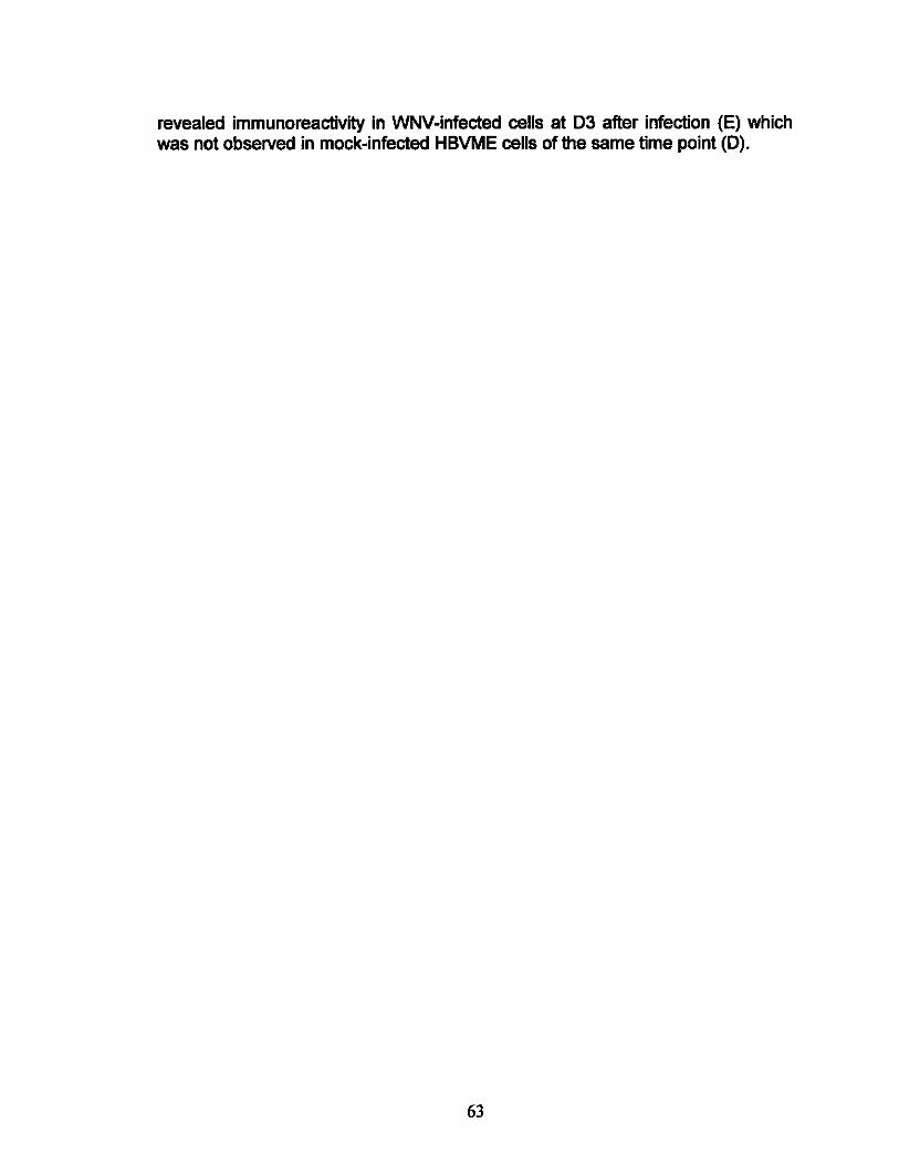
revealed immunoreactivity in WNV-infected cells at 03 after infection (E) which was not observed in mock-infected HBVME cells of the same time point (0).
63

4.4 Infection of HBMVE cells with WNV did not result in MMP or TIMP
expressions
MMP and its inhibitor TIMP are important matrix modulators and have been
documented to play roles in T JP degradation and BBB disruption leading to
increased vascular permeability during inflammatory conditions (Samuel &
Diamond, 2005). Since endothelial cells under pathological conditions have been
shown to secrete MMP-2, -3, -7 and -9 induced by inflammatory cytokines
(A~ona at al., 2007a; Wang at al., 2004), we examined the expressions of MMP-
2, -3, -9 and TIMP-1 by qRT-PCR. The same RNA derived-cDNA from cells
harvested at 6, 12 h, and at 01 through 4 after infection described under T JP and
CAM expression analyses was used for MMP and TIMP expression fold-change
analysis using qRT-PCR. All fold-change was determined in comparison to
mocked-infected control with GAPDH being the house keeping gene for
expression normalization. In contrast to our expectation, qRT-PCR analyses of
MMP and TIMP did not yield any changes in MMP and TIMP expressions
compared to mock-infected control (data not shown). In fact, the basal levels of
MMP and TIMP were very low when compared to a positive control derived from
stimulated blood cells. ELISA for MMP-9 activity was performed on non
cumulative and cumulative supematants collected from 0, 6, and 12 h, and at 01
through 4 after infection. No difference in MMP-9 activity was observed between
experimental infected-samples and mock-infected control samples (data not
shown).
64

Chapter 5. Discussion
WNND occur in approximately 1 % of the infected individuals. While
meningoencephalitis is acute and can be fatal if not treated promptly, recovery
from other forms of WNND is often lengthy and symptoms may persistent
through life. Therefore, it is of most value to prevent WNV from entering the
CNS; however, the mechanism by which WNV enters the brain is not well
understood. While WNV can exploit multiple mechanisms to enter the CNS,
recent studies in mice have implicated the likelihood of WNV transmigration via
the BBB through hematogenous spread (Samuel & Diamond, 2005; Wang et a/.,
2004). Similar to polio virus CNS-invasion, Samuel et al has most recently
demonstrated that WNV is able to spread through antero- and retro-grade
transport from peripheral nerve axon into the CNS in hamsters (Samuel et a/.,
2007). As the various pathways and mechanisms of WNV CNS-entry are being
uncovered, we aimed to further elucidate the role of the BBB by examining the
response of HBMVE cells, the key component of the BBB, to WNV infection. Our
in vitro data are the first report of direct cellular and molecular modulations
induced by WNV infection in HBMVE cells. We demonstrated that HBMVE cells
are susceptible to WNV infection and are able to support viral replication without
causing noticeable cytopathic effects and cell death during the duration of our
study - five days. Further, we report increased expression of a tight junction
protein, claudin-1, and up-regulations of cell adhesion molecules, E-selectin and
VCAM-1, induced directly by WNV infection and replication.
65

WNV replication kinetics varies among cell types and has never been
characterized in HBMVE cells. In neurons, WNV titers determined by plaque
assay in vitro reached as high as 1x10⁷ PFUlmL at day 6 after infection and
remained high until day 14 after infection while WNV titers in astrocytes and
microglia were only around 1 x1 05 and 1 x1 03 PFUlmL, respectively, which
dramatically decreased by day 5 after infection (Cheeran et s/., 2005). Our data
reveal WNV titers in HBMVE cells to be relatively high, like that in neurons, but
the replication cycle lasted only for five days, similar to that of astrocytes and
microglia. Therefore, infection of HBMVE cells, or of the BBB, is transient,
similar to the transient nature of WNV viremia, and detection of which may easily
be missed during the course of WNV infection.
Experimental productive infection of brain endothelial cells have been
documented in many neurotropic viruses, such as simian immunodeficiency virus
(SIV) (Mankowski et s/., 1994; Strelow et s/., 2002; Strelow et s/., 1998), human
cytomegalovirus (HCMV) (Bentz et 81., 2006; Lathey et s/., 1990), measles virus
(MV) (Cosby & Brankin, 1995), and equine herpes virus-1 (EHV-1) (Hasebe et
s/., 2006), which suggest transcellular migration of cell-free virus from the blood
into the CNS. When progeny virions are released on the abluminal side of the
infected endothelial cells, cell-free virus can make its way through the basal
lamina and have direct contact to resident brain cells such as astrocytes and
microglia and eventually neurons. Our demonstration of productive infection of
WNV in HBMVE cells precisely supports the same CNS entry mechanism.
Further, our result is supported by the occasional demonstration of WNV antigen
66

staining in gray owls (Lopes at a/., 2007) and crows (Wunschmann et a/., 2004)
in endothelial cells. Infection of human brain endothelium by Japanese
encephalitis virus, another closely related f1avivirus to WNV, also suggests the
positive role of HBMVE cells in virus spread (German et a/., 2006)
Infection with whole virus or exposure to certain viral proteins can elicit cellular
and molecular changes in HBMVE cells, such as SIV (Luabeya at a/., 2000;
Maclean at a/., 2005), HSV (Kim at a/., 2000) and HIV (Andras at a/., 2003;
Boven at a/., 2000; Dallasta at a/., 1999; Kanmogne at a/., 2005; Kanmogne at
a/., 2007). We examined the expressions of various important tight junction
proteins and cell adhesion molecules which have been commonly found to be
modulated in a variety of viral infections in the context of increased BBB
permeability. The continuous presence of tight junctions along the entire length
of brain vasculature is crucial to BBB property, thus the fundamental integrity of
brain endothelial tight junctions, which is in part governed by the expression and
distribution of TJP, are also important in conferring the BBB property of brain
endothelium. Unlike other viruses, WNV induced increased expression of
claudin-1 tight junction protein in HBMEV cells, but no change in claudin-5, ZO-1
and occludin. Interestingly, this unique up-regulation of claudin-1 was temporally
correlated with maximal virus replication and release as seen in our plaque assay
and qRT-PCR data, indicating that this tight junction modulation was mediated by
replication of WNV, rather than by the mere presence of viral proteins; no
increase was observed in HBMVE cells infected with UV-inactivated WNV.
67

Typically, viral infections of brain endothelial cells are associated with BBB
disruption mediated partly by reduction and/or disorganization of T JP as
described in HIV-1 (Andras et al., 2003; Oallasta et al., 1999; Kanmogne et al.,
2005), SIV (Luabeya et al., 2000; Maclean et al., 2005), and HCMV (Bentz et al.,
2006). To our knowledge, up-regulation of TJP has never been reported in the
context of viral infections; yet has been reported in cancers such as human
primary colon carcinoma (Ohawan et al., 2005; Kinugasa et al., 2007), oral
squamous cell carcinoma cells (Oku et al., 2006), and HPV-associated cervical
cancer (Vazquez-Ortiz et al., 2005). It is therefore perplexing for us to observe
an increase in claudin-1 expression. Based on the limited functional studies of
claudin-1 in the literature, we speculate that in the absence of immune
modulation, HBMVE cell's primary defensive response to WNV infection is
enhanced tight junction integrity via claudin-1 increase, possibly to prevent
further paracellular influx of virus. This conjecture is supported by in vitro
demonstration of approximately four times higher transendothelial resistance in
claudin-1-expressing cells than wild-type MOCK cells (Inai et al., 1999).
Furthermore, over-expression of claudin-1 was found to be associated with
reduced paracellular flux, suggesting major barrier function of claudin-1 protein
(Inai et al., 1999). Similar protective role of endothelial cells was previously
suggested for HIV-1 infection where in a BBB monolayer model, HBMVE cells
prevented HIV-1 from crossing the monolayer form one side to another (Fiala et
al., 1997). To determine the precise consequence of WNV-induced claudin-1
68

increase in HBMVE cells, further examination with an in vitro BBB model deems
necessary.
In addition to cell-free WNV transcellular migration through the BBB, it is also
possible that WNV crosses in cell-associated manner, i.e., the Trojan horse
mechanism. Infection of blood monoctye-derived macrophages by WNV has
been previously demonstrated in vitro (Rios et a/., 2006) and infection with
various viruses including HIV-1 (Kanmogne et a/., 2007; Nottet, 1999; Nottet et
a/., 1996; Song et a/., 2007) and HCMV (Bentz et a/., 2006) are often associated
with increased infiltration of infected or un infected leukocytes which may
contribute to endothelial tight junction disruption.
We report selective inductions of E-selectin and VCAM-1 in HBMVE cells by
WNV and that the induction temporally correlated with maximal virus replication
and release, indicating that endothelial cell activation is mediated by WNV
replication. Together, E-selectin and VCAM-1 are involved in increased
basophils, eosinophils and neutrophils adhesion (Bevilacqua et a/., 1989;
Bochner et a/., 1991; Hakkert et a/., 1991) to endothelial cells as well as
monocytes, macrophages and lymphocytes transmigration (Bentz et a/., 2006;
Nottet et a/., 1996; Wong et a/., 1999) across endothelium. Increased
expressions of E-selectin and VCAM-1 in HBMVE cells are likely to promote
interaction between circulating leukocytes and HBMVE cells given an in vivo
system. Thus, it is of potential that WNV may transmigrate through the BBB by
69
infecting leukocytes. Further, increased infiltration of macrophages and
lymphocytes are cardinal features documented in WNV encephalitic brain
histological studies in human (Hayes at al., 2005b), however, the underlying
mechanism of these immune cells influx are unclear. Based on our finding, we
believe that the perivascular immune cells infiltration can be partly modulated by
the increased expression of E-selectin and VCAM-1.
Increased BBB permeability can result from compromised tight junction as well
as leaky basal lamina, both mediated by increased MMP activities. As
suggested elsewhere, HBMVE cells can secrete MMP-2, -3, -7 and -9 in the
presence of pro-inflammatory stimuli such as TNF-a and IL-1 (Hummel at al.,
2001; Leppert at al., 2001). We also analyzed for the expressions of MMP-2, -3,
and -9, as well as an inhibitor of MMP-9, TIMP-1. Various MMP have been found
to cause T JP degradation as well as decomposition of the extra cellular matrix,
leading to increased permeability of the endothelium (Hawkins at al., 2007;
Rosenberg & Yang, 2007; Yang at al., 2007). In contrast to our expectation
based on the literature of other neurotropic viruses such as HIV-1 (Conant at al.,
1999; Eugenin at al., 2006b; Lafrenie at al., 1997; Sporer at al., 2000), analysis
of MMP-2, -3, -9 and TIMP-1 expressions revealed no change in HBMVE cells
infected with WNV. However, our MMP data are consistent with our T JP result
where no reduction was observed. Together, our result indicates that induction
of MMP is not a direct response of HBMVE cells to WNV infection but possibly an
indirect response under pathological conditions when pro-inflammatory mediators
70

are present. Therefore, further in vivo examination of the contribution of MMP in
BBB permeability changes with respect to WNV infection is warranted. Based on
our cell culture system, we have successfully delineated the direct cellular and
molecular response of HBMVE cells to WNV infection without complication of
host immune influence.
Taken together, our results suggest that HBMVE cells can play a role in the
dissemination of WNV from blood-to-brain. Direct WNV infection and replication
of HBMVE cells support transcellular crossing of cell-free WNV virus. As a direct
result of WNV replication, HBMVE cells up-regulated claudin-1 expression,
possibly to enhance tight junction integrity in order to fend off further infiltration of
virus and to prevent cell-t~1I spread. Also, WNV-induced expressions of E
selectin and VCAM-1 are likely to promote adhesion and diapedesis of circulating
leukocytes across the BBB, thereby supporting paracellular entry of WNV via
"Trojan horse". Our study has characterized, on the cellular and molecular
levels, tight junction integrity of HBMVE cells infected with WNV. However, our
findings warrant further investigation on the functional increase of claudin-1, E
selectin and VCAM-1 expressions with in vitro BBB model. In conclusion, our
study suggests an active role of HBMVE cells of the BBB in the dissemination of
WNV from the blood into the brain.
71

References
(2002a). Intrauterine West Nile virus infection-New York, 2002. Mmwr 51, 1135-
1136.
(2002b). Laboratory-acquired West Nile virus infections-United States, 2002.
Mmwr51,1133-1135.
(2002c). Possible West Nile virus transmission to an infant through breast
feeding-Michigan, 2002. Mmwr 51,877-878.
Afonso, P. V., Ozden, 5., Prevost, M. C., Schmitt, C., Seilhean, D., Weksler, B.,
Couraud, P.O., Gessain, A., Romero, I. A. & Ceccaldi, P. E. (2007).
Human blood-brain barrier disruption by retroviral-infected lymphocytes:
role of myosin light chain kinase in endothelial tight-junction
disorganization. J Immunol179, 2576-2583.
Aktas, 0., U.llrich, 0., Infante-Duarte, C., Nitsch, R. & Zipp, F. (2007). Neuronal
damage in brain inflammation. Archives of neurology 64, 185-189.
Alexopoulou, L., Holt, A. C., Medzhitov, R. & Flavell, R. A. (2001). Recognition of
double-stranded RNA and activation of NF-kappaB by Toll-like receptor 3.
Nature 413, 732-738.
Anderson, J. M., Fanning, A. 5., Lapierre, L. & Van ltallie, C. M. (1995). Zonula
occludens (ZO)-1 and ZO-2: membrane-associated guanylate kinase
homologues (MAGuKs) of the tight junction. Biochemical Society
transactions 23, 470-475.
72

Andras, I. E., Pu, H., Deli, M. A., Nath, A., Hennig, B. & Toborek, M. (2003). HIV-
1 Tat protein alters tight junction protein expression and distribution in
cultured brain endothelial cells. Journal of neuroscience research 74,255-
265.
Annunziata, P. (2003). Blood-brain barrier changes during invasion of the central
nervous system by HIV-1. Old and new insights into the mechanism.
Journal of neurology 250,901-906.
Araki, T., Miki, C. & Kusunoki, M. (2001). Biological implications of circulating
soluble intercellular adhesion molecule-1 in colorectal cancer patients.
Scandinavian journal of gastroenterology 36, 399-404.
Argenbright, l. W. & Barton, R. W. (1992). Interactions of leukocyte integrins with
intercellular adhesion molecule 1 in the production of inflammatory
vascular injury in vivo. The Shwartzman reaction revisited. The Journal of
clinical investigation 89, 259-272.
Arjona, A., Foellmer, H. G., Town, T., Leng, l., McDonald, C., Wang, T., Wong,
S. J., Montgomery, R. R., Fikrig, E. & Bucala, R. (2007a). Abrogation of
macrophage migration inhibitory factor decreases West Nile virus lethality
by limiting viral neuroinvasion. The Journal of clinical investigation 117,
3059-3066.
Arjona, A., Ledizet, M., Anthony, K., Bonafe, N., Modis, Y., Town, T. & Fikrig, E.
(2007b). West Nile Virus Envelope Protein Inhibits dsRNA-lnduced Innate
Immune Responses. J Immunol179, 8403-8409.
73

Armah, H. B., Wang, G., Omalu, B. I., Tesh, R. B., Gyure, K. A, Chute, D. J.,
Smith, R. D., Dulai, P., Vinters, H. V., Kleinschmidt-DeMasters, B. K. &
Wiley, C. A (2007). Systemic distribution of West Nile virus infection:
postmortem immunohistochemical study of six cases. Brain pathology
(Zurich, Switzerland) 17, 354-362.
Arndt, U., Wennemuth, G., Barth, P., Nain, M., AI-Abed, Y., Meinhardt, A,
Gemsa, D. & Bacher, M. (2002). Release of macrophage migration
inhibitory factor and CXCL8/interleukin-8 from lung epithelial cells
rendered necrotic by influenza A virus infection. Journal of virology 76,
9298-9306.
Bacher, M., Eickmann, M., Schrader, J., Gemsa, D. & Heiske, A. (2002). Human
cytomegalovirus-mediated induction of MIF in fibroblasts. Virology 299,
32-37.
Bagai, R., Valujskikh, A., Canaday, D. H., Bailey, E., Lalli, P. N., Harding, C. V. &
Heeger, P. S. (2005). Mouse endothelial cells cross-present Iymphocyte
derived antigen on class I MHC via a TAP1- and proteasome-dependent
pathway. J Immunol174, 7711-7715.
Banks, W. A. (2004). The source of cerebral insulin. European journal of
pharmacology 490,5-12.
Barnard, D. R. & Xue, R. D. (2004). Laboratory evaluation of mosquito repellents
against Aedes albopictus, Culex nigripalpus, and Ochierotatus triseriatus
(Diptera: Culicidae). Journal of medical entomology 41, 726-730.
74

Barthel, S. R., Gavino, J. D., Descheny, l. & Dimitroff, C. J. (2007). Targeting
selectins and selectin ligands in inflammation and cancer. Expert opinion
on therapeutic targets 11, 1473-1491.
Beasley, D. W., Davis, C. T., Whiteman, M., Granwehr, B., Kinney, R. M. &
Barrett, A. D. (2004). Molecular determinants of virulence of West Nile
virus in North America. Arch Virol Suppl, 35-41.
Ben David, D., Reznick, A. Z., Srouji, S. & Livne, E. (2008). Exposure to pro
inflammatory cytokines upregulates MMP-9 synthesis by mesenchymal
stem cells-derived osteoprogenitors. Histochemistry and cell biology.
Bentz, G. l., Jarquin-Pardo, M., Chan, G., Smith, M. S., Sinzger, C. & Yurochko,
A. D. (2006). Human cytomegalovirus (HCMV) infection of endothelial
cells promotes naive monocyte extravasation and transfer of productive
virus to enhance hematogenous dissemination of HCMV. Journal of
virology 80, 11539-11555.
Bevilacqua, M. P., Stengelin, S., Gimbrone, M. A., Jr. & Seed, B. (1989).
Endothelial leukocyte adhesion molecule 1: an inducible receptor for
neutrophils related to complement regulatory proteins and lectins. Science
(New York, NY243,1160-1165.
Bochner, B. S., Luscinskas, F. W., Gimbrone, M. A., Jr., Newman, W.,
Sterbinsky, S. A., Derse-Anthony, C. P., Klunk, D. & Schleimer, R. P.
(1991). Adhesion of human basophils, eosinophils, and neutrophils to
interleukin 1-activated human vascular endothelial cells: contributions of
75
endothelial cell adhesion molecules. The Journal of experimental medicine
173, 1553-1557.
Bode, A. V., Sejvar, J. J., Pape, W. J., Campbell, G. L. & Martin, A. A. (2006).
West Nile virus disease: a descriptive study of 228 patients hospitalized in
a 4-county region of Colorado in 2003. Clin Infect Dis 42, 1234-1240.
Boven, L. A., Middel, J., Verhoef, J., De Groot, C. J. & Nottet, H. S. (2000).
Monocyte infiltration is highly associated with loss of the tight junction
protein zonula occludens in HIV-1-associated dementia. Neuropathology
and applied neurobiology 26, 356-360.
Brabers, N. A. & Nottet, H. S. (2006). Role of the pro-inflammatory cytokines
TNF-alpha and IL-1beta in HIV-associated dementia. Europeanjoumal of
clinical investigation 36, 447-458.
Bragin-Sanchez, D. & Chang, P. P. (2005). West Nile virus encephalitis infection
in a heart transplant recipient: a case report. J Heart Lung Transplant 24,
621-623.
Bratt, J. & Palmblad, J. (1997). Cytokine-induced neutrophil-mediated injury of
human endothelial cells. J Immunol159, 912-918.
Brien, J. D., Uhrlaub, J. L. & Nikolich-Zugich, J. (2007). Protective capacity and
epitope specificity of CD8(+) T cells responding to lethal West Nile virus
infection. European journal of immunology 37, 1855-1863.
Brinton, M. A. (2002). The molecular biology of West Nile Virus: a new invader of
the western hemisphere. Annual review of microbiology 56,371-402.
76

Butcher, E. C. (1991). Leukocyte-endothelial cell recognition: three (or more)
steps to specificity and diversity. Cell 67, 1033-1036.
Campbell, G. L., Marfin, A. A., Lanciotti, R. S. & Gubler, D. J. (2002). West Nile
virus. The Lancet infectious diseases 2,519-529.
Carlos, T., Kovach, N., Schwartz, B., Rosa, M., Newman, B., Wayner, E.,
Benjamin, C., Osbom, L., Lobb, R. & Harlan, J. (1991). Human monocytes
bind to two cytokine-induced adhesive ligands on cultured human
endothelial cells: endothelial-leukocyte adhesion molecule-1 and vascular
cell adhesion molecule-1. Blood 77, 2266-2271.
Carter, W. G., Kaur, P., Gil, S. G., Gahr, P. J. & Wayner, E. A. (1990). Distinct
functions for integrins alpha 3 beta 1 in focal adhesions and alpha 6 beta
4/bullous pemphigoid antigen in a new stable anchoring contact (SAC) of
keratinocytes: relation to hemidesmosomes. The Joumal of cell biology
111,3141-3154.
Chambers, T. J. & Diamond, M. S. (2003). Pathogenesis of flavivirus
encephalitis. Advances in virus research 60, 273-342.
Chan, C. K., Limstrom, S. A., Tarasewicz, D. G. & Lin, S. G. (2006). Ocular
features of west nile virus infection in North America: a study of 14 eyes.
Ophthalmology 113,1539-1546.
Cheeran, M. C., Hu, S., Sheng, W. S., Rashid, A., Peterson, P. K. & Lokensgard,
J. R. (2005). Differential responses of human brain cells to West Nile virus
infection. Joumal of neurovirology 11, 512-524.
77

Chu, J. J. & Ng, M. L. (2004a). Infectious entry of West Nile virus occurs through
a clathrin-mediated endocytic pathway. Journal of virology 78, 10543-
10555.
Chu, J. J. & Ng, M. L. (2004b). Interaction of West Nile virus with alpha v beta 3
integrin mediates virus entry into cells. The Journal of biological chemistry
279,54533-54541.
Chu, J. J., Rajamanonmani, R., Li, J., Bhuvanakantham, R., Lescar, J. & Ng, M.
L. (2005). Inhibition of West Nile virus entry by using a recombinant
domain III from the envelope glycoprotein. The Journal of general virology
86,405-412.
Chung, K. M., Thompson, B. S., Fremont, D. H. & Diamond, M. S. (2007).
Antibody recognition of cell surface-associated NS1 triggers Fc-gamma
receptor-mediated phagocytosis and clearance of West Nile Virus-infected
cells. Journal of virology 81, 9551-9555.
Conant, K., McArthur, J. C., Griffin, D. E., Sjulson, L., Wahl, L. M. & Irani, D. N.
(1999). Cerebrospinal fluid levels of MMP-2, 7, and 9 are elevated in
association with human immunodeficiency virus dementia. Annals of
neurology 46,391-398.
Cordenonsi, M., D'Atri, F., Hammar, E., Parry, D. A., Kendrick-Jones, J., Shore,
D. & Citi, S. (1999). Cingulin contains globular and coiled-coil domains
and interacts with ZO-1, ZO-2, ZO-3, and myosin. The Journal of cell
biology 147, 1569-1582.
78

Cosby, S. L. & Brankin, B. (1995). Measles virus infection of cerebral endothelial
cells and effect on their adhesive properties. Veterinary microbiology 44,
135-139.
Crawford, J. M. & Hopp, B. (1990). Junctional epithelium expresses the
intercellular adhesion molecule ICAM-1. Joumal of periodontal research
25, 254-256.
Cushing, M. M., Brat, D. J., Mosunjac, M. I., Hennigar, R. A., Jemigan, D. B.,
Lanciotti, R., Petersen, L. R., Goldsmith, C., Rollin, P. E., Shieh, W. J.,
Guamer, J. & Zaki, S. R. (2004). Fatal West Nile virus encephalitis in a
renal transplant recipient. American joumal of clinical pathology 121, 26-
31.
Custer, B., Tomasulo, P. A., Murphy, E. L., Caglioti, S., Harpool, D., McEvoy, P.
& Busch, M. P. (2004). Triggers for switching from minipool testing by
nucleic acid technology to individual-donation nucleic acid testing for West
Nile virus: analysis of 2003 data to inform 2004 decision making.
Transfusion 44, 1547-1554.
D'Atri, F., Nadalutti, F. & Citi, S. (2002). Evidence for a functional interaction
between cingulin and ZO-1 in cultured cells. The Joumal of biological
chemistry 277,27757-27764.
Dallasta, L. M., Pisarov, L. A., Esplen, J. E., Werley, J. V., Moses, A. V., Nelson,
J. A. & Achim, C. L. (1999). Blood-brain barrier tight junction disruption in
human immunodeficiency virus-1 encephalitis. The American joumal of
pathology 155. 1915-1927.
79

Davis, C. W., Nguyen, H. Y., Hanna, S. L., Sanchez, M. D., Doms, R W. &
Pierson, T. C. (2006a). West Nile virus discriminates between DC-SIGN
and DC-SIGNR for cellular attachment and infection. Journal of virology
80,1290-1301.
Davis, L. E., DeBiasi, R, Goade, D. E., Haaland, K. Y., Harrington, J. A., Hamar,
J. B., Pergam, S. A., King, M. K., DeMasters, B. K. & Tyler, K. L. (2006b).
West Nile virus neuroinvasive disease. Annals of neurology 60, 286-300.
Deas, T. S., Binduga-Gajewska, I., Tilgner, M., Ren, P., Stein, D. A., Moulton, H.
M., Iversen, P. L., Kauffman, E. B., Kramer, L. D. & Shi, P. Y. (2005).
Inhibition of flavivirus infections by antisense oligomers specifically
suppressing viral translation and RNA replication. Journal of virology 79,
4599-4609.
del Zoppo, G. J. & Hallenbeck, J. M. (2000). Advances in the vascular
pathophysiology of ischemic stroke. Thrombosis research 98, 73-81.
Dhawan, P., Singh, A. B., Deane, N. G., No, Y., Shiou, S. R, Schmidt, C., Neff,
J., Washington, M. K. & Beauchamp, R D. (2005). Claudin-1 regulates
cellular transformation and metastatic behavior in colon cancer. The
Journal of clinical investigation 115, 1765-1776.
Diamond, M. S., Shrestha, B., Mehlhop, E., Sitati, E. & Engle, M. (2003a). Innate
and adaptive immune responses determine protection against
disseminated infection by West Nile encephalitis virus. Viral immunology
16,259-278.
80

Diamond, M. S., Sitati, E. M., Friend, L. D., Higgs, S., Shrestha, B. & Engle, M.
(2003b). A critical role for induced IgM in the protection against West Nile
virus infection. The Joumal of experimental medicine 198, 1853-1862.
Dittmar, T., Heyder, C., Gloria-Maercker, E., Hatzmann, W. & Zanker, K. S.
(2008). Adhesion molecules and chemokines: the navigation system for
circulating tumor (stem) cells to metastasize in an organ-specific manner.
Clinical & experimental metastasis 25, 11-32.
Dowlati, A., Gray, R, Sandler, A. B., Schiller, J. H. & Johnson, D. H. (2008). Cell
Adhesion Molecules, Vascular Endothelial Growth Factor, and Basic
Fibroblast Growth Factor in Patients with Non-Small Cell Lung Cancer
Treated with Chemotherapy with or without Bevacizumab-an Eastem
Cooperative Oncology Group Study. Clin Cancer Res 14,1407-1412.
Ebnet, K., Schulz, C. U., Meyer Zu Brickwedde, M. K., Pendl, G. G. & Vestweber,
D. (2000). Junctional adhesion molecule interacts with the PDZ domain
containing proteins AF-6 and ZO-1. The Joumal of biological chemistry
275, 27979-27988.
Engle, M. J. & Diamond, M. S. (2003). Antibody prophylaxis and therapy against
West Nile virus infection in wild-type and immunodeficient mice. Joumal of
virology 77, 12941-12949.
Eugenin, E. A., Gamss, R, Buckner, C., Buono, D., Klein, R S., Schoenbaum, E.
E., Calderon, T. M. & Berman, J. W. (2006a). Shedding of PECAM-1
during HIV infection: a potential role for soluble PECAM-1 in the
pathogenesis of NeuroAIDS. Joumal of leukocyte biology 79, 444452.
81

Eugenin, E. A., Osiecki, K., Lopez, L., Goldstein, H., Calderon, T. M. & Berman,
J. W. (2006b). CCL2Imonocyte chemoattractant protein-1 mediates
enhanced transmigration of human immunodeficiency virus (HIV)-infected
leukocytes across the blood-brain barrier: a potential mechanism of HIV
CNS invasion and NeuroAIDS. J Neurosci 26,1098-1106.
Fanning, A. S., Jameson, B. J., Jesaitis, L. A. & Anderson, J. M. (1998). The tight
junction protein ZO-1 establishes a link between the transmembrane
protein occludin and the actin cytoskeleton. The Joumal of biological
chemistry 273,29745-29753.
Ferguson, D. D., Gershman, K., LeBailly, A. & Petersen, L. R. (2005).
Characteristics of the rash associated with West Nile virus fever. Clin
Infect Dis 41,1204-1207.
Fiala, M., Looney, D. J., Stins, M., Way, D. D., Zhang, L., Gan, X., Chiappelli, F.,
Schweitzer, E. S., Shapshak, P., Weinand, M., Graves, M. C., Witte, M. &
Kim, K. S. (1997). TNF-alpha opens a paracellular route for HIV-1 invasion
across the blood-brain barrier. Molecular medicine (Cambridge, Mass 3,
553-564.
Fradin, M. S. (1998). Mosquitoes and mosquito repellents: a clinician's guide.
Annals of intemal medicine 128, 931-940.
Fradin, M. S. & Day, J. F. (2002). Comparative efficacy of insect repellents
against mosquito bites. The New England joumal of medicine 347, 13-18.
Fredericksen, B. L. & Gale, M., Jr. (2006). West Nile virus evades activation of
interferon regulatory factor 3 through RIG-I-dependent and -independent
82
pathways without antagonizing host defense signaling. Journal of virology
80,2913-2923.
Fredericksen, B. L., Keller, B. C., Fornek, J., Katze, M. G. & Gale, M., Jr. (2008).
Establishment and maintenance of the innate antiviral response to West
Nile Virus involves both RIG-I and MDA5 signaling through IPS-1. Journal
of virology 82, 609-616.
Gallicchio, M., Rosa, A. C., Dianzani, C., Brucato, L., Benetti, E., Collino, M. &
Fantozzi, R. (2008). Celecoxib decreases expression of the adhesion
molecules ICAM-1 and VCAM-1 in a colon cancer cell line (HT29). British
journal of pharmacology 153, 870-878.
Garcia-Tapia, D., Hassett, D. E., Mitchell, W. J., Jr., Johnson, G. C. & Kleiboeker,
S. B. (2007). West Nile virus encephalitis: sequential histopathological and
immunological events in a murine model of infection. Journal of
neurovirology 13, 130-138.
German, A. C., Myint, K. S., Mai, N. T., Pomeroy, I., Phu, N. H., Tzartos, J.,
Winter, P., Collett, J., Farrar, J., Barrett, A., Kipar, A., Esiri, M. M. &
Solomon, T. (2006). A preliminary neuropathological study of Japanese
encephalitis in humans and a mouse model. Transactions of the Royal
Society of Tropical Medicine and Hygiene 100, 1135-1145.
Girard, Y. A., Popov, V., Wen, J., Han, V. & Higgs, S. (2005). Ultrastructural
study of West Nile virus pathogenesis in Culex pipiens quinquefasciatus
(Diptera: Culicidae). Journal of medical entomology 42, 429-444.
83

Glass, W. G., Lim, J. K., Cholera, R., Pletnev, A. G., Gao, J. L. & Murphy, P. M.
(2005). Chemokine receptor CCR5 promotes leukocyte trafficking to the
brain and survival in West Nile virus infection. The Journal of experimental
medicine 202,1087-1098.
Glass, W. G., McDermott, D. H., Lim, J. K., Lekhong, S., Yu, S. F., Frank, W. A.,
Pape, J., Cheshier, R. C. & Murphy, P. M. (2006). CCR5 deficiency
increases risk of symptomatic West Nile virus infection. The Journal of
experimental medicine 203, 35-40.
Golias, C., Tsoutsi, E., Matziridis, A., Makridis, P., Batistatou, A. &
Charalabopoulos, K. (2007). Review. Leukocyte and endothelial cell
adhesion molecules in inflammation focusing on inflammatory heart
disease. In vivo (Athens, Greece) 21, 757-769.
Goodell, J. R., Puig-Basagoiti, F., Forshey, B. M., Shi, P. Y. & Ferguson, D. M.
(2006). Identification of compounds with anti-West Nile Virus activity.
Journal of medicinal chemistry 49, 2127-2137.
Green, M. S., Weinberger, M., Ben-Ezer, J., Bin, H., Mendelson, E., Gandacu,
D., Kaufman, Z., Dichtiar, R., Sobel, A., Cohen, D. & Chowers, M. Y.
(2005). Long-term Death Rates, West Nile virus epidemic, Israel, 2000.
Emerging infectious diseases 11, 1754-1757.
Guarner, J., Shieh, W. J., Hunter, S., Paddock, C. D., Morken, T., Campbell, G.
L., Martin, A. A. & Zaki, S. R. (2004). Clinicopathologic study and
laboratory diagnosis of 23 cases with West Nile virus encephalomyelitis.
Human pathology 35, 983-990.
84

Gubler, D. J. (2007). The continuing spread of West Nile virus in the western
hemisphere. Clin Infect Dis 45, 1039-1046.
Gubler, D. J., Campbell. G. L., Nasci. R.. Komar, N., Petersen. L. & Roehrig, J. T.
(2000). West Nile virus in the United States: guidelines for detection,
prevention, and control. Viral immunology 13, 469-475.
Hakkert, B. C., Kuijpers, T. W., Leeuwenberg, J. F., van Mourik, J. A. & Roos, D.
(1991). Neutrophil and monocyte adherence to and migration across
monolayers of cytokine-activated endothelial cells: the contribution of
CD18, ELAM-1, and VLA-4. Blood 78, 2721-2726.
Haorah, J., Schall, K., Ramirez, S. H. & Persidsky, Y. (2008). Activation of
protein tyrosine kinases and matrix metalloproteinases causes blood-brain
barrier injury: Novel mechanism for neurodegeneration associated with
alcohol abuse. Glia 56, 78-88.
Hasebe, R., Kimura, T., Nakamura, K., Ochiai, K., Okazaki, K., Wada, R. &
Umemura, T. (2006). Differential susceptibility of equine and mouse brain
microvascular endothelial cells to equine herpesvirus 1 infection. Archives
of virology 151, 775-786.
Hawkins, B. T. & Davis, T. P. (2005). The blood-brain barrier/neurovascular unit
in health and disease. Pharmacological reviews 57,173-185.
Hawkins, B. T., Lundeen, T. F., Norwood, K. M., Brooks, H. L. & Egleton, R. D.
(2007). Increased blood-brain barrier permeability and altered tight
junctions in experimental diabetes in the rat contribution of
hyperglycaemia and matrix metalloproteinases. Diabetologia 50, 202-211.
85

Hayes, E. B. & Gubler, D. J. (2006). West Nile virus: epidemiology and clinical
features of an emerging epidemic in the United States. Annual review of
medicine 57,181-194.
Hayes, E. B., Komar, N., Nasci, R S., Montgomery, S. P., O'Leary, D. R &
Campbell, G. L. (2005a). Epidemiology and transmission dynamics of
West Nile virus disease. Emerging infectious diseases 11, 1167-1173.
Hayes, E. B., Sejvar, J. J., Zaki, S. R, Lanciotti, R S., Bode, A. V. & Campbell,
G. L. (2005b). Virology, pathology, and clinical manifestations of West Nile
virus disease. Emerging infectious diseases 11, 1174-1179.
Hidalgo, A., Peired, A. J., Wild, M. K., Vestweber, D. & Frenette, P. S. (2007).
Complete identification of E-selectin ligands on neutrophils reveals distinct
functions of PSGL-1, ESL-1, and CD44. Immunity 26, 477-489.
Hirase, T., Kawashima, S., Wong, E. Y., Ueyama, T., Rikitake, Y., Tsukita, S.,
Yokoyama, M. & Staddon, J. M. (2001). Regulation of tight junction
permeability and occludin phosphorylation by Rhoa-p160ROCK
dependent and -independent mechanisms. The Journal of'biological
chemistry 276, 10423-10431.
Hummel, V., Kallmann, B. A., Wagner, S., Fuller, T .• Bayas. A.. Tonn. J. C .•
Benveniste, E. N., Toyka, K. V. & Rieckmann, P. (2001). Production of
MMPs in human cerebral endothelial cells and their role in shedding
adhesion molecules. Journal of neuropathology and experimental
neurology 60, 320-327.
86

Hynes, R. O. (1992). Integrins: versatility, modulation, and signaling in cell
adhesion. Cell 69, 11-25.
Hynes, R. O. (2002). Integrins: bidirectional, allosteric signaling machines. Cell
110,673-687.
Ichiyama, T., Morishima, T., Kajimoto, M., Matsushige, T., Matsubara, T. &
Furukawa, S. (2007). Matrix metalloproteinase-9 and tissue inhibitors of
metalloproteinases 1 in influenza-associated encephalopathy. The
Pediatric infectious disease joumal 26, 542-544.
Inai, T., Kobayashi, J. & Shibata, Y. (1999). Claudin-1 contributes to the epithelial
barrier function in MOCK cells. European jot,lmal of cell biology 78, 849-
855.
lrie, S. & Tavassoli, M. (1991). Transendothelial transport of macromolecules: the
concept of tissue-blood barriers. Cell Bioi Rev 25.317-333.340-311.
Jean CM, H. S., Louie JK, Glaser CA. Risk factors for West Nile virus
neuroinvasive disease, Califomia, 2005. Emerging infectious diseases
[Epub; ahead of print].
Jian Liu, K. & Rosenberg, G. A. (2005). Matrix metalloproteinases and free
radicals in cerebral ischemia. Free radical biology & medicine 39, 71-80.
Johnston, B. & Butcher, E. C. (2002). Chemokines in rapid leukocyte adhesion
triggering and migration. Seminars in immunology 14, 83-92.
Johnston, L. J., Halliday, G. M. & King, N. J. (2000). Langerhans cells migrate to
local lymph nodes following cutaneous infection with an arbovirus. The
Joumal of investigative dermatology 114, 560-568.
87

Kanmogne, G. D., Primeaux, C. & Grammas, P. (2005). HIV-1 gp120 proteins
alter tight junction protein expression and brain endothelial cell
permeability: implications for the pathogenesis of HIV-associated
dementia. Journal of neuropathology and experimental neurology 64, 498-
505.
Kanmogne, G. D., Schall, K., Leibhart, J., Knipe, B., Gendelman, H. E. &
Persidsky, Y. (2007). HIV-1 gp120 compromises blood-brain barrier
integrity and enhances monocyte migration across blood-brain barrier:
implication for viral neuropathogenesis. J Cereb Blood Flow Metab 27,
123-134.
Katayama, Y., Hidalgo, A., Cha~g, J., Peired, A. & Frenette, P. S. (2005). CD44
is a physiological E-selectin ligand on neutrophils. The Journal of
experimental medicine 201, 1183-1189.
Katayama, Y., Hidalgo, A., Furie, B. C., Vestweber, D., Furie, B. & Frenette, P. S.
(2003). PSGL-1 participates in E-selectin-mediated progenitor homing to
bone marrow: evidence for cooperation between E-selectin ligands and
alpha4 integrin. Blood 102, 2060-2067.
Kelley, T. W., Prayson, R. A., Ruiz, A. I., lsada, C. M. & Gordon, S. M. (2003).
The neuropathology of West Nile virus meningoencephalitis. A report of
two cases and review of the literature. American journal of clinical
pathology 119, 749-753.
Kim, Y. C., Bang, D., Lee, S. & Lee, K. H. (2000). The effect of herpesvirus
infection on the expression of cell adhesion molecules on cultured human
88

dermal microvascular endothelial cells. Journal of dermatological science
24,38-47.
Kinugasa, T., Huo, Q., Higashi, D., Shibaguchi, H., Kuroki, M., Tanaka, T.,
Futami, K., Yamashita, Y., Hachimine, K., Maekawa, S., Nabeshima, K.,
Iwasaki, H. & Kuroki, M. (2007). Selective up-regulation of claudin-1 and
claudin-2 in colorectal cancer. Anticancer research 27, 3729-3734.
Kleiboeker, S. B., Loiacono, C. M., Rottinghaus, A., Pue, H. L. & Johnson, G. C.
(2004). Diagnosis of West Nile virus infection in horses. J Vet Diagn Invest
16,2-10.
Klein, R. S., Lin, E., Zhang, B., Luster, A. D., Tollett, J., Samuel, M. A., Engle, M.
& Diamond, M. S. (2005). Neuronal CXCL 10 directs CD8+ T-cell
recruitment and control of West Nile virus encephalitis. Joumal of virology
79, 11457-11466.
Kleinman, S., Glynn, S. A., Busch, M., Todd, D., Powell, L., Pietrelli, L., Nemo,
G., Schreiber, G., Bianco, C. & Katz, L. (2005). The 2003 West Nile virus
United States epidemic: the America's Blood Centers experience.
Transfusion 45, 469-479.
Kleinschmidt-DeMasters, B. K., Marder, B. A., Levi, M. E., Laird, S. P., McNutt, J.
T., Escott, E. J., Everson, G. T. & Tyler, K. L. (2004). Naturally acquired
West Nile virus encephalomyelitis in transplant recipients: clinical,
laboratory, diagnostic, and neuropathological features. Archives of
neurology 61, 1210-1220.
89

Kramer, L. D., Styer, L. M. & Ebel, G. D. (2008). A Global Perspective on the
Epidemiology of West Nile Virus. Annu Rev Entomol53, 61-81.
Kubota, K., Furuse, M., Sasaki, H., Sonoda, N., Fujita, K., Nagafuchi, A. &
Tsukita, S. (1999). Ca(2+)-independent cell-adhesion activity of claudins,
a family of integral membrane proteins localized at tight junctions. Curr
BioI 9, 1035-1038.
Kuno, G., Chang, G. J., Tsuchiya, K. R, Karabatsos, N. & Cropp, C. B. (1998).
Phylogeny of the genus Flavivirus. Joumal of virology 72, 73-83.
Lafrenie, R M., Wahl, L. M., Epstein, J. S., Yamada, K. M. & Dhawan, S. (1997).
Activation of monocytes by HIV-Tat treatment is mediated by cytokine
expression. J Immunol159, 4077-4083.
Lanciotti, R S., Ebel, G. D., Deubel, V., Kerst, A. J., Murri, S., Meyer, R, Bowen,
M., McKinney, N., Morrill, W. E., Crabtree, M. B., Kramer, L. D. & Roehrig,
J. T. (2002). Complete genome sequences and phylogenetic analysis of
West Nile virus strains isolated from the United States, Europe, and the
Middle East Virology 298,96-105.
Lanciotti, R S., Roehrig, J. T., Deubel, V., Smith, J., Parker, M., Steele, K., Crise,
B., Volpe, K. E., Crabtree, M. B., Scherret, J. H., Hall, R A., MacKenzie, J.
S., Cropp, C. B., Panigrahy, B., Ostlund, E., Schmitt, B., Malkinson, M.,
Banet, C., Weissman, J., Komar, N., Savage, H. M., Stone, W.,
McNamara, T. & Gubler, D. J. (1999). Origin of the West Nile virus
responsible for an outbreak of encephalitis in the northeastem United
States. Science (New York, NY 286,2333-2337.
90

Lathey, J. L., Wiley, C. A., Verity, M. A. & Nelson, J. A. (1990). Cultured human
brain capillary endothelial cells are permissive for infection by human
cytomegalovirus. Virology 176, 266-273.
Lee, J. W., Chu, J. J. & Ng, M. L. (2006). Quantifying the specific binding
between West Nile virus envelope domain III protein and the cellular
receptor aiphaVbeta3 integrin. The Journal of biological chemistry 281,
1352-1360.
Leppert, D., Lindberg, R. L., Kappos, L. & Leib, S. L. (2001). Matrix
metalloproteinases: multifunctional effectors of inflammation in multiple
sclerosis and bacterial meningitis. Brain research 36, 249-257.
Lewis, M. & Amsden, J. R. (2007). Successful treatment of West Nile virus
infection after approximately 3 weeks into the disease course.
Pharmacotherapy 27, 455-458.
Lockwood, C. J., Oner, C., Uz, Y. H., Kayisli, U. A., Huang, S. J., Buchwalder, L.
F., Murk, W., Funai, E. F. & Schatz, F. (2008). Matrix Metalloproteinase 9
(MMP9) Expression in Preeclamptic Decidua and MMP9 Induction by
Tumor Necrosis Factor Alpha and Interleukin 1 Beta in Human First
Trimester Decidual Cells. Biology of reproduction.
Lopes, H., Redig, P., Glaser, A., Armien, A. & Wunschmann, A. (2007). Clinical
findings, lesions, and viral antigen distribution in great gray owls (Strix
nebulosa) and barred owls (Strix varia) with spontaneous West Nile virus
infection. Avian diseases 51,140-145.
91

Luabeya, M. K., Dallasta, L. M., Achim, C. L., Pauza, C. D. & Hamilton, R. L.
(2000). Blood-brain barrier disruption in simian immunodeficiency virus
encephalitis. Neuropathology and applied neurobiology 26, 454-462.
Lyons, A. J. & Jones, J. (2007). Cell adhesion molecules, the extracellular matrix
and oral squamous carcinoma. International journal of oral and
maxillofacial surgery 36,671-679.
Maclean, A. G., Belenchia, G. E., Bieniemy, D. N., Moroney-Rasmussen, T. A. &
Lackner, A. A. (2005). Simian immunodeficiency virus disrupts extended
lengths of the blood-brain barrier. Journal of medical primatology 34, 237-
242.
Man, S., Ubogu, E. E. & Ransohoff, R. M. (2007). Inflammatory cell migration into
the central nervous system: a few new twists on an old tale. Brain
pathology (Zurich, Switzerland) 17, 243-250.
Mandai, M., Mandai, A., Das, S., Chakraborti, T. & Sajal, C. (2003). Clinical
implications of matrix metalloproteinases. Molecular and cellular
biochemistry 252, 305-329.
Mandell, K. J. & Parkos, C. A. (2005). The JAM family of proteins. Advanced
drug delivery reviews 57,857-867.
Mankowski, J. L., Spelman, J. P., Ressetar, H. G., Strandberg, J. D., Laterra, J.,
Carter, D. L., Clements, J. E. & Zink, M. C. (1994). Neurovirulent simian
immunodeficiency virus replicates productively in endothelial cells of the
central nervous system in vivo and in vitro. Journal of virology 68, 8202-
8208.
92

Martin-Padura, I., Lostaglio, S., Schneemann, M., Williams, L., Romano, M.,
Fruscella, P., Panzeri, C., Stoppacciaro, A., Ruco, L., Villa, A., Simmons,
D. & Dejana. E. (1998). Junctional adhesion molecule. a novel member of
the immunoglobulin superfamily that distributes at intercellular junctions
and modulates monocyte transmigration. The Joumal of cell biology 142,
117-127.
McCarthy. K. M .• Skare. I. B .• Stankewich. M. C., Furuse, M., Tsukita, S., Rogers,
R. A .• Lynch, R. D. & Schneeberger, E. E. (1996). Occludin is a functional
component of the tight junction. Joumal of cell science 109 ( Pt 9), 2287-
2298.
McEver, R. P. (2002). Selectins: lectins that initiate cell adhesion under flow.
Current opinion in cell biology 14, 581-586.
McEver, R. P. & Cummings, R. D. (1997). Role of PSGL-1 binding to selectins in
leukocyte recruitment. The Joumal of clinical investigation 100, S97-103.
Mehlhop. E. & Diamond, M. S. (2008). The molecular basis of antibody protection
against West Nile virus. Current topics in microbiology and immunology
317, 125-153.
Mehlhop, E., Whitby, K., Oliphant, T., Marri, A., Engle, M. & Diamond, M. S.
(2005). Complement activation is required for induction of a protective
antibody response against West Nile virus infection. Joumal of virology 79,
7466-7477.
Moos, T., Trinder, D. & Morgan, E. H. (2000). Cellular distribution of ferric iron,
ferritin. transferrin and divalent metal transporter 1 (DMT1) in substantia
93

nigra and basal ganglia of normal and beta2-microglobulin deficient
mouse brain. Cellular and molecular biology (Noisy-Ie-Grand, France) 46,
549-561.
Mukhopadhyay, S., Kim, B. S., Chipman, P. R., Rossmann, M. G. & Kuhn, R. J.
(2003). Structure of West Nile virus. Science (New York, NY 302, 248.
Mukhopadhyay, S., Kuhn, R. J. & Rossmann, M. G. (2005). A structural
perspective of the flavivirus life cycle. Nature reviews 3, 13-22.
Murgue, B., Zeller, H. & Deubel, V. (2002). The ecology and epidemiology of
West Nile virus in Africa, Europe and Asia. Current topics in microbiology
and immunology 267,195-221.
Murray, K., Baraniuk, S., Resnick, M., Arafat, R., Kilborn, C., Cain, K.,
Shallenberger, R., York, T. L., Martinez, D., Hellums, J. S., Hellums, D.,
Malkoff, M., Elgawley, N., McNeely, W., Khuwaja, S. A. & Tesh. R. B.
(2006). Risk factors for encephalitis and death from West Nile virus
infection. Epidemiology and infection 134, 1325-1332.
Niu, J. X., Zhang, W. J., Ye, L. Y., Wu, L. Q., Zhu, G. J., Yang, Z. H., Grau, G. E.
& Lou, J. N. (2007). The role of adhesion molecules, alpha v beta 3, alpha
v beta 5 and their ligands in the tumor cell and endothelial cell adhesion.
Eur J Cancer Prev 16, 517-527.
Nottet, H. S. (1999). Interactions between macrophages and brain microvascular
endothelial cells: role in pathogenesis of HIV-1 infection and blood - brain
barrier function. Journal of neurovirology 5, 659-669.
94

Nottet, H. 5., Persidsky, Y., Sasseville, V. G., Nukuna, A. N., Bock. P., Zhai, Q.
H., Sharer, l. R., McComb, R. D., Swindells, 5., Soderland, C. &
Gendelman, H. E. (1996). Mechanisms for the transendothelial migration
of HIV-1-infected monocytes into brain. J Immunol156, 1284-1295.
Nybakken, G. E., Oliphant, T., Johnson,S., Burke,S., Diamond, M. S. &
Fremont, D. H. (2005). Structural basis of West Nile virus neutralization by
a therapeutic antibody. Nature 437,764-769.
Oku, N., Sasabe, E., Ueta, E., Yamamoto, T. & Osaki, T. (2006). Tight junction
protein claudin-1 enhances the invasive activity of oral squamous cell
carcinoma cells by promoting cleavage of laminin-5 gamma2 chain via
matrix metalloproteinase (MMP)-2 and membrane-type MMP-1. Cancer
research 66, 5251-5257.
Oldendorf, W. (1976). Blood-brain barrier. New York: Spectrum publications.
Pealer, l. N., Martin, A. A., Petersen, l. R., lanciotti, R. 5., Page, P. l., Stramer,
S. l., Stobierski, M. G., Signs, K., Newman, B., Kapoor, H., Goodman, J.
l. & Chamberland, M. E. (2003). Transmission of West Nile virus through
blood transfusion in the United States in 2002. The New England joumal
of medicine 349,1236-1245.
Persidsky, Y., Ramirez, S. H., Haorah, J. & Kanmogne, G. D. (2006). Blood-brain
barrier: structural components and function under physiologic and
pathologic conditions. J Neuroimmune Pharmacol 1, 223-236.
Petri, B. & Bixel, M. G. (2006). Molecular events during leukocyte diapedesis.
The FEBS joumal 273, 4399-4407.
95
Petty, M. A. & Lo, E. H. (2002). Junctional complexes of the blood-brain barrier:
permeability changes in neuroinflammation. Progress in neurobiology 68,
311-323.
Pober, J. 5., Kluger, M.S. & 5chechner, J. 5. (2001). Human endothelial cell
presentation of antigen and the homing of memory/effector T cells to skin.
Annals of the New York Academy of Sciences 941,12-25.
Puig-Basagoiti, F., Tilgner, M., Forshey, B. M., Philpott,S. M., Espina, N. G.,
Wentworth, D. E .• Goebel,S. J., Masters. P. 5., Falgout, B., Ren, P.,
Ferguson, D. M. & 5hi, P. Y. (2006). Triaryl pyrazoline compound inhibits
f1avivirus RNA replication. Antimicrobial agents and chemotherapy 50,
1320-1329.
Purtha, W. E., Myers, N., Mitaksov, V., 5itati, E., Connolly, J., Fremont, D. H.,
Hansen, T. H. & Diamond, M.S. (2007). Antigen-specific cytotoxic T
lymphocytes protect against lethal West Nile virus encephalitis. European
journal of immunology 37, 1845-1854.
Rao, R. M., Yang, L., Garcia-Cardena, G. & Luscinskas, F. W. (2007).
Endothelial-dependent mechanisms of leukocyte recruitment to the
vascular wall. Circulation research 101,234-247.
Rappole, J. H., Derrickson,S. R. & Hubalek, Z. (2000). Migratory birds and
spread of West Nile virus in the Western Hemisphere. Emerging infectious
diseases 6, 319-328.
Rascher, G., Fischmann, A., Kroger,S., Duffner, F., Grote, E. H. & Wolburg, H.
(2002). Extracellular matrix and the blood-brain barrier in glioblastoma
96

multiforme: spatial segregation of tenascin and agrin. Acta
neuropathologica 104, 85-91.
Reese, T. S. & Kamovsky, M. J. (1967). Fine structural localization of a blood
brain barrier to exogenous peroxidase. The Journal of cell biology 34, 207-
217.
Rios, M., Zhang, M. J., Grinev, A., Srinivasan, K., Daniel, S., Wood, 0., Hewlett,
I. K. & Dayton, A. I. (2006). Monocytes-macrophages are a potential target
in human infection with West Nile virus through blood transfusion.
Transfusion 46, 659-667.
Rosenberg, G. A. (2002). Matrix metalloproteinases in neuroinflammation. Glia
39, 279-291.
Rosenberg, G. A. & Yang, Y. (2007). Vasogenic edema due to tight junction
disruption by matrix metalloproteinases in cerebral ischemia.
Neurosurgical focus 22, E4.
Rubas, W., Cromwell, M. E., Shahrokh, Z., Villagran, J., Nguyen, T. N., Wellton,
M., Nguyen, T. H. & Mrsny, R. J. (1996). Flux measurements across
Caco-2 monolayers may predict transport in human large intestinal tissue.
Journal of pharmaceutical sciences 85, 165-169.
Saadoun, D., Bieche, I., Authier, F. J., Laurendeau, I., Jambou, F., Piette, J. C.,
Vidaud, M., Maisonobe, T. & Cacoub, P. (2007). Role of matrix
metalloproteinases, proinflammatory cytokines, and oxidative stress
derived molecules in hepatitis C virus-associated mixed cryoglobulinemia
vasculitis neuropathy. Arthritis and rheumatism 56, 1315-1324.
97

Sakakibara, A., Furuse, M., Saitou, M., Ando-Akatsuka, Y. & Tsukita, S. (1997).
Possible involvement of phosphorylation of occludin in tight junction
formation. The Journal of cell biology 137,1393-1401.
Samuel, M. A. & Diamond, M. S. (2005). Alphalbeta interferon protects against
lethal West Nile virus infection by restricting cellular tropism and
enhancing neuronal survival. Journal ofvirology 79,13350-13361.
Samuel, M. A. & Diamond, M. S. (2006). Pathogenesis of West Nile Virus
infection: a balance between virulence, innate and adaptive immunity, and
viral evasion. Journal of virology 80, 9349-9360.
Samuel, M. A., Wang, H., Siddharthan, v., Morrey, J. D. & Diamond, M. S.
(2007). Axonal transport mediates West Nile virus entry into the central
nervous system and induces acute flaccid paralysis. Proceedings of the
National Academy of Sciences of the United States of America 104,
17140-17145.
Savettieri, G., Di Liegro, I., Catania, C., Licata, L., Pitarresi, G. L., D'Agostino, S.,
Schiera, G., De Caro, V., Giandalia, G., Giannola, L. I. & Cestelli, A.
(2000). Neurons and ECM regulate occludin localization in brain
endothelial cells. Neuroreport 11,1081-1084.
Scholle, F. & Mason, P. W. (2005). West Nile virus replication interferes with both
poly(I:C)-induced interferon gene transcription and response to interferon
treatment. Virology 342,77-87.
98

Seguin, C. A., Pilliar, R. M., Madri, J. A. & Kandel, R. A. (2008). TNF-alpha
induces MMP2 gelatinase activity and MT1-MMP expression in an in vitro
model of nucleus pulposus tissue degeneration. Spine 33, 356-365.
Shen, J., SS, T. T., Schrieber, L. & King, N. J. (1997). Early E-selectin, VCAM-1,
ICAM-1, and late major histocompatibility complex antigen induction on
human endothelial cells by flavivirus and comodulation of adhesion
molecule expression by immune cytokines. Joumal of virology 71, 9323-
9332.
Shimizu, Y., Shaw, S., Graber, N., Gopal, T. V., Horgan, K. J., Van Seventer, G.
A. & Newman, W. (1991). Activation-independent binding of human
memory T cells to adhesion molecule ELAM-1. Nature 349, 799-802.
Shirai, A., Furukawa, M. & Yoshizaki, T. (2003). Expression of intercellular
adhesion molecule (ICAM)-1 in adenoid cystic carcinoma of the head and
neck. The Laryngoscope 113,1955-1960.
Shrestha, B. & Diamond, M. S. (2004). Role of CD8+ T cells in control of West
Nile virus infection. Joumal of virology 78, 8312-8321.
Shrestha, B., Samuel, M. A. & Diamond, M. S. (2006). CD8+ T cells require
pertorin to clear West Nile virus from infected' neurons. Joumal of virology
80,119-129.
Sitati, E., McCandless, E. E., Klein, R. S. & Diamond, M. S. (2007). CD40-CD40
ligand interactions promote trafficking of CD8+ T cells into the brain and
protection against West Nile virus encephalitis. Joumal of virology 81,
9801-9811.
99
Sitati, E. M. & Diamond, M. S. (2006). CD4+ T-cell responses are required for
clearance of West Nile virus from the central nervous system. Journal of
virology 80, 12060-12069.
Skelding, K. A., Barry, R. D. & Shafren, D. R. (2008). SystemiC targeting of
metastatic human breast tumor xenografls by Coxsackievirus A21. Breast
cancer research and treatment.
Smithbum K, H. T., Burke A, Paul J (1940). A neurotropic virus isolated from the
blood of a native of Uganda. The American journal of tropical medicine
and hygiene 20,471-492.
Solomon, T. (2004). Flavivirus encephalitis. The New England journal of
medicine 351, 370-378.
Song, H. Y., Ryu, J., Ju, S. M., Park, L. J., Lee, J. A., Choi, S. Y. & Park, J.
(2007). Extracellular HIV-1 Tat enhances monocyte adhesion by up
regulation of ICAM-1 and VCAM-1 gene expression via ROS-dependent
NF-kappaB activation in astrocytes. Experimental & molecular medicine
39,27-37.
Sperandio, M., Smith, M. L., Forlow, S. B., Olson, T. S., Xia, L., McEver, R. P. &
Ley, K. (2003). P-selectin glycoprotein Iigand-1 mediates L-selectin
dependent leukocyte rolling in venules. The Journal of experimental
medicine 197,1355-1363.
Sporer, B., Koedel, U., Paul, R., Kohleisen, B., Erfle, V., Fontana, A. & Pfister, H.
W. (2000). Human immunodeficiency virus type-1 Nef protein induces
100

blood-brain barrier disruption in the rat: role of matrix metalloproteinase-9.
Journal of neuroimmunology 102,125-130.
Springer, T. A. (1990). Adhesion receptors ofthe immune system. Nature 346,
425-434.
Springer, T. A. (1994). Traffic signals for lymphocyte recirculation and leukocyte
emigration: the multistep paradigm. Cell 76, 301-314.
Steegmaier, M., Levinovitz, A., lsenmann, S., Borges, E., Lenter, M., Kocher, H.
P .• Kleuser. B. & Vestweber. D. (1995). The E-selectin-Iigand ESL-1 is a
variant of a receptor for fibroblast growth factor. Nature 373, 615-620.
Stramer, S. L., Fang, C. T .• Foster, G. A., Wagner, A. G., Brodsky, J. P. & Dodd,
R. Y. (2005). West Nile virus among blood donors in the United States,
2003 and 2004. The New England journal of medicine 353, 451-459.
Strelow, L., Janigro. D. & Nelson, J. A. (2002). Persistent SIV infection of a
blood-brain barrier moclel. Journal of neurovirology 8, 270-280.
Strelow, L. I., Watry, D. D., Fox, H. S. & Nelson, J. A. (1998). Efficient infection of
brain microvascular endothelial cells by an in vivo-selected neuroinvasive
SIVmac variant. Journal of neurovirology 4, 269-280.
Tardei. G .• Ruta. S .. Chitu, V., Rossi, C .• Tsai. T. F. & Cernescu. C. (2000).
Evaluation of immunoglobulin M (lgM) and IgG enzyme immunoassays in
serologic diagnosis of West Nile Virus infection. Journal of clinical
microbiology 38, 2232-2239.
Tesfamariam, B. & DeFelice, A. F. (2007). Endothelial injury in the initiation and
progression of vascular disorders. Vascular pharmacology 46. 229-237.
101

Tilleux, S. & Hermans, E. (2007). Neuroinflammation and regulation of glial
glutamate uptake in neurological disorders. Journal of neuroscience
research 85, 2059-2070.
Tilley, P. A., Fox, J. D., Jayaraman, G. C. & Preiksaitis, J. K. (2007).
Maculopapular rash and tremor are associated with West Nile fever and
neurological syndromes. Journal of neurology, neurosurgery, and
psychiatry 78, 529-531.
Torrence, P. F., Gupta, N., Whitney, C. & Morrey, J. D. (2006). Evaluation of
synthetic oligonucleotides as inhibitors of West Nile virus replication.
Antiviral research 70, 60-65.
Tu, L., Murphy, P. G., Li, X. & Tedder, T. F. (1999). L-selectin ligands expressed
by human leukocytes are HECA-452 antibody-clefined carbohydrate
epitopes preferentially displayed by P-selectin glycoprotein Iigand-1. J
ImmunoI163,5070-5078.
van Dam-Mieras, M. C., Muller, A. D., van Hinsbergh, V. W., Muliers, W. J.,
Bomans, P. H. & Bruggeman, C. A. (1992). The procoagulant response of
cytomegalovirus infected endothelial cells. Thrombosis and haemostasis
68, 364-370.
van der Meulen, K. M., Pensaert, M. B. & Nauwynck, H. J. (2005). West Nile
virus in the vertebrate world. Archives of virology 150, 637-657.
van Marie, G., Antony, J., Ostermann, H., Dunham, C., Hunt, T., Halliday, W.,
Maingat, F., Urbanowski, M. D., Hobman, T., Peeling, J. & Power, C.
(2007). West Nile virus-induced neuroinflammation: glial infection and
102

capsid protein-mediated neurovirulence. Journal of virology 81, 10933-
10949.
Van Waes, C. & Carey, T. E. (1992). Overexpression of the A9 antigen/alpha 6
beta 4 integrin in head and neck cancer. Otolaryngologic clinics of North
America 25,1117-1139.
Vazquez-Ortiz, G., Ciudad, C. J., Pina, P., Vazquez, K., Hidalgo, A., Alatorre, B.,
Garcia, J. A., Salamanca, F., Peralta-Rodriguez, R., Rangel, A. &
Salcedo, M. (2005). Gene identification by cDNA arrays in HPV-positive
cervical cancer. Archives of medical research 36, 448-458.
Vercellotti, G. M. (1990). Proinflammatory and procoagulant effects of herpes
simplex infection on human endothelium. Blood cells 16, 209-215;
discussion 215-206.
Vestweber, D. (2007). Adhesion and signaling molecules contrOlling the
transmigration of leukocytes through endothelium. Immunological reviews
218, 178-196.
Visseren, F. L., Bouwman, J. J., Bouter, K. P., Diepersloot, R. J., de Groot, P. H.
& Erkelens, D. W. (2000). Procoagulant activity of endothelial cells after
infection with respiratory viruses. Thrombosis and haemostasis 84,319-
324.
Vorbrodt, A. W., Dobrogowska, D. H. & Tamawski, M. (2001). Immunogold study
of interendothelial junction-associated and glucose transporter proteins
during postnatal maturation of the mouse blood-brain barrier. Journal of
neurocytology 30,705-716.
103

Wachtel. M .• Frei. K .• Ehler. E.. Fontana. A.. Winterhalter. K. & Gloor. S. M.
(1999). Occludin proteolysis and increased permeability in endothelial
cells through tyrosine phosphatase inhibition. Journal of cell science 112 (
Pt 23).4347-4356.
Wadei. H .• Alangaden. G. J .• Sillix. D. H .• EI-Amm. J. M .• Gruber. S. A.. West. M.
S .• Granger. D. K .• Garnick. J .• Chandrasekar. P .• Migdal. S. D. & Haririan.
A. (2004). West Nile virus encephalitis: an emerging disease in renal
transplant recipients. Clinical transplantation 18.753-758.
Wang. T .• Scully. E.. Yin. Z .• Kim. J. H .• Wang. S .• Van. J .• Mamula. M .•
Anderson. J. F .• Craft. J. & Fikrig. E. (2003a). IFN-gamma-producing
gamma delta T cells help control murine West Nile virus infection. J
ImmunoI171.2524-2531.
Wang. T .• Town. T .• Alexopoulou. L.. Anderson. J. F.. Fikrig. E. & Flavell. R. A.
(2004). Toll-like receptor 3 mediates West Nile virus entry into the brain
causing lethal encephalitis. Nature medicine 10. 1366-1373.
Wang. T. D .• Wang. V. H .• Huang. T. S .• Suo T. C .• Pan. S. L. & Chen. S. V.
(2007). Circulating levels of markers of inflammation and endothelial
activation are increased in men with chronic spinal cord injury. Journal of
the Formosan Medical Association = Taiwan yi zhi 106. 919-928.
Wang. V .• Lobigs. M .• Lee. E. & Mullbacher. A. (2003b). CD8+ T cells mediate
recovery and immunopathology in West Nile virus encephalitis. Journal of
virology n. 13323-13334.
104

Weiss, D., Carr, D., Kellachan, J., Tan, C., Phillips, M., Bresnitz, E. & Layton, M.
(2001). Clinical findings of West Nile virus infection in hospitalized
patients, New York and New Jersey, 2000. Emerging infectious diseases
7,654-658.
Wrtz, I. P. (2008). The selectin-selectin ligand axis in tumor progression. Cancer
Metastasis Rev.
Wong, D., Prameya, R. & Dorovini-Zis, K. (1999). In vitro adhesion and migration
of T lymphocytes across monolayers of human brain microvessel
endothelial cells: regulation by ICAM-1, VCAM-1, E-selectin and PECAM-
1. Joumal of neuropathology and experimental neurology 58, 138-152.
Wunschmann, A., Shivers, J., Carroll, L. & Bender, J. (2004). Pathological and
immunohistochemical findings in American crows (Corvus
brachyrhynchos) naturally infected with West Nile virus. J Vet Diagn Invest
16,329-333.
Xiang, J., Ennis, S. R., Abdelkarim, G. E., Fujisawa, M., Kawai, N. & Keep, R. F.
(2003). Glutamine transport at the blood-brain and blood-cerebrospinal
fluid barriers. Neurochemistry intemational 43, 279-288.
Yang, Y., Estrada, E. Y., Thompson, J. F., Liu, W. & Rosenberg, G. A. (2007).
Matrix metalloproteinase-mediated disruption of tight junction proteins in
cerebral vessels is reversed by synthetic matrix metalloproteinase inhibitor
in focal ischemia in rat. J Cereb Blood Flow Metab 27, 697-709.
Zhang, W., Yue, B., Wang, G. Q. & Lu, S. L. (2002). Serum and ascites levels of
macrophage migration inhibitory factor, TNF-alpha and IL-6 in patients
105

with chronic virus hepatitis B and hepatitis cirrhosis. Hepatobiliary
Pancreat Dis Int 1, 577-580.
Zheng, M., Lu, R., Che, X., Li, J., Zhou, C., Wang, L., Xu, Q., Cao, H., Li, Q. &
Yao, Z. (2006). Tyroservatide therapy for tumor growth, invasion and
metastasis of Lewis lung carcinoma and human lung carcinoma A549.
Oncology 70, 418-426.
106