in vitro culturing and harvesting of human plucked hair
TRANSCRIPT
Journal of Bioresource Management Journal of Bioresource Management
Volume 2 Issue 3 Article 9
In Vitro Culturing and Harvesting of Human Plucked Hair Follicles Culturing and Harvesting of Human Plucked Hair Follicles
Iram Rehman Quaid-i-Azam University, Islamabad, Pakistan
Attika Rehman Quaid-i-Azam University, Islamabad, Pakistan, [email protected]
Samina Jalali Quaid-i-Azam University, Islamabad, Pakistan
Follow this and additional works at: https://corescholar.libraries.wright.edu/jbm
Part of the Biodiversity Commons, and the Biology Commons
Recommended Citation Recommended Citation Rehman, I., Rehman, A., & Jalali, S. (2015). In Vitro Culturing and Harvesting of Human Plucked Hair Follicles, Journal of Bioresource Management, 2 (3). DOI: 10.35691/JBM.5102.0033 ISSN: 2309-3854 online
This Article is brought to you for free and open access by CORE Scholar. It has been accepted for inclusion in Journal of Bioresource Management by an authorized editor of CORE Scholar. For more information, please contact [email protected].

In Vitro Culturing and Harvesting of Human Plucked Hair Follicles Culturing and Harvesting of Human Plucked Hair Follicles
© Copyrights of all the papers published in Journal of Bioresource Management are with its publisher, Center for Bioresource Research (CBR) Islamabad, Pakistan. This permits anyone to copy, redistribute, remix, transmit and adapt the work for non-commercial purposes provided the original work and source is appropriately cited. Journal of Bioresource Management does not grant you any other rights in relation to this website or the material on this website. In other words, all other rights are reserved. For the avoidance of doubt, you must not adapt, edit, change, transform, publish, republish, distribute, redistribute, broadcast, rebroadcast or show or play in public this website or the material on this website (in any form or media) without appropriately and conspicuously citing the original work and source or Journal of Bioresource Management’s prior written permission.
This article is available in Journal of Bioresource Management: https://corescholar.libraries.wright.edu/jbm/vol2/iss3/9

Rehman et al.,: In vitro culturing of hair follicle J. Bioresource Manage. (2015) 2(3): 68-81.
68
IN VITRO CULTURING AND HARVESTING OF HUMAN PLUCKED HAIR FOLLICLES
Iram Rehman, Attika Rehman*, Samina Jalali Quaid-i-Azam University, Islamabad *Email: [email protected]
ABSTRACT Human hair follicles are miniature hair growing organs. A common human hair disorder
is androgenetic alopecia (AGA), which becomes a medical problem only when the hair loss is subjectively seen as excessive, premature, and distressing on the scalp. The objectives of the present study were to culture the hair stem cells in vitro and to study the morphology of the cultured cells for the treatment of AGA. The present study proposes that plucked human hairs are a cheap source to treat male baldness and in vitro culturing of hair follicle cells is the potential method to apply the cultured cells back into the balding scalp. It may be possible to create thousands of hair follicles from that original follicle. In this study human hair follicle cells of normal and AGA male groups were taken by plucking the hair follicle cells. The hair follicles cells of normal and AGA were cultured in vitro without a feeder layer, as the feeder layer has many drawbacks. The plucked hair follicles which were in the anagen stage were selected from both groups. These hair follicles were digested by enzymatic disaggregation using trypsin/EDTA. Then these cells were cultured in a FAD medium ( Dulbecco’s Modified Eagle Medium and Ham’s F 12 medium, 3:1) plus a 17% serum and incubated in a CO2 Incubator at 37 °C in 5% CO2 without a feeder layer. The whole procedure was performed under sterile conditions. The morphology of cultured and subcultured cells was observed daily under a phase contrast microscope for 14 days. The viable cultured cells of both groups refracted light, while dead cells appeared black. Keratinocytes appeared after 24 hours, Melanocytes appeared at 48 hours, and stem cells appeared in 7 to 10 days. Shelf life of cultured and subcultured cells of normal and AGA group was 12 and 7 days, respectively. Live cell counting was done by using an improved Neubauer chamber. DNA extraction and optical density (OD) assay of cultured and subcultured cells was undertaken using the plucked hair follicles of normal and AGA human male subject. The plucked human hair follicles cells were harvested and cultured successfully without a feeder layer. Their genomic DNA was extracted successfully. This hair cloning technique is an alternative to the usual method of hair transplantation. The most positive aspect of the new technique compared to hair transplantation is the preservation of the 'donor hair area'. This technique will be cheaper and more 'patient friendly'. Keywords: Hair stem cells, androgenetic alopecia, keratinocytes, melanocytes, regenerative medicine.
INTRODUCTION
In the past few years, the hair follicle has started revealing its beauty and mysteries. Androgenetic alopecia (AGA) is characterized by progressive, patterned hair loss from the scalp. Synonyms of AGA are calvities hippocratica, male pattern baldness, and androgenetic effluvium. As baldness progresses, the hair follicles go through several hair cycles faster, and with each one
the follicles become shorter, finer, and less pigmented until the initially large terminal hair follicle has become a small vellus hair follicle containing thin, non-pigmented hair with a much shorter growth period (Randall et al., 1996; Trueb, 2002).
By the age of 30 years, some 30% of men have AGA and by the age of 50 years, 50% of men have AGA. Over time there is complete hair loss centrally on the

Rehman et al.,: In vitro culturing of hair follicle J. Bioresource Manage. (2015) 2(3): 68-81.
69
vertex, producing a bald patch. The patch enlarges and joins the receding frontal hair line, leaving behind an island of hair on the frontal scalp. Currently there are two treatments approved by the Food and Drug Administration in the United States for the treatment of androgenetic alopecia in men.
The androgen receptor antagonists used to treat women are not suitable for men because of the potential risks of gynaecomastia, feminisation, and impotence. Topical minoxidil also has a few side effects, like skin irritation and contact allergic dermatitis (Savin, 1987). Finasteride, a potent 5 -reductase type II inhibitor, received approval from the Food and Drug Administration for treating androgenetic alopecia in men in December 1997.
Hair transplantation involves harvesting small pieces of occipital hair-bearing scalp grafts because they maintain their resistance to androgenetic alopecia from a donor site, and relocating them to the vertex or bald area (Norstrom, 1979). In the hair transplantation technique, a strip of (head) hair with approximately 1000 to 1500 hairs is removed. These hairs then are separated and planted into tiny holes on the head which the doctor previously made with a laser or drill. Negative aspects of hair transplantation method is that some people, such as patients with several degree burns, do not have enough donor skin tissue with head hair. Besides, that form of hair transplantation is very labor-intensive and therefore expensive (Kopan, 2002).
Plucked human hairs contain proliferating cells within the hair sheath in vitro. They are therefore potentially attractive as an easily accessible tissue (Moll, 1995; Gho et al., 2004). Hair follicle stem cells, located in the hair follicle bulge,
possess stem cell characteristics, including multipotency, high proliferative potential and the ability to enter quiescence. Lineage analysis has demonstrated that all epithelial layers within the adult follicle and hair originate from bulge cells (Cotsarelis et al., 1990; Cotsarelis, 2006). Therefore, the hair follicle stem cells appear to be responsible for regenerating the hair follicle in each hair cycle. Recently, it was demonstrated that follicular stem cells are involved in the formation of the hair follicle (Taylor et al., 2000).
Serum-free methods have been established for the cultivation and expansion of human keratinocytes. Generally, this method requires that the keratinocytes are grown on a layer of feeder cells, typically mouse 3T3 fibroblasts (Wu and Morris, 2005). However, this method is also undesirable in that keratinocytes grown in this manner cannot be used therapeutically in humans. The mouse feeder cells can induce genetic changes in the human keratinocytes. Thus, the present study was carried out to harvest and culture the hair follicle cells without a feeder layer.
MATERIALS AND METHODS
The present study on the in vitro growth of human plucked hair follicle cells was carried out in normal and androgenetic alopecia (AGA) affected male subjects. The subculturing of hair follicle cells of Normal and AGA was also done.
Grouping of human subjects / Sampling
About 2-3 hair follicles were plucked from each of 4 normal and each 3 AGA human males.
Isolation of the tissue: Plucking of hair follicles

Rehman et al.,: In vitro culturing of hair follicle J. Bioresource Manage. (2015) 2(3): 68-81.
70
Optimal recovery of the Outer Root Sheath (ORS) tissue and hair follicles of the normal and AGA male subjects of human during the plucking procedure was achieved by carefully observing the following protocol: the hairs to be plucked (normal and alopecia) were exposed by pulling up the adjacent hair. A few of the hairs (approximately 3-4) were gripped with gross sterile forceps (blunt nosed forceps) as close to the skin surface as possible. The hairs were pulled out by a jerky movement made perpendicular to the skin surface. Plucked hairs without visible bulbs were discarded and not processed.
The follicular material was then directly collected into a Petri plate containing a 5ml immersion medium (total volume 2ml) that was DMEM in HEPES 1900 µl, Pen/strep 20 µl and Amphotericin β 80 µl and were cut with fine sterile scissors. The remaining distal keratinized hair shaft was discarded. The follicles in the anagen phase were selected under the dissecting microscope and transferred into a new falcon tube containing 5ml of the immersion medium.
Micro-dissection of Plucked Human Hair Follicles
Hair follicles having a bulbar part as well as bulge of the follicular length (corresponding to the infundibular part), were dissected out using miniscalpels, ensuring that the only living cell population in the remaining follicle constituted Outer Root Sheath (ORS) cells and dermal papilla (DP) 2-3 mm. The micro dissection was carried out in a sterile cabin (Yang et al, 1993). The follicles were washed twice with the immersion media at 2500 revolutions per minute (rpm) for five minutes or at 1500 rpm for 10 minutes. The follicles after the last wash were taken to the laminar flow
hood. Excess immersion media was removed with a pipette without disturbing the pellet.
Enzymatic dissociation
A volume of 200µL digestion solution (0.1 % Trypsin and 0.02 % EDTA) was added per 10 follicles. After, a gentle shaking mixture was incubated at 37 °C for 20 min and 5 ml of suspension media was added. Subsequently trypsin (trypsin and EDTA, 0.125 % and 0.01 % respectively) was inactivated with fresh suspension medium. When 5 ml suspension media was added, a small amount 200µl of pellet was picked with the help of the micropipette and applied on the sterile culture plate and observed under a phase contrast microscope. If there were still cells attached to the root, the digestion was repeated. Once the majority of the cells had been removed from the root, centrifugation was done in 5 ml suspension media for 10 min at 2500 rpm.
Culture following seeding into the culture flask
The media was aspirated from the pellet and gently resuspended in a culture flask containing 4 ml culture media DF or FAD medium (a 3:1 mixture of DMEM (3ml) and Ham’s F12 (1ml), supplemented with 17 % FBS (680µl), insulin (5ug/ml, 4 µl), adenine (0.135mM, 27 µl), L-Glutamine (3.4mM, 680µl ), Epidermal Growth Factor (EGF) (10ng/ml, 4 µl), and pen/strep (100U/ml Pen,100ug/ml Strep, 40 µl ) in a CO2 incubator in 5% CO2 and 95% air at 37 °C. 20 µL of cell suspension was removed and the cells were counted on the improved Neubauer chamber. The whole procedure was done thrice on normal as well as androgenic alopecia plucked hair follicles. After 24 hours, the cell culture was observed under the phase contrast microscope.

Rehman et al.,: In vitro culturing of hair follicle J. Bioresource Manage. (2015) 2(3): 68-81.
71
Photography was done and photographs after every 24 hours were recorded. Photographs were taken by a fixed digital camera on the phase contrast microscope. Viable cell counting was also done after every 24 hours on the improved Neubauer chamber.
Subculturing of Primary Culture of Human Plucked Hair Follicle Cells
After 2 weeks in primary cultures of both groups, cells were collected by incubation with a 2ml, 1:1 mixture of 0.125% Trypsin and 0.02% EDTA for 15 min at 37 °C. The dispersed cells were centrifuged for 10 min at 1500 rpm and replanted in tissue culture flasks with a fresh culture medium (Yi et al., 2006). For study of cell morphology of subcultured cells, the cell cultures were examined daily under the phase contrast microscope (100X). Photographs were taken by a fixed digital camera on phase contrast microscope.
Cell Counting and Viability Measurements
The growth of normal and AGA cells in culture was monitored by measuring the viability of cultured and subcultured cells using both a direct (image analysis; 10X, 40X and 100X computer recorded microphotographs taken consecutively after every 24 hours, for 13 days and dye exclusion) and indirect method.
a) Dye Exclusion Method (Cell Counting by Improved Neubauer Chamber)
Equal volumes of trypan blue to a cell suspension were added and left for two minutes at room temperature. Samples were introduced on the improved Neubauer chamber by a Pasteur pipette. Cells were counted on five grid blocks defined by triple
lines in the Neubauer chamber. The cell concentration (cells/ml) in the original sample was determined. The cells, which were not stained with Trypan blue, were a measure of viability. Counting was done in conjunction with the determination of cell concentration. Staining tests were performed at pH 7.0. Counting was done after every 24 hours for 13 days consecutively.
c = n x 5 x 10⁴
Where c is the concentration (cells /ml), n= number of cells counted
c) Cell growth curve for hair follicle cells
At 24 hours after the start of culture, the cells were treated with Trypsin, and then seeded into a 25 cm2 culture flask at a density of 1x10⁴ cells/culture flask. Then, the cells were cultured for 12 days, while the numbers of cells in three culture flasks were calculated once a day. These were counted daily for 13 days. The mean of the calculated values was taken, and a growth curve was plotted between days and cells/ml on the basis of the calculation results. Cell counting of the normal group and AGA group of cultured and subcultured cells was done by using the improved Neaubuer chamber.
A) Indirect Method
DNA extraction Optical density (OD) assay was done as an indirect method for measuring the cells viability.
RESULTS
All experiments were designed with normal group (plucked hair follicles of normal human male subjects), diseased group (plucked hair follicles of androgenetic alopecia (AGA) human male subjects) and

Rehman et al.,: In vitro culturing of hair follicle J. Bioresource Manage. (2015) 2(3): 68-81.
72
Culture flask containing only culture media acting as control group.
Digestion of human plucked hair follicle
Hair follicles were ruptured and their cells released in both the groups (Figure 1).
Figure 1: Digested hair follicle of plucked human hair after incubation in trypsin 0.1% EDTA 0.02%). Arrows showing successfully digested hair follicle. Phase contrast photography 100 X.
Cell Growth and Morphology
A) Morphology of cultured cells
At the start of cell culture, the cells grew slowly, and then the cells proliferated rapidly and formed a rounded cell sheet (Figure 2). The rapid growth was observed for 10 to 12 days. Cell morphology of the primary cultured cells of plucked human hair follicles of the normal and AGA group showed different cells morphologies in vitro. After 24 hours of culture some small round cells attached to the base of culture flask. These cells were transient amplifying cells, which are shown in Figure 3 a and b.
After 48 hours of culture, X like growth of cells was observed in the cell culture of the normal and AGA group in
vitro. Growth of cultured cells was also observed (Figure 4 a and b). It was also observed that within 3-4 weeks in cultures of both groups, the melanocytes continue to grow and pure melanocyte cultures were obtained (Figure 5).
Figure 2: Formation of round cells sheet after 24 hours of cell culture of human plucked hair follicle. Arrows showing the round cell sheets. Phase contrast photography, 100X
After 72 hours, suspension culture contained a heterogeneous cell population in the normal and AGA group. Some cells displayed a relatively large spindle shape, whereas others were small round cells in both groups. It was observed that small cells proliferated to form cell cluster or clusters like appearances in both groups (Figure 6 a and b).
After 7 to 10 days of culture, highly refracted compact colonies appeared. These were stem cells. Compact colonies were

Rehman et al.,: In vitro culturing of hair follicle J. Bioresource Manage. (2015) 2(3): 68-81.
73
Figure 3: (a) Cultured cells of human (AGA) plucked hair follicles after 24 hours. Arrows showing the small round cells proliferation and 2- to 3- cell cluster formation in suspension culture. Phase contrast photography, 40 X. (b) Cultured cells of human (Normal) plucked hair follicles at 24 hours. Arrows showing the small round cells proliferation and formation of cell clusters in suspension culture Phase contrast photography, 40 X.
more in the normal group as compared to the AGA group (Figure 7 a and b). After two weeks, it was observed that a large number of black granules and vacuoles appeared in the cytoplasm in the cell culture of the
normal and AGA group. The cultured cells retained
Figure 4: (a) Primary culture of bulge cells from AGA human hair follicles in culture media at 48 hours. Arrows showing X like growth of cultured cells. Named Melanocytes, Phase contrast photography 40X. (b) The primary culture of bulge cells from Normal human hair follicles in culture media at 48 hours. Arrows showing the X like growth of Melanocytes, Phase contrast photography 40X.
b
a
a
b

Rehman et al.,: In vitro culturing of hair follicle J. Bioresource Manage. (2015) 2(3): 68-81.
74
proliferative potential in vitro after two weeks (Figure 8a and b). The cells of the primary culture after a period of more than
Figure 5: Cultures of pure melanocytes obtained within 3-4 weeks.
two weeks changed morphologically into an oval-shape, followed by a decrease in cell numbers in both the normal and AGA group, which was determined by cell count as described below in the cell counting and viability measurements (Figure 9 a and b).
Shelf Life of Cultured Cells
The shelf life of cultured cells was analyzed by observing daily; the cultured cells were observed under a green filter on the phase contrast microscope so that the viable cells refracted light, while dead cells appeared as black. At day 16, large numbers of dead cells were observed compared to viable cells in primary cultured cells of the normal and AGA group. It was observed that the first population, i.e. primary culture, was composed of small basal cells and was of a long generation of time.
Morphology of subculture cells
Subcultured cells of the normal and AGA groups had large basal cells with a large, round and black nucleus. It was observed that the first population, the primary culture, had small basal cells and the subpopulation had large basal cells of the normal and AGA group.
Figure 6: (a) Cultured cells of human (normal) plucked hair follicles at 72 hours. Arrows showing the heterogeneous and highly light refracted cell population in suspension culture. Phase contrast photography, 40 X (b) Cultured cells of human (AGA) plucked
b
a

Rehman et al.,: In vitro culturing of hair follicle J. Bioresource Manage. (2015) 2(3): 68-81.
75
hair follicles at 72 hours. Arrows showing the heterogeneous and highly light refracted cell population in suspension culture. Phase contrast photography, 40 X.
Figure 7: (a) Highly refracted compact colonies (Stem cells) appeared after 7 to 10 days of primary culture of normal group. Phase contrast
Photography, 40X (b) Highly refracted compact colonies (Stem cells) appeared after 7 to 10 days of primary culture of AGA group. Phase contrast Photography, 40X.
Figure 8: (a) A large number of black granules and vacuoles appeared in the cytoplasm in cell culture of AGA group after two weeks. Phase contrast
a
b
a
b
Rehman et al.,: In vitro culturing of hair follicle J. Bioresource Manage. (2015) 2(3): 68-81.
76
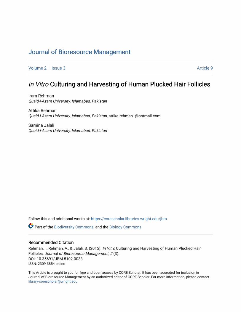
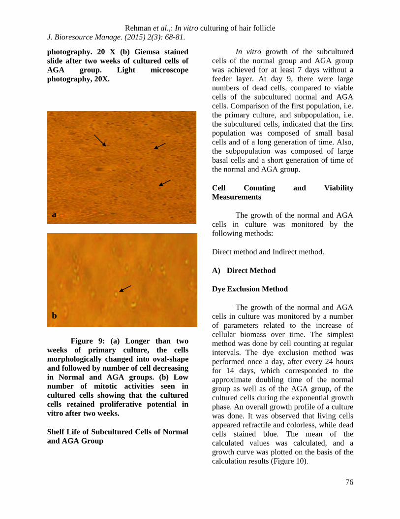
photography. 20 X (b) Giemsa stained slide after two weeks of cultured cells of AGA group. Light microscope photography, 20X.
Figure 9: (a) Longer than two weeks of primary culture, the cells morphologically changed into oval-shape and followed by number of cell decreasing in Normal and AGA groups. (b) Low number of mitotic activities seen in cultured cells showing that the cultured cells retained proliferative potential in vitro after two weeks.
Shelf Life of Subcultured Cells of Normal and AGA Group
In vitro growth of the subcultured cells of the normal group and AGA group was achieved for at least 7 days without a feeder layer. At day 9, there were large numbers of dead cells, compared to viable cells of the subcultured normal and AGA cells. Comparison of the first population, i.e. the primary culture, and subpopulation, i.e. the subcultured cells, indicated that the first population was composed of small basal cells and of a long generation of time. Also, the subpopulation was composed of large basal cells and a short generation of time of the normal and AGA group.
Cell Counting and Viability Measurements
The growth of the normal and AGA cells in culture was monitored by the following methods:
Direct method and Indirect method.
A) Direct Method
Dye Exclusion Method
The growth of the normal and AGA cells in culture was monitored by a number of parameters related to the increase of cellular biomass over time. The simplest method was done by cell counting at regular intervals. The dye exclusion method was performed once a day, after every 24 hours for 14 days, which corresponded to the approximate doubling time of the normal group as well as of the AGA group, of the cultured cells during the exponential growth phase. An overall growth profile of a culture was done. It was observed that living cells appeared refractile and colorless, while dead cells stained blue. The mean of the calculated values was calculated, and a growth curve was plotted on the basis of the calculation results (Figure 10).
a
b

Rehman et al.,: In vitro culturing of hair follicle J. Bioresource Manage. (2015) 2(3): 68-81.
77
Cell growth curve for cultured hair follicle cells
The growth curve of the present study shows the results of counting the viable cells of cultured cells of human plucked hair follicles. The growth curve shows that 1.5 X 10⁴ cells of the AGA group were viable. In the case of the normal sample, 2.0 X 10⁴ cells were found to be viable on the first day. In both samples, i.e. AGA and normal, maximum viable cells were observed on the fifth day with 2.0 X 10⁴ cells/ml and 2.5 X 10⁴ cells/ml respectively. Gradual decline was observed at day 6 and minimum cells were observed at day 13, i.e. 1.3 X 10⁴ cells/ml of the AGA group and 2.0 X10⁴ cells/ml of the normal group, which shows the death of cells (Figure 11). Statistical analysis showed that there was significant (P = 0.0001) differences between the cell numbers of cultured cells of the normal and AGA group.
Figure 10: Cultured cells of various morphologies from human plucked hair follicle cells placed in
improved Neubauer chamber. Arrows showing the various types of cultured cells morphologies like keratinocytes, large polygonal cells, large and small spindle-shaped cells and compact colonies. Light microscope photography 100 X.
Figure 11: In Vitro Cell Growth Curve of Viable cultured cells of human hair follicle cells, plotted by collecting cells 24 hours after the start of culture, calculating the cell number every day for 1-15 days of culture of the cells, and calculating the mean values of the calculated cell number each day. Increase in viable cultured cells showing comparison between cultured cells of Normal human plucked hair follicle cells and AGA human plucked hair follicle cells. Number of cells is given in cells per ml by using dye exclusion method.
Cell growth curve for subcultured hair follicle cells
Figure 12 shows the results of counting the viable cells of subcultured cells of human plucked hair follicles by the dye exclusion method. The growth curve shows that 1.6 X 10³ cells of the AGA group were viable, while in the case of the normal sample, 2.0 X 10³ cells were viable on the first day. Although in both samples, i.e. the AGA and normal, maximum viable cells
0
1000000
2000000
3000000
4000000
5000000
6000000
0 5 10 15 20
days
cells
/ml
NORMAL AGA

Rehman et al.,: In vitro culturing of hair follicle J. Bioresource Manage. (2015) 2(3): 68-81.
78
were observed on the fifth day with 2.0 X 10⁴ cells/ml and 2.5 X 10⁴ cells/ml respectively. Gradual decline was observed at day 6 and minimum cells were observed at day 7 to 8, i.e. 1.4 X 10³ cells/ml of the AGA group and 1.9 X 10³ cells/ml of the normal group, which shows the death of cells.
Figure 12: In Vitro Cell Growth Curve of Viable subcultured cells of human hair follicle cells, plotted by collecting cells 24 hours after the start of subculture, calculating the cell number every day for 1-9 days of culture of the cells, and calculating the mean values of the calculated cell number each day. Increase in viable subcultured cells showing comparison between subcultured cells of Normal human plucked hair follicle cells and AGA human plucked hair follicle cells. Number of cells is given in cells /ml by using dye exclusion method.
B) Indirect Methods of Cell Determination
Indirect methods of estimating cell growth rely on the measurement of an intracellular cell component such as DNA or Protein. In the present study, DNA was extracted by using the protocol of DNA extraction of cultured cells. The OD of DNA samples of cultured and subcultured cells of the normal and AGA were measured at 260 nm and at 280 nm.
a) DNA Determination
Extraction of DNA from cultured cells of normal and AGA
Extraction of DNA from subcultured cells of normal and AGA
b) Optical Density Assay (OD of Cultured and Subcultured Cells of Normal and AGA Groups)
Optical density (OD) of DNA samples of cultured and subcultured cells of the normal and AGA were measured at 260 nm and at 280 nm. DNA was calculated using the formula. The concentration of DNA of both the groups was determined in µg/ml. The purity of DNA was also calculated by a 260/280 ratio of cultured and subcultured cells of both the groups, as shown in Table 1.
Table 1: Optical Density at 260nm, concentration of DNA in µg/ml and purity of DNA of cultured and subcultured cells of normal and AGA group. Data shown are mean ± SD from three independent experiments.
Groups Optical density (260nm)
DNA Concentration
(µg/ml)
DNA Purity (260/280)
Cultured Cells Normal group 0.29 ±0.01 0.220 1.82 AGA group 0.30±0.01 0.230 1.82
0
5000
10000
15000
20000
25000
30000
0 2 4 6 8 10
DAYS
Cel
ls/m
l
NORMAL AGA

Rehman et al.,: In vitro culturing of hair follicle J. Bioresource Manage. (2015) 2(3): 68-81.
69
Subcultured Cells
Normal group 0.53±0.017 0.365 1.81 AGA group 0.51±0.015 0.255 1.81
DISCUSSION
Since methods for the isolation and culture of human hair follicles were first published (Kondo et al., 1990; Philpott et al., 1990), many researchers have used these model systems to investigate hair follicle biology (Philpott et al., 1996). The major application of these models has been to investigate the possible role of growth factors in controlling hair follicle growth and differentiation (Philpott, 2000). The importance of in vitro hair follicle culture as a tool to assist hair biology research can best be demonstrated by referring to studies that give an indication of the variety of uses that can be made of these models. The in vitro cultured follicle is a powerful tool for the hair biologist. Keeping in view the importance of this model, the plucked hair follicle was chosen as an experimental model in this study.
Milner et al. (2002) reported that hair is formed by rapidly proliferating matrix keratinocytes in the bulb located at the base of the growing (anagen) follicle. The duration of anagen varies drastically between hairs of differing lengths, and scalp follicles can remain in anagen for many years. Yang et al. (1993) proposed that follicular stem cells are located in the bulge and during the anagen phase, these bulge cells are stimulated by the dermal papilla cells to undergo transient proliferation, yielding new follicular down growth. Yu et al. (2006) used human plucked anagen hair follicles for isolating the novel population of
multipotent adult stem cells from human hair follicles.
In the present study, we used the growth potential and properties of the cells originating from the bulge, which is a major repository of dermal papilla cells and stem cells. After digestion, we used a modified method of microdissected hair follicle that was precisely cut down, and the bulge was placed in dispersed cell cultures. The advantages of this approach were that the bulge avoided being damaged and the activity of the cells was kept in good condition.
Results of the present study showed the shelf life of the human plucked hair follicle cell culture was 13-15 days. Wesgate et al. previously reported that the in vitro growth of human hair follicles was up to 4 days in a partially defined medium containing serum. The same studies report that the prolonged in vitro growth of isolated human hair follicles was at least 9 days. This was achieved after an analysis of the contribution of certain components of the original medium and, by a process of elimination, deriving a completely defined medium supplemented only with antibiotics L-Glutamine, insulin and hydrocortisone. Wesgate et al. (2008) had shown that by [methyl-3H] thymidine autoradiography, the hair follicles grown in the defined medium maintained an in vivo pattern of DNA synthesis, and that the gross morphology and histology of the maintained hair follicles remained similar to that of the freshly isolated hair follicles. It was also shown that the patterns of keratin synthesis, as determined by [35S] methionine labelling, did not alter with maintenance.

Rehman et al.,: In vitro culturing of hair follicle J. Bioresource Manage. (2015) 2(3): 68-81.
79
In the present study, it was observed that the subcultured cells of the plucked normal and AGA human hair were larger in size then the cultured cells and had a significantly shorter generation time. It was observed that the number of subcultured cells was low as compared to cultured cells of both groups. Wells and Sieber, (2006) studied the morphological characteristics of cells derived from human plucked hair follicles in vitro. They observed that the first population of hair follicle cells in vitro, i.e. basal keratinocytes, were larger cells in size and had a shorter generation time in the second population.
The hair follicle bulge cells are an easily available human tissue as it is a cheap source of human tissue to study. The conclusion of the present study is that the plucked human hair follicles from normal and AGA groups can be harvested and cultured successfully without a feeder layer.
REFERENCES
Cotsarelis G (2006). Epithelial stem cells:a folliculocentric view. J. Invest. Dermatol. 126, 1459-1468
Cotsarelis G, Sun TT, Lavker RM (1990). Label-retaining cells reside in the bulge area of pilosebaceous unit: implications for follicular stem cells, hair cycle and skin carcinogenesis. Cell. 61, 1329-1337
Gho GC, Braun JE, Tilli CM, Neumann (2004). Human hair follicle stem cells; Their prescence in plucked hair and follicular cell culture. Brit. J. Dermatol.150, 860-868.
Kondo S, Hozumi Y, Aso K (1990). Organ culture of human scalp hair follicles: Effect of testosterone and oestrogen
on hair growth. Arch. Dermatol. Res. 282, 442–445.
Kopan R, Lee J, Lin MH, Syder AJ, Kesterson J, Crutchfield N, Li CR, Wu W, Books J, Gordon JI (2002). Genetic mosaic analysis indicates that the bulb region of coat hair follicles contains a resident population of several active multipotent epithelial lineage progenitors. Dev. Biol. 242 (1), 44-57.
Moll I (1995). Proliferative potential of different keratinocytes of plucked human hair follicles. J. Invest. Dermatol. 105, 14–21.
Norstrom RE (1979). Synchronous balding of scalp and hair bearing grafts of scalp transplanted to the skin of the arm in male pattern baldness. Acta. Derm. Venereol. 59, 266-268.
Philpott MP, Green MR, Kealey T (1990). Human hair growth in vitro. J. Cell Sci. 97, 463-471.
Philpott MP, Sanders DA, Kealey T (1996). Whole human hair follicle culture. In: Whiting DA (Ed). Dermatologic Clinics. Philadelphia: W.B. Saunders Press, 595-607.
Philpott MP (2000). The roles of growth factors in hair follicles: Investigations using cultured hair follicles. In: Camacho FM, Randall VA, Price VH (eds). Hair and its Disorders: Biology, Pathology and Management. London, Martin Dunitz, 103-113
Randall VA, Hibberts NA, Hamada K (1996). A comparison of the culture and

Rehman et al.,: In vitro culturing of hair follicle J. Bioresource Manage. (2015) 2(3): 68-81.
80
growth of dermal papilla cells from hair follicles from non-balding and balding (androgenetic alopecia) scalp. Brit. J. Dermatol. 134, 437-444.
Savin RC (1987). Use of topical minoxidil in the treatment of male pattern baldness. J. Am. Acad. Dermatol. 16, 696-704.
Taylor G, Lehrer MS, Jensen PJ, Sun TT, Lavker RM (2000). Involvement of follicular stem cells in forming not only the follicle but also the epidermis. Cell. 102, 451-461.
Trueb RM (2002). Molecular mechanisms of androgenetic alopecia. Exp. Gerontol. 37, 981–990.
Wells J, Sieber VK (2006). Morphological characteristics of cells derived from plucked human hair in vitro. Brit. J. Dermatol. 113 (6), 669-675.
Westgate GE, Gibson WT, Kealey T, and Philpott MP 2008 Prolonged maintenance of human hair follicles in vitro in a serum-free medium. Brit. J. Dermatol. 129 (4), 372 – 379.
WU W, RJ Morris (2005). Method for the harvest and assay of In vitro clonogenic keratinocytes stem cells from mice. Methods Mol. Biol. 289, 79-86.
Yang JS, Robert ML, Sun TT (1993). Upper human hair follicle contains a subpopulation of keratinocytes with superior in vitro proiferative potential. J. Investigat. Dermatol. 101, 652-659.
Yi Z, Mingming X, Yun W, Jun Y, Yijun Z, Jin Y, Tian Y (2006). Bulge cells of human hair follicles: segregation,
cultivation and properties. Elsevier B.V. Available online 4 January 2006.
Yu H, Dong F, Suresh MK, Ling L, Thiennga KN, Geza A, Meenhard H, Xiaowei X (2006). Isolation of a novel population of multipotent adult stem cells from human hair follicles. Am. J. Pathol. 168, 1879-1888.