in vitro methodologies for assessing cutaneous irritation ... · in vitro methodologies for...
TRANSCRIPT

In Vitro Methodologies for Assessing Cutaneous Irritation, Sensitization
and PhotosensitivityDonald V. Belsito, MD
Leonard C. Harber Professor of DermatologyColumbia University Medical Center
New York, NY

Conflict of Interests
None – but in the spirit of full disclosureI am a member of the Cosmetic Ingredient Review’s
Expert Panel I am a member and chair of the Expert Panel for
Fragrance SafetyI am reimbursed for my travel & time for both from
the Personal Care Products Council (Washington, DC) and the Research Institute for Fragrance Materials (Woodcliff Lake, NJ), respectively

IRRITANT CONTACT DERMATITIS
1) Acute primary irritation
2) Cumulative irritation
3) Corrosion
4) Phototoxicity

CHEMICAL IRRITANCY
Inherent chemical properties
Concentration
Amount
Duration / frequency
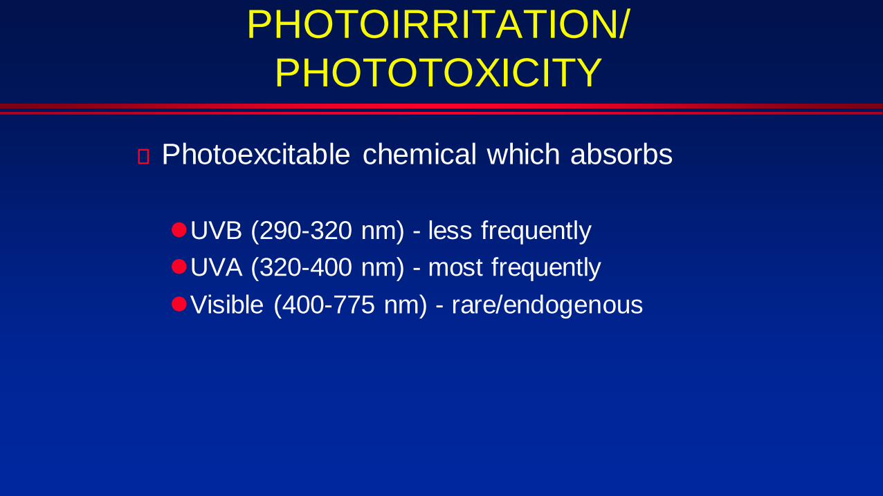
PHOTOIRRITATION/PHOTOTOXICITY
Photoexcitable chemical which absorbs
UVB (290-320 nm) - less frequentlyUVA (320-400 nm) - most frequentlyVisible (400-775 nm) - rare/endogenous

IN VITRO METHODS FOR CORROSION
rat skin transcutaneous electrical resistance (OECD TG 430) liquids (150 υl) & solids (“sufficient to cover surface”) x 24h TER > 5 kΩ, non-corrosive; < 5 kΩ, corrosive not applicable to surfactants or neutral organics
– need to use dye (sulforhodamine B) penetration http://www.oecd-ilibrary.org/environment/test-no-430-in-vitro-skin-corrosion-transcutaneous-
electrical-resistance-test-method-ter_9789264203808-en. Corrositex (reconstituted collagen matrix; OECD TG431)
liquids (500 υl) & solids (500 mg) breakthrough time correlated to corrosivity applicable only to materials that produce a color or physical change in the
“chemical detection system” w/in 5 mins http://www.oecd-ilibrary.org/environment/test-no-431-in-vitro-skin-corrosion-human-skin-model-
test_9789264071148-en
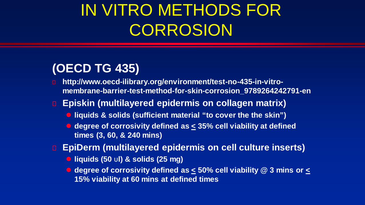
IN VITRO METHODS FOR CORROSION
(OECD TG 435) http://www.oecd-ilibrary.org/environment/test-no-435-in-vitro-
membrane-barrier-test-method-for-skin-corrosion_9789264242791-en Episkin (multilayered epidermis on collagen matrix)
liquids & solids (sufficient material “to cover the the skin”) degree of corrosivity defined as < 35% cell viability at defined
times (3, 60, & 240 mins) EpiDerm (multilayered epidermis on cell culture inserts)
liquids (50 υl) & solids (25 mg) degree of corrosivity defined as < 50% cell viability @ 3 mins or <
15% viability at 60 mins at defined times

IN VITRO METHODS FOR ACUTE IRRITATION
Acute: (OECD TG 439) http://www.oecd-ilibrary.org/environment/test-no-439-in-
vitro-skin-irritation-reconstructed-human-epidermis-test-method_9789264242845-en
EpiSkin™ EpiDerm™ SIT (EPI-200) Modified EpiDerm (EPI-200) SkinEthic™ RHE

IN VITRO: ACUTE IRRITATION
Dose: minimum of 25 μL/cm2 (liquid) or 25 mg/cm2 (solid) Incubation: 3 hrs Viability: mitochondrial dehydrogenases to reduce the vital
dye MTT The test substance is considered to be: irritant to skin if the tissue viability after exposure and
post-treatment incubation is ≤ to 50 %.no category if the tissue viability after exposure and
post-treatment incubation is > 50 %.

IN VITRO: CHRONIC CUMULATIVE IRRITATION
Chronic cumulative irritationno ECVAM / OECD acceptable in vitro methodUnder development:
– Reconstructed organotypic skin model (ROSM) w/ keratinocytes, basement membrane & fibroblasts
– Cultured for varying lengths of time w/ chemical in ?– Assay for ↑ heat shock protein (HSP)-27– Methodology assessed w/ SLS and acute irritants
(acids & bases) Chen, et al. Toxicology Letters. 226: 124 = 131, 2014
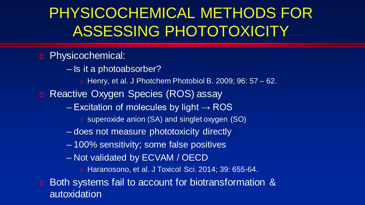
PHYSICOCHEMICAL METHODS FOR ASSESSING PHOTOTOXICITY
Physicochemical:– Is it a photoabsorber?
Henry, et al. J Photchem Photobiol B. 2009; 96: 57 – 62.
Reactive Oxygen Species (ROS) assay– Excitation of molecules by light → ROS
superoxide anion (SA) and singlet oxygen (SO)– does not measure phototoxicity directly– 100% sensitivity; some false positives– Not validated by ECVAM / OECD
Haranosono, et al. J Toxicol Sci. 2014; 39: 655-64.
Both systems fail to account for biotransformation & autoxidation

ROS (Japanese Center for the Validation of Alternative Methods): PHOTOTOXICITY
Judgment Conc. SO (mean X 3) SA (mean X3)(% ↑, irradiated / non-irradiated)
Photoreactive 200 μM ≥25 and ≥70<25 and ≥70≥25 and <70
Weakly photoR 200 μM <25 and ≥20, <70Non-photoR 200 μM <25 and <20Inconclusive: The results do not meet the above-mentioned criterion.
Solvents: DMSO or 20 mM of NaPBControls: Quinine hydrochloride (+); Sulisobenzone (-)Irradiation: Solar stimulator x 1h @ controlled temperature
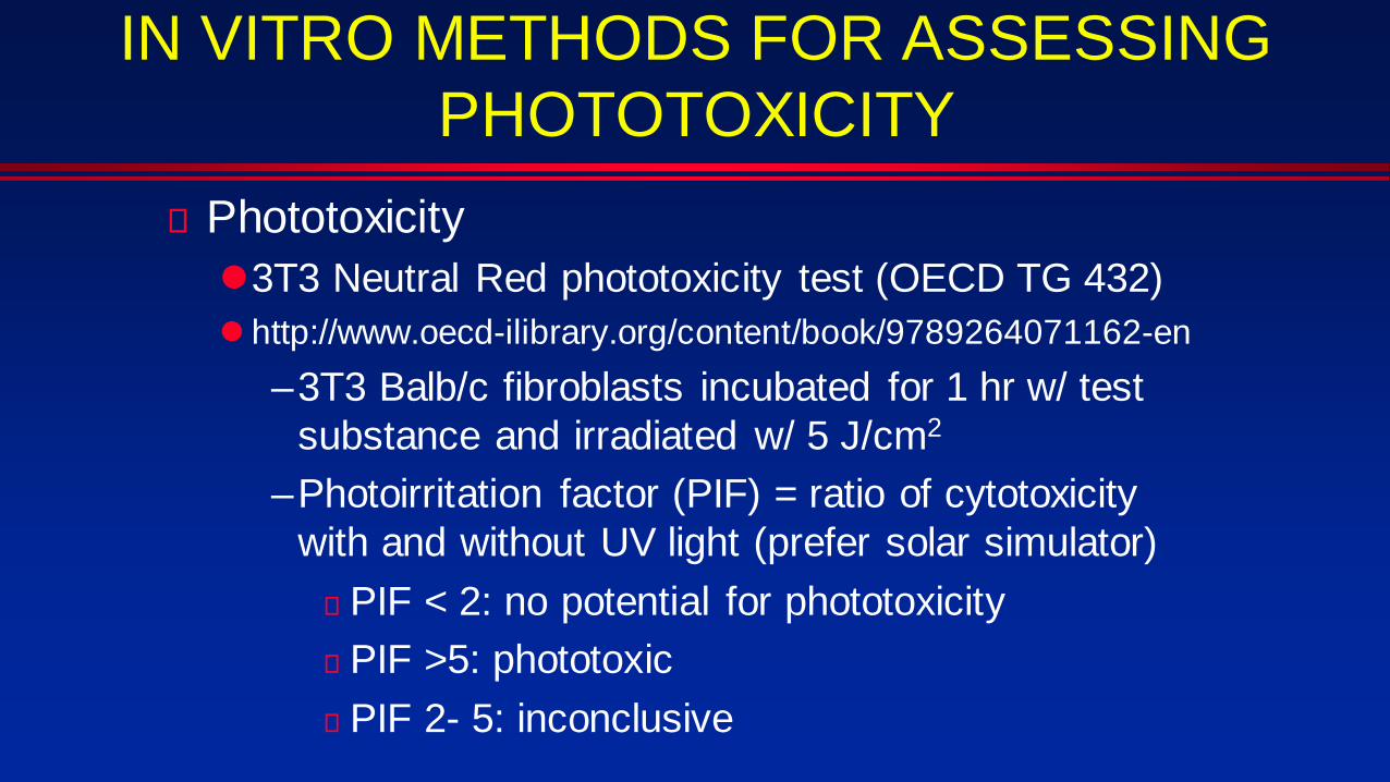
IN VITRO METHODS FOR ASSESSING PHOTOTOXICITY
Phototoxicity3T3 Neutral Red phototoxicity test (OECD TG 432) http://www.oecd-ilibrary.org/content/book/9789264071162-en
–3T3 Balb/c fibroblasts incubated for 1 hr w/ test substance and irradiated w/ 5 J/cm2
–Photoirritation factor (PIF) = ratio of cytotoxicity with and without UV light (prefer solar simulator) PIF < 2: no potential for phototoxicity PIF >5: phototoxic PIF 2- 5: inconclusive

DRAWBACKS TO 3T3
false negativestoxicological hazard Primarily due to a mechanism of
action not captured by the cell line usedthe cell lines lack of metabolic capacity, compounds
metabolized in vivo to biologically active forms may be missed
false positive limited bioavailability in vivo due to poor absorption and
distribution, or rapid biotransformation and excretion

IN VITRO METHODS FOR ASSESSING PHOTOTOXIC METABOLITES
Enhanced Phototoxicity Assay in Reconstituted Skin (EPARS) -- Portes, et al. Toxicol In Vitro. 2002; 16: 765-70.
Enzymatic reactive oxygen species assay (eROS) -- Kato, et al. Enzymatic reactive oxygen species assay to evaluate phototoxic risk of metabolites. Toxicol Lett. 2017; 278: 59-65.

Enhanced Phototoxicity Assay in Reconstituted Skin (EPARS)
100 μl test substance directly to tissue surface; incubated at 37ºC x 18-24 h
UV Irradiation (solar simulator): 6 J/cm2; control = non-irradiated
Tissues and incubated x 18-24 h at 37ºC Tissue viability measured using the MatTek MTT Viability
Assay protocolCurrently uses cell viability (< 30%) and PIF > 2 PGE2 levels as alternative to viability
Prevalidation suggests under-prediction and a precautionary factor of 10 for extrapolation to man
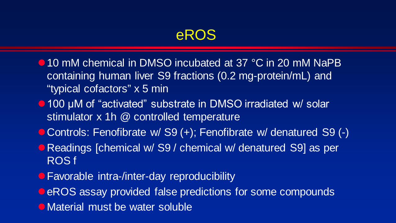
eROS
10 mM chemical in DMSO incubated at 37 °C in 20 mM NaPB containing human liver S9 fractions (0.2 mg-protein/mL) and “typical cofactors” x 5 min 100 μM of “activated” substrate in DMSO irradiated w/ solar
stimulator x 1h @ controlled temperatureControls: Fenofibrate w/ S9 (+); Fenofibrate w/ denatured S9 (-)Readings [chemical w/ S9 / chemical w/ denatured S9] as per
ROS fFavorable intra-/inter-day reproducibility eROS assay provided false predictions for some compoundsMaterial must be water soluble

ALLERGIC CONTACT DERMATITIS
AllergicPre-haptensPro-haptens
PhotoallergicPre-haptensPro-haptens

IN VITRO METHODS FOR ASSESSING ALLERGY: AOP
1: Molecular Initiation• Direct Peptide Reactivity Assay (DPRA)
2: Keratinocyte Activation• KeratinoSens™ (ARE-Nrf2 Luciferase Test Method)• IL-8 Luc Assay (not ECVAM; +JaCVAM)
3: Dendritic Cell Activation• Human Cell line Activation Test (h-CLAT)• Myeloid U937 skin sensitization test (MUSST; ECVAM,
not OECD)4: T cell activation
• none

DPRA (OECD TG 442C) peptide to chemical ratio used is 1:10 (cysteine)
and 1:50 (lysine) [controls: cinnamic aldehyde (+); vehicle (-)]Cys & Lys: % depletion (mean)
Minimal Reactivity < 6.38% Low Reactivity: > 6.38 - < 22.62% Moderate Reactivity: > 22.62 - < 42.47% High Reactivity: > 42.47% http://www.oecd-ilibrary.org/environment/test-no-442c-in-
chemico-skin-sensitisation_9789264229709-en

LIMITATIONS of DPRADPRA test method does not contain a metabolic/bioactivation system, therefore pro-haptens& pre-haptens are not detected pre-haptens (i.e. simple chemical transformation, for
example oxidation by air or photo-activation in the presence of UV light)
pro-haptens (i.e. chemicals requiring enzymatic activation) peroxidase peptide reactivity assay “PPRA” identifies pro-
haptens via the use of peroxidase in the DPRA– Gerberick. Altern Lab Anim. 2016; 44: 437-442.

KeratinoSens ASSAY (OECD TG 442D) Based on a stable reporter construct consisting of a luciferase gene under
the control of the antioxidant response element (ARE) in a HaCaT (immortalized keratinocyte) cell line.
The luciferase gene allows for the detection of sensitization potential based on the bioluminescent activity of ATP: light is produced after the breakdown of the protein luciferin by luciferase.
Inserted genes allow for the exploitation of the signaling pathway of Keap1-Nrf2, which has previously been shown to be activated during skin sensitization events (Natsch & Emter, 2008; Natsch et al., 2009)
Cell viability also assessed > 2/3 trials show 50% increased induction of luciferase gene; viability not a
criterion; (-) control, solvent; (+) control, cinnamic aldehyde http://www.oecd-ilibrary.org/environment/test-no-442d-in-vitro-skin-
sensitisation_9789264229822-en.

IL-8 Luc AssayTHP-1 cell line is a human monocyte line; not keratinocyte

H-CLAT (OECD TG 442E) / MUSST*
CD86 >150% or CD54: >200% = positive; *MUSST similar; uses Myeloid U937 cell line and only CD86; ECVAM validated; awaiting OECD TG.
http://www.oecd-ilibrary.org/content/book/9789264264359-en
DNCB = (+) control;Medium = (-) control

IN VITRO METHODS FOR ASSESSING Pro-HAPTEN PHOTOSENSITIZATION
No accepted methodology; no photo-activated DPRA has been proposed.
in vitro testing methodologies utilizing KeratinoSens and h-CLAT with the addition of exposure to 5J/cm2 of UVA
– photo-KeratinoSensTM assay Tsujita-Inoue et al. J Appl Toxicol. 2016; 36: 956-968
– photo-h-CLAT assay Hoya, et al. Toxicol In Vitro. 2009; 23: 911-918.
In vitro systems to assess photosensitization are critical– EU ban on testing cosmetic ingredients in animals (sunscreens are
cosmetic in the EU!)– testing for photoallergenicity in humans unethical due to potential for the
induction of persistent light reactivity (PLR): extreme sensitivity to UVB in absence of inducing photoallergen
Sensitization Hazard (not risk) Integration of results of testing on the first three
steps of AOP to predict sensitization hazard not resolved
Various models proposedBayesian networkArtificial Neural Network Weight of Evidence
– Weight of Evidence or best “two out of three” to assess sensitization hazard AOP has found the greatest success
But what about risk??

The picture can't be displayed.
Thank you!