in vitro production of haploid cells after coculture of ... · in vitro production of haploid cells...
TRANSCRIPT
ORIGINAL ARTICLE: ANDROLOGY
In vitro production of haploid cellsafter coculture of CD49fD withSertoli cells from testicular spermextraction in nonobstructiveazoospermic patients
Marcia Riboldi, B.Sc.,a,b Carmen Rubio, Ph.D.,c Antonio Pellicer, M.D., Ph.D.,d Manuel Gil-Salom, Ph.D.,dand Carlos Sim�on, M.D., Ph.D.a,b,c,d
a Valencia Node of the Spanish Stem Cell Bank, Prince Felipe Research Centre (CIPF); b Fundaci�on IVI-InstitutoUniversitario IVI; c Iviomics; and d Instituto Valenciano de Infertilidad (IVI), Valencia, Spain
Objective: To isolate CD49fþ cells from testicular sperm extraction (TESE) samples of azoospermic patients and induce meiosis bycoculturing these cells with Sertoli cells.Design: Prospective analysis.Setting: Research center.Patient(s): Obstructive azoospermic (OA) and nonobstructive azoospermic (NOA) patients.Intervention(s): TESE, with enzymatic dissociation of samples to obtain a cell suspension, which was cultured for 4 days with 4 ng/mLGDNF. The CD49fþ cells were sorted using fluorescence-activated cell sorting (FACS) as a marker to identify spermatogonial stem cells(SSCs), which were cocultured with Sertoli cells expressing red fluorescent protein (RFP) in knockout serum replacement (KSR) mediawith addition of 1,000 IU/mL of follicle-stimulating hormone (FSH), 1 mM testosterone, 40 ng/mL of GDNF, and 2 mM retinoic acid (RA)for 15 days in culture at 37�C and 5% CO2 to induce meiotic progression. Cells were collected and analyzed by immunofluorescence formeiosis progression with specific markers SCP3 and CREST, and they were confirmed by fluorescence in situ hybridization (FISH).Main OutcomeMeasure(s): Isolation of CD49fþ cells and coculture with Sertoli cells, meiosis progression in vitro, assessment of SSCsand meiotic markers real-time polymerase chain reaction (RT-PCR), immunohistochemical analysis, and FISH.Result(s): The CD49fþ isolated from the of total cell count in the TESE samples of azoospermic patients varied from 5.45% in OA to2.36% in NOA. Sertoli cells were obtained from the same TESE samples, and established protocols were used to characterize them aspositive for SCF, rGDNF, WT1, GATA-4, and vimentin, with the presence of tight junctions and lipid droplets shown by oil redstaining. After isolation, the CD49fþ cells were cocultured with RFP Sertoli cells in a 15-day time-course experiment. Positiveimmunostaining for meiosis markers SCP3 and CREST on days 3 to 5 was noted in the samples obtained from one NOA patient.A FISH analysis for chromosomes 13, 18, 21, X, and Y confirmed the presence of haploid cells on day 5 of the coculture.Conclusion(s): In vitro coculture of SSCs from TESE samples of NOA patients along with Sertoli cells promotedmeiosis induction and re-
Use your smartphone
sulted in haploid cell generation. These results improve the existing protocols to generate sper-matogenesis in vitro and open new avenues for clinical translation in azoospermic patients.(Fertil Steril� 2012;-:-–-. �2012 by American Society for Reproductive Medicine.)Key Words: CD49fþ cells, coculture, gametes, germ line, in vitro, meiosis, Sertoli cells,spermatogenesis, spermatogonial stem cell
Discuss: You can discuss this article with its authors and with other ASRM members at http://www.ASRM.org/fns-xx-x-xxx
to scan this QR codeand connect to thediscussion forum forthis article now.*
* Download a free QR code scanner by searching for “QRscanner” in your smartphone’s app store or app marketplace.
Received December 8, 2011; revised April 15, 2012; accepted May 25, 2012.M.R. has nothing to disclose. C.R. has nothing to disclose. A.P. has nothing to disclose. M.G.-S. has
nothing to disclose. C.S. has nothing to disclose.Reprint requests: Marcia Riboldi, B.Sc., Fundaci�on IVI, CIPF Bank Stem Cell, Paterna, Valencia 46980,
Spain (E-mail: [email protected]).
Fertility and Sterility® Vol. -, No. -, - 2012 0015-0282/$36.00Copyright ©2012 American Society for Reproductive Medicine, Published by Elsevier Inc.http://dx.doi.org/10.1016/j.fertnstert.2012.05.039
VOL. - NO. - / - 2012
O bstructive (OA) and nonob-structive (NOA) azoospermiaare major causes of male
infertility. Testicular sperm extraction(TESE) is usually performed to searchfor sperm that will be later used in the in-tracytoplasmic sperm injection (ICSI)technique (1). Spermatogenesis is normal
1

ORIGINAL ARTICLE: ANDROLOGY
in OA patients, so TESE usually solves the problem (2). However,in patients whose NOA is caused by a variety of pathologies thatlead to spermatogenesis arrest, their only chance for paternitylies in sperm banks with donor sperm (3).
Essentially, spermatogenesis is a process by which germcells (GCs) proliferate and differentiate into haploid sperm.Germ cells initially migrate to the basement membrane ofthe seminiferous tubules of the testis, where they maintaintheir ability to proliferate and differentiate to sperm underthe influence of Sertoli cells throughout a lifetime (4–6).
Accumulated evidence demonstrates the existence ofa stem cell population in human adult testes (7–12).Spermatogonial stem cells (SSCs) are the adult stem cellpopulation that renews itself and produces spermatogeniccells after transplantation into the seminiferous tubules ofinfertile recipient males, constituting a model system tounderstand spermatogenesis (13). Isolation of the SSCpopulation can be achieved by sorting them by the presenceof the cell surface markers CD49f, GPR-125, and GFRA-1 (7,11, 14, 15) and by further culturing them with the addition ofgrowth factors such as GDNF, LIF, EGF, GFRA-1, or bFGF (11).
In humans, in vitro spermatogenesis from SSCs to obtainhaploid sperm has yet to be achieved. Researchers have inves-tigated this process via various strategies such as combinationsof growth factors or hormones or/and coculture of germ cellswith testicular tissue (16–22). Recently, important advancesin the in vivo production of sperm in mice that have resultedin healthy offspring have used two different approaches: thefirst consists of starting with an epiblast-like cell intermediatethat is transplanted into mouse testes (23); the second com-prises an organ culture system of SSCs in prepubertal gonadaltissue (24). Our study isolated SSCs from the TESE samples ofazoospermic men to induce the meiosis process in vitro byuse of a coculture system with Sertoli cells.
MATERIALS AND METHODSHuman Samples
Our study was approved by the institutional review board ofthe Instituto Valenciano de Infertilidad (IVI) in accordancewith Spanish legislation (NCT: 01375662). The samples wereobtained after signed, informed consent from the patients.The samples, composed of seminiferous tubules and interstitialtissue, were obtained from20 azoospermic patients (OA, n¼ 9;NOA, n ¼ 11) as part of their diagnosis/therapeutic workup.The spermobtained from the TESE sampleswas frozen for sub-sequent use in ICSI, and the residual tissue was transported inbasic media and maintained on ice for 60 to 90 minutes aftersurgery. The tissue was washed three times in Hank's balancedsalt solution 1x (HBSS; PAA)with penicillin/streptomycin (In-vitrogen); the samples were mechanically dissected out intosmall pieces (1 mm3) and enzymatically dissociated with col-lagenase type IA (1,000 IU/mL; Sigma) for 20 minutes at 37�Con a shaker. Then the samples were incubated with TrypLE Se-lect (Invitrogen) for 10 minutes at 37�C on a shaker, filteredthrough a 50-mm mesh (Partec), and centrifuged at 1,000rpm for 5 minutes. The cell pellet was seeded on a plate (max-imum of 2� 106 cell/cm2 for a culture dish) containing mediasupplemented with 20% embryonic stem-cell qualified fetal
2
bovine serum (ES-qualified FBS; Invitrogen), plus 4 to 40ng/mL of GDNF (Sigma); this was incubated at 32�C in 5%CO2 for 4 days.
CD49fD Cell Isolation
After 4 days of culture, the media were collected, and the cul-tured cells were gently washed twice with Dulbecco's modi-fied Eagle medium (DMEM; Invitrogen). They were thenwashed once with phosphate-buffered saline (PBS; PAA) torecover all the unattached cells and to discard the monolayeradherent cells. The collected cells were centrifuged at 1,000rpm for 5 minutes, suspended in basic media, and filteredthrough a 30-mm filter (Partec). The single-cell suspensionobtained was then incubated with 5% bovine serum albumin(BSA; Sigma) in PBS for 30 minutes at 4�C, washed with 0.1%BSA in PBS, and incubated with an Alexa Fluor 488 1:100conjugated antibody against the CD49f surface marker(a6-integrin; Biolegend) on ice for 45 minutes. Peripheralblood lymphocytes were used as positive controls for CD49f,while human skin fibroblast feeder cells were employed asthe negative control. The cells that expressed positive forCD49f were separated from the whole population by fluores-cence-activated cell-sorting (FACS) (MoFlo Modular FlowCytometer; Beckman Coulter).
Sertoli Cell Purification
After we had obtained the cell suspension from the TESE sam-ples, the cells were resuspended in Dulbecco's modified Eaglemedium/nutrient mixture F-12 (DMEM/F-12; GIBCO, Invitro-gen) supplemented with 10% FBS; these were seeded intoculture plates and incubated at 37�C in 5% CO2 for 10 min-utes. After incubation, the media containing different sper-matogenic cell types were removed, and the attached cellswere cultured for approximately 1 week; the media werechanged every other day until reaching confluence. The cellmonolayer was trypsinized with TrypLE Select diluted (1:1)in PBS (PAA) for 5 minutes at 37�C (25). The cell numberwas calculated, and 1.106 cells were then incubated with 0.5mg/mL Nile red (Sigma) for 15 minutes in PBS. The cellswere filtered through a 50-mm sieve (Partec), and cell purifi-cation was performed using a cell sorter (MoFlo; BeckmanCoulter). The Sertoli cells positive for Nile red staining wereseeded at a density of 50,000 cells/well with a collagen matrix(Biocoat Collagen I; Becton Dickinson) in DMEM/F-12 sup-plemented with 10% FBS, and were cultured for 10 days at37�C in 5% CO2. Another demonstration of their nature wasperformed by oil red staining, electron microscopy for lipiddroplet and tight junctions, and polymerase chain reactionfor specific markers such as the GDNF receptor (GAF), stemcell factors (SCF), and transcription factors GATA-4 andWilms tumor (WT-1).
Viral Transduction of Sertoli Cells with RedFluorescent Protein (RFP)
Sertoli cells were transfectedwith a lentiviral vector coding forred fluorescent protein (RFP), as previously described by ourgroup (26). Briefly, lentiviral stocks were obtained using thepackaging cell line 293-T (American Type Culture Collection,
VOL. - NO. - / - 2012
Fertility and Sterility®
Manassas, Virginia) by the transient cotransfection of 1 mg ofplasmidsA179-Helix-UBC-RFP (kindly provided by theGeno-micsUnit, SpanishNational CancerCentre, CNIO), 1mgofGAGPOL-BAM HI (virus survival), and 1 mg of VSVG-ECORT (en-capsulation virus) plasmids using Fugene HD (Roche Diagnos-tics). The lentiviral supernatantwas harvested at 48 hours aftertransfection and used to infect Sertoli cells for 12 hours in thepresence of 10 mg/mL of polybrene (Sigma-Aldrich), whichwas then incubated at 37�C in 5%CO2. After 48 hours, the cellswere analyzed under a fluorescence microscope to observe thetransduced cells' efficiency to express RFP.
Coculture System of CD49fD and Sertoli-RFP Cells
Sertoli-RFP cells were seeded on 12-well plates with a colla-genmatrix (Biocoat Collagen I; Becton Dickinson) at a densityof 10,000 cells/well with DMEM/F-12 (GIBCO, Invitrogen)supplemented with 10% FBS. The plates were incubated for24 hours at 37�C in 5% CO2 to allow for cell attachment.The CD49fþ-sorted cells were directly seeded onto a Ser-toli-RFP feeder layer at a density of 3,000 cells/well andwere grown in knockout D-MEM (Ko-DMEM; GIBCO, Invitro-gen) supplemented with 20% knockout serum replacement)(KSR; GIBCO, Invitrogen), 1,000 IU/mL of follicle-stimulating hormone (FSH; Sigma), 1 mM testosterone(Qpharma), 40 ng/mL of GDNF (Sigma), and 2 mM retinoicacid (RA) (Sigma) at 37�C in 5% CO2. The cells were collectedat 48 hours for further analysis.
RNA Extraction and RT-PCR Analysis
The RNA extraction was performed using the Mini RNAIsolation I Kit (Zymo Research) according to the manufac-turer's protocol, and the integrity was analyzed in a Nano-Drop Spectrophotometer (NanoDrop Technologies) for RNAquantification and purity. Complementary DNA (cDNA) wassynthesized by use of real-time polymerase chain reaction(RT-PCR) with an MMLV enzyme (Clontech; BD Biosci-ences) and 500 ng of RNA per sample. The RT-PCR analysisof CD49fþ cells was performed using the Advantage RT-PCR Kit (Clontech; BD Biosciences) following the manufac-turer's protocol. The cDNA from each sample was used asa PCR template to detect the expression of those geneswith the primer sequences presented in SupplementalTable 1 (available online). The program used was 95�C de-naturation for 10 minutes, 35 cycles in 94�C for 30 seconds,57�C for 30 seconds, and 72�C for 1 minute. The positivecontrol was human testis tissue, and the negative controlwas human foreskin, which were included in each reaction.The PCR products were resolved on 2% agarose gels,stained with ethidium bromide and visualized in a transillu-minator (BioRad).
Immunocytochemistry
The CD49fþ and Sertoli cells were seeded on a chamber slide,cultured for 3 days, fixed with 4% paraformaldehyde for 20minutes, and washed with PBS. Two protocols were followedfor staining. For the membrane surface markers, PBS was dis-carded, and the cells were washed three times with rinse buffer
VOL. - NO. - / - 2012
1X—20 mM Tris (Fluka), HCl (J.T. Baker), 0.15 NaCl (Sigma-Aldrich), 0.05% Tween-20 (Sigma-Aldrich)—with PBS for per-meabilization. Cells were then incubated for 10 minutes withTriton X100 0.1% (Sigma-Aldrich) and blocked with 4% nor-mal goat serum (NGS; Sigma-Aldrich) for 30 minutes. Next,the primary antibodies CD49f (1:100) (Alexa Fluor 488 anti-human/mouse CD49f; Biolegend), GPR125 (1:200) (rabbitpolyclonal GPR125; Abcam), THY-1 (1:100) (mouse anti-human THY-1 monoclonal antibody R-PE; Chemicon), andVimentin (1:200) (mouse monoclonal vimentin antibody; Ab-cam) were added for 1 hour at room temperature; after incu-bation, this was washed three times with rinse buffer 1X andthen was incubated for 1 hour with the corresponding second-ary antibody (1:500) (goat anti-rabbit IgG Alexa Fluor 488;Molecular Probes/goat anti-mouse IgG1-Alexa Fluor 568;Molecular Probes). Finally, the cells were washed with rinsebuffer 1X and Prolong Gold Antifade Reagent with DAPI(Molecular Probes). For the nuclear markers, the PBS was dis-carded, and the cells were washed three times with BSA 0.1%(Sigma-Aldrich) for permeabilization and blocked by incu-bating for 45 minutes at room temperature with 0.1% TritonX100 (Sigma-Aldrich), 10% normal donkey serum (NDS;Sigma-Aldrich), or normal goat serum (NGS; Sigma-Aldrich) in 1% BSA (Sigma-Aldrich). After incubation, thecells were washed three times with BSA 0.1% and incubatedovernight at 4�Cwith primary Gata-4 antibody (1:200) (rabbitpolyclonal Gata-4 antibody; Santa Cruz Biotechnology). Thenext day, cells were washed three times with BSA 0.1% andincubated with the corresponding secondary antibody(1:500) (rabbit anti-goat IgG-Alexa Fluor 568; MolecularProbes) for 1 hour at room temperature. Finally, they werewashed with BSA 0.1%, and Prolong Gold Antifade Reagentwith DAPI (Molecular Probes) was added.
Oil Red Staining
Sertoli cells were seeded in six-well plates, cultured for 5 days,fixed with 4% paraformaldehyde (PFA) (Electron MicroscopySciences) for 20 minutes, and then washed with PBS (PAA).After the first wash, 0.5% oil red solution (Sigma) in isopropa-nol 100% (J.T. Baker) was prepared and filtered twice througha 0.45-mm pore mesh (Partec); then the stock solution was di-luted (3:2) to be filtered again twice. For staining, Sertoli cellswere incubated for 5 minutes at 150 rpm, washed three timeswith PBS (PAA) at 4�C, counterstained with hematoxylin-filtered samples for 3 minutes, and washed with H2O. Finally,cells were observed under the microscope.
Electron Microscopy
For the electron microscopy analysis, cells were washed threetimes with 0.1 M phosphate buffer (PB; Electron MicroscopySciences), fixed with 3.5% glutaraldehyde (Electron Micros-copy Sciences) for 45 minutes at 37�C, and subsequentlywashed with 0.1 M PB. Samples were then post-fixed with0.5% osmium, rinsed, dehydrated, and embedded in Araldite(Durcupan; Fluka). Semithin sections (1.5 mm) were cut witha diamond knife and stained lightly with 1% toluidine blue.Semithin sections were reembedded in an Araldite block
3

ORIGINAL ARTICLE: ANDROLOGY
and detached from the glass slide by repeated freezing withliquid nitrogen and thawing. The block with the semithin sec-tions was cut into ultrathin (0.05 mm) sections with a diamondknife, stained with lead citrate, and examined under an elec-tron microscope (Jeol 100CX).
Analysis of Meiotic Markers SCP3, MLH1, andCREST
Cells from the coculture were harvested every 2 to 3 days bycollecting the supernatant and trypsinizing the adherent cells.Cells were then centrifuged for 5 minutes at 12,000 rpm, andthe supernatant was discarded. The pellet was then resus-pended in 700 mL of prewarmed 0.075 mol/L potassium chlo-ride solution (KCl) (GIBCO) and incubated for 15 minutes at37�C. Cells were fixed with 700 mL of 1% paraformaldehyde(PFA; Electron Microscopy Sciences) for preparation on slides(Thermo Scientific Superfrost Slides). Slides were dried for ap-proximately 24 hours at 37�C in a humid chamber andwashed for 2 minutes in 0.4% Photo-Flo (Kodak). Cells weresubjected to immunofluorescence following the protocol de-scribed by Sun et al. (27) using antibodies against SCP3 (lat-eral elements of the SC marker) (Novus Biologicals), MLH1(Becton Dickinson), and CREST (centromere marker) (FisherScientific) to identify the meiosis stage of the cells at eachtime point. As internal controls, both the CD49fþ-sorted cellsand the Sertoli-RFP cells were analyzed separately beforestarting the coculture with the indicated antibodies.
FIGURE 1
(A) Flow cytometry isolation of CD49f cells. (B) (I) Real-time polymerase cPIWILL2, BOULE, DCM1, SCP3, PRM1, TNP2, and Blimp1 in CD49fþ cells.Immunocytochemistry in CD49f positive cells: (I) CD49f, (II) dapi, and (III)and (IX) merge.Riboldi. Haploid cells after coculture of CD49f+ with Sertoli cells. Fertil Steril 2012.
4
Fluorescence in Situ Hybridization Technique(FISH)
Cocultured cells were analyzed by fluorescence in situ hy-bridization (FISH) every 48 hours. For this purpose, cellswere collected from both the supernatant and the mono-layer by trypsinization and were then centrifuged for 5minutes at 12,000 rpm; the supernatant was discarded.The pellet was then resuspended and incubated in 500 mLof prewarmed 0.075 mol/L potassium chloride solution(KCl; GIBCO) for 15 minutes at 37�C. Cells were prefixedwith 500 mL of Carnoy fixative solution at �20�C. Finally,the sample was centrifuged, the supernatant was discarded,and the cells were resuspended in 500 mL of Carnoy forpreparation on slides (Thermo Scientific Superfrost Slides)for the analysis. The FISH analysis was assessed for the sig-nals of chromosomes 18, X, and Y (ADN Poseidon; Krea-tech Diagnostics), and 13 and 21 (Vysis Inc.) (28). As aninternal experiment control, 3,000 CD49fþ freshly isolatedcells were FISH analyzed before the coculture started, aswere 10,000 RFP-Sertoli cells.
RESULTSIsolation of CD49fD Cells from TESE
Enzymatic digestion of the TESE samples yielded 75% of via-ble cells after disaggregation, and they were cultured for 4days in the media containing 20% ES cell-qualified FBS
hain reaction (RT-PCR) for THY-1, GAF, C-KIT, DAZL, VASA, STELLA,(IIa) RT-PCR for positive control. (IIb) RT-PCR for negative control. (C)merge; (IV) THY-1, (V) dapi, and (VI) merge; (VII) GPR125, (VIII) dapi,
VOL. - NO. - / - 2012
Fertility and Sterility®
with 40 ng/mL of GDNF. To isolate the SSC population, non-adherent cells were FACS-sorted for CD49fþ selection(Fig. 1A), resulting in the separation of a mean of 5.45%CD49fþ in the OA patients, and of 2.36% in the NOA patients(Supplemental Table 2, available online).
Molecular characterization of CD49fþ cells was performedbyRT-PCRand immunofluorescence. Their transcriptional pro-file revealed a positive expression for the genes involved in thegerm cell lineage, such as Thy-1 (a spermatogonial stem cellmarker), Blimp-1 (a primordial germ cell marker), c-Kit(a marker for spermatogonia), GAF (a GDNF receptor), BOULEand Stella (as germ cell markers), as well as the meiotic geneSCP3 (Fig. 1B). At the protein level, immunofluorescenceshowed the cytoplasmic localization of CD49f, Thy-1, andGPR125 in the sorted population (Fig. 1C). These molecular re-sults indicate that the isolatedCD49fþ cells are compatiblewiththe SSC population as they displayed specific surface markersand expressed a genetic pattern that was compatible withgerm line progenitor cells, as previously demonstrated (7–9).
Isolation of Sertoli Cells and Transfection with RFP
After monolayer formation, Sertoli cells were isolated byflow cytometry by use of a lipid-droplet marker identifiedby Nile red (Fig. 2A). Isolated cells showed the typical struc-tural features of Sertoli cells, such as lipid droplets and tightjunctions, which were further assessed by oil red stainingand electron microscopy (Fig. 2B). With RT-PCR, we de-tected the expression of specific markers of Sertoli cells,such as GDNF receptor (GAF), stem cell factor (SCF), tran-scription factors GATA-4 and Wilms tumor (WT-1)(Fig. 2C). Immunocytochemistry studies further showed theexpression of GATA-4 and vimentin by fluorescence in theputative Sertoli cells (Fig. 2D), as previously demonstratedelsewhere (29).
Because Sertoli cells are used as feeders for the SSC cocul-ture, we decided to transfect the RFP protein into Sertoli cellsto differentiate them from the germinal population. Sertolicells were transfected with 1 mg of lentiviral vector A179-Helix-UBC-RFP, 1 mg of GAG POL-BAM HI (virus survival),and 1 mg of VSVG-ECORT (encapsulation virus) plasmids.The mixture was gently shaken and incubated for 10 minutes.The DNAmixture was then gently applied in the 100 mL of Op-timenmedium (Invitrogen)with 10mL of FugeneHD to be sub-sequently incubated for 30 minutes. The 293-T packagingcells, cultured in 60 mm diameter dishes (Falcon; BD Biosci-ences Discovery Labware) at 70% confluents in 2 mL of freshwarmDMEMmedium, were cotransfected by dropping the en-tire Fugene-DNA mixture into the dishes. Afterward, thedishes were slowly shaken. The levels of transfection in the293-T cells were analyzed after 24 hours by immunofluores-cence microscopy for RFP expression. The lentiviral superna-tant was recovered at 48 hours after cotransfection. For viraltransduction, Sertoli cells (50,000 cells/well in six-well plates;Falcon) were incubated at 37�C in a humidified 5% CO2
atmosphere for 12 hours. The transfection cells were analyzedunder a fluorescence microscope to observe the efficiency ofthe method to express the RFP protein (Fig. 1E). TheseSertoli-RFP cells were used as feeders for the SSC coculture.
VOL. - NO. - / - 2012
Coculture of CD49fD Cells with RFP-Sertoli Cells toPromote Meiosis
The CD49fþ cells were seeded directly onto the Sertoli-RFPmonolayer in collagen matrix-coated plates at a proportionof 3,000 CD49fþ cells/10,000 Sertoli-RFP cells. Cocultureswere allowed to grow in the presence of 1,000 IU/mL ofFSH, 1 mM testosterone, 40 ng/mL GDNF, and 2 mM RA at37�C in 5% CO2. Finally, the cells from all the cocultureswere collected at 48 hours in a time course experiment lasting15 days, and they were subjected to a immunocytochemicalmeiosis stage analysis to check their meiosis progressionand FISH.
The CD49fþ and Sertoli-RFP cells were interrogated sep-arately for their meiotic status before we initiated the cocul-ture. In each sample, 1,500 cells were isolated for testingbefore the coculture; 20% to 30% of them were lost duringprocessing, and approximately 1,000 cells per sample wereanalyzed for meiosis markers and FISH. In the controls, as ex-pected, the immunocytochemical analysis of the meiosismarkers showed no expression of SCP3, CREST, or MLH1, in-dicating that no meiosis had initiated in these cells (Fig. 3Aand B). Moreover, the FISH results obtained for chromosomes18, X, and Y further demonstrated that all the CD49fþ cells,regardless of their OA or NOA origin, and Sertoli cells werediploid (Fig. 3C and D).
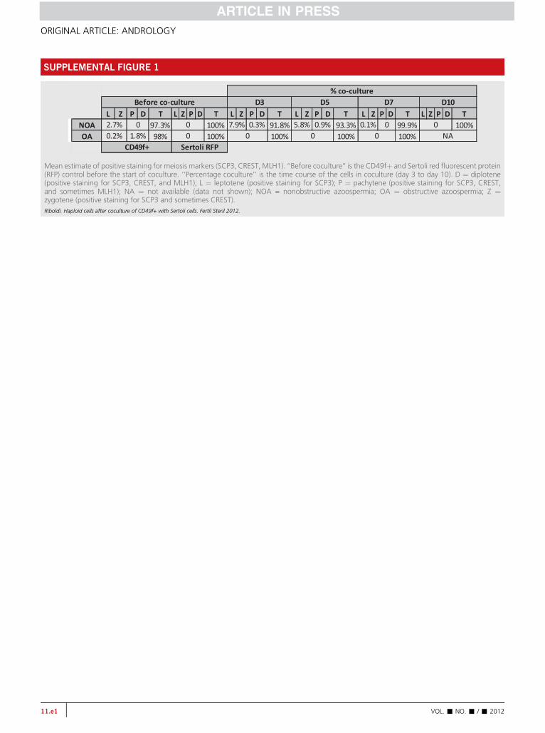
With the CD49þ cocultured cells, an immunocytochemi-cal analysis of proteins SCP3, CREST, and MLH1 demon-strated the existence of patterns that are compatible withthe different meiotic stages. After 3 days of coculturing theCD49fþ cells from NOA patients with Sertoli cells, SCP3 ex-pression was detected along with low CREST expression(Fig. 4A). This concomitant staining of SCP3 and CREST re-mained until day 5 of the coculture and disappeared by day7 (Fig. 4B and C). Supplemental Figure 1 (available online)summarizes the results of the meiosis progression in CD49þfrom the OA and NOA patients.
On day 5 of coculturing CD49þ cells from a NOA patient,we demonstrated the development of haploid cells, as shownby the detection of chromosomes 13, 18, 21, X, and Y via FISH(Fig. 4D). Supplemental Table 3 (available online) provides theFISH results.
DISCUSSIONSince the 1960s (30, 31), many researchers have examined andattempted meiosis progression in human spermatogenesisin vitro under various conditions. To date, complete in vitrospermatogenesis that provides human viable sperm has yetto be achieved, although different groups have studied thisprocess in animal models, including mice and primates.Different strategies have been adopted, such as use ofcombinations of growth factors and hormones, the 3D agarculture system, and immature testicular tissues (16–22).Recent studies in rodents have demonstrated that a crucialprerequisite to pass the meiotic barrier is the presence of theniche, the appropriate in vivo microenvironment providedby the seminiferous tubule (32–34).
Our study isolated SSCs in TESE samples obtained fromazoospermic patients with (NOA) or without (OA)
5
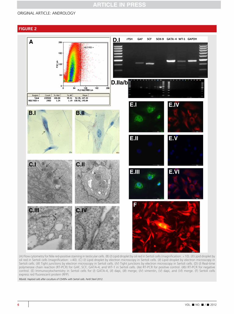
FIGURE 2
(A) Flow cytometry for Nile red-positive staining in testicular cells. (B) (I) Lipid droplet by oil red in Sertoli cells (magnification:�10). (II) Lipid droplet byoil red in Sertoli cells (magnification: �40). (C) (I) Lipid droplet by electron microscopy in Sertoli cells. (II) Lipid droplet by electron microscopy inSertoli cells. (III) Tight junctions by electron microscopy in Sertoli cells. (IV) Tight junctions by electron microscopy in Sertoli cells. (D) (I) Real-timepolymerase chain reaction (RT-PCR) for GAF, SCF, GATA-4, and WT-1 in Sertoli cells. (IIa) RT-PCR for positive control. (IIb) RT-PCR for negativecontrol. (E) Immunocytochemistry in Sertoli cells for (I) GATA-4, (II) dapi, (III) merge; (IV) vimentin, (V) dapi, and (VI) merge. (F) Sertoli cellsexpress red fluorescent protein (RFP).Riboldi. Haploid cells after coculture of CD49f+ with Sertoli cells. Fertil Steril 2012.
6 VOL. - NO. - / - 2012
ORIGINAL ARTICLE: ANDROLOGY

FIGURE 3
(A) Fluorescence in situ hybridization (FISH) in CD49fþ cells: (I) dapi, (II) chromosome 18, (III) chromosome X, and (IV) chromosome Y. (B) FISH inSertoli cells: (I) dapi, (II) chromosome 18, (III) chromosome X, and (IV) chromosome Y. (C) Immunocytochemistry for meiosis in CD49fþ cells(staining: negative for SCP3, CREST, and MLH1; positive for DAPI). (D) Immunocytochemistry for meiosis in Sertoli cells red fluorescent protein(RFP) (staining: negative for SCP3, CREST, and MLH1; positive for DAPI).Riboldi. Haploid cells after coculture of CD49f+ with Sertoli cells. Fertil Steril 2012.
VOL. - NO. - / - 2012 7
Fertility and Sterility®
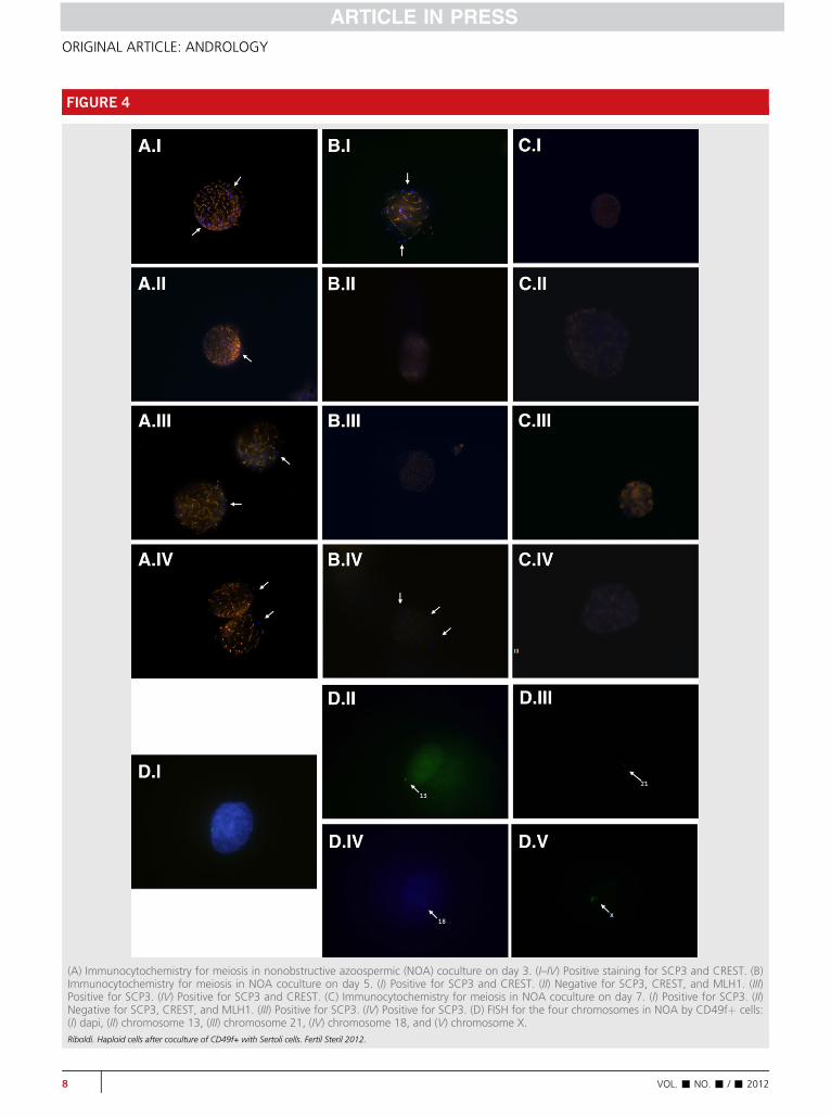
FIGURE 4
(A) Immunocytochemistry for meiosis in nonobstructive azoospermic (NOA) coculture on day 3. (I–IV) Positive staining for SCP3 and CREST. (B)Immunocytochemistry for meiosis in NOA coculture on day 5. (I) Positive for SCP3 and CREST. (II) Negative for SCP3, CREST, and MLH1. (III)Positive for SCP3. (IV) Positive for SCP3 and CREST. (C) Immunocytochemistry for meiosis in NOA coculture on day 7. (I) Positive for SCP3. (II)Negative for SCP3, CREST, and MLH1. (III) Positive for SCP3. (IV) Positive for SCP3. (D) FISH for the four chromosomes in NOA by CD49fþ cells:(I) dapi, (II) chromosome 13, (III) chromosome 21, (IV) chromosome 18, and (V) chromosome X.Riboldi. Haploid cells after coculture of CD49f+ with Sertoli cells. Fertil Steril 2012.
8 VOL. - NO. - / - 2012
ORIGINAL ARTICLE: ANDROLOGY

Fertility and Sterility®
spermatogenic arrest (diagnosis proved by a pathologist) andcocultured them in vitro with RFP-Sertoli cells to mimic theseminiferous tubule microenvironment that results in haploidcell generation. Testicular size varied among the samples(�1 mm3) and was composed of seminiferous tubules andinterstitial tissue. After mechanical dissection into smallpieces, the samples were enzymatically dissociated with colla-genase type IA and selected on the basis of previous reports(35, 36). The concentration and time were defined via dose-response and time-course preview experiments. The totalnumber of isolated cells from the testicular samples variedfrom 250,000 to 7,200,000 (see Supplemental Table 2).Depending on the number of cells collected, they were distrib-uted in a varying number of wells to a maximum of 2.106 cellsper well. Our strategy is based on simulating the proper nichefor SSC differentiation to lead to spermatogenesis.
The putative spermatogonial stem cells were separatedfrom the TESE samples by isolating the cells that tested pos-itive for surface marker CD49f, which is expressed in the sper-matogonia and primary spermatocytes attached to the basalmembrane of seminiferous tubules (9). Once the cells werecharacterized, their transcriptional profile revealed positiveexpression for the genes involved in germ-cell lineage suchas Thy-1 (a spermatogonial stem cell marker), Blimp-1 (a pri-mordial germ cell marker), c-Kit (a marker for spermatogo-nia), GAF (a GDNF receptor), BOULE, Stella (germ cellmarkers), and the meiotic gene SCP3. This transcriptional pro-file clearly demonstrates no cross-reactivity with Sertoli cells.It is interesting that the percentage of SSCs in the TESE sam-ples from OA patients was twice that of the NOA patients(5.45% vs. 2.36%, respectively), indicating the existence ofthis germinal primordial population in all the azoospermicpatients, even those with Sertoli cell-only syndrome (seeSupplemental Table 2).
Sertoli cells cultured on collagen matrix after growthwere sorted by Nile red staining, which identified the exis-tence of characteristic lipid droplets that were further con-firmed by the expression of GATA-4, vimentin, WT-1, SCF,and GAF as well as by the presence of tight junctions, thusconfirming that they match the characteristics of functionalhuman Sertoli cells (29). We then cocultured the SSC popula-tion onto a monolayer of Sertoli cells and collagen matrixcomponents of the basal layer of seminiferous tubules, whichprovided structural support to SSCs. In addition, the Sertolicells were stimulated with FSH (30, 37) and testosterone,the two main players in the differentiation and maturationof spermatogonia toward sperm (38–40). Because thepresence of Leydig cells was demonstrated to not bemandatory in a bovine coculture SSC system, Aponte et al.(41) grew SSCs in a mix of somatic cells (e.g., Sertoli,Leydig, peritubular), and the presence of Leydig cells wasanecdotal and not stimulated by luteinizing hormone (LH).The absence of Leydig cells was compensated for by thedirect addition of testosterone. Sertoli cells based incollagen matrix were provided as coculture, and they weresufficient for meiotic differentiation of the SSCs with theaddition of growth factors and hormones. The role of RA inthe regulatory mechanisms implicated in gametogenesis hasbeen extensively demonstrated (42–44), as has the
VOL. - NO. - / - 2012
important role of GDNF in maintaining the testicular niche(45). The aim of adding specific hormones (FSH andtestosterone) and factors (GDNF and RA) to the coculture isto confer the microenvironment conditions of the testicularniche and to contribute to the SSCs' maturation andmeiosis progression.
In the first part of the meiotic process, a primary sper-matocyte divides to produce two haploid cells; the chromo-somes pair up in a synaptic process to form a structureknown as a bivalent (two chromosomes) or a tetrad (fourchromatids). Prophase I is the meiosis stage in which the ho-mologous chromosomes pair up and exchange their DNA bygenetic recombination. This step comprises four different sub-phases: leptotene, where chromosomes begin to condense andpositive staining for SCP3 can be observed; zygotene, wherechromosomes become closely paired and can be identifiedby positive staining for SCP3 with low CREST expression;pachytene, when crossing over occurs, as evidenced by posi-tive staining for SCP3 and CREST with low MLH1 expression;and diplotene, where homologous chromosomes begin toseparate yet remain attached by the chiasmata, with positivestaining for SCP3, CREST, and MLH1. On day 3 of the cocul-ture, the SSCs from NOA patients presented SCP3 stainingalong with low CREST expression, suggesting that they hadentered meiosis and progressed through the leptotene phase.On day 5 after coculture, SCP3 staining was maintained inthe SSCs from the NOA patients along with stronger CRESTstaining, suggesting continuation to the zygotene meiosisstage. No staining was detected from day 7 onward, implyingthat meiosis had stopped in those cells after zygotene for somereason. This observation is coincident in time with the detec-tion of haploid cells on day 5, which is expected to occur justafter the zygotene phase. It is interesting that progressthrough the zygotene phase and generation of haploid cellshave been observed in the SSCs isolated from the TESE sam-ples of NOA patients.
Other researchers have already reported on the usefulnessof coculture systems togetherwith the addition of specific hor-mones and factors to attempt human spermatogenesis in vitro(16–20). Our work presents evidence for the first time thatSSCs isolated from TESE samples of NOA patients can entermeiosis and produce haploid cells in vitro under theindicated culture conditions. Thus, our study incorporatesnew perspectives in the field of male infertility. Althoughmore studies with tissues, stem cells, or induced cells are stillnecessary before any application in assisted reproduction,the maturation of germ cells could allow the acquisition ofsperm, and genetic and epigenetic characteristics (46).
It is important to recall that the spontaneous onset of mei-osis was determined by positive staining for SCP3 in theCD49fþ cells obtained from the OA patients. The cellsobtained from the NOA patients could not initiate meiosisspontaneously, as demonstrated by the absence of SCP3,CREST, and MLH1 under noninducing conditions. It is clearthat the efficiency of spermatogenesis progression is verylow, and the haploid cell generation demonstrated by FISHoccurred in only one of the NOA samples on day 5 of cocul-ture. Further work is needed to ascertain the proper cultureconditions that avoid meiosis arrest.
9
ORIGINAL ARTICLE: ANDROLOGY
The putative clinical strategy that emanates from thiswork involves the use of autologous CD49fþ and Sertoli cellsin TESE samples of NOA patients in which no sperm is foundfor in vitro maturation of their own SSC. The same alternativecan be offered to patients with other types of spermatogenicarrest and those who are undergoing chemotherapy duringchildhood, at which time TESE can be performed to preservetesticular tissue in the prepubertal stage so that they can latterattempt to have their own biological offspring. To develop thetechnique in the future, the safety of CD49fþ antibodies onsperm function must be properly tested.
Acknowledgments: The authors thank for all professionalthe Andrology Laboratory from IVI-Valencia and IVIOMICSgroup in specially Nasser Al-Asmar Pinar for help with tech-niques for FISH. The Inmaculada Moreno and Felipe VilellaMitjana for their assistance and review of the manuscript. Areview by Emily Rutledge language and Eva Gomez for helpwith protocols and culture.
REFERENCES1. Devroey P, Liu J, Nagy Z, Tournaye H, Silber SJ, Van Steirteghem AC. Normal
fertilization of human oocytes after testicular sperm extraction and intracy-toplasmic sperm injection. Fertil Steril 1994;62:639–41.
2. Weidner W, Colpi GM, Hargreave TB, Papp GK, Pomerol JM, Ghosh C. EAUWorking Group on Male Infertility. EAU guidelines on male infertility. EurUrol 2002;42:313–22.
3. Diedrich K, Fauser BC, Devroey P. Evian Annual Reproduction (EVAR) Work-shopGroup 2009. Cancer and fertility: strategies to preserve fertility. ReprodBiomed Online 2011;22:232–48.
4. Print CG, Loveland KL. Germ cell suicide: new insights into apoptosis duringspermatogenesis. Bioessays 2000;22:423–30.
5. Grootegoed JA, Siep M, Baarends WM. Molecular and cellular mechanismsin spermatogenesis. Baillieres Best Pract Res Clin Endocrinol Metab 2000;14:331–43.
6. Dunkel L, Taskinen S, Hovatta O, Tilly JL, Wikstr€om S. Germ cell apoptosisafter treatment of cryptorchidism with human chorionic gonadotropin is as-sociated with impaired reproductive function in the adult. J Clin Invest 1997;100:2341–6.
7. Conrad S, Renninger M, Hennenlotter J, Wiesner T, Just L, Bonin M, et al.Generation of pluripotent stem cells from adult human testis. Nature2008;456:344–9.
8. Kossack N, Meneses J, Shefi S, Nguyen HN, Chavez S, Nicholas C, et al. Iso-lation and characterization of pluripotent human spermatogonial stem cell-derived cells. Stem Cells 2009;27:138–49.
9. Golestaneh N, Kokkinaki M, Pant D, Jiang J, DeStefano D, Fernandez-Bueno C, et al. Pluripotent stem cells derived from adult human testes.Stem Cells Dev 2009;18:1115–26.
10. Mizrak SC, Chikhovskaya JV, Sadri-Ardekani H, van Daalen S, Korver CM,Hovingh SE, et al. Embryonic stem cell-like cells derived from adult humantestis. Hum Reprod 2010;25:158–67.
11. He Z, Kokkinaki M, Jiang J, Dobrinski I, Dym M. Isolation, characterization,and culture of human spermatogonia. Biol Reprod 2010;82:363–72.
12. Izadyar F, Wong J, Maki C, Pacchiarotti J, Ramos T, Howerton K, et al. Iden-tification and characterization of repopulating spermatogonial stem cellsfrom the adult human testis. Hum Reprod 2011;26:1296–306.
13. Brinster RL, Zimmermann JW. Spermatogenesis following male germ-celltransplantation. Proc Natl Acad Sci USA 1994;91:11298–302.
14. Costoya JA, Hobbs RM, Barna M, Cattoretti G, Manova K, Sukhwani M,et al. Essential role of PLZF in maintenance of spermatogonial stem cellsNat. Genet 2004;36:551–3.
15. Dym M, Kokkinaki M, He Z. Spermatogonial stem cells: mouse and humancomparisons. Birth Defects Res C Embryo Today 2009;87:27–34.
10
16. Staub C, Hue D, Nicolle JC, Perrard-Sappori MH, Segretain D, Durand P. Thewholemeiotic process can occur in vitro in untransformed rat spermatogeniccells. Exp Cell Res 2000;260:85–95.
17. Kita K, Watanabe T, Ohsaka K, Hayashi H, Kubota Y, Nagashima Y, et al.Production of functional spermatids from mouse germline stem cells inectopically reconstituted seminiferous tubules. Biol Reprod 2007;76:211–7.
18. Kanatsu-Shinohara M, Miki H, Inoue K, Ogonuki N, Toyokuni S, Ogura A,Shinohara T. Germline niche transplantation restores fertility in infertilemice. Hum Reprod 2005;20:2376–82.
19. Gohbara A, Katagiri K, Sato T, Kubota Y, Kagechika H, Araki Y, Araki Y,Ogawa T. In vitro murine spermatogenesis in an organ culture system. BiolReprod 2010;83:261–7.
20. Sousa M, Cremades N, Alves C, Silva J, Barros A. Developmental potential ofhuman spermatogenic cells co-culturedwith Sertoli cells. Hum Reprod 2002;17:161–72.
21. Cremades N, Bernabeu R, Barros A, Sousa M. In-vitro maturation ofround spermatids using co-culture on Vero cells. Hum Reprod 1999;14:1287–93.
22. Cremades N, Sousa M, Bernabeu R, Barros A. Developmental potential ofelongating and elongated spermatids obtained after in-vitro maturation ofisolated round spermatids. Hum Reprod 2001;16:1938–44.
23. Hayashi K, Ohta H, Kurimoto K, Aramaki S, Saitou M. Reconstitution of themouse germ cell specification pathway in culture by pluripotent stem cells.Cell 2011;146:519–32.
24. Sato T, Katagiri K, Gohbara A, Inoue K, Ogonuki N, Ogura A, et al. In vitroproduction of functional sperm in cultured neonatal mouse testes. Nature2011;471:504–7.
25. Shi B, Zhang S, Wang Y, Zhuang Y, Chu J, Zhang S, Shi X, Bi J, Guo M. Ex-pansion of mouse Sertoli cells on microcarriers. Cell Prolif 2010;43:275–86.
26. Ruiz-Vela A, Aggarwal M, de la Cueva P, Treda C, Herreros B, Martín-P�erez D, Dominguez O, Piris MA. Lentiviral (HIV)-based RNA interferencescreen in human B-cell receptor regulatory networks reveals MCL1-induced oncogenic pathways. Blood 2008;111:1665–76.
27. Sun F, Kozak G, Scott S, Trpkov K, Ko E, Mikhaail-Philips M, et al. Meioticdefects in a man with non-obstructive azoospermia: case report. Hum Re-prod 2004;19:1770–3.
28. Sarrate Z, Anton E. Fluorescence in situ hybridization (FISH) protocol in hu-man sperm. J Vis Exp 2009;31:1405.
29. Chui K, Trivedi A, Cheng CY, Cherbavaz DB, Dazin PF, Huynh AL, et al. Char-acterization and functionality of proliferative human Sertoli cells. Cell Trans-plant 2011;20:619–35.
30. Steinberger E, Steinberger A, Perloff WH. Initiation of spermatogenesisin vitro. Endocrinology 1964;74:788–92.
31. Steinberger A, Steinberger E. In vitro culture of rat testicular cells. Exp CellRes 1966;44:443–52.
32. Guan K, Nayernia K, Maier LS, Wagner S, Dressel R, Lee JH, et al. Pluripo-tency of spermatogonial stem cells from adult mouse testis. Nature 2006;440:1199–203.
33. Lee DR, Kim KS, Yang YH, Oh HS, Lee SH, Chung TG, et al. Isolation of malegerm stem cell-like cells from testicular tissue of non-obstructive azoosper-mic patients and differentiation into haploid male germ cells in vitro. HumReprod 2006;21:471–6.
34. Stukenborg JB, Wistuba J, Luetjens CM, Elhija MA, Huleihel M, Lunenfeld E,et al. Coculture of spermatogonia with somatic cells in a novel three-dimensional soft-agar-culture-system. J Androl 2008;29:312–29.
35. Crabb�e E, Verheyen G, Tournaye H, Van Steirteghem A. The use of enzy-matic procedures to recover testicular germ cells. Hum Reprod 1997;12:1682–7.
36. Salzbrunn A, Benson DM, Holstein AF, Schulze W. A new concept for theextraction of testicular spermatozoa as a tool for assisted fertilization(ICSI). Hum Reprod 1996;11:752–5.
37. Walker WH, Cheng J. FSH and testosterone signaling in Sertoli cells. Repro-duction 2005;130:15–28.
38. Niederberger C, Agulnik AI, Cho Y, Lamb D, Bishop CE. In situ hybridizationshows that Dazl expression in mouse testis is restricted to premeiotic stagesIV–VI of spermatogenesis. Mamm Genome 1997;8:277–8.
VOL. - NO. - / - 2012
Fertility and Sterility®
39. Hofmann MC. Gdnf signaling pathways within the mammalian spermato-gonial stem cell niche. Mol Cell Endocrinol 2008;288:95–103.
40. Maki CB, Pacchiarotti J, Ramos T, Pascual M, Pham J, Kinjo J, et al. Pheno-typic and molecular characterization of spermatogonial stem cells in adultprimate testes. Hum Reprod 2009;24:1480–91.
41. Aponte PM, Soda T, Teerds KJ, Mizrak SC, van de Kant HJ, de Rooij DG.Propagation of bovine spermatogonial stem cells in vitro. Reproduction2008;136:543–57.
42. Anderson EL, Baltus AE, Roepers-Gajadien HL, Hassold TJ, de Rooij DG, vanPelt AM, Page DC. Stra8 and its inducer, retinoic acid, regulate meiotic ini-tiation in both spermatogenesis and oogenesis in mice. Proc Natl Acad SciUSA 2008;105:14976–80.
VOL. - NO. - / - 2012
43. Zhou Q, Li Y, Nie R, Friel P, Mitchell D, Evanoff RM, et al. Expression of stim-ulated by retinoic acid gene 8 (Stra8) and maturation of murine gonocytesand spermatogonia induced by retinoic acid in vitro. Biol Reprod 2008;78:537–45.
44. Nayernia K, Nolte J, Michelmann HW, Lee JH, Rathsack K, Drusenheimer N,et al. In vitro-differentiated embryonic stem cells give rise to male gametesthat can generate offspring mice. Dev Cell 2006;11:125–32.
45. Rooij DG. The spermatogonial stem cell niche. Microsc Res Tech 2009;72:580–5.
46. Neri QV, Palermo GD, Rosenwaks Z. Fertility preservation. In: Seli E,Agarwal A, editors. Fertility preservation: emerging technologies and clinicalapplications. New York: Springer; 2012.
11
SUPPLEMENTAL FIGURE 1
Mean estimate of positive staining for meiosis markers (SCP3, CREST, MLH1). ‘‘Before coculture’’ is the CD49fþ and Sertoli red fluorescent protein(RFP) control before the start of coculture. ‘‘Percentage coculture’’ is the time course of the cells in coculture (day 3 to day 10). D ¼ diplotene(positive staining for SCP3, CREST, and MLH1); L ¼ leptotene (positive staining for SCP3); P ¼ pachytene (positive staining for SCP3, CREST,and sometimes MLH1); NA ¼ not available (data not shown); NOA = nonobstructive azoospermia; OA ¼ obstructive azoospermia; Z ¼zygotene (positive staining for SCP3 and sometimes CREST).Riboldi. Haploid cells after coculture of CD49f+ with Sertoli cells. Fertil Steril 2012.
ORIGINAL ARTICLE: ANDROLOGY
11.e1 VOL. - NO. - / - 2012
SUPPLEMENTAL TABLE 1
Primers sequence for real-time polymerase chain reaction.
Gene Primer sequence 50–30 Base pair
THY1 TGCCGCCATGAGAATACCATCAGAGAAGTAGGATCTCTGCA
439
GAF AAGCACAGCTACGGAATGCTATTGCCAAAGGCTTGAATTG
426
C-KIT GCACGGTTGAATGTAAGGCTTCATGGCCGCATCTGACTTA
396
STELLA GTTACTGGGCGGAGTTCGTATGAAGTGGCTTGGTGTCTTG
174
DAZL ATGTTAGGATGGATGAAACTGAGATTACCATGGAAATTTATCTGTGATTCTACT
178
VASA AGAAAGTAGTGATACTCAAGGACCAATGACAGAGATTAGCTTCTTCAAAAGT
199
PIWIL2 TCTATGGGGCCATCAAGAAGCCATCCCGATCACCATTAAC
195
BOULE ATGTAGCTCCCCTGTGATGGGTGATGGCACTTGGAGCATA
300
DCM1 CTTTCAGGCAGATCCCAAAACCCAATTCCTCCAGCAGTTA
172
SCP3 GCCGTCTGTGGAAGATCAGTTGGTTAAGCTTCTGCCTTTGA
327
PRM1 CACCATGGCCAGGTACAGATGTCTTCTACATCGCGGTCTG
155
TNP2 CACAGGCAAGAAGGAAGAGGAGCCAATGCATTCTTCCAAC
235
BLIMP1 GCCAAGTTCACCCAGTTTGTGATTCGGGTCAGATCTTCCA
183
GATA4 AGACATCGCACTGACTGAGAACGACGGGTCACTATCTGTGCAAC
475
WT1 TCCTTCATCAAACAGGAGCCGAGCCTGTAGGGCGTCCTCAGCAGCAAAG
450
rFSH CACAGTCCCCAGGTTCCTTAATGCTGCTGGCTTTTTCACT
166
SCF GCTCCAGAACAGCTAAACGGTCTTTGACGCACTCCACAAG
417
SOX9 GAGGAAGTCGGTGAAGAACGAGACAGCCCCCTATCGACTT
276
GAPDH TGAGCTGAACGGGAAGCTCAGTCTACATGGCAACTGTGAGGA
470
Riboldi. Haploid cells after coculture of CD49f+ with Sertoli cells. Fertil Steril 2012.
Fertility and Sterility®
VOL. - NO. - / - 2012 11.e2
SUPPLEMENTAL TABLE 2
Total number of cells isolated from testicular sperm extraction andnumber of CD49fD isolated, percentage (%), and mean.
Total cells CD49fD % CD49fD Mean
OA 250,000 16,300 0.5% 5.45%7,200,000 500,000 6.25%1,750,000 235,000 15%700,000 17,500 3%730,000 19,400 3.6%
13,920,000 20,000 1.04%2,250,000 28,000 2%9,600,000 50,845 3%1,080,000 33,000 1.8%
NOA 4,600,000 5,000 2.5% 2.36%3,200,000 13,000 4.35%850,000 8,000 1.30%
3,000,000 25,000 2.6%6,000,000 193,000 10.19%2,000,000 32,000 2.66%485,000 7,000 1.10%270,000 400 0.19%735,000 850 0.2%250,000 795 0.7%525,000 1,200 0.16%
Note: NOA ¼ nonobstructive azoospermia (11); OA ¼ obstructive azoospermia (9).
Riboldi. Haploid cells after coculture of CD49f+ with Sertoli cells. Fertil Steril 2012.
ORIGINAL ARTICLE: ANDROLOGY
11.e3 VOL. - NO. - / - 2012
SUPPLEMENTAL TABLE 3
Average estimate of cell ploidy from fluorescence in situ hybridization analysis for chromosomes 18, X, and Y.
Before coculture Percentage coculture
CD49fD Sertoli RFP D3 D5 D7 D10 D12 D15
2n 1n 2n 1n 2n 1n 2n 1n 2n 1n 2n 1n 2n 1n 2n 1n
NOA 100% 0 100% 0 100% 0 99.3% 0.57% 100% 0 NA NA 100% 0OA 100% 0 100% 0 100% 0 100% 0 100% 0 100% 0 100% 0 100% 0Note: ‘‘Before coculture’’ is the CD49fþ and Sertoli red fluorescent protein (RFP) control. ‘‘Percentage coculture’’ is the time course of the cells in coculture (day 3 to day 15). NA ¼ not available(data not shown); NOA ¼ nonobstructive azoospermia; OA ¼ obstructive azoospermia.
Riboldi. Haploid cells after coculture of CD49f+ with Sertoli cells. Fertil Steril 2012.
Fertility and Sterility®
VOL. - NO. - / - 2012 11.e4