in vitro propagation of citrus indica tanaka—an endangered progenitor...
TRANSCRIPT

Indian Journal of Biotechnology
Vol 8, July 2009, pp 311-316
In vitro propagation of Citrus indica Tanaka—An endangered progenitor species
M A Laskar*, M Hynniewta and C S Rao1
Department of Biotechnology and Department of Botany1, St Anthony’s College, Shillong 793 001, India
Received 17 April 2008; revised 26 December 2008; accepted 27 February 2009
A method for in vitro propagation of Citrus indica Tanaka by shoot organogenesis from leaf-derived callus was
developed. Regenerative calli were induced on MS medium supplemented with 0.01 mg L-1 TDZ and 0.1 mg L-1 NAA.
Shoots were regenerated on WPM medium supplemented with 0.5 mg L-1 BAP, 0.25 mg L-1 TDZ and 0.25 mg L-1 NAA.
Regenerated shoots were rooted on MS medium supplemented with 1.0 mg L-1 NAA. Sixty per cent of the rooted plantlets
were acclimatized successfully under ex situ conditions.
Keywords: Citrus indica endangered, ex situ adaptibility, leaf-derived callus, progenitor, shoot organogenesis
Introduction Citrus indica Tanaka is one of the most primitive
species of citrus and is considered to be the progenitor
of cultivated citrus1. It is endemic to the Tura range of
Garo Hills of Meghalaya2,3
; this area falls within the
buffer zone of Nokrek Biosphere Reserve. Villagers,
belonging to the Garo tribe, who live around the
biosphere reserve, attribute medicinal and religious
values to C. indica. The fruit is used to treat jaundice
and stomach diseases of humans and domestic
animals. Powder made from the fruit as well as raw
fruits is taken as a cure for small pox. Fruits are
placed on dead bodies during the last rites with the
belief that it will ward off ghosts of the departed4.
Absence of organised cultivation and destruction of
natural habitat by deforestation and jhum activities
have severely depleted the population of C. indica.
The endemic nature of this species has further
contributed to its decline. It is essential to prevent the
extinction of C. indica for its taxonomic and
ethnobotanical importance as well as for the fact that
it can be used as rootstock for propagation of
commercial citrus species for being hardy and free
from pests and diseases in its natural habitat4. The
buffer zone comprising the natural habitat of C. indica
has been established as Citrus Gene Sanctuary1.
However, this in situ conservation effort has had
limited impact on halting the decline in population. It
is, therefore, necessary to establish ex situ conservation
methods like micropropagation as supplemental
measures. The present in vitro propagation study on
C. indica was taken to develop a method for
multiplication of this endangered progenitor species.
Materials and Methods Plant Material
Fruits of C. indica were collected during January,
2004 from plants growing around the Tura Peak,
which is situated inside the Nokrek Biosphere
Reserve, at 1,412 m above MSL. The fruits (Fig. 1a)
have orange coloured outer covering and bear one to
three monoembryonic seeds (Fig. 1b) in each fruit
segment. The seeds take up most of the space in a
segment. Due to scarcity of plant material, the work
was started with only 20 seeds.
Seeds were surfaced sterilized by soaking in 5%
(w/v) calcium hypochorite for 20 min. The surface
sterilant suspension containing the seeds was kept
agitated during the duration of treatment. The calcium
hypochlorite suspension was then decanted, seeds
shifted to a fresh container and washed five times
with sterilized distilled water to remove all traces of
the surface sterilant. Surface sterilized seeds were
germinated on 0.7% agar, gelled in distilled water and
sterilized by autoclaving at 15 psi for 18 min.
Seedlings were considered as a genetic variants and
were marked as V (Variant) 1 to V 20.
Leaf explants were prepared 6 d after germination
when seedlings were 5-7 cm long. Petiole was
removed from a leaf and 1 cm2 portion of lamina
comprising the midrib was excised. Throughout the
study, the marking scheme as described above was
____________________
*Author for correspondence:
Tel: 91-364-2211955, 09436118118; Fax: 91-364-2229558
E-mail: [email protected]

INDIAN J BIOTECHNOL, JULY 2009
312
used to maintain the identity of leaf explants, calli and
regenerants that originated from each variant.
Media
To induce callusing form leaf explants, the
constituents of MS5 and WPM
6 media were
supplemented with NAA (α-napthalene acetic acid),
2,4-D (2,4-dichlorophenoxy acetic acid) and TDZ
(thidiazuron). NAA was used at concentrations from
0.2 to 1.25 mg L-1
, 2,4-D at concentrations from 0.1 to
1 mg L-1
and TDZ at concentrations from 0.005 to 0.1
mg L-1
. NAA and TDZ were used alone and in
various combinations, while 2,4-D was used alone.
The regenerative ability of calli was tested by
inoculating them on MS or WPM media
supplemented with BAP (6-benzyl amino purine), Kn
(kinetin), TDZ and NAA singly and in various
combinations. BAP was used at concentrations from
0.1 to 1 mg L-1
, Kn at concentrations from 0.05 to 0.5
mg L-1
, TDZ at concentrations from 0.0025 to 0.05
mg L-1
and NAA at concentrations from 0.25 to 0.5
mg L-1
. To induce multiple shoots from axillary buds,
shoots regenerated from leaf-derived calli were
excised into nodal segments bearing one axillary bud
and inoculated on MS- or WPM-based media
supplemented with growth regulators mentioned
above. To induce rooting from regenerated plantlets,
MS- or WPM-based media supplemented with IBA
(indole 3-butyric acid) or NAA, both at
concentrations from 0.25 to 1 mg L-1
were tested. The
pH of all media was adjusted to 5.8 before
autoclaving at 15 psi for 18 min. Media, except those
tested for root induction, were gelled with 0.7% agar.
Media used for testing root induction were gelled with
0.6% agar.
Rooted plantlets were removed from culture vessels.
The roots were washed under tap water to remove
adherent media and planted in plastic pots containing
river sand, leaf-mould compost and garden soil in the
ratio, 3:1:1. The potted plantlets were kept in the shade
covered with perforated polythene bags. The plantlets
were irrigated with tap water. The irrigation schedule
and volume of water was calibrated to keep the pot
mixture saturated and prevent flooding. The covers
were removed after 10 d. Physical Parameters
The source of light was cool fluorescent tubes.
Germination of seedlings and the experiments on
callus induction and rooting were done under 200 lux
luminance and 12 h photoperiod. Regeneration
experiments were done under 400 lux luminance and
12 h photoperiod. The temperature inside the culture
room was maintained at 25±1°C during all stages of
the study.
Data Analysis
Experiments were set in the completely
randomized block design (CRD). A single leaf
explant inoculated on 10 mL of callusing media
contained in a culture tube (25 cm × 1.5 cm) formed a
replicate; 15 such replications were provided for
Figs (1-6)—1: a. Ripe Fruits, b: Seeds; 2: Leaf-derived callus;
3: Shoot organogenesis; 4: Root development; 5: Rooted plantlet
in pot; and 6: acclimatized plant.
LASKAR et al: PROPAGATION OF C. INDICA TANAKA BY SHOOT ORGANOGENESIS
313
every variant. Similarly, 10 replications were
provided during the trials on shoot organogenesis
from leaf-derived calli, axillary shoot proliferation
and on root induction from shootlets. However, media
in these cases were dispensed in Erlenmeyer flasks of
150 mL capacity and 30 mL of a medium was poured
in each flask.
The parameters studied were percentage of
explants that underwent callusing, average number of
shoots regenerated from each callus, mean shoot
length, average number of shoots developed per
axillary bud, percentage of shoot from which roots
developed, average number of roots that developed
per shoot and mean root length. Data were subjected
to Analysis of Variance (ANOVA) followed by
Duncan’s Multiple Range Test (DMRT), using SPSS
10.0.
Results and Discussion Sixty per cent, i.e., 12 out of 20, seeds of C. indica
germinated in 7-10 d. Thus, there were 12 seedlings
or variants from the leaves, of which induction of
callus (Fig. 2) occurred. The seedlings were used as
sources of explants. Since seedlings are the products
of sexual reproduction and since cross-pollination
exists in citrus, care was taken to maintain separate
identities of leaf explants as well as the calli and
regenerants derived from each seedling. Calli were
induced in 2,4-D or NAA and TDZ-supplemented MS
and WPM media. NAA when used alone induced
callus growth at concentrations 0.5 to 1.25 mg L-1;
however, these calli were hard and did not undergo
shoot regeneration. Similarly, 2,4-D when present in
media at concentrations 0.125 to 1.0 mg L-1 induced
calli that were hard and non-regenerative. Friable
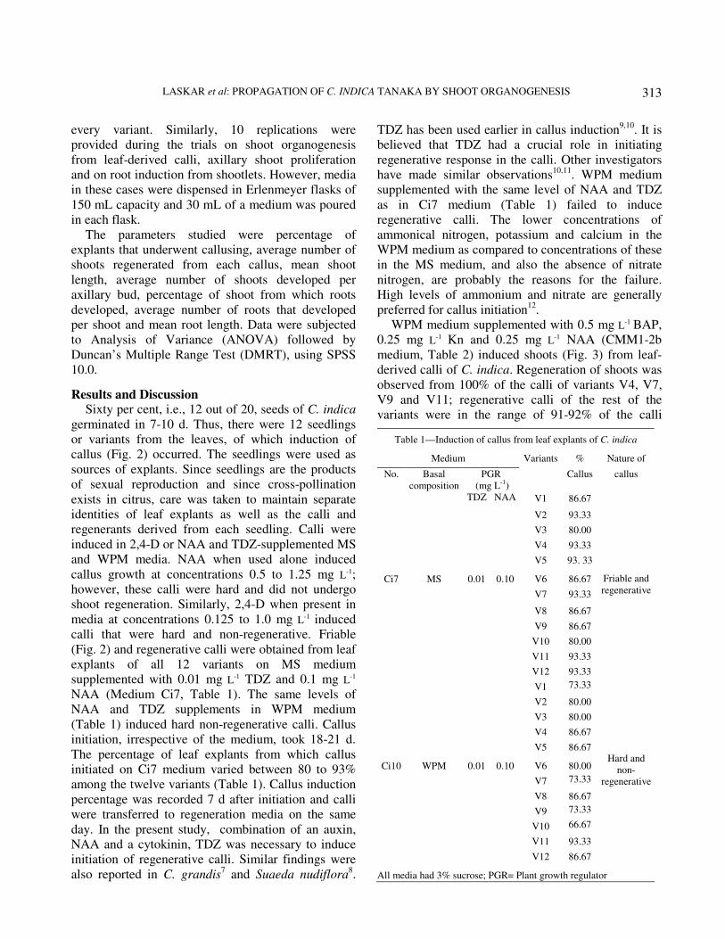
(Fig. 2) and regenerative calli were obtained from leaf
explants of all 12 variants on MS medium
supplemented with 0.01 mg L-1 TDZ and 0.1 mg L-1
NAA (Medium Ci7, Table 1). The same levels of
NAA and TDZ supplements in WPM medium
(Table 1) induced hard non-regenerative calli. Callus
initiation, irrespective of the medium, took 18-21 d.
The percentage of leaf explants from which callus
initiated on Ci7 medium varied between 80 to 93%
among the twelve variants (Table 1). Callus induction
percentage was recorded 7 d after initiation and calli
were transferred to regeneration media on the same
day. In the present study, combination of an auxin,
NAA and a cytokinin, TDZ was necessary to induce
initiation of regenerative calli. Similar findings were
also reported in C. grandis7 and Suaeda nudiflora
8.
TDZ has been used earlier in callus induction9,10
. It is
believed that TDZ had a crucial role in initiating
regenerative response in the calli. Other investigators
have made similar observations10,11
. WPM medium
supplemented with the same level of NAA and TDZ
as in Ci7 medium (Table 1) failed to induce
regenerative calli. The lower concentrations of
ammonical nitrogen, potassium and calcium in the
WPM medium as compared to concentrations of these
in the MS medium, and also the absence of nitrate
nitrogen, are probably the reasons for the failure.
High levels of ammonium and nitrate are generally
preferred for callus initiation12
.
WPM medium supplemented with 0.5 mg L-1 BAP,
0.25 mg L-1 Kn and 0.25 mg L-1 NAA (CMM1-2b
medium, Table 2) induced shoots (Fig. 3) from leaf-
derived calli of C. indica. Regeneration of shoots was
observed from 100% of the calli of variants V4, V7,
V9 and V11; regenerative calli of the rest of the
variants were in the range of 91-92% of the calli
Table 1—Induction of callus from leaf explants of C. indica
Medium Variants % Nature of
No.
Basal
composition
PGR
(mg L-1
)
Callus
callus
TDZ NAA V1 86.67
V2 93.33
V3 80.00
V4 93.33
V5 93. 33
Ci7 MS 0.01 0.10 V6 86.67
V7 93.33
V8 86.67
V9 86.67
V10 80.00
V11 93.33
V12 93.33
Friable and
regenerative
V1 73.33
V2 80.00
V3 80.00
V4 86.67
V5 86.67
Ci10 WPM 0.01 0.10 V6 80.00
V7 73.33
V8 86.67
V9 73.33
V10 66.67
V11 93.33
V12 86.67
Hard and
non-
regenerative
All media had 3% sucrose; PGR= Plant growth regulator

INDIAN J BIOTECHNOL, JULY 2009
314
induced (Table 2). Initiation of shoot regeneration
was noticed 12-16 d after transfer of calli to CMM1-
2b medium. Comparative study between the twelve
variants done 15 d after initiation on the mean number
of shoots that regenerated from a callus revealed
differences, some of them significant (Table 2). The
highest mean shoot induction frequency, 4.9±0.23
shoots/callus, was observed in calli of V1. This was
significantly higher from the mean shoot induction
frequency in calli of all other variants, except V2
(4.7±0.37 shoots/callus) and V12 (4.6±0.27
shoots/callus). It was necessary to supplement the
WPM basal medium with the cytokinins (BAP, Kn)
and the auxin (NAA) to induce shoot regeneration.
The combination of BAP, Kn and NAA was used for
the regeneration of plants from the callus cultures of
S. nudiflora8. As in the present study, other workers
have reported the use of combination of cytokinins
and auxins for inducing shoot regeneration where the
concentrations of cytokinins were higher than that of
auxins13
.
Significant differences also existed among the
variants in the average length of shoots measured 20 d
after induction (Table 2). The highest mean shoot
length was 4.63±0.19 cm recorded from V3
regenerants. This value was significantly different
from the mean shoot length recorded from all other
regnerents, barring V1 regenerants (4.42±0.22 cm).
The frequency of shoot organogenesis and the mean
length of shoot did not show similar trends. For
example, the regeneration frequency from V4 calli
(2.3 ±0.21) was among the lowest but the average
shoot length of these shoots (4.11±0.19) was among
highest (Table 2). On the other hand, regeneration
frequency from V2 calli (4.7 ±0.37) was
insignificantly different from the highest value
(4.9 ±0.23 recorded from V1 calli) but the mean shoot
length of these shoots was 2.92 ±0.15, comparable to
the lowest mean value (2.78 ±0.13) recorded from V9
shoots.
Proliferation of axillary shoot was achieved in
three different media (Table 3), but the best results
were recorded on the same medium (CMM1-2b) in
which shoot organogenesis was also induced, i.e.,
WPM basal medium supplemented with 0.5 mg L-1
BAP, 0.25 mg L-1 Kn, 0.25 mg L-1 NAA. The mean
number of shoots regenerated per axillary bud of
variants V1 to V11 was significantly higher in
CMM1-2b medium than that in either CMM1-2a or
CMM1-2d media (Table 3). Frequency of shoot
proliferation from V12 axillary buds was a little
different; the mean number of shoots regenerated per
axillary bud on CMM1-2b was significantly higher
(5.1±0.31) than that in CMM1-2d (1.2±0.2); but there
was no significant difference between the
proliferation obtained in CMM1-2b and CMM1-2a
media (Table 3). The time required for initiation of
multiple shoot was the same (8-10 d) on both CMM1-
2a and CMM1-2b but was more (18-21 d) on CMM1-
d. As in the present study, use of the same medium for
shoot regeneration and also for proliferation of
axillary shoots has been reported in Capparis
Table 2—Regenertion of plantlets from leaf-derived callus of C. indica
Medium
No.
Basal
composition
PGR
(mg L-1)
Variants
*% regenerative
callus
Shoots
regnerated/callus
Shoot length
(cm)
BAP Kn NAA V1 92.31 4.9 ±0.23d 4.42 ±0.22cd
V2 92.86 4.7 ±0.37cd 2.92 ±0.15a
V3 91.67 3.4 ±0.16b 4.63 ±0.19d
V4 100.00 2.3 ±0.21a 4.11 ±0.19bc
V5 92.86 2.1 ±0.41a 2.98 ±0.09a
CMM1-2b WPM 0.50 0.25 0.25 V6 92.31 3.9 ±0.23bc 2.94 ±0.06a
V7 100.00 3.4 ±0.22b 3.93 ±0.2b
V8 92.31 1.7 ±0.21a 2.8 ±0.09a
V9 100.00 1.5 ±0.17a 2.78 ±0.13a
V10 91.67 1.8 ±0.2a 3.04 ±0.13a
V11 100.00 1.9 ±0.23a 2.95 ±0.07a
V12 92.86 4.6 ±0.27cd 3.02 ±0.05a
All media had 3% sucrose; *Response obtained only from callus induced on Ci7 medium (vide text & Table 1); Means ± S.E. within
columns followed by the same letters are not significant according to DMRT (p=0.05); PGR= Palnt Growth Regulator

LASKAR et al: PROPAGATION OF C. INDICA TANAKA BY SHOOT ORGANOGENESIS
315
spinosa14
, Terminalia bellerica15
and in
Chrysanthemum16
. In the present study, the WPM
basal medium was found better than the MS basal
medium for shoot proliferation. Similar results have
also been reported in Tinospora cordifolia17
and in
Quercus semecarpifolia18
. Axillary shoot proliferation
was poor in CMM1-2d when compared to that in
CMM1-2b, both in terms of mean number of shoots
regenerated per axillary bud and the time required for
initiation of multiple shoots, due to lower
concentrations of BAP, Kn, and NAA (Table 3).
Studies on in vitro propagation of other Citrus
species19,20
also reported similar findings.
MS medium supplemented with 1.00 mg L-1 NAA
and gelled with 6% agar (CCM3 medium, Table 4)
induced roots (Fig. 4). NAA has previously been used
for induction of roots from in vitro regenerated shoots
of citrus7,21,22
and other species23
. Root induction was
observed 10-12 d after transfer of shoots to CCM3
medium; observations as detailed below were
recorded 10 d after induction of roots (Table 4). In
V7, 92.86% of shoots had root growth; this was the
highest incidence of rooting. The lowest percentage of
root regeneration was 71.43%, recorded in V11. The
variation in the average number of roots developed
per shoot was not very wide. The highest average
value (2.5±0.17 roots/shoot) was recoded in V7
shoots. This frequency of root induction was not
significantly different from the values recorded from
shoots of other variants. However, significant
differences were present in the mean length of roots.
The longest roots were recorded from the shoots of
V6; the mean value, 3.87 ±0.08 was not significantly
different from mean values recorded from the shoots
Table 3—Proliferation of axillary shoots of C. indica
*CMM1-2a
medium
CMM1-2b
medium
CMM1-2d
medium
Variants Shoots/bud 1DI Shoots/bud DI Shoots/bud DI
V1 3.6 ±0.43g 6.3 ±0.42a 1.2 ±0.2h
V2 4.0 ±0.26fg 5.1 ±0.28cde 1.2 ±0.13h
V3 3.6 ±0.22g 4.9 ±0.43cdef 1.4 ±0.16h
V4 3.8 ±0.2g 5.8 ±0.42abc 0.9 ±0.23h
V5 3.5 ±0.31g 5.1 ±0.31cde 1.0 ±0.21h
V6 3.7 ±0.3g 5.0 ±0.47cde 1.3 ±0.15h
V7 4.1 ±0.32efg 5.7 ±0.42abc 1.1 ±0.23h
V8 4.2 ±0.25efg 5.3 ±0.3bcd 1.2 ±0.13h
V9 4.5 ±0.37defg 5.5 ±0.5abc 1.0 ±0.26h
V10 4.0 ±0.26fg 5.3 ±0.3bcd 1.0 ±0.21h
V11 4.4 ±0.43defg 6.1 ±0.38ab 1.1 ±0.23h
V12 4.2 ±0.33defg
8-10
d
5.1 ±0.31cde
8-10
d
1.2 ±0.2h
18-
21 d
*CMM1-2a: MS + 0.5 mg L-1
BAP + 0.25 mg L-1
Kn + 0.25 mg L-1
NAA; CMM1-2b: WPM+ 0.5 mg L-1
BAP + 0.25 mg L-1
Kn + 0.25 mg L-1
NAA; CMM1-2d: WPM+ 0.25 mg L-1
BAP + 0.1
mg L-1
Kn + 0.05 mg L-1
NAA
All media had 3% sucrose 1DI: Days required for initiation of multiple shoots
Means ± S.E. followed by the same letters across rows & columns
are not significant according to DMRT (p= 0.05)
Table 4—Rooting from plantlets of C indica
Medium
No. Basal composition PGR
(mg L-1)
Variants % plantlets from which
rooting occurred
Roots/shoot Root length
(cm)
NAA V1 83.33 1.9 ±0.17a 3.8 ±0.17d
V2 76.92 1.9 ±0.06a 3.24 ±0.06b
V3 81.82 2.2 ±0.25a 2.83 ±0.07a
V4 85.71 1.9 ±0.18a 3.15 ±0.06bc
V5 76.92 1.7 ±0.21a 3.56 ±0.01cd
CCM3 MS 1.00 V6 83.33 2.0 ±0.21a 3.87 ±0.08d
V7 92.86 2.5 ±0.17a 3.28 ±0.06bc
V8 75.00 1.9 ±0.18a 3.23 ±0.07bc
V9 84.62 2.2 ±0.13a 3.57 ±0.14cd
V10 90.91 2.1 ±0.18a 2.98 ±0.12ab
V11 71.43 2.3 ±0.26a 3.78 ±0.1d
V12 92.31 1.6 ±0.22a 3.61 ±0.12d
All media had 3% sucrose; Means ±S.E. within columns followed by the same letters are not significant according to DMRT
(p=0.05); PGR= Plant growth regulator
INDIAN J BIOTECHNOL, JULY 2009
316
of V1, V5, V9, V11 and V12, but significantly higher
than that recorded from the shoots of rest of the
variants (Table 4).
Sixty per cent of the rooted plantlets were
acclimatized (Fig. 5) to the ambient conditions of
Shillong. A hardened plant of C. indica maintained in
the Botanical Garden of St Anthony’s College,
Shillong is shown in Fig. 6.
The significant differences observed for some of
the parameters as described above might be
manifestations of genetic differences between the
seedlings. The authors are developing DNA-based
markers to evaluate the genetic homogeneity of intra-
and inter-group regenerants. Nonetheless, the present
report provides a method for in vitro multiplication of
C. indica and also demonstrates its adaptability to ex
situ conditions. Both aspects will have useful impact
on the conservation efforts of this species and also on
its use as a rootstock for propagation of commercial
varieties of citrus, given its hardy and pest resistant
qualities.
Acknowledgement We express our gratitude for the support and
encouragement provided by Fr I Warpakma, the
Principal, and Fr J Nellanat and Fr J Joseph, Vice-
Principals, St Anthony’s College, Shillong.
References 1 Singh B, Establishment of first gene sanctuary for Citrus in
Garo hills (Concept Publishing Company, New Delhi), 1981.
2 Upadhyay R C & Sundriyal R C, Crop gene pools in the
Northeast Indian Himalayas and threats, in Managing
agrobiodiversity-farmers changing perspective and
institutional responses in the Hindu Kush-Himalayan
Region, edited by T Pratap & B Sthapit (ICIMOD & IPGRI,
Kathmandu) 1998, 167-173.
3 Singh I P & Singh S, Exploration, collection and mapping of
citrus genetic diversity in India (National Research Centre
for Citrus, Nagpur) Technical Bull No. 7, 2003.
4 Malik S K, Chaudhury R, Dhariwal O P & Kalia R R,
Collection and characterization of Citrus indica Tanaka and C.
macroptera Montr.: Wild endangered species of northeastern
India, Genet Resour Crop Evol, 53 (2006) 1485-1493.
5 Mursahige T & Skoog F, A revised medium for rapid growth
and bioassay with tobacco tissue culture, Physiol Plant, 15
(1962) 473-497.
6 McCown B M & Llyod G, Woody plant medium: A mineral
nutrient formulation for microculture of woody plant species,
Hortic Sci, 16 (1981) 89.
7 Begum F, Amin I S, Azad M A K & Rehman M M, In vitro
plant regeneration from cotyledon-derived callus of three
varieties of Pummelo [Citrus grandis (L.) Osb.], Online J
Biol Sci (Pak), 3 (2003) 751-759.
8 Singh A, Chikara J & Pandya J B, Plant regeneration from
callus cultures in Suaeda nudiflora (Wild.) Moq., Indian J
Exp Biol, 3 (2004) 454-456.
9 Zhang C G, Li W, Mao Y F, Zhao D L, Dong W et al,
Endogenous hormonal levels in Scutellaria baicalensis calli
induced by thidiazuron, Russian J Plant Physiol, 52 (2005)
345-351.
10 Ipekci Z & Gozukirmizi N, Indirect somatic embryogenesis
and plant regeneration from leaf and internode explants of
Paulownia elongata, Plant Cell Tissue Organ Cult, 79
(2005) 341-345.
11 Shankala D, Davis T D & Shankala N, Thidiazuron-induced
in vitro shoot formation from roots of intact seedlings of
Albizzia julibrissin, Plant Growth Regul, 14 (1994) 267-272.
12 Vasil I K & Thorpe T A, Plant cell and tissue culture
(Kluwer Academic Publishers, Dordrecht, The Netherlands)
1994, 6-7.
13 Vidya S M, Krishna V, Manjunatha B K & Shankarmurthy
K, Micropropagation of Entada pursaetha DC—An
endangered medicinal plant of Western Ghats, Indian J
Biotechnol, 4 (2005) 561-564.
14 Chalak L & Elbitar A, Micropropagation of Capparis
spinosa L. subsp. rupestris Sibth. & Sm. by nodal cuttings,
Indian J Biotechnol, 5 (2006) 555-558.
15 Mishra P & Dutta S K, Standardization of in vitro protocol in
Chrysanthemum cv. Madame E. Roger for development of
quality planting material and to induce genetic variability
using γ-irradiation, Indian J Biotechnol, 6 (2007) 121-124.
16 Rathore P, Suthar R & Purohit S D, Micropropagation of
Terminalia bellerica Roxb. from juvenile explants, Indian J
Biotechnol, 7 (2008) 246-249.
17 Raghu A V, Geetha S P, Martin G, Balachandran I &
Ravindran P N, In vitro clonal propagation through mature
nodes of Tinospora cordifolia (Willd.) Hook. F. & Thoms.:
An important ayurvedic medicinal plant, In Vitro Cell Dev
Biol (Plant), 42 (2006) 584-588.
18 Tamta S, Palni L M, Purohit V K & Nandi S H, In vitro
propagation of brown oak (Quercus semecarpifolia Sm.)
from seedling explants, In Vitro Cell Dev Biol (Plant), 44
(2008) 136-141.
19 Hassanein A M & Azooz M M, Propagation of Citrus
reticulata via in vitro seed germination and shoot cuttings,
Plant Cell Tissue Organ Cult, 47 (2004) 173-177.
20 Beloualy N, Plant regeneration from callus culture of three citrus
rootstocks, Plant Cell Tissue Organ Cult, 24 (2004) 29-34.
21 Normah M N, Hamidah S & Ghani F D, Micropropagation of
Citrus halimii—An endangered species of South-east Asia,
Plant Cell Tissue Organ Cult, 50 (1997) 225-227.
22 Begum F, Amin, Islam S & Azad M A K, A comparative
study of axillary shoot proliferation from the nodal explants
of three varieties of Pummelo (Citrus grandis [L.] Osb.),
Biotechnology, 3 (2004) 56-62.
23 Siddique I & Anis M, Thidiazuron induced high frequency
shoot bud formation and plant regeneration from
cotyledonary node explants of Capsicum annuum L., Indian
J Biotechnol, 5 (2006) 303-308.