in vitro transdifferentiation of human peripheral blood
TRANSCRIPT

RESEARCH ARTICLE
In vitro transdifferentiation of human peripheral bloodmononuclear cells to photoreceptor-like cellsYukari Komuta1,*, Toshiyuki Ishii2,*, Makoto Kaneda2, Yasuji Ueda3, Kiyoko Miyamoto1, Masashi Toyoda4,Akihiro Umezawa5 and Yuko Seko1,‡
ABSTRACTDirect reprogramming is a promising, simple and low-cost approachto generate target cells from somatic cells without using inducedpluripotent stem cells. Recently, peripheral blood mononuclear cells(PBMCs) have attracted considerable attention as a somatic cellsource for reprogramming. As a cell source, PBMCs have anadvantage over dermal fibroblasts with respect to the ease ofcollecting tissues. Based on our studies involving generation ofphotosensitive photoreceptor cells from human iris cells and humandermal fibroblasts by transduction of photoreceptor-relatedtranscription factors via retrovirus vectors, we transduced thesetranscription factors into PBMCs via Sendai virus vectors. We foundthat retinal disease-related genes were efficiently detected in CRX-transduced cells, most of which are crucial to photoreceptorfunctions. In functional studies, a light-induced inward current wasdetected in someCRX-transduced cells. Moreover, by modification ofthe culture conditions including additional transduction of RAX1 andNEUROD1, we found a greater variety of retinal disease-relatedgenes than that observed in CRX-transduced PBMCs. These datasuggest that CRX acts as a master control gene for reprogrammingPBMCs into photoreceptor-like cells and that our inducedphotoreceptor-like cells might contribute to individualized drugscreening and disease modeling of inherited retinal degeneration.
KEY WORDS: Direct reprogramming, Human peripheral bloodmononuclear cell (PBMC), Photoreceptor, Retinitis pigmentosa
INTRODUCTIONInherited retinal degenerative diseases deteriorate the quality of life(QOL) of patients mostly owing to a lack of efficient therapies.Owing to recent establishment of neural retina-induction in a self-organizing manner (Nakano et al., 2012; Ohlemacher et al., 2015),the disease-specific induced pluripotent stem cell (iPSC) model
is thought to be ideal to overcome such incurable retinal diseases(Li et al., 2014; Schwarz et al., 2015). Induced photoreceptor cellsgenerated from disease-specific iPSCs of retinitis pigmentosa (RP)patients were reported to reproduce pathogenic phenotypes (Jinet al., 2011, 2012; Phillips et al., 2014; Tucker et al., 2011; Yoshidaet al., 2014). Although methods to generate photoreceptors fromiPSCs have been established (Ohlemacher et al., 2015; Osakadaet al., 2009), they are expensive and time-consuming.
For the past few decades, studies on transdifferentiation into othercell types have been performed (Freytag et al., 1994; Lassar et al.,1986; Weintraub et al., 1989; Xie et al., 2004); these have revealed afew transcriptional factors called master control genes (Lewis,1992) that developmentally induce and maintain cell-specific geneexpressions, lineages, and states (Takeuchi and Bruneau, 2009;Zhou et al., 2008). In most of these studies, source and target cellswere derived from the same germ layer for developmental concepts.Direct reprogramming methods originated from these studies;therefore direct reprogramming methods are based on the databaseof cell-fate determining transcription factors, which can alsodirectly reprogram somatic cells into other targeted cell typeswithout the need for pluripotency (Feng et al., 2008; Ieda et al.,2010; Sekiya and Suzuki, 2011). Recently, other successful studieson transdifferentiation into other germ cell types were reported(Huang et al., 2011; Marro et al., 2011; Vierbuchen et al., 2010; Xieet al., 2004). Because direct reprograming is a fast and simple low-cost method of obtaining target cells, this method is potentiallyapplicable to research on many human diseases.
We therefore employed the strategy of direct reprogramming togenerate retinal photoreceptor cells from human somatic cells,defining a combination of transcription factors CRX (Morrowet al., 1999), RAX (Mathers et al., 1997) and NEUROD (Furukawaet al., 1997), that induce light responsive photoreceptor cells(Seko et al., 2012). In that study we induced ‘iris cells’ intophotoreceptor cells. The iris and the retina share a commondevelopmental origin. We then demonstrated that the samecombination of genes used for human iris cells, i.e. CRX, RAXand NEUROD, generate human photoreceptor cells from humandermal fibroblasts and that additional OTX2 (Nishida et al., 2003)gene transduction further amplifies the expression of retina-specific genes (Seko et al., 2014).
Though dermal fibroblasts are often utilized for directreprogramming, sampling of adult human dermal biopsies actuallyrequires surgical intervention and expertise. Recently, peripheralblood mononuclear cells (PBMCs) were used as a source of iPSCs(Kunisato et al., 2011; Seki et al., 2010; Staerk et al., 2010), andretinal cells were generated from human blood-derived iPSCs(Phillips et al., 2012). Indeed PBMC proliferation can be induced byIL-2, and these cells are easier and safer to harvest than dermalfibroblasts because collection of PBMCs does not require surgicalintervention and expertise. Moreover, unlike that observed withReceived 10 December 2015; Accepted 17 April 2016
1Visual Functions Section, Department of Rehabilitation for Sensory Functions,Research Institute, National Rehabilitation Center for Persons with Disabilities,Tokorozawa, Saitama 359-8555, Japan. 2Department of Physiology, NipponMedical School, Sendagi, Bunkyo, Tokyo 113-8602, Japan. 3ID Pharma Co. Ltd,Tsukuba, Ibaraki 300-2611, Japan. 4Department of Vascular Medicine, TokyoMetropolitan Institute of Gerontology, Itabashi-ku, Tokyo 173-0015, Japan.5Department of Reproductive Biology, Center for Regenerative Medicine, NationalInstitute for Child Health and Development, Okura, Setagaya, Tokyo 157-8535,Japan.*These authors contributed equally to this work
‡Author for correspondence ([email protected])
Y.S., 0000-0003-0933-8350
This is an Open Access article distributed under the terms of the Creative Commons AttributionLicense (http://creativecommons.org/licenses/by/3.0), which permits unrestricted use,distribution and reproduction in any medium provided that the original work is properly attributed.
709
© 2016. Published by The Company of Biologists Ltd | Biology Open (2016) 5, 709-719 doi:10.1242/bio.016477
BiologyOpen

fibroblasts, irrelevance of the origin difference of the donor’s bodyto collect and the non-requirement of sampling expertise will reduceindividual variations in PBMCs biopsies (Chang et al., 2002).Here, based on the results of our studies using a direct
reprogramming method to generate photoreceptors from humaniris cells and dermal fibroblasts (Seko et al., 2014, 2012), weexamined whether human PBMCs can be directly reprogrammedinto photoreceptor-like cells in vitro.
RESULTSPBMCs started to express cone-related genes sufficientlyafter transduction of CRX alone via Sendai virus vectorsExpression of photoreceptor-related genes was examined by RT-PCR 7 days after transduction of the CRX gene alone by retrovirusvectors or Sendai virus vectors (SeV vectors) in PBMCs isolatedfrom the blood of three healthy donors, designated No. 1-3. Wefound that the CRX gene was effective in inducing photoreceptor-related genes, blue opsin and red/green opsin, in the PBMCs. Aftertransduction of the CRX gene by SeV-CRX at 20 or 50 MOI,PBMCs efficiently expressed the blue opsin and red/green opsingenes (Fig. 1A). When PBMCs were transduced with retrovirusvectors, the blue opsin gene was not detected. The red/green opsingene was specifically expressed in cone-photoreceptor cells.Compared with that observed in PBMCs, human dermalfibroblasts expressed these photoreceptor-related genes at a muchlower level following transduction of CRX alone via retrovirus orSeV vectors. However, rhodopsin was not detected followingtransduction of CRX alone.Induction of the expression of photoreceptor-related proteins was
studied by immunocytochemistry (Fig. 1B,Fig. S1). On the thirdday after transduction, blue opsin-positive cells in the No. 2 PBMCswere counted (Fig. 1B). Among the CRX-transduced cells, 20.4%were PBMCs bearing blue opsin signals and HN-positive cells (HN,hemagglutinin-neuraminidase glycoprotein used as Sendai virusinfection marker) accounted for 90.9% of blue opsin-expressingcells and 33.2% of the total cells. Among the control cells, blueopsin-expressing cells accounted for 6.7% of the total cells and HN-positive cells accounted for 12.7% of the total cells. HN- or blueopsin-expressing control cells were too few to reliably calculate thepercentage of HN-positive cells in blue opsin-expressing cells.These results demonstrated that CRX-transduced cells efficientlyexpressed blue opsin. However, CRX-transduced cells exhibitedround shape, unlike genuine photoreceptors. To investigate the timecourse of photoreceptor-related gene expression levels afterCRX-transduction, PDE6H (phosphodiesterase 6H, CGMP-specific, cone, gamma), blue opsin, and SAG were evaluated byquantitative real-time PCR, using sequentially harvested CRX-transduced No. 2 PBMCs (Fig. 1C). All of the genes wereupregulated 2 days after transduction. Expression of PDE6H andblue opsin genes peaked 1 week later. At 2 weeks after transduction,expression levels of all these genes declined to very low orundetectable levels. We also confirmed that the expression ofendogenous CRX genes was detected at 1 week and 2 weeks aftertransduction (Fig. 1D). These results showed that at least some cellstransduced with CRX-SeV were reprogrammed to formphotoreceptor-like cells within approximately 1 week.
Induced photoreceptor-like cells from PBMCs showedphotoresponse in vitroWe examined whether the CRX-transduced No. 2 PBMCsresponded to light stimuli. In some cells, we could record adetectable light-induced inward current, although light-induced
responses were undetectable in most cells (Fig. 2A, bottom panel;Fig. 2B). In contrast, no detectable inward current was observed incontrol 1 (non-transduced cells) or control 2 (RAX1-transducedcells) in all cells examined (Fig. 2A). To confirm whether theinward current observed in CRX-transduced human PBMCs wasreally triggered by light stimuli, we calculated the total chargegenerated during light stimulation (Fig. 2B). In control 1 andcontrol 2, light responses showed a maximum charge of ±3 pC.Since control 1 and control 2 did not express any markers forphotoreceptors and intrinsically photosensitive retinal ganglioncells (ipRGC) at the mRNA level, light responses with a chargeless than ±3 pC were considered to reflect a fluctuation of thebaseline during the recordings. Distribution of light responses ofCRX-transduced PBMCs was classified into distinct two clusters.Light responses in most CRX-transduced cells (33 out of 37) wereless than ±3 pC, suggesting that they were not photosensitive(Fig. 2B). In four cells of CRX-transduced cells, we observed lightresponses higher than −3 pC (Fig. 2B, arrow). Therefore, weconcluded that reprogramming at the functional level probablyoccurs only in some parts of the CRX-transduced human PBMCs.We further examined whether the light responses in CRX-transduced human T-cell were reproducible in other donors (No.1 and No. 3). In both donors, CRX-transduced PBMCs respondedto light stimuli, while no detectable inward current was observedin non-transduced PBMCs (Fig. S3). As mentioned in No. 2PBMCs, efficiency of reprogramming is not so high in bothdonors’ PBMCs (3 out of 15 cells for No. 1, 2 out of 14 cells forNo. 3; arrow).
These results were confirmed by RT-PCR of phototransduction-related genes (Fig. 2C). Photostimulation of the rod or cone pathwayproduces hyperpolarizing responses, while activation of themelanopsin pathway produces depolarizing responses (Hattaret al., 2002). In the present study, melanopsin was not detected incontrols or CRX-transduced cells. Downstream genes of themelanopsin cascade, TRPCs (transient receptor potential cationchannel, subfamily C) and Gqα subunits [GNAQ (guaninenucleotide binding protein (G protein), GNA11 (G protein, Alpha11), and GNA14 (G protein, Alpha 14)] (Hughes et al., 2015) wereabundantly expressed in both controls and CRX-transduced cells; inparticular, GNA14 was detected in CRX-transduced cells. Bycontrast, expression of phototransduction-related genes, CNGs, andGαt subunits (GNAT1 and GNAT2) was relatively insufficient.Though GNAT2 [guanine nucleotide-binding protein G(T), alpha-2subunit] was sufficiently expressed in all samples, CNGA1 (cyclicnucleotide gated channel alpha 1) was slightly expressed in controlsand CRX-transduced cells. CNGB1 and GNAT1 [guaninenucleotide-binding protein G(T), alpha-1 subunit] were notdetected, while CNGA3 and CNGB3 were detected in CRX-transduced cells; however CNGA3 expression was very low.
Numerous retinal disease-related genes were expressed inphotoreceptor-directed PBMCs transduced with CRXBecause retinal disease-related genes (listed on the RetNet website;https://sph.uth.edu/retnet/, defects of these genes are associated withretinal diseases) are considered to manage important functions ofphotoreceptor cells, we analyzed the expression of some retinaldiseases-related genes in CRX-transduced Nos 1-3 PBMCs by RT-PCR to investigate the applicability of our induced photoreceptor-like cells for basic and clinical studies on photoreceptor cells.Detected genes are indicated in Fig. 3. The expression of most ofthese genes was more efficiently increased in CRX-transduced cellsprepared using SeV vectors than in those prepared using retrovirus
710
RESEARCH ARTICLE Biology Open (2016) 5, 709-719 doi:10.1242/bio.016477
BiologyOpen

Fig. 1. PBMCs transduced with CRX via Sendai virus (SeV) expressed photoreceptor-related genes. (A) Comparison between induced photoreceptor-relatedgenes inCRX-transducedPBMCs and those in commercially available human dermal fibroblasts prepared by using SeVor retrovirus vectors. All PBMCs collected fromthe three volunteers were analyzed, and showed figures of blue opsinwas detected fromNo. 2 PBMCs sample, red/green opsin was fromNo. 3 sample, rhodopsinwasfromNo. 1 sample (nestedPCR). Bands at the far right in the rhodopsin lanes showpositive controls from theY79 sample (positive control).CRX (total): total expressionofCRX (35PCRcycles);CRX (endo): endogenous expression ofCRX (40PCRcycles);G3PDH: housekeeping gene as internal control. (B) Immunocytochemistryandpopulations of cells expressing blue opsin (green) or HN (red). White arrows indicate a representative double-stained cell (enlarged in the left square). Nuclei werestained with DAPI (blue). Scale bar: 20 μm. Populations of blue opsin- or HN-positive cells were displayed in graphs (bottom). (n=4, **P<0.01; Student’s t-test).(C) Temporal patterns of detected photoreceptor-related genesPDE6H, blue opsin, andSAG determined by real-timePCR. No. 2 PBMCswere analyzed for 6 h, 1 day,2 days, 3 days, 5 days, 1 week and 2 weeks after CRX transduction. Samples from controls at 6 h were used as references (n=3; *P<0.05, **P<0.01; Dunnett’s test).(D) Induced endogenous CRX expression. Specific primer sets were designed to distinguish endogenous from exogenous expression of CRX. Endogenous CRXexpression was detected in CRX-transduced PBMCs at 1 week after transduction (endo: endogenous CRX, G3: G3PDH). All error bars represent s.d.
711
RESEARCH ARTICLE Biology Open (2016) 5, 709-719 doi:10.1242/bio.016477
BiologyOpen

vectors. Most retinal disease-related genes that were detected inCRX-transduced cells are specifically expressed in photoreceptorcells (Cremers et al., 2002; Ikeda et al., 2002; Rachel et al., 2012;Roosing et al., 2014; Smith et al., 2009; Trifunovic et al., 2008; Yuand Hazlett, 2006). Among genes that were detected by CRXtransduction, retinal disease-related genes such as GUCA1A(guanylate cyclase activator 1A), GUCA1B (guanylate cyclaseactivator 1B), GUCY2D (guanylate cyclase 2D), PDE6A(phosphodiesterase 6A, CGMP-specific, rod, alpha), PDE6H,SAG, CNGA1, CNGA3 and CNGB3 are ordinarily expressed inthe retinal outer segments, RP1, RP1L1, MAK (male germ cell-associated kinase), RPGRIP1 (retinitis pigmentosa GTPaseregulator interacting protein 1), NPHP1 (nephronophthisis 1),C2orf71 (chromosome 2 open reading frame 71), CC2D2A (coiled-coil and C2 domain containing 2A), and GPR98 (ADGRV1;adhesion G protein-coupled receptor V1) are expressed in the cilia,TLR3, RDH12 and TUB (tubby bipartite transcription factor) areexpressed in the retinal inner segments, and FZD4 is expressed inthe inner nuclear layer. Expression of the transcription factorCOUPTF1 increased as well. TSPAN12 (Tetraspanin 12) was alsodetected but was previously reported to be expressed in the retinalvascular endothelial cells (Tummala et al., 2010). Genes thatremained undetected here were analyzed again in Fig. 4 underimproved culture conditions.
Several retinal disease-related genes thatwere not detectedin CRX-transduced PBMCs after transduction in the order ofRAX1 and CRX followed by NEUROD1 were expressedExpression of the rhodopsin gene was not detected in PBMCstransduced with CRX alone. Because effective photoreceptor-directed induction by transduction of a combination of CRX, RAX1,and NEUROD1 (Seko et al., 2014, 2012) has been reported usinghuman iris cells and human dermal fibroblasts, we transduced thesegenes by using SeV vectors in this experiment. We examinedwhether transduction of additional transcription factors, RAX1 andNEUROD1, or modifications of the differentiation medium couldincrease the expression levels of retinal disease-related genes. Weanalyzed rhodopsin expression by nested PCR, using inducedphotoreceptor-like cells at 1 week after the first transduction byusing Nos 1-3 PBMCs (Fig. 4A). In Fig. 4A, relatively effectiveconditions and the frequencies of rhodopsin detection are listed.RAX1 and NEUROD1 did not trigger the expression of rhodopsin orblue opsin. NEUROD1 repressed the inducible effect of CRX,particularly when transduced before CRX; hence, NEUROD1 wastransduced simultaneously with CRX or after CRX. Photoreceptordifferentiation was the most effective when CRX was transducedfirst, followed by transduction of NEUROD1 1 or 2 days later, orwhen RAX1 and CRX were transduced simultaneously first,followed by transduction of NEUROD1 1 or 2 days later.
Fig. 2. Responses to light incontrol 1 (non-transducedcells), control 2 (RAX1-transducedcells), andCRX-transducedcells. (A) Responses to light in control1 (non-transduced cells, top), control 2 (RAX1-transduced cells, middle), andCRX-transduced cells (bottom). No. 2 PBMCswere used. Timing and duration of lightstimulation are shownunder the current trace. Holding potential was−40 mV. Scale bar: 20 μm. (B) Summaryof the light responses. Amplitude of the responseswasnormalized to the total charge. Details of the data analysis are provided in Fig. S4. (C) Expressions of phototransduction-related genes inRAX1- orCRX-transducedPBMCs. Expression of melanopsin cascade genes (melanopsin, TRPCs, GNAQ, GNA11 and GNA14) and cone/rod photoreceptor-related opsin cascade(phototransduction cascade) genes (CNGA1,3,CNGB1,3 andGNAT1,2) was analyzed. Each positive control was extracted from the human brain (Ambion) or Y79cell line. Downstream genes of the melanopsin cascade were abundantly expressed in controls and RAX1- and CRX-transduced PBMCs.
712
RESEARCH ARTICLE Biology Open (2016) 5, 709-719 doi:10.1242/bio.016477
BiologyOpen

We also examined the effects of modification of the differentiationmedium including all combinations of Activin A (100 ng/ml),Dkk (100 ng/ml), Shh (200 ng/ml), Lefty2 (500 ng/ml), and theconditioned medium of primary cultured retinal pigment epithelium(RPE) cells of rats by half. In these modified media, CRX wastransduced into No. 2 PBMCs first, followed byNeuroD1 1 or 2 dayslater and expression of rhodopsin was analyzed 1 week after CRXtransduction (Fig. 4A). Gene expression of rhodopsin was morefrequently detectable inmodifiedmediumcontainingActivinA,Dkk,and Lefty2 or in the RPE-conditioned medium. These results suggestthat suchmodifications facilitate the differentiation of human PBMCsinto photoreceptor-like cells.Several retinal disease-related genes absent in cells transduced
with CRX alone were also significantly detected (Fig. 4B, Fig. 5) incells transduced withCRX beforeNeuroD1 or those transduced with
RAX1+CRX before NeuroD1 in a modified medium containingActivin A, Dkk, and Lefty2 or the RPE-conditioned medium. Thedetected genes included CNGB1, EFEMP1 (EGF containingfibulin-like extracellular matrix protein 1), GDF6 (growthdifferentiation factor 6) (Asai-Coakwell et al., 2013), KCNV2(potassium channel, voltage gated modifier subfamily V, member2), NDP (Norrie disease protein, Norrin) (Braunger et al., 2013),OTX2, RBP3, RD3 (retinal degeneration 3) (Azadi, 2013), RPE65(Cremers et al., 2002; Znoiko et al., 2002), USH1G (Maerker et al.,2008; Sahly et al., 2012), and ZNF423 (zinc finger protein 423).However, NR2E3, RAX2, RGR, SLC7A14, TTPA [tocopherol(alpha) transfer protein] and USH2A (Usher syndrome 2A) werenot detected following the treatments carried out in this study.
DISCUSSIONWe found that retinal disease-related genes, most of which arecrucial to photoreceptor functions, were efficiently expressed inPBMCs transduced with CRX via SeV vectors. In functionalphysiological studies, light-responsive cells could be found amongCRX-transduced cells. Moreover, by modification of cultureconditions, a greater variety of retinal disease-related genes wasdetected in photoreceptor-like cells generated from PBMCs.
PBMCs started to express cone-photoreceptor-related genes aftertransduction of CRX alone using SeV vectors. Blue opsin and red/green opsin were more efficiently and intensely expressed in CRX-transduced PBMCs prepared using SeV vectors than in thoseprepared using retrovirus vectors (Fig. 1A, Fig. 3) becausetransduction by SeV vectors could be more efficient than that byretrovirus. However the expression level of the blue opsin geneincreased in CRX-transduced PBMCs but not in fibroblasts,although transduction was performed by SeV vectors in bothcells. Endogenous CRX expression was detected in dermalfibroblasts transduced with CRX by both retrovirus and SeVvectors, but was detected in PBMCs transduced only by SeVvectors. These differences might be attributed to variablereprogramming efficiencies based on different methylationsignatures dependent on cell types as previously reported(Bramswig et al., 2013; Nishino et al., 2011). We also confirmedthat the expression of endogenous CRX genes was detected at1 week and 2 weeks (Fig. 1A,D). This result shows that at leastsome CRX-SeV transduced cells were reprogrammed tophotoreceptor-like cells within approximately 1 week. Time-course analysis revealed that upregulated expression of the blueopsin and PDE6H genes in CRX-transduced cells prepared by SeVvectors peaked at 1 week after transduction and declined by 2 weeksafter transduction (Fig. 1C). Expression of S-antigen was detectedand peaked immediately after transduction. The observed decline ofgene expression might be attributed to the fact that the genetransduced by SeV vectors is not inherited in the daughter cells,causing a relative decrease in the population of the transduced cells.Therefore, if a longer incubation was needed, a sorting systemshould be established to separate transduced cells.
Induced photoreceptor-like cells from PBMCs werephotoresponsive in vitro, at least in part. To investigate whetherour induced photoreceptor-like cells from PBMCs have thefunctional nature as well as their genes expression profiling ofphotoreceptors, we examined whether CRX-transduced No. 2PBMCs responded to light stimuli. In a few cells, we could record adetectable light-induced inward current although light-inducedresponses were not detectable in most cells (Fig. 2A, bottom;Fig. 2B). This scarcity of light-responsive cells raised a possibilitythat the observed light-induced inward current in these cells might
Fig. 3. Many retinal disease-related genes were induced in CRX-transduced PBMCs prepared by SeV. Expression of retinal disease-relatedgenes in CRX-transduced and control (non-transduced) PBMCs by RT-PCR.Results of PDE6H, CNGA1, RPGRIP1, C2orf71, CC2D2A, COPTF1 andTSPAN12 are fromNo. 1 PBMCs sample, those ofRBP4,GUCA1A,GUCA1B,GUCY2D, PDE6A, SAG and GPR98 are from No. 2 sample, and those ofRP1L1, MAK, NPHP1, TLR3, RDH12, TUB and FZD4 are from No. 3 sample.PBMCs transduced with CRX via SeV vectors more efficiently induced retinaldisease-related genes than those transduced with CRX via retrovirus vectors,and most of the detected genes were related to photoreceptor functions.CS/ECM, cytoplasm/extracellular matrix; OS, outer segment; C, cilia-related;IS, inner segment; ONL, outer nuclear layer; TF, transcription factor.
713
RESEARCH ARTICLE Biology Open (2016) 5, 709-719 doi:10.1242/bio.016477
BiologyOpen

have been generated by an occasional deflection due to cell damagesuch as that following viral transduction. However this is not likelythe case owing to the following reasons: first, the occasionaldeflection in non-transduced cells was limited to ±3 pC from thebaseline. Second, the occasional deflection in RAX-induced cellswas also limited to ±3 pC from the baseline, indicating that viraltransduction itself did not increase the amplitude of occasionaldeflection. In the present study, as we judged cells with normalizedtotal charge higher than 3 pC as light-responsive, it is likely thatfunctional reprogramming of CRX-transduced cells occurs inlimited populations. Such a scarcity of light responsive cells wasrepeatedly observed in the other two cell lines. The low frequencyof light responses in reprogrammed CRX-transduced cells might beexplained by the possibility that only a part of blue opsin-expressing cells possess the mature signaling cascade to evoke lightresponses, the small size of the cells, efficiency of proteinexpression, etc.In this study, we detected light-induced inward current and
could not detect the typical outward current of photoreceptors.Since the light-induced inward current seemed to be mediatedby melanopsin-associated phototransduction as observed in iris-
derived photoreceptor-like cells (Hartwick et al., 2007; Hattar et al.,2002; Seko et al., 2012), we investigated the expression ofmelanopsin by RT-PCR. However, expression of melanopsin wasnot detected in photoreceptor-directed PBMCs. We thereforeexamined photoreceptor-related and melanopsin-related genes thatfunction in phototransduction. We detected strong expression ofdownstream genes of the melanopsin cascade such as TRPC andGqα, but not GNA14, which are commonly expressed in the controland CRX-transduced cells, indicating expression of these genesresulted from the original PBMCs. CNGB3 expression was detectedin abundance in CRX-transduced cells, but CNGA3, whichcollaborates with CNGB3, was not sufficiently expressed. Thisfact might be the reason why the phototransduction cascade couldnot mediate the light stimuli as the typical outward current ofphotoreceptors. Proteins involved in the signal transduction cascadeof melanopsin, such as TRPC and Gqα proteins, which inducedepolarization, were expressed abundantly while Gαt and CNGproteins, which induce hyperpolarization, were not sufficientlyexpressed. The reason why an inward current was detected in CRX-transduced cells expressing photoreceptor-related genes is unknownat this time; however it might be possible that signals passed from
Fig. 4. Transduction of RAX1 and NEUROD1 in addition to CRX, with incubation in a modified medium improved the expression of retinal disease-related genes. (A) Relatively effective modified conditions and frequencies of rhodopsin detection. Modified conditions tended to induce rhodopsin expression.Nos 1-3 PBMCs were used for the upper table, while No. 2 PBMCs were used for the lower table. The numerators show the number of experiments inwhich rhodopsin expression was detected, and the denominators show the total number of inductions. C, CRX; R, RAX1; Nd, NEUROD1. The schematic showsthe experimental protocol, with transduction and culture conditions indicated by symbols; for example, C.Nd, CRX and NEUROD1 were simultaneouslytransduced, R.C-Nd, RAX1 and CRX were simultaneously transduced followed by transduction of NEUROD1. A, Activin A added; D, DKK1 added; L, Lefty2added; RPE, including RPE-conditioned medium; diff.med, unmodified differentiation medium. (B) Detection of some retinal disease-related genes expressionthat was absent in PBMCs transduced withCRX alone. Positive controls were derived from the human brain (Ambion) or Y79 cell line (human brain samples wereused for NDP, PCDH15 and TTPA). ECM, extracellular matrix; OS, outer segment; C, cilia-related; IS, inner segment; ONL, outer nuclear layer; TF, transcriptionfactor.
714
RESEARCH ARTICLE Biology Open (2016) 5, 709-719 doi:10.1242/bio.016477
BiologyOpen

Fig. 5. An illustrationshowing rolesor functionsofanalyzed retinal disease-relatedgenes.Localization and functions of analyzed retinal disease-related genesare illustrated based on previously reported papers. Gene names written in black and red here indicate that expression of those genes was detected in CRX-transduced PBMCs and in modified culture conditions, respectively (Figs. 3,4). Expression of genes written in white was not detected in any culture conditions.Localization is categorized into disc, cilium, inner segment (IS) and nucleus. Phototransduction: in opsins (rhodopsin, blue opsin, red/green opsins), 11-cis retinal isisomerized to all-trans-retinal by absorption of photons. All-trans-retinol is converted from all-trans-retinal by RDH12 and carried by RBP3/4. Activated opsins in turnactivateGprotein transducinα subunit (GNAT1/2), andGNAT1/2 activatesPDE6A/H.PDE6A/Hhydrolyses cGMP toGMP. cGMP is synthesizedbyguanylyl cyclase(GUCY2D), which reaction is regulated by guanylyl cyclase activating protein (GUCA1A/B) andRD3. CNG channels are composed of CNGA (CNGA1/3) andCNGB(CNGB1/3) subunits.CNGchannels are kept openby cGMP for influxof potassiumandcalcium ion (denHollanderet al., 2010; Larhammaret al., 2009).RBP3 (equalto IRBP) and RBP4 bind and protect 11-cis or all-trans retinoids, and transport these retinoids between the retinal pigment epithelium and photoreceptors (denHollander et al., 2009). RPE65 is expressed in mammalian cone cells as well as retinal pigment epithelium and synthesize 11-cis-retinol from all-trans-retinyl ester(Znoiko et al., 2002). RGR (retinal G protein coupled 2eceptor) binds all-trans-retinal and converts it to 11-cis retinal (Wenzel et al., 2005). RDH12 (retinoldehydrogenase 12) was reported to play roles for retinoid cycle (Maeda et al., 2006). Cilium constructing proteins: RP1, RP1L1, MAK, RPGRIP1, NPHP1, CC2D2Aand C2ORF71 are relating to ciliogenesis and/or function of the photoreceptor cilium. Those were reported about their expressions around transition zone (Rachelet al., 2012). Ectodomains ofUSH2A (Aliases areUsherin orUSH2) andGPR98 (equal to ADGRV1,USH2B,USH2C, VLGR1) connect USH1G (equal to SANS) antthese complexes construct USH protein network and are related to vesicle delivery along microtubules. PCDH15 (equal to USH1F or CDHR15) is essential formaintenance photoreceptors (Maerker et al., 2008; Sahlyet al., 2012). Others: KCNV2 is voltage-gated potassiumchannel subunit (Wu et al., 2006). CRB1 (Crumbshomolog 1) constructs zonula adherens and maintains photoreceptor morphogenesis (Pellikka et al., 2002). The autosomal recessive mutation tubby (tub), whichshow retinal degeneration, occurred spontaneously inC57BL/6J (B6)mice (Sahlyet al., 1998). TLR3 (toll-like receptor 3) protect photoreceptors fromoxidative stress(Patel and Hackam, 2014). Norrin, FZD4 and TSPAN12 have been suggested to participate in Norrin signal pathway and their protective roles from cell death by lightdamage (Braunger et al., 2013). OTX2 and CRX are expressed in photoreceptor precursors (Furukawa et al., 1997; Nishida et al., 2003). NR2E3 promotesdifferentiation to rod photoreceptors (Cheng et al., 2004) and COUPTFs regulate S- or M-opsin expression (Satoh et al., 2009).
715
RESEARCH ARTICLE Biology Open (2016) 5, 709-719 doi:10.1242/bio.016477
BiologyOpen

blue, red, or green opsin to a downstream point in the melanopsinsignaling cascade in CRX-transduced cells, leading to theproduction of depolarization by light stimuli. In a future study wewill investigate the presence of a novel ‘phototransduction’ cascadein CRX-transduced cells.We found that numerous retinal disease-related genes were
detected in CRX-transduced PBMCs, and most of them werereported to be expressed in photoreceptors, in which they playessential roles (den Hollander et al., 2010; Rachel et al., 2012;Smith et al., 2009; Trifunovic et al., 2008). We did not evaluatethe expression of several retinal disease-related genes such asUNC119 and BBS5 because of their strong expression in primaryPBMCs (Fairfax et al., 2012; Zhao et al., 2014) or their ubiquitousexpression in other regions besides the retina. Furthermore, wefound a greater variety of retinal disease-related genes bymodification of culture conditions compared to that observed inCRX-transduced PBMCs. Transduction of RAX1 and NeuroD1 inaddition to CRX and modification of the differentiation mediumfacilitated induction of a greater variety of retinal disease-relatedgenes. Improvement of induction of retinal disease-related genesexpression by additional transduction of RAX1 and NEUROD1supports our previous reports, in which those three factors areessential for generation of photoreceptor-like cells from humansomatic cells (Seko et al., 2014, 2012). Although RAX1 andNEUROD1 are not master genes of photoreceptors because theyfailed to induce major photoreceptor markers such as rhodopsinand blue opsin by themselves, they would be essential fordifferentiation of cells into rod-photoreceptors. However, severalphotoreceptor-related genes were not detected and the expressionlevel of the rhodopsin gene was very low in photoreceptor-directed PBMCs; hence other factors may be necessary fordifferentiation of cells into mature photoreceptor cells. The roundmorphology of the cells we generated, the deficiency ofexpression of several photoreceptor-related genes, and thedifference in the direction of photo-response indicate that ourcells are not completely differentiated. However, the fact that ourimmature photoreceptor-like cells express many retinal disease-related genes indicates our methods may be useful for the study ofthe target RNAs or proteins which are expressed in human retina,and analysis of abnormalities in signaling, mRNA sequence orprotein structure (Fig. 5).In conclusion, PBMCs, whose proliferation is induced by IL-2,
are collected in a much easier manner and are safer to use comparedto dermal fibroblasts; these cells have potential for use as a cellsource of differentiation into photoreceptors. CRX transduced bySeV acts as a master control gene for reprogramming of PBMCs intophotoreceptors and the induced photoreceptor-like cells wegenerated might contribute to individualized drug screening anddisease modeling of inherited retinal degeneration.
MATERIALS AND METHODSIsolation and culture of human PBMCsPBMCs were extracted from 5 ml samples of blood from three healthyvolunteers under the approval of the Ethics Committee of the NationalRehabilitation Center for Persons with Disabilities (NRCPD). Signedinformed consent was obtained from three healthy donors (48 year-female,63 year-male, and 53 year-male), and samples were irreversibly de-identified. All experiments involving human cells and tissues wereperformed in line with the Declaration of Helsinki. PBMCs were isolatedusing BD Vacutainer® Cell Preparation Tube according to themanufacture’s recommended procedure (BD Pharmingen, San Diego,CA, USA) (Corkum et al., 2015), and the three samples were designated asNo. 1, No. 2, and No. 3 PBMC, respectively. Collected PBMCs were
suspended, cultured and expanded in an IL-2 containing culture medium(KBM502, Kohjin Bio, Saitama, Japan) on anti-CD3 antibody-coated (BDPharmingen) 10 cm dishes. Proliferated PBMCs were stored in TC-protector medium (DS Pharma Biomedical, Osaka, Japan) at −130°C.RetroNectin/laminin/anti-CD3 antibody-coated plates were prepared beforevirus infection. A 1 ml mixture of RetroNectin (25 μg/ml, Takara, Shiga,Japan), laminin (10 μg/ml, Sigma-Aldrich, St. Louis, USA), and anti-CD3antibody (5 μg/ml) in PBS was incubated in 1 well of 6-well plate at37°C for a minimum of 3 h or at 4°C overnight. Before use, excesssolution was aspirated and the 6-well plate was dried thoroughly. Formodification of the differentiation medium, Activin A (100 ng/ml; R&DSystems, Minneapolis, MN, USA), Dkk (100 ng/ml; R&D Systems), Shh(200 ng/ml; R&D Systems), and Lefty-2 (500 ng/ml; R&D Systems) wereadded to the differentiation medium. Alternatively, RPE cells were isolatedand cultured as previously described (Mayerson et al., 1985; Seko et al.,2001) and the modified differentiation medium contained 50% RPE-conditioned medium.
Generation of Sendai virus vectorsInsert sequences containing open reading frames of human CRX,NEUROD1 and RAX were amplified from cDNAs prepared from the totalRNA of the adult human retina (Clontech Laboratories, Mountain View,CA, USA) by using NotI-tagged gene-specific forward and reverse primerscontaining SeV-specific transcriptional regulatory signal sequences that arelisted in Table S1. Amplified fragments were inserted into SeV/ΔF vectors.Recovery and propagation of SeV/ΔF vectors were performed as follows.First, 293T cells were transduced with template pSeV/ΔF carrying eachtransgene and pCAGGS plasmids carrying the T7 RNA polymerase and NP,P, F5R, and L genes. Cells were maintained in DMEM supplemented with10% heat-inactivated fetal bovine serum (FBS) and cultured for 1-3 daysto generate the seed SeV/ΔF vector. Vectors were propagated using SeVF-expressing LLC-MK2/F7/A cells (Li et al., 2000) in MEM containingtrypsin (2.5 μg/ml). Vector titers (cell infectious units/ml) of the recoveredSeV/ΔF vector were determined by immunostaining using anti-SeV rabbitpolyclonal serum as described previously (Fusaki et al., 2009). To generatenew TS SeV vectors, mutations were introduced into conventional TS SeV/ΔF vectors (Inoue et al., 2003) by oligonucleotide-directed mutagenesis(QuikChange; Stratagene, La Jolla, CA, USA) with primer pairs listed inTable S1.
Sendai virus infectionPBMCs were cultured at a concentration of 2×10⁶ cells/well in RetroNectin/laminin/anti-CD3 antibody-coated 6-well plates in 750 µl/well of KBM502media for 30 min. Cells were incubated with Sendai virus solutions atapproximately MOI=20 (for Fig. 1 and Fig. 4) or 50 (for Fig. 2 and Fig. 3)for 1 day, and media were then changed to differentiation media with 5%KBM502. For electrophysiological recordings, PBMCswere incubated withSendai virus solutions at MOI=50 for 1 day. Media were changed todifferentiation media without KBM502 to restrict proliferation, andrecordings were performed at 5 and 6 days after transduction. For humandermal fibroblasts, after cells were cultured at a concentration of 3×10⁴ cells/well in laminin-coated 6-well plates for 1 day, Sendai virus solutions wereadded at adjusted MOI and cells were further incubated for 1 day, followingwhich media were changed to differentiation media. These media wererenewed every 2 days for 1 week until cells were collected for analysis.
Retrovirus infectionRecombinant retrovirus was prepared as previously reported (Seko et al.,2012). Briefly, the full-length transcription factor CRX was amplified fromcDNAs prepared from total RNA of adult human retina (Clontech, CA,USA) by PCR, and cloned into the XmnI-EcoRV sites of pENTR11(Invitrogen). The resulting pENTR11-transcription factor was recombinedwith pMXs-DEST by use of the LR recombination reaction as instructed bythe manufacturer (Invitrogen). Retroviral DNAs were then transfected into293T cells, and 3 days later, media were collected and concentrated. Beforeretroviral infection, PBMCs were cultured without FBS at least for 3 days.PBMCswere prepared at a concentration of 2×10⁶ cells/well of RetroNectin/laminin/anti-CD3 antibody-coated 6-well plates and incubated for 3 h in
716
RESEARCH ARTICLE Biology Open (2016) 5, 709-719 doi:10.1242/bio.016477
BiologyOpen
KBM502 media with 10% FBS. Incubated media were changed todifferentiation media (DMEM/F12/B27 medium supplemented with40 ng/ml bFGF, 20 ng/ml EGF, fibronectin, and 1% FBS) with 5%KBM502. These media were renewed every 2 days for 1 week before cellswere collected for analysis. Retroviral infection of human PBMCs anddermal fibroblasts were performed as previously reported (Seko et al., 2012,2014). To measure the efficiency of transduction by retroviral vectors, wetransduced retroviral eGFP under the same conditions used for retroviralCRX. The frequency of eGFP-positive cells was approximately 80% offibroblast cells and 40% PBMCs at 48 h after transduction by retrovirus atthe same MOI.
Reverse transcription-PCR (RT-PCR)Total RNA was isolated with the ARCTURUS® PicoPure® RNA IsolationKit (Life Technologies, Carlsbad, CA, USA) or PureLink® RNA Mini Kit(Life Technologies). cDNAs were synthesized from 1 µg of total RNAwitholigo (dT) primers and SuperScript® III (Life Technologies). PCR wasperformed using GoTaq® polymerase (Promega, Fitchburg, WI, USA) orKOD FX polymerase (Toyobo, Osaka, Japan). Primer sets are listed inTable S2 or in our previous report (Seko et al., 2012). Only rhodopsin wasanalyzed by nested PCR. These procedures were carried according tomanufacturer’s instructions. For all primer sets used in this study, PCRproducts were purified by Wizard® SV Gel and PCR Clean-Up System(Promega). Direct sequencing of each PCR product was analyzed using aBigDye H Terminator Cycle Sequencing Kit (Life Technologies) and anautomated capillary sequencer (3130xl Genetic Analyzer; LifeTechnologies). Positive or negative RT-PCR results were confirmed byduplicate experiments and representative data are shown in all figures.
Quantitative RT-PCRThe cDNA template was amplified (ABI PRISM 7900HT SequenceDetection System, Life Technologies) using Platinum Quantitative PCRSuperMix-UDG with ROX (11743-100, Life Technologies). Fluorescencewas monitored in every PCR cycle at the annealing step. The authenticityand size of PCR products were confirmed using melting curve (withsoftware provided by Life Technologies) and gel analyses. mRNA levelswere normalized using GAPDH as a housekeeping gene. The design of PCRprimer sets is shown in our previous paper (Seko et al., 2012).
ImmunocytochemistryAfter mixing with SeVsolutions, 50 μl PBMC suspension was dropped on aRetroNectin/laminin/anti-CD3 antibody-coated Cell Desk LF1 (SumitomoBakelite, Tokyo Japan) plates and incubated for 30 min. Differentiationmedia were added and cells were cultured for 3 days. Cells were fixed inchilled acetone at −20°C for 20 min followed by rinsing and incubated with4% PFA in PBS for 10 min. Cells were rinsed in PBS and incubated for 1 hwith mouse monoclonal anti-HN antibody (Sendai virus expressingglycoprotein, 1:5000; ID Pharma, Ibaraki, Japan) and rabbit polyclonalanti-blue opsin antibody (1:50; Abcam, Tokyo, Japan). After washing, cellswere incubated with Alexa Fluor 488 conjugated anti-rabbit IgG and AlexaFluor 568 anti-mouse IgG (1:300; Life Technologies). Nuclei were stainedwith DAPI and TO-PRO®-3 (1:1000; Life Technologies). Cells weremounted on MAS-coated glass slides (Matsunami Glass, Osaka, Japan) byusing fluorescence mounting medium (Dako, Tokyo, Japan). The antibodyused in Fig. 1B was rabbit anti-blue opsin (Abcam), while in Fig. S1A wasgoat anti-blue opsin (1:100, Santa Cruz, CA, USA). Because rabbit anti-blueopsin antibody showed stronger signal than goat, we used it for automatedcounting analysis. We used the goat anti-blue opsin antibody in Fig. S1Abecause of co-staining with anti-RAX or anti-CRX antibodies derived fromrabbit. Detection of digital images and counting of immunostained cellswere performed by a Nikon ECLIPSE TE300 and analyzed by NIS-Elements AR3.0 (Nikon, Tokyo, Japan).
Light stimulation and electrophysiological recordingsFor light stimulation, a high pressure UV lamp (USH-102D, Ushio, Tokyo,Japan) was used as a light source. Diffuse, unpolarized blue light wasgenerated through bandpass filters attached to the fluorescent emissionsystem (BX-FLA, Olympus, Tokyo, Japan). Wavelength of light used for
stimulation was 460-490 nm. Duration and timing of light stimulation werecontrolled by an electrically controlled shutter attached to the UV lamp box.The electrically controlled shutter was triggered by commercially availablesoftware (pCLAMP 9, Axon Instruments, Foster City, CA, USA) throughAD/DA. Light intensity used for stimulation was 390 W/m2. To activate thephototransduction cascade, 11-cis retinal (gift from the Vision ResearchCommunity, National Eye Institute, National Institutes of Health, USA) wasadded to culture media of human PBMCs approximately 20 min before theelectrical recording (final concentration: 37.5 µM). Electrical recordingswere obtained in the whole-cell patch-clamp configuration. Thecomposition of the intrapipette solution was (in mM) KCl, 135; CaCl2,0.5; HEPES, 5; EGTA, 5; ATP-2Na, 5; and GTP-3Na, 1 and pH wasadjusted to 7.3 with KOH. The resistance of patch pipettes was 5-13 MΩwhen filled with an intrapipette solution. The membrane current wasrecorded with a patch-clamp amplifier (Axopatch-200B; Axon Instruments)at 500 Hz through a DigiData 1322A interface using the pCLAMP software.Recorded data were pooled for further analysis (for details, see Fig. S4).
Statistical analysisStudent’s t-test or Dunnett’s post hoc test (1-way ANOVA) was used for allstatistical analyses (GraphPad Software Inc., La Jolla, CA, USA). Forquantitative PCR, mean data (n=3) of non-transduced PBMCs after 6 h ofcultivation were used as reference. Differences with P<0.05 were consideredsignificant. In immunocytochemistry experiments (Fig. 1B), differencesbetween means of control and blue opsin positive- or HN positive-cells weretested by Student’s t-test (n=4, 1823 control cells and 2531 CRX-transducedcells). In time-course experiments (Fig. 1C,D), Dunnett’s tests were used formultiple comparisons (n=3). Means+s.d. are shown in bar graphs.
AcknowledgementsWe would like to express our sincere thanks to Akiko Ogawa for their technicalassistance of this work.
Competing interestsThe authors declare no competing or financial interests.
Author contributionsY.K. performed all of the experiments; T.I. and M.K. performed electrophysiologicalanalyses; Y.U., Y.S. and K.M. prepared viral vectors; Y.S., Y.K., M.T. and A.U. madeexperimental designs, and Y.S., Y.K., M.K. and T.I. wrote the manuscript.
FundingThis research was supported by grants from the Japan Society for the Promotion ofScience [Grant-in-Aid for challenging Exploratory Research 25670741, Grant-in-Aidfor Young Scientists 26830037, Grant-in-Aid for Scientific Research 15H04998]; bygrants of National Rehabilitation Center for Persons with Disabilities.
Supplementary informationSupplementary information available online athttp://bio.biologists.org/lookup/suppl/doi:10.1242/bio.016477/-/DC1
ReferencesAsai-Coakwell, M., March, L., Dai, X. H., DuVal, M., Lopez, I., French, C. R.,
Famulski, J., De Baere, E., Francis, P. J., Sundaresan, P. et al. (2013).Contribution of growth differentiation factor 6-dependent cell survival to early-onset retinal dystrophies. Hum. Mol. Genet. 22, 1432-1442.
Azadi, S. (2013). RD3: a challenge and a promise. JSMBiotechnol. Biomed. Eng. 1,pii: 1016.
Bramswig, N. C., Everett, L. J., Schug, J., Dorrell, C., Liu, C., Luo, Y., Streeter,P. R., Naji, A., Grompe, M. and Kaestner, K. H. (2013). Epigenomic plasticityenables human pancreatic alpha to beta cell reprogramming. J. Clin. Invest. 123,1275-1284.
Braunger, B. M., Ohlmann, A., Koch, M., Tanimoto, N., Volz, C., Yang, Y., Bosl,M. R., Cvekl, A., Jagle, H., Seeliger, M. W. et al. (2013). Constitutiveoverexpression of Norrin activates Wnt/beta-catenin and endothelin-2 signalingto protect photoreceptors from light damage. Neurobiol. Dis. 50, 1-12.
Chang, H. Y., Chi, J.-T., Dudoit, S., Bondre, C., van de Rijn, M., Botstein, D. andBrown, P. O. (2002). Diversity, topographic differentiation, and positional memoryin human fibroblasts. Proc. Natl. Acad. Sci. USA 99, 12877-12882.
Cheng, H., Khanna, H., Oh, E. C. T., Hicks, D., Mitton, K. P. and Swaroop, A.(2004). Photoreceptor-specific nuclear receptor NR2E3 functions as atranscriptional activator in rod photoreceptors. Hum. Mol. Genet. 13, 1563-1575.
717
RESEARCH ARTICLE Biology Open (2016) 5, 709-719 doi:10.1242/bio.016477
BiologyOpen

Corkum, C. P., Ings, D. P., Burgess, C., Karwowska, S., Kroll, W. and Michalak,T. I. (2015). Immune cell subsets and their gene expression profiles from humanPBMC isolated by Vacutainer Cell Preparation Tube (CPT™) and standarddensity gradient. BMC Immunol. 16, 48.
Cremers, F. P. M., van den Hurk, J. A. and den Hollander, A. I. (2002). Moleculargenetics of Leber congenital amaurosis. Hum. Mol. Genet. 11, 1169-1176.
den Hollander, A. I., McGee, T. L., Ziviello, C., Banfi, S., Dryja, T. P., Gonzalez-Fernandez, F., Ghosh, D. and Berson, E. L. (2009). A homozygous missensemutation in the IRBP gene (RBP3) associated with autosomal recessive retinitispigmentosa. Invest. Ophthalmol. Vis. Sci. 50, 1864-1872.
den Hollander, A. I., Black, A., Bennett, J. and Cremers, F. P. M. (2010). Lightinga candle in the dark: advances in genetics and gene therapy of recessive retinaldystrophies. J. Clin. Invest. 120, 3042-3053.
Fairfax, B. P., Makino, S., Radhakrishnan, J., Plant, K., Leslie, S., Dilthey, A.,Ellis, P., Langford, C., Vannberg, F. O. and Knight, J. C. (2012). Genetics ofgene expression in primary immune cells identifies cell type-specific masterregulators and roles of HLA alleles. Nat. Genet. 44, 502-510.
Feng, R., Desbordes, S. C., Xie, H., Tillo, E. S., Pixley, F., Stanley, E. R. andGraf,T. (2008). PU.1 and C/EBPalpha/beta convert fibroblasts into macrophage-likecells. Proc. Natl. Acad. Sci. USA 105, 6057-6062.
Freytag, S. O., Paielli, D. L. and Gilbert, J. D. (1994). Ectopic expression of theCCAAT/enhancer-binding protein alpha promotes the adipogenic program in avariety of mouse fibroblastic cells. Genes Dev. 8, 1654-1663.
Furukawa, T., Morrow, E. M. and Cepko, C. L. (1997). Crx, a novel otx-likehomeobox gene, shows photoreceptor-specific expression and regulatesphotoreceptor differentiation. Cell 91, 531-541.
Fusaki, N., Ban, H., Nishiyama, A., Saeki, K. and Hasegawa, M. (2009). Efficientinduction of transgene-free human pluripotent stem cells using a vector based onSendai virus, an RNA virus that does not integrate into the host genome. Proc.Jpn. Acad. Ser. B Phys. Biol. Sci. 85, 348-362.
Hartwick, A. T. E., Bramley, J. R., Yu, J., Stevens, K. T., Allen, C. N., Baldridge,W. H., Sollars, P. J. and Pickard, G. E. (2007). Light-evoked calcium responsesof isolated melanopsin-expressing retinal ganglion cells. J. Neurosci. 27,13468-13480.
Hattar, S., Liao, H.-W., Takao, M., Berson, D. M. and Yau, K.-W. (2002).Melanopsin-containing retinal ganglion cells: architecture, projections, andintrinsic photosensitivity. Science 295, 1065-1070.
Huang, P., He, Z., Ji, S., Sun, H., Xiang, D., Liu, C., Hu, Y., Wang, X. and Hui, L.(2011). Induction of functional hepatocyte-like cells from mouse fibroblasts bydefined factors. Nature 475, 386-389.
Hughes, S., Jagannath, A., Hickey, D., Gatti, S., Wood, M., Peirson, S. N.,Foster, R. G. and Hankins, M. W. (2015). Using siRNA to define functionalinteractions between melanopsin and multiple G Protein partners. Cell. Mol. LifeSci. 72, 165-179.
Ieda, M., Fu, J.-D., Delgado-Olguin, P., Vedantham, V., Hayashi, Y., Bruneau,B. G. and Srivastava, D. (2010). Direct reprogramming of fibroblasts intofunctional cardiomyocytes by defined factors. Cell 142, 375-386.
Ikeda, A., Nishina, P. M. andNaggert, J. K. (2002). The tubby-like proteins, a familywith roles in neuronal development and function. J. Cell Sci. 115, 9-14.
Inoue, M., Tokusumi, Y., Ban, H., Kanaya, T., Tokusumi, T., Nagai, Y., Iida, A.and Hasegawa, M. (2003). Nontransmissible virus-like particle formation by F-deficient sendai virus is temperature sensitive and reduced by mutations in M andHN proteins. J. Virol. 77, 3238-3246.
Jin, Z.-B., Okamoto, S., Osakada, F., Homma, K., Assawachananont, J., Hirami,Y., Iwata, T. and Takahashi, M. (2011). Modeling retinal degeneration usingpatient-specific induced pluripotent stem cells. PLoS ONE 6, e17084.
Jin, Z.-B., Okamoto, S., Xiang, P. and Takahashi, M. (2012). Integration-freeinduced pluripotent stem cells derived from retinitis pigmentosa patient for diseasemodeling. Stem. Cells Transl. Med. 1, 503-509.
Kunisato, A., Wakatsuki, M., Shinba, H., Ota, T., Ishida, I. and Nagao, K. (2011).Direct generation of induced pluripotent stem cells from human nonmobilizedblood. Stem Cells Dev. 20, 159-168.
Larhammar, D., Nordstrom, K. and Larsson, T. A. (2009). Evolution of vertebraterod and cone phototransduction genes. Philos. Trans. R Soc. Lond. B Biol. Sci.364, 2867-2880.
Lassar, A. B., Paterson, B. M. and Weintraub, H. (1986). Transfection of a DNAlocus that mediates the conversion of 10T1/2 fibroblasts to myoblasts. Cell 47,649-656.
Lewis, E. B. (1992). Clusters of master control genes regulate the development ofhigher organisms. JAMA 267, 1524-1531.
Li, H.-O., Zhu, Y.-F., Asakawa, M., Kuma, H., Hirata, T., Ueda, Y., Lee, Y.-S.,Fukumura, M., Iida, A., Kato, A. et al. (2000). A cytoplasmic RNAvector derivedfrom nontransmissible Sendai virus with efficient gene transfer and expression.J. Virol. 74, 6564-6569.
Li, Y., Wu, W.-H., Hsu, C.-W., Nguyen, H. V., Tsai, Y.-T., Chan, L., Nagasaki, T.,Maumenee, I. H., Yannuzzi, L. A., Hoang, Q. V. et al. (2014). Gene therapy inpatient-specific stem cell lines and a preclinical model of retinitis pigmentosa withmembrane frizzled-related protein defects. Mol. Ther. 22, 1688-1697.
Maeda, A., Maeda, T., Imanishi, Y., Sun, W., Jastrzebska, B., Hatala, D. A.,Winkens, H. J., Hofmann, K. P., Janssen, J. J., Baehr, W. et al. (2006). Retinol
dehydrogenase (RDH12) protects photoreceptors from light-induceddegeneration in mice. J. Biol. Chem. 281, 37697-37704.
Maerker, T., van Wijk, E., Overlack, N., Kersten, F. F. J., McGee, J., Goldmann,T., Sehn, E., Roepman, R., Walsh, E. J., Kremer, H. et al. (2008). A novel Usherprotein network at the periciliary reloading point between molecular transportmachineries in vertebrate photoreceptor cells. Hum. Mol. Genet. 17, 71-86.
Marro, S., Pang, Z. P., Yang, N., Tsai, M.-C., Qu, K., Chang, H. Y., Sudhof, T. C.and Wernig, M. (2011). Direct lineage conversion of terminally differentiatedhepatocytes to functional neurons. Cell Stem Cell 9, 374-382.
Mathers, P. H., Grinberg, A., Mahon, K. A. and Jamrich, M. (1997). The Rxhomeobox gene is essential for vertebrate eye development. Nature 387,603-607.
Mayerson, P. L., Hall, M. O., Clark, V. and Abrams, T. (1985). An improvedmethodfor isolation and culture of rat retinal pigment epithelial cells. Invest. Ophthalmol.Vis. Sci. 26, 1599-1609.
Morrow, E. M., Furukawa, T., Lee, J. E. and Cepko, C. L. (1999). NeuroD regulatesmultiple functions in the developing neural retina in rodent. Development 126,23-36.
Nakano, T., Ando, S., Takata, N., Kawada, M., Muguruma, K., Sekiguchi, K.,Saito, K., Yonemura, S., Eiraku, M. and Sasai, Y. (2012). Self-formation of opticcups and storable stratified neural retina from human ESCs. Cell Stem Cell 10,771-785.
Nishida, A., Furukawa, A., Koike, C., Tano, Y., Aizawa, S., Matsuo, I. andFurukawa, T. (2003). Otx2 homeobox gene controls retinal photoreceptor cell fateand pineal gland development. Nat. Neurosci. 6, 1255-1263.
Nishino, K., Toyoda, M., Yamazaki-Inoue, M., Fukawatase, Y., Chikazawa, E.,Sakaguchi, H., Akutsu, H. andUmezawa, A. (2011). DNAmethylation dynamicsin human induced pluripotent stem cells over time. PLoS Genet. 7, e1002085.
Ohlemacher, S. K., Iglesias, C. L., Sridhar, A., Gamm, D. M. and Meyer, J. S.(2015). Generation of highly enriched populations of optic vesicle-like retinal cellsfrom human pluripotent stem cells. Curr. Protoc. Stem Cell Biol. 32, 1H 8 1-1H 820.
Osakada, F., Jin, Z.B., Hirami, Y., Ikeda, H., Danjyo, T., Watanabe, K., Sasai, Y.and Takahashi, M. (2009). In vitro differentiation of retinal cells from humanpluripotent stem cells by small-molecule induction. J. Cell Sci. 122, 3169-3179.
Patel, A. K. and Hackam, A. S. (2014). A novel protective role for the innateimmunity Toll-Like Receptor 3 (TLR3) in the retina via Stat3. Mol. Cell. Neurosci.63, 38-48.
Pellikka, M., Tanentzapf, G., Pinto, M., Smith, C., McGlade, C. J., Ready, D. F.and Tepass, U. (2002). Crumbs, the Drosophila homologue of human CRB1/RP12, is essential for photoreceptor morphogenesis. Nature 416, 143-149.
Phillips, M. J., Wallace, K. A., Dickerson, S. J., Miller, M. J., Verhoeven, A. D.,Martin, J. M., Wright, L. S., Shen, W., Capowski, E. E., Percin, E. F. et al.(2012). Blood-derived human iPS cells generate optic vesicle-like structures withthe capacity to form retinal laminae and develop synapses. Invest. Ophthalmol.Vis. Sci. 53, 2007-2019.
Phillips, M. J., Perez, E. T., Martin, J. M., Reshel, S. T., Wallace, K. A., Capowski,E. E., Singh, R., Wright, L. S., Clark, E. M., Barney, P. M. et al. (2014). Modelinghuman retinal development with patient-specific induced pluripotent stem cellsreveals multiple roles for visual system homeobox 2. Stem Cells 32, 1480-1492.
Rachel, R. A., Li, T. and Swaroop, A. (2012). Photoreceptor sensory cilia andciliopathies: focus on CEP290, RPGR and their interacting proteins. Cilia 1, 22.
Roosing, S., Thiadens, A. A. H. J., Hoyng, C. B., Klaver, C. C.W., den Hollander,A. I. and Cremers, F. P. M. (2014). Causes and consequences of inherited conedisorders. Prog. Retin. Eye Res. 42, 1-26.
Sahly, I., Gogat, K., Kobetz, A., Marchant, D., Menasche, M., Castel, M.-N.,Revah, F., Dufier, J.-L., Guerre-Millo, M. and Abitbol, M. M. (1998). Prominentneuronal-specific tub gene expression in cellular targets of tubby mice mutation.Hum. Mol. Genet. 7, 1437-1447.
Sahly, I., Dufour, E., Schietroma, C., Michel, V., Bahloul, A., Perfettini, I.,Pepermans, E., Estivalet, A., Carette, D., Aghaie, A. et al. (2012). Localizationof Usher 1 proteins to the photoreceptor calyceal processes, which are absentfrom mice. J. Cell Biol. 199, 381-399.
Satoh, S., Tang, K., Iida, A., Inoue, M., Kodama, T., Tsai, S. Y., Tsai, M.-J.,Furuta, Y. and Watanabe, S. (2009). The spatial patterning of mouse cone opsinexpression is regulated by bone morphogenetic protein signaling throughdownstream effector COUP-TF nuclear receptors. J. Neurosci. 29, 12401-12411.
Schwarz, N., Carr, A.-J., Lane, A., Moeller, F., Chen, L. L., Aguila, M., Nommiste,B., Muthiah, M. N., Kanuga, N., Wolfrum, U. et al. (2015). Translational read-through of the RP2 Arg120stop mutation in patient iPSC-derived retinal pigmentepithelium cells. Hum. Mol. Genet. 24, 972-986.
Seki, T., Yuasa, S., Oda, M., Egashira, T., Yae, K., Kusumoto, D., Nakata, H.,Tohyama, S., Hashimoto, H., Kodaira, M. et al. (2010). Generation of inducedpluripotent stem cells from human terminally differentiated circulating T cells. CellStem Cell 7, 11-14.
Sekiya, S. and Suzuki, A. (2011). Direct conversion of mouse fibroblasts tohepatocyte-like cells by defined factors. Nature 475, 390-393.
Seko, Y., Pang, J., Tokoro, T., Ichinose, S. and Mochizuki, M. (2001). Blue light-induced apoptosis in cultured retinal pigment epithelium cells of the rat. GraefesArch. Clin. Exp. Ophthalmol. 239, 47-52.
718
RESEARCH ARTICLE Biology Open (2016) 5, 709-719 doi:10.1242/bio.016477
BiologyOpen

Seko, Y., Azuma, N., Kaneda, M., Nakatani, K., Miyagawa, Y., Noshiro, Y.,Kurokawa, R., Okano, H. and Umezawa, A. (2012). Derivation of humandifferential photoreceptor-like cells from the iris by defined combinations of CRX,RX and NEUROD. PLoS ONE 7, e35611.
Seko, Y., Azuma, N., Ishii, T., Komuta, Y., Miyamoto, K., Miyagawa, Y., Kaneda,M. and Umezawa, A. (2014). Derivation of human differential photoreceptor cellsfrom adult human dermal fibroblasts by defined combinations of CRX, RAX, OTX2and NEUROD. Genes Cells 19, 198-208.
Smith, A. J., Bainbridge, J. W. and Ali, R. R. (2009). Prospects for retinal genereplacement therapy. Trends Genet. 25, 156-165.
Staerk, J., Dawlaty, M. M., Gao, Q., Maetzel, D., Hanna, J., Sommer, C. A.,Mostoslavsky, G. and Jaenisch, R. (2010). Reprogramming of humanperipheral blood cells to induced pluripotent stem cells. Cell Stem Cell 7, 20-24.
Takeuchi, J. K. and Bruneau, B. G. (2009). Directed transdifferentiation of mousemesoderm to heart tissue by defined factors. Nature 459, 708-711.
Trifunovic, D., Karali, M., Camposampiero, D., Ponzin, D., Banfi, S. and Marigo,V. (2008). A high-resolution RNA expression atlas of retinitis pigmentosa genes inhuman and mouse retinas. Invest. Ophthalmol. Vis. Sci. 49, 2330-2336.
Tucker, B. A., Scheetz, T. E., Mullins, R. F., DeLuca, A. P., Hoffmann, J. M.,Johnston, R. M., Jacobson, S. G., Sheffield, V. C. and Stone, E. M. (2011).Exome sequencing and analysis of induced pluripotent stem cells identify thecilia-related gene male germ cell-associated kinase (MAK) as a cause of retinitispigmentosa. Proc. Natl. Acad. Sci. USA 108, E569-E576.
Tummala, P., Mali, R. S., Guzman, E., Zhang, X. and Mitton, K. P. (2010).Temporal ChIP-on-Chip of RNA-Polymerase-II to detect novel gene activationevents during photoreceptor maturation. Mol. Vis. 16, 252-271.
Vierbuchen, T., Ostermeier, A., Pang, Z. P., Kokubu, Y., Sudhof, T. C. andWernig, M. (2010). Direct conversion of fibroblasts to functional neurons bydefined factors. Nature 463, 1035-1041.
Weintraub, H., Tapscott, S. J., Davis, R. L., Thayer, M. J., Adam, M. A., Lassar,A. B. and Miller, A. D. (1989). Activation of muscle-specific genes in pigment,nerve, fat, liver, and fibroblast cell lines by forced expression of MyoD. Proc. Natl.Acad. Sci. USA 86, 5434-5438.
Wenzel, A., Oberhauser, V., Pugh, E. N., Jr, Lamb, T. D., Grimm, C., Samardzija,M., Fahl, E., Seeliger, M.W., Reme, C. E. and von Lintig, J. (2005). The retinal Gprotein-coupled receptor (RGR) enhances isomerohydrolase activity independentof light. J. Biol. Chem. 280, 29874-29884.
Wu, H., Cowing, J. A., Michaelides, M., Wilkie, S. E., Jeffery, G., Jenkins, S. A.,Mester, V., Bird, A. C., Robson, A. G., Holder, G. E. et al. (2006). Mutations inthe gene KCNV2 encoding a voltage-gated potassium channel subunit cause“cone dystrophy with supernormal rod electroretinogram” in humans. Am. J. Hum.Genet. 79, 574-579.
Xie, H., Ye,M., Feng, R. andGraf, T. (2004). Stepwise reprogramming of B cells intomacrophages. Cell 117, 663-676.
Yoshida, T., Ozawa, Y., Suzuki, K., Yuki, K., Ohyama, M., Akamatsu, W.,Matsuzaki, Y., Shimmura, S., Mitani, K., Tsubota, K. et al. (2014). The use ofinduced pluripotent stem cells to reveal pathogenic gene mutations and exploretreatments for retinitis pigmentosa. Mol. Brain 7, 45.
Yu, F.-S. and Hazlett, L. D. (2006). Toll-like receptors and the eye. Invest.Ophthalmol. Vis. Sci. 47, 1255-1263.
Zhao, S., Fung-Leung, W.-P., Bittner, A., Ngo, K. and Liu, X. (2014). Comparisonof RNA-Seq and microarray in transcriptome profiling of activated T cells. PLoSONE 9, e78644.
Zhou, Q., Brown, J., Kanarek, A., Rajagopal, J. and Melton, D. A. (2008). In vivoreprogramming of adult pancreatic exocrine cells to beta-cells. Nature 455,627-632.
Znoiko, S. L., Crouch, R. K., Moiseyev, G. and Ma, J. X. (2002). Identification ofthe RPE65 protein in mammalian cone photoreceptors. Invest. Ophthalmol. Vis.Sci. 43, 1604-1609.
719
RESEARCH ARTICLE Biology Open (2016) 5, 709-719 doi:10.1242/bio.016477
BiologyOpen

Figure S1. Expression of transfected genes and blue opsin in CRX- or RAX-transfected
PBMCs.
A. Results of immunocytochemistry. CRX (1:100; Santa Cruz, CA, USA) and RAX (1:300; Abcam)
are shown in green and blue opsin (1:100; Santa Cruz) and HN are shown in red. Nuclei were
stained with DAPI (blue). Scale bar in the bottom right figure indicates 10 μm.
B. Results of RT-PCR to verify the results of immunocytochemistry. Expression of transduced genes
(CRX and RAX) and blue opsin were analyzed. cDNA of human retina (Clontech, CA, USA) were
used as a positive control. Bio
logy
Ope
n •
Sup
plem
enta
ry in
form
atio
n
Biology Open (2016): doi:10.1242/bio.016477: Supplementary information

Figure S2: Temporal patterns of detected photoreceptor-related genes using No.1 and No.3
PBMCs samples
Temporal patterns of detected photoreceptor-related genes PDE6H, blue opsin, and SAG determined
by real-time PCR using No.1 and No.3 PBMCs samples. PBMCs were analyzed for 6 h, 1 day, 2
days, 3 days, 5 days, 1 week, and 2 weeks after CRX transduction. Samples from controls at 6h were
used as references (n = 3, *: p < 0.05, **: p < 0.01, Dunnett’s test). In both PBMCs, PDE6H and
SAG were peaked at 3 days, while Blue opsin was peaked at 7 days after CRX transduction.
Bio
logy
Ope
n •
Sup
plem
enta
ry in
form
atio
n
Biology Open (2016): doi:10.1242/bio.016477: Supplementary information

Figure S3: Responses to light in two different additional donors’ PBMCs
A, C. Responses to light in control (non-transduced cells, top) and CRX-transduced cells (bottom) in
No. 1 (A) and No. 3 PBMCs (C).Timing and duration of light stimulation are shown under the
current traces. Holding potential was -40 mV. B, D. Summary of light responses in No. 1 (B) and No.
3 PBMCs (D). Amplitude of responses was normalized to the total charge. Details of the data
analysis are provided in Figure S4. E. Expressions of phototransduction-related genes in two
additional donors’ PBMCs by RT-PCR.
Bio
logy
Ope
n •
Sup
plem
enta
ry in
form
atio
n
Biology Open (2016): doi:10.1242/bio.016477: Supplementary information
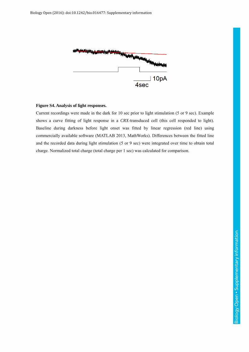
Figure S4. Analysis of light responses.
Current recordings were made in the dark for 10 sec prior to light stimulation (5 or 9 sec). Example
shows a curve fitting of light response in a CRX-transduced cell (this cell responded to light).
Baseline during darkness before light onset was fitted by linear regression (red line) using
commercially available software (MATLAB 2013, MathWorks). Differences between the fitted line
and the recorded data during light stimulation (5 or 9 sec) were integrated over time to obtain total
charge. Normalized total charge (total charge per 1 sec) was calculated for comparison.
Bio
logy
Ope
n •
Sup
plem
enta
ry in
form
atio
n
Biology Open (2016): doi:10.1242/bio.016477: Supplementary information

Table S1
Primer name Sequence
Not1 CRX N atatgcggcc gccaaggttc acttatgatg gcgtatatga ac
CRX EIS Not1 C atatgcggcc gcgatgaact ttcaccctaa gtttttctta ctacggctac aagatctaaa cttc
Not1 NeuroD1 N atatgcggcc gccaaggttc aatgaccaaa tcgtacag
NeuroD1 EIS Not1 C atatgcggcc gcgatgaact ttcaccctaa gtttttctta ctacggctaa tcatgaaata tggc
Not1 RAX N atatgcggcc gccaaggttc aatgcacctg ccgggctgcg cg
RAX EIS Not1 C atatgcggcc gcgatgaact ttcaccctaa gtttttctta ctacggctag agggcctgcc acggc
Bio
logy
Ope
n •
Sup
plem
enta
ry in
form
atio
n
Biology Open (2016): doi:10.1242/bio.016477: Supplementary information

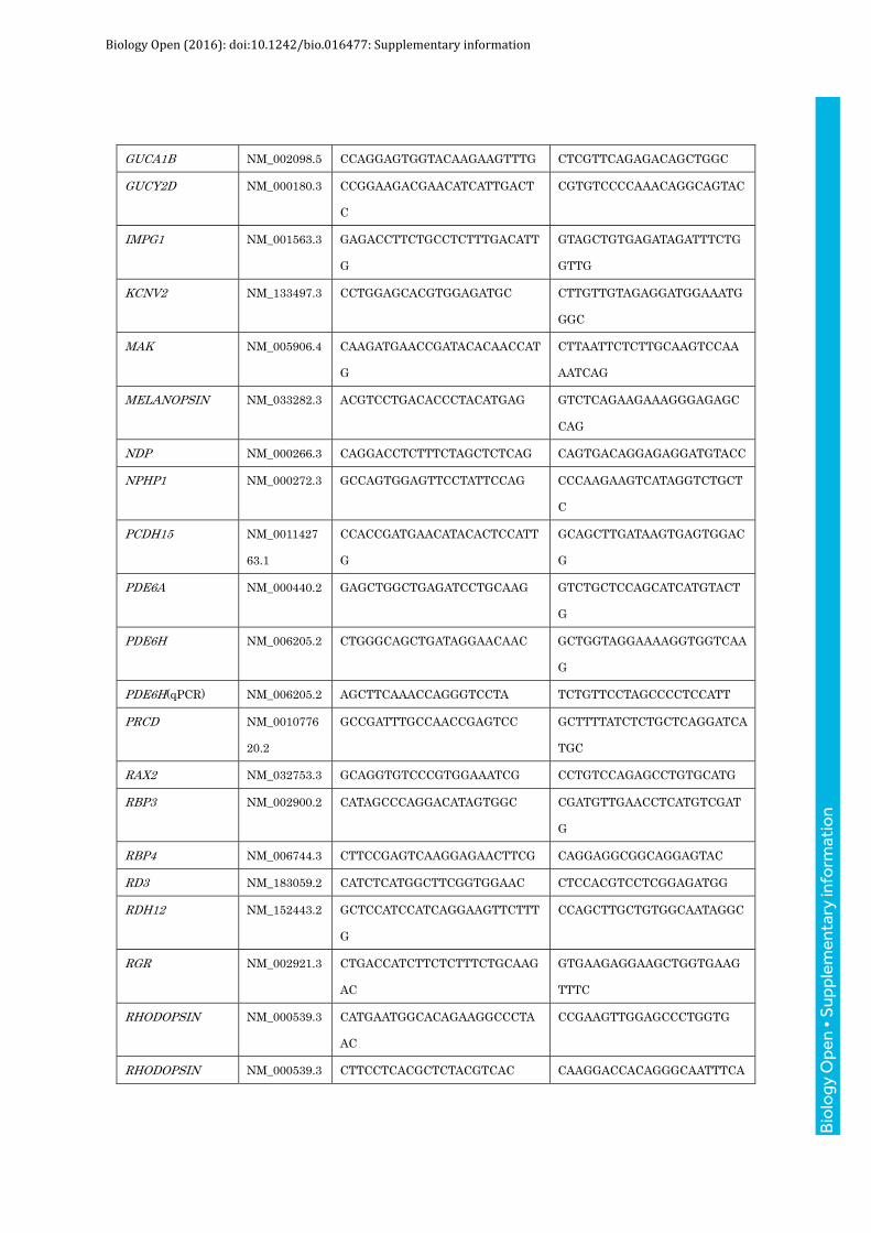
Table S2
Gene Reference Forward Reverse
BlueOpsin(qPCR) NM_001708.2 GCTTGTCACCATTCCTTCATTC TTCATCTGTCATGGCCTTCC
C2orf71 NM_0010298
83.2
GAAGCTAAACCGCCACTCTCAAC CGCCTCTGGCTGGGATTTGTC
CC2D2A NM_0010805
22.2
GCTAAGCTGGTGTTTTTGCTCCAA
G
CTTGGGCTCCTCCTGCACAG
CNGA1 NM_0011425
64.1
GGCTCATTTTGGATTACGTATCAG CTAGCCAAACGGCCAAATTCAG
CNGA3 NM_001298.2 CTGGACTACTCGGCAGATGTC GGACCAGTAGAGACTGTAAATG
TAC
CNGB1 NM_001297.4 CAGGATGTTGGGCTGGGTCC CTCTTTCCAGATTCTGCTCCAG
C
COUPTF1 NM_005654.5 CAGCCCAATCCAGGCCAGTAC GGCAGTCGCAGCAGCAGTTTG
CRB1 NM_201253.2 CAGAGGACGCTGCATCAACTTG CCTAGGTTTTGTGAAGACTGAT
ACAG
EFEMP1 NM_0010393
48.2
GAATGCAGAACCTCAAGCTACCT
G
CACAGAGCTTGTGCGGAAGGTC
FZD4 NM_012193.3 GGCTACAACGTGACCAAGATGC GCCTCTTCAAAATCACAGGATA
TCC
GDF6 NM_0010015
57.2
GTGCCCCACGAGTACATGCTG GAACAGGTTCTTGCGCTGGGAT
C
GNA11 NM_002067.4 CAAGGCCAATGCGCTCCTGATC GACTCCACCAGGACTTGGTCG
GNA14 NM_004297.3 GACAGGAGGAGGGAGTACCAG GATGACTTTCTCTTTGTCAGGA
TTCTG
GNAQ NM_002072.4 GCTTTTGAGAATCCATATGTAGAT
GC
GGAATACATGATTTTCTCCTCTA
GAAG
GNAT1 NM_144499.2 CACGATGCCCAAGGAGATGTC CATGTGGGAATAGATCTCCTTC
AC
GNAT2 NM_005272.3 GATAAGGAAGCCAAGACTGTCAA
GC
GACTCCCTCGAAGCAGTGGATC
GPR98 NM_032119.3 CTAAACTTCTGACTCACATGATGG
C
GAGAACCAACATCCAGAAGTGT
C
GUCA1A NM_000409.3 GTGGAGGAGCTGAGCAGCAC GTCAGTGTGTCCAGGAGCATCT
G
Bio
logy
Ope
n •
Sup
plem
enta
ry in
form
atio
n
Biology Open (2016): doi:10.1242/bio.016477: Supplementary information
GUCA1B NM_002098.5 CCAGGAGTGGTACAAGAAGTTTG CTCGTTCAGAGACAGCTGGC
GUCY2D NM_000180.3 CCGGAAGACGAACATCATTGACT
C
CGTGTCCCCAAACAGGCAGTAC
IMPG1 NM_001563.3 GAGACCTTCTGCCTCTTTGACATT
G
GTAGCTGTGAGATAGATTTCTG
GTTG
KCNV2 NM_133497.3 CCTGGAGCACGTGGAGATGC CTTGTTGTAGAGGATGGAAATG
GGC
MAK NM_005906.4 CAAGATGAACCGATACACAACCAT
G
CTTAATTCTCTTGCAAGTCCAA
AATCAG
MELANOPSIN NM_033282.3 ACGTCCTGACACCCTACATGAG GTCTCAGAAGAAAGGGAGAGC
CAG
NDP NM_000266.3 CAGGACCTCTTTCTAGCTCTCAG CAGTGACAGGAGAGGATGTACC
NPHP1 NM_000272.3 GCCAGTGGAGTTCCTATTCCAG CCCAAGAAGTCATAGGTCTGCT
C
PCDH15 NM_0011427
63.1
CCACCGATGAACATACACTCCATT
G
GCAGCTTGATAAGTGAGTGGAC
G
PDE6A NM_000440.2 GAGCTGGCTGAGATCCTGCAAG GTCTGCTCCAGCATCATGTACT
G
PDE6H NM_006205.2 CTGGGCAGCTGATAGGAACAAC GCTGGTAGGAAAAGGTGGTCAA
G
PDE6H(qPCR) NM_006205.2 AGCTTCAAACCAGGGTCCTA TCTGTTCCTAGCCCCTCCATT
PRCD NM_0010776
20.2
GCCGATTTGCCAACCGAGTCC GCTTTTATCTCTGCTCAGGATCA
TGC
RAX2 NM_032753.3 GCAGGTGTCCCGTGGAAATCG CCTGTCCAGAGCCTGTGCATG
RBP3 NM_002900.2 CATAGCCCAGGACATAGTGGC CGATGTTGAACCTCATGTCGAT
G
RBP4 NM_006744.3 CTTCCGAGTCAAGGAGAACTTCG CAGGAGGCGGCAGGAGTAC
RD3 NM_183059.2 CATCTCATGGCTTCGGTGGAAC CTCCACGTCCTCGGAGATGG
RDH12 NM_152443.2 GCTCCATCCATCAGGAAGTTCTTT
G
CCAGCTTGCTGTGGCAATAGGC
RGR NM_002921.3 CTGACCATCTTCTCTTTCTGCAAG
AC
GTGAAGAGGAAGCTGGTGAAG
TTTC
RHODOPSIN NM_000539.3 CATGAATGGCACAGAAGGCCCTA
AC
CCGAAGTTGGAGCCCTGGTG
RHODOPSIN NM_000539.3 CTTCCTCACGCTCTACGTCAC CAAGGACCACAGGGCAATTTCA
Bio
logy
Ope
n •
Sup
plem
enta
ry in
form
atio
n
Biology Open (2016): doi:10.1242/bio.016477: Supplementary information

(nested) CC
RP1 NM_006269.1 GGTCAACCCTCGCTCCTTTAAG CCTGGTTTAAATGGCTCCCTTC
C
RP1L1 NM_178857.5 CAGCAGACAGTGGTTCTCAGTC CAGGCTGCCGTCCTCATTCATG
RPE65 NM_000329.2 GTAGAGGTTACTGACAATGCCCTT
G
GTAGTTGGCTCCCCAAAGACTC
RPGRIP1 NM_020366.3 GAATCCTCTGAACAAGGTTCTGAA
G
CATCCAGAGGATCACTTACCAC
TG
SLC7A14 NM_020949.2 CGCAGGCCTCTTCTTCATCAATGG CTATGAGCATCTCATTGTCTCCC
AAG
TLR4 NM_003265.2 CTTACCCATAATCAACTCAGAAGA
TTAC
GACAGATTCCGAATGCTTGTGT
TTG
TRPC3/6/7 NM_003305.2
NM_004621.5
NM_0011675
77.1
CATGAAGTTTGTAGCTCACGCAG CATAATGAATATGACCATGAACT
TGAAG
TSPAN12 NM_012338.3 CTGTTGCTTCTTGCATGGTACTTT
G
GCAGAGTAATGGTGAGAATCAT
GGC
TTPA NM_000370.3 GAAATAAGTGCAGATCTACACCCT
AG
CCTGACAAATGTCCTCCATGGA
G
TUB NM_003320.4 CCAATTACCTCATCTCTGTGGACC GTAGTTGTAATCCATGGTGAAC
ACATC
USH1G NM_173477.4 CCACCCGAAAGGAGCTGAATGC CTTGGGATTGGCGTAGGTGCC
USH2A NM_007123.5 GCAACCTCCAATAAAAGTAATGAC
AC
GGCTTCAGGATTCAACCGTGAC
ZNF423 NM_0012716
20.1
CGAAGACCTGGAGAGCCACATG GCCTTCTGCGGAGAGGTGTC B
iolo
gy O
pen
• S
uppl
emen
tary
info
rmat
ion
Biology Open (2016): doi:10.1242/bio.016477: Supplementary information