in vivo characterization of a new transpedicular...
TRANSCRIPT

1
Amministrative office: University of Padova
Department of Comparative Biomedicine and Food Science
___________________________________________________________________
DOCTORAL SCHOOL IN VETERINARY SCIENCE
SERIES XXVII
IN VIVO CHARACTERIZATION OF A NEW TRANSPEDICULAR APPROACH
TO THE INTERVERTEBRAL DISC IN A LARGE ANIMAL MODEL
Director of the School: Prof. Gianfranco Gabai
Supervisor: Prof. Marco Bernardini
PhD student: Francesca de Strobel

2
SUMMARY - Back pain (BP) is a common clinical condition that leads to high morbidity
with significant psychosocial and economic effects. It is the leading cause of disability in
people under 45 years of age, and it results in enormous national economic losses in
developed countries. The wide majority of BP is associated with degenerative changes of
the intervertebral disc (IVD). Intervertebral disc degeneration (IVDD) is an age-related
chronic process that is characterized by a progressive reduction of proteoglycans and
water content in the nucleus pulposus (NP) with loss of the IVD's ability to resist
compressive forces. Current treatment options for BP and IVDD range from conservative
measures to invasive procedures however, these treatment modalities have limited
efficacy and do not produce predictable and reliable outcomes. Therefore, there is a clear
need for more effective early treatment of BP that may prevent, slow down, or reverse the
degenerative changes. To evaluate the efficacy of novel IVD regenerative treatments,
prior to translation into humans, ex vivo and in vivo animal model systems are needed.
In the present study, a novel transpedicular approach to the IVD was validated in 12
sheep. Under fluoroscopy, a 2-mm Kirshner wire was introduced through the vertebral
body with a cranio-medial inclination of approximately 45° in all plans direction to reach
the center of the NP. In each animal four IVDs, from L1 to L5, were addressed by
performing different surgical techniques, respectively: (I) nucleotomy, (II) tunnel, (III)
nucleotomy + polyurethane (PU) scaffold, and (IV) tunnel + PU scaffold. Intact IVDs (L5-6)
were used as controls. Intra- and post-surgical morbidity rates were low; CSF leakage
was recorded in two spinal segments whereas discospondylitis and vertebral luxation
occurred respectively at L1-2 and L3-4 in two different animals. The quantitative and
qualitative analysis of MRI, radiologic, histologic and macroscopic data, collected at four
different time points (before and 1, 3 and 6 months after surgery), suggested that the
injury induced in the present model represents a reliable method for initiating a
progressive IVDD process, obtaining different degrees of IVDD depending on the type of
lesion performed. The endplate damage itself, caused by the realization of the

3
transpedicular tunnel, led to IVD degenerative changes, although to a lesser extent than
those caused by performing nucleotomy. Furthermore, the sealing of the tunnel with the
PU scaffold resulted in a lack of cells leakage throughout the tunnel.
The transpedicular approach represents a feasible alternative route to the IVD, with
respect to the traditional ventral and ventrolateral approaches through the annulus
fibrosus. This new pathway to the IVD provides a new valid model to study biologic and
biomechanical alterations in relation either, to IVD degenerative processes and to
potential NP regenerative therapies.
RIASSUNTO - La lombalgia è una condizione clinica molto frequente associata ad
elevata morbidità, con effetti sia dal punto di vista psicosociale che della spesa sanitaria
nazionale. La lombalgia é la principale causa di disabilità nella popolazione al di sotto dei
45 anni, e rappresenta una della principali voci di spesa nei paesi sviluppati. La grande
maggioranza dei casi di lombalgia è associata alla degenerazione del disco
intervertebrale (IVDD). L'IVDD è un processo cronico età-correlato caratterizzato da una
progressiva riduzione del contenuto di proteoglicani ed acqua nel nucleo polposo (NP),
con perdita della capacità del disco di resistere a forze compressive. Le attuali opzioni di
trattamento disponibili per lombalgia e IVDD comprendono sia approcci conservativi che
procedure invasive. Tuttavia, queste modalità di trattamento hanno un'efficacia limitata e
non producono risultati prevedibili e riproducibili. Per queste ragioni, vi è una chiara
necessità di trattamenti che siano efficaci nel prevenire, rallentare o invertire le
modificazioni degenerative causate dal processo di IVDD. Al fine di valutare l’efficacia di
nuove tecniche di rigenerazione discale e prima di trasferirle all’uomo, é necessario
testarle su modelli animali ex vivo ed in vivo.
Questo studio ha permesso di validare, in 12 pecore, un nuovo approccio al disco
intervertebrale (IVD) per via transpeduncolare. Tramite guida fluoroscopica, un filo di
Kirshner di 2-mm di diametro é stato introdotto attraverso il corpo vertebrale con
un’inclinazione di circa 45° in tutti i piani dello spazio e in direzione cranio-mediale, tale

4
da raggiungere il centro del NP. In ciascun animale sono stati trattati quattro IVD, da L1 a
L5, effettuando in ciascuno una diversa tecnica chirurgica: (I) nucleotomia, (II) tunnel, (III)
nucleotomia + scaffold in poliuretano (PU) e (IV) tunnel + scaffold PU. I dischi non trattati
(L5-6) sono stati considerati come gruppo controllo. Le percentuali di morbiditá intra- e
post-operatoria si sono rivelate basse; in due occasioni é stata riportata fuoriuscita di
liquor in fase chirurgica, inoltre lussazione di L3-4 e discospondilite a livello di L1-2 si
sono verificate in due soggetti. Le analisi quantitative e qualitative, effettuate sui dati
ricavati in quattro diversi time point (prima e 1, 3 e 6 mesi dopo la chirurgia) dalle
immagini radiografiche e di risonanza magnetica e dai campioni macroscopici e istologici,
hanno rivelato l’efficacia del metodo presentato nell’ottenere un modello progressive di
IVDD. Inoltre, attraverso l’approccio transpeduncolare é stato possibile ottenere diversi
gradi di IVDD, dipendentemente dalla tipologia di lesione effettuata. Il solo danno al piatto
vertebrale ha determinato alterazioni degenerative del IVD, tuttavia inferiori rispetto a
quelle causate effettuando la nucleotomia. Inoltre, l’utilizzo dello scaffold in PU per
sigillare il tunnel é servito a prevenire la fuoriuscita di cellule attraverso il tunnel.
L’approccio transpeduncolare rappresenta una via alternativa per raggiungere il IVD
rispetto ai tradizionali approcci ventrali e ventrolaterali attraverso l’anello fibroso. Questa
nuova tecnica fornisce un nuovo modello per lo studio di alterazioni biologiche e
biomeccaniche in relazione sia ai processi di IVDD sia a potenziali terapie regenerative.

5
INDEX
INTRODUCTION 9
O INTERVERTEBRAL DISC 11
EMBRYOGENESIS AND POSTNATAL DEVELOPMENT 11
INTERVERTEBRAL DISC MORPHOLOGY AND BIOCHEMISTRY 14
INTERVERTEBRAL DISC INNERVATION AND VASCULARIZATION 18
INTERVERTEBRAL DISC DEGENERATION 21
AETIOLOGY OF DISC DEGENERATION 25
O DISCOGENIC PAIN 27
O HISTOLOGIC AND MRI CLASSIFICATION OF IVDD 29
THOMPSON GRADING SCALE 29
PFFIRMANN GRADING SCALE 30
O CURRENT TREATMENT OPTIONS: MEDICAL / SURGICAL 31
O NOVEL THERAPEUTIC APPROACHES 33
REGENERATIVE STRATEGIES 33
REPAIR STRATEGIES 34
O ANIMAL MODELS 36
O SHEEP MODEL IN SPINE RESEARCH 40
SURGICAL AND MRI ANATOMY OF THE OVINE LUMBAR SPINE 45
O THE TRANSPEDICULAR APPROACH 47
OBJECTIVES OF THE STUDY 52
SECTION 1: IN VIVO CHARACTERIZATION AND VALIDATION OF A NEW
INTERVERTEBRAL DISC DEGENERATION MODEL
MATERIALS AND METHODS 53

6
O CASES SELECTION 53
O IMAGING 53
O SURGICAL PROCEDURE 54
O PAIN MANAGEMENT AND POSTOPERATIVE CARE 58
O EUTHANASIA 59
O COMPLICATIONS 59
RESULTS 60
O SURGICAL PROCEDURE 60
O IMAGING 60
O PERI- AND POST-OPERATIVE CARE AND COMPLICATIONS 61
INTRAOPERATIVE DATA 61
POSTOPERATIVE DATA 61
DISCUSSION 63
SECTION 2: EVALUATION OF QUALITATIVE AND QUANTITATIVE DATA OF THE
INTERVERTEBRAL DISC DEGENERATION MODEL
MATERIALS AND METHODS 69
O QUALITATIVE ANALYSIS 69
MAGNETIC RESONANCE IMAGES ANALYSIS 69
GROSS ANATOMY AND HISTOLOGICAL EVALUATION 69
O QUANTITATIVE ANALYSIS 70
DISC HEIGHT INDEX (DHI) 71
MRI INDEX (MRI) 71
O STATISTICAL ANALYSIS 72
RESULTS 74
O QUALITATIVE ANALYSIS 74

7
MRI ANALYSIS 74
GROSS ANATOMY AND HISTOLOGY 74
O QUANTITATIVE ANALYSIS 79
DISCUSSION 84
O QUALITATIVE ANALYSIS 84
O QUANTITATIVE ANALYSIS 87
CONCLUSIONS 91
BIBLIOGRAPHY 92

8
9
INTRODUCTION
Back pain (BP) is a common clinical condition that leads to high morbidity with significant
psychosocial and economic effects. It is the leading cause of disability in people under 45
years of age, and it results in enormous national economic losses in developed countries.
The wide majority of BP is associated with degenerative changes of the intervertebral disc
(IVD) (Luo et al., 2004). Intervertebral disc degeneration (IVDD) is an age-related chronic
process that is characterized by a progressive reduction of proteoglycans (PGs) and water
content in nucleus pulposus (NP) with loss of the IVD’s ability to resist compressive
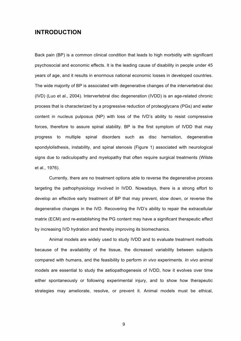
forces, therefore to assure spinal stability. BP is the first symptom of IVDD that may
progress to multiple spinal disorders such as disc herniation, degenerative
spondylolisthesis, instability, and spinal stenosis (Figure 1) associated with neurological
signs due to radiculopathy and myelopathy that often require surgical treatments (Wilste
et al., 1976).
Currently, there are no treatment options able to reverse the degenerative process
targeting the pathophysiology involved in IVDD. Nowadays, there is a strong effort to
develop an effective early treatment of BP that may prevent, slow down, or reverse the
degenerative changes in the IVD. Recovering the IVD’s ability to repair the extracellular
matrix (ECM) and re-establishing the PG content may have a significant therapeutic effect
by increasing IVD hydration and thereby improving its biomechanics.
Animal models are widely used to study IVDD and to evaluate treatment methods
because of the availability of the tissue, the dicreased variability between subjects
compared with humans, and the feasibility to perform in vivo experiments. In vivo animal
models are essential to study the aetiopathogenesis of IVDD, how it evolves over time
either spontaneously or following experimental injury, and to show how therapeutic
strategies may ameliorate, resolve, or prevent it. Animal models must be ethical,

10
controllable, reproducible, and cost-effective; in addition, they should be able to model
human pathologic processes.
Figure 1
Spinal stenosis - Progressive narrowing of the spinal canal could be either a congenital or acquired condition. It is most common in the cervical and lumbar areas. The canal components that contribute to acquired stenosis include the facets (hypertrophy, arthropathy), ligamentum flavum (hypertrophy), posterior longitudinal ligament, vertebral body (spondylosis), IVD, and epidural fat. Spinal stenosis implies spinal canal narrowing with possible subsequent neural compression. Congenital stenosis may predispose an individual with mild degenerative changes to become symptomatic earlier in life.
Spondylolisthesis – It is defined as the translation of one vertebra in relation to the adjacent level and is commonly referenced as an anterolisthesis of the cranial segment on the caudal segment. Five different types of spondylolisthesis have been described: dysplastic, isthmic, degenerative, traumatic, and pathologic (Wilste et al., 1976). Spondylolisthesis can be either congenital or acquired.
Numerous in vivo animal models for IVDD are described in literature, and each
model has its own advantages and disadvantages. There are several important aspects to
consider in relation to using animals for studying IVDD such as development and anatomy
11
of the spine, loading and size differences, the mechanical, biochemical and nutritional
conditions.
o INTERVERTEBRAL DISC
§ EMBRYOGENESIS AND POSTNATAL DEVELOPMENT
The embryonic development of the vertebral column centers on the notochord, a rod-like
mesoderm-derived structure (Fleming et al., 2001; Stemple, 2005). The notochord is
important both as a signaling center that mediates cell migration, differentiation and
survival, and as a structure that physically gives rise to the NP (Peacock, 1951; Walmsley,
1953; Choi et al., 2008). Embryonic morphogenesis of the disc, as well as key molecules
implicated in this process, are illustrated schematically in Figure 2.
The annulus fibrosus (AF) and NP regions of the IVD arise concurrently along
distinct developmental pathways. During human fetal gestation, at approximately 30 days
(12 days in the mouse), cells of the sclerotome migrate medially from pairs of paraxial
somites (Figure 2A) to condense around the notochord (Figure 2B) (Peacock, 1951;
Hunter et al., 2003a), adopting a metameric pattern of more condensed and less
condensed regions (Figure 2C), which later give rise to the AF and vertebral bodies,
respectively (Aszodi et al., 1998). Cells in the future AF region adopt a fibroblastic
morphology. These cells align and orient to form the template for ECM deposition that
later defines the AF angle-ply lamellar structure (Figure 2E) (Rufai et al., 1995).
Concurrently with AF morphogenesis, the notochord contracts within the forming vertebral
body rudiments, simultaneously expanding within the intervertebral regions to form the NP
(Figure 2D) (Peacock, 1951; Pazzaglia et al., 1989; Aszodi et al., 1998).

12
Figure 2. Schematic representation of embryonic morphogenesis of the mammalian IVD. Colours represent origins and destinations of cell populations. Also indicated are key morphogens and transcriptional regulators implicated in the growth and differentiation of the disc structures at each developmental stage. (A) The notochord adjacent to pairs of paraxial somites, which contain sclerotome cells. (B) Sclerotome cells condense around the notochord. (C) Cells adopt a metameric pattern of more condensed (green) and less condensed (brown) regions that give rise to the AF and vertebral bodies, respectively. (D) The notochord contracts within the vertebral body rudiments and expands within the future intervertebral disc to form the NP. (E) Basic structures of the disc are established, and AF cells adopt orientations and alignments that form the template for the lamellar structure. VB, vertebral body.
Once the basic structures of the IVD are established, several developmental
changes occur. In humans, during the early postnatal years, blood vessels that have
penetrated the AF and cartilage endplates (EPs) from as early as 35 weeks gestation
begin to recede, determining the avascular structure of the IVD (Urban and Roberts, 1995;
Nerlich et al., 2007). The vascular regression has been hypothesisied to be a
consequence of decreased nutrient requirements following the initial period of rapid
growth or, more likely, of the inability of circulatory pressure to compete with large
physiological stresses in the surrounding ECM. Poor nutritional supply to the cells of the
avascular IVD has been implicated in the pathogenesis of IVDD.
Further changes characterising the resident cell populations, and specifically those of
the NP, begin to occur very early in life. Soon after birth, the cells that populate the NP
exhibit morphological characteristics that are similar to the cells that populate its
notochord precursor (Peacock, 1952; Wolfe et al., 1965; Trout et al., 1982a; Trout et al.,
1982b). Notochordal cells (NCs) in the postnatal NP are large (30-40 µm in diameter),
frequently appear in clusters and possess actin-filament-bounded intracellular vacuoles

13
that occupy more than 25% of the cell area (Hunter et al., 2003b; Hunter et al., 2004)
(Figure 3b).
In the first 10 years following birth, the number of NCs declines, and no NCs are present
by adulthood (Peacock, 1952; Trout et al., 1982a; Hunter et al., 2004). Concurrently, a
second population of chondrocyte-like cells appears, characterised by apparent
morphological similarities with cartilage chondrocytes (Urban and Roberts, 1995) (Figure
3). In comparison with NCs, the chondrocyte-like NP cells are smaller (~10 µm in
diameter) and lack intracellular vacuoles (Hunter et al., 2004).
In many species (mouse, rat, cat, mink, chondrodystrophoid dog, pig and rabbit) the NCs
persist through most of adult life, whereas in other species they gradually disappear
during aging (human, sheep, non-condrodystrophoid dog, cow) (Hunter et al., 2004).
Horses apparently have no NCs at birth (Table 1). The exact mechanism of transition from
NCs to chondrocytic-like cells is not precisely known; however, developmental changes to
both mechanical and nutritional microenvironment have been implicated (Rastogi et al.,
2009; Guehring et al., 2010). It is unclear whether this change in cell populations is due to
continued differentiation of the NCs into chondrocytic phenotype (Choi et al., 2008;
Risbud et al., 2010), or due to apoptosis of the resident cells with the subsequent
migration to the NP by cells derived from the cartilaginous EPs or AF (Kim et al., 2003).
Considered that altered cellularity represents a hallmark of IVDD, the relevance of
NCs in animal models used to study IVDD can be great since this is a completely different
cell type in terms of morphology and function to the cells populating the adult human NP
(Hunter et al., 2003a).
Another key factor in early IVDD is the decrease in the PG content in the NP. NCs
have been shown to synthesize ECM in a distinct manner respect the mature NP cells
(Cappello et al., 2006). PGs synthesized by NCs are evenly distributed between the inter-
and pericellular regions, compared with mature NP cells, in which the majority of PGs are
intercellular. Additionally, the rate at which PGs migrate to the intercellular regions is
significantly greater for NCs than for mature NP cells (Cappello et al., 2006).

14
Table 1. Age of loss of notochordal cells in different species (Hunter et al., 2004)
Species Age of skeletal maturity
Age of loss of notochordal cells References
Dog (c) 12 months 12 months Hansen, 1952; Hutton et al., 2000; Ganey et al., 2003
Dog (n/c) 12 months 60 months
Frick et al., 1994; Hansen 1952; Maldonado and Oegema, 1992; Katsuura and Hukuda, 1994; Matsuzaki et al., 1996; Hunter et al., 2004
Rabbit 10 months 6 months
Anderson et al., 2002; Smith and Serafini-Fracassini, 1968; Scott et al., 1980; Nomura et al., 2001; Hunter et al., 2004
Pig 12 months Unknown Holm et al., 2004; Kawchuck et al., 2001; Hunter et al. ,2004
Cat 24 months Never Hansen 1959; Butler 1989; Kathmann et al., 2000; Hunter et al., 2004
Ferret n/d Never Hunter et al., 2004
Sheep 12 months Unknown Kadoya et al., 2001 ; Hunter et al., 2004
Rat 2 months 12 months
Adler et al., 1983; Moskowitz et al., 1990; Nishimura and Mochida, 1998; Iatridis et al., 1999; Mente et al., 1999; MacLean et al., 2003; Hunter et al., 2004
Mouse 4 months n/d Ariga et al., 2001; Lotz et al., 1998; Lotz and Chin, 2000; Wlash and Lotz, 2004; Hunter et al., 2004
Human 20 years 6-10 years Horwitz, 1977
C, chondrodystrophoid (beagles); n/c, non-chondrodystrophoid (mongrels); n/d: no data available.
§ INTERVERTEBRAL DISC MORPHOLOGY AND BIOCHEMISTRY
The human vertebral column consists of 24 mobile vertebrae: 7 cervical, 12 thoracic, and
5 lumbar. With the exception of the first and second cervical vertebrae, all vertebral bodies
are connected by IVDs. IVDs form a strong and flexible connection between the
vertebrae, allowing flexion, extension, and rotation of the spinal column and forming an
amphiartrosis type of joint. Moreover, the IVDs constantly transmit loads arising from body
weight and muscle activity through the spinal column (Twomey and Taylor, 1987; Roberts
et al., 1989).

15
As described previously, developmentally the IVD is a unique structure formed
from cells of at least two embryonic origins: the notochord and the somites. These
lineages give rise to a tissue that is complex and specialized in terms of its microstructure,
mechanical function and cell types (Smith et al, 2011). IVDs consist of a thick outer ring of
fibrous cartilage (AF), which surrounds a more gelatinous core known as the NP; the NP
is delimited cranially and caudally by cartilage EPs (Figure 4A), forming a symphysis type
of cartilagineous joint.
Nucleus pulposus - The central NP contains large quantities of aggrecan, the major PG,
which aggregates along chains of hyaluronan (Urban, 1996). The glycosaminoglycan
(GAG) side chains of these PGs carry a fixed negative charge and generate an osmotic
swelling pressure within an irregular meshwork of collagen II fibrils (Inoue, 1981) and
elastin fibres, which are arranged radially (Yu et al., 2002). Interspersed at a low density
(approximately 5000/mm3) (Maroudas et al, 1975) are chondrocytes-like cells (Figure 3c;
4b).
Annulus fibrosus - Outside the NP is the AF, characterized by heterogeneous
composition and architecture. The highly organized outer regions consist of concentric
distinct lamellae (Figure 5A-B), which are composed of bundles of collagen I fibers
oriented at oblique angles that alternate within each consecutive lamella to form an angle-
ply structure (Marchand and Ahmed, 1990). In the inner AF, there is a transition to
collagen II that, together with increasing PGs concentration, gives rise to a less fibrous
and less organized structure (Humzah and Soames, 1988). Elastin fibres lie between the
lamellae, possibly helping the IVD to return to its original arrangement following bending.
They may also bind the lamellae together as elastin fibres pass radially from one lamella
to the next (Yu et al., 2002). The cells of the AF, particularly in the outer region, tend to be
fibroblast-like, elongated, thin and aligned parallel to the collagen fibres (Figure 3a), while
toward the inner AF the cells can be more oval. Cells of the IVD, both in the AF and NP,
can have several long, thin cytoplasmatic projections, which may be more than 30 µm

16
long (Errington et al., 1998; Bruehlmann et al., 2002). Their function in the IVD is unknown
but it has been suggested that they may act as sensors and communicators of mechanical
strain within the tissue (Bruehlmann et al., 2002).
Figure 3. Morphology of IVD cells. Cells of the intervertebral disc differ in morphology according to the region of origin, age and species. (a) Annulus fibrosus cells (bovine disc): the cells in the outer fraction are fibroblast-like, therefore much more bipolar and elongated (arrow) than those in the inner fraction that are more rounded; (b) Notochordal cells (bovine disc): large highly vacuolated cells; (c) Nucleus pulposus cells (human disc) are rounded, often sitting inside a capsule (thin arrow) that may be surrounded by an obvious pericellular matrix (thick arrow) (Roberts et al., 2006; Alini et al., 2008).
Endplate - The third morphologically distinct region is the cartilage EP, a thin horizontal
layer, usually less than 1 mm thick, of hyaline cartilage. The EPs extend cranially and
caudally over the inner AF and NP to interface with the vertebral bodies (Figure 4), and
function to regulate nutrient diffusion between the IVD and the vertebral bodies. In the
outer regions of the AF, collagen fibres anchor directly into the vertebral bone, interfacing
the IVD and the vertebral body. The collagen fibres within it run horizontal and parallel to
the vertebral bodies, with the fibres continuing into the IVD (Roberts et al., 1989). During
childhood and adolescence, the EP functions also as a growth plate of the adjacent
vertebrae in humans, whereas the vertebral bodies of other mammals contain separate
growth plates (Adam and Roughley, 2006).

17
A B Figure 4. Schematic representations of the adult intervertebral disc. (A) Midsagittal cross-section showing anatomical regions. (B) Three-dimensional view illustrating the AF lamellar structure.
Extracellular matrix - The mechanical functions of the IVD are determined by the
composition and organization of the ECM. The main mechanical role is provided by the
two major macromolecular components, collagen and PGs. The collagen network, formed
mostly of type I and type II collagen fibrils and making up approximately 70% and 20% of
the dry weight of the AF and NP respectively (Eyre and Muir, 1977), provides tensile
strengh to the IVD and anchors the tissue to the bone. Aggrecan, the major PG of the IVD
(Johnstone and Bayliss, 1995), is responsible for maintaining tissue hydration through the
osmotic pressure provided by its constituent chondroitin and keratan sulphate chains
(Urban et al, 1979) (Figure 5B). The PGs and water content of the NP (around 50% and
80% of the wet weight, respectively) is greater than in the AF (approximately 20% and
70% of the wet weight, respectively) (Figure 5A). In addition, there are many other minor
components, such as collagen types III, V, VI, IX, X, XI, XII and XIV; small PGs such as
lumican, biglycan, decorin and fibromodulin; and other glycoproteins such as fibronectin
and amyloid (Roberts et al., 1991; Melrose et al., 2001). The functional role of many of
these additional matrix proteins and glycoproteins is not yet clear. Collagen IX, however,
is thought to be involved in forming cross-links between collagen fibrils and is thus
important in maintaining network integrity (Eyre et al., 2001).

18
A B Figure 5. (A) Distribution of the main IVD constituents within the AF, NP and EPs (Prithvi, 2008). (B) Structure of an aggrecan type complex PG. Aggrecan is a high molecular weight PG, in which chondroitin sulfate and keratan sulfate GAG chains are attached to an extended protein core.
The ECM is a dynamic structure. Its molecules are continually being broken down
by proteinases such as the matrix metalloproteinases (MMPs) and aggrecanases, which
are also synthesized by IVD cells (Sztrolovics et al., 1997; Roberts et al., 2000; Weiler et
al., 2002). The balance between synthesis, breakdown and accumulation of ECM
macromolecules determines the quality and integrity of the ECM, and thus the mechanical
behaviour of the IVD itself. The integrity of the ECM is also important for maintaining the
relatively avascular and aneural nature of the healthy IVD.
§ INTERVERTEBRAL DISC INNERVATION AND VASCULARIZATION
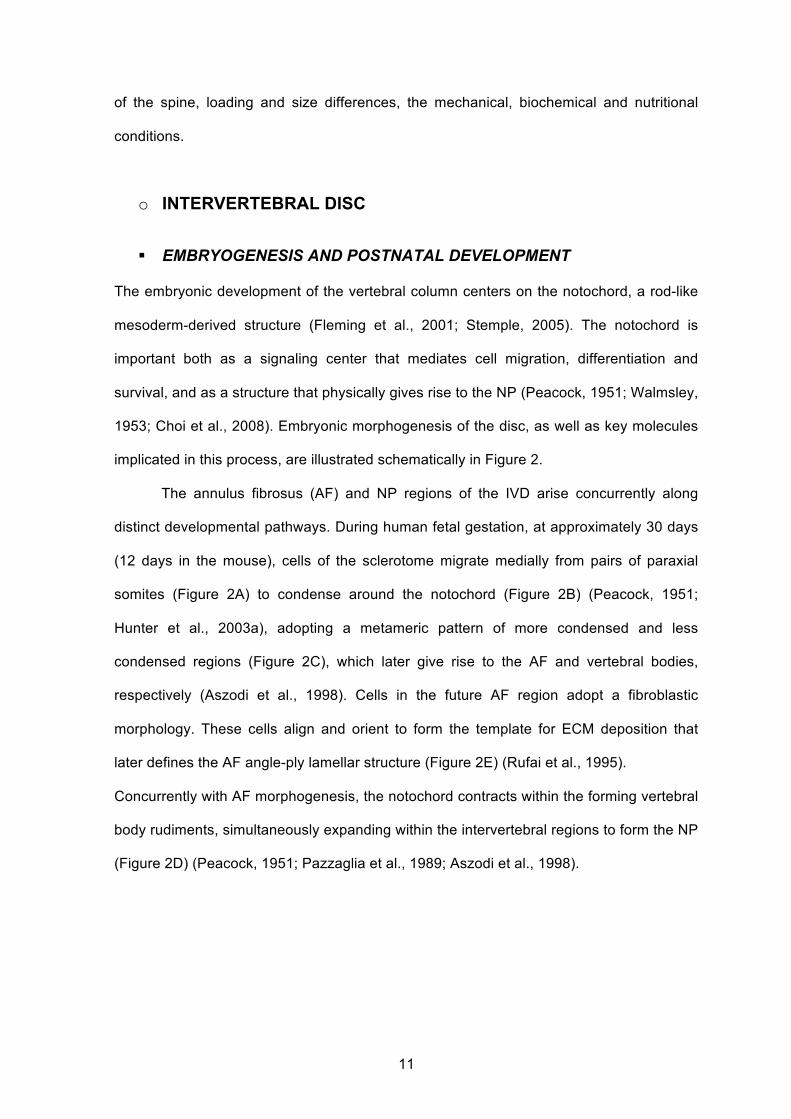
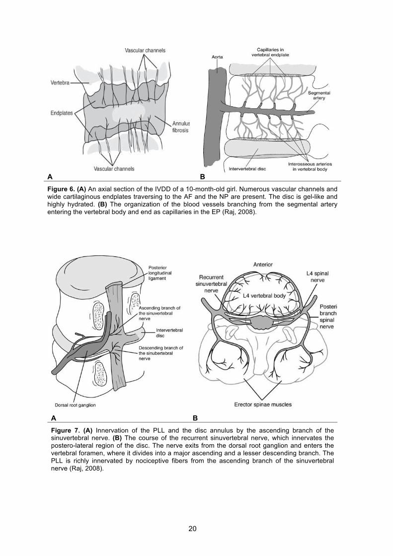
The adult IVD is almost completely avascular (Figure 6B) (Nerlich et al., 2007), so
resident cells must survive and function in an environment that is low in nutrients and
oxygen (Urban et al., 2004). In the longitudinal ligaments adjacent to the IVD and in young
cartilage EPs (less than about 12 months old) are present blood vessels which represent
branches of the spinal artery (Crock et al., 1991) (Figure 6A). The cartilagineous EP, like
other hyaline cartilages, is normally totally avascular and aneural in the healthy adult
PATHOPHYSIOLOGY
Loss of Proteoglycan
The most significant biochemical change to occur in discdegeneration is loss of proteoglycan (Figure 11A and
B).29 The aggrecan molecules become degraded, withsmaller fragments being able to leach from the tissuemore readily than larger portions. This results in loss ofglycosaminoglycans; this loss is responsible for a fall in
A
B
Figure 10. (A) A line drawing of the intervertebral disc structure.(B) A line drawing of a disc proteoglycan aggregate.
Figure 11. (A) The proteoglycan aggregates are depicted as thecentral hyaluronan molecule (dashed line) substituted withaggrecan molecules possessing a central core protein (open line)and sulfated glycosaminoglycan side chains (solid lines) Thehydration properties of the glycosaminoglycan chains of aggre-can cause the tissue to swell until an equilibrium is reached,where the swelling potential is balanced by tensile forces in thecollagen network. (B) Degradation of proteoglycan with thedegeneration of the intervertebral disc. This results in loss ofwater pressure and disc dehydration.
Intervertebral Disc • 23

19
(Groen et al., 1990).
The nerve supply of the IVD is complex. The healthy adult IVD innervation is
mainly restricted to the outer lamellae where some branches terminate in proprioceptors
(Roberts et al., 1995) (Figure 6). A meningeal branch of the spinal nerve, known as the
recurrent sinuvertebral nerve, originates near the IVD space and innervates the dorsal
circumference of the AF (Figure 7). This nerve stems from the rami communicantes, runs
ventral to the nerve root, enters the foramen back to the spinal canal, where the nerve
splits into finer branches (ascending and descending branches), which form nerve
networks – one in the posterior longitudinal ligament (PLL) and a network in the ventral
dura (Groen et al., 1990). It has been shown in animal studies that further afferent
contributions to the sinuvertebral nerve arises via the rami communicantes from multiple
superior and inferior dorsal root ganglia (Figure 7B). In addition, the anterior longitudinal
ligament (ALL) also receives afferent innervation from branches originating in the dorsal
root ganglion. The PLL is richly innervated by nociceptive fibers from the ascending
branch of the sinovertebral nerve, which also innervate the adjacent outer layers of the AF
(Figure 8). The sensory innervation of the IVD occurs via branches of the truncus
sympathicus (Groen et al., 1990), supplying the ventral and lateral sides of the IVD.
Some of the nerves in the IVD have glial support cells, or Schwann cells, alongside them
(Johnson et al., 2001).
Afferent fibers from the IVD travel along with the sympathetic nerve and both nerve
networks consist of interverconnected nerves with somatic and autonomic branches from
various lumbar spinal nerves (Groen et al., 1990; Suseki et al., 1998; Oh and Shim, 2004).
20
A B Figure 6. (A) An axial section of the IVDD of a 10-month-old girl. Numerous vascular channels and wide cartilaginous endplates traversing to the AF and the NP are present. The disc is gel-like and highly hydrated. (B) The organization of the blood vessels branching from the segmental artery entering the vertebral body and end as capillaries in the EP (Raj, 2008).
A B Figure 7. (A) Innervation of the PLL and the disc annulus by the ascending branch of the sinuvertebral nerve. (B) The course of the recurrent sinuvertebral nerve, which innervates the postero-lateral region of the disc. The nerve exits from the dorsal root ganglion and enters the vertebral foramen, where it divides into a major ascending and a lesser descending branch. The PLL is richly innervated by nociceptive fibers from the ascending branch of the sinuvertebral nerve (Raj, 2008).
original arrangement following bending, whether it isflexion or extension. They may also bind the lamellaetogether as elastin fibers pass radially from one lamellato the next.9 The cells of the annulus, particularly in theouter region, tend to be fibroblast-like, elongated, thin,and aligned parallel to the collagen fibers. Toward theinner annulus the cells can be more oval. Cells of thedisc, both in the annulus and nucleus, can have severallong, thin cytoplasmic projections, which may be morethan 30 mm long.12,13 Such features are not seen incells of articular cartilage.12 Their function in disc isunknown but it has been suggested that they may act assensors and communicators of mechanical strain withinthe tissue.12
The Structure of the Endplate
The third morphologically distinct region is the cartilageendplate, a thin horizontal layer, usually less than 1 mmthick, of hyaline cartilage (Figure 4). This interfaces thedisc and the vertebral body. The collagen fibers within itrun horizontal and parallel to the vertebral bodies, withthe fibers continuing into the disc.7 The appearance of theintervertebral disc at young (10 months old) and adultperiods are shown in Figure 5A and B, respectively.
Blood Vessels and Nerve Supply of the Disc
The healthy adult disc has few (if any) blood vessels, butit has some nerves, mainly restricted to the outer lamel-
lae (Figure 6), some of which terminate in proprio-ceptor.14 The cartilaginous endplate, like other hyalinecartilages, is normally totally avascular and aneural inthe healthy adult. Blood vessels present in the longitu-dinal ligaments adjacent to the disc and in young carti-lage endplates (less than about 12 months old) arebranches of the spinal artery.15
Figure 4. The organization of the vertebral endplate containinghyaline cartilage bonded to the perforated cortical bone of thevertebral body and collagen fibers of the annulus and thenucleus. Arrows indicate routes for nutrient transport fromblood vessels into the central portion of the disc (Adapted fromJ Orthop Res. 1993;11:747–757).
A
B
Figure 5. (A) An axial section of the intervertebral disc in a10-month-old girl. Note the numerous vascular channels andwide cartilaginous endplates traversing to the annulus fibrosisand Nucleus Pulposus. The disc is more gel-like and highlyhydrated. (B) An axial section of the intervertebral disc of a50-year-old adult. Note the thin cartilaginous plate and lesservascular channels traversing to less hydrated distinct AnnulusFibrosis and Nucleus Pulposus.
20 • raj
Nerves in the disc have been demonstrated, oftenaccompanying these vessels, but they can also occurindependently, being branches of the sinuvertebral nerveor derived from the ventral rami or gray rami com-municantes. A meningeal branch of the spinal nerve,known as the recurrent sinovertebral nerve, originatesnear the disc space (Figure 7). This nerve exits fromthe dorsal root ganglion and enters the foramen, whenit then divides into a major ascending and a lesserdescending branch. It has been shown in animal studiesthat further afferent contributions to the sinovertebralnerve arises via the rami communicantes from multiplesuperior and inferior dorsal root ganglia (Figure 8). Inaddition, the anterior longitudinal ligament also receivesafferent Innervation from branches that originate in thedorsal root ganglion. The posterior longitudinal liga-ment (PLL) is richly innervated by nociceptive fibersfrom the ascending branch of the sinovertebral nerve.These nerves also innervate the adjacent outer layers ofthe annulus fibrosis. Some of the nerves in discs alsohave glial support cells, or Schwann cells, alongsidethem.16
PATHOPHYSIOLOGY
Changes in the Disc due to Aging
During growth and skeletal maturation, the boundarybetween annulus and nucleus becomes less obvious,and with increasing age the nucleus generally becomesmore fibrotic and less gel-like.17 With increasing age anddegeneration, the disc changes in morphology, becom-ing more and more disorganized. Often the annularlamellae become irregular, bifurcating, and interdigitat-ing and the collagen and elastin networks also appear tobecome more disorganized. There is frequently cleftformation with fissures forming within the disc, par-ticularly in the nucleus. Nerves and blood vessels areincreasingly found with degeneration.14 Cell prolifera-tion occurs, leading to cluster formation (Figure 9), par-ticularly in the nucleus.18,19 Cell death also occurs, withthe presence of cells with necrotic and apoptotic appear-ance.20,21 It has been reported that more than 50% ofcells in adult discs are necrotic.20 With discs from indi-viduals as young as 2 years of age having some very mildcleft formation and granular changes to the nucleus.With increasing age comes an increased incidence ofdegenerative changes, including cell death, cell prolif-
Figure 6. The organization of the blood vessels branchingfrom the segmental artery entering the vertebral body andend as capillaries in the endplate (adapted from http://www.Medscape.com).
Figure 7. Innervation of the PLL and the disc annulus by theascending branch of the Sino-vertebral nerve.
Intervertebral Disc • 21
eration, mucous degeneration, granular change, andconcentric tears. It is difficult to differentiate changesthat occur solely due to aging from those that might be“pathological.”
PHYSIOLOGYBiochemistry of the Normal Disc
The mechanical functions of the disc are served by theextracellular matrix; its composition and organizationgovern the disc’s mechanical responses. The mainmechanical role is provided by the two major macro-molecular components.
Collagen Fibers
The collagen network, formed mostly of type I andtype II collagen fibrils and making up approximately70% and 20% of the dry weight of the annulusand nucleus, respectively,22 provides tensile strengthto the disc and anchors the tissue to the bone(Figure 10A).
Aggrecan
Aggrecan, the major proteoglycan of the disc,23 isresponsible for maintaining tissue hydration through theosmotic pressure provided by its constituent chondroitinand keratan sulfate chains.24 The proteoglycan andwater content of the nucleus (around 15% and 80%of the wet weight, respectively) is greater than in theannulus (approximately 5% and 70% of the wet weight,respectively) (Figure 10B).
Matrix
The matrix is a dynamic structure. Its molecules arecontinually being broken down by proteiniases, suchas the matrix metalloproteinase (MMPs) and aggreca-nases, which are also synthesized by disc cells.25–27 Thebalance between synthesis, breakdown, and accumula-tion of matrix macromolecules determines the qualityand integrity of the matrix, and thus the mechanicalbehavior of the disc itself. The integrity of the matrix isalso important for maintaining the relatively avascularand aneural nature of the healthy disc.
The intervertebral disc is often likened to articularcartilage. However, there are significant differencesbetween the two tissues, one of these being the compo-sition and structure of aggrecan. Disc aggrecan is morehighly substituted with keratan sulfate than that foundin the deep zone of articular cartilage. In addition, theaggrecan molecules are less aggregated (30%) and moreheterogeneous, with smaller, more degraded fragmentsin the disc than in articular cartilage (80% aggregated)from the same individual.28 Disc proteoglycans becomeincreasingly difficult to extract from the matrix withincreasing age.23
Figure 8. The course of the recurrent Sino-vertebral nerve, whichinnervates the Postero-lateral region of the disc. The nerve exitsfrom the dorsal root ganglion and enters the vertebral foramen,where it divides into a major ascending and a lesser descendingbranch. The posterior longitudinal ligament is richly innervatedby nociceptive fibers from the ascending branch of theSino-vertebral nerve.
Figure 9. A nerve bundle in human intervertebral disc stainedwith an antibody to neurofilament (with permission from:Roberts S, Evans H, Menage J et al. Eur Spine J. 2005;14:36–42).
22 • raj
Nerves in the disc have been demonstrated, oftenaccompanying these vessels, but they can also occurindependently, being branches of the sinuvertebral nerveor derived from the ventral rami or gray rami com-municantes. A meningeal branch of the spinal nerve,known as the recurrent sinovertebral nerve, originatesnear the disc space (Figure 7). This nerve exits fromthe dorsal root ganglion and enters the foramen, whenit then divides into a major ascending and a lesserdescending branch. It has been shown in animal studiesthat further afferent contributions to the sinovertebralnerve arises via the rami communicantes from multiplesuperior and inferior dorsal root ganglia (Figure 8). Inaddition, the anterior longitudinal ligament also receivesafferent Innervation from branches that originate in thedorsal root ganglion. The posterior longitudinal liga-ment (PLL) is richly innervated by nociceptive fibersfrom the ascending branch of the sinovertebral nerve.These nerves also innervate the adjacent outer layers ofthe annulus fibrosis. Some of the nerves in discs alsohave glial support cells, or Schwann cells, alongsidethem.16
PATHOPHYSIOLOGYChanges in the Disc due to Aging
During growth and skeletal maturation, the boundarybetween annulus and nucleus becomes less obvious,and with increasing age the nucleus generally becomesmore fibrotic and less gel-like.17 With increasing age anddegeneration, the disc changes in morphology, becom-ing more and more disorganized. Often the annularlamellae become irregular, bifurcating, and interdigitat-ing and the collagen and elastin networks also appear tobecome more disorganized. There is frequently cleftformation with fissures forming within the disc, par-ticularly in the nucleus. Nerves and blood vessels areincreasingly found with degeneration.14 Cell prolifera-tion occurs, leading to cluster formation (Figure 9), par-ticularly in the nucleus.18,19 Cell death also occurs, withthe presence of cells with necrotic and apoptotic appear-ance.20,21 It has been reported that more than 50% ofcells in adult discs are necrotic.20 With discs from indi-viduals as young as 2 years of age having some very mildcleft formation and granular changes to the nucleus.With increasing age comes an increased incidence ofdegenerative changes, including cell death, cell prolif-
Figure 6. The organization of the blood vessels branchingfrom the segmental artery entering the vertebral body andend as capillaries in the endplate (adapted from http://www.Medscape.com).
Figure 7. Innervation of the PLL and the disc annulus by theascending branch of the Sino-vertebral nerve.
Intervertebral Disc • 21

21
Figure 8. Schematic representation of the innervation of the normal IVD. The ventral and lateral sides of the IVD are supplied by branches of the rami communicantes, direct branches of the truncus sympathicus, and the ligamentum longitudinale anterius and ligamentum longitudinale posterius nerve plexus. In healthy adult animals and human beings, nerves extend no further into the IVD than the outer third of the annulus fibrosus.
§ INTERVERTEBRAL DISC DEGENERATION
During growth and skeletal maturation the boundary between AF and NP becomes less
obvious, the NP becomes progressively more fibrotic and less gel-like and the disc
morphology becomes more disorganized (Buckwalter, 1995). The IVD degeneration
process involves four main stages: dehydration, fissuring, neovascularization, and bony
changes.
Dehydration of the IVD results from the reduced synthesis of the PG matrix. The
morphological changes associated with IVDD are characterised by increased incidence of
cell death with necrotic and apoptotic appearance (Trout et al., 1982b; Gruber and
Hanley, 1998), cell proliferation leading to cluster formation particularly in the NP

22
(Johnson et al., 2001; Hastreiter, 2001), reduced ECM turnover and accelerated loss of
PGs due to the production of interleukin-1 (IL-1) and tumour necrosis factor in response to
repeated injury (Boos et al., 2002).
Over time, the AF becomes thickened and may develop radial fissures. The EPs
are prone to fracturing under repeated loading. The nuclear material may leak out into
either the AF or the EP. As nuclear material contains proinflammatory cytokines, this
leads to an inflammatory response within this parts of the IVD. As these injuries heal, the
healing process is accompanied by new vessel formation, or neovascularization within the
AF (Figure 9) or EP. In conjunction with this neovascularization, sensory nerve endings
spread into the inner layers of the AF and into the EPs. In severely degenerate IVDs,
nerves fibers may extend all the way into the NP (Roberts et al., 1995).
IVDD is radiologically and histologically characterized by a loss of disc height (DH)
which leads to altered spinal mechanics. Furthermore, subchondral sclerosis of the EP,
osteophite formation, facet joint atrophy, radial bulging may occur over time (Lotz et al.,
1998).
It is difficult to differentiate changes that occur solely due to ageing from those that
might be considered “pathological”. Examples of IVDs with advancing degrees of
degeneration, as visualized by magnetic resonance imaging, are shown in Figure 10.
As mentioned previously, the most significant biochemical change that occurs in
IVDD is loss of PGs (Lyons et al., 1981). The aggrecan molecules become degraded, with
smaller fragments being able to leach from the tissue more readily than larger portions.
This results in loss of GAG, which leads to a fall in the osmotic pressure of the disc ECM,
therefore a loss of hydration. In degenerate IVDs, however, the IVD cells can retain the
ability to synthesize large aggrecan molecules, with intact hyaluronan-binding regions,
which have the potential to form aggregates (Johnstone and Bayliss, 1995).

23
Although the collagen population of the IVD also changes with degeneration of the
ECM, the changes are not as obvious as those of the PGs. The main changes are related
to the types and distribution of collagens rather than to the absolute quantity. In addition,
the fibrillar collagens, such as type II collagen, become progressively more denatured with
degeneration, apparently because of enzymatic activity (Antoniou et al., 1996; Hollander
et al., 1996). However, as with PGs, new collagen molecules may be synthesized, at least
early in IVDD, possibly in an attempt at repair (Duance et al., 1998).
At the molecular level, increased levels of cytokines and catabolic enzymes are
associated with IVDD (Gruber et al, 2002). The catabolic process in IVDD is thought to be
mediated by several cytokines: IL1β, IL6, IL8, IL10, and TNFα (Hansen, 1951; Lotz,
2004). The biochemistry of IVDD indicates that enzymatic activity contributes to this
disorder, with increased fragmentation of the collagen, PG and fibronectin populations.
Figure 9. Schematic representation of the innervation of the injured IVD. The innervation of IVD is more extensive. Sensory nerve structures have been demonstrated in the AF, extending into the inner layers of AF, and even reaching up to NP.

24
Several families of enzymes are capable of breaking down the various matrix molecules of
IVD, including cathepsins, MMPs and aggrecanases; they may be produced by the cells
of the IVD themselves as well as by the cells of the invading blood vessels (Eyre and
Muir, 1977; Roberts et al., 2000; Ariga et al., 2001).
Figure 10. Magnetic resonance images illustrating different stages of human lumbar disc degeneration. (A) A healthy disc exhibiting distinct AF lamellae (AF) and central NP region (NP). (B) A disc exhibiting early stages of degeneration, inlcuding moderate height reduction, decreased NP signal intensity and inward bulging of AF lamellae (*). (C) A disc exhibiting advanced stages of degeneration, including severely reduced height, large fissure (*) and generalized structural deterioration. Images obtained using 7T Siemens scanner and a turbo spin echo sequence at 200 µm isotropic voxel resolution.

25
§ AETIOLOGY OF DISC DEGENERATION
Several mechanisms have been postulated to be involved in IVDD, however a clear
understanding of the basic mechanisms of its pathogenesis and specific therapeutic
agents is still lacking. Unquestionably, IVDD is a multifactorial process influenced by
genetics, lifestyle conditions (e.g. obesity, occupation, smoking and alcohol consumption),
biomechanical loading and activities, and other health factors (e.g. diabetes, aging) (Ariga
et al., 2001; Boos et al., 2002; Gruber and Hanley, 2002; Lotz et al., 2002; Roughley et
al., 2002; Ferguson and Steffen, 2003).
Nutritional pathways to IVDD – The pathway from the blood supply to the NP cells is
precarious because these cells are supplied virtually entirely by capillaries that originate in
the vertebral bodies, penetrating the subchondral plate and terminating just above the
cartilaginous EP (Urban et al., 1978; Crock et al., 1991). Nutrients must then diffuse from
the capillaries through the cartilaginous EP and the dense ECM of the NP to the cells.
Considering that, like all cell types, IVD cells require nutrients such as glucose and
oxygen to remain alive and active, failure of the nutrient supply has been proposed to be
one of the primary causes of IVDD (Nachemson et al., 1970). In vitro, it has been
demonstrated that IVD cells activity (ECM synthesis) is very sensitive to low extracellular
oxygen and acidic pH (Ishihara and Urban, 1999; Ohshima and Urban, 1992), and that
cells do not survive prolonged exposure to low pH or glucose concentrations (Horner and
Urban, 2001). Several factors have been reported to affect the nutrient supply to the NP.
Sclerosis of the subchondral bone is a well known characteristic of IVDD (Thompson et
al., 1990). Atherosclerosis (Kauppila et al., 1997a; Kauppila, 1997b), sickle cell anaemia,
Caisson disease and Gaucher’s disease all appear to lead to a significant increase in
IVDD, due to impaired blood supply to the vertebral body. Long-term exercise or lack of it
appears to have an effect on diffusion of nutrients into the IVD, and thus on their
concentration in the tissue (Holm and Nachemson, 1982; Holm and Nachemson, 1983).
The mechanism is not known but could be related to an alteration of the architecture of

26
the capillary bed at the disc-bone interface. Finally, even if the blood supply remains
undisturbed, nutrients may not reach the IVD cells if the cartilaginous EP calcifies or
becomes sclerotic (Nachemson et al., 1970; Roberts et al., 1993; Roberts et al., 1996),
due to a decreased subchondral bone’s permeability (Holm and Nachemson, 1988; Urban
et al., 2001).
Mechanical load and injury – Epidemiologic data suggest an associaton between spinal
force and IVDD. In attempt to understand mechanisms for this phenomenon, several
animal models have been reported where forces across the normal IVD are altered. While
regular loading of the IVDs is assumed to be essential for maintaining a normal phenotype
of IVD cells, excessive and repetitive loading can lead to the biochemical and radiological
changes that are associated with IVDD (Sztrolovics et al., 1997; Eyre et al., 2001). Both
occupational loading of the lumbar spine and obesity have been reported to increase the
risk of IVD degeneration; instead, intense exercise does not appear to affect IVDs
adversely (Puustjarvi et al, 1993) and IVDs are reported to respond to some long-term
moderate loading regimens by increasing PG content (Iatridis et al, 1999). Further support
for the role of abnormal mechanical forces in IVDD comes from findings that disc levels
adjacent to a fused segment tend to degenerate more rapidly (Eck et al., 1999).
Genetic factors in IVDD – Although genetic factors by themselves are not a cause of
disease, IVDD seems to have a strong genetic background (Ghosh et al., 1976;
Silberberg, 1988). Several studies have reported familial predisposition for IVDD and
herniation (Heikkila et al., 1989; Varlotta et al., 1991; Matsui et al., 1998). MRI twin studies
have shown that the hereditability of low back pain ranges between 52%-81% (Eijkelkamp
et al., 2002).

27
o DISCOGENIC PAIN
The mechanisms for the provocation of discogenic pain have been associated to
five main different causes:
1. stretching of the fibers of an abnormal AF;
2. extravasation of extradurally irritating substances such as
glycosaminoglycans, lactic acid, and acidic media;
3. pressure on nerves posteriorly caused by bulging of the AF;
4. hyperflexion of posterior joints on disc injection (during discography) (Wiley
et al., 1986);
5. presence of vascular granulation tissue, with pain caused by scar
distension (Heggeness and Doherty, 1993).
The pathways for discogenic pain are still very controversial. The spreading of
sensory nerve endings into the inner layers of the AF, the EPs and NP during the
degenerative process seems to play an important role in the development of
discogenic pain (Coppes et al., 1997; Freemont et al., 1997; Freemont et al., 2002;
Hurri and Karppinen, 2004; Peng et al., 2005; Peng et al., 2006; Peng et al., 2009;
Freemont et al., 2009). Indeed, as a consequence of annular fissure and tears
formation, chronic pain may occur if the outermost third of the AF is involved.
Furthermore, smaller insults within the IVD with repeated small extravasation of
proinflammatory nuclear material lead to sensitization of nerve endings and pain.
The release of inflammatory mediators from the nuclear material onto neural
structures within the spinal canal is believed to promote the development of
radicular pain as a result of IVD herniation (Rea et al., 2012). Herniation-induced
pressure on the nerve root is not the only cause of pain because more than 70% of
“normal”, asymptomatic people have disc prolapses pressurizing the nerve roots but
no pain (Boden et al., 1990; Boos et al., 1995). A hypothesis is that, in symptomatic

28
individuals, the nerves are somehow sensitized to the pressure (Cavanaugh, 1995),
possibly by molecules arising from an inflammatory cascade from arachidonic acid
through to prostaglandin E2, thromboxane, phospolipase A2, TNF-α, the interleukins
and matrix metalloproteinasis (MMPs) (Kang et al., 1996). These molecules can be
produced by cells of herniated IVDs, and because of the close physical contact
between the nerve root and IVD following herniation they may be able to sensitize
the nerve root (Kawakami et al., 1996; Olmarker et al., 2002).
Analyzing the innervation pattern, which has been described previously, it is
possible to observe that the presence of the left-right connections in the nerve plexuses
suggests that lateralized disorders, in which nociceptive stimuli reach the spinal cord via
sinuvertebral nerve from the other side, can cause pain at the side that is controlateral to
its origin. Another implication is that the majority of spinal structures, including the IVDs,
are innervated multisegmentally (Groen et al., 1990). Via the mechanism of deep somatic
referred pain, this innervations pattern lead to an overlap in distribution of referred pain
areas from adjacent structures. As a result, the pain projections are not always reliable for
determining the source of the pain. Traditionally, pain signals that originate in the nerve
roots adjacent to the IVD move from that root, into the corresponding dorsal root ganglion
and into the spinal cord (Figure 11A). However, has been suggested that pain signals
from the lower lumbar IVDs (L4 and L5) are detoured up the sympathetic nerves (gray
ramus communicans) and into the upper lumbar dorsal root ganglions—especially at the
L2 level (Figure 11B) (Oh and Shim, 2004; Morinaga et al., 1996; Ohtori et al., 1999).
Therefore, it would be possible that some patiens with L4 and L5 IVD pathology, manifest
L1 or L2 dermatomal pain (groin and anterior thigh pain).

29
Figure 11. Pain pathways for discogenic pain. (A) Pain signals that generate from the IVD traverse pass into the corresponding dorsal root ganglion and into the spinal cord. (B) Pain signals from the lower lumbar IVDs (L4-L5) detour up the sympathetic nerves (grey ramus communicans) into the upper lumbar dorsal root ganglion, especially at the L2 level (Raj, 2008).
o HISTOLOGIC AND MRI CLASSIFICATION OF IVDD
§ THOMPSON GRADING SCALE
A fine-category grading scheme for assessing the gross morphology of midsagittal
sections of the human lumbar IVD has been developed by Thompson et al. (1990) (Table
2). The scheme has been applied to IVDs sectioned in midsagittal plane and permits to
observe all four tissues of the IVD: NP, AF, EP and adjacent vertebral bodies, providing a
valid overall assessment of the tissue.
Table 2. Description of morphologic histologic grades
Grade NP AF EP Vertebral body Histology
Gadolinium-DTPA Enhanced MRI
Although provocation discography with CT discogra-phy is the “gold standard” to make the diagnosis ofsymptomatic IDD, the procedure itself can damage thedisc and spread the degenerative disc disease.97–102
As an alternative, the use of gadolinium (contrast)enhancement may be considered. Gadolinium-dimethoxypropane, when injected into the vein duringthe MRI, will “light up” the granulation tissue thatforms within a healing/healed full thickness annular disctear (Figure 15A and B).
Hyper Intensity Zone
A hyper intensity zone is a focal high intensity signal inthe posterior annulus fibrosis distinct from the nucleuswithout disc protrusion. This phenomenon also givesanother clue that IDD might be involved in the patient’spain syndrome, although this T2-weighted MRI findingis highly controversial (Figure 16).
OPTIONS FOR TREATMENT OF DISCOGENIC PAIN
The Pathological Basis for some Low Back Pain maybe due to internally disrupted intervertebral discs andin particular, sensitized annular tears. Treatmentsdescribed to manage IDD and discogenic pain includesurgical intervention with total disc excision andarthrodesis or more conservative measures such as in-tradiscal steroids, chemonucleolysis, intradiscal decom-pression, annuloplasty, and the use of intradiscal laserdevices.
Two minimally invasive procedures have been pro-moted as alternatives to major surgical interven-tions. Both involve the introduction of a flexibleelectrode into the painful disc, with the aim ofcoagulating the posterior annulus. In addition, newminimally invasive procedures have been introducedto decompress the disc with painful pathology. Aclassification of such percutaneous procedures is givenbelow.
1. AnnuloplastyA. Intradiscal electrothermal therapy (IDET)B. Radiofrequency posterior annuloplasty (RFA)C. Biacuplasty
2. Percutaneous disc decompressionA. Laser discectomyB. Radiofrequency coblation (plasma discec-
tomy)
A
B
Figures 14. Pain pathways for discogenic pain. (A) Pain signalsthat generate from the disc traverse pass into the correspondingDRG and into the spinal cord. (B) Pain signals from the lowerlumbar discs (L4-L5) detour up the sympathetic nerves (greyramus communicans) into the upper lumbar DRG, especially atthe L2 level. DRG, dorsal root ganglion.
28 • raj

30
I Bulging gel Discrete fibrous lamellas
Hyaline, uniformly thick Margins rounded
II White fibrous tissue peripherally
Mucinous material between lamellas
Thickness irregular Margins pointed
III Consolidated fibrous tissue
Extensive mucinous infiltration; loss of anular-nuclear demarcation
Focal defects in cartilage
Early chondrophytes or osteophytes at margins
IV Horizontal clefts parallel to EP Focal disruption
Fibrocartilage from subchondral bone, irregularity and sclerosis in subchondral bone
Osteophytes less than 2 mm
V Clefts extend through NP and AF
Diffuse sclerosis Osteophytes greater than 2 mm
§ PFFIRMANN GRADING SCALE
A morphologic grading system relating to the MRI pathologic changes in the lumbar IVDs
has been developed by Pfirrmann et al. (2001) (Table 3). This MRI grading system is
based on a gross morphology grading scheme and evaluates, on sagittal T2-weighted
(T2w) images, variations in MRI signal intensity, IVD structure, distinction between NP
and AF, and IVD height. Degeneration of the NP and the anatomical structure of the IVD
itself is readily seen in T2w sagittal images of the spinal column. Normal hydrated NP has
a hyperintense signal compared to the AF. As the NP loses hydration, the signal becomes
less intense and may appear iso- or hypointense relative to the AF.
Table 3. MRI classification of disc degeneration (Pfirrmann et al., 2001)
Grade Structure Distinction NP/AF Signal intensity Height of IVD MRI

31
I Homogeneous, bright white Clear Hyperintense,
isointense to CSF Normal
II Inhomogeneous with or without horizontal bands
Clear Hyperintense, isointense to CSF Normal
III Inhomogeneous, gray Unclear Intermediate
Normal to slightly decreased
IV Inhomogeneous, gray to black Lost Intermediate to
hypointense
Normal to moderately decreased
V Inhomogeneous, black Lost Hypointense Collapsed disc
space
The signal intensity of the IVD on T2w images correlates with the progressive
degenerative changes of the IVD (Modic et al., 1988) and the brightess of the NP
has been shown to correlate directly with the PG concentration, but not with the
water or collagen content (Pearce et al., 1991). The DH is important mainly for
distinguishing between Grades IV and V, while for Grades III and IV, the DH is not a
discriminative feature.
o CURRENT TREATMENT OPTIONS: MEDICAL / SURGICAL
Current treatments for IVDD still remain a subject of debate. Conservative therapy of
chronic low back pain may involve a vast group of treatment modalities, such as
physiotherapy, analgesic and anti-inflammatory medications, acupuncure and
chiropractics (Mirza and Deyo, 2007). Approximately 75-90% of all chronic low back pain
patients obtain satisfactory results with conservative treatment (Burkus et al., 2004; Smith
et al., 2011; Highes et al., 2012; Ludwinski et al., 2012). Analgesia, such as non-steroidal

32
anti-inflammatory drugs and muscle relaxants are very effective for both acute and chronic
pain (Pye et al., 2004; Ludwinski et al., 2013). In the patients that remain symptomatic
surgery becomes an option (Blummenkrantz et al., 2004). The two main surgical
treatment alternative for IVDD are spinal fusion and the placement of an IVD prosthesis.
Despite spinal fusion is considered the gold standard (Radin et al., 1986; Blummenkrantz
et al., 2004), the results of three randomized controlled trials, which compared spinal
fusion with conservative treatment, showed substantial clinical improvement in only a
limited number of patients (Diamant et al., 1986; Thompson et al., 1990; Nguyen-minh et
al., 1998; Haefeli et al., 2006). Furthermore, spinal fusion could accelerate the
degenerative process in adjacent levels (Wang et al., 2007; Rutges et al., 2010) and it
seeks only to alleviate painful symptoms without restoring disc mechanics and structure.
Implantation of an IVD prosthesis (disc arthroplasty) has been introduced more recently to
restore mobility. Randomized controlled trials, where prosthesis has been compared with
spinal fusion, revealed comparable results with both techniques (Sandtrom, 1951; Radin
et al., 1986; Feinberg et al., 1990; Prescher, 1998; Drissi et al., 2005). This technique has
been reported to provide a statistically better clinical outcome when compared to
conservative treatment (Huang et al., 2008), although with minor improvement; therefore,
the peri-operative risks and possible complications do not out-weight these limited
benefits of total disc prosthesis placement (Huang et al., 2008). Furthermore, disc
arthroplasty do not reestablish the mechanical function of the native joint, is subject to
wear and failure, and resection is a complex surgical procedure (Hanley et al., 2010).
Current surgical treatment strategies for chronic low BP yield far from optimal results and
there is, therefore, a strong need for therapies that both alleviate painful symptoms and
restore IVD structure and mechanical function by directly addressing the underlying
biological causes of IVDD.

33
o NOVEL THERAPEUTIC APPROACHES
The aim of therapeutic approaches for IVDD is both to alleviate painful symptoms and to
restore mechanical function. Depending on the stage of degeneration during which
treatment strategies are designed to act, they can be classified as regenerative or
reparative. In general, in the early stage of IVDD (grades II-III, Figure 12), protein factors
such as growth factors or proteinase inhibitors may be effective. In the intermediate stage
of degeneration (grade IV), cell or gene therapy may be required; in the advanced stage
of IVDD (grade V, Figure 12), tissue engineering approaches are needed (Zhang et al.,
2011).
§ REGENERATIVE STRATEGIES
Regenerative strategies for the treatment of IVDD are focused on reviving or healing
extant IVD tissue. This can be done either by altering the phenotype of cells native to the
degenerate IVD or by introducing new cell populations (Smith et al., 2011).
Injections of growth factors into the IVD has been widely studied as a means to
stimulate extracellular matrix production and cell proliferation (Masuda, 2008). Despite
succesfull results obtained in certain animal models of IVDD (Masuda, 2008), the
translation of such treatments to human application and clinical use is hampered by the
inability to accurately recreate the progressive, life-long degenerative transformation of the
disc in an animal model (Alini et al., 2008). Moreover, the potential success of anabolic
factors injected directly into the disc might be limited, both owing to the short biological
half-life of the factors and their rapid diffusion away from the delivery state (Smith et al.,
2011).
Alternatively, cell populations within the IVD can be manipulated through gene
therapy approaches, which involve the delivering of genes into cells through viral-vector-
mediated gene transfer (Sobajima et al., 2004). The NP represents a promising site for
gene introduction, as its avascular encapsulated nature can protect the vector form body’s

34
own immune system, preventing damaging immune reaction against the transfected cells
and prolonging gene expression (Kalson et al., 2008). Questions remain as to the best
method for gene delivery (Wallach et al., 2006). Adenoviruses are currently the most
commonly used vector owing to their high transfection rate, although safety concerns
preclude their use in clinical trials. Alternatives are represented by non viral vectors such
as the gene gun or liposomes, which deliver the gene as an episome into the host cell
cytoplasm (Wells, 2004). These methods have the disadvantage of reduced duration of
gene expression and lower transfection success rate.
Finally, a more recent regenerative approach under investigation is cell therapy,
whereby cells are delivered locally to the degenerated IVD. The purpose of these cells is
to either provide signaling cues that ameliorate the effects of IVDD, or adopt and/or
maintain disc-like phenotypes themselves, producing extracellular matrix intended to re-
establish healthy disc function (Sakai, 2005; Leung et al., 2006).
§ REPAIR STRATEGIES
Reparative strategies are focused on, either augmenting or replacing degenerate IVD
tissue to re-establish healthy IVD function. In IVDs with higher grades of degeneration
(grade IV-V) (Figure 12), the number of cells responsive for example to growth factor
stimulation diminishes (Zhang et al., 2011). In this setting, has been recently observed an
increased interest in the tissue engineering approach. Cell therapy and cell-based gene
therapy may be useful treatment strategies at most advanced stages of IVDD (grade V)
(Figure 12), with the aim of restoring the IVD function (Zhang et al., 2011). The appeal of
tissue engineering strategies is that, unlike non-biological materials that can wear with
time, cell-generated tissues retain their capacity for remodeling and growth. A cell therapy
approach has been described using different type of cells such as IVD cells, cartilaginous
chondrocytes, and progenitor cells.

35
Autologous IVD cell transplantation are currently under clinical investigation, and
two-year follow-up has shown a decrease of BP and prevention of IVD narrowing.
However, this approach is limited by the poor expansion rates or the loss of phenotypic
characteristic when expanded in monolayer cell culture, and it is applicable only when
discectomy is required (Meisel et al., 2002; Meisel et al., 2006).
Autologous articular chondrocytes are phenotypically similar to IVD cells and
can be obtained from non-weight bearing areas of the knee without significant donor site
morbidity (Brittberg et al., 1994). Hydrogels sich as alginate-, collagen-, and hyaluronan-
based gels, among others, have been shown to support the survival of mature NP cells
and to be conducive to ECM deposition (O’Halloran and Pandit, 2007). However, although
NP tissue engineering has been a particular focus over the years, interest has more
recently turned to the AF and to whole IVD composite tissues (Bowles et al., 2010; Mizuno
et al., 2006; Nerurkar et al., 2010; Nesti et al., 2008).
Adult stem cells are self-renewing cells that, because of their ability to
differentiate into different cell types and to secrete a range of cytokines, have received
considerable interest and shown promise in respect of treating chronic conditions such as
IVDD. The potential use of different adult stem cells has been described for IVD
regeneration, and recent studies have demonstrated that bone marrow mesenchymal
stem/stromal cells (MSCs) therapies might be a feasible and effective approach to prevent
and cure IVDD (Sakai et al., 2006). In vivo studies have demonstrated proof of efficacy of
MSCs transplantation in reproducible animal models in terms of enhanced PG content,
DH and hydration (Sakai et al., 2006). Therefore, adult stem cell therapy may be
considered a powerful tool in the future treatment of IVDD. If stem cell therapy is
convenient for the low harvest site morbidity, ease of ex vivo cell expansion, and favorable
modulation of the cell phenotype before or after transplantation, many open questions
remain in the translation of this new cell therapy in the medical arsenal, such as the most

36
reliable transplantation method including the surgical route to approach the IVD, the
carrier choice and the optimal cell dose.
Figure 12. Human IVD with different degrees of degeneration classification according to the Thompson grading scale and Pfirrmann grading scale (Zhang et al., 2011)
o ANIMAL MODELS
Animal models are essentials in making the transition from scientific concepts to clinical
applications. The scarse availability of primary human degenerate disc tissue, and the
almost non-existence of healthy tissue for comparison in in vitro studies, means that
model systems, despite their limitations, are indispensable for investigating the molecular
and cellular pathways that maintain healthy IVDs and that characterize the degenerative
cascade (Smith et al., 2011).

37
Certain fundamental principles must be followed for an animal model to be
effective. The animal model must be ethical, controllable, reproducible and cost-effective
and must adequately model the human pathologic process that is under investigation.
Ethical issus are always of concern. The Animal Welfare Act and the Public Health
Service Animal Welfare Policy require that an Institutional Animal Care and Use
Committee review and approve each protocol. The use of appropriate technologies to
eliminate or reduce pain and to humanely euthanize the animals is required. The use of
the minimum number of animals from which significant conclusions can be inferred is not
only ethically necessary but also cost-effective. In addition, the species selected should be
carefully chosen with serious consideration given to all applicable federal regulations,
Public Health Service Policy and institutional policies (Singh et al., 2005).
The degree of IVDD obtained in an animal model should be controllable and
selectable to aid the researcher in proving the hypothesis. The validation of the
reproducibility of an animal model allows results from different scientific researchers to be
compared. Furthermore, for validation, the interobserver variability of outcome measures
should be fully studied. If a surgical technique or other environmental change is used, the
procedure should be standardized in detail to increase the transferability of models to
other groups (Singh et al., 2005).
The time period required to generate IVDD and the size of the animal used are
other factors related to cost-effectiveness. The cost and availability of a specific strain of
animal, the housing and husbandry requirements and the ease of handling by researchers
and animal technicians are other factors to be taken into consideration. Generally, animal
models of IVDD that use species higher in the phylogenetic tree and less invasive
procedures show a slower progression. However, although these models are desirable to
study the natural course of IVDD, the associated costs can be prohibitive. Ultimately, the
selection of the proper animal model of IVDD depends on the stage of development of the
investigational drugs or devices (Singh et al., 2005).

38
Most important, the animal model must be similar in nature to the human
pathologic process that it is intended to mimic. Otherwise, conclusions made from
dissimilar animal and human pathologic states may not be clinically appropriate (Singh et
al., 2005).
The development and application of model systems in which to study the
pathogenesis of IVDD and evaluate associated treatments is extremely challenging (Alini
et al., 2008), due to the natural slow progression of the condition, multifactorial underlying
causes and a poor understanding of the circumstances under which degenerative
changes are associated with painful symptoms.
In vivo, animal models are important to study how degeneration evolves over time
either spontaneously or following experimental injury, and to determine how constitutive,
environmental, or biomechanical risk factors may initiate, promote, or otherwise regulate
these changes (Lotz, 2004). Animal models are also needed to show how therapeutic
strategies may ameliorate, resolve, or prevent IVDD. These models can be classified as
either experimentally induced or spontaneous (Table 4) (Lotz, 2004). In IVDD research,
experimentally induced models can be further subdivided into mechanical or structural
perturbations. Mechanical perturbation is the alteration of the magnitude or distribution of
forces on the normal joint. By contrast, structural models perturb tissue functioning by
means of injury or chemical alterations. Spontaneous models include animals that develop
IVDD naturally in a systematic fashion, including those that have been genetically altered
or specially bred to develop this condition (Hansen, 1951; Gruber et al., 2002).

39
Table 4 In vivo animal models used to study disc degeneration (Alini et al., 2008).
Model type Animal Stimulus References
Spontaneous Mouse Pintail mouse-genetic Berry, 1960
Mouse Cmd aggrecan knockout Watanabe et al., 1997; Watanabe and Yamada, 2002
Mouse Inherited kyphoscoliosis Mason and Palfrey, 1984
Mouse Collagen II mutation Sahlman et al., 2001
Mouse Collagen IX mutation Kimura et al., 1996
Mouse Myostatin knockout Hamrick et al., 2003
Mouse HLA-B27 transgenic spondylolisthesis Weinreich et al., 1995
Mouse Defect at ank locus, ankylosing spondylitis Sweet and Green, 1981
Mouse twy mouse—IVD calcification and ankylosis Furuya et al., 2000
Rat HLA B27 transgenic, spondylolisthesis Hammer et al., 1990; Taurog et al., 1999
Sand rat Accelerated ageing Moskowitz et al., 1990
Dog Chondrodystrophy Braund, 1974
Baboon Ageing Lauerman et al., 1992
Structural (induced)
Chemical
Rat Norcross et al., 1991
Rabbit Chondroitinase ABC Ando et al., 1995
Dog Yamada et al., 2001
Rabbit Fibronectin fragments Anderson et al., 2003
Dog Chymopapain, krill proteases Melrose et al. 1995; Melrose et al., 1996; Suguro et al., 1986
Antigen induced inflammation
Mouse (BALB/c)
Immunized with aggrecan and/or versican, develops spondylitis
Shi et al., 2003, Mikecz et al., 1987; Glant et al., 1987
Mechanical
Disc compression Rabbit Static compression Kroeber et al., 2002
Pig Compression injury, lumbar spine and caudal disc compression
Lundin et al., 1998; Lundin et al., 2000
Bipedal Mouse, rat Amputation of upper limbs and tail Higuchi et al., 1983; Cassidy et
al., 1988
EP damage Pig EP perforation Holm et al., 2004
Hyperactivity (running) Dog Long distance running training
Puustjarvi et al., 1993; Puustjarvi et al., 1994; Saamanen et al., 1993
Fusion Dog Cole et al., 1985 a,b
Rabbit Lumbar arthrodesis Phillips et al., 2002
Sheep Foster et al., 2002
Spinal distraction Rabbit Controlled dynamic distraction Kroeber et al., 2005
Spinal instability Mouse Resection of spinous processes, Resection of facet joints
Ariga et al., 2001; Peng et al., 2001
Rabbit Stokes et al., 1989
Pig Kaigle et al., 1995
Rabbit Bilateral facet joint resection at L7S1 and rotational anipulation Osterman and Osterman, 1996
Rat Facetectomy/capsulotomy torsional lumbar injury Latorre et al., 1998
Rabbit Hadjipavlou et al., 1998b
Rabbit Distraction, rib resection and spinal rotation Thometz et al., 2000

40
Rabbit Surgical narrowing of intervertebral neural foramen, vibrational stimulation of dorsal root ganglia
Pedrini-Mille et al., 1990
Disc lesions Rabbit Full depth anterior annular stab Lipson and Muir, 1980; Lipson and Muir, 1981
Rabbit Multiple 5 mm stab incisions using 16, 18 or 21G needles
Kim et al., 2005 a,b, Masuda et al., 2005; Sobajima et al., 2005
Rabbit NP removal Urayama, 1986
Rabbit Surgical resection of NP Takaishi et al., 1997
Sheep 3–5 mm outer anterolateral annular incision (rim-lesion)
Osti et al., 1990a; Melrose et al. , 1992; Melrose et al., 1997 a,b,c; 1997; Melrose et al., 2002 a,b
Sheep Circumferential annular tear (delamellation)
Fazzalari et al., 2001; Thompson et al., 2004
Pig 5 mm outer annular incision Kaapa et al., 1994; Kaapa et al., 1995
Dog Full depth posterior annulotomy Olsewski et al., 1996
Chronic AF, NP, facet joint lesion model
Pig Combined lesions in AF, NP, facet joint and capsule Kaigle et al., 1997
Pinealectomy models of scoliosis Chicken Pinealectomy Cheung et al., 2003; Machida et
al. 1993; Machida et al., 1995
Rat Pinealectomy + bipedal Machida et al., 1999
o SHEEP MODEL IN SPINE RESEARCH
Sheep have been widely used as models in spinal research and several comparative
studies have indicated the adequacy of this species as a model for IVD research
questions due to specific biologic, anatomical and biomechanical characteristics similar to
those present in human spine. (Wilke et al., 1997a; Wilke et al., 1997b, Kandziora et al.,
2002; Reid et al., 2002; Smit, 2002; Hunter et al., 2004; O’Connell et al., 2007; Melrose et
al., 2009; Schmidt et al., 2013).
Notochordal cells - In sheep as in humans, NCs predominate in the very young
NP, their number decreases very rapidly after birth and no NCs are present by adulthood
(Trout et al., 1982a; Trout et al., 1982b; Hunter et al., 2003). This is important because
IVDs exhibiting NCs may respond differently to a degenerative stimulus than IVDs without
NCs (Alini et al., 2008). As mentioned previously, NCs represent a completely different
cell type in terms of morphology and function to the cells populating the adult NP (Hunter
et al., 2004), and their disappearance seems to precede the onset of IVDD (Hunter et al.,
2003).
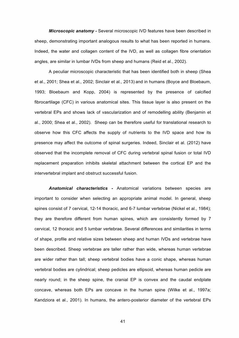
41
Microscopic anatomy - Several microscopic IVD features have been described in
sheep, demonstrating important analogous results to what has been reported in humans.
Indeed, the water and collagen content of the IVD, as well as collagen fibre orientation
angles, are similar in lumbar IVDs from sheep and humans (Reid et al., 2002).
A peculiar microscopic characteristic that has been identified both in sheep (Shea
et al., 2001; Shea et al., 2002; Sinclair et al., 2013) and in humans (Boyce and Bloebaum,
1993; Bloebaum and Kopp, 2004) is represented by the presence of calcified
fibrocartilage (CFC) in various anatomical sites. This tissue layer is also present on the
vertebral EPs and shows lack of vascularization and of remodelling ability (Benjamin et
al., 2000; Shea et al., 2002). Sheep can be therefore useful for translational research to
observe how this CFC affects the supply of nutrients to the IVD space and how its
presence may affect the outcome of spinal surgeries. Indeed, Sinclair et al. (2012) have
observed that the incomplete removal of CFC during vertebral spinal fusion or total IVD
replacement preparation inhibits skeletal attachment between the cortical EP and the
intervertebral implant and obstruct successful fusion.
Anatomical characteristics - Anatomical variations between species are
important to consider when selecting an appropriate animal model. In general, sheep
spines consist of 7 cervical, 12-14 thoracic, and 6-7 lumbar vertebrae (Nickel et al., 1984);
they are therefore different from human spines, which are consistently formed by 7
cervical, 12 thoracic and 5 lumbar vertebrae. Several differences and similarities in terms
of shape, profile and relative sizes between sheep and human IVDs and vertebrae have
been described. Sheep vertebrae are taller rather than wide, whereas human vertebrae
are wider rather than tall; sheep vertebral bodies have a conic shape, whereas human
vertebral bodies are cylindrical; sheep pedicles are ellipsoid, whereas human pedicle are
nearly round; in the sheep spine, the cranial EP is convex and the caudal endplate
concave, whereas both EPs are concave in the human spine (Wilke et al., 1997a;
Kandziora et al., 2001). In humans, the antero-posterior diameter of the vertebral EPs

42
steadily increases from the cervical to the lumbar region, while in sheep this diameter
almost stays the same over the whole spine (Wilke et al., 1997a). Therefore, compared to
humans, this species has larger EP diameters in the cervical but smaller diameters in the
lumbar spine. Despite the differences listed above, the trends of the two species in spinal
canal width are nearly identical (Wilke et al., 1997a; Mageed et al., 2013) and similarities
in the major dimensions have been reported to be strongest for the motion segment C5-
C6 (Kandziora et al., 2001) and for the thoracic and lumbar regions (Wilke et al., 1997b).
When comparing the macroscopic appearance of a sheep and a human motion segment,
it is evident that their sizes greatly differ; in particular IVD height and endplate cross-
section area of sheep IVDs are smaller than those of human (Schmidt and Reitmaier,
2013). This difference in sheep IVD size in comparison to human IVD may influence
results interpretation (Elliott and Sarver, 2004); in fact size affects rates of biologic
processes (West et al., 1997) and is influenced by competing biomechanical
(Nakatsukasa and Hirose, 2003) and nutritional (Horner and Urban, 2001) demands.
However, radiographic evaluation of IVD space height (IVD-SH) has shown an average of
6 mm in the sheep cervical spine, which is only approximately 1 mm (15%) higher than
the IVD-SH in the human cervical spine (Kandziora et al., 26). In the lumbar spine,
differences in intervertebral IVD-SH are higher (Wilke et al., 1997a; Green et al., 1993;
Nachemson et al., 1979). Therefore, with regard to IVD-SH, the sheep cervical spine may
represent a better model for human studies than sheep lumbar spine.
Biomechanical properties - Although the several variations of anatomical shapes
and dimensions listed above influence the biomechanical behavior of the spinal
segments, in vivo and in vitro studies have indicated that sheep may represent a valid
model in spinal research. Sheep spinal range of motions (ROMs) present smaller absolute
values in particular in flexion/extension of the lumbar spine, however the ROMs of sheep
spines for different loads directions (flexion/extension, axial rotation right/left, lateral
bending right/left) are qualitatively similar in their cranio-caudal trends to those of human

43
specimens reported in the literature (Ahlgren et al., 1994; Oxland et al., 1992; Panjabi,
1988; Panjabi et al., 1994; Wen et al., 1993; Wilke et al., 1995). Further similarities
between sheep and humans are related to the relative DH loss and internal NP pressure
after compression tests (Schmidt et al., 2013). It is nevertheless important to underline
that differences in compressive forces are present between the two species; therefore, for
example, pedicle screw systems and interspinous implants can be transferred from animal
model to human but neither magnitude of the effect nor the statistical results can be
compared.
Loading on the spine and IVDs - Another important aspect that needs to be
considered is the fact that sheep are quadrupeds therefore, their spines supposedly are
subjected to loads that differ considerably from those in humans (Bogdanffy et al., 1995;
Frick et al., 1994; Goel and Gilbertson, 1997; Herkowitz, 1994; Robin and Stein, 1975;
Yoganandan et al., 1996). In humans, the weight of the upper body acts on the lumbar
spine, therefore the loading of the lumbar spine is often assumed to be larger than in
quadrupeds. This, however, may not be correct since additional tensile forces from
muscle contraction and tension of passive structures such as ligaments are necessary to
control the posture of a quadruped spine as it cannot withstand substantial bending
moments (Smit, 2002; Wilke et al., 2003). A finite element study has shown that the stress
distributions in ovine and human motion segments under physiological loads are similar,
thus strengthening the justification for their use as in vivo models for the study of the spine
(Schmidt and Reitmaier, 2013). Furthermore, Smit (2002) demonstrated that quadrupeds’
spine is mainly loaded by axial compression and that trabecular distribution in the
vertebral bodies courses horizontally between their caudal and cranial EPs, implying that
the main load within the vertebral body is indeed an axial compression force, as well as in
human spine. An important point of difference, however, is that sheep are subjected to
higher axial compression stress than humans (Smit, 2002), leading to a bone mineral
density in the vertebrae up to fourfold higher than in humans (Wilke et al., 1996). Although

44
these differences should be considered on the transferability of the results of animal
experiments, Kandziora et al. (2001) did not find significant differences between the bone
mineral density value of human and sheep lower cervical spine, which therefore
represents a suitable model for the testing of fixation devices such as plates, screws, and
interbody fusion cages (Kandziora et al., 2001).
IVD changes with age and degeneration - The age of animals used for spine
research must be closely monitored as naturally occurring changes, both histological and
biochemical, occur with age. Several histologic, radiologic and biochemical changes, that
are generally associated with pathology in human IVDs, have also been observed in
sheep (Bayliss et al., 1988; Maeda et al., 2000). For example, common features of IVDD
in both sheep and humans are represented by altered metabolism and PGs synthesis by
the IVD fibrochondrocytes, decreased PGs aggregation levels with hyaluronan, increased
PG hydrodynamic size and relative keratan sulphate content, increased serine proteinase
activity and decreased serine proteinase inhibitory protein levels (Bayliss et al., 1988;
Maeda and Kokubun, 2000). However, an important variation that should be considered is
related to the fact that, in the aging sheep, the composition of IVD varies with the IVD
location within the spine. In fact, lower PG levels are present in IVDs at the level of the
thoracolumbar and lumbosacral junction (Melrose et al., 1994).
Disc calcification is a degenerative process, which has been described to occur in
the IVDs of both humans and sheep (Melrose et al., 2009; Miller et al., 1988; Feinberg et
al., 1990; Cheng et al., 1996; Chanchairujira et al., 2004). The aetiology remains
unknown. It is characterized by hydroxyapatite (HA) crystals deposition and a decrease in
IVD PGs content and size, suggesting that IVD calcification may be involved in the IVD
degenerative process in ageing IVDs. The HA deposition seems to affect mainly the
transitional zone between the AF and NP (Melrose et al., 2009).
Sheep provides a useful, naturally occurring model for investigation of the
aetiology and pathogenesis of HA deposition, however direct comparison with human IVD

45
is inappropriate due to dissimilarities in IVD calcification between sheep and humans
(Melrose et al., 2009). In sheep, HA deposition is restricted mainly to the lumbar region
(L4-L5; L5-L6) whereas in humans the thoracic and upper lumbar regions are
predominantly affected (Taylor et al., 1981). In sheep, IVD calcification seems to be
secondary to a degenerative process whereas histological studies have indicated that
metaplasia is the event, which leads to this form of calcification in humans (Depalma and
Kruper, 1961; Uhthoff et al., 1976).
§ SURGICAL AND MRI ANATOMY OF THE OVINE LUMBAR SPINE
A variation concerning the number of lumbar vertebrae is normal in the ovine species, as
they can be either 6 or 7 (Lori et al., 2005). The curvature of sheep spine has a kyphotic
shape (Barone, 2004). The vertebral body heights commonly exceed 40 mm (mean
42.49, SD 2.36) whilst DHs are usually only 4-5 mm (mean 4.48, SD 0.66) (Wilke et al.,
1997). The IVDs and EPs appear as bulbous convex expansions in between concave
elongated vertebral bodies. The tip of sheep transverse processes are large and easily
palpable, and are visible in the flank region, serving as useful landmarks when performing
surgery. Radicular veins and arteries run approximately 1 cm below the inferior endplates
across the vertebral bodies and are variable in size and number. Muscular insertions into
the lower lumbar vertebral bodies are usually thick and tendinous, whilst those higher in
the lumbar spine are thin and easily divided. The spinal cord continues into the sacral
region in the sheep (Baramki et al., 2000).
On MRI (1.5 Tesla) (Figure 13), the bone marrow has a characteristic
distribution on sagittal T2w in all vertebrae with a zone of hyperintensity parallel to the
endplate (Nisolle et al., 2013). This might be due to the characteristic presentation of
secondary ossification centres that arise at the cranial and caudal aspects of the vertebral
bodies, forming complete osseous plates above and below the physical plates (Alini et al.,
2008). The facet joints appear as “J-shaped” lines, with the surfaces of the caudal and
cranial articular processes being respectively concave and convex. The articular facet

46
cartilage, characterized in humans by a linear focus of high signal in T2w (synovial fluid),
is not always visible in sheep due to the fact that are thin (Harnsberger et al., 2006). The
ligaments appear hypointense in T2w, however only the supraspinous and interspinous
ligaments can be well identified, whereas it is not always possible to differentiate the
dorsal and ventral longitudinal ligaments from the low signal of the vertebral cortical bone
or the AF (Harnsberger et al., 2006). This may be due to the fact that the ventral
longitudinal ligament is very thin and narrow, while the dorsal ligament is thin but wider
and spreading on the floor of the spinal canal (Nisolle et al., 2013). In the sheep, the
ligamentum flavum is also difficult to identify by MR imaging due to the small space
present between the capsule of the apophyseal joint and the junction of tha lamina with
the spinous process, where the ligamentum generally extends (Nisolle et al., 2013). In
high field MRI, in both T1w and T2w images, IVDs signal mirrors the water content that
increases from outer to inner AF (74-82%), while the NP consists of 86% of water (Reid et
al., 2002). Indeed, the moderately high signal for both NP and inner AF may be explained
by these high percentages of water content. On transverse planes through the
intervertebral foramen it is possible to identify the dorsal and ventral roots, the dorsal
spinal ganglion and the ventral and dorsal branches of the spinal nerve. Due to the fact
that in sheep the spinal cord ends at S1, a small subarachnoid space and conus
medullaris are visible below the level of S1 vertebral body. Peripheral nerves can be
identified in the centre of the neural foramen surrounded by hyperintense fat, with the
same signal intensiy, in T1w and T2w images, of the spinal cord (Nisolle et al., 2013).

47
Figure 13. Sagittal view obtained at the median plane. 1. Spinal cord, 2. Cerebrospinal fluid, 3. First sacral vertebra, 4. Spinous process, 5. Supraspinous ligament, 6. Interspinous ligament, 7. Basivertebral veins, 8. Subcutaneous fat, 9. Epidural fat, 10. Nucleus pulposus and inner part of annulus fibrosus, 11. Outer part of annulus fibrosus, 12. Vertebral body, 13. End of spinal cord, 14. Vertebral plate. Note in T2w the nuclear cleft that appears as a hypointense spot in the centre of the nucleus pulposus (Nisolle et al., 2013).
o THE TRANSPEDICULAR APPROACH
As described above, IVD pathological changes affect all areas of the IVD. The NP is
particularly affected in this degenerative process, therefore regenerative strategies are
primarily focused on NP regeneration. The relatively homogeneous structure of NP tissue
and its avascular compartmentalized microenvironment make easy to conceive molecular
and engineering strategies toward tissue regeneration (Sowa et al., 2008). However,
many issues still have to be overcome taking into account the biological, biophysical, and
biomechanical environment.

48
Delivery of therapeutic growth factors, stem cells, and/or bioactive hydrogels for
NP regeneration might be considered a straightforward task because of the easy
accessibility of the NP through the AF. However, even a very modest AF injury may
induce or enhance a degenerative cascade of the IVD (Elliott et al., 2008; Iatridis et al.,
2009), affecting IVD biomechanics, cellularity, and biosynthesis (Korecki et al., 2008;
Hsieh et al, 2009). A retrospective study, with ten years follow-up, showed how
discography performed through small needle puncture resulted in accelerated IVDD,
higher IVD herniation rate on the same side of the disc injection, and changes in the EP
compared with matched control IVDs (Carragee et al., 2009). Furthermore, it has been
recently shown on a rabbit IVDD model that the injection of MSCs into the NP through the
AF route may lead to cell leakage and accelerated osteophyte formation (Vadalà et al,,
2012). Taking into account that the majority of regenerative strategies for IVD (molecular
therapy, cell therapy, and biomaterial based tissue engineering) are traditionally delivered
through the AF route, it should be considered that the resulting lesion might impair the
expected outcomes leading to further degeneration, leakage of the biologically delivered
material, and potential failure of the regenerative treatment. Therefore, alternative delivery
methods and/or AF regeneration strategies have been considered.
The transpedicular approach has been recently described in a descriptive
anatomical study on ovine and human cadaveric lumbar spinal segments, as an
alternative route to deliver therapeutic agents to the IVD without disruption to the AF
(Vadalà et al., 2013) (Figure 14).
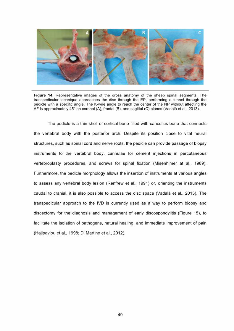
49
Figure 14. Representative images of the gross anatomy of the sheep spinal segments. The transpedicular technique approaches the disc through the EP, performing a tunnel through the pedicle with a specific angle. The K-wire angle to reach the center of the NP without affecting the AF is approximately 45° on coronal (A), frontal (B), and sagittal (C) planes (Vadalà et al., 2013).
The pedicle is a thin shell of cortical bone filled with cancellus bone that connects
the vertebral body with the posterior arch. Despite its position close to vital neural
structures, such as spinal cord and nerve roots, the pedicle can provide passage of biopsy
instruments to the vertebral body, cannulae for cement injections in percutaneous
vertebroplasty procedures, and screws for spinal fixation (Misenhimer at al., 1989).
Furthermore, the pedicle morphology allows the insertion of instruments at various angles
to assess any vertebral body lesion (Renfrew et al., 1991) or, orienting the instruments
caudal to cranial, it is also possible to access the disc space (Vadalà et al., 2013). The
transpedicular approach to the IVD is currently used as a way to perform biopsy and
discectomy for the diagnosis and management of early discospondylitis (Figure 15), to
facilitate the isolation of pathogens, natural healing, and immediate improvement of pain
(Hajipavlou et al., 1998; Di Martino et al., 2012).

50
Figure 15. Lateral view of diagrammatic representation of transpedicular access at L4-L5 level that shows the guide pin introduced into the intervertebral disc space though the caudally located pedicle (A). Axial diagram illustrating the instrumentation for creating transpedicular channel for the insertion of discectomy instrumentation. A guide pin is first inserted percutaneously through the pedicle and subchondral bone into the disc space (a). Subsequently, a tissue dilator (b) is introduced over the guide pin, and over the tissue dilator, a sleeve (c) is placed to be used as a working channel. Then the dilator is removed and a toothed cutting bone tool (d) is inserted over the guide pin into the intended target (Hadjipavlou et al, 2004).
In a descriptive study (Vadalà et al., 2013) the transpedicular injection of a contrast
agent demonstrated that drugs can diffuse into the whole NP tissue. Nucleotomy was
obtained performing a very small diameter tunnel (2-mm) into the pedicle using an
arthroscopic and/or endoscopic spine surgery device and the NP was replaced by an
agarose gel. To avoid material leakage through the tunnel, biomechanical impairment,
and diffusion of serum or blood cells into the disc space, a porous polyurethane
biomaterial, with the ability to regenerate both bone tissue and cartilage tissue (Eglin et
al., 2010), was used to seal the tunnel. Therefore, the small EP break generated by the 2-
mm drill could be potentially closed/repaired by the biomaterial. The EP, which damage
could trigger a degenerative cascade (Alini et al., 2008), is a paramount structure that
supplies nutrients to the avascular IVD, therefore its impairment represents a potential
limitation that needs to be carefully evaluated. Blood vessels in soft tissues provide the
nutrients for the outer AF cells, while NP and inner AF cells rely on a capillary network in
the subchondral EP through which nutrients diffuse. The reduction in nutritional supply is
known to be one of the leading causes of IVDD (Urban et al., 2004). Holm et al. have
described a technique to create IVDD in porcine lumbar spine by drilling a hole through

51
the central part of the EP into the NP. This model has been shown to trigger similar
degenerative changes observed in degenerated human IVDs, such as alteration of water,
PG, and cellular contents. However, this IVDD model is based on large damage to the EP
(3.5 mm), which is quite extensive considering the relatively small geometry of the IVDs of
the animal model used.
On the basis of different IVDD stages, the transpedicular approach could be used
to deliver growth factors or drugs in early stages, or progenitor cells in moderate stages,
of disc degeneration. Furthermore, it might also be used to introduce osteoinductive
and/or osteoconductive agents to achieve interbody fusion in late stages of IVDD.

52
OBJECTIVES OF THE STUDY
The objectives of the study were divided into two main sections:
1. The first aim was to describe in detail and validate in vivo the transpedicular
approach as an alternative route to the IVD, highlighting possible surgical-
related difficulties and complications. The purpose of this novel approach was
not only to provide a new model of IVDD without disruption of the AF, but also
a new system for delivering therapeutic agents into the NP.
2. The second purpose was to collect qualitative and quantitative data from MRI,
radiologic, macroscopic and histologic findings, with the aim of assessing the
type and degree of IVDD changes obtained by performing four different
surgical approaches to the IVD.
53
Section 1: in vivo characterization and validation of a new intervertebral disc degeneration model
MATERIALS AND METHODS
o CASES SELECTION
Twelve skeletally mature (approximately three years of age), female Brogna breed sheep,
weighing 34 to 42 kg, were included in the study. Ethical approval for the use of animals in
this study was granted by the Italian Ministry of Health. All animal procedures and
surgeries were performed at an accredited facility (Department of Animal Medicine,
Productions and Health, Veterinary Teaching Hospital, University of Padua, Padua, Italy)
where sheep were monitored at least once a day.
After arrival, a two-week period was provided to each animal to acclimate prior to any use
for the experiments; this is important to reduce the stress associated with transportation
and new environment which can have widespread effects on animals’ physiological
systems, including changes in cardiovascular, endocrine, immune, central nervous, and
reproductive system. Blood analysis were performed as part of a general screening;
subcutaneous injection with ivermectin (200 µg/kg) was administered to all animals, on
arrival and 2 weeks later, to treat any possible infection and infestation due to
gastrointestinal roundworms, lungworms, and larval stages of the nasal bot.
Sheep were followed serially before surgery (T0) and at 1 (T1), 3 (T2) and 6 (T3) months
post surgical procedure.
o IMAGING
X-RAY - Before the surgery and at each time point, conventional lateral plain radiographs
of the lumbar spine were taken in each animal under sedation. The radiographs were
analyzed qualitatively for evidence of changes in the vertebrae adjacent to the disc

Section 1: in vivo characterization and validation of a new intervertebral disc degeneration model
54
spaces, looking for possible post-surgical alterations such as luxations, fractures,
discospondylitis.
MRI - MR images of the lumbar spine were performed serially in 6 sheep, before surgery
and at 1, 3, and 6 months post surgery. MR imaging scans were obtained using a 0,25
Tesla clinical magnet (Esaote Vet MR). Sheep were premedicated and anaesthetised and
placed in lateral recumbency within the open magnet (Figure 16). A dorsal T1-weighted
(T1w) localizer image (TR, 770 milliseconds; TE, 18 milliseconds) was obtained to
establish the position of the lumbar discs from L1–L2 to L5–L6. A 4-mm thick midsagittal
section was imaged, using a T2-weighted imaging sequence (TR, 2500 milliseconds; TE,
120 milliseconds) to highlight the signal from the NP. T1w (TR, 770 milliseconds; TE, 18
milliseconds) and 3D HYCE transverse images (TR, 10 milliseconds; TE, 5 milliseconds)
were also obtained.
Figure 16. Sheep under anaesthesia placed in lateral recumbency within the open magnet
o SURGICAL PROCEDURE
Preparation - Sheep were fasted for 24 hours in order to prevent abdominal distension
and aspiration of rumen fluids during surgery. Medetomidine hydrochloride (5-10 µg/kg IV)

Section 1: in vivo characterization and validation of a new intervertebral disc degeneration model
55
was administrated to facilitate transport to the operating theatre, followed by intravenous
injection of propofol (2-4 mg/kg IV) for anaesthetic induction. An endotracheal tube was
inserted and anaesthesia maintained by isoflurane (2-3%) in oxygen and nitrous oxide.
During surgery, replacement fluids (lactate Ringer’s solution or 0.9% NaCl, 10mL/kg/h)
and analgesia (alfentanil 60 µg/kg/h) were provided.
The lumbar region of each sheep was clipped from T10 to approximately the level of S3
vertebra. The sheep were placed on the operating table in sternal recumbency. The
surgical site was aseptically prepared with chlorhexidine and alcoholic-iodide antiseptic
wash followed by sterile draping. Local anaesthetic (lidocaine 0.5%) was subcutaneously
injected around the incision site. Strict sterile precautions were maintained at all times.
Dorso-lateral approach to the lumbar IVD – Landmarks used for the incision were
easily palpable; these were iliac crest, lumbar transverse processes and the costo-
vertebral angle (Figure 17A); in addition a backhaus towel forcep was placed on one of
the lumbar dorsal processes which was then identified on lateral X-Ray performed before
starting surgery (Figure 17B).
A B Figure 17. (A) Preoperative photo of sheep demonstrating lumbar spinous processes (lower dashed line), left lumbar transverse processes (upper dashed line), iliac crest (left) and costal margin (right) (Oeheme et al., 2012). (B) Radiographic landmark obtained by placing a backhause preoperatively (red circle); T13 can be easily identified due to the presence of the last pair of ribs.
A dorsal midline skin incision was extended from T12 to S1, using fluoroscopy to verify the
correct localization in each animal. Following incision and retraction of the skin and
superficial fascia, the deep fascia was incised unilaterally (left) around the tip of the
spinous processes and retracted. Following left side dissection to the level of the synovial

Section 1: in vivo characterization and validation of a new intervertebral disc degeneration model
56
joint of the articular processes, the incision was continued laterally and ventrally, by
elevating the multifidus muscle attachments from the pedicles, allowing exposure of the
articular and part of the transverse processes from L1 to L6. Electrocautery was used
throughout the exposure to minimize bleeding. Once the procedure had been performed
and haemostasis achieved, the wound was irrigated with Ringers’ solution. Then, the
transpedicular approach to the NP was performed by one surgeon. Five lumbar discs
were assigned to different groups of treatment:
• L1-2: EP tunnel + nucleotomy;
• L2-3: EP tunnel;
• L3-4: EP tunnel + nucleotomy + EP repair with the polyurethane (PU) scaffold;
• L4-5: EP tunnel + EP repair with the PU scaffold;
• L5-6: intact control.
The different treatments are described below.
End-plate tunnel - Throughout a dorso-lateral surgical access to the lumbar spine, a 2-
mm tunnel was drilled using a Kirschner wire (k-wire) into the pedicle of the vertebra to
access the NP space of the cranial spinal segment under fluoroscopy guidance (Figure
18), checking the wire direction (mean angle degree ± standard deviation) in latero-lateral
(Figure 18A) and dorso-ventral (Figure 18B) images, as already described (Vadalá et al.,
2013). The k-wire was directed cranially and medially to reach the center of the NP, with
an inclination of approximately 45°. A manual 2-mm drill was used to hole the EP and
access the NP space.

Section 1: in vivo characterization and validation of a new intervertebral disc degeneration model
57
A B Figure 18. Representative intraoperative fluoroscopic images on the lateral (A) and dorsal (B) planes of the lumbar spine. It is possible to appreciate the k-wire reaching the center of the disc space through the transpedicular approach.
Nucleotomy - A 2-mm diameter shaver resector (Smith & Nephew Shaver Blades, 2.0
mm Full Radius, 7 cm length), powered by an arthroscopy shaver unit (Smith & Nephew,
Dionics Inc. PS3500, Germany) and connected to a vacuum pump, was introduced
through the 2-mm tunnel. NP tissue was removed maintaining the shaver blade in the IVD
space oscillating at 4000 rpm for 5 minutes (Figure 19).
Figure 19. Surgical pictures showing placement of the 2-mm shaver dissector through the transpedicular approach in the disc space, used to perform nucleotomy under aspiration.
End-plate repair - The EP was repaired by sealing the edge of the tunnel using a press-fit
porous polyurethane (PU) cylinder (2.2 mm diameter and 10 mm length) (Figure 20A) that
was placed at the EP edge using a 14G cannula (Bonopty® Bone Biopsy System,
AprioMed, Sweden) inserted through the tunnel (Figure 20B). The elastomeric PU cylinder
was synthesized as previously described (Eglin et al., 2010; Laschke et al., 2010; Laschke
et al., 2009). The scaffold was sterilized by cold ethylene oxide process and stored until
the time of surgery.

Section 1: in vivo characterization and validation of a new intervertebral disc degeneration model
58
At the end of the surgical procedure, the deep fascia, the subcutaneous layers and the
skin were closed in a simple continuous pattern.
A B Figure 20. (A) A porous polyurethane (PU) cylinder was inserted using 14-G cannula (Bonopty - Bone Biopsy System, AprioMed, Sweden) at the edge of the tunnel performed through the vertebral body to repair the end-plate and seal the disc space. (B) Representative fluoroscopy image control showing the cannula placed into the transpedicular tunnel during the scaffold positioning (arrow).
o PAIN MANAGEMENT AND POSTOPERATIVE CARE
As soon as each sheep was breathing spontaneously, following cessation of isofluorane
anaesthesia, it was extubated, transferred to a heated room and returned to the sheep
housing, where it was given food when fully alert and standing. The sheep were observed
for approximately one hour following surgery. All sheep received prophylactic antibiotics
(Amoxicillin 15 mg/kg/q48 hours IM) perioperatively and postoperatively for 10 days.
Tramadol (4 mg/kg/q12h) and Carprofen (4 mg/kg/q24h) were administered for
postoperative analgesia at least for the first 5 days after surgery. Vital signs were
monitored, neurological examination was performed postoperatively on a routine basis,
and standard nursing care was provided. The sheep were monitored for normal feeding
behaviour and cage activity twice daily to determine their general health. A wound
inspection to check for possible infection or wound openings was performed on a daily
basis until a complete healing.

Section 1: in vivo characterization and validation of a new intervertebral disc degeneration model
59
o EUTHANASIA
At each time point (before and 1, 3, and 6 months post surgery), 3 sheep were
anaesthetized with medetomidine (5-10 µg/kg IV) and propofol (2-4 mg/kg IV), and then
euthanized with a solution of embutramide, mebenzonio ioduro and tetracaine
chlorhydrate (0.3 ml/kg IV).
o COMPLICATIONS
Intra- and post-operative complications were recorded. Potential intraoperative
complications included:
• Improper identification of the surgical site
• Inability to reach correctly the NP
• Excessive haemorrhage
• Incorrect k-wire inclination
• Iatrogenic damage to the spinal cord and nerve roots
Postoperative complications included:
• Persistent kyphosis
• Vertebral fracture and instability
• Pelvic limbs paresis/paralysis
• Seroma
• Wound infection
• Cutaneous fistula
• Discospondylitis

Section 1: in vivo characterization and validation of a new intervertebral disc degeneration model
60
RESULTS
o SURGICAL PROCEDURE
The transpedicular approach was feasible in all animals included in the study. In all the
animals the K-wire was inserted correctly in the 4 discs treated (L1 to L5), allowing the
correct placement of the shaver within the center of the NP.
The K-wire mean orientation angles (angle ± standard deviation) assessed on the
latero-lateral and dorso-ventral fluoroscopy images were 50.6° ± 3.1 on the dorsal plane
and 50.1° ± 6 on the sagittal plane. The average operation time was 92 minutes (range
74-110). The duration of the procedure to reach the NP, perform nucleotomy and place
the PU scaffold became shorter over the course of the experimental period.
o IMAGING
In animals that underwent MR imaging it was possible to detect the transpedicular
tunnel throughout the vertebral body and pedicle of each treated disc, confirming the
correct positioning of the k-wire (Figure 21).
Figure 21. Representative middle-sagittal T2-weighted MR images showing the L3-L4 disc before the surgical approach (a) and after nucleotomy and scaffold placement at 3-month follow-up (b). The tunnel is visible on both sagittal (a) and transverse (c) images; furthermore the transverse MR image shows that the tunnel entry point on the end-plate is in its central area (c).

Section 1: in vivo characterization and validation of a new intervertebral disc degeneration model
61
On parasagittal and transverse T2w images the tunnel appeared hypointense to the rest
of the vertebral body (Figure 21b). Furthermore, loss of the NP signal intensity in the discs
that underwent nucleotomy was visible in transverse images. The PU scaffold inserted in
two of the treated discs in each sheep was not discernible from the surrounding bone.
o PERI- AND POST-OPERATIVE CARE AND COMPLICATIONS
§ INTRAOPERATIVE DATA
The only intraoperative complication was CSF leakage from the tunnel during the drilling
process, that occurred in 2 of the 48 IVD spaces treated (complication rate 2.1%). Blood
loss was minimal and no clinically apparent neural or great vessel injury was caused by
any of the surgical procedures. There were no anaesthetic complications as confirmed by
stable cardiovascular parameters.
§ POSTOPERATIVE DATA
In the early post-operative period, no significant problems with mobility or pain have been
encountered; all animals tolerated the procedure and within two hours from the end of the
surgery were able to walk. After surgery all the sheep presented kyphosis, which had
resolved after 3-5 days in 10/12 sheep. Post-operative complications verified in 3/12
sheep (25%). One sheep developed a sterile seroma, which reabsorbed completely after
7 days without causing any clinical problem. In none of the animal postoperative wound
dehiscence did occur. A fracture to the cranial aspect of L4 vertebral body was detected
radiologically in one sheep at 1-month follow-up (Figure 22a). The fracture evolved into
L3-4 spondylarthritis after 3 months (Figure 22b). Apart from mild kyphosis the animal did
not develop further clinical signs, such as discomfort and paresis, however was
euthanized at 3-month follow-up. Through the analysis of the X-ray (Figure 22c) and MR
images (Figure 22d) performed one month after surgery, discospondylitis at L1-2 (2.1%
complication rate) was diagnosed in one sheep. The animal presented mild paraparesis,

Section 1: in vivo characterization and validation of a new intervertebral disc degeneration model
62
ataxia and kyphosis and for ethical reasons was euthanized. Macroscopic changes
attributed to discospondylitis were confirmed during autopsy.
Post-operative pain management was considered adecuate in all the sheep, with
exception of the one that developed discospondylitis. Indeed, all the animals were
walking, eating and drinking regularly and no evident reactions were elicited on spinal
palpation. No weight loss was detected in any of the animal at their last time point; after
the experimental period the animals weighed approximately 41 to 50 kg.
Figure 22. X-ray images showing a fracture of the cranial aspect of the vertebral body observed 1 month after the transpedicular approach (a) and that evolved into spondylarthritis after 3 months (b). Discospondylitis developed at L1-L2 as shown by the erosion of both end-plates and the consequent widening of disc space in letero-lateral X-ray image (c); T2-weighted MR image shows increase intensity of the disc and end-plates and apparent extension of the infection into the vertebral canal causing ventral compression of the spinal cord (d).

Section 1: in vivo characterization and validation of a new intervertebral disc degeneration model
63
DISCUSSION
The recent consensus about the importance of maintaining the AF structural integrity
(Elliott et al., 2008; Iatridis et al., 2009) has brought attention within the field of IVD
regeneration on both novel AF repair strategies (Vadalá et al., 2012) and alternative ways
to access the NP space. Indeed, taking into account that the majority of regenerative
strategies for the IVD are traditionally delivered through the AF route, it should be
considered that the resulting lesion might impair the expected outcomes leading to further
degeneration, leakage of the biologically delivered material, and potential failure of the
regenerative treatment (Vadalá et al., 2011).
The transpedicular approach has been described in a preliminary ex vivo study,
which demonstrated that injections, nucleotomy and nucleoplasty were feasible by
realizing a tunnel passing through the pedicle and the EP (Vadalá et al., 2013). The
present study has proved that this alternative approach to the IVD is feasible in vivo with a
relatively low morbidity rate. Indeed, both the transpedicular tunnel and nucleotomy were
performed successfully in 48 and 24 IVD, respectively.
Technical surgical skills and a good knowledge of the regional anatomy are needed,
however a remarkable progressive shortening of the surgical time was recorded, reflecting
a quick rate of improvement of the learning curve.
The transpedicular approach provides good visualization and surgical access to
the IVDs. The procedure can be performed from either the right or the left side of the
lumbar spine. The only limitation is related to the realization of the technique caudally to
L5, where the pelvis and the pelvic limbs can interfere with the acquisition of fluoroscopic
lateral images.
Particular care must be taken to avoid any damage to the spinal cord. The
inclination of the k-wire through the pedicle represents a key-point to perform this
technique correctly and to reach the NP without causing any significant damage. On the

Section 1: in vivo characterization and validation of a new intervertebral disc degeneration model
64
preliminary study (Vadalá et al., 2013) performed on lumbar ovine and human spinal
segments, the k-wire angles which enabled to reach the centre of the NP were 47.5° ±
5.1° on the dorsal plane and 49.6° ± 6.2° on the sagittal plane. The angles recorded in the
present study are very similar (50.6° ± 3.1° on the dorsal plane and 50.1° ± 6.0° on the
sagittal plane), however it is possible that even a subtle difference in the inclination could
explain the occurence of CSF leakage. Indeed, pedicles are paired, strong, tubular bony
structures, made of hard cortical bone outside and cancellous bone inside and acting as
thin lateral walls of the spinal canal. Due to the thin conformation of the pedicles and their
proximity to the spinal cord even a small angle variation could cause severe
consequences; it is therefore essential the careful evaluation of the anatomical landmarks
and the k-wire inclination. Furthermore, when performing the transpedicular approach and
before considering its application in human medicine, interspecies anatomical variations
should be considered. In the light of this consideration and of the need of an experimental
animal model able to reproduce the disc degenerative process, sheep spine was selected
to minimize the differences with human spine. Sheep are readily available and show great
homogeneity when selected for age, breed, and sex (Wilke, 1997a). Sheep have been
widely used in spinal research and several comparative studies have pointed to
similarities between the sheep and human IVD: (1) proportions are similare, though
absolute size is smaller in sheep (Wilke et al., 1996), (2) the sheep spine is loaded along
its long axis, like humans (Smit, 2002) and, (3) similar to adult human IVDs, no notochord
cells are present in mature sheep discs (Trout et al., 1982 a,b). The skeletally mature
sheep used in the present study showed uniform anatomical disc and vertebral patterns.
Several surgical approaches to the lumbar IVDs have been described in spinal
research, however they can carry some limitation related to anatomical characteristics.
For example, dorsal approaches to the lumbar spine, commonly used in human surgery,
are difficult in sheep due to the risks related to the presence of the spinal cord within the
lumbar spinal canal and due to the ossification of the dorsal longitudinal ligament
(Oeheme et al., 2012). For this reason, the ovine lumbar IVD have traditionally been

Section 1: in vivo characterization and validation of a new intervertebral disc degeneration model
65
accessed via a ventral or ventrolateral approach through the retroperitoneal or
transperitoneal regions (Melrose et al., 2012; Baramki et al., 2000). These approaches
carry significant risks, including bowel and great vessel injury, neural injury, and hernia
formation. The procedure also requires a large abdominal incision with greater retraction
of abdominal viscera, which can be harmful to the animal (Baramki et al., 2000). Recently,
a minimally invasive retroperitoneal lateral approach has been described in an ovine
model, allowing for a small focused incision and causing minimal morbidity (Oheme et al.,
2012). Lateral approaches to the human lumbar spine, such as the extreme lateral
interbody fusion procedure, have gained popularity as minimally invasive approaches to
the lumbar IVDs. In the lateral body fusion procedure, a retroperitoneal transpsoas route
is employed, requiring real-time neuromonitoring to ensure safe passage through the
psoas without damaging the lumbar plexus (Youssef et al., 2010), which represents the
main concern with this procedure. The transpedicular approach to the IVD is currently
used, in humans, as a route to perform biopsy and discectomy for the diagnosis and
management of early spondylodiscitis, to facilitate the isolation of pathogens, natural
healing, and immediate improvement of pain (Hadjipavlou et al., 1998a; Di Martino et al.,
2012). The technique is performed through a dorsal percutaneous approach; the
advantage of this procedure is its minimal invasiveness that allows bacteriological and
histological testing, drainage of infected material, as well as injection of therapies,
permitting early patient mobilization (Arya et al., 1996; Hadjipavlou et al., 2004). The
approach is simple, cost-effective and its diagnostic accuracy is comparable to that of
open biopsy (Arya et al., 1996; Hadjipavlou et al., 2004). The transpedicular approach has
been validated in spinal research both ex vivo by Vadalá et al. (2013) and in vivo with the
present study, providing a model to perform biological and biomechanical studies of NP
regenerative therapies (Vadalá et al., 2013). Further future perspectives are represented
by the validation of CT/fluoroscopy guided minimally invasive approaches and by the
clinical translation of new regenerative strategies for biological restoration of early and
mild degenerative changes in IVD, which is crucial to improve present clinical treatments

Section 1: in vivo characterization and validation of a new intervertebral disc degeneration model
66
and life quality of several patients.
Most of the structural injury IVDD models are based on a direct damage to the AF
by performing different tear types (rim lesions, concentric and radiating annular tears) of
different sizes, evaluating the evolution and progression of the biologic intradiscal
alterations and demonstrating how these may affect the mechanics of the joint (Osti,
1990a; Melrose et al., 1992; Moore et al., 1992; Gunzburg et al., 1993; Ahlgren et al.,
1994; Moore et al., 1996; Fazzalari et al., 2001; Melrose et al., 2002; Elliott et al., 2008). A
porcine experimental model of IVDD, based on EP fractures or NP herniation through the
EP, was described by Holm et al. (2004). Through a retroperitoneal approach, the cranial
EP of the lumbar vertebra was perforated using a 3.5-mm drill bit inserted from the lateral
cortex at mid-height, angulated at 45° so as to reach the central part of the EP. This
model highlighted how the EP damage can trigger similar degenerative changes as those
observed in degenerated human IVD, such as alteration of water, PG and cellular
contents (Holm et al., 2004; Salo et al., 2008a,b). In contrast with this model, the
transpedicular approach performed in the present study is based on a smaller EP damage
(2-mm) and, in 2 of the 4 treated discs, the EP tunnel was sealed using conductive
scaffolds with the aim of limiting the degradation cascade and preventing Schmorl’s nodes
formation or material leakage. The histological evaluation of the degeneration obtained in
the IVDs treated and of the changes related to the use of the scaffold will be discussed in
the second part.
A moderately low rate of intraoperative (2.1%) and postoperative complications
(25%) occurred in the present study. The CSF leakage reported in two sheep is likely to
be secondary to dural tear provoked by the incorrect k-wire positioning during the
realization of the transpedicular tunnel. To our knowledge, neural complications related to
the perforation of the dura have not occurred as the animals recovered uneventfully.
Vertebral subluxation and discospondylitis, localized at L3-4 and L1-2 IVDs respectively,
were observed during the postoperative period in two different sheep; the two lesions
were likely to be iatrogenic. The fact that both the affected discs were nucleotomized may

Section 1: in vivo characterization and validation of a new intervertebral disc degeneration model
67
imply that nucleotomy could contribute to increasing the risk of potential complications.
Experimentally, it is generally agreed that the structural damage due to in vitro nucleotomy
significantly destabilizes the motion segment (Zollner et al., 2000). However, in vivo
biomechanical tests suggested that post-operative instability might not be exclusively a
consequence of the trauma to the IVD, but that dorsal passive-spine supportive structures
integrity (articular facets, spinous processes) play a crucial role in maintaining motion
segments stability (Reitmaier et al., 2014). In the current study, dorsal vertebral structures
remained intact, however we cannot rule out that the retraction of the lumbar epaxial
muscles may have contributed to reducing the strength of the motion segments involved.
Iatrogenic IVD space infection represents one of the possible complications
occurring after spinal surgery. In humans, discospondylitis represents a common
complication following microsurgical discectomy, percutaneous laser disc decompression
(Farrar et al., 1998), automated percutaneous lumbar nucleotomy operations (Dullerud et
al., 1997), and discography (Guyer et al., 1997; Osti et al., 1990b). In the present case,
the occurence of discospondylitis could represent a consequence of either iatrogenic
bacterial inoculation into the NP or hematogenous spread. Further laboratory and
histologic analysis were not performed to confirm the diagnosis and identify a pathogen,
however the characteristic radiologic and post-mortem macroscopic changes are highly
supportive of IVD infection. MRI analysis has high sensitivity and specificity for the
evaluation of pyogenic discospondylitis (Lucio et al., 2000). Characteristic MRI findings of
discospondylitis include decreased signal from the disc and adjacent portion of vertebral
bodies on T1w sequences and an increased signal from these structures on T2w
sequences; contrast uptake can be detected in the adjacent vertebral bone marrow, in the
disc space and in the dorsal AF (Lucio et al., 2000). In the present study post contrast
images were not available, however T1w and T2w images showed alterations similar to
those described. The peculiar radiographic changes, characterized by erosion of the EPs
and widening of the IVD space, and the macroscopic bone remodeling observed on the
ventral aspect of L1-2 vertebral bodies during necroscopy, supported the diagnosis of disc

Section 1: in vivo characterization and validation of a new intervertebral disc degeneration model
68
infection.
Summarizing, the transpedicular approach represents an alternative route to the
traditional ventral and ventrolateral approaches through the AF. This new pathway to the
IVD provides a new model to study biologic and biomechanical alterations in relation to
both IVD degenerative processes and potential NP regenerative therapies.

69
Section 2: evaluation of quantitative and qualitative data of the
intervertebral disc degeneration model
MATERIALS AND METHODS
o QUALITATIVE ANALYSIS
MR images, macroscopic and histologic samples of the intact and injured discs were
analysed qualitatively for evidence of degenerative changes.
§ MAGNETIC RESONANCE IMAGES ANALYSIS
MRI images, acquired as described in the first part of the study, were analysed
qualitatively. Disc morphologic changes, as seen on sagittal T2w images, were recorded
and divided into 5 categories according to the Pfirrmann grading scale (Pfirrmann et al.,
2001). Variations of signal intensity, IVD structure, distinction between NP and AF, and
IVD height were evaluated for each treated and control disc at each time point.
§ GROSS ANATOMY AND HISTOLOGICAL EVALUATION
At sacrifice, the lumbar spines were removed from the sheep (Figure 23a), and the discs
(L1 to L6) together with intact adjacent vertebral body bone (Figure 23b) were fixed in
10% neutral buffered formalin for at least 1 week, decalcified in
ethylenediaminetetraacetic acid, and processed for paraffin sectioning. Blocks of tissue (5
mm in thickness) were embedded in paraffin. The blocks of tissue were then cut into
sections of 5 µm in thickness using a microtome and stained with Safranin O/Fast Green
and hematoxylin and eosin (H&E).

Section 2: evaluation of quantitative and qualitative data of the intervertebral disc degeneration model
70
A B Figure 23. (A) Post-operative photo of one of the sheep spine (L1-S3) harvested after euthanasia; perispinal muscles have been resected. (B) Representative picture of treated (L1-L5) and control (L5-L6) removed from the spine.
Based on the previously described grading scale (Thompson et al., 1990),
all midsagittal sections of the harvested discs within each single sheep were compared to
each others, to discs of all other sheep and to the control discs. Anatomical characteristics
and degenerative changes were evaluated for the NP, AF, EPs and vertebral bodies of
each disc. More in detail, in the NP the progressive replacement of the bulging gel with
fibrous tissue and the presence of clefts formation were evaluated; in the AF and EP,
changes in the organization of the lamellae in the composition and structure of the hyaline
cartilage were observed; finally, margins shape and osteophytes formation were recorded
for the vertebral bodies.
Qualitative histological analysis was performed under a light microscope
(Nikon Eclipse E800, Nikon, Melville, NY), at magnifications ranging from 10 to 200X. In
particular, evidence of changes in NP cells composition and the presence of possible cells
leaking from the tunnels were recorded.
o QUANTITATIVE ANALYSIS
The effect of the different degeneration modalities performed on each disc was evaluated
quantitatively through the analysis of X-ray and MR images performed at each time point.
Quantitative analysis of the images was performed by transferring the images to a

Section 2: evaluation of quantitative and qualitative data of the intervertebral disc degeneration model
71
commercially available image processing software (OsiriX Imaging Software, www.osirix-
viewer.com) as follows.
§ DISC HEIGHT INDEX (DHi)
Under lateral view, vertebral body and DHs were measured in each segment from L1 to
L6. Disc and vertebral body heights were derived by averaging the distance between EPs
at 3 places obtained from the dorsal, middle, and ventral portions of the disc normalized to
the average length of 2 adjacent vertebral bodies in each section (Figure 24). The
preoperative radiograph was used as a baseline measurement. The data were then
transferred to the Excel sofware program (Microsoft Excel, 2010) and the DHi was
calculated using the method described by Masuda et al. (2004) (Figure 24).
DHi = 2 x (D+E+F) / (A+B+C+G+H+I)
Figure 24. Using digitized radiographs, measurements including the vertebral body heights and DH were analyzed by Osirix software. IVD height was calculated by averaging the measurements obtained from the dorsal, middle, and ventral portions of the IVD and dividing that by the average of adjacent vertebral body heights (Masuda et al., 2004).
§ MRI INDEX (MRi)
The T2w midsagittal images of the intact and treated discs were analyzed quantitavely
calculating the MRi (Sobajiama et al., 2004). The NP of each disc was outlined manually
to define the region of interest (ROI), as shown in Figure 25. The area and average signal
intensity (gray scale value) of this ROI were then computed automatically using the Osirix
surement. A C-arm image intensifier (Philips Med System Ser:15128, !55kV, 3mA, 30 seconds, 48 cm) was used. Effortswere also made to keep the spine in a flexed position. To de-crease the error from axial rotation of the spine and from beamdivergence, radiographs were repeated at least twice on eachanimal in the lateral decubitus position with the beam centeredat 4 cm from the rabbit iliac crest. Radiographs were scanned(Epson Expression 636) and digitally stored using an image-capture software program.
Image Analysis. All radiographic images were independentlyinterpreted by an orthopedic researcher, who was blinded, ex-cept for the initial validation study, to the following: studypurpose, surgical procedure, and whether the films were pre-surgery or postsurgery. Using digitized radiographs, measure-ments including the vertebral body height and IVD height wereanalyzed using the public domain image analysis program de-veloped at the U.S. National Institutes of Health (Scion soft-ware, Scion Corp., Frederick, MD). The data were transportedto Excel software and the IVD height was expressed as the discheight index (DHI) based on the method of Lu et al29 with aslight modification (Figure 1B). The average IVD height (DHI)was calculated by averaging the measurements obtained fromthe anterior, middle, and posterior portions of the IVD anddividing that by the average of adjacent vertebral body heights.Changes in the DHI of injected discs were expressed as %DHIand normalized to the measured preoperative IVD height(%DHI " postoperative DHI/preoperative DHI x 100).
Magnetic Resonance Imaging Methods. Magnetic reso-nance imaging examinations were performed on all the rabbitsin the needle puncture study using a 0.3-T imager (Airis II,version 4.0 A; Hitachi Medical System America, Inc., USA)with a quadrature extremity coil receiver. T2-weighted sectionsin the sagittal plane were obtained in the following settings: fastspin echo sequence with time to repetition (TR) of 4000 msecand time to echo (TE) of 120 msec; 256 (h) # 128 (v) matrix;field of view of 260; and 4 excitations. The section thicknesswas 2 mm with a 0-mm gap. The MRIs were evaluated by 2blinded observers using the modified Thompson classificationbased on changes in the degree and area of signal intensity fromGrade 1 to 4 (1, normal; 2, minimal decrease of signal intensitybut obvious narrowing of high signal area; 3, moderate de-crease of signal intensity; and 4, severe decrease of signal inten-sity).
Reliability of Image Analyses
Disc Height Index. Two examiners, each of whom has over 5years experience as an orthopedic researcher and as a boardcertified orthopedic surgeon, participated in the image analysisfor DHI assessment.
To estimate interobserver error, 2 observers, who wereblinded to the treatment and follow-up period, independentlyconducted the measurement of DHI. For intraobserver agree-ment, DHI was remeasured in 158 discs, which were selected ina randomized manner 1 month after the initial measurement. Inthe DHI analysis, the intraobserver and interobserver error wascalculated by the Bland and Altman method and the coefficientof variation. The within-subject standard deviation (Sw) wascalculated by the equation
!"$x1 ! x2)2#2n
where x1 is the first measurement value, x2 is the second mea-surement value, and n " 158.30 The percent coefficient of vari-ance (%CV) was calculated as (Sw/means of all measurementsx 100).
Magnetic Resonance Imaging. Intraobserver reliability inclassifying disc degeneration on MRI was assessed by havingthe same examiner re-evaluate all images more than 1 monthafter the initial assessment. To assess interobserver reliability, 2senior authors who have over 10 years experience as boardcertified surgeons independently performed the rating. The de-gree of agreement beyond chance was determined using Cohenkappa statistics.31
Histologic Preparation and Development of a HistologicGrading Scale. After the final radiograph was obtained andthe rabbit killed, control and stabbed/punctured discs wereharvested for histologic studies. The tissues were fixed in 10%neutral buffered formalin containing 10% cetylpyridiniumchloride, decalcified in Cal-Ex II Fixative/Decalcifier (FisherScientific, Pittsburgh, PA), paraffin-embedded, and sectioned toa 6-"m thickness. The sections were stained with hematoxylinand eosin for cellular constituents and Safranin-O for proteo-glycans.32 In addition, collagen fiber orientation was assessedusing picrosirius red staining and polarizing microscopy.33
Based on a literature review of disc degeneration stud-ies22,27,34–39 and the review of 100 slides of rabbit discs invarious stages of degeneration, we developed a grading scale
Figure 1. The needle with a stopper (A) and the schematic representation of measurement of disc height index (DHI) (B). A, a hand-madestopper using a regular cap from a commercially available needle was designed to control the depth of the needle puncture (5 mm depth).B, Using digitized radiographs, measurements including the vertebral body height and intervertebral disc (IVD) height were analyzed byScion image software. Intervertebral disc height (DHI) was calculated by averaging the measurements obtained from the anterior, middle,and posterior portions of the IVD and dividing that by the average of adjacent vertebral body heights.
7Disc Degeneration by Anulus Needle Puncture • Masuda et al

Section 2: evaluation of quantitative and qualitative data of the intervertebral disc degeneration model
72
software, and their product resulted in an MRi for each IVD (Figure 25). Considering that
MRI images of a NP of a degenerating disc potentially could exhibit changes in area,
signal intensity, or both, the MRi was computed to serve as a more comprehensive
measure of NP degenerative changes.
MRi = Area (mm2) x Signal Intensity (GY)
Figure 25. T2-weighted MRI image of a representative rabbit lumbar disc with NP outlined to define ROI. The measured NP area, signal intensity, and MRi (product of area and signal intensity) are shown (Sobajima et al., 2004).
o STATISTICAL ANALYSIS
Data regarding DHI and MRi were collected in a spreadsheet (Microsoft Excel 2016,
Microsoft Corporation, Redmond, WA, USA) and analyzed using commercially available
software (JMP Pro 11, SAS Institute, Cary, NC, USA). Normal distribution of the residuals,
symmetrical distribution of differences, and homoscedasticity of data were visually
assessed. When necessary, a Shapiro-Wilk test and a Levene test were used to confirm
or reject the aforementioned assumptions.
Two different sets of analyses were performed both on MRi and on DHI data in order to
identify differences between treatments (discs) and differences between time-points.
Difference between techniques – Each individual DHI and MRi score at T1, T2, and T3
were subtracted from the corresponding T0 value from the same individual, in order to
obtain a score independent from individual variations in magnitude of DHI and MRi

Section 2: evaluation of quantitative and qualitative data of the intervertebral disc degeneration model
73
possibly present at T0. The different treatments (discs) were therefore compared to the
control group using a Dunn’s test.
Difference between time-points – For each treatment group (discs) a Wilcoxon Signed
Rank was used to identify differences in DHI and MRi scores of the same disc at different
time-points (T1 vs. T0, T2 vs. T0, T3 vs. T0, T2 vs. T1, T3 vs. T1, T3 vs. T2).
P-values were Bonferroni adjusted when necessary and a p<0.05 was considered
significant for all the tests performed.

Section 2: evaluation of quantitative and qualitative data of the intervertebral disc degeneration model
74
RESULTS
o QUALITATIVE ANALYSIS
§ MRI ANALYSIS
Representative serial MRI scans of the lumbar spine of one sheep are shown in figure 26,
consisting of T2w, midsagittal images obtained before and 1, 3 and 6 months after
surgery. Progressive decrease in NP area and signal intensity was evident for each of the
4 treated discs (L1-2, L2-3, L3-4, L4-5) over the 6-month period. Furthermore, different
degrees (Pfirrmann scale) of disc degeneration could be assigned to each disc at time
point 3 (6 months), depending on the surgical technique used. As shown in figure 26,
lower degrees of IVDD (Grade II and III) were achieved by performing only the tunnel
through the vertebral body, while higher degrees of degeneration were obtained by
performing nucleotomy. The sealing of the tunnel with the PU scaffold seemed to lead to a
lesser degree of disc degeneration at L3-4 and L4-5, if compared to L2-3 and L1-2
respectively (Figure 26). MRI appearance of the NP of the intact control disc (L5-6)
remained relatively constant over the same period. Qualitative MRI observations are
provided in Table 4.
§ GROSS ANATOMY AND HISTOLOGY
On gross inspection of the lumbar spines removed at sacrifice variable degenerative
changes, depending on the surgical technique, were seen. In figure 27 are shown
representative lumbar IVD specimens of three sheep collected at time point 3. Five
categories (Thompson grading scale) of IVDD were observed for each IVD
considered. In the two nucleotomized IVDs (L1-2, L3-4) macroscopic signs of
degeneration, such as defects in EP cartilage, fibrous appearance of the NP, focal
disruption of the AF and pointed vertebral bodies margins were observed. L2-3 and

Section 2: evaluation of quantitative and qualitative data of the intervertebral disc degeneration model
75
L4-5 IVDs were degenerated as well, although to a lesser degree (Table 5). In figure
27 it is possible to observe how control IVDs were showing the characteristic bulging
gel NP, with uniform hyaline EPs in the three sheep represented. These macroscopic
findings seemed to overlap with the MRI changes and the five grades of degeneration
attributed to each treated IVD (Figure 30).
The Safranin O/Fast Green (figure 28) and H&E histologic sections revealed a
range of morphologies from normal to severely degenerated which were consistent in
relation to the IVD observed, therefore to the surgical technique used. Normal-looking
IVDs matched with the control group and displayed an intact AF with a normal pattern
of well-organized fibrocartilage lamellar sheets and a well-defined border between the
AF and the NP. The NP was rounded, bloated-looking and consisted of numerous
chondrocyte-like cells. With progression of the degree of IVDD, the AF aquired a wavy
appearance due to its disorganization and infolding and gradual loss of NP cells was
observed. Cracks and fissures were progressively more evident in the AF, and a
general loss of definition between the AF and NP was noticed. The NP was gradually
occupied by disorganized, hypocellular fibrocartilagineous tissue. The qualitative
histologic findings are summarized in Table 5. Further observations were made about
possible differences between the discs where the tunnel was sealed with the PU
scaffold (L3-4, L4-5) and those where the cylinder was not inserted (L1-2, L2-3). An
important aspect observed was the presence of leakage of NP cells through the tunnel
in non-sealed discs (Figure 29); moreover remodeling of the scaffold was noted.

Section 2: evaluation of quantitative and qualitative data of the intervertebral disc degeneration model
76
•
Figure 26. Representative serial MRI scans of lumbar spine of one sheep, showing Tw, midsagittal plane images obtained before and 1, 3 and 6 months after surgery. On L1-2, L2-3, L3-4 and L4-5 IVDs four different surgical techniques were performed; L5-6 was considered as control disc. Progressive decrease in NP area and signal intensity for each of the four treated discs over the 6-month period is present. In contrast, area and signal intensity of the NP of the intact control disc (L5-6) seem to remain relatively constant over the same period.
•
Figure 27. Midsagittal plane IVD samples of three sheep are represented. The specimen were collected at 6-month follow-up.

Section 2: evaluation of quantitative and qualitative data of the intervertebral disc degeneration model
77
Figure 28. Representative histologic sections (Safranin O/Fast Green staining) of L1 to L6 IVDs, 6 months after surgery. A progressive narrowing of the EPs can be observed; disorganization and fragmentation of the AF and NP matrix appear gradually increased depending on the surgical technique performed.
Figure 29. NP tissue leakage within the tunnel without PU scaffold (H&E) (yellow arrows).
Summarizing, the qualitative MRI, macroscopic and histologic findings, similar and
consistent results were observed for each disc evaluated (Figure 30). With the model of
IVDD described in this study, four progressive degree of IVDD were obtained by
performing either a transpedicular tunnel or nucleotomy technique, with or without the use
of a PU scaffold.

Section 2: evaluation of quantitative and qualitative data of the intervertebral disc degeneration model
78
Figure 30. Representative MRI, macroscopic and histologic images of L1 to L6 IVDs at time point 3. Correspondence between the findings is observed and each grade of degeneration reflects a differet surgical technique, demonstrating progressive and reproducible changes similar to changes seen in human IDD.
Table 5. Summary of MRI, macroscopic and histologic observations of the treated and control discs at 6-month follow-up
IVD Surgical technique MRI Gross anatomy Histology
L1-2 Nucleotomy Extensive decrease in NP area and signal intensity and increasing dark signal of the NP; loss of distinction between AF and NP
Fibrotic appearance of both NP and AF, diffuse EP sclerosis and pointed vertbral body’s margins
Loss of definition between NP and AF; cracks and fissures in AF; NP tissue leakage within the tunnel
L2-3 Tunnel Irregular NP and reduced area and signal intensity; decreased distinction between AF and NP
Reduction in anular-nuclear demarcation; defects in EP cartilage
Apparent decrease in number off chondrocyte-like cells; infolding of inner AF
L3-4 Nucleotomy+PU scaffold
Dark/grey signal of NP and reduction of its area; low definition between AF and NP
Progressive fibrous appearance of the NP; irregularity and sclerosis in subchondral bone; smoothed vertebral body’s margins
Hypocellular fibrocartilage evident in NP; loss of definition between AF and NP
L4-5 Tunnel + PU scaffold
Mild decrease in NP signal intensity
Altered NP structure, irregular EP thickness
Reduced number of chondrocyte-like cells in NP
L5-6 Control Homogeneous, bright NP; clear distinction between NP and AF
Bulging gel NP with fibrous lammellar AF and uniform hyaline EPs
Homogeneous population of chondrocyte-like cells in NP

Section 2: evaluation of quantitative and qualitative data of the intervertebral disc degeneration model
79
o QUANTITATIVE ANALYSIS
DHi and MRi were calculated at each time point (T0 to T3) in 6 and 5 cases, respectively.
In 4 sheep X-Ray were available until T1 or T2. The two animals that developed
discospondylitis and vertebral fracture were omitted from the study (Table 6).
Table 6. Survival time and diagnostic test performed
Case N. Survival time (months) Diagnostic test
22063 6 Rx+MRI 84480 6 Rx+MRI 22752 6 Rx+MRI 85936 6 Rx+MRI 83734 6 Rx+MRI 84320 6 Rx 85184 3 Rx 85233 3 Rx 22002 1 Rx 22005 1 Rx 23389* 3 Rx 22542* 1 Rx+MRI
* Cases eliminated from the study
In table 6 are represented the differences, expressed in percentage, of DHi and MRi of
each time point (T1, T2, T3) in comparison to the baseline (T0). Each IVD showed an
overall reduction of both indices over the 6 months, apart from L4-5 IVD, which presented
an increased DHi when T3 was compared to T0 (Table 7).
MRi DHi Table 7. Percentage of variation of MRi and DHi comparing each time point to the baseline (T0)

Section 2: evaluation of quantitative and qualitative data of the intervertebral disc degeneration model
80
In figure 31 it is represented the trend of the two indices evaluated over the 6-month
experimental period. Each index is inversely proportional to the degree of degeneration
obtained, therefore the overall negative trend shown in both DHi and MRi curves indicates
a progressive increasing in IVDD over time (Figure 31). In the control IVDs was observed
a gradual and progressive decrease in DHi and MRi among time points; this regular trend
was not seen in the treated discs, which exhibited fluctuations over the time points. In the
treated discs, major disc space narrowing was observed 3 months after surgery (time
point 2), followed by an apparent widening of the disc heights at 6 months. MRi scores
exhibited similar results in term of progressive reduction of the values noted at T3 if
compared to T0.
Figure 31. Graphic representation of the trend of DHi and MRi averages for each disc over 6 months
DHi MRi
Differences between techniques - Figure 32 shows the mean DHi and MRi of the
pooled data of the treated discs (L1 to L5) and the intact control discs (L5–L6), plotted
against time. Each IVD treated was then compared to the control disc (L5-6). No
statistically significant differences were detected in these data for DHi (P > 0.05).

Section 2: evaluation of quantitative and qualitative data of the intervertebral disc degeneration model
81
However, when the treated discs data where compared to the control disc, significantly
higher higher values were observed for L4-5 at T3.
On MRi analysis was observed that the nuclotomy+scaffold technique significantly
affected some results throughout the experimental period. The MRi was significantly lower
at L3-4 than control group at T2 and T3. Further differences among the remaining discs
were not detected.
Differences between time points – DHi and MRi were evaluated for each disc by
comparing the results obtained in two different time points (T0 to T1, T0 to T2, T0 to T3,
T1 to T2, T1 to T3, T2 to T3) to judge which of each entitity had the lowest amount
(increased degeneration) of the quantitative data (Figure 33). Disc height (Figure 33a)
exhibited a significant narrowing at L2-3 IVD space between T0 and T2 and T1 and T2.
The disc height of the control disc was significantly reduced when T3 was compared to
T0. Statistically significant results were observed in the degree of degeneration obtained
by calculating MRi (Figure 34b) for L1-2 (p< 0.03: T1-T0), L2-3 (p< 0.03: T2-T0), L3-4 (p<
0.03: T1-T0; T2-T0; T2-T1; T3-T0; T3-T1) and L4-5 (p< 0.03: T2-T0; T3-T0) (Figure 33b).
The degree of IVDD significantly increased throughout the experimental period in the four
treated disc levels. Not all the time points where involved in the changes and in none of
the discs significant degeneration was observed between T2 and T3.

Section 2: evaluation of quantitative and qualitative data of the intervertebral disc degeneration model
82
Figure 32. Difference between techniques (Dunn’s test)
(a) DHi. Changes in the IVD height were assessed quantitaviley and postsurgical results (T1, T2, T3) were normalized to the baseline (T0). When the effect of each technique was analyzed (L1-2: nucleotomy; L2-3: tunnel; L3-4: nucleotomy+scaffold; L4-5: tunnel +scaffold) a narrowing of the disc space was observed at T2 when compared to control; however this was not significant. At T3 the curve shows increasing results, with a significant difference between L4-5 and the control disc.
(b) MRi. Statistically significant differences were detected in disc L3-4 (nucleotomy+scaffold) when compared to the control disc. The disc was significantly and progressively more degenerated at time point 2 and 3.

Section 2: evaluation of quantitative and qualitative data of the intervertebral disc degeneration model
83
Figure 33. Difference between time points (Wilcoxon test)
(a) DHi. Statistically significant results (P < 0.05) were detected for L3-4 and L5-6 (red asterisk)
(b) MRi. Statistically significan results where detected for the four disc levels where surgery was performed.

Section 2: evaluation of quantitative and qualitative data of the intervertebral disc degeneration model
84
DISCUSSION
o QUALITATIVE ANALYSIS
In the present study, four different IVDD techniques were performed through the
transpedicular route in lumbar IVDs of skeletally mature sheep. Different degrees of
degeneration were obtained by performing distinct types of surgical approaches to four
lumbar IVDs. Through the transpedicular route, the IVDs from L1 to L5 were addressed by
performing (1) nucleotomy, (2) tunnel, (3) nucleotomy + PU scaffold and (4) tunnel + PU
scaffold, respectively. Variations in IVD degenerative changes were detected by
quantitative MRI, macroscopic and histologic evaluations, in order to follow the IVDD
process secondary to EP and NP injury.
The 4 transpedicular IVDD models led to slowly progressive MRI changes,
similar to those observed in human IVDD. Through the analysis of MRI images,
quantitative alterations in IVD hydration or composition can be detected non-invasively
with a sufficient degree of accuracy (Boos et al., 1995). Indeed, the signal loss of IVDs on
T2w images correlates with progressive degenerative changes of IVDs (Modic et al.,
1988), reflecting true biochemical IVDD (Tertti et al., 1991). Furthermore, the brightness of
the NP has been demonstrated to correlate directly with PG concentration (Pearce et al.,
1991). Over the 6-month experimental period, the NP of all the treated IVDs displayed a
progressive decrease of MRI signal intensity in T2w mid-sagittal image, exhibiting different
degrees of degeneration in line with the type of treatment performed. According to
Pfirrmann grading scale, the control group (L5-6) maintained a grade I of degeneration at
all time points; at T3, group L1-2 appeared as grade V, group L2-3 looked as grade III and
group L3-4 and L4-5 appeared as grade IV and II, respectively.
These observations were further supported by macroscopic and histologic
analysis. Morphologically, progressive increasing in IVD narrowing, fragmentation of the

Section 2: evaluation of quantitative and qualitative data of the intervertebral disc degeneration model
85
NP matrix and loss of definition between NP and AF were observed at T3 in correlation to
each technique performed. All stages of the degenerative process, from Thompson grade
I to grade V, were observed with the same association of the Pfirrmann degenerative
grade. The Thompson score reviews different aspects of the IVD (NP, EP and vertebral
body) and subsequently a grade is assigned, ranging between I (normal disc) and V
(severe degeneration) (Thompson et al., 1990). This scale has been widely accepted as a
tool to measure IVDD (Battie and Videman, 2006; Bogduk, 1995), therefore represents a
reliable parameter to evaluate IVD changes.
The histologic evaluation to assess IVDD showed a consistency to MRI and
macroscopic findings at T3. The correlation between histologic and MRI findings
supported the concept that the MRI findings correlate with changes, such as cellularity,
and matrix of the NP, and border changes of the NP and AF. Although the MRI machine
available for this research had a small magnet, the data obtained suggested that in vivo
MRI might hold promise for assessing the NP integrity.
The histologic evaluation provided interesting considerations regarding the efficacy
of the PU scaffold. Unlike the spinal segments where the tunnel was sealed with the
scaffold (L3-4, L4-5), in those segments where the device was not introduced at the EP
edge (L1-2 and L2-3), NP tissue was observed within the tunnel with infiltration of
inflammatory cells. This observation indicates that the PU scaffold prevents the NP from
leaking within the tunnel. This is important for several reasons. The EP damage, caused
by the 2-mm tunnel, represents a potential limitation of this technique. Indeed, the
importance of the EP for IVD nutrition is well known and a lesion created by the proposed
approach might induce Schmorl’s nodes, alter the nutrition supply to the centre of the IVD
and cause further degeneration (Holm et al., 2004; Urban et al., 2004; Papalia et al.,
2015). These changes could be prevented or at least reduced by the use of the scaffold.
Furthermore, when drugs, growth factors, stem cells, and viscoelastic biomaterials are
delivered into the NP, the sealing of the tunnel would prevent the material from leaking

Section 2: evaluation of quantitative and qualitative data of the intervertebral disc degeneration model
86
and might promote the restoration of intradiscal pressure, allowing the realization of
biomechanical studies. Scaffolds should provide a three-dimensional matrix for the cells to
attach, proliferate and finally form a functional tissue construct. They should exhibit a
porous structure with interconnecting pores to allow a rapid and sufficient vascularization
(Lashke, 2000). In particular, PU is regarded as a kind of bone repair material for its
mechanical property and its special shape memory function. PUs have controlled
degradation rate and the degradation time can take few months, which is compatible with
the growth rate of osteoblasts (Guelcher et al., 2005; Guelcher et al., 2008). In the present
study, evident signs of bone formation, in relation to the presence of the PU scaffold, were
not observed; this could be related either to inadequate time for the surrounding cells to
proliferate or to inadequate scaffold characteristics. Indeed, the biological quality of the
scaffold determines to a great extent the regenerative potential of a tissue. In vitro, among
the factors determining the cell-scaffold interactions are the biocompatibility of the
scaffold’s material and its degradation products, the physicochemical characteristics of the
scaffold’s surface, and the morphology, density, and size of pores in the scaffold (Gugala
and Gogolewski, 2005).
The data collected from the qualitative findings suggest that the injury
induced in this study is a reliable method for initiating a progressive form of IVDD in the
adult sheep, obtaining different grades of IVDD depending on the type of lesion
performed. The EP damage itself, caused by the realization of the transpedicular tunnel,
led to IVD degenerative changes, although less severe than those caused by performing
nucleotomy, which represents a more invasive and aggressive approach to the IVD.
This stepwise model could be suitable for studying pathogenesis and
pathophysiology of IDD evaluated at the different stages of degeneration. Moreover, the
model could be used to test safety and efficacy of novel treatments for IDD. Since has
been proved that the sealing of the tunnel prevents the NP leakage into the cavity, growth
factors and drugs could be tested in grade II model (tunnel + PU scaffold), while

Section 2: evaluation of quantitative and qualitative data of the intervertebral disc degeneration model
87
biomaterials and cells as well as tissue engineering constructs might be tested in grade IV
model (nucleotomy + PU scaffold) (Zhang et al., 2011). Moreover the intact AF would
prevent leakage trough the AF allowing also reliable biomechanical tests (Vadalá et al.,
2013).
The above mentioned parameters, excluding the histology measures,
represent indirect measurements of degeneration; therefore, despite the good correlation
disclosed between the different scales, further quantitative analysis were performed to
better determine the degenerative changes obtained.
o QUANTITATIVE ANALYSIS
The parameters analyzed in the present experiment are commonly used to study IVDD
(Krijnen et al., 2006; Loeser, 2002; Masuda et al., 2005; Sobajiama et al., 2005; Sakai et
al., 2006). Both the DHi and the MRi are useful tools to quantify the results based on
imaging techniques. Over the 6-month duration of the present study, the IVDs exhibited a
progressive reduction in both indices if compared to the preoperative values.
MRI currently is the gold standard to evaluate IVDD in the (pre)clinical
setting (Vaga et al., 2008). The MRI score and the MRi have been reported to correlate
well with IVDD and are used to analyze the MRI images (Lu et al., 2008; Fan et al., 2012).
The MRi assesses both the area and the signal intensity of the NP, which are both
affected by IVDD (Fan et al., 2012). Three of the treated IVDs (L1-2, L2-3, L4-5)
presented a similar reduction in mean MRi at T3 (approximately 30%), whereas the
“nucleotomy + PU scaffold” group showed a mean decrease of 57%, which was
significantly higher than the control group at T2 and T3, when compared to the baseline.
These results may indicate that the degree of degeneration induced was major and more
consistent at L3-4, however no significant differences were found when treated discs were
compared to each other. The intact control discs (L5-6) of the present model underwent
some mild degenerative changes (15%) as well, possibly due to their proximity to and

Section 2: evaluation of quantitative and qualitative data of the intervertebral disc degeneration model
88
interaction with the degenerated IVDs (Philips et al., 2002). When the variations of the
MRi were evaluated for each treated IVD between the different time points, significant
degree of degeneration were detected in all of them, demonstrating the potential
effectiveness of each corresponding IVDD model. Interestingly, none of the IVDs
presented significant indices variations between T2 and T3, which might indicate that
most of the changes occurred during the first months of the study or may be related to the
onset of reversal changes or spontaneous recovery between T2 and T3. When
interpreting MRi results it is important to consider that several variables may influence the
NP average signal intensity and its area. High signal intensity in the NP can be
determined not only by the water content, but also by localized fluid collection in a fissure
or the vacuum phenomenon (Schweitzer et al., 1998), that could represent rare but
potential pitfalls for the assessment of the IVD. Increased NP signal intensity, secondary
to the potential inflammatory process triggered by the nucleotomy, may also represent a
limit for results interpretation. Limits linked to the outlined NP area could be related to the
low field MRI machine and to the fact that the line was drawn manually, leading to
reduced images detail and precision, respectively. In addition, the application of a
phantom-based method (Luoma et al., 1997) to normalize the signal intensity at the
different time points would have increased the accuracy of the MRI measurements.
Controversial results were observed for the DHi. At T3, the DH lost was
only between 2 and 9% in 3 of the treated IVDs, whereas L4-5 presented a greater DH.
Loss of DH is widely accepted as a measure of IVDD and the DHi enables the
quantification of this phenomenon (Miyakoshi et al., 2000; Sasaki et al., 2001; Shao et al.,
2002; Masuda et al., 2005). The DHi neutralizes the effects of the sheep size and the
magnification of the X-ray, allowing an inter-animal control of these values (Miyakoshi et
al., 2000; Sasaki et al., 2001; Shao et al., 2002; Masuda et al., 2004). The curves in
Figure 33a are represented by “v-shape” lines, indicating an increased DHi when T3 was
compared to T0. This result may imply the presence of spontaneous recovery or reversal

Section 2: evaluation of quantitative and qualitative data of the intervertebral disc degeneration model
89
changes with consequent widening of the space. However, this variation must be
evaluated cautiously; indeed, for practical reasons a consistent level of anaesthesia was
not maintained during radiography of each animal and at each time point, leading to
different degrees of muscle relaxation, which may have affected the DHi by causing a
false increasing in the DH.
Animal models play a central role in current regenerative or reparative disc
response studies (Alini et al., 2008). Different animal species, anatomical sites (spine or
tale), and methods to induce IVDD are currently considered adequate to represent human
IVD degenerative changes. In the present study, a variable degree of IVDD was induced
by performing distinct type of injury, providing different models suitable to study different
stages of IVDD in human. The nucleotomy produced a direct and fast damage to the NP,
whereas the realization of the transpedicular tunnel through the EP induced a less severe
and slower IVD damage. The time course of the modeled IVDD is relevant from several
perspectives. The classic stab model developed by Lipson and Muir (1981) results in fast
degeneration by immediate herniation of the NP and therefore might not mimic the
process of human IVDD. Further, the rapid development of severe degenerative changes
in this model does not allow the study of the mild changes occurring in early phase IVDD.
Recently, new models have been developed to induce mild and slowly progressive
degeneration by a needle puncture (Masuda et al., 2005; Sobajiama et al., 2005). These
models provide an excellent and cost-effective method to study new regenerative
therapies. However, the rabbit, used in these models, has some shortcomings in its
comparability to humans, as it is much smaller and also retains the notochordal cells in
the NP in maturity (Hunter et al., 2004). Therefore, a large animal model is needed to
study the feasibility of newly developed therapies before moving towards clinical
application. The present transpedicular sheep model is suitable for this purpose as it lacks
notochordal cells at the skeletally mature age (Hunter et al., 2004) and its IVD has
comparable proportions as humans (Reid et al., 2002); furthermore, it provides different

Section 2: evaluation of quantitative and qualitative data of the intervertebral disc degeneration model
90
progressive stages of IVDD. To determine whether the induced degeneration is truly
mimicking human IVDD, biochemical and biological parameters need to be evaluated.
The present study has a number of limitations that have been partially discussed
throughout the text (e.g. EP damage, use of low field MRI, possible imprecisions related to
quantitative measures). Further limitations of this model are represented by the fact that,
being an injury model, may not truly represent human IVDD, by the measurable costs and
by time to generate a consistent IVD injury. In order to study a sufficient number of
animals to test a new intervention procedure or a drug, this model is a reasonable testing
model, although the results obtained should be confirmed in a larger number of animals.

91
CONCLUSIONS
The present study has demonstrated the feasibility of the transpedicular approach in vivo
and its potential for studying IVDD physiopathology and for future clinical applications for
delivering drugs, growth factors, stem cells, and viscoelastic biomaterials directly into the
NP without affecting the AF. By sealing the transpedicular tunnel with a PU scaffold, will
be also possible to perform additional biomechanical studies of the new NP regenerative
therapies. Mechanical EP injury and nucleotomy of skeletally mature sheep lumbar IVDs
has been performed via the EP route through the transpedicular approach, resulting in
different degrees of MRI, radiographic, macroscopic and histologic IVDD changes. The
IVDD seemed to occur with specific degrees of degeneration according to the group of
surgical treatment at 6-month follow-up. The present IVDD model will be a significant
contribution towards the translation to the therapeutic arsenal of new regenerative
strategies for biological restoration of degenerative changes in the IVD, which is crucial to
improve present clinical treatments and life quality of several patients.

92
BIBLIOGRAPHY
1. Adams MA, Roughley PJ. What is intervertebral disc degeneration, and what causes it ?. Spine (Phila Pa 1976) 2006; 31, 2151- 2161
2. Adler JH, Schoenbaum M, Silberberg R. Early onset of disk degeneration and spondylosis in sand rats (Psammomys obesus). Vet Pathol 1983; 20, 13–22
3. Ahlgren BD, Vasava MS, Brower RS, Lydon C, Herkowitz MD, Panjabi MM. Anular incision technique on the strength and multidirectional flexibility of the healing intervertebral disc. Spine 1994; 19: 948-54
4. Alini M, Eisenstein SM, Ito K, Little C, Kettler AA, Masuda K, Melrose J, Ralphs J, Stokes I, Wilke HJ. Are animal models useful for studying human disc disorders/degeneration?. Eur Spine J 2008; 17: 2–19
5. Anderson DG, Izzo MW, Hall DJ, Vaccaro AR, Hilibrand A, Arnold W, Tuan RS, Albert TJ. Comparative gene expression profiling of normal and degenerative discs: analysis of a rabbit annular laceration model. Spine 2002; 27, 1291–1296
6. Anderson GD, Li X, Tannoury T, Beck G, Balian G. A fibronectin fragment stimulates intervertebral disc degeneration in vivo. Spine 2003; 28: 2338–2345
7. Ando T, Kato F, Mimatsu K, Iwata H. Effects of chon- droitinase ABC on degenerative intervertebral discs. Clin Orthop Relat Res 1995; 318: 214–221
8. Antoniou J, Steffen T, Nelson F, Winterbottom N, Hollander AP, Poole RA, Aebi M, Alini M. The human lumbar intervertebral disc: evidence for changes in the biosynthesis and denaturation of the extracellular matrix with growth, maturation, ageing, and degeneration. J Clin Invest 1996; 98: 996-1003
9. Ariga K, Miyamoto S, Nakase T, Okuda S, Meng W, Yonenobu K, Yoshikawa H. The relationship between apoptosis of endplate chondrocytes and aging and degeneration of the intervertebral disc. Spine, 2001; 26: 2414 –20
10. Arya S, Crow WN, Hadjipavlou AG, Nauta HJW, Borowski AM, Vierra LA, Walser E. Percutaneous transpedicular management of discitis. J Vasc Interv Radiol 1996; 7: 921-927
11. Aszodi A, Chan D, Hunziker E, Bateman JF, Fassler R. Collagen II is essential for the removal of the notochord and the formation of intervertebral discs. J Cell Biol 1998; 143, 1399-1412
12. Baramki HG, Papin P, Steffen T. A surgical approach to the ventral aspect of the lumbar vertebrae in the sheep model. Surg Radiol Anat 2000; 22: 25-27
13. Barone R (2004). Anatomie comparée des mammiféres domestiques. Paris, Vigot
14. Battié MC, Videman T, Gibbons LE, Manninen H, Gill K, Pope M, Kaprio J. Occupational driving and lumbar disc degeneration: a case-control study. Lancet 2002; 360: 1369-74
15. Bayliss MT, Johnstone B, O’Brien JP. 1988 Volvo award in basic science. Proteoglycan synthesis in the human intervertebral disc. Variation with age, region and pathology. Spine 1988; 13: 972-81.

93
16. Benjamin M, Rufal A, Ralphs JR. The mechanism of formation of bony spurs (enthesophytes) in the Achilles tendon. Arthritis Rheum 2000; 43: 576-583
17. Berry RJ. Genetical studies on the skeleton of the mouse 26. Pintail. Genet Res 1960; 1:439–451
18. Bloebaum RD, Kopp DV. Remodeling capacity of calcified fibrocartilage of the hip. Anat Rec 2004; 279: 736-739
19. Blummenkrantz G, Lindsey CT, Dunn TC, Jin H, Ries MD, Link TM, Steinbach LS, Majumdar S. A pilot, two-year longitudinal study of the interrelationship between trabecular bone and articular cartilage in the osteoarthritic knee. Osteoarthritis Cartilage 2004; 12, 977-1005
20. Boden SD, Davis DO, Dina TS, Patronas NJ, Wiesel SW. Abnormal magnetic-resonance scans of the lumbar spine in asymptomatic subjects. A prospective investigation. J Bone Joint Surg Am 1990; 72: 403–408
21. Bogdanffy GM, Ohnmeiss DD, Guyer RD. Early changes in bone mineral density above a combined antero-posterior L4-S1 lumbar spinal fusion: a clinical investigation. Spine 1995; 20:1674-8
22. Bogduk N. The anatomical basis for spinal pain syndromes. J.Manipulative Physiol Ther 1995; 18: 603-5
23. Boos N, Rieder R, Schade V, Spratt KF, Semmer N, Aebi M. Volvo Award in clinical sciences. The diagnostic accuracy of magnetic resonance imaging, work perception, and psychosocial factors in identifying symptomatic disc herniations. Spine 1995; 20: 2613-2625
24. Boos N, Weissbach S, Rohrbach H, Weiler C , Spratt KF, Nerlich AG. Classification of age-related changes in lumbar intervertebral discs: 2002 Volvo Award in basic science. Spine 2002; 27:2631-2644
25. Boos N, Weissbach S, Rohrbach H, Weiler C, Spratt KF, Nerlich AG. Classification of age-related changes in lumbar intervertebral discs: 2002 Volvo Award in basic science. Spine 2002; 27: 2631–44
26. Bowles, R. D., Williams, R. M., Zipfel, W. R. and Bonassar, L. J. (2010). Self-assembly of aligned tissue-engineered annulus fibrosus and intervertebral disc composite via collagen gel contraction. Tissue Eng. Part A. 16, 1339-1348
27. Boyce TM, Bloebaum RD. Cortical aging differences and fracture implications for the human femoral neck. Bone 1993; 14: 769-778
28. Braund KG (1974). Some aspects of structure and function of the canine intervertebral disc. Ph.D. thesis, University of Sydney
29. Brittberg M, Lindahl A, Nilsson A, Ohlsson C, Isaksson O, Peterson L. Treatment of deep cartilage defects in the knee with autologous chondrocyte transplantation. N Engl J Med 1994; 331: 889-895
30. Bruehlmann SB, Rattner JB, Matyas JR, Duncan NA. Regional variations in the cellular matrix of the annulus fibrosus of the intervertebral disc. J Anat 2002; 201: 159-171
31. Buckwalter JA. Aging and degeneration of the human intervertebral disc. Spine 1995; 20: 1307–14

94
32. Burkus JK, Zdeblick TA (2004). Lumbar disc disease. Frymoyer JW, Wiesel SW, editors. The adult & pediatric spine. 844-899. Philadelphia, Lippincott, Williams & Wilkins
33. Butler WF (1989) .Comparative anatomy and development of the mammalian disc. In The Biology of the Intervertebral Disc (ed. Gosh P), pp. 84–108. Boca Raton: CRC Press
34. Cappello R, Bird JL, Pfeiffer D, Bayliss MT, Dudhia J. Notochordal cell produce and assemble extracellular matrix in a distinct manner, which may be responsible for the maintenance of healthy nucleus pulposus. Spine 2006; 31, 873-882
35. Carragee EJ, Don AS, Hurwitz EL, Cuellar JM, Carrino JA, Herzog R. 2009 ISSLS prize winner: does discography cause accelerated progression of degeneration changes in the lumbar disc: a ten-year matched cohort study. Spine (Phila Pa 1976) 2009; 34: 2338 – 45
36. Cassidy JD, Yong-Hing K, Kirkaldy-Willis WH, Wilkinson AA. A study of the effects of bipedism and upright posture on the lumbosacral spine and paravertebral muscles of the Wistar rat. Spine 1988; 13: 301–308
37. Cavanaugh JM. Neural mechanisms of lumbar pain. Spine 1995; 20: 1804-1809
38. Chanchairujira K, Chung CB, Kim JY, Papakonstantinou O, Lee MH, Clopton P, Resnick D. Intervertebral disk calcification of the spine in an elderly population: radiographic prevalence, location, and distribution and correlation with spinal degeneration. Radiology 2004; 230: 499-503
39. Cheng XG, Brys P, Nijs J, Nicholson P, Jiang Y, Baert AL, Dequeker J. Radiological prevalence of lumbar intervertebral disc calcification in the elderly: an autopsy study. Skeletal Radiol 1996; 25: 231-5
40. Cheung KM, Lu DS, Poon AM, Wang T, Luk KD, Leong JC. Effect of melatonin suppression on scoliosis develop ment in chickens by either constant light or surgical pinealectomy. Spine 2003; 28: 1941–1944
41. Choi KS, Cohn MJ, Harfe BD. Identification of nucleus pulposus precursor cells and notochordal remnants in the mouse: implications for disk degeneration and chordoma formation. Dev Dyn 2008; 237, 3953-3958
42. Cole TC, Burkhardt D, Frost L, Ghosh P. The proteo- glycans of the canine intervertebral disc. Biochim Biophys Acta 1985 (a); 839: 127–138
43. Cole TC, Burkhardt D, Ghosh P, Ryan M, Taylor T. Effects of spinal fusion on the proteoglycans of the canine intervertebral disc. J Orthop Res 1985 (b); 3: 277–291
44. Coppes MH, Marani E, Thomeer RT, Groen GJ. Innervation of “painful” lumbar discs. Spine, 1997; 22: 2342– 2349.
45. Crock HV, Goldwasser M, Yoshizawa H (1991). Vascular anatomy related to the intervertebral disc. In: Biology of the Intervertebral Disc. Edited by Ghosh P. Boca Raton: CRC Press
46. Depalma AF, Kruper JS. Long-term study of shoulder joints afflicted with and treated for calcific tendinitis. Clin Orthop Relat Res 1961; 20:61-72

95
47. Di Martino A, Papapietro N, Lanotte A, Russo F, Vadalá G, Denaro V. Spondylodiscitis standards of current treatment. Curr Med Res Opin 2012; 2: 689–99
48. Diamant B, Karlsson, Nachemson A. Correlation between lactate levels and pH in discs of patients with lumbar rhizopathies. Experientia 1986; 24, 1159-1196
49. Drissi H, Zuscik M, Rosier R, O’Keefe R. Transcriptional regulation of chondrocyte maturation: potential involvement of transcription factors in OA pathogenesis. Mol Aspects Med 2005; 26, 169-179
50. Duance VC, Crean JK, Sims TJ, Avery N, Smith S, Menage J, Eisenstein SM, Roberts S. Changes in collagen cross-linking in degenerative disc disease and scoliosis. Spine 1998; 23: 2545-2551
51. Dullerud R, Nakstad PH. Side effects and complications of automated percutaneous lumbar nucleotomy. Neuroradiology 1997; 39: 282-285
52. Eck JC, Humphreys SC, Hodges SD. Adjacent-segment degeneration after lumbar fusion: a review of clinical, biomechanical, and radiologic studies. Am J Orthop, 1999, 28: 336-340
53. Eglin D, Griffon S, Alini M. Thiol-containing degradable poly(thiourethane-urethane)s for tissue engineering . J Biomater Sci Polym Ed, 2010; 21: 477 – 91
54. Eijkelkamp MF, et al. (2002) On the development of an artificial intervertebral disc. Faculty of Medical Sciences, Thesis. Groningen, The Netherlands: University of Groningen; 2002: 106
55. Elliott DM, Sarver JJ. Young investigator award winner: validation of the mouse and rat disc as mechanical models of the human lumbar disc. Spine 2004; 29: 713-22
56. Elliott DM, Yerramalli CS, Beckstein JC, Boxberger JI, Johannessen W, Vresilovic EJ. The effect of relative needle diameter in puncture and sham injection animal models of degeneration. Spine (Phila Pa 1976) 2008; 33: 588–96
57. Errington RJ, Puustjarvi K, White IR, Roberts S, Urban JP. Characterisation of cytoplasm-filled processes in cells of the intervertebral disc. J Anat 1998; 192: 369-378
58. Eyre DR, Muir H. Quantitative analysis of types I and II collagens in human intervertebral discs at various ages. Biochimi Biophys Acta 1977; 27: 29-42
59. Eyre DR, Wu JJ, Fernandes RJ, Pietka TA, Weis MA. Recent developments in cartilage research: matrix biology of the collagen II/IX/XI heterofibril network. Biochem Soc Trans 2001; 30: 93-899
60. Eyre DR, Wu JJ, Fernandes RJ, Pietka TA, Weis MA. Recent developments in cartilage research: matrix biology of the collagen II/IX/XI heterofibril network. Biochem Soc Trans 2001; 30: 893-899
61. Fan SW, Zhou ZJ, Hu ZJ, Fang XQ, Zhao FD, Zhang J. Quantitative MRI analysis of the surface area, signal intensity and MRI index of the central bright area for the evaluation of early adjacent disc degeneration after lumbar fusion. Eur Spine J 2012; 21: 1709-1715

96
62. Farrar MJ, Walker A, Cowling P. Possible salmonella osteomyelitis of spine following laser disc decompression. Eur Spine J 1998; 7: 509-511
63. Fazzalari NL, Costi JJ, Hearn TC, Fraser RD, Vernon-Roberts B, Hutchinson J, Manthey BA, Parkinson IH, Sinclair C. Mechanical and pathologic consequences of induced concentric anular tears in an ovine model. Spine 2001; 26: 2575– 81
64. Feinberg J, Boachie-Adjei O, Bullough PG, Boskey AL. The distribution of calcific deposits in intervertebral discs of the lumbosacral spine. Clin Orthop Relat Res 1990; 254, 303-310
65. Feinberg J, Boachie-Adjei O, Bullough PG, Boskey AL. The distribution of calcific deposits in intervertebral discs of the lumbosacral spine. Clin Orthop Relat Res 1990; 254: 303-10.
66. Ferguson SJ, Steffen T. Biomechanics of the aging spine. Eur Spine J 2003; 12 (suppl 2): S97–103
67. Fleming A, Keynes RJ, Tannahill D. The role of the notochord in vertebral column formation. Anat 2001; 199, 177-180
68. Foster MR, Allen MJ, Schoonmaker JE, Yuan HA, Kanazawa A, Park SA, Liu B. Characterization of a developing lumbar arthrodesis in a sheep model with quantitative instability. Spine J 2002; 2: 244-50
69. Freemont AJ, Peacock TE, Goupille P, Hoyland JA, O’Brien J, Jayson MI. Nerve ingrowth into diseased intervertebral disc in chronic back pain. Lancet, 1997; 350: 178–181
70. Freemont AJ, Watkins A, Le Maitre C, Baird P, Jeziorska M, Knight MT, Ross ER, O’Brian JP, Hoyland JA. Nerve growth factor expression and innervation of the painful intervertebral disc. J Pathol 2002; 197: 286–292
71. Freemont AJ. The cellular pathobiology of the degenerate intervertebral disc and discogenic back pain. Rheumatology (Oxford), 2009; 48: 5–10
72. Frick SL, Hanley EN Jr, Meyer RA Jr, Ramp WK, Chapman TM. Lumbar intervertebral disc transfer. A canine study. Spine 1994; 19: 1826-34.
73. Frick SL, Hanley EN, Meyer RA, Ramp WK Jr, Chapman TM Jr. Lumbar intervertebral disc transfer. A canine study. Spine 1994; 19, 1826–1834
74. Furuya S, Ohtsuki T, Yabe Y, Hosoda Y. Ultrastructural study on calcification of cartilage: comparing ICR and twy mice. J Bone Miner Metab 2000; 18:140–147
75. Ganey T, Libera J, Moos V, Alasevic O, Fritsch KG, Meisel HJ, Hutton WC. Disc chondrocyte transplantation in a canine model: a treatment for degenerated or damaged intervertebral disc. Spine 2003; 28, 2609–2620
76. Ghosh P, Taylor TK, Braund KG, Larsen LH. The collagenous and non-collagenous protein of the canine intervertebral disc and their variation with age, spinal level and breed. Gerontology 1976; 22: 124–34
77. Glant TT, Mikecz K, Arzoumanian A, Poole AR. Proteoglycan-induced arthritis in BALB/c mice. Clinical features and histopathology. Arthritis Rheum 1987; 30: 201–212

97
78. Goel VK, Gilbertson LG. Basic science of spinal instrumentation. Clin Orthop 1997; 335: 10-31
79. Green TP, Adams MA, Dolan P. Tensile properties of the anulus fibrosus: Ultimate tensile strength and fatigue life. Eur Spine J 1993; 2: 209 -14
80. Groen GJ, Baljet B, Drukker J. Nerves and nerve plexuses of the human vertebral column. Am J Anat 1990; 188: 282 – 296
81. Gruber HE, Hanley EN Jr. Ultrastructure of the human intervertebral disc during aging and degeneration: comparison of surgical and control specimens. Spine 2002; 27: 798–805
82. Gruber HE, Hanley EN. Analysis of aging and degeneration of the human intervertebral disc - Comparison of surgical specimens with normal controls. Spine 1998; 23:751-757
83. Guehring T, Nerlich A, Kroeber M, Richter W, Omlor GW. Sensitivity of notochordal disc cells to mechanical loading: an experimental animal study. Eur Spine J 2010; 19, 113-121
84. Guelcher SA, Gallagher K. Synthesis of biocompatible segmented polyurethanes from aliphatic diisocyanates and diureadiol chain extenders. Acta Biomaterialia 2005; 1, 471�484
85. Guelcher SA. Synthesis, mechanical properties, bio- compatibility, and biodegradation of polyurethane networks from lysine polyisocyanates. Biomaterials 2008; 29, 1762�1775
86. Gugala Z, Gogolewski S. The in vitro growth and activity of sheep osteoblasts on three-dimensional scaffolds from poly(L/DL-lactide) 80/20%. J Biomed Mater Res A 2005; 75: 702-9
87. Guyer RD, Ohnmeiss DD, Mason SL, Shelokov QP. Complications of cervical discography: findings in a large series. J Spinal Disord 1997; 10: 95-101
88. Hadjipavlou AG, Crow WN, Borowski A, Mader JT, Adesokan A, Jensen RE. Percutaneous transpedicular discectomy and drainage in pyogenic spondylodiscitis. Am J Orthop (Belle Mead NJ) 1998 (a); 27: 188–97
89. Hadjipavlou AG, Simmons JW, Yang JP, Bi LX, Ansari GA, Kaphalia BS, Simmons DJ, Nicodemus CL, Necessary JT, Lane R, Esch O. Torsional injury resulting in disc degeneration: I. An in vivo rabbit model. J Spinal Disord 1998 (b); 11: 312–317
90. Haefeli M, Kalberer F, Saefesser D, Nerlich AG, Boos N, Paesolf G. The course of macroscopic degeneration in the human lumbar intervertebral disc. Spine 2006; 31, 1522-1531
91. Hammer RE, Maika SD, Richardson JA, Tang JP, Taurog JD. Spontaneous inflammatory disease in transgenic rats expressing HLA-B27 and human beta 2m: an animal model of HLA-B27-associated human disorders. Cell 1990; 63: 1099–1112
92. Hamrick MW, Pennington C, Byron CD. Bone architecture and disc degeneration in the lumbar spine of mice lacking GDF-8 (myostatin). J Orthop Res 2003; 21: 1025–1032

98
93. Hansen HJ. A pathologic-anatomical interpretation of disc degeneration in dogs. Acta Orthop Scand 1952; 20, 280-293
94. Hansen HJ. A pathologic-anatomical interpretation of disc degeneration in dogs. Acta Orthop Scand 1951; 20: 280 – 93
95. Hansen HJ. Comparative views on the pathology of disk degeneration in animals. Lab Invest 1959; 8, 1242–1265
96. Harnsberger HR, Osborn AG, Macdonald AJ, Ross JS (2006). Diagnostic and surgical Imaging anatomy. Salt Lake City Utah: Amirsys
97. Hastreiter D, Ozuna RM, Spector M. Regional variations in certain cellular characteristics in human lumbar intervertebral discs, including the presence of alpha-smooth muscle actin. J Orthop Res 2001; 19:597-604
98. Heggeness MH, Doherty BJ. Discography causes end plate deflection. Spine 1993; 18: 1050–1053
99. Heikkila JK, Koskenvuo M, Heliovaara M, Kurppa K, Riihimaki H, Heikkila K, Rita H, Videman T. Genetic and environmental factors in sciatica. Evidence from a nationwide panel of 9365 adult twin pairs. Ann Med 1989; 21: 393-398
100. Herkowitz HN. Point of view. Spine 1994; 19: 1444
101. Higuchi M, Abe K, Kaneda K. Changes in the nucleus pulposus of the intervertebral disc in bipedal mice. A light and electron microscopic study. Clin Orthop Relat Res 1983; 175:251–257
102. Hollander AP, Heathfield TF, Liu JJ, Pidoux I, Roughley PJ, Mort JS, Poole AR. Enhanced denaturation of the alpha (II) chains of type-II collagen in normal adult human intervertebral discs compared with femoral articular cartilage. J Orthop Res 1996, 14:61-66
103. Holm S, Holm AK, Ekstro ̈m L, Karladani A, Hansson T. Experimental disc degeneration due to endplate injury. J Spinal Disord Tech 2004; 17:64–71
104. Holm S, Nachemson A. Nutrition of the intervertebral disc: acute effects of cigarette smoking. An experimental animal study. Ups J Med Sci 1988; 93: 91-99
105. Holm S, Nachemson A. Nutritional changes in the canine intervertebral disc after spinal fusion. Clin Orthop 1982; 169: 243-258
106. Holm S, Nachemson A. Variation in the nutrition of the canine intervertebral disc induced by motion. Spine 1983; 8: 866-874
107. Horner HA, Urban JP. 2001 Volvo Award Winner in Basic Science Studies: effect of nutrient supply on the viability of cells from the nucleus pulposus of the intervertebral disc. Spine 2001; 26: 2543-9
108. Horner HA, Urban JP. 2001 Volvo Award Winner in Basic Science Studies: effect of nutrient supply on the viability of cells from the nucleus pulposus of the intervertebral disc. Spine 2001; 26: 2543-9
109. Horwitz T (1977) The Human Notochord: a Study of its Development and Regression, Variations, and Pathologic Derivative, Chordoma. Indianapolis: Horwitz

99
110. Hsieh AH, D Hwang, DA Ryan, AK Freeman, H Kim. Degenerative anular changes induced by puncture are associated with insufficiency of disc biomechanical function. Spine (Phila Pa 1976) 2009; 34: 998 – 1005
111. Huang CY, Gu WY. Effects of mechanical compression on metabolism and distribution of oxygen and lactate in intervertebral disc. J Biomech 2008; 41, 1184-1196
112. Hughes SPF, Freemont AJ, DWL Hukins, McGregor AH, Roberts S. The pathogenesis of degeneration of the intervertebral disc and emerging therapies in the management of back pain. J Bone Joint Surg Br 2012; 94-b, 1298-1304
113. Hunter CJ, Matyas JR, Duncan NA. Cytomorphology of notochordal and chondrocytic cells from the nucleus pulposus: a species comparison. J Anat 2004; 205, 357-362
114. Hunter CJ, Matyas JR, Duncan NA. Cytomorphology of notochordal and chondrocytic cells from the nucleus pulposus: a species comparison. J Anat 2004; 205: 357-62
115. Hunter CJ, Matyas JR, Duncan NA. The notochordal cell in the nucleus pulposus: a review in the context of tissue engineering. Tissue Eng 2003 (a); 9, 667- 677
116. Hunter CJ, Matyas JR, Duncan NA. The three-dimensional architecture of the notochordal nucleus pulposus: novel observations on cell structures in the canine intervertebral disc. J Anat 2003b; 202, 279-291
117. Hurri H, Karppinen J. Discogenic pain. Pain 2004; 112: 225–228
118. Hutton WC, Ganey TM, Elmer WA, Kozlowska E, Ugbo JL, Doh ES, Whitesides TE Jr. Does long-term compressive loading on the intervertebral disc cause degeneration? Spine 2000; 25, 2993–3004
119. Iatridis JC, Mente PL, Stokes IA, Aronsson DD, Alini M. Compression-induced changes in intervertebral disc proper- ties in a rat tail model. Spine 1999; 24, 996–1002
120. Iatridis JC, Mente PL, Stokes IA, Aronsson DD, Alini M. Compression-induced changes in intervertebral disc properties in a rat tail model. Spine 1999; 24: 996-1002.
121. Iatridis JC, Michalek AJ, Purmessur D, Korecki CL. Localized intervertebral disc injury leads to organ level changes in structure, cellularity, and biosynthesis. Cell Mol Bioeng 2009; 2: 437 – 47
122. Inoue H. Three-dimensional architecture of lumbar intervertebral discs. Spine 1981; 6:139-146
123. Ishihara H, Urban JP. Effects of low oxygen concentrations and metabolic inhibitors on proteoglycan and protein synthesis rates in the intervertebral disc. J Orthop Res 1999; 17:829-835
124. Johnson WE, Evans H, Menage J, Eisenstein SM, El Haj A, Roberts S. Immunohistochemical detection of Schwann cells in innervated and vascularized human intervertebral discs. Spine, 2001; 26: 2550-2557

100
125. Johnstone B, Bayliss MT. The large proteoglycans of the human intervertebral disc. Changes in their biosynthesis and structure with age, topography, and pathology. Spine 1995; 20: 674-684.
126. Kaapa E, Han X, Holm S, Peltonen J, Takala T, Vanharanta . Collagen synthesis and types I, III, IV, and VI collagens in an animal model of disc degeneration. Spine 1995; 20: 59–66
127. Kaapa E, Holm S, Han X, Takala T, Kovanen V, Vanharanta H. Collagens in the injured porcine intervertebral disc. J Orthop Res 1994; 12: 93–102
128. Kadoya K, Kotani Y, Abumi K, Takada T, Shimamoto N, Shikinami Y, Kadosawa T, Kaneda K. Biomechanical and morphologic evaluation of a three-dimensional fabric sheep artificial intervertebral disc: in vitro and in vivo analysis. Spine 2001; 26, 1562–1569
129. Kaigle AM, Holm SH, Hansson TH. 1997 Volvo Award winner in biomechanical studies. Kinematic behavior of the porcine lumbar spine: a chronic lesion model. Spine 1997; 22: 2796– 2806
130. Kaigle AM, Holm SH, Hansson TH. Experimental instability in the lumbar spine. Spine 1995; 20: 421–430
131. Kalson NS, Richardson S, Hoyland JA. Strategies for regeneration of the intervertebral disc. Regen Med 2008; 3: 717-29
132. Kandziora F, Pflugmacher R, Scholz M, Schnake K, Lucke M, Schröder R, Mittlmeier T. Comparison between sheep and human cervical spines. Spine 2001; 26: 1028-37
133. Kang JD, Georgescu HI, McIntyre-Larkin L, Stefanovic-Racic M, Donaldson WF, Evans CH. Herniated lumbar intervertebral discs spontaneously produced matrix metalloproteinases, nitric oxide, interleukin-6, and prostag- landin E2. Spine 1996; 21: 271-277
134. Kathmann I, Cizinauskas S, Rytz U, Lang J, Jaggy A. Spontaneous lumbar intervertebral disc protrusion in cats: literature review and case presentations. J Feline Med Surg 2000; 2, 207–212
135. Katsuura A, Hukuda S. Experimental study of intervertebral disc allografting in the dog. Spine 1994; 19, 2426–2432
136. Kauppila LI, McAlindon T, Evans S, Wilson PW, Kiel D, Felson DT. Disc degeneration/back pain and calcification of the abdominal aorta. A 25-year follow-up study in Framingham. Spine, 1997 (a); 22 1642-1647
137. Kauppila LI. Prevalence of stenotic changes in arteries supplying the lumbar spine. A postmortem angiographic study on 140 subjects. Ann Rheum Dis, 1997 (b); 56: 591-595
138. Kawakami M, Tamaki T, Weinstein JN, Hashizume H, Nishi H, Meller ST. Pathomechanism of pain-related behavior r produced by allografts of intervertebral disc in the rat. Spine 1996; 21: 2101-2107
139. Kawchuk GN, Kaigle AM, Holm SH, Rod FO, Ekstrom L, Hansson T. The diagnostic performance of vertebral displacement measurements derived from ultrasonic indentation in an in vivo model of degenerative disc dis- ease. Spine 2001; 26, 1348–1355

101
140. Kim KS, Yoon ST, Li J, Park JS, Hutton WC. Disc degeneration in the rabbit: a biochemical and radiological comparison between four disc injury models. Spine 2005 (a) 30: 33–37
141. Kim KW, Ha KY, Park JB, Woo YK, Chung HN, An HS. Expressions of membrane-type I matrix metalloproteinase, Ki- 67 protein, and type II collagen by chondrocytes migrating from cartilage endplate into nucleus pulposus in rat intervertebral discs: a cartilage endplate-fracture model using an intervertebral disc organ culture. Spine 2005 (b); 30: 1373–1378
142. Kim KW, Lim TH, Kim JG, Jeong ST, Masuda K, An HS. The origin of chondrocytes in the nucleus pulposus and histologic findings associated with the transition of a notochordal nucleus pulposus to a fibrocartilaginous nucleus pulposus in intact rabbit intervertebral discs. Spine 2003; 28, 982-990
143. Kimura T, Nakata K, Tsumaki N, Miyamoto S, Matsui Y, Ebara S, Ochi T. Progressive degeneration of articular cartilage and intervertebral discs. An experimental study in transgenic mice bearing a type IX collagen mutation. Int Orthop 1996; 20: 177– 181
144. Korecki CL, Costi JJ, Iatridis JC. Needle puncture injury affects intervertebral disc mechanics and biology in an organ culture model. Spine (Phila Pa 1976) 2008; 33: 235 – 41
145. Krijnen MR, Valstar ER, Smit TH, Wuisman OI. Does bioresorbable cage material influence segment stability in spinal interbody fusion?. Clin Orthop Relat Res 2006; 448: 33-8
146. Kroeber M, Unglaub F, Guehring T, Nerlich A, Hadi T, Lotz J, Carstens C. Effects of controlled dynamic disc distraction on degenerated intervertebral discs: an in vivo study on the rabbit lumbar spine model. Spine 2005; 30: 181–187
147. Kroeber MW, Unglaub F, Wang HL, Schmid C, Thomsen M, Nerlich A, Richter W. New in vivo animal model to create intervertebral disc degeneration and to investigate the effects of therapeutic strategies to stimulate disc regeneration. Spine 2002; 27: 2684–2690
148. Laschke MW, Strohe A, Scheuer C, Eglin D, Verrier S, Alini M, Pohlemann T, Menger MD. In vivo biocompatibility and vascularization of biodegradable porous polyurethane scaffolds for tissue engineering. Acta Biomater 2009; 5: 1991-2001
149. Latorre A, Albareda J, Castiella T, Lasierra JM, Seral F. Experimental model of multidirectional disc hernia in rats. Int Orthop 1998; 22: 44–48
150. Lauerman WC, Platenberg RC, Cain JE, Deeney VF. Age-related disk degeneration: preliminary report of a naturally occurring baboon model. J Spinal Disord 1992; 5: 170–174
151. Leung VY, Chan D, Cheung KMC. Regeneration of intervertebral disc by mesenchymal stem cells: potentials, limitations, and future direction. Eur Spine J 2006; 15: S406-413
152. Lipson SJ, Muir H. 1980 Volvo award in basic science. Proteoglycans in experimental intervertebral disc degeneration. Spine 1981; 6: 194–210

102
153. Lipson SJ, Muir H. Vertebral osteophyte formation in experimental disc degeneration. Morphologic and proteoglycan changes over time. Arthritis Rheum 1980; 23: 319–324
154. Loeser RF. Integrins and cell signaling in chondrocytes. Biorheology 2002; 39:119-24
155. Lori DN, MacLeay JM, Turner AS. Variation in the lumbar spine of the mature ewe: a descriptive study. Vet Radiol Ultrasound 2005; 46: 105–107
156. Lotz J, Colliou O, Chin J, Duncan NA, Liebenberg E. Compression-induced degeneration of the intervertebral disc: an in vivo mouse model and finite-element study. Spine 1998; 23: 2493–506
157. Lotz JC, Chin JR. Intervertebral disc cell death is dependent on the magnitude and duration of spinal loading. Spine 2000; 25, 1477–1483
158. Lotz JC, Hsieh AH, Walsh AL, Palmer EI, Chin JR. Mechanobiology of the intervertebral disc. Biochem Soc Trans 2002; 30: 853–8
159. Lotz JC. Animal models of intervertebral disc degeneration: lessons learned. Spine, 2004; 29: 2742 – 50
160. Lu ZF, Zandieh DB, Wuisman PI, Bank RA, Helder MN. Influence of collagen type II and nucleus pulposus cells on aggregation and differentiation of adipose tissue-derived stem cells. J Cell Mol Med 2008; 12: 2812-22
161. Lucio E, Adesokan A, Hadjipavlou AG, Crow WN, Adegboyega PA. Pyogenic spondylodiskitis: A radiologic/pathologic and culture correlation studies. Arch Pathol Lab Med 2000; 124: 712-716
162. Ludwinski FE, Gnanalingham K, Richardson SM, Hoyland JA. Understanding the native nucleus pulposus cell phenotype has important implications for intervertebral disc regeneration strategies. Regen Med 2013; 8: 75-87. 2013
163. Lundin O, Ekstrom L, Hellstrom M, Holm S, Sward L. Exposure of the porcine spine to mechanical compression: differences in injury pattern between adolescents and adults. Eur Spine J 2000; 9: 466–471
164. Lundin O, Ekstrom L, Hellstrom M, Holm S, Sward L. Injuries in the adolescent porcine spine exposed to mechanical compression. Spine 1998; 23: 2574–2579
165. Luo X, Pietrobon R, Sun SX, Liu GG, Hey L. Estimates and patterns of direct health care expenditures among individuals with back pan in the United States. Spine, 2004; 29: 79-86
166. Luoma EK, Raininko R, Nummi PJ, Luukkonen R, Manninen HI, Riihimaki. Suitability of cerebrospinal fluid as a signal-intensity reference on MRI: evaluation of signal-intensity variations in the lumbosacral dural sac. Neuroradiology 1997; 39: 728-732
167. Lyons G, Eisenstein SM, Sweet MB. Biochemical changes in intervertebral disc degeneration. Biochim Biophys Acta 1981; 673:443-453

103
168. Machida M, Dubousset J, Imamura Y, Iwaya T, Yamada T, Kimura. Role of melatonin deficiency in the develop- ment of scoliosis in pinealectomised chickens. J Bone Joint Surg Br 1995; 77:v134–138
169. Machida M, Dubousset J, Imamura Y, Iwaya T, Yamada T, Kimura J. An experimental study in chickens for the pathogenesis of idiopathic scoliosis. Spine 1993; 18: 1609–1615
170. Machida M, Murai I, Miyashita Y, Dubousset J, Yamada T, Kimura J. Pathogenesis of idiopathic scoliosis. Experimental study in rats. Spine 1999; 24: v1985–1989
171. MacLean JJ, Lee CR, Grad S, Ito K, Alini M, Iatridis JC. Effects of immobilization and dynamic compression on intervertebral disc cell gene expression in vivo. Spine 2003; 28, 973–981
172. Maeda S, Kokubun S. Changes with age in proteoglycan synthesis in cells cultured in vitro from the inner and outer rabbit annulus fibrosus. Responses to interleukin-1 and interleukin-1 receptor antagonist protein. Spine 2000; 25: 166-9.
173. Mageed M, Berner D, Jülke H, Hohaus C, Brehm W, Gerlach K. Is sheep lumbar spine a suitablet alternative model for human spinal research? Morphometrical comparison study. Lab Anim Res 2013; 29: 183-189
174. Maldonado BA, Oegema TRJ. Initial characterization of the metabolism of intervertebral disc cells encapsulated in microspheres. J Orthop Res 1992; 10, 677–690
175. Marchand F, Ahmed AM. Investigation of the laminate structure of lumbar disc anulus fibrosus. Spine 1990, 15: 402-410
176. Maroudas A, Nachemson A, Stockwell R, Urban J. Some factors involved in the nutrition of the intervertebral disc. J Anat 1975; 120: 113-130
177. Mason RM, Palfrey AJ. Intervertebral disc degeneration in adult mice with hereditary kyphoscoliosis. J Orthop Res 1984; 2: 333–338
178. Masuda K, Aota Y, Muehleman C, Imai Y, Okuma M, Thonar EJ, Andersson GB, An HS. A novel rabbit model of mild, reproducible disc degeneration by an anulus needle puncture: correlation between the degree of disc injury and radiological and histological appearances of disc degeneration. Spine (Phila Pa 1976) 2005; 30: 5 – 14
179. Matsui H, Kanamori M, Ishihara H, Yudoh K, Naruse Y, Tsuji H. Familial predisposition for lumbar degenerative disc disease. A case-control study. Spine 1998; 23: 1029-1034
180. Matsuzaki H, Wakabayashi K, Ishihara K, Ishikawa H, Ohkawa A. Allografting intervertebral discs in dogs: a possible clinical application. Spine 1996; 21, 178–183
181. Meisel HJ, Ganey T, Hutton WC, Libera J, Minkus Y, Alasevic O. Clinical experience in cell-based therapeutics: Intervention and outcome. Eur Spine J 2006; 15: S397-S405
182. Meisel HJ, Siodla V, Ganey T, Minkus Y, Hutton WC, Alasevic OJ. Clinical experience in cell-based therapeutics: Disc chondrocyte transplantation: A

104
treatment for degenerated or damaged intervertebral disc. Biomol Eng 2007; 24: 5-21
183. Melrose C, Shu C, Young AA, Smith SS, Gooden B, Dart A, Podadera J, Appleyard RC, Little CB. Mechanical destabilization induced by controlled annular incision of the intervertebral disc dysregulates metalloproteinase expression and induces disc degeneration. Spine (Phila Pa 1976) 2012; 37: 18–25
184. Melrose J, Burkhardt D, Taylor TKF, Dillon CT, Read R, Cake M, Little CB. Calcification in the ovine intervertebral disc: a model of hydroxyapatite deposition disease. Eur Spine J 2009; 18: 479-89
185. Melrose J, Ghosh P, Taylor TK, Hall A, Osti OL, Vernon- Roberts B, Fraser RD. A longitudinal study of the matrix changes induced in the intervertebral disc by surgical damage to the annulus fibrosus. J Orthop Res 1992; 10: 665–676
186. Melrose J, Ghosh P, Taylor TK, Latham J, Moore R. Topographical variation in the catabolism of aggrecan in an ovine annular lesion model of experimental disc degeneration. J Spinal Disord 1997 (a); 10: 55–67
187. Melrose J, Ghosh P, Taylor TK, McAuley L. Variation in the composition of the ovine intervertebral disc with spinal level and in its constituent proteoglycans. Vet Comp Orthop Traumatol 1994; 7: 70-6
188. Melrose J, Ghosh P, Taylor TK, Vernon-Roberts B, Latham J, Moore R. Elevated synthesis of biglycan and decorin in an ovine annular lesion model of experimental disc degeneration. Eur Spine J 1997 (b); 6:376–384
189. Melrose J, Ghosh P, Taylor TK. A comparative analysis of the differential spatial and temporal distributions of the large (aggrecan, versican) and small (decorin, biglycan, fibromodulin) proteoglycans of the intervertebral disc. J Anat 2001; 198: 3-15
190. Melrose J, Hall A, Macpherson C, Bellenger CR, Ghosh P. Evaluation of digestive proteinases from the Antarctic krill Eu-280 phasia superba as potential chemonucleolytic agents. In vitro and in vivo studies. Arch Orthop Trauma Surg 1995; 114: 145–152
191. Melrose J, Roberts S, Smith S, Menage J, Ghosh P. Increased nerve and blood vessel ingrowth associated with proteoglycan depletion in an ovine anular lesion model of experimental disc degeneration. Spine 2002; 27:1278–1285
192. Melrose J, Smith S, Little CB, Kitson J, Hwa SY, Ghosh P. Spatial and temporal localization of transforming growth factor-beta, fibroblast growth factor-2, and osteonectin, and identification of cells expressing alpha-smooth muscle actin in the injured anulus fibrosus: implications for extracellular matrix repair. Spine 2002; 27: 1756–1764
193. Melrose J, Taylor TK, Ghosh P, Holbert C, Macpherson C, Bellenger CR. Intervertebral disc reconstitution after chemonucleolysis with chymopapain is dependent on dosage. Spine 1996; 21: 9–17
194. Melrose J, Taylor TK, Ghosh P. The serine proteinase inhibitory proteins of the chondrodystrophoid (beagle) and non- chondrodystrophoid (greyhound) canine intervertebral disc. Electrophoresis 1997 (c); 18: 1059–1063
105
195. Mente PL, Aronsson DD, Stokes IA, Iatridis JC. Mechanical modulation of growth for the correction of vertebral wedge deformities. J Orthop Res 1999; 17, 518–524
196. Mikecz K, Glant TT, Poole AR. Immunity to cartilage proteoglycans in BALB/c mice with progressive polyarthritis and ankylosing spondylitis induced by injection of human cartilage proteoglycan. Arthritis Rheum 1987; 30: 306–318
197. Miller JA, Schmatz C, Schultz AB. Lumbar disc degeneration: correlation with age, sex, and spine level in 600 autopsy specimens. Spine 1988; 13: 173-8
198. Mirza SK, Deyo RA. Systematic review of randomized trials comparing lumbar fusion surgery to nonoperative care for treatment of chronic back pain. Spine 2007; 32, 816-823
199. Misenhimer GR, Peek RD, Wiltse LL, Rothman SL, Widell EH Jr. Anatomic analysis pedicle cortical and cancellous diameter as related to screw size. Spine (Phila Pa 1976) 1989; 14: 367–72
200. Miyakoshi N, Abe E, Shimada Y, Okuyama K, Suzuki T, Sato K. Outcome of one-level posterior lumbar interbody fusion for spondylolisthesis and postoperative intervertebral disc degeneration adjacent to the fusion. Spine 2000; 25: 1837-1842
201. Mizuno H, Roy AK, Zaporojan V, Vacanti CA, Ueda M, Bonassar LJ. Biomechanical and biochemical characterization of composite tissue-engineered intervertebral discs. Biomaterials 2006; 27, 362-370
202. Modic MT, Masaryk TJ, Ross JS, Carter JR. Imaging of degenerative disk disease. Radiology 1988; 168: 177– 86
203. Morinaga T, Takahashi K, Yamagata M, Chiba T, Tanaka K, Takahashi Y, NakamuraS, Suseki K, Moriya H. Sensory innervation to the anterior portion of lumbar intervertebral disc. Spine 1996; 21: 1848–1851
204. Moskowitz RW, Ziv I, Denko CW, Boja B, Jones PK, Adler JH. Spondylosis in sand rats: a model of intervertebral disc degeneration and hyperostosis. J Orthop Res 1990; 8: 401–411
205. Nachemson A, Lewin T, Maroudas A, Freeman MAF. In vitro diffusion of dye through the end-plates and annulus fibrosus of human lumbar intervertebral discs. Acta Orthop Scand 1970; 41: 589-607
206. Nachemson A, Schiltz AB, Berkson MH. Mechanical properties of human lumbar spine motion segments: Influence of age, sex, disc level, and degeneration. Spine 1979; 4: 1-8
207. Nakatsukasa M, Hirose Y. Scaling of lumbar vertebrae in anthropoids and implications for evolution of the hominoid axial skeleton. Primates 2003; 44:127-35
208. Nerlich AG, Schaaf R, Walchli B, Boos N. Temporo-spatial distribution of blood vessels in human lumbar intervertebral discs. Eur Spine J 2007; 16, 547-555
209. Nerurkar NL, Elliott DM, Mauck RL. Mechanical design criteria for intervertebral disc tissue engineering. J. Biomech 2010; 43, 1017-1030

106
210. Nesti LJ, Li WJ, Shanti RM, Jiang YJ, Jackson W, Freedman BA, Kuklo TR, Giuliani JR, Tuan RS. Intervertebral disc tissue engineering using a novel hyaluronic acid-nanofibrous scaffold (HANFS) amalgam. Tissue Eng. Part A 2008; 14, 1527-1537
211. Neufeld JH, Machado T, Margolin L. Variables affecting disc size in the lumbar spine of rabbits: anesthesia, paralysis, and disc injury. J Orthop Res 1991; 9:104–112
212. Nguyen-minh C, Haughton VM, Papke RA, An H, Censky CS. Measuring diffusion of solutes into intervertebral disks with MR imaging and paramagnetic contrast medium. Am J Neuroradiol 1998; 19, 1781-1784
213. Nickel R, Schummer A, Seiferle E. Lehrbuch der Anatomie der Haustiere. Vol 1. Berlin und Hamburg: Verlag Paul Parey, 1984
214. Nishimura K, Mochida J. Percutaneous reinsertion of the nucleus pulposus. An experimental study. Spine 1998; 23, 1531–1538
215. Nisolle JF, Wang XQ, Squélart M, Hontoir F, Kirschvink N, Clegg P, Vandeweerd JM. Magnetic resonance imaging (MRI) anatomy of the ovine lumbar spine. Anat Histol Embryol 2013; 43: 203-209
216. Nomura T, Mochida J, Okuma M, Nishimura K, Sakabe K. Nucleus pulposus allograft retards intervertebral disc degeneration. Clin Orthop 2001; 1, 94–101
217. O’Connell GD, Vresilovic EJ, Elliott DM. Comparison of animals used in disc research to human lumbar disc geometry. Spine (Phila Pa 1976) 2007; 32: 328-33
218. O’Halloran DM, Pandit AS. Tissue-engineering approach to regenerating the intervertebral disc. Tissue Eng 2007; 13: 1927-54
219. Oeheme D, Goldschlager T, Rosenfeld J, Danks A, Ghosh P, Gibbon A, Jenkin G. Lateral surgical approach to lumbar intervertebral disc in an ovine model. Sci World J 2012; 2012: 873726
220. Oh WS, Shim JC. A randomized controlled trial of radiofrequency denervation of the ramus communicans nerve for chronic discogenic low back pain. Clin J Pain 2004; 20: 55–60
221. Ohshima H, Urban JPG. Effect of lactate concentrations and pH on matrix synthesis rates in the intervertebral disc. Spine 1992; 17 1079-1082
222. Ohtori S, Takahashi K, Chiba T, Yamagata M, Sameda H, Moriya H. Sensory innervation of the dorsal portion of the lumbar intervertebral disc in rats. Spine 1999; 24: 2295–2299
223. Olmarker K, Rydevik B (2002). Disc Herniation and sciatica; the basic science platform. In: Gunzburg R, Szpalski M, eds. Lumbar Disc Herniation. Philadelphia: Lippincott. Williams & Wilkins
224. Olsewski JM, Schendel MJ, Wallace LJ, Ogilvie JW, Gundry CR. Magnetic resonance imaging and biological changes in injured intervertebral discs under normal and increased mechanical demands. Spine 1996; 21: 1945–1951
107
225. Osterman K, Osterman H. Experimental lumbar spondylolisthesis in growing rabbits. Clin Orthop Relat Res 1996; 332: 274–280
226. Osti O, Vernon-Roberts B, Fraser R. Annulus tears and intervertebral disc degeneration. Spine 1990 (a); 15: 762–7
227. Osti OL, Fraser RD, Vernon-Roberts B. Discitis after discography. The role of prophylactic antibiotics. J Bone Joint Surg Br 1990 (b); 72: 271-274
228. Oxland TR, Lin R-M, Panjabi MM. Three dimensional mechanical properties of the thoracolumbar junction. J Orthop Res 1992; 10: 573-80
229. Panjabi MM, Oda T, Crisco JJ, Dvorak J, Grob D. Posture affects motion coupling patterns of the upper cervical spine. Spine 1993; 11: 525-36
230. Panjabi MM, Oxland TR, Yamamoto I, Crisco JJ. Mechanical behavior of the human lumbar and lumbosacral spine as shown by three-dimensional load-displacement curves. J Bone Joint Surg [Am] 1994; 76: 413-24
231. Panjabi MM. Biomechanical evaluation of spinal fication devices: I. A conceptual framework. Spine 1988: 13: 1129-34
232. Papalia R, Albo E, Vadalá G, D’Adamio S, Lanotte A, Di Martino A, Denaro V. Is there a role for endothelial dysfunction in the pathogenesis of lumbar disc degeneration? A hypothesis that needs to be tested. Med Hypotheses 84: 249-51
233. Pazzaglia UE, Salisbury JR, Byers PD. Development and involution of the notochord in the human spine. J R Soc Med 1989; 82, 413-415
234. Peacock A. Observations on the postnatal development of the intervertebral disc in man. J Anat 1952; 86, 162-179
235. Peacock A. Observations on the prenatal development of the intervertebral disc in man. J Anat 1951; 85, 260-274
236. Pearce RH, Thompson JP, Bebault GM, Flak B. Magnetic resonance imaging reflects the chemical changes of aging degeneration in the human intervertebral disk. J Rheumatol Suppl 1991; 27: 42–3
237. Pedrini-Mille A, Weinstein JN, Found EM, Chung CB, Goel VK. Stimulation of dorsal root ganglia and degradation of rabbit annulus fibrosus. Spine 1990; 15: 1252–1256
238. Peng B, Chen J, Kuang Z, Li D, Pang X, Zhang X. Expression and role of connective tissue growth factor in painful disc fibrosis and degeneration. Spine (Phila Pa 1976), 2009; 34: E178–E182
239. Peng B, Hao J, Hou S, Wu W, Jiang D, Fu X, Yang Y. Possible pathogenesis of painful intervertebral disc degeneration. Spine (Phila Pa 1976) 2006; 31: 560–566
240. Peng B, Wu W, Hou S, Li P, Zhang C, Yang Y. The pathogenesis of discogenic low back pain. J Bone Joint Surg Br 2005; 87: 62–67
241. Peng BG, Hou SX, Shi Q, Jia LS. The relationship be- tween cartilage end-plate calcification and disc degeneration: an experimental study. Chin Med J 2001; 114:308–312

108
242. Pfirrmann CW, Metzdorf A, Zanetti M, Hodler J, Boos N. Magnetic resonance classification of lumbar intervertebral disc degeneration. Spine 2001; 1873-1878
243. Phillips FM, Reuben J, Wetzel FT. Intervertebral disc degeneration adjacent to a lumbar fusion. An experimental rabbit model. J Bone Joint Surg Br 2002; 84: 289-94
244. Prescher A. Anatomy and pathology of the aging spine. Eur J Radiol 1998; 27, 181-195
245. Puustjarvi K, Lammi M, Helminen H, Inkinen R, Tammi M. Proteoglycans in the intervertebral disc of young dogs following strenuous running exercise. Connect Tissue Res 1994; 30: 225–240
246. Puustjarvi K, Lammi M, Kiviranta I, Helminen HJ, Tammi M. Proteoglycan synthesis in canine intervertebral discs after long-distance running training. J Orthop Res 1993; 11: 738–746
247. Puustjarvi K, Takala T, Wang W, Tammi M, Helminen H, Inkinen R. Proteoglycans in the interverterbal disc of young dogs following strenuous running exercise. Conn Tiss Res 1993; 30: 1-16 �
248. Radin EL, Rose RM. Role of subchondral bone in the initiation and progression of cartilage damage. Clin Orthop Relat Res 1986; 231, 34-40
249. Raj PP. Intervertebral disc: anatomy-physiology-pathophysiology-treatment. Pain Pract 2008; 8: 18-44
250. Rastogi A, Thakore P, Leung A, Benavides M, Machado M, Morschauser MA, Hsieh AH. Environmental regulation of notochordal gene expression in nucleus pulposus cells. J. Cell Physiol 2009; 220, 698-705
251. Rea W, Kapur S, Mutagi H. Intervertebral disc as a source of pain. Contin Educ Anaesth Crit Care Pain 2012; 12: 279-282
252. Reid JE, Meakin JR Robins, SP, Skakle JM, Hukins DW. Sheep lumbar intervertebral discs as models for human discs. Clin Biomech (Bristol, Avon) 2002; 17: 312–14
253. Renfrew DL, Whitten CG, Wiese JA, el-Khoury GY, Harris KG. CT-guided percutaneous transpedicular biopsy of the spine. Radiology 1991; 180: 574 – 6
254. Retimaier S, Volkheimer D, Berger-Roscher N, Wilke HJ, Ignatius A. Increase or decrease in stability after nucleotomy? Conflicting in vitro and in vivo results in the sheep model. J R Soc Interface 2014; 11: 20140650
255. Risbud MV, Schaer TP, Shapiro IM. Toward an understanding of the role of notochordal cells in the adult intervertebral disc: from discord to accord. Dev. Dyn 2010; 239, 2141-2148.
256. Roberts S, Caterson B, Menage J, Evans EH, Jaffray DC, Eisenstein SM. Matrix metalloproteinases and aggrecanase: their role in disorders of the human intervertebral disc. Spine 2000; 25: 3005-3013
257. Roberts S, Eisenstein SM, Menage J, Evans EH, Ashton IK. Mechanoreceptors in intervertebral discs. Morphology, distribution, and neuropeptides. Spine 1995; 20: 2645-2651

109
258. Roberts S, Evans H, Trivedi J, Menage J. Histology and pathology of the human intervertebral disc. J Bone Joint Surg Am 2006; 88: 10-4
259. Roberts S, Menage J, Duance V, Wotton S, Ayad S. 1991 Volvo award in basic sciences: Collagen types around the cells of the intervertebral disc and cartilage end plate: An immunolocalization study. Spine 1991; 16: 1030-1038
260. Roberts S, Menage J, Eisenstein SM. The cartilage end-plate and intervertebral disc in scoliosis: calcification and other sequelae. J Orthop Res 1993; 11: 747-757
261. Roberts S, Menage J, Urban JP. Biomechanical and structural properties of the cartilage end-plate and its relation to the intervertebral disc. Spine (Phila Pa 1976) 1989; 14: 166-74
262. Roberts S, Menage J, Urban JPG. Biochemical and structural properties of the cartilage end-plate and its relation to the intervertebral disc. Spine 1989; 14: 166-174
263. Roberts S, Urban JPG, Evans H, Eisenstein SM. Transport properties of the human cartilage endplate in relation to its composition and calcification. Spine 1996; 21: 415-420
264. Robin GC, Stein H. Experimental scoliosis in primates. Failure of a technique. J Bone Joint Surg 1975; 57-A: 142-5
265. Roughley PJ, Alini M, Antoniou J. The role of proteoglycans in aging, degeneration and repair of the intervertebral disc. Biochem Soc Trans 2002; 30: 869–74
266. Rufai A, Benjamin M, Ralphs JR. The development of fibrocartilage in the rat intervertebral disc. Anat Embryol (Berl) 1995; 192, 53-62
267. Rutges JP, Duit RA, Kummer JA, Oner FC, van Rijen MH, Verbout AJ, Castelein RM, Dhert WJA, Creemers LB. Hypertrophic differentiation and calcification during intervertebral disc degeneration. Osteoarthritis Cartilage 2010; 18, 1487-1495
268. Saamanen AM, Puustjarvi K, Ilves K, Lammi M, Kiviranta I, Jurvelin J, Helminen HJ, Tammi M. Effect of running exercise on proteoglycans and collagen content in the interver- tebral disc of young dogs. Int J Sports Med 1993; 14:48–51
269. Sahlman J, Inkinen R, Hirvonen T, Lammi MJ, Lammi PE, Nieminen J, Lapvetelainen T, Prockop DJ, Arita M, Li SW, Hyttinen MM, Helminen HJ, Puustjarvi K. Premature vertebral endplate ossification and mild disc degeneration in mice after inactivation of one allele belonging to the Col2a1 gene for Type II collagen. Spine 2001; 26: 2558–2565
270. Sakai D, Mochida J, Iwashina T et al. Differentiation of mesenchymal stem cells transplanted to a rabbit degenerative disc model: potential and limitations for stem cell therapy in disc regeneration. Spine 2005; 30:2379-87
271. Sakai D, Mochida J, Iwashina T, Hiyama A, Omi H, Imai M, Najai T, Ando K, Hotta T. Regenerative effects of transplanting mesenchymal stem cells embedded in atelocollagen to the degenerated intervertebral disc. Biomaterials 2006; 27:335-345

110
272. Salo J, Kaigle Holm A, Indahl A, Mackiewicz Z, Sukura A, Holm S, Jämsen E, Konttinen YT. Expression of vascular endothelial growth factor receptors coincide with blood vessel in-growth and reactive bone remodelling in experimental intervertebral disc degeneration. Clin Exp Rheumatol 2008 (a); 26: 1018–26
273. Salo J, Mackiewicz Z, Indahl A, et al. Plasmin-matrix metallopro- teinase cascades in spinal response to an experimental disc lesion in pig. Spine (Phila Pa 1976) 2008 (b); 33: 839–44
274. Sandstrom C. Calcifications of the intervertebral discs and the relationship between various types of calcifications in the soft tissues of the body. Acta radiol 1951; 36, 217-233
275. Sasaki M, Takahashi T, Miyahara K, Hirose T. Effects of chondroitinase ABC on intradiscal pressure in sheep. An in vivo study. Spine 2001; 26: 463-468
276. Schmidt H, Reitmaier S. Is the ovine intervertebral disc a small human one? A finite element model study. J Mech Behav Biomed Mater 2013; 17: 229-41
277. Schweitzer ME, el-Noueam KI. Vacuum disc: frequency of high signal intensity on T2-weighted MR images. Skeletal Radiol 1998; 27: 83-6
278. Scott NA, Harris PF, Bagnall KM. A morphological and histological study of the postnatal development of intervertebral discs in the lumbar spine of the rabbit. J Anat 1980; 130, 75–81
279. Shao Z, Rompe G, Schiltenwolf M. Radiographic changes in the lumbar intervertebral discs and lumbar vertebrae with age. Spine 2002; 27: 263-268
280. Shea JE, Hallows RK, Bloebaum RD. Experimental confirmation of the sheep model for studying the role of calcified fibro-cartilage in hip fractures and tendon attachments. Anat Rec 2002 (a); 266:177-183.
281. Shea JE, Hallows RK, Ricks S, Bloebaum RD. Microvascularization of the hypermineralized calcified fibrocartilage and cortical bone in the sheep proximal femur. Anat Rec 2002 (b); 268: 365-370
282. Shea JE, Vajda EG, Bloebaum RD. Evidence of a hypermineralized calcified fibrocartilage on the human femoral neck and lesser trochanter. J Anat 2001; 198: 153-162.
283. Shi S, Ciurli C, Cartman A, Pidoux I, Poole AR, Zhang Y. Experimental immunity to the G1 domain of the proteoglycan versican induces spondylitis and sacroilitis, of a kind seen in human spondylarthropathies. Arthritis Rheum 2003; 48: 2903–2915
284. Silberberg R. Histologic and morphometric observations on vertebral bone of aging sand rats. Spine 1988; 13: 202-8
285. Sinclair SK, Bell S, Epperson RT, Bloebaum RD. The significance of calcified fibrocartilage on the cortical endplate of the translational sheep spine model. Anat Rec 2013; 296: 736-44
286. Sinclair SK, Konz GJ, Dawson JM, Epperson RT, Bloebaum RD. Host bone response to polyetheretherketone versus porous tantalum implants for cervical spinal fusion in a goat model. Spine (Phila Pa 1976) 2012; 37-E: 571-80

111
287. Singh K, Masuda K, An HS. Animal models for human disc degeneration. Spine J 2005; 5: 267S-279S
288. Smit TH. The use of a quadruped as an in vivo model for the study of the spine - biomechanical considerations. Eur Spine J 2002; 11: 137-44
289. Smith JW, Serafini-Fracassini A. The distribution of the protein-polysaccharide complex in the nucleus pulposus matrix in young rabbits. J Cell Sci 1968; 3, 33–40
290. Smith LJ, Nerurjar NL, Choi KS, Harfe BD, Elliott DM. Degeneration and regeneration of the intervertebral disc: lessons from development. Dis Model Mech 2011; 4, 31-41
291. Sobajima S, Kim JS, Gilbertson LG, Kang JD. Gene therapy for degenerative disc disease. Gene Ther 2004; 11: 390-401
292. Sobajima S, Kompel JF, Kim JS et al. A slowly progressive and reproducible animal model of intervertebral disc degeneration characterized by MRI, X-ray, and histology. Spine 2005; 30: 15-24
293. Sowa G, Vadalà G, Studer R, Kompel J, Iucu C, Georgescu H, Gilbertson L, Kang J. Characterization of intervertebral disc aging: longitudinal analysis of a rabbit model by magnetic resonance imaging, histology, and gene expression. Spine (Phila Pa 1976), 2008; 33 : 1821 – 8
294. Stemple DL Structure and function of the notochord: an essential organ for chordate development. Development 2005; 132, 2503-2512
295. Stokes IA, Counts DF, Frymoyer JW. Experimental instability in the rabbit lumbar spine. Spine 1989; 14: 68–72
296. Suguro T, Oegema TR Jr, Bradford DS. Ultrastructural study of the short-term effects of chymopapain on the inter- vertebral disc. J Orthop Res 1986; 4: 281–287
297. Suseki K, Takahashi Y, Takahashi K, Chiba T, Yamagata M, Moriya H. Sensory nerve fibres from lumbar intervertebral discs pass through rami communicantes. A possible pathway for discogenic low back pain. J Bone Joint Surg Br 1998; 80: 737–742
298. Sweet HO, Green MC. Progressive ankylosis, a new skeletal mutation in the mouse. J Hered 1981; 72: 87–93
299. Sztrolovics R, Alini M, Roughley PJ, Mort JS. Aggrecan degradation in human intervertebral disc and articular cartilage. Biochem J 1997; 326: 235-241
300. Taher F, Essig D, Lebl DR, Hughes AP, Sama AA, Cammisa FP, Girardi FP. Lumbar degenerative disc disease: current and future concepts of diagnosis and management. Adv Orthop 2012; 970752
301. Takaishi H, Nemoto O, Shiota M, Kikuchi T, Yamada H, Yamagishi M, Yabe Y. Type-II collagen gene expression is transiently upregulated in experimentally induced degeneration of rabbit intervertebral disc. J Orthop Res 1997; 15:528–538
302. Taurog JD, Maika SD, Satumtira N, Dorris ML, McLean IL, Yanagisawa H, Sayad A, Stagg AJ, Fox GM, Le O’Brien A, Rehman M, Zhou M, Weiner AL,

112
Splawski JB, Richardson JA, Hammer RE. Inflammatory disease in HLA-B27 trans- genic rats. Immunol Rev 1999; 169: 209–223
303. Taylor TKF, Ghosh P, Bushell GR, Stephens RW. Scientific basis of the treatment of intervertebral disc disorders. London: Heinemann Medical Books, 1981
304. Tertti M, Paajanen H, Laato M, Aho H, Komu M, Kormano M. Disc degeneration in magnetic resonance imaging: a comparative biochemical, histologic, and radiologic study in cadaver spines. Spine 1991; 16: 629 –34
305. Thometz JG, Liu XC, Lyon R. Three-dimensional rota- tions of the thoracic spine after distraction with and without rib resection: a kinematic evaluation of the apical vertebra in rabbits with induced scoliosis. J Spinal Disord 2000; 13: 108–112
306. Thompson JP, Pearce RH, Schechter MT, Adams ME, Tsang IKY, Bishop PB. Preliminary evaluation of a scheme for grading the gross morphology of the human intervertebral disc. Spine 1990; 15, 411-415
307. Thompson RE, Pearcy MJ, Barker TM. The mechanical effects of intervertebral disc lesions. Clin Biomech 2004; 19:v448–455
308. Trout JJ, Buckwalter JA, Moore KC, Landas S K. (1982a). Ultrastructure of the human intervertebral disc. I. Changes in notochordal cells with age. Tissue Cell 1982 (a); 14, 359-369
309. Trout JJ, Buckwalter JA, Moore KC. Ultrastructure of the human intervertebral disc. II. Cells of the nucleus pulposus. Anat Rec 1982 (b); 207, 307-314
310. Twomey LT, Taylor JR. Age changes in lumbar vertebrae and intervertebral discs. Clin Orthop 1987; 224: 97-104.
311. Uhthoff HK, Sarkar K, Maynard JA. Calcifying tendinitis: a new concept of its pathogenesis. Clin Orthop Relat Res 1976; 118:164-8
312. Urayama. Histological and ultrastructural study of degeneration of the lumbar intervertebral disc in the rabbit following nucleotomy, with special reference to the topographical distribution pattern of the degeneration. Nippon Seikeigeka Gakkai Zasshi 1986; 60: 649–662
313. Urban J (1996). Disc biochemistry in relation to function. In The Lumbar Spine (ed. S. W. Wiesel, J. N. Weinstein, H. N. Herkowitz, J. Dvorak and G. Bell), pp. 271-280. Philadelphia: Saunders
314. Urban JP, Holm S, Maroudas A. Diffusion of small solutes into the intervertebral disc: as in vivo study. Biorheology 1978; 15: 203-221
315. Urban JP, Maroudas A, Bayliss MT, Dillon J. Swelling pressures of proteoglycans at the concentrations found in cartilaginous tissues. Biorheology 1979; 16: 447-464
316. Urban JP, Roberts S. Development and degeneration of the intervertebral discs. Mol Med Today 1995; 1, 329-335
317. Urban JP, Smith S, Fairbank JC. Nutrition of the intervertebral disc. Spine (Phila Pa 1976) 2004; 29: 2700-9

113
318. Urban MR, Fairbank JC, Etherington PJ, Loh FL, Winlove CP, Urban JP. Electrochemical measurement of transport into scoliotic intervertebral discs in vivo using nitrous oxide as a tracer. Spine 2001; 26: 984-990
319. Vadalá G, Mozetic P, Rainer A, Centola M, Loppini M, Trombetta M, Denaro V. Bioactive electrospun scaffold for annulus fibrosus repair and regeneration. Eur Spine J 2012; 21: S20-6
320. Vadalà G, Russo F, Pattappa G, Peroglio M, Stadelmann VA, Roughley P, Grad S, Alini M, Denaro V. A nucleotomy model with intact annulus fibrosus to test intervertebral disc regeneration strategies. Tissue Eng Part C Methods 2015; 21: 1117-24
321. Vadalà G, Russo F, Pattappa G, Schiuma D, Peroglio M, Benneker L, Grad S, Alini M, Denaro V. The transpedicular approach as an alternative route for intervertebral disc regeneration. Spine 2013; 38-E: 319-24
322. Vadalà G, Sowa G, Hubert M, Gilbertson LG, Denaro V, Kang JD. Mesenchymal stem cells injection in degenerated intervertebral disc: cell leakage may induce osteophyte formation. J Tissue Eng Regen Med 2011; 6: 348 – 55
323. Vaga S, Raimondi MT, Caiani EG, Costa F, Giordano C, Perona F, Zerbi A, Fornari M. Quantitative assessment of intervertebral disc glrycosaminoglycan distribution by gadolinium-enhanced MRI in orthopedic patients. Magn Reson Med 2008; 59: 85-95
324. Varlotta GP, Brown MD, Kelsey JL, Golden AL. Familial predisposition for herniation of a lumbar disc in patients who are less than twenty-one years old. J Bone Joint Surg Am 1991; 73:124-128
325. Wallach CJ, Kim JS, Sobajima S, Wallach CJ, James W, Larson III JW, MD, Robbins PD, Xiao X, Juan L, Vadalá G, Gilbertson LG, Kang JD. Safety assessment of intradiscal gene transfer: a pilot study. Spine J 2006; 6, 107–112
326. Walmsley R. The development and growth of the intervertebral disc. Edinb Med J 1953; 60, 341-364
327. Walsh AJ, Lotz JC. Biological response of the intervertebral disc to dynamic loading. J Biomech 2004; 37, 329–337
328. Wang H, Zhang J, Sub Q, Yang X. Altered gene expression in articular chondrocytes of Smad3ex8/ex8 mice, revealed by gene profiling using microarrays. J Genet Genomics 2007; 34, 698-708
329. Watanabe H, Nakata K, Kimata K, Nakanishi I, Yamada Y. Dwarfism and age-associated spinal degeneration of heterozygote cmd mice defective in aggrecan. Proc Natl Acad Sci USA 1997; 94: 6943–6947
330. Watanabe H, Yamada Y. Chondrodysplasia of gene knockout mice for aggrecan and link protein. Glycoconj J 2002; 19: 269–273
331. Weiler C, Nerlich AG, Zipperer J, Bachmeier BE, Boos N. 2002 SSE Award Competition in Basic Science: expression of major matrix metalloproteinases is associated with intervertebral disc degradation and resorption. Eur Spine J 2002; 11: 308-320

114
332. Weinreich S, Hoebe B, Ivanyi P. Maternal age influences risk for HLA-B27 associated ankylosing enthesopathy in trans- genic mice. Ann Rheum Dis 1995; 54:754–756
333. Wells DJ. Gene therapy progress and prospects: electroporation and other physical methods. Gene Ther 2004; 11, 1363–1369
334. Wen N, Lavaste F, Santin JJ, Lassau JP. Three-dimensional biomechanical properties of the human cervical spine in vitro. Eur Spine J 1993; 2: 2-47
335. West GB, Brown JH, Enquist BJ. A general model for the origin of allometric scaling laws in biology. Science 1997; 276: 122-6
336. Wiley JJ, Macnab I, Wortzman G. Lumbar discography and its clinical applications. Can J Surg 1986; 11: 280– 289
337. Wilke H-J, Wolf S, Claes LE, Arand M, Wiesend A. Stability increase of the lumbar spine with different muscle groups. Spine 1995: 20 192-8
338. Wilke HJ, Kettler A, Gosh P, Claes LE. Are sheep spines a valid biomechanical model for human spines? Spine 1997 (b); 22: 2365-74
339. Wilke HJ, Kettler A, Wenger KH, Claes LE. Anatomy of the sheep spine and its comparison to the human spine. Anat Rec 1997 (a); 247: 542-55
340. Wilke HJ, Krischak S, Claes L. Biomechanical comparison of calf and human spines. J Orthop Res 1996; 14: 500-3
341. Wilke HJ, RohlmannA, Neller S, Graichen F, Claes L, Bergmann G. ISSLS prize winner: a nove approach to determine trunk muscle forces during flexion and extension: a comparison of data from an in vitro experiment and in vivo measurements. Spine 2003; 28: 2585-93
342. Wilste LL, Newman PH, Macnab I. Classification of spondylolisis and spondylolisthesis. Clin Orthop Relat Res 1976; 117: 23-29
343. Wolfe HJ, Pustchar WGJ, Vickery AL. Role of the notochord in human intervertebral disk. I. Human and infant. Clin Orthop Relat Res 1965; 39, 205-212
344. Yamada K, Tanabe S, Eueno H, Oinuma A, Ytakahashi T, Mityauchi S, Shigeno S, Hirose T, Miyahara K, Sato M. Investigation of the short term effect of chemonucleolysis with chondroitinase ABC. J Vet Med Sci 2001; 63: 521–525
345. Yoganandan N, Kumaresan S, Voo L, Pintar FA. Finite element applications in human cervical spine modeling. Spine 1996; 21:1824-34
346. Youssef JA, McAfee PC, Patty CA, Raley E, DeBauche S, Shucosky E, Chotikul L. Minimally invasive surgery: lateral approach interbody fusion: results and review. Spine 2010; 35: S302–S311
347. Yu J, Winlove CP, Roberts S, Urban JP. Elastic fibre organization in the intervertebral discs of the bovine tail. J Anat 2002; 201: 465-475
348. Zhang Y, Chee A, Thonar E, An HS. Intervertebral disk repair by protein, gene, or cell injection: a framework for rehabilitation-focused biologics in the spine. PM R 2011; 3: S88-S95

115
349. Zollner J, Heine J, Eysel P. Effect of enucleation on the biomechanical behavior of the lumbar motion segment. Zentralbl Neurochir 2000; 61, 138–42