in vivo dopamine metabolism is altered in iron- deficient...
TRANSCRIPT

In Vivo Dopamine Metabolism Is Altered in Iron- Deficient Anemic Rats
By: Christopher Nelson, Keith Erikson, Domingo J. Pinero and John L. Beard
Nelson, C.L., Erikson, K.M., Pinero, D.J. and Beard J.L. (1997) In vivo dopamine metabolism in iron
deficiency anemia. J. Nutr. 127(12):2282-2288.
Made available courtesy of American Society for Nutrition: http://www.nutrition.org/
*** Note: Figures may be missing from this format of the document
Abstract:
Previous studies of dopamine metabolism in iron-deficient rats demonstrated an elevation in extra-neuronal
levels of dopamine and a depression in the number of dopamine D2 receptors; however, the importance of
anemia per se and the reversibility of these observations are not completely resolved. The purpose of this study
was to determine if in vivo reuptake of caudate dopamine is altered by iron deficiency anemia, if it is reversible
with iron therapy, and if anemia per se produced the same effects on dopamine metabolism. Male Sprague-
Dawley rats (21-d old) were fed an iron-deficient diet (4 mg Fe/kg diet) and then iron repleted (5 mg iron
dextran), or were fed an iron adequate diet (35 mg Fe/kg diet) and then given phenylhydrazine to induce
hemolytic anemia. In vivo microdialysis was performed in steady-state conditions both before and after iron or
no therapy and was followed by an intraperitoneal injection of a dopamine reuptake blocker (cocaine-HCl 30
mg/kg), Thirty percent higher extracellular dopamine levels in the caudate-putamen were observed in iron-
deficient rats compared with control rats, but no differences were observed in tissue levels. Hemolytic anemic
and iron-repleted rats had normal extracellular dopamine levels. The response to dopamine reuptake blockade
was significantly attenuated in iron-deficient rats compared with control, iron-repleted, or hemolytic anemic
rats. These experiments provide evidence that iron deficiency blunts the dopamine reuptake mechanism, that
this is a reversible process in postweaning rats, and that anemia per se does not cause the increased extracellular
dopamine levels.
Key Words: iron deficiency anemia, rat brain, dopamine, cocaine, microdialysis
Article:
Iron deficiency is one of the most common nutritional disorders in the world, affecting ~15% of the world's
population with likely functional consequences in many of those individuals (Baynes and Bothwell 1990). An
important question relating to the effects of iron deficiency in early life on brain development, neural
functioning and behavioral development remains unanswered (Beard 1996, Felt and Lozoff 1996, Lozoff 1990).
Little is known about the biological consequences of iron deficiency in early life on brain functioning, or even if
brain iron deficits exist in humans.lt is quite clear from animal studies that dietary iron deficiency can quickly
change brain iron content and have behavioral consequences, regardless of the timing of this iron deprivation
(Chen et al. 1995b, Felt and Lozoff 1996, Yehuda and Youdim 1989). Importantly, the staging of iron
deficiency relative to brain development has received comparatively little attention. Most studies have been
conducted in postweaning rats, demonstrating reversibility of alterations with long-term iron refeeding (Yehuda
1990, Yehuda and Youdim l989,Youdim 1990). In contrast, iron deficiency before postnatal day (PND)4
21 is
associated with irreversible changes in brain iron content and in behavior (Dallman and Spirito 1977, Felt and
Lozoff 1996). Differentiation between iron deficiency anemia and anemia per se is also lacking and is critical to
our understanding of causality. Phenylhydrazine (PHZ) causes a hemolytic anemia when injected repeatedly in
rats but is without an effect on dopamine (DA) D2 receptor density or on striatum iron content (Ashkenazi et al.
1982, Youdim et al. 1989).
There is a strong correlation between increased peripheral sympathetic nervous system activity and severity of
iron deficiency, with a resulting increase in circulating norepinephrine (NE), tissue NE turnover, and
appearance of NE in urine (Beard 1987). Indeed, the first publications on developmental delays in iron-deficient
infants showed increased excretion of NE before the administration of iron therapy (Oski al, 1983). Although
there is some colocalization of brain iron and catecholaminergic neurons in adult rodent brain (Hill and Switzer
1984), relationships between changes in brain iron concentration and changes in local brain catecholamine
metabolism are lacking or have been unexplored. The exception to this is the elevation in dopamine and a
depression in the density of dopamine D2 receptors in the striatum of iron-deficient rats (Beard et al. 1993, Chen
et al. 1995a, Youdim et al. 1989). In vitro measurements fail to consistently reveal alterations in the
concentration of monoamines or in the activity of the iron-dependent enzymes tyrosine hydroxylase or
tryptophan hydroxylase (Yehuda and Youdim 1989), although clear behavioral consequences have been
observed in both the preweaning and postweaning iron deficiency models (Felt and Lozoff 1996, Youdim
1990). Behavioral responses returned to normal quickly after iron repletion therapy (Youdim et al. 1979 and
1981) as do dopamine D2 receptor Bmax levels (Ashkenazi et al, 1982, Ben-Shachar et al. 1986). In vivo
measurements of dopamine metabolism, however, have not yet shown reversibility with iron therapy, nor has
the possibility of altered catabolism of dopamine been carefully examined.
We were interested in several related questions regarding dopamine metabolism in young growing iron-
deficient rats. 1) Is the elevation in extracellular dopamine readily reversible with iron therapy? 2) What is the
role of tissue iron deficiency versus anemia per se on this elevation in DA? 3) Could the elevation in
extracellular dopamine be caused by decreased reuptake of the neurotransmitter because the in vitro data did not
suggest any alteration in synthesis? We employed the method of in vivo microdialysis to examine these
questions in postweaning male rats and utilized pharmacologic blockade of dopamine reuptake to evaluate the
hypothesis that clearance of dopamine from the extracellular space was altered in iron deficiency.
MATERIALS AND METHODS
Animals. Male Sprague-Dawley rats (Harlan Sprague Dawley, Indianapolis, IN), 21-d old, were randomly
divided into two dietary treatment groups: control (CN; 35 mg Fe/kg diet) and iron-deficient (ID; 4 mg Fe/kg
diet). These diets were prepared as described in Borel et al. (1991). Rats had free access to food and water 24
h/d, and the lights were turned off between 1100 and 2300 h. The temperature was maintained at 25 ± 1°C.
After 4 wk of dietary treatment, the rats were prepared for surgery. All animal procedures were approved by
The Pennsylvania State University Animal Care and Use Committee.
Surgery. On the morning of the surgery day, each rat was anesthetized with an intramuscular injection of a
combination of ketamine HCl and xylazine (0.75 and 0.38 mL/kg, respectively); stereotaxic placement of a
CMA/12 microdialysis guide was perforated as previously described (Beard et al. 1994). The guide was located
as follows: 0.4-mm anterior, 3.0-mm lateral and 4.0-mm vertical from bregma, thus located in the middle of the
caudate-putamen (Beard et al, 1994). Location of the probe was verified at death.
Microdialysis. Four days after surgery, and after all rats were again gaining weight, rats were prepared for
microdialysis. Each rat was placed in a Plexiglas cage on the fourth evening after surgery. The cage was devoid
of food, but rats had access to water and the temperature was maintained at 25 ± 1°C. A microdialysis probe
(CM A/12, BioAnalytical Systems, West Lafayette, IN) was inserted into the guide with the dialysis exchange
membrane extending 2 mm into the brain tissue. The probe was connected to a high precision syringe pump
(CMA/100, BioAnalytical Systems). Sterile synthetic cerebral spinal fluid (CSF, 128 mmol/L NaCl, 2.7
mmol/L KCl, 1 mmol/L CaCl2, 2 mmol/L MgCl2, pH 7.3) was pumped at 0.9 μL/min, and the fluid was allowed
to perfuse overnight with the lights off. At ~0900 h the following morning, the perfusion rate was increased to
1.5 μL/min, and collections began 1 h afterwards. Previous studies demonstrated that this protocol is sufficient
to provide steady-state neurotransmitter levels in the dialysate (Chen et al. 1995a). A total of 13 15-min samples
of dialysate were collected. Samples were collected in microdialysis tubes, with 3 μl of 100 μmol/L acetic acid
used as a preservative. The first two collections were base-line data. The rats were then injected
intraperitoneally with 100 μL of sterile saline, and three more collections were taken. The rats were then
injected intraperitoneally with cocaine with a dose sufficient to elicit a maximal response (35 mg/kg body
weight in 100 μL saline) (Nicolaysen et al, 1988), and the remaining collections were taken. Other studies
demonstrate that cocaine appears in the brain in rats within several minutes (Morse et al. 1995) and is then
rapidly oxidized and cleared.
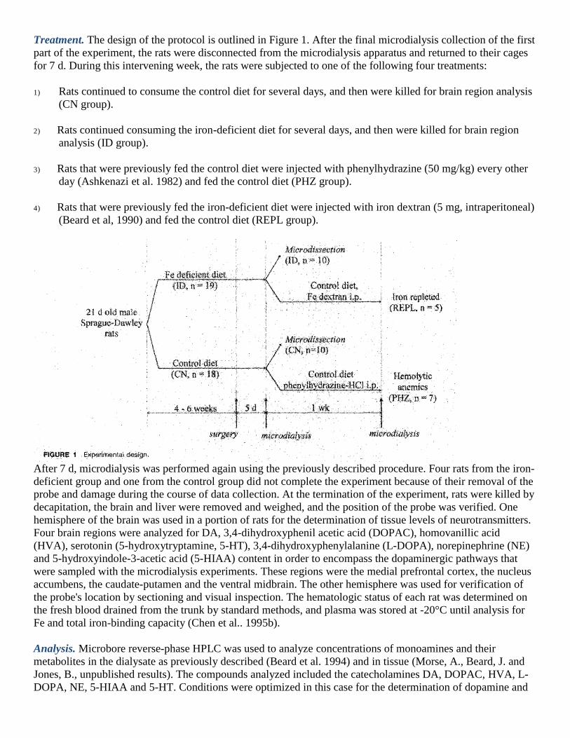
Treatment. The design of the protocol is outlined in Figure 1. After the final microdialysis collection of the first
part of the experiment, the rats were disconnected from the microdialysis apparatus and returned to their cages
for 7 d. During this intervening week, the rats were subjected to one of the following four treatments:
1) Rats continued to consume the control diet for several days, and then were killed for brain region analysis
(CN group).
2) Rats continued consuming the iron-deficient diet for several days, and then were killed for brain region
analysis (ID group).
3) Rats that were previously fed the control diet were injected with phenylhydrazine (50 mg/kg) every other
day (Ashkenazi et al. 1982) and fed the control diet (PHZ group).
4) Rats that were previously fed the iron-deficient diet were injected with iron dextran (5 mg, intraperitoneal)
(Beard et al, 1990) and fed the control diet (REPL group).
After 7 d, microdialysis was performed again using the previously described procedure. Four rats from the iron-
deficient group and one from the control group did not complete the experiment because of their removal of the
probe and damage during the course of data collection. At the termination of the experiment, rats were killed by
decapitation, the brain and liver were removed and weighed, and the position of the probe was verified. One
hemisphere of the brain was used in a portion of rats for the determination of tissue levels of neurotransmitters.
Four brain regions were analyzed for DA, 3,4-dihydroxyphenil acetic acid (DOPAC), homovanillic acid
(HVA), serotonin (5-hydroxytryptamine, 5-HT), 3,4-dihydroxyphenylalanine (L-DOPA), norepinephrine (NE)
and 5-hydroxyindole-3-acetic acid (5-HIAA) content in order to encompass the dopaminergic pathways that
were sampled with the microdialysis experiments. These regions were the medial prefrontal cortex, the nucleus
accumbens, the caudate-putamen and the ventral midbrain. The other hemisphere was used for verification of
the probe's location by sectioning and visual inspection. The hematologic status of each rat was determined on
the fresh blood drained from the trunk by standard methods, and plasma was stored at -20°C until analysis for
Fe and total iron-binding capacity (Chen et al.. 1995b).
Analysis. Microbore reverse-phase HPLC was used to analyze concentrations of monoamines and their
metabolites in the dialysate as previously described (Beard et al. 1994) and in tissue (Morse, A., Beard, J. and
Jones, B., unpublished results). The compounds analyzed included the catecholamines DA, DOPAC, HVA, L-
DOPA, NE, 5-HIAA and 5-HT. Conditions were optimized in this case for the determination of dopamine and
its metabolites; hence, inconsistent data on 5-HIAA and 5-HT were obtained and are not reported.
Concentrations were determined by comparison of peak heights with known concentrations of standards
analyzed with each set of samples (correlation coefficient >97% for all standard curves). Determinations of
tissue neurotransmitters were performed on microdissections of brain sections as defined in an atlas for rats
(Paxinos and Watson 1986). These dissections were performed immediately at death, and brain sections frozen
in dry ice and then stored at -80°C. Brain regions were thawed slightly, homogenized with a Teflon pestle in 50
mmol/L HClO4 (5:1, v/wt) at 4°C, 250 pg of dihydroxy benzylamine (DHBA) added as an internal standard,
and then analyzed by HPLC. All analyses were performed at electrode potentials of +800 mV relative to the
reference Ag-AgCl electrode. The minimum detectable concentration was ~3 μg/L for all of the monoamines
and metabolites. Microdialysate and plasma were also analyzed for amino acid concentration by HPLC
(Sizemore et al. 1995) to test for possible alterations in substrate availability.
Statistical analysis. Data were corrected for day-to-day variation in column conditions by analyzing an external
DHBA standard each day and then correcting the sample values accordingly. All data were subjected to a box-
plot analysis in Minitab (State College, PA) to determine the presence of outliers; tests for normality also were
performed before ANOVA. ANOVA was used for statistical analysis of hematologic and dialysis data when
data from all four groups were compared; the Type III sums of squares was used to calculate F ratios. When
group variances were unequal, data were log-transformed before analysis. Tukey's test for multiple pairwise
comparisons was performed to compare specific cell means for treatment effects (Hinkle et al 1988).
Significance was assumed at P ≤ 0.05.
RESULTS
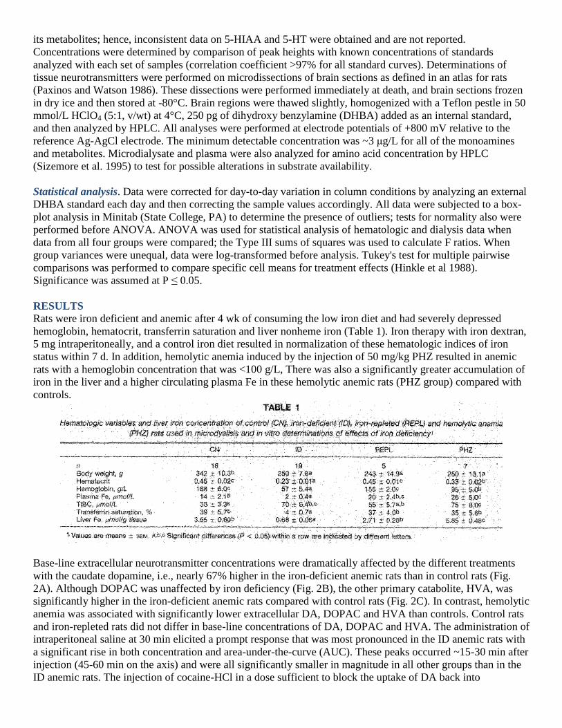
Rats were iron deficient and anemic after 4 wk of consuming the low iron diet and had severely depressed
hemoglobin, hematocrit, transferrin saturation and liver nonheme iron (Table 1). Iron therapy with iron dextran,
5 mg intraperitoneally, and a control iron diet resulted in normalization of these hematologic indices of iron
status within 7 d. In addition, hemolytic anemia induced by the injection of 50 mg/kg PHZ resulted in anemic
rats with a hemoglobin concentration that was <100 g/L, There was also a significantly greater accumulation of
iron in the liver and a higher circulating plasma Fe in these hemolytic anemic rats (PHZ group) compared with
controls.
Base-line extracellular neurotransmitter concentrations were dramatically affected by the different treatments
with the caudate dopamine, i.e., nearly 67% higher in the iron-deficient anemic rats than in control rats (Fig.
2A). Although DOPAC was unaffected by iron deficiency (Fig. 2B), the other primary catabolite, HVA, was
significantly higher in the iron-deficient anemic rats compared with control rats (Fig. 2C). In contrast, hemolytic
anemia was associated with significantly lower extracellular DA, DOPAC and HVA than controls. Control rats
and iron-repleted rats did not differ in base-line concentrations of DA, DOPAC and HVA. The administration of
intraperitoneal saline at 30 min elicited a prompt response that was most pronounced in the ID anemic rats with
a significant rise in both concentration and area-under-the-curve (AUC). These peaks occurred ~15-30 min after
injection (45-60 min on the axis) and were all significantly smaller in magnitude in all other groups than in the
ID anemic rats. The injection of cocaine-HCl in a dose sufficient to block the uptake of DA back into

presynaptic neurons resulted in a similar size response (both peak height and AUC) as the saline injection in ID
rats. The response of control rats to cocaine injection was much more pronounced than that of iron-deficient
rats. The net AUC for control rats was 32-fold greater than that of iron-deficient rats (16 vs. 0.5 cm2, Fig. 2). All
other treatment groups, however, had much larger dopamine responses to cocaine than to saline injection. In
addition, the response was delayed by 15 min compared with control rats. Iron-repleted rats did not differ from
controls in their dopamine response to cocaine. Phenylhydrazine anemic rats showed a temporal profile similar
to that of iron-deficient anemics but with a much larger effect of cocaine than saline. The intra- and extra-
cellular production of HVA within the paradigms demonstrated higher concentrations of HVA in iron-deficient
anemic rats and generally lower levels in hemolytic anemic rats compared with control rats (Fig, 2C). There
was no difference between iron-repleted and control rats. In contrast, DOPAC was lower in hemolytic anemia
rats, dropping to concentrations less than 30% of normal with a blunted response to cocaine injection (Fig. 2B).
Because one explanation of the changes in dopamine metabolism in iron-deficient anemic rats could be the
altered availability of substrate amino acids for synthesis, we measured the steady-state concentrations of
certain amino acids in the dialysate (Fig. 3). There was no effect of iron deficiency on the levels of amino acids
in brain extracellular fluid or plasma (data not shown).

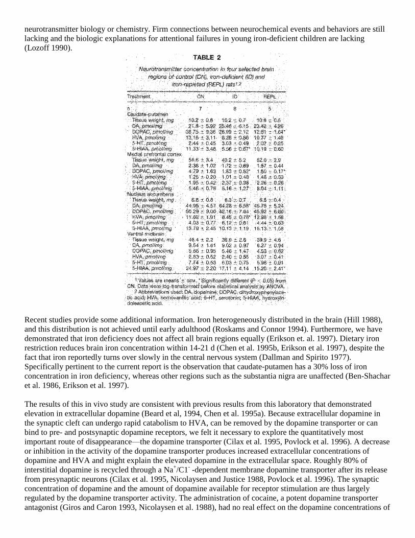
Our final effort to explore neurotransmitter metabolism in this experiment was the measurement of
neurotransmitters and metabolites in postmortem brain tissue of iron-deficient, control and iron-replete rats
(Table 2). As expected, there was a >10-fold variation in dopamine concentration and metabolite concentration
across the regions studied, i.e., caudate-putamen, medial prefrontal cortex nucleus accumbens and ventral
midbrain, with caudate-putamen and nucleus accumbens having the highest concentrations. Iron deficiency
anemia was associated with an elevation in dopamine in the nucleus accumbens but not in the other brain
regions, although there was a clear tendency (P = 0.08) for an elevation to occur in most regions examined. It is
interesting to note that in caudate-putamen, the region in which microdialysis had shown a significant in vivo
effect of iron deficiency on extracellular DA, there was no significant effect on tissue concentration of DA,
DOPAC or HVA.
DISCUSSION
Several new and important observations are reported in these experiments: 1) extracellular dopamine in the
caudate-putamen returned to normal with the normalization of iron status; 2) elevations in dopamine or other
neurotransmitters are not due to anemia per se; :and 3) dopaminergic transporters may be altered in iron
deficiency. This report extends our previous observations (Beard et al. 1994, Chen et al. 1995a) of elevations of
extracellular dopamine and metabolites by demonstrating reversibility with iron therapy in a short period of
time and suggests further that it is removal of dopamine from the interneuronal cleft that is responsible for this
elevation. Availability of substrate amino acids is not altered in this brain region, thus eliminating this
possibility for the explanation of altered neurotransmitter concentrations.
One research group (see review of Youdim et al. 1989) established that dopamine is perturbed by iron
deficiency in the rat animal model. They observed a decrease in D2 receptor density in the caudate-putamen that
was irreversible if iron deficiency is begun in early preweaning or intrauterine life. These investigators also note
that certain behaviors, such as poor responses to adverse stimuli (foot shock), decreased temperature regulation
after apomorphine and a reversal of the diurnal cycle, are likely related to this alteration in dopamine
metabolism. Iron deficiency postweaning did not affect tyrosine hydroxylase or tryptophan hydroxylase, both
iron-containing enzymes, nor were there effects on serotonin, adrenergic or gabaminergic receptor populations
(Youdim et al. 1989). Thus, for many years, the focus and attention in this area of work resided on the D2
receptor in one brain region despite a lack of critical studies by other research groups into other aspects of
neurotransmitter biology or chemistry. Firm connections between neurochemical events and behaviors are still
lacking and the biologic explanations for attentional failures in young iron-deficient children are lacking
(Lozoff 1990).
Recent studies provide some additional information. Iron heterogeneously distributed in the brain (Hill 1988),
and this distribution is not achieved until early adulthood (Roskams and Connor 1994). Furthermore, we have
demonstrated that iron deficiency does not affect all brain regions equally (Erikson et. al. 1997). Dietary iron
restriction reduces brain iron concentration within 14-21 d (Chen et al. 1995b, Erikson et al. 1997), despite the
fact that iron reportedly turns over slowly in the central nervous system (Dallman and Spirito 1977).
Specifically pertinent to the current report is the observation that caudate-putamen has a 30% loss of iron
concentration in iron deficiency, whereas other regions such as the substantia nigra are unaffected (Ben-Shachar
et al. 1986, Erikson et al. 1997).
The results of this in vivo study are consistent with previous results from this laboratory that demonstrated
elevation in extracellular dopamine (Beard et al, 1994, Chen et al. 1995a). Because extracellular dopamine in
the synaptic cleft can undergo rapid catabolism to HVA, can be removed by the dopamine transporter or can
bind to pre- and postsynaptic dopamine receptors, we felt it necessary to explore the quantitatively most
important route of disappearance—the dopamine transporter (Cilax et al. 1995, Povlock et al. 1996). A decrease
or inhibition in the activity of the dopamine transporter produces increased extracellular concentrations of
dopamine and HVA and might explain the elevated dopamine in the extracellular space. Roughly 80% of
interstitial dopamine is recycled through a Na+/C1
- -dependent membrane dopamine transporter after its release
from presynaptic neurons (Cilax et al. 1995, Nicolaysen and Justice 1988, Povlock et al. 1996). The synaptic
concentration of dopamine and the amount of dopamine available for receptor stimulation are thus largely
regulated by the dopamine transporter activity. The administration of cocaine, a potent dopamine transporter
antagonist (Giros and Caron 1993, Nicolaysen et al. 1988), had no real effect on the dopamine concentrations of

the iron-deficient rat beyond what was seen with a placebo saline injection. We know that there is a very rapid
appearance of cocaine and its metabolites in the brain after peripheral injection (Morse et al. 1995). Injection of
cocaine caused an increase in the levels of dopamine and its metabolites in the first post-cocaine collection in
control rats. In iron-deficient rats, the increase was not seen until 30 min post-cocaine injection, indicating a
delay in the onset effect of the blockade. This delay could he interpreted as an indication of a decreased
appearance of cocaine in the brain of iron-deficient rats, a decreased binding of cocaine to these transporters
and/or a decreased number of functioning dopamine transporters in the striatum of iron-deficient rats,
Experiments with mice, however, demonstrate no effect of iron deficiency on the rate of appearance of cocaine
or its metabolites in brain after an intraperitoneal injection (Morse, A., Beard, J. and Jones, B., unpublished
results). Thus we can tentatively conclude that dopamine clearance by this mechanism is altered in iron
deficiency. Iron therapy rapidly normalized these metabolite patterns as well as the recovery of a normal
hematologic status, demonstrating a clear iron responsive process. Direct measurements of the amount of
dopamine transporter and its functioning are necessary before a clear role for iron is firmly established.
The in vitro data suggest that other brain regions may also be adversely affected by iron status, although none
were examined by the in vivo method. These data contrast with Youdim's experiments, which showed no
significant effect in vitro of iron deficiency anemia on dopamine, serotonin norepinephrine concentrations in
brain regions (Youdim et al, 1989). Peripheral sympathetic nervous system activity is altered by iron deficiency,
with increased concentrations of norepinephrine in plasma and urine and decreased concentrations in tissue (see
review of Brigham and Beard 1996), an observation that could also be explained by a decreased reuptake of that
catecholamine (Kanner and Schulinder 1987).
Fourteen days of dietary iron therapy are sufficient to restore brain iron concentration to normal (Chen et al.
1995b), and this study also shows rapid normalization of neurochemical alterations. These results support
previous studies showing that rat behavioral responses return to normal after 7 d of iron repletion therapy
(Youdim et al, 1979 and 1981) and that D2 receptor Bmax levels are restored rapidly by iron therapy in
postweaning rats (Ashkenazi et al. 1982, Ben-Shachar et al. 1986). Iron therapy probably will not correct the
alterations in brain neurochemistry suspected in preweaning iron deficiency, which have irreversible
consequences (Felt and Lozoff 1996), although this was not tested in the current study. Because it is speculation
at this time to surmise that the most important "critical period" is the time of active myelinogenesis, PND 8- 14
in rats, and the first year of postnatal life in humans, continued active investigations using the developmental
perspective must be conducted (Dobbing 1990).
Previous studies from our laboratory (Beard et al.1994, Chen et al. 1995a) did not resolve whether anemia per
se was, responsible for the elevation of CSF dopamine in the striatum, because the dietary model of severe iron
deficiency anemia cannot itself distinguish and separate tissue iron deficiency effects from oxygen transport
effects. Several protocols are available, such as exchange transfusion or rapid repletion, to differentiate these
effects (Beard et al. 1990). In this study, we used hemolytic anemia, induced with phenylhydrazine, an alternate
form of anemia that should not have direct effects on brain iron metabolism (Ashkenazi et al. 1982).
Extracellular levels of dopamine and its metabolites, DOPAC and HVA, in hemolytic anemic rats were below
control values, demonstrating that anemia per se is not responsible for the elevated extracellular dopamine
levels found in ID anemic rats and that oxygen transport to brain is not a likely cause for the alterations.
In conclusion, we demonstrated in this experiment that iron deficiency is associated with a significant elevation
of dopamine in young rats and that anemia itself is not the cause. This functional pool of dopamine is
normalized within 7 d of iron therapy. Additional evidence suggests that proper functioning of the dopamine
transporter is faulty in iron deficiency. These animal studies thus provide some further biologic support for a
causal relationship of iron status to cognitive functioning via the dopaminergic system.
Notes: 4 Abbreviations used: AUC, area under the curve; CN, control; CSF, cerebral spinal fluid; DA, dopamine;
OHBA, dihydroxybenzylamine acid; DOPAC. dlhydroxyphenyl acetic acid; L-DOPA, 3,4-

dihydroxyphenylalanine; 5-HIAA, 5-hydroxyindole-3-acetic acid; 5-HT, serotonin (5-hydroxytryptamine);
HVA, homovanillic acid; ID, iron deficient; NE, norepinephrine; PHZ, phenylhydrazine; PND, postnatal day:
REPL, iron repleted.
LITERATURE CITED
Ashkenazi, R., Ben-Shachar, D. & Youdim, M. B. (1982) Nutritional Fe and dopamine binding sites in the rat
brain. Pharm. Biochem. Behay. 17 (suppl): 43-47.
Baynes, R. D, & Bothwell, T. H. (1990) Iron deficiency. Annu. Rev. Nutr. 10: 133-148.
Beard, J. L. (1987) Feed efficiency and norepinephrine turnover in Iron deficiency. Proc. Soc. Exp. Biol. Med.
184: 337-344.
Beard, J. L, (1996) Nutrient status and central nervous system function. In: Present Knowledge in Nutrition, 7th
ed. (Ziegler, E. E. & Filer, L. J., Jr., eds.), pp. 612-614, LSI Press, Washington, DC.
Beard, J. L.. Chen, Q., Connor, J. R. & Jones, B, C. (1994) Altered monoamine metabolism in caudate-putamen
of iron deficient rats. Pharm. Biochem. Behav. 48: 621-624.
Beard, J. L,, Connor, J. R. & Jones, B. C. (1993) Iron in the brain. Nutr. Rev, 51: 157-170.
Beard, J. L., Tobin, B. VV. & Smith, S. M. (1990) Effects of iron repletion and correction of anemia on
norepinephrine turnover and thyroid metabolism in Iron deficiency. Proc. Soc. Exp. Biol. Med. 193: 306-312.
Ben-Shachar, D., Ashkenazi, R. & Youdim, M,B,H, (19B6) Long term consequences of early iron deficiency on
dopaminergic neurotransmission, J. Dev. Neurosci. 4: 81-88.
Borel, M. J., Smith, S. H., Brigham, D.& Beard, J. (1991) The impact of varying degrees of iron nutrition on
several functional consequences of iron deficiency in rats. J. Nutr. 121: 729-736,
Brigham, D. & Beard, J. L. (1996) Iron and thermoregulation: a review. Crlt. Rev. Food Sci. Nutr. 36: 747-763.
Chen, Q„ Beard, J. L. & Jones, B. C. (1995a) Abnormal rat brain metabolism in iron deficiency anemia. Nutr.
Biochem. 6: 486-493,
Chen, Q., Connor, J. R. & Beard, J. L. (1995b) Brain iron transferrin and ferritin concentrations are altered in
developing iron-deficient rats. J. Nutr. 125: 1529-1535.
Ciliax, B. J., Heilman, C., Demchyshyn, L, L., Pristupa. Z. B., Ince, E., Hersch, S. M„ NIzrilk, H. B, & Levey,
A. (1995) The dopamine transporter: immunochemical characterization and localization in brain, J. Neurosci.
15: 1714-1723.
Dallman, P. R. & Spirito, R. A. (1977) Brain iron in the rat: extremely slow turn-over in normal rats may
explain long-lasting effects of early iron deficiency, J. Nutr. 107: 1075-1081.
Dobbing, J. (1990) Vulnerable periods in developing brain. In: Brain, Behaviour and Iron in the Infant Diet
(Dobbing, J., ed.), pp. 1-15. Springer-Verlag, Lon-don, UK.
Erikson, K. M., Pinero, D. J., Connor, J. R, & Beard, J. L. (1997) Regional brain iron, ferritin, and transferrin
concentrations change due to Iron deficiency and iron repletion in developing rats. J. Nutr. (in press).
Felt, B. & Lozoff, B. (1996) Brain iron and behavior of rats are not normalized by treatment of iron deficiency
anemia during early development. J. Nutr. 126: 693-701.
Giros, B. & Caron, M,(1993) Molecular characterization of the dopamine transporter, Trends Pharmacol. Sci.
14: 43-49.
Hill, J, M, (1988) The distribution of iron in the brain, In: Brain Iron: Neurochemistry and Behavioural Aspects
(Youdim, M.B.H., ed.), pp. 1-24, Taylor & Francis, London, UK.
Hill, J. M. & Switzer, R. C., III (1984) The regional distribution and cellular localization of iron in the rat brain.
Neuroscience 11: 595-603,
Hinkle, D. E., Wiersma, W, & Jurs, S. a (1988). Applied Statistics for the Behavioral Sciences, 2nd ed.
Houghton Mifflin Co., Boston, MA,
Kanner, B. I, & Schuldiner, S. (1987) Mechanism of transport and storage of neurotransmitters. CRC Grit. Rev.
Biochem. 22: 1-38.
Lozoff, B. (1990) Has iron deficiency been shown to cause altered behavior in Infants? In: Brain, Behaviour
and Iron in the Infant Diet (Dabbing, J., ed.), pp. 107-123. Springer-Verlag, London, UK.
Morse, A, C., Erwin, V. G, & Jones, 6, C, (1995) Pharmacogenetics of cocaine: a critical review.
Pharmacogenetics 5: 183-192.

Nicolaysen, L. C. & Justice, J. B., Jr. (1988) Effects of cocaine on release and uptake of dopamine in vivo:
differentiation by mathematical modeling. Pharmacol. Biochem. Behay. 31: 327-335.
Nicolaysen, L. C., Pan, H. T. & Justice, J. B., Jr. (1988) Extracellular cocaine and dopamine concentrations are
linarly related in rat striatum, Brain Res. 456: 317-323.
Oski, F. A., Honing, A. S., Helu, B. & Howanitz, P. H. (1983) Effect of iron therapy on behavior performance
in non-anemic, iron deficient infants. Pediatrics 71: 877-880.
Paxinos, G. & Watson. C. (1986) The Rat Brain in Stereotaxic Coordinates, 2nd ed„ Fig, 18, Academic Press,
San Diego, GA.
Povlock, S. L., Meiergerd, S, M. & Schenk, J. O. (1996) Kinetic mechanisms of the dopamine transporter: a
comparison with other biogenic amine transporters. In: CNS Neurotransmitters and Neuromodulators of
Dopamine (Stone, T. W., ed.), pp. 21-39. CRC Press, Boca Raton, FL.
Roskams, A. J. & Connor, J. R. (1994) Iron, transferrin, and ferritin in the rat brain during development and
aging. J. Neurochem. 63: 709-716.
Sizemore, F. G„ Leach, G. R. & Barbato, G, F. (1995) Neonatal MSG administration eliminates hypothalamic
lateralization of biogenic amines in chickens selected for divergent growth rates. Biogenic Amines 11: 19-29.
Yehuda, S. (1990) Neurochemical basis of behavioural effects of brain iron deficiency In rats. In: Brain,
Behaviour and Iron in the Infant Diet (Jobbing, J. ed.). pp. 63-74. Springer-Verlag, London, UK.
Yehuda, S. & Youdim, M.B.H. (1989) Brain Iron: a lesson from animal models. Am. J. Clin. Nutr. 50: 618S-
629S.
Youdim, M,B.H. (1990) Neuropharmacological and neurobiological aspects of iron deficiency, In: Brain,
Behaviour and Iron in the Infant Diet (Dobbing, ed.), pp. 83-97. Springer-Verlag, London, UK.
Youdim, M.B.H., Ben-Shachar, D. & Yehuda, S, (1989) Putative biological mechanisms on the effects of iron
deficiency on brain biochemistry. Arr. J. Clin. Nutr. 50: 607S-617S.
Youdim, M.B.H„ Green, A, R., Bloomfield, M. R., Mitchell, B. D., Heal, D. J. & Grahame-Smith, D. G. (1979)
The effect of iron deficiency on brain biogenic monoamine biochemistry and function in rats,
Neuropharmacology 19: 259-267.
Youdim, M.B.H., Yehuda, S. & Ben-Uriah, Y. (1981) Iron deficiency induced circadian rhythm reversal of
dopaminergic-mediated behaviours and thermoregulation in rats, Eur. J, Pharmacol. 74: 295-301,